Article

Bacterial Biofilm Characterization and Microscopic Evaluation of the Antibacterial Properties of a Photocatalytic Coating Protecting Building Material

1
LMDC, Université de Toulouse, INSA/UPS Génie Civil, 135 Avenue de Rangueil, 31077 Toulouse Cédex 04, France
2
LGC, Université de Toulouse, UOS-CNRS Dép. BioSyM, 4 allée Emile Monso, 31029 Toulouse, France
*
Author to whom correspondence should be addressed.
Coatings 2018, 8(3), 93; https://doi.org/10.3390/coatings8030093
Submission received: 2 February 2018 / Revised: 28 February 2018 / Accepted: 4 March 2018 / Published: 5 March 2018
(This article belongs to the Special Issue Advanced Coatings for Buildings)

Abstract

:
Use of photocatalytic paint-like coatings may be a way to protect building materials from microbial colonization. Numerous studies have shown the antimicrobial efficiency of TiO 2 photocatalysis on various microorganisms. However, few have focused on easy-to-apply solutions and on photocatalysis under low irradiance. This paper focuses on (a) the antibacterial properties of a semi-transparent coating formulated using TiO 2 particles and (b) the microscopic investigations of bacterial biofilm development on TiO 2 -coated building materials under accelerated growth conditions. Results showed significant antibacterial activity after few hours of testing. The efficiency seemed limited by the confinement of the TiO 2 particles inside the coating binder. However, a pre-irradiation with UV light can improve efficiency. In addition, a significant effect against the formation of a bacterial biofilm was also observed. The epifluorescence approach, in which fluorescence is produced by reflect rather than transmitted light, could be applied in further studies of microbial growth on coatings and building materials.

1. Introduction

Since microorganisms are ubiquitous and dispersible from soil and water as well as from air, building materials are permanently exposed to them and may easily become targets for contamination and growth. The requirements’ microorganism development, i.e., an energy source, carbon and water, can be satisfied by building materials (organic and/or inorganic) in a large variety of contexts [1,2,3]. The growth of microorganisms can have various detrimental consequences, including the biodeterioration of materials, which may concern structural and aesthetic properties, and/or the release of aerial contaminants that can be deleterious for human health. Their proliferation on building materials is of growing concern to the scientific community [3,4,5,6].
A substantial number of man-made constructions have been reported to be widely contaminated by microorganisms (mainly fungi, bacteria and algae). They include pipes carrying aggressive aqueous media such as sewers [3], moisture-damaged buildings [1,7,8], historical monuments [9], etc. Contaminated building materials are usually visually deteriorated by the activity of fungi and bacteria, which are known to be the most harmful organisms inducing physical and chemical changes in materials.
Microbial contamination of building materials in indoor environments has also been much studied. The degradation of indoor air quality is a growing public health concern worldwide and microorganisms or their components are major polluting agents [2]. Their contribution to the Sick Building Syndrome (SBS) and Building Related Illness (BRI) is widely emphasized in the literature. Microorganisms may release aerial particles such as spores, allergens, toxins and other metabolites that can be harmful to human health [10,11,12,13,14]. Serious health troubles such as irritations and toxic effects, infections, allergies, etc. have been experienced by building occupants frequently exposed to microbial contaminants [15,16]. Several studies have already reported that indoor building materials can become major sites of microbial growth [13,15,17]. Actually, growth-promoting conditions, i.e., high humidity and presence of nutrients, are easily fulfilled in damp environments such as water-damaged, damp, and/or badly-insulated buildings. Microorganisms grow on interfacial zones (air–substrates, water–air, etc.). Under specific environmental conditions, they develop on surfaces in complex three-dimensional structures, in which adherent cells are embedded within a self-produced matrix of extracellular polymeric substance (EPS). The overall organization, commonly called a microbial biofilm, provides the microorganisms with particular resistance towards physical and chemical aggressions, be they environmental or from disinfection treatments [18,19,20].
Different solutions have been studied to prevent, stop or at least reduce microbial biofim development on building materials, including core treatments and coatings applied to surfaces. Antimicrobial products can be formulated using biocides [21], metal oxide nanoparticles [22,23,24] or bio-based products [25]. A substantial amount of literature has been published on the effect of photocatalytic TiO 2 nanoparticles on microorganisms [26,27,28,29]. Studies have shown high efficiency of photocatalysis on the viability of a wide variety of microorganisms and on microbial contaminants, including bacteria, endospores, fungi, algae, protozoa, viruses and prions (shown to be the organisms most resistant to disinfectants [30]) as well as on microbial toxins. Photocatalysis needs to be activated by a light irradiation of sufficient energy to create electron–hole pairs. The electron–hole pairs may react with electron donors or acceptors previously adsorbed on the photocatalyst, e.g., O 2 , H 2 O. The resulting redox reactions lead to the formation of oxide radicals that are highly reactive and degrade organic matter by non-selective action. Additionally, irradiated TiO 2 presents photo-induced superhydrophilicity [31] and may be incorporated in materials to provide self-cleaning properties.
The use of titanium dioxide as a self-cleaning agent in building materials is common for some years now. Photocatalytic concretes have been developed to prevent fouling and biofouling [32,33] or to purify ambient air (VOC, NO x …) [34,35,36,37,38]. Regarding bacterial and fungal disinfection, research has mainly focused on the efficiency of photocatalytic TiO 2 particles used alone, powdered or immobilized in the form of thin film coatings by complex processes (usually involving calcination). It seems that only few studies have investigated easy-to-apply devices such as paint-like coatings. In addition, one of the main advantages of such coatings are their passive aspect: once applied to a surface, the photocatalysis is activated by natural light. Furthermore, efficiencies of TiO 2 photocatalysis found in the literature are usually related to very high irradiances (>10 W/m 2 ) [28,29,39] that are far from the real-world conditions. Only recent studies have begun to investigate on the photocatalytic activity under low irradiances [40].
The main objectives of the present paper were (a) to focus on the antibacterial efficiency under low irradiance of a photocatalytic semi-transparent coating that is easy to apply on existing materials and (b) to carry out microscopic observations (epifluorescence and SEM) of bacterial biofilm development on a coated building materials under accelerated growth conditions.

2. Materials and Methods

2.1. Bacterial Cultivation

The microorganisms the most frequently detected on indoor building materials are (i) fungi genera Cladosporium, Penicillium, Aspergillus and Stachybotrys, and (ii) Gram negative bacteria and mycobacteria [2].
In this study, the model bacterium Escherichia coli was chosen for methodological reasons, i.e., its relative ease of culture and its high speed of growth. E. coli CIP 53126 was obtained from the Collection of Institute Pasteur (CIP), Paris, France. The strain was preserved at 80 C in Eugon medium (Biomérieux, Craponne, France) supplemented with 10% glycerol. The E. coli strain identification was checked by Gram staining and biochemical characterization (oxydase reaction and Analitical Profile Index 20E) (Biomérieux, Craponne, France). Before each experiment, bacterial cells were pre-cultured on a nutrient agar Petri dish. Colonies were then transferred to trypcase soy agar (TSA) (Biomérieux) and incubated at a temperature of 36 C for 16 to 24 h. A second culture on TSA was incubated at a temperature of 36 C for 16 to 20 h prior to the test. For testing, one plastic loop of bacteria was dispersed evenly in a small amount of sterile distilled water and the suspension for inoculation was adjusted to about 10 8 cells/mL using a spectrophotometer (640 nm). A 10-fold dilution of the cell suspension was then prepared to obtain the final concentration of the test suspensions, depending on the type of evaluation (antibacterial activity or resistance to biofilm formation).

2.2. Coatings and Preparation of Supports

2.2.1. Coatings

Two semi-transparent coatings were formulated using water and acrylic resin. The formulation contained 5 wt % TiO 2 dispersion (Kronos type 7454, trial product, KRONOS/Société Industrielle du Titane, Paris, France), 2 wt % acrylic resin and 93 wt % water. A control coating was made of 2 wt % acrylic resin and 98 wt % water. The TiO 2 dispersion contained TiO 2 particles (Kronos VLP7050), some physical characteristics of which are presented in Table 1.

2.2.2. Nature and Preparation of Supports

Three types of support were used in this study: glass slides, polycarbonate membranes, and cementitious matrices.

Glass Slides

Preliminary measurements of the antibacterial activity were carried out on glass microscope slides ( 26 × 76 mm 2 ) covered by pipetting the TiO 2 coating or the control coating (without TiO 2 ). Regarding the coating of glass slides, while drying, the water left an even film of acrylic binder scattered with TiO 2 particles. Although the distribution of TiO 2 was not optimal on these supports, it was rather homogeneous over the entire surfaces of samples.

Polycarbonate Membranes

Isopore hydrophilic polycarbonate membranes (Millipore, 47 mm diameter, 0.4 μ m filter pore size, 5%–20% porosity) were also used as supports for the coating. Membranes were placed in sterile Petri dishes, covered by pipetting the coatings and placed under a sterile flow hood for air drying. Then, samples were pre-irradiated with UV light (5 W/m 2 ) for different durations, from 0 h to 109 h. The role of the pre-irradiation was to increase the antibacterial activity of the coating. It is discussed later in the paper.

Cementitious Supports

Cement paste was chosen as reference building material because of its availability, its ease of casting, and because it is morphologically representative of many building materials (porosity and roughness). The cement paste samples were made of ordinary Portland cement CEM I 52.5R were cast with a 0.55 Water/Cement ratio in cylindrical molds (d = 2.8 cm, h = 1 cm). Samples were demolded after 24 h and stored for 27 days in a moisture chamber (100 %RH, room temperature). It is widely known that the pH of materials is a determining factor in microbial contamination. The pH of unaltered cementitious materials is around 13. In their work on an accelerated laboratory test to study fungal biodeterioration, Wiktor et al. [41,42] showed that, for cementitious materials, a low pH of the surface was essential to increase the bioreceptivity of samples. Recently-cast cementitious materials are thus not so subject to microbial colonization, probably because of their relatively high alkalinity incompatible with microbial growth. Usually, the bioreceptivity of these materials increases over time as carbonation tends to decrease the surface pH, but this phenomenon takes place very slowly (typically, a few weeks in natural conditions). Consequently, accelerated aging of the cementitious mortars was performed. After 28 days, samples were placed in a 2-liter-borosilicate reactor for a leaching operation, as follows: the reactor was continuously supplied with a leaching solution by a peristaltic pump (Masterflex L/S, Cole-Palmer, Vernon Hills, IL, USA). The flow rate was set to 1.4 mL/m in order to renew the reactor content in 24 h. The leaching solution was composed of water and acetic acid (CH 3 COOH) 0.12 mol/L and the pH was adjusted to 4.7 with sodium hydroxide (NaOH) at 0.076 mol/L. The operation was carried out for 15 days. Then, samples were removed from the reactor and a pH-paper was applied on their wet surfaces. The pH measured was around 7 for all samples. After this leaching operation, samples were washed with distilled water and stored at 100% RH until tested.
Prior to the experiments, samples were covered with the coatings (TiO 2 coating or control coating) by pipetting 500 μ L over the entire surface (6.16 cm 2 ). They were placed under a sterile flow hood for over 12 h for air drying. Then, samples coated with TiO 2 and control samples coated without TiO 2 , were pre-aged by UV irradiation (5 W/m 2 ) for different durations, from 0 h to 109 h.

2.3. Photocatalysis Activation

The TiO 2 photocatalyst (anatase) is naturally activated by light irradiation in the UV-range. The light intensity, or irradiance, greatly influences photocatalysis efficiency. In the literature, intensity values are usually chosen between 10 and 500 W/m 2 [43,44]. These values are quite different from natural light intensity values, in indoor or outdoor conditions. For example, studies have reported outdoor intensity around 30 W/m 2 on sunny days [36,45,46] and between 5 and 10 W/m 2 on cloudy days [47,48]. The few studies that observed inactivation of microorganisms by photocatalysis with light intensities around 10 3 W/m 2 showed much lower inactivation rates [49], confirming the major impact of light intensity on the process. Regarding the resistance to microbial biofilm, a recent study also showed significant efficiency of TiO 2 coatings against the development of microbial biofilms under relatively low irradiance (13 W/m 2 ) closer to real-world light intensity [40].
UV light in the 325–400 nm range can strongly damage microorganisms and lead to their death. Standards usually recommend the use of low intensities for photocatalytic activity evaluation [50]. The position and the power of the lamp must be chosen so as activate the photocatalyst while making sure to avoid any lethal action of the UV light on bacterial cells. In our experiment, 8 W black light bulbs were chosen (peak wave 365 nm). The distance between lamps and samples was adjusted to obtain 2–4 W/m 2 at the bottom of the Petri dish (irradiance measured with a UV-A radiometer Gigahertz-Optik, GmbH, Türkenfeld, Germany, in the 310–400 nm range).
In order to assess the effect of UV irradiation on the survival of Escherichia coli, a drop of a bacterial suspension in phosphate buffer (about 1 mL of a 10 6 CFU/mL suspension) was deposited in a borosilicate Petri dish and irradiated for several hours. Different configurations were tested: irradiation with a borosilicate lid on the Petri dish, irradiation without the lid, and kept in the dark (control). After the test, bacteria were collected with a recovery solution (9 mL of Soybean Casein Lecithin Polysorbate 80 medium, SCDLP broth, as described in [51]) and numerated (CFU) in TSA after a 24 h incubation at 36 C . Results are presented in Figure 1.
For the three configurations, no significant evolution in the number of CFU was observed in the first 6 h of experiment. Control samples kept in the dark and irradiated in the presence of the borosilicate lid showed an increase of 2 log and 0.75 log , respectively, in 24 h, while no CFUs were detected on samples irradiated without the lid (limit of detection = 2 log ). The borosilicate lid allowed UV light to penetrate while keeping the hygrometry constant in the Petri dish and preventing the drop from drying.

2.4. Evaluation of Antibacterial Activity

The antibacterial activity characterizes the ability of a substrate to inactivate bacteria cells, i.e., to kill them or to inhibit their growth. It is a quantitative test that provides a bacterial reduction in terms of Colony Forming Units (CFUs).
The experiment was based on the contact between an antibacterial surface and a bacterial suspension for a given period of time, according to standards JIS Z 2801:2010 [52] and ISO 27447:2009 [50]. The test was carried out on polycarbonate membranes covered with TiO 2 coating, or with control coating, and placed in Petri dishes under a sterile flow hood. Each membrane was inoculated with 0.4 ml of bacterial suspension (cell concentration adjusted between 8 × 10 4 and 2 × 10 5 CFU/mL ). A transparent sterile film (9–10 cm 2 ) was carefully placed on the suspension in order to increase the contact area between bacterial cells and coatings [51]. Then, some membranes were UV-irradiated as described earlier (Section 2.3) and others were kept in the dark. The irradiation times were 2 h, 4 h, and 6 h.
After a certain time of contact (t), bacteria were recovered using the SCDLP broth. Quantitative evaluation was performed by CFU counting as described earlier (Section 2.4). The antibacterial activity was then calculated as the difference between the average logarithm of the number of viable bacteria on the control without TiO 2 and the average logarithm of the number of viable bacteria on samples coated with TiO 2 :
A = log N T i O 2 log N c o n t r o l = log N T i O 2 N c o n t r o l ,
with A: antibacterial activity; N T i O 2 : average number of CFU on TiO 2 -coated samples; N c o n t r o l : average number of CFU on control-coated samples.

2.5. Development of a Bacterial Biofilm under Accelerated Conditions

This experiment was designed to observe the effect of the photocatalytic coating on bacterial growth and spread once coated on a building material. Conditions favorable to microbial growth were chosen to obtain a biofilm very rapidly (24 h).
The test was carried out in aqueous media and in static conditions as previously described [53,54,55]. The media was chosen so that bacterial development was favored in the form of biofilm without planktonic growth. Direct observations of samples were carried out by epifluorescence microscopy and by scanning electron microscopy in order to highlight the distribution of bacteria.

2.5.1. Biofilm Nutrient Broth

The biofilm nutrient broth used to promote bacterial adhesion and biofilm formation was prepared as described in [53,54,55]. The composition of the broth is given in Table 2.

2.5.2. Procedure

The test was carried out with the cementitious supports covered with TiO 2 coating or with control coating. Cement supports were placed in 6-well culture plates filled with approximately 6 mL per well of biofilm nutrient broth at room temperature. Then, each sample was inoculated with 300 μ L of the bacterial test suspension adjusted to about 10 3 CFU/mL . The plates were covered with UV-transparent pyrex lids and incubated at 36 C . One plate was irradiated with UV light and the other was kept in the dark.
Light intensity on the samples was settled around 3 W/m 2 . In order to avoid planktonic cells (non adherent or released from the biofilm), the wells were washed with sterile distilled water and the biofilm broth was renewed after 4 h, 6 h and 24 h of incubation. Rapid assessments with pH paper was carried out before and after the experiment. The broths of different samples were between 7 and 9. No jump of pH was detected, indicating that the pre-treatment (leaching) of the cementitious supports was efficient. After 24 h, the samples were washed and removed with sterile tweezers and placed in new culture plates. Then, 6 mL of phosphate buffer was added to the wells and the top surface of each sample was gently scraped with a steel spatula. After homogenization of the solution, 1 mL was collected and diluted in sterile distilled water.

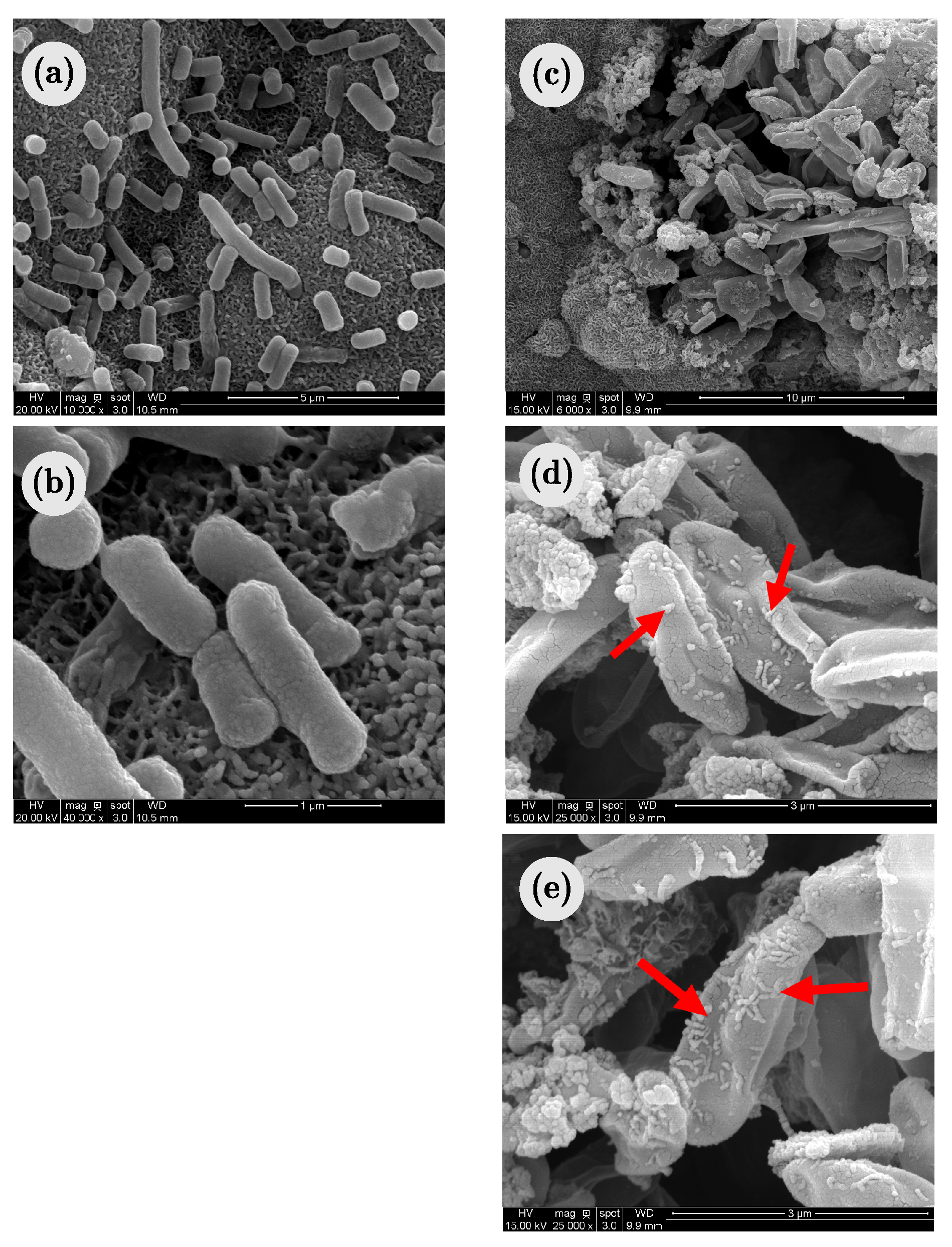
2.5.3. Scanning Electron Microscopy

Microstrucutural observations and chemical analyses were performed using a scanning electron microscope (JSM-6380LV, Jeol, Tokyo, Japan) fitted with an Energy Dispersive Spectroscopy detector (XFlash ® 3001, Röntec, Berlin, Germany).

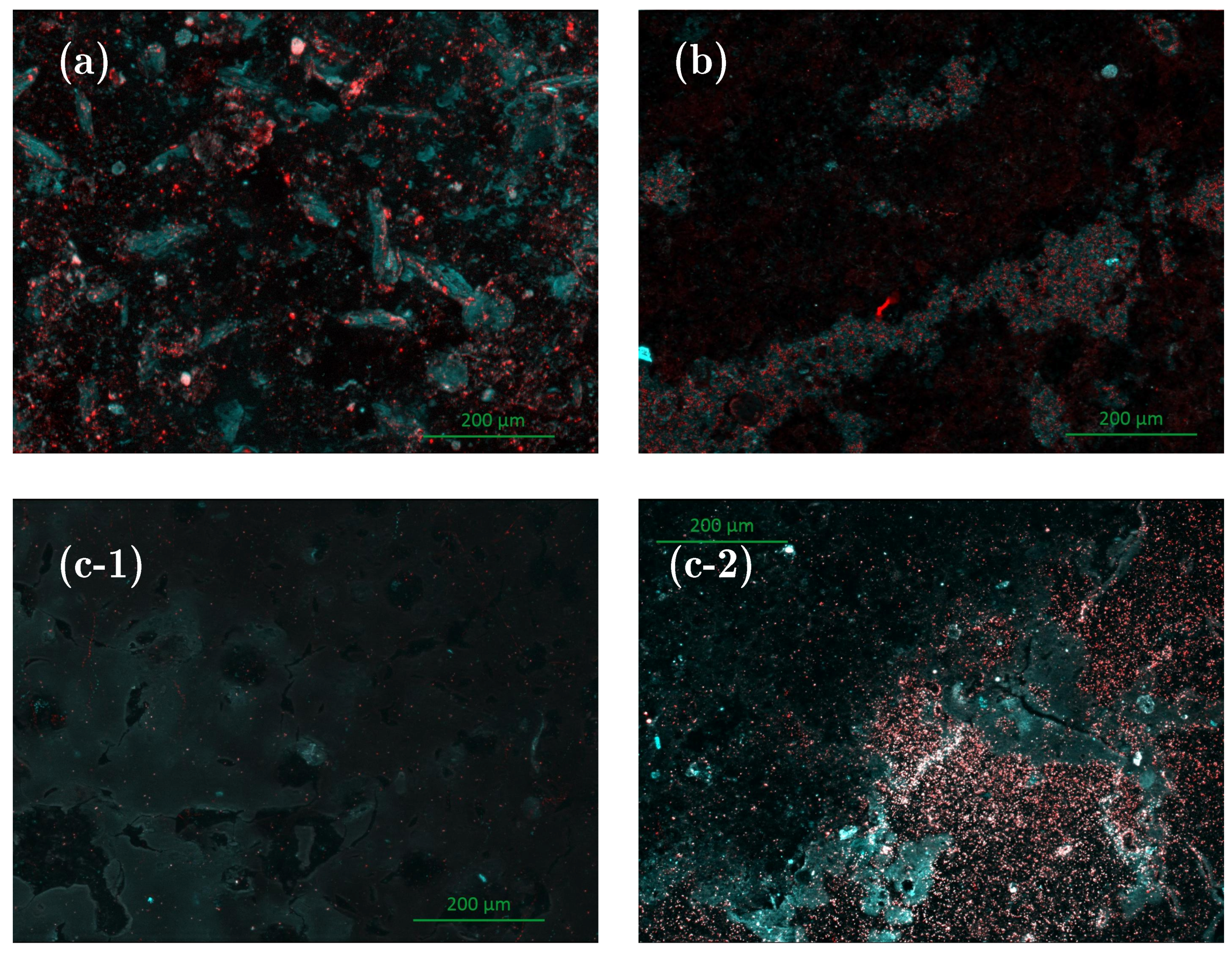
2.5.4. Epifluorescence Microscopy

Epifluorescence microscopy was used for rapid assessment of the bacterial colonization of sample surfaces. Following the addition of phosphate buffer at the end of the experiment, 0.75 μ L of Syto9 was used as a fluorochrome to stain the bacterial biofilm (Syto9 excitation range: 470–520 nm ; emission ange: 510–540 nm). After 10 min, samples were imaged with a Carl Zeiss Axio Imager-M2 microscope (Carl Zeiss, Oberkochen, Germany) equipped for epifluorescence with an HXP 200 C light source and the Zeiss 09 filter (excitor HP450r HP450200 C light source). Images were acquired with a digital camera (Zeiss AxioCam MRm) every 0.5 mm along the Z-axis and the set of images was processed with the Zen (Carl Zeiss) ® software. It should be emphasized that the Syto9 fluorochrome penetrates through damaged membranes as well as through whole membranes. Consequently, the method used here does not differentiate viable and non-viable cells but provides a visualization of global microbial colonization.

3. Results and Discussions

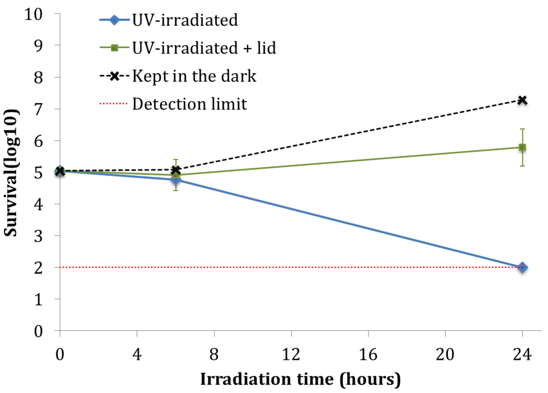
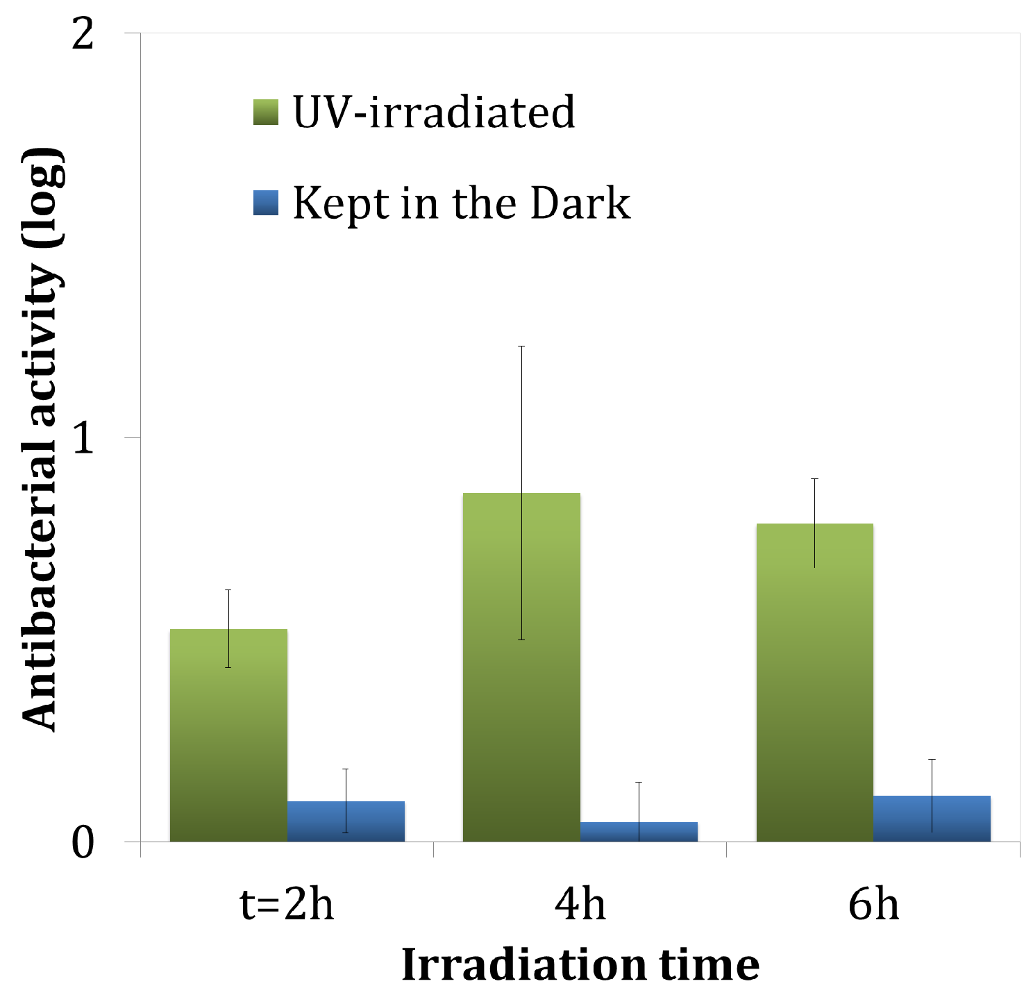
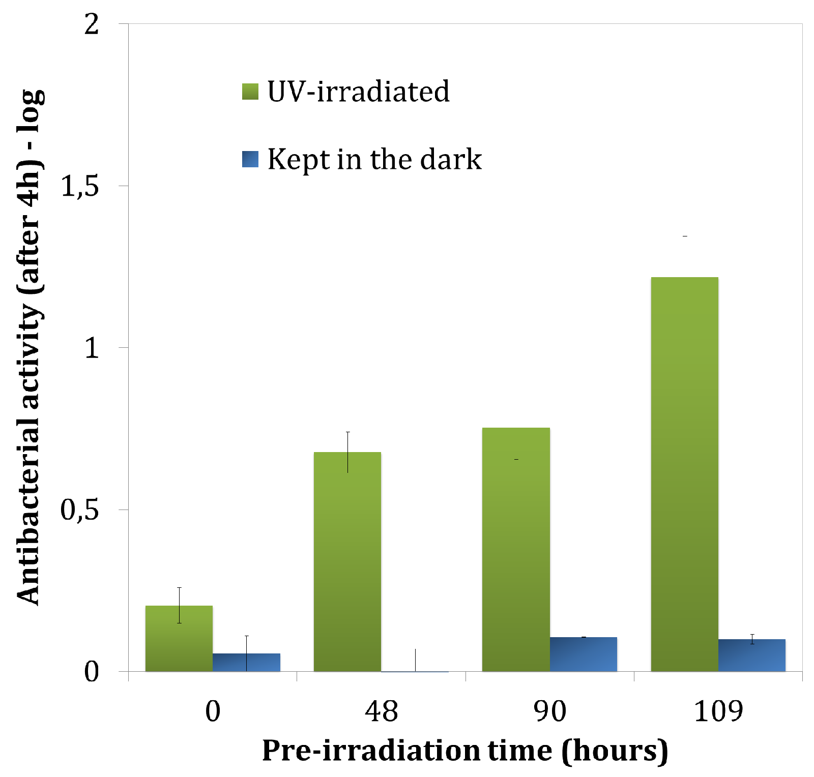
3.1. Antibacterial Activity

The results obtained from the analysis of TiO 2 antibacterial activity on coated glass are summarized in Figure 2.
The TiO 2 coating showed no antibacterial activity in the dark. Irradiation led to lower CFU values and activities were defined at 0.53 ± 0.1 log (p = 0.241), 0.96 ± 0.43 log (p = 0.018) and 0.79 ± 0.11 log (p = 0.03) after 2 h, 4 h, and 6 h of UV irradiation, respectively.
These inactivation values are relatively low when compared to the inactivation values found in the literature on photocatalytic TiO 2 coatings. Many factors strongly impact the microbial inactivation of TiO 2 : the inoculum concentration, the nature of aqueous medium of the inoculum, the support material of the coating, the contact between TiO 2 particles and microbial cells, etc. [1,2,3]. However, two factors are quite often not highlighted: Irradiance and TiO 2 implementation. Regarding irradiance, in the frame of building engineering, it is desired to set up ‘passive’ antibacterial solutions, meaning that can be activated by low irradiances (daylight or conventional indoor lightning). This is why we chose to work under such values (<5 W/m 2 ). Regarding the photocatalyst implementation, its optimization often leads to expensive processes or processes that are impossible to implement on building (calcination process generally involved). The most interesting solutions are therefore inclusion in the mass (photocatalytic concrete) or paint-like coatings. One purpose of this study was to emphasize that such photocatalytic paint-like coating presents significant inactivation rates. Even if these values are far from those found in the literature, they are encouraging especially because they are observed in extreme contamination conditions (still far from the contamination that can occur in real conditions).
Nevertheless, these activities are quite a lot lower than those obtained from the commercial TiO 2 powder alone (VLP5070) alone in a previous study [51]: 2.62 ± 0.20 log (p < 0.001) after 4 h and 3.73 ± 0.24 log (p = 0.001) after 6 h of experiments. This can be explained by the presence of organic binder, which can interfere in the photocatalytic disinfection by directly reacting with TiO 2 particles. Various studies have shown that the presence of organic compounds could reduce photocatalytic efficiency [51,56,57,58,59,60]. Moreover, such results could be explained by the encapsulation of TiO 2 particles in the organic binder of the coating substantially reducing the antibacterial efficiency. The encapsulation of particles inside the acrylic binder might hamper the UV incidence on TiO 2 particles, which would reduce photocatalytic reactions.
Figure 3 presents the antibacterial activity of the photocatalytic coating (with TiO 2 ) on membrane after 4 h of experiment under UV irradiation, for several times of pre-irradiation of the coating (0 h, 48 h, 90 h, and 109 h prior to the test). The antibacterial activity increased with the pre-irradiation time. Samples that had not been pre-irradiated (0 h) showed an antibacterial activity of 0.20 ± 0.06 log (p = 0.016) after 4 h of experiment. The activities increased for samples that had been pre-irradiated for 48 h ( 0.68 ± 0.06 log ) or for 90 h ( 0.75 ± 0.10 log ) with the higher detected activity of 1.22 ± 0.13 log (p < 0.001) for a 109 h pre-irradiation.
After a few hours of UV-irradiation, the TiO 2 coating showed significant antibacterial activities that could reflect bactericidal action and/or growth inhibition (bacteriostatic action). Moreover, the antibacterial activity increased significantly after 48 h of pre-irradiation.
A possible explanation for these results may be the degradation of a first layer of the coating that covers most of TiO 2 particles. When samples were irradiated before the test (pre-irradiation), some of the UV beams may have reached encapsulated particles near the surface and activated the photocatalytic process. The organic coating would thus be partially degraded, generating additional porosity in the coating surface. The apparition of this new porosity would favor:
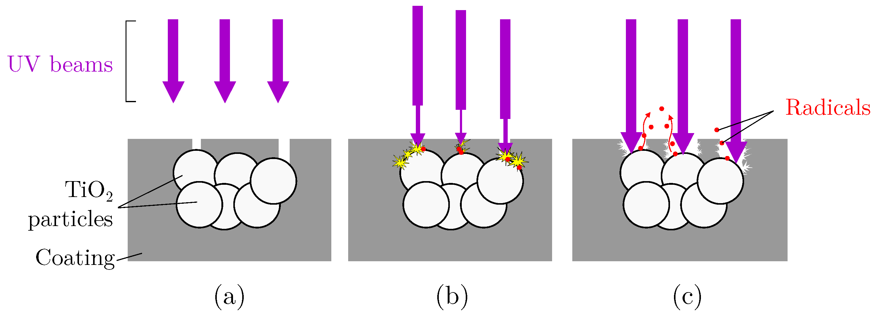
  • The access of UV beams for photocatalytic activation.
  • The access of the pollutants (here bacteria cells) to the particles.
  • Diffusion of the reactive radicals produced by photocatalysis toward pollutants, as shown in Figure 4.
Regarding this phenomenon of self-degradation of the coating, it is important to specify that this study follows previous work on photocatalytic coatings for air purifying [36] and for bacteria inactivation [51]. Coatings’ formulation was initially engineered in the thesis of Martinez [48] on air purifying. In his work, TiO 2 coatings included a part of silicates as inorganic binder, promoting adhesion on building materials and sustainability of coatings. In the frame of bacterial inactivation, the presence of alkaline silicates (pH = 11–12) has a major impact on bacteria survival. In order to prevent overlapping between pH and photocatalysis effects, we decided to use a ‘partial’ coating, silicate free. Regarding the silicate-based coatings, no degradation phenomenon of the coating was observed over time (same samples were tested after several weeks). It seems possible that only the surface layer of the coating is degraded and that TiO 2 particles will subsequently react with external pollutants or other molecules from ambient air. Further work on pre-irradiation times greater than 109 h would be useful to evaluate if the activity increases, is maintained or decreases.
The antibacterial activity experiment, carried out under low UV irradiation (2.5–3 W/m 2 ) confirmed that the efficiency on the bacterial inactivation of the TiO 2 coating increased after 48 h of pre-irradiation. The combined effect of photoinduced hydrophilicity and antibacterial activity of TiO 2 coating suggests an antibacterial action that is effective immediately on contact, providing resistance to bacterial adhesion.

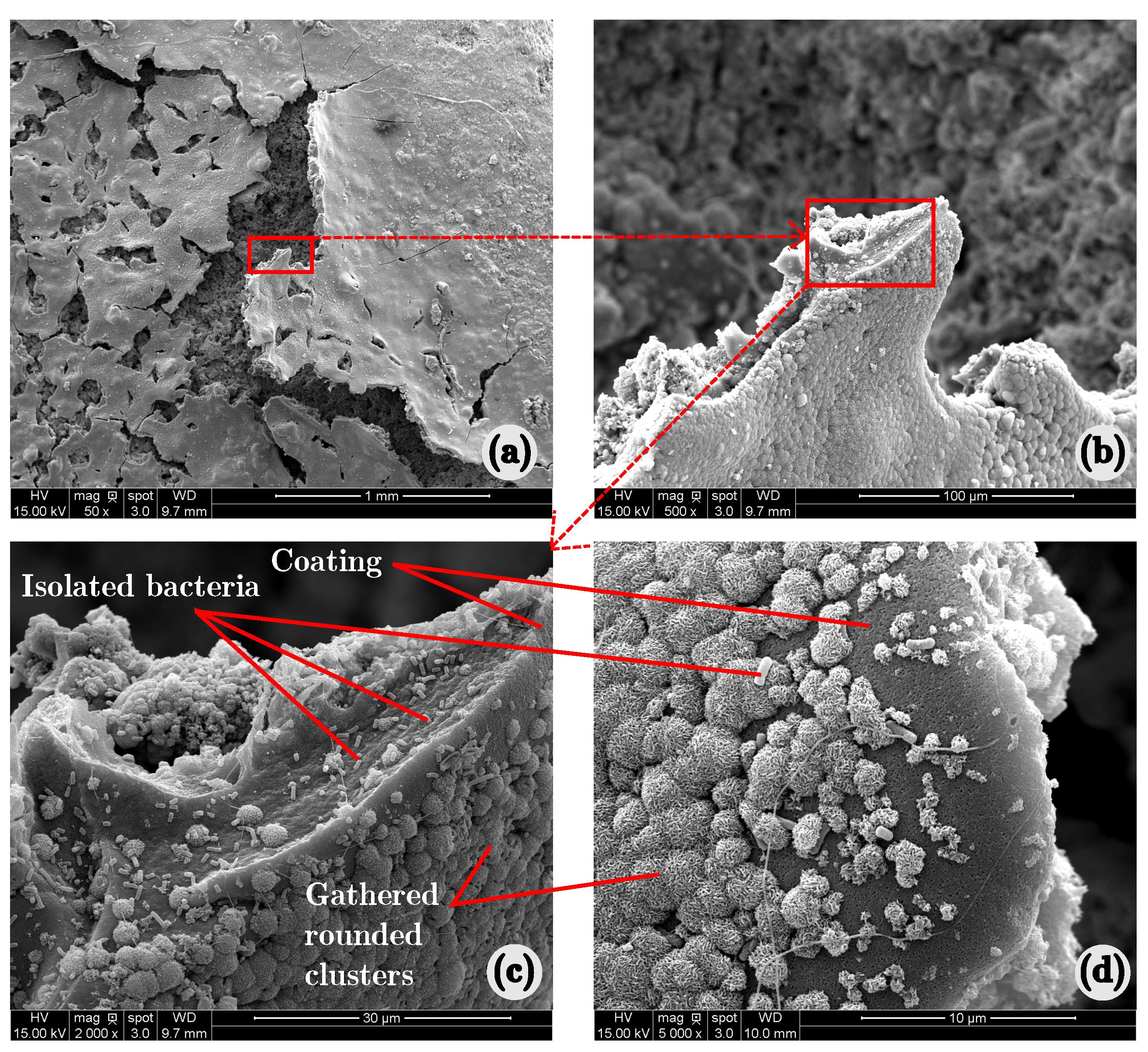
3.2. Developement of a Bacterial Biofilm under Accelerated Conditions

Epifluorescence observations were carried out on the surface of samples after 24 h of experiment. Epifluorescence observation gave the best images of the spread of microbial colonization on samples. Figure 5 shows epifluorescence images of the surface of samples after 24 h of experiment. A light blue color was chosen to represent the coating and red to highlight the bacteria. Images taken on control coating either stored in the dark or exposed to UV irradiation showed bacterial colonization spread over the entire surface of samples (Figure 5a,b).
As can be seen from Figure 5c-1, TiO 2 -coated samples that had been irradiated for 24 h showed areas of bacterial colonization spread over the entire surface but with very low density. Only a few red spots were visible in the near-center area of samples, meaning that bacterial cells were somewhat isolated from each other. Figure 5c-2 was taken on the edge area of the sample surface. The figure shows high concentrations of bacterial cells (red spots) surrounding TiO 2 coating (blue areas). It can be clearly seen that bacteria have developed mainly around the coating (directly on cementitious support) and very little on it. This ‘repulsive’ effect seems to confirm the results from antibacterial activity of the coating. Even under very severe conditions of microbial contamination, the coating was still active and prevented the biofilm development on its surface. These results are also in accordance with a recent study that showed a significant antibacterial effect of TiO 2 coatings against the formation of microbial biofilms under low irradiance [40].
More tests would be required to validate the methodology. However, the presented images of early trials encourages the use of such device to study microbial growth on coated building materials. Moreover, further experimental investigations could be carried out with bacteria genetically modified to emit their own fluorescence, which would allow the evolution of the microbial colonization of material to be observed in real time, using a time lapse method [61,62]. In addition, the use of a fluorochrome such as propidium iodide, which is able to penetrate through damaged membrane only, would enable live cells, damaged membrane and dead cells to be quantified as previously described by Gregori et al. [63].
Figure 6 shows SEM observations of control-coated samples irradiated with UV (Figure 6a,b). Other samples that have been kept in the dark, i.e., control-coated samples and TiO 2 -coated samples, presented the same colonization pattern (as did replicate samples, not shown). On Figure 6a,b, rounded protuberances can be observed, which gather to form a cohesive substance. This substance was not detected on control samples that had not been contaminated (Figure 6c,d). It was likely to be organic-based and produced by bacteria. A similar matrix was observed in the form of a dense network of curli fibers organized as a ‘basket’-like structure around the cells [64]. On a smaller scale (Figure 6b), many bacterial cells were visible evolving near the surface or encased by the dense curli network.
Observations of TiO 2 -coated samples irradiated with UV light for 24 h of experiment showed colonization patterns. As can be seen from Figure 7, bacteria also colonized the coating in some areas. Figure 7c,d also show that parts of the coating were almost completely covered by the oragnic matrix network.
Damaged bacteria were also detected on the photocatalytic coating that had been UV-irradiated for 24 h. Figure 8a,b show intact bacterial cells from control samples kept in the dark. The natural shape of the bacillus, the classic physical appearance of E. coli, is quite visible. Figure 8c,d,e show damaged bacteria that were detected on UV-irradiated TiO 2 -coated samples. Several similar clusters of damaged bacteria were found on the coating. The cells were always gathered in clusters. In addition, it can be seen from Figure 8d,e that small aggregates were attached on the cell walls of some bacteria. These particles were obviously submicrometer sized. They were detected only on visibly damaged bacteria. Moreover, such clusters of damaged bacteria were not detected on TiO 2 -coated samples kept in the dark nor on control-coated samples. These aggregates thus might be TiO 2 particles that may have participated in the degradation of the cells.

4. Conclusions

In this paper, two types of experiments were carried out in order to evaluate the antibacterial properties of a semi-transparent coating incorporating photocatalytic TiO 2 particles. Experimentation focused on the measurement of: (a) the photoinduced antibacterial activity of TiO 2 coating, and (b) its resistance to the formation of bacterial biofilm under accelerated growth conditions.
The tested coating showed significant effects in terms of antibacterial activity and resistance to biofilm formation under low irradiation (2.5 W/m 2 ). The antibacterial activity was induced by photocatalysis. SEM analyses confirmed the lethal activity of the coating on bacterial cells, even in favorable conditions of biofilm formation considered as a ‘worst case’ in comparison to the main conditions of use.
The Epifluorescence approach carried out to evaluate biofilm formation was suitable for TiO 2 -coated cementitious supports. Fluorescence observations provided good pictures of the colonization patterns on the surface of samples. This work can be seen as a preliminary study exploring the potential of epifluorescence microscopy in a field in which it is little exploited: microbial contamination of building materials. Further tests are necessary to validate the methodology, e.g., quantitative measurements by chemical or molecular methods. Subsequently, the methodology could be used in studies on microbial growth and spread on coatings and/or on building materials.
Regarding the coating, further research should be undertaken to explore the optimal formulation, the distribution of TiO 2 particles and the application of the coating to building materials. More work should also be carried out to evaluate the antimicrobial properties towards gram positive bacteria, including Staphylococcus aureus (also recommended by JIS Z 2801) and molds. The molds will be chosen among the most detected in indoor environments such as Penicillium, Aspergillus, and Cladosporium species.
References

Acknowledgments

The authors would like to thank Université Toulouse III—Paul Sabatier for its financial support.

Author Contributions

T.V. conceived and designed the experiments with the help of C.R.; T.V. performed the experiments; T.V. and B.E. performed the Epifluorescence observations and the corresponding analysis; T.V., A.B. and C.R. analyzed the data; T.V. wrote the paper.

Conflicts of Interest

The authors declare no conflict of interest.

References

  1. Gutarowska, B.; Piotrowska, M. Methods of mycological analysis in buildings. Build. Environ. 2007, 42, 1843–1850. [Google Scholar] [CrossRef]
  2. Verdier, T.; Coutand, M.; Bertron, A.; Roques, C. A review of indoor microbial growth across building materials and sampling and analysis methods. Build. Environ. 2014, 80, 136–149. [Google Scholar] [CrossRef]
  3. Bertron, A. Understanding interactions between cementitious materials and microorganisms: A key to sustainable and safe concrete structures in various contexts. Mater. Struct. 2014, 47, 1787–1806. [Google Scholar] [CrossRef]
  4. Guidelines for Indoor Air Quality; WHO: Geneva, Switzerland, 2006.
  5. Escadeillas, G.; Bertron, A.; Blanc, P.; Dubosc, A. Accelerated testing of biological stain growth on external concrete walls. Part 1: Development of the growth tests. Mater. Struct. 2007, 40, 1061–1071. [Google Scholar] [CrossRef]
  6. Verdier, T.; Bertron, A.; Johansson, P. Overview of Indoor Microbial Growth on Building Materials. In Proceedings of the International RILEM Symposium on the Microorganisms-Cementitious materials Interactions (TC253-MCI), Delft, The Netherlands, 23 June 2016. [Google Scholar]
  7. Johansson, P.; Ekstrand-Tobin, A.; Svensson, T.; Bok, G. Laboratory study to determine the critical moisture level for mould growth on building materials. Int. Biodeterior. Biodeterior. 2012, 73, 23–32. [Google Scholar] [CrossRef]
  8. Pasanen, A.L. A Review: Fungal Exposure Assessment in Indoor Environments. Indoor Air 2001, 11, 87–98. [Google Scholar] [CrossRef] [PubMed]
  9. Warscheid, T.; Braams, J. Biodeterioration of stone: A review. Int. Biodeterior. Biodeterior. 2000, 46, 343–368. [Google Scholar] [CrossRef]
  10. Samson, R.; Flannigan, B.; Flannigan, M.; Verhoeff, A.; Adan, O.; Hoekstra, E. Health Implications of Fungi in Indoor Environments; Elsevier: Amsterdam, The Netherlands, 1994. [Google Scholar]
  11. Torvinen, E.; Meklin, T.; Torkko, P.; Suomalainen, S.; Reiman, M.; Katila, M.L.; Paulin, L.; Nevalainen, A. Mycobacteria and Fungi in Moisture-Damaged Building Materials. Appl. Environ. Microbiol. 2006, 72, 6822–6824. [Google Scholar] [CrossRef] [PubMed]
  12. Andersson, M.A.; Nikulin, M.; Köljalg, U.; Andersson, M.C.; Rainey, F.; Reijula, K.; Hintikka, E.L.; Salkinoja-Salonen, M. Bacteria, molds, and toxins in water-damaged building materials. Appl. Environ. Microbiol. 1997, 63, 387–393. [Google Scholar] [PubMed]
  13. Lacaze, I. Etude des Mécanismes de Colonisation des Produits de Construction Par les Micromycètes. Ph.D. Thesis, Microbiologie Procaryote Et Eucaryote, Université Paris-Diderot, Paris, France, 2016. (in French). [Google Scholar]
  14. Sharpe, R.A.; Cocq, K.L.; Nikolaou, V.; Osborne, N.J.; Thornton, C.R. Identifying risk factors for exposure to culturable allergenic moulds in energy efficient homes by using highly specific monoclonal antibodies. Environ. Res. 2016, 144, 32–42. [Google Scholar] [CrossRef] [PubMed] [Green Version]
  15. Flannigan, B.; Samson, R.A.; Miller, J.D. Microorganisms in Home and Indoor Work Environments: Diversity, Health Impacts, Investigation and Control, 2nd ed.; CRC Press: Boca Raton, FL, USA, 2011; p. 529. [Google Scholar]
  16. Parat, S.; Perdrix, A.; Mann, S.; Cochet, C. A study of the relationship between airborne microbiological concentrations and symptoms in office in buildings. In Proceedings of the Healthy Buildings ’95: An International Conference on Healthy Buildings in Mild Climate, Milano, Italy, 10–14 September 1995; Volume III, pp. 1481–1486. [Google Scholar]
  17. Spilak, M.P.; Madsen, A.M.; Knudsen, S.M.; Kolarik, B.; Hansen, E.W.; Frederiksen, M.; Gunnarsen, L. Impact of dwelling characteristics on concentrations of bacteria, fungi, endotoxin and total inflammatory potential in settled dust. Build. Environ. 2015, 93, 64–71. [Google Scholar] [CrossRef]
  18. Prescott, L.M.; Harley, J.P.; Klein, D.A.; Willey, J.M.; Sherwood, L.M.; Woolverton, C.J. Microbiology, 7th ed.; McGraw-Hill: New York, NY, USA, 2007. [Google Scholar]
  19. Costerton, J.W.; Lewandowski, Z.; DeBeer, D.; Caldwell, D.; Korber, D.; James, G. Biofilms, the customized microniche. J. Bacteriol. 1994, 176, 2137–2142. [Google Scholar] [CrossRef] [PubMed]
  20. Davies, D.G.; Parsek, M.R.; Pearson, J.P.; Iglewski, B.H.; Costerton, J.W.; Greenberg, E.P. The Involvement of Cell-to-Cell Signals in the Development of a Bacterial Biofilm. Science 1998, 280, 295–298. [Google Scholar] [CrossRef] [PubMed]
  21. Shirakawa, M.A.; Tavares, R.G.; Gaylarde, C.C.; Taqueda, M.E.S.; Loh, K.; John, V.M. Climate as the most important factor determining anti-fungal biocide performance in paint films. Sci. Total Environ. 2010, 408, 5878–5886. [Google Scholar] [CrossRef] [PubMed]
  22. Gutarowska, B.; Pietrzak, K.; Machnowski, W.; Danielewicz, D.; Szynkowska, M.; Konca, P.; Surma-Slusarska, B. Application of Silver Nanoparticles for Disinfection of Materials to Protect Historical Objects. Curr. Nanosci. 2014, 10, 277–286. [Google Scholar] [CrossRef]
  23. Shirakawa, M.A.; Gaylarde, C.C.; Sahão, H.D.; Lima, J.R.B. Inhibition of Cladosporium growth on gypsum panels treated with nanosilver particles. Int. Biodeterior. Biodeterior. 2013, 85, 57–61. [Google Scholar] [CrossRef]
  24. De Niederhãusern, S.; Bondi, M.; Bondioli, F. Self-Cleaning and Antibacteric Ceramic Tile Surface. Int. J. Appl. Ceram. Technol. 2013, 10, 949–956. [Google Scholar] [CrossRef]
  25. Valentin, R.; Alignan, M.; Giacinti, G.; Renaud, F.N.R.; Raymond, B.; Mouloungui, Z. Pure short-chain glycerol fatty acid esters and glycerylic cyclocarbonic fatty acid esters as surface active and antimicrobial coagels protecting surfaces by promoting superhydrophilicity. J. Colloid Interface Sci. 2012, 365, 280–288. [Google Scholar] [CrossRef] [PubMed]
  26. Carp, O.; Huisman, C.; Reller, A. Photoinduced reactivity of titanium dioxide. Prog. Solid State Chem. 2004, 32, 33–177. [Google Scholar] [CrossRef]
  27. Chong, M.N.; Jin, B.; Chow, C.W.K.; Saint, C. Recent developments in photocatalytic water treatment technology: A review. Water Res. 2010, 44, 2997–3027. [Google Scholar] [CrossRef] [PubMed]
  28. Dalrymple, O.K.; Stefanakos, E.; Trotz, M.A.; Goswami, D.Y. A review of the mechanisms and modeling of photocatalytic disinfection. App. Catal. B Environ. 2010, 98, 27–38. [Google Scholar] [CrossRef]
  29. Foster, H.A.; Ditta, I.B.; Varghese, S.; Steele, A. Photocatalytic disinfection using titanium dioxide: Spectrum and mechanism of antimicrobial activity. Appl. Microbiol. Biotechnol. 2011, 90, 1847–1868. [Google Scholar] [CrossRef] [PubMed]
  30. Taylor, D.M. Transmissible Degenerative Encephalopathies: Inactivation of the Unconventional Causal Agents; John Wiley & Sons: Hoboken, NJ, USA, 2008; p. 324. [Google Scholar]
  31. Fujishima, A.; Rao, T.N.; Tryk, D.A. Titanium dioxide photocatalysis. J. Photochem. Photobiol. C Photochem. Rev. 2000, 1, 1–21. [Google Scholar] [CrossRef]
  32. Maury-Ramirez, A.; Muynck, W.D.; Stevens, R.; Demeestere, K.; Belie, N.D. Titanium dioxide based strategies to prevent algal fouling on cementitious materials. Cem. Concr. Compos. 2013, 36, 93–100. [Google Scholar] [CrossRef]
  33. Guo, M.Z.; Maury-Ramirez, A.; Poon, C.S. Self-cleaning ability of titanium dioxide clear paint coated architectural mortar and its potential in field application. J. Clean. Prod. 2016, 112, 3583–3588. [Google Scholar] [CrossRef]
  34. Maury-Ramirez, A.; Demeestere, K.; Belie, N.D.; Mäntylä, T.; Levänen, E. Titanium dioxide coated cementitious materials for air purifying purposes: Preparation, characterization and toluene removal potential. Build. Environ. 2010, 45, 832–838. [Google Scholar] [CrossRef]
  35. Boonen, E.; Beeldens, A. Recent Photocatalytic Applications for Air Purification in Belgium. Coatings 2014, 4, 553–573. [Google Scholar] [CrossRef]
  36. Martinez, T.; Bertron, A.; Ringot, E.; Escadeillas, G. Degradation of NO using photocatalytic coatings applied to different substrates. Build. Environ. 2011, 46, 1808–1816. [Google Scholar] [CrossRef]
  37. Hot, J.; Martinez, T.; Wayser, B.; Ringo, E.; Bertron, A. Photocatalytic degradation of NO/NO2 gas discharge in a 10 m3 experimental chamber. Environ. Sci. Pollut. Res. Int. 2016, 24, 12562–12570. [Google Scholar] [CrossRef] [PubMed]
  38. Maury-Ramirez, A.; Demeestere, K.; Belie, N.D. Photocatalytic activity of titanium dioxide nanoparticle coatings applied on autoclaved aerated concrete: Effect of weathering on coating physical characteristics and gaseous toluene removal. J. Hazard. Mater. 2012, 211–212, 218–225. [Google Scholar] [CrossRef] [PubMed]
  39. Verdier, T. Élaboration de Revêtements Pour Matériaux de Construction Visant à Lutter Contre la Prolifération Microbienne à L’intérieur des Bâtiments: Efficacité et Mode D’action. Ph.D. Thesis, Toulouse III-Paul Sabatier, Toulouse, France, 2015. (in French). [Google Scholar]
  40. Falco, G.D.; Porta, A.; Petrone, A.M.; Gaudio, P.D.; Hassanin, A.E.; Commodo, M.; Minutolo, P.; Squillace, A.; D’Anna, A. Antimicrobial activity of flame-synthesized nano-TiO2 coatings. Environ. Sci. Nano 2017, 4, 1095–1107. [Google Scholar] [CrossRef]
  41. Wiktor, V.; De Leo, F.; Urzì, C.; Guyonnet, R.; Grosseau, P.; Garcia-Diaz, E. Accelerated laboratory test to study fungal biodeterioration of cementitious matrix. Int. Biodeterior. Biodeterior. 2009, 63, 1061–1065. [Google Scholar] [CrossRef] [Green Version]
  42. Wiktor, V.; Grosseau, P.; Guyonnet, R.; Garcia-Diaz, E.; Lors, C. Accelerated weathering of cementitious matrix for the development of an accelerated laboratory test of biodeterioration. Mater. Struct. 2010, 44, 623–640. [Google Scholar] [CrossRef] [Green Version]
  43. Maness, P.C.; Smolinski, S.; Blake, D.M.; Huang, Z.; Wolfrum, E.J.; Jacoby, W.A. Bactericidal Activity of Photocatalytic TiO2 Reaction: toward an Understanding of Its Killing Mechanism. Appl. Environ. Microbiol. 1999, 65, 4094–4098. [Google Scholar] [PubMed]
  44. Sunada, K.; Watanabe, T.; Hashimoto, K. Studies on photokilling of bacteria on TiO2 thin film. J. Photochem. Photobiol. A Chem. 2003, 156, 227–233. [Google Scholar] [CrossRef]
  45. Blöß, S.P.; Elfenthal, L. Doped titanium dioxide as a photocatalyst for UV and visible light. In Proceedings of the International RILEM Symposium on Photocatalysis, Environment and Construction Materials TDP, Florence, Italy, 8–9 October 2007; pp. 31–38. [Google Scholar]
  46. Fujishima, A.; Zhang, X. Titanium dioxide photocatalysis: present situation and future approaches. Comptes Rendus Chim. 2006, 9, 750–760. [Google Scholar] [CrossRef]
  47. Husken, G.; Hunger, M.; Brouwers, H.J.H. Experimental study of photocatalytic concrete products for air purification. Build. Environ. 2009, 44, 2463–2474. [Google Scholar] [CrossRef]
  48. Martinez, T. Revêtements Photocatalytiques Pour les Matériaux de Construction: Formulation, Évaluation de L’efficacité de la Dépollution de L’air et Écotoxicité. Ph.D. Thesis, Toulouse III-Paul Sabatier, Toulouse, France, 2012. (in French). [Google Scholar]
  49. Ishiguro, H.; Nakano, R.; Yao, Y.; Kajioka, J.; Fujishima, A.; Sunada, K.; Minoshima, M.; Hashimoto, K.; Kubota, Y. Photocatalytic inactivation of bacteriophages by TiO2-coated glass plates under low-intensity, long-wavelength UV irradiation. Photochem. Photobiol. Sci. 2011, 10, 1825–1829. [Google Scholar] [CrossRef] [PubMed]
  50. ISO 27447 Fine Ceramics (Advanced Ceramics, Advanced Technical Ceramics)—Test Method for Antibacterial Activity of Semiconducting Photocatalytic Materials; International Organization for Standardization: Geneva, Switzerland, 2009.
  51. Verdier, T.; Coutand, M.; Bertron, A.; Roques, C. Antibacterial Activity of TiO2 Photocatalyst Alone or in Coatings on E. coli: The Influence of Methodological Aspects. Coatings 2014, 4, 670–686. [Google Scholar] [CrossRef]
  52. JIS Z 2801 Antibacterial Products—Test for Antibacterial Activity And Efficacy; Biosan Laboratories, Inc.: Warren, MI, USA, 2010.
  53. Alasri, A.; Roques, C.; Michel, G.; Cabassud, C.; Aptel, P. Bactericidal properties of peracetic acid and hydrogen peroxide, alone and in combination, and chlorine and formaldehyde against bacterial water strains. Can. J. Microbiol. 1992, 38, 635–642. [Google Scholar] [CrossRef]
  54. Pineau, L.; Roques, C.; Luc, J.; Michel, G. Automatic washer disinfector for flexible endoscopes: A new evaluation process. Endoscopy 1997, 29, 372–379. [Google Scholar] [CrossRef] [PubMed]
  55. Campanac, C.; Pineau, L.; Payard, A.; Baziard-Mouysset, G.; Roques, C. Interactions between Biocide Cationic Agents and Bacterial Biofilms. Antimicrob. Agents Chemother. 2002, 46, 1469–1474. [Google Scholar] [CrossRef] [PubMed]
  56. Ortelli, S.; Blosi, M.; Albonetti, S.; Vaccari, A.; Dondi, M.; Costa, A.L. TiO2 based nano-photocatalysis immobilized on cellulose substrates. J. Photochem. Photobiol. A Chem. 2014, 276, 58–64. [Google Scholar] [CrossRef]
  57. Saito, T.; Iwase, T.; Horie, J.; Morioka, T. Mode of photocatalytic bactericidal action of powdered semiconductor TiO2 on mutans streptococci. J. Photochem. Photobiol. B Biol. 1992, 14, 369–379. [Google Scholar] [CrossRef]
  58. Gogniat, G.; Thyssen, M.; Denis, M.; Pulgarin, C.; Dukan, S. The bactericidal effect of TiO2 photocatalysis involves adsorption onto catalyst and the loss of membrane integrity. FEMS Microbiol. Lett. 2006, 258, 18–24. [Google Scholar] [CrossRef] [PubMed]
  59. Dunlop, P.; Byrne, J.; Manga, N.; Eggins, B. The photocatalytic removal of bacterial pollutants from drinking water. J. Photochem. Photobiol. A Chem. 2001, 148, 355–363. [Google Scholar] [CrossRef]
  60. Rincón, A.G.; Pulgarin, C. Effect of pH, inorganic ions, organic matter and H2O2 on E. coli K12 photocatalytic inactivation by TiO2: Implications in solar water disinfection. Appl. Catal. B Environ. 2004, 51, 283–302. [Google Scholar] [CrossRef]
  61. Gogoi, S.K.; Gopinath, P.; Paul, A.; Ramesh, A.; Ghosh, S.S.; Chattopadhyay, A. Green Fluorescent Protein-Expressing Escherichia coli as a Model System for Investigating the Antimicrobial Activities of Silver Nanoparticles. Langmuir 2006, 22, 9322–9328. [Google Scholar] [CrossRef] [PubMed]
  62. Erable, B.; Vandecandelaere, I.; Faimali, M.; Delia, M.L.; Etcheverry, L.; Vandamme, P.; Bergel, A. Marine aerobic biofilm as biocathode catalyst. Bioelectrochemistry 2010, 78, 51–56. [Google Scholar] [CrossRef] [PubMed] [Green Version]
  63. Gregori, G.; Citterio, S.; Ghiani, A.; Labra, M.; Sgorbati, S.; Brown, S.; Denis, M. Resolution of Viable and Membrane-Compromised Bacteria in Freshwater and Marine Waters Based on Analytical Flow Cytometry and Nucleic Acid Double Staining. Appl. Environ. Microbiol. 2001, 67, 4662–4670. [Google Scholar] [CrossRef] [PubMed]
  64. Serra, D.O.; Richter, A.M.; Klauck, G.; Mika, F.; Hengge, R. Microanatomy at Cellular Resolution and Spatial Order of Physiological Differentiation in a Bacterial Biofilm. mBio 2013, 4, e00103-13. [Google Scholar] [CrossRef] [PubMed]
Figure 1. Direct effect of the time of UV irradiation on the survival of Escherichia coli CIP 53126 expressed as the log residual viable bacteria, light intensity ≈2.5 W/m 2 .
Coatings 08 00093 g001
Figure 2. Antibacterial activity on E. coli of TiO 2 -coating (glass slides), after 2 h, 4 h, and 6 h of contact. Mean ± s.e., n = 3.
Coatings 08 00093 g002
Figure 3. Antibacterial activity of TiO 2 -coating on E. coli after 4 h of test, depending on the time of pre-irradiation of the coating (3 W/m 2 ). Mean ± s.e., n = 3.
Coatings 08 00093 g003
Figure 4. Schematic illustration of the disencapsulation mechanism of TiO 2 particles from acrylic coating. (a) starting UV pre-irradiation; (b) part of the UV beams reach particles near the surface, activating the photocatalytic reaction: the coating starts to degrade around particles; (c) apparition of additional porosity, favoring access of UV beams, diffusion of radicals outside the coating and diffusion of pollutants inside the coating.
Coatings 08 00093 g004
Figure 5. Epifluorescence pictures of surface samples after 24 h of experiment. (a) control-coated sample kept in the dark; (b) control-coated sample under UV irradiation; (c) TiO 2 -coated samples under UV irradiation. For TiO 2 -coated samples, one image was taken at the center of the sample (c-1) and another at the edge (c-2).
Coatings 08 00093 g005
Figure 6. SEM images (secondary electron mode) of samples after 24 h of experiment. (a,b) SEM images of control samples without TiO 2 ( × 500 and × 3000 ) showing an organic matrix and ovoid bacteria encased by or near surface of this dense network of curli fibers. (c,d) SEM images of TiO 2 -coated samples that have not been inoculated ( × 500 and × 3000 ) showing the surface of coating bacteria-free.
Coatings 08 00093 g006
Figure 7. SEM images (secondary electron mode) of TiO 2 -coated samples irradiated with UV light for 24 h.
Coatings 08 00093 g007
Figure 8. SEM images (secondary electron mode) of intact cells on control-coated samples kept in the dark (a,b) and of damaged cells observed on TiO 2 -coated samples irradiated with UV light for 24 h experiment (c,d,e).
Coatings 08 00093 g008
Table 1. Physical characteristics of the VLP5070 (Kronos) TiO 2 particles
DescriptionVLP5070
TiO 2 -Content>85 %
Crystal modificationanatase
Crystallite sizeapprox. 15 nm
Specific surface area>225 m 2 /g
Table 2. Composition of the biofilm nutrient broth.
ComponentsConcentration (g/L)
FeSO 4 , 7H 2 O0.0005
Na 2 HPO 4 1.25
KH 2 PO 4 0.5
(NH 4 ) 2 SO 4 0.1
Glucose0.05
MgSO 4 , 7H 2 O0.02

