Photoactivated Self-Sanitizing Chlorophyllin-Containing Coatings to Prevent Microbial Contamination in Packaged Food

Packaging Group, Instituto de Agroquímica y Tecnología de Alimentos, IATA-CSIC, Av. Agustin Escardino 7, 46980 Paterna, Spain
*
Author to whom correspondence should be addressed.
Coatings 2018, 8(9), 328; https://doi.org/10.3390/coatings8090328
Submission received: 27 August 2018
/
Revised: 12 September 2018
/
Accepted: 19 September 2018
/
Published: 19 September 2018
(This article belongs to the Special Issue Functional Coatings for Food Packaging Applications)
Abstract
:Chlorophyllins are semi-synthetic porphyrins obtained from chlorophyll that—when exposed to visible light—generate radical oxygen substances with antimicrobial activity. In this work, chlorophyllins incorporated with polyethylene (PE), polyvinyl alcohol (PVOH), (hydroxypropyl)methyl cellulose (HPMC), and gelatin (G) were formulated for application as coatings in packages providing antimicrobial activity after photoactivation. First, the antimicrobial properties of two porphyrins (sodium magnesium chlorophyllin, E-140, and sodium copper chlorophyllin, E-141) were analyzed against L. monocytogenes and Escherichia coli. The results indicated that E-140 was more active than E-141 and that chlorophyllins were more effective against Gram-positive bacteria. In addition, both chlorophyllins were more efficient when irradiated with halogen lamps than with LEDs, and they were inactive in dark conditions. Then, coatings on polyethylene terephthalate (PET) film were prepared, and their effect against the test bacteria was similar to that shown previously with pure chlorophyllins, i.e., greater activity in films containing E-140. Among the coating matrices, those based on PE presented the least effect (1 log reduction), whereas PVOH, HPMC, and G were lethal (7 log reduction). The self-sanitizing effect of these coatings was also analyzed by contaminating the surface of the coatings and irradiating them through the PET surface, which showed high efficiency, although the activity of the coatings was limited to L. monocytogenes. Finally, coated films were applied as separators of bologna slices. After irradiation, all the films showed count reductions of L. monocytogenes and the usual microbial load; the gelatin coating was the most effective, with an average of 3 log reduction.
Keywords:
porphyrin; chlorophyllin; active coating; antimicrobial; photoactivation; self-sanitizing; bologna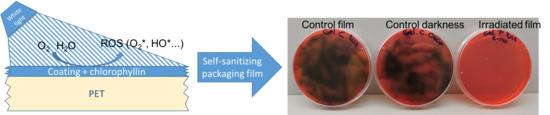
1. Introduction
Recent reports from the World Health Organization (WHO) have pointed out the high incidence of foodborne diseases as a public health problem [1]. The globalization of food supply has presented new challenges for food safety and has contributed to the internationalization of the public health problem of foodborne diseases [1]. Foodborne pathogens are usually eliminated by thermal treatment during food processing [2]. These methods can inactivate pathogenic microorganisms but could also reduce nutrients or modify organoleptic and sensory properties. To overcome these disadvantages, nonthermal treatments, such as high pressure, ultraviolet or pulsed light, or irradiation, have been developed. However, they are not as effective as thermal ones, have a high application cost [3], and are not always accepted by consumers [4].
In the last few years, many research groups have been paying attention to antimicrobial food packaging, a nonthermal treatment that involves the development of active antimicrobial films and coatings to act as protective barriers against pathogens present in food products. Polymer films and coatings can be used as carriers for a wide range of food additives, including various antimicrobials that could reduce the risk of pathogen growth on food surfaces and thus prolong product shelf life. In this context, there is increasing demand for natural, healthier additives. Chlorophyllins (water-soluble sodium magnesium chlorophyllin, E-140, and water-soluble sodium copper chlorophyllin, E-141) are semisynthetic porphyrins obtained from chlorophyll and used as colorants in dietary supplements and in cosmetics [2].
When these molecules are exposed to visible light in air, they generate singlet oxygen and radical oxygen substances, which have antimicrobial activity [2,5]. The cationic 5,10,15,20-tetrakis(1-methylpyridinium-4-yl) porphyrin tetra-iodide as photosensitizer has been reported to provide effective photoinactivation of Pseudomonas syringae pv. actinidiae in kiwifruit leaves under sunlight irradiation without damaging the plant [5]. Other authors have shown that chlorophyllin-based photosensitization reduced mesophilic bacteria and inoculated strains of food pathogens on cherry tomatoes without producing harmful effects on the nutritional quality of the tomatoes or their antioxidant activity [6].
These molecules are not toxic, and they do not present any effect in dark conditions. There are few studies about the use of porphyrins in antimicrobial applications. Porphyrin films with nylon or cellulose have been developed for industrial, household, and medical applications [7,8]. Antimicrobial activity of phthalocyanine-dyed paper has also been tested against E. coli and A. baylyi by illuminating for 1 h under lights of various intensities.
However, the application of polymer films or coatings carrying these antimicrobial compounds to avoid microbial contamination and improve the shelf life of food products has scarcely been reported. Previous studies with chlorophyllin-based films and coatings have shown their effectiveness in preventing microbial contamination of cooked frankfurters inoculated with S. aureus and L. monocytogenes [2]. The antimicrobial activity of chlorophyllins has been found to be dependent on the choice of light source: quartz lamps, near-infrared lamps, halogen lamps, slide projector lamps, special photodynamic lamps, or special light-emitting diode lights [9].
The aim of this work was the development of antimicrobial photosensitizer coatings based on synthetic and biological polymer matrices with various degrees of hydrophilicity, such as gelatin (G), polyvinyl alcohol (PVOH), polyethylene (PE) and (hydroxypropyl) methyl cellulose (HPMC), incorporated with two chlorophyllins and exposed to the radiation of halogen lamps and LED lamps as activation sources. These films were applied on bologna slices to test their antimicrobial effectiveness.
2. Materials and Methods
2.1. Reactive Agents
Water-soluble sodium magnesium chlorophyllin, E-140, and water-soluble sodium copper chlorophyllin, E-141, were obtained from Natracol (ROHA Europe S.L.U., Torrente, Spain). Gelatin from porcine skin (G) and (hydroxypropyl) methyl cellulose (HPMC) were supplied by Sigma (Barcelona, Spain). Low-density polyethylene (LDPE) emulsion (50% w/w) Aquaseal® 2200 (PE) was kindly provided by Paramelt B.V. (Heerhugowaard, The Netherlands), and Gohsenol AH17 polyvinyl alcohol was kindly provided by the Nippon Synthetic Chemical Company (Osaka, Japan).
2.2. Bacterial Strains
Bacterial strains were obtained from the Spanish Type Culture Collection (Valencia, Spain): L. monocytogenes CECT 911 (ATCC 19112) was used as a model of a Gram-positive bacterium and Escherichia coli CECT 434 (ATCC 25922) as a model of a Gram-negative bacterium. The strains were stored in Tryptone soy broth (TSB) with glycerol at −80 °C. For experimental use, the stock cultures were maintained by regular subculture on agar medium slants at 4 °C and transferred every month. A loopful of each strain was transferred to 10 mL of TSB and incubated at 37 °C overnight to obtain early stationary phase cells.
Bacteria were spread on selective media, PALCAM agar and Mueller Hinton agar, for L. monocytogenes and E. coli, respectively.
2.3. Methods
2.3.1. Minimum Inhibitory Concentration and Minimum Bactericidal Concentration
Minimum inhibitory concentration (MIC) and minimum bactericidal concentration (MBC) of porphyrin were determined for E. coli and L. monocytogenes. The MIC is defined as the amount or concentration of active compound in which growth inhibition is observed. The MBC is the concentration in which there is no growth of microorganisms [10]. The procedure, which we have called the “drops method”, consisted of adding 100 μL of microorganism (107 CFU/mL) and 10 μL of chlorophyllin prepared at different concentrations ranging between 0.0016 mg/mL and 15 mg/mL. Chlorophyllins were activated by two lighting systems: three 500 W Haloline 64702 halogen lamps (Osram) or five 50 W 81.575/Dia LED lights (Electro DH) for 15 min. Their light spectra differ greatly: LED has its maximum radiation in a thin band close to 450 nm and a medium-intensity wide band with a maximum at 550 nm and an unimportant contribution in the infrared region, while halogen light increases in intensity with wavelength, having its maximum in the infrared region [11]. As control samples, identical experiments were also carried out with (a) samples with chlorophyllins stored in dark conditions and (b) an illuminated sample without chlorophyllins. After photosensitization, the suspension of microorganisms was recovered, plated in agar medium, and incubated overnight at 37 °C. Cytotoxicity was calculated as the difference between colony forming units (CFU) counted with (photosensitizer and illumination) and without photodynamic treatment. Experiments were carried out in triplicate.
2.3.2. Antimicrobial Coating Preparation
Various matrices were employed as chlorophyllin carriers: gelatin (G), polyethylene (PE), polyvinyl alcohol (PVOH), and (hydroxypropyl) methyl cellulose (HPMC).
G coating-forming solution was prepared by dissolving 10 g of gelatin in 100 mL of 50% (v/v) aqueous ethanol for 2 h at 75 °C and adding 25% of glycerol (with respect to polymer dry mass) as plasticizer.
PE coating emulsion was prepared from a commercial emulsion, previously homogenized for 10 min in an ultrasonic bath. Then, 5% of 2-propanol was added to improve wettability, and the mixture was stirred for 10 min using a magnetic stirrer at room temperature.
PVOH coating-forming solution was prepared by dissolving 4 g in 100 mL of deionized water with 10% of glycerol as plasticizer and stirring for 2 h at 75 °C.
HPMC coating-forming solution was prepared by dissolving 2.5 g of HPMC in 100 mL of 50% (v/v) aqueous ethanol with 20% of glycerol as plasticizer and stirring for 2 h at 75 °C.
G, PE, PVOH, and HPMC coatings were prepared on a 30 µm poly (ethylene terephthalate) (PET) film. The PET film was set on a glass surface and corona-treated (Model BD-20V corona treater, Electro-Technic Products, Chicago, IL, USA) to improve coating adherence. Ten grams of the film-forming solutions was spread using a 50 µm extension bar (Linlab, Logroño, Spain), and the samples were placed in a homemade drying tunnel equipped with a 2500 W heat source and a 30 W fan for 3 min until they were completely dry. Coating thickness was calculated as the difference between the film thicknesses measured with a micrometer before and after the coating process. Finally, the coatings were stored in glass desiccators containing silica gel at 22 ± 2 °C prior to use.
2.3.3. Color Measurements
The color of the coated films was measured using a Konica Minolta CM-3500d spectrophotometer (Konica Minolta Business Technologies, Inc., Tokyo, Japan) set to D65 illuminant/10° observer. Film samples were measured against the surface of a standard white plate, and the CIELAB color space was used to obtain the color coordinates L* (lightness) [black (0) to white (100)], a* [green (−) to red (+)], and b* [blue (−) to yellow (+)]. The color was expressed using the polar coordinates L*, C*, h°, and ΔE*, where L* is the same as above, C* is chroma, h° is hue angle, and ΔE* = [(ΔL*)2 + (Δa*)2 + (Δb*)2]1/2. Ten measurements were taken of each sample, and three samples of each film were measured.
2.3.4. Antimicrobial Activity of Coatings
Agar Diffusion
First, 100 µL of a bacterial suspension containing approximately 103 CFU/mL was spread on agar medium, and then a disk of film (2.5 cm diameter) was put over the agar with the coating surface facing the agar. Samples with and without chlorophyllins were irradiated with five 50 W LED lights for 15 min. At the same time, samples with and without active agent were treated in dark conditions by covering the Petri dishes with aluminum foil.
All the plates were incubated at 37 °C for 24 h, and the diameter of the resulting inhibition zone in the bacterial lawn was measured. The experiment was carried out in triplicate.
Surface Disinfection
The microorganisms to be tested were spread directly on the G, HPMC, PVOH, and PE coatings (with 1% E-140, and some without antimicrobial agent as controls). For this purpose, the coated films were placed on Petri dishes with the coating facing up and 100 μL of 105 CFU/mL of L. monocytogenes or E. coli was spread over them, ensuring direct contact between the bacteria and the coating. In the case of the E. coli bacteria, 20 or 40 mM of ethylenediaminetetraacetic acid (EDTA) was previously added to ease mass transport through the outer membrane. They were then irradiated with 5 LED lights for 15 min. The bacteria were collected with 10 mL of peptone water, and serial dilutions were made and plated on solid agar medium. The incubation was carried out at 37 °C for 24 h and CFU/mL was counted. The experiments were carried out in triplicate.
2.3.5. Release of Chlorophyllins
According to Council Directive 85/572/EEC, which determines the list of simulants that should be used to control the migration of components from materials and objects of plastic material intended to come into contact with food products, the simulant that best represents a fatty food such as bologna is vegetable oil. However, since the two chlorophyllins selected are water soluble, we decided to test the films with 50% ethanol, which is a valid simulant for oil-in-water emulsions. In this case, the exposure conditions were 10 mL of simulant and an exposure area of 12.5 cm2 (5 cm × 2.5 cm rectangles) at a refrigeration temperature of 5 °C for 16 days. At 1 h, 2 h, 4 h, 6 h, 24 h, 4 days, 8 days, and 16 days, samples were analyzed at 655 nm by UV-vis spectrometry (Agilent UV-Visible 8453 Spectroscopy System, Agilent Technologies, Wilmington, DE, USA). To quantify the concentrations of migrated porphyrin, a previous calibration with known concentrations in 50% ethanol was prepared.
2.3.6. Application to Bologna Slices
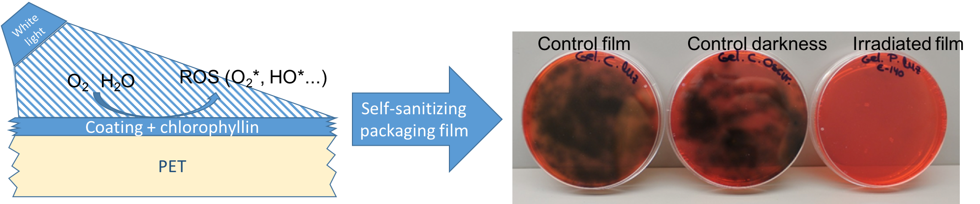
Coatings on PET were used as separators of slices of a real food-bologna. In this test, the antibacterial activity of PVOH, G, PE, and HPMC with 1% porphyrin E-140 coatings was tested against the usual microbial load and against L. monocytogenes, which had been previously inoculated. An 800 g piece of classic bologna containing 50% pork and 15% turkey was purchased and sliced. Slices that were 3 mm thick and 6.8 cm in diameter were prepared, with an average weight per slice of 10.12 ± 0.13 g. Slices of bologna were inoculated with 100 μL of 105 CFU/mL of L. monocytogenes spread over their surface. Then, the active- or control-coated film (with/without porphyrin) was placed on the bologna slices and irradiated for 15 min with 5 LED lamps on a tray with ice to avoid sample overheating (Figure 1). In parallel, control samples with films with and without antimicrobial were kept in darkness for 15 min.
After the treatment, bologna slices were placed in a Stomacher bag together with 10 mL of peptone water and homogenized in a Stomacher (Bagmix, Interscience, St. Nom, France) for 4 min. Serial dilutions were carried out, and the enumeration of particular microbial groups was performed using the following culture conditions and media (from Scharlab, Barcelona, Spain): (a) PALCAM Listeria selective agar for L. monocytogenes incubated at 37 °C for 48 h; (b) plate count agar for total aerobic plate count, pour-plated and incubated at 30 °C for 48 h and plate count agar for total aerobic psychrotrophic count, pour-plated and incubated at 10 °C for 10 days; (c) Violet Red Bile Glucose agar for total enterobacteria, pour-plated and incubated at 37 °C for 48 h; (d) Eosin Methylene Blue agar for coliform bacteria, pour-plated and incubated at 37 °C for 48 h; (e) Brilliant Green agar for Salmonella isolation, pour-plated and incubated at 37 °C for 48 h; (f) King agar for Pseudomonas spp. count, pour-plated and incubated at 25 °C for 4 days; (g) De Man, Rogosa and Sharpe agar for lactic bacteria count, pour-plated and incubated at 25 °C for 4 days. The counts were performed in triplicate.
Once the antimicrobial activity had been evaluated, the possible impact on the color characteristics of the food due to active agent release was studied. The color of bologna samples before and after irradiation was determined using a Konica Minolta CM-3500d spectrophotometer and the same procedure as described in Section 2.3.3. Eight measurements were taken of each sample, and three samples of each film were measured.
2.3.7. Statistical Analysis
All the experiments were conducted at least in triplicate. The data were subjected to an analysis of main effects and interaction factors by multifactorial ANOVA using the StatGraphics Plus 5.1 program.
3. Results and Discussion
3.1. Minimum Inhibitory Concentration (MIC) and Minimum Bactericidal Concentration (MBC) of Chlorophyllins
Antimicrobial activity of E-140 and E-141 chlorophyllins was tested against L. monocytogenes and E. coli using two types of irradiation: halogen and LED lights. The results obtained are shown in Table 1 (halogen light) and Table 2 (LED light).
The first important data were that the MIC values were higher against E. coli than against L. monocytogenes. In general, Gram-positive bacteria are more sensitive than Gram-negative bacteria to chemical compounds owing to the protection of the outer membrane surrounding the Gram-negative cell wall, which restricts the diffusion of antimicrobial substances [12,13]. Caires et al. [14] reported on the antimicrobial activity of two photosensitizers—eosin methylene blue and chlorophyllin sodium copper salt (E-141)—and showed that eosin methylene blue was able to photoinactivate E. coli and S. aureus, while E-141 was only active against S. aureus. In that test, the maximum concentration tested was 50 µM, which was much lower than the 4.15 mM obtained in this study. However, it is difficult to compare the published results because the important parameters of the photoactivation process—intensity, time, and type of radiation—were either different or were not provided. In this test, 15 min was enough to induce photoactivation, much less than the 120 min employed elsewhere [14].
It is also important to note that although both chlorophyllins presented high antimicrobial activity, porphyrin E-140 was much more effective (with much lower MIC and MBC values) than E-141. With regard to the type of photoactivation, halogen lights were more effective than LED lights. The use of LEDs slightly reduced the efficacy of E-140 against L. monocytogenes and E. coli. Other authors have reported the antimicrobial effectiveness of zinc complexes of tetrakis (N-methylpyridinium-4-yl) tetraiodide porphyrin and tetrakis (N-methylpyridinium-4-yl) tetraiodide phthalocyanine impregnated in paper using an inexpensive consumer LED lamp as activation mechanism [9].
In the case of E-141, porphyrin appeared to be inactive when exposed to the radiation of LED lamps, and neither the MBC nor the MIC of the two tested bacteria was found. Our hypothesis is that the LED lamps employed did not emit radiation of sufficient intensity in the wavelength range necessary for E-141 activation. Moreover, copper chlorophyillins have been reported to present low yield of singlet oxygen [15].
Finally, two control tests were carried out. In one, the two microorganisms were exposed to the chlorophyllins in darkness; in the other, they were exposed to radiation without porphyrin. The two microorganisms tested reflected the absence of antimicrobial effect, indicating that chlorophyllins were nontoxic for the two model bacteria, and light radiation alone did not produce antimicrobial activity.
3.2. Development of Coated Films
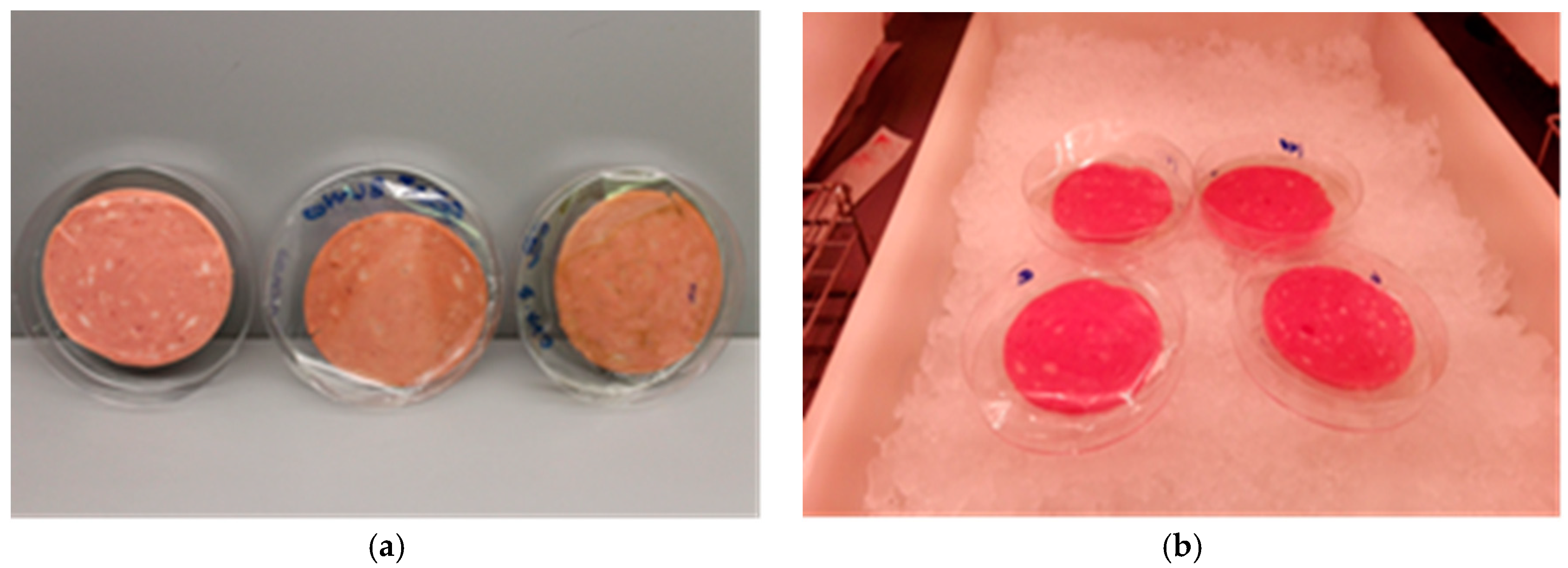
G, PE, PVOH, and HPMC coatings on PET film with and without 1% of E-140 or E-141 were successfully obtained. They were homogeneous, without discontinuities, flexible, transparent, had a light green color when the chlorophyllins were added, and with thicknesses that are shown in Table 3. As can be seen, the coating thicknesses varied according to the polymer material as a consequence of the different solid content of the film-forming solution and the polymer density; the PE-based materials were the thickest, while PVOH and HPMC were the thinnest. The incorporation of chlorophyllins in the coatings did not significantly affect thickness.
The most significant effect of the addition of the chlorophyllins to the coatings was the yellowish (E-140) or greenish (E-141) color induced. Table 4 shows the color coordinates in the CIELAB system for different coatings, including chromaticity (C*) and tone (h). As can be seen, in general, the coated films presented high luminosity as revealed by the L* values ranging between 83 and 90. The only exceptions were the coatings based on PE-incorporated chlorophyllins, which presented a considerable reduction in luminosity. The addition of E-140 provided coatings with a yellowish color, which is characterized in CIELAB coordinates by positive b* values and low negative a* values. As a consequence of this, the tone of the coatings was characterized by values ranging between 95 and 110°. The incorporation of E-141 provided a greener color, with higher negative a* values and positive b* and tone values ranging between 107 and 135°. With respect to saturation or chromaticity, the values were greater for E-141 samples than for E-140. Comparing the polymeric materials, C* values were greatest for the thickest material (PE).
Figure 2 shows, as examples, photos of the different coatings. As can be seen, in the PE coatings there was a clearly visible alteration of the color of the material after incorporation of both chlorophyllins. On the other hand, the color of the HPMC coatings with E-140 or E-141 was hardly distinguishable from the control.
3.3. Antimicrobial Activity of Films Coated with Chlorphyllins
From the results shown above, it was inferred that chlorophyllin E-140 was more efficient than E-141, provided less color change to the PET film, reduced energy consumption and, overall, was active when photoactivated with LED lights as it reduced the heating on the film and on the product. Therefore, coated films containing E-140 were selected to verify the effectiveness of porphyrin incorporated in various polymer matrices. For this purpose, two methods were used: the agar diffusion method and the surface disinfection method.
3.3.1. Agar Diffusion Method
The antimicrobial capacity of G, PVOH, PE, and HPMC films incorporating 1% of chlorophyllin E-140 was studied against L. monocytogenes and E. coli using the agar diffusion method. The results obtained showed that the G, PVOH, and HPMC films had a bactericidal effect against both microorganisms, while the PE film had a certain degree of inhibition, as can be seen in Table 5.
The results showed that the most hydrophilic films, i.e., with a higher degree of affinity for water, presented a greater antibacterial capacity. It is possible that the excited oxygen reacts with water molecules to produce hydroxyl radicals that have a longer lifetime and are more active against bacteria [16]. Accordingly, it was observed that photoexcited G, PVOH, and HPMC films with 1% chlorophyllin E-140 had a lethal effect on L. monocytogenes or E. coli in the area covered by the films. In the case of PE-coated films, a certain degree of inhibition was seen, but it was less than that with the other films, despite the higher amount of porphyrin present owing to the greater thickness of the coating. The absence of water in this hydrophobic polymer may limit the photoactivation to the formation of singlet oxygen, which rapidly reduces to triplet oxygen without interacting with the cells. Another explanation is that the low affinity between chlorophyllin and PE causes itsaggregation, and this results in a lower production of singlet oxygen. The films that developed acted mainly by contact, although in the case of the PVOH, G, and HPMC matrices, an inhibition halo of ca. 5 mm was observed around the films.
Bozja et al. (2003) tested the antimicrobial activity of porphyrins grafted onto nylon-6 fibers with polyacrylic acid. They found that the antimicrobial system developed was very effective against S. aureus but had no effect on E. coli bacteria at any light intensity [7]. There have been some similar studies, such as the one carried out in 2008 in which the effectiveness of E-140 porphyrin-bearing gelatin films as antibacterial agents [2] was demonstrated against S. aureus and L. monocytogenes, but was found to have no effect on Salmonella spp. and E. coli bacteria after photoactivation with 30,000 luxes for five minutes.
3.3.2. Surface Disinfection
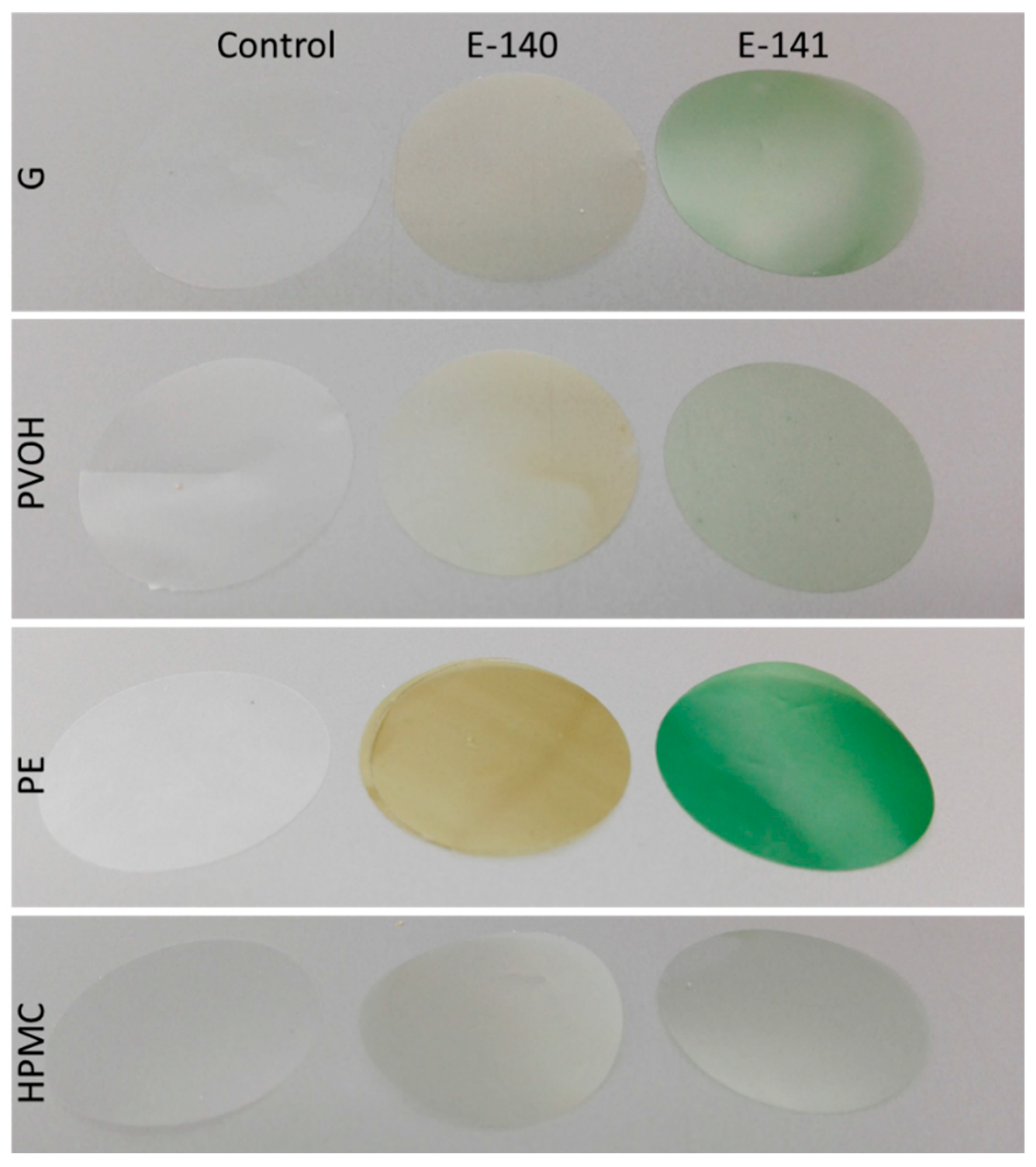
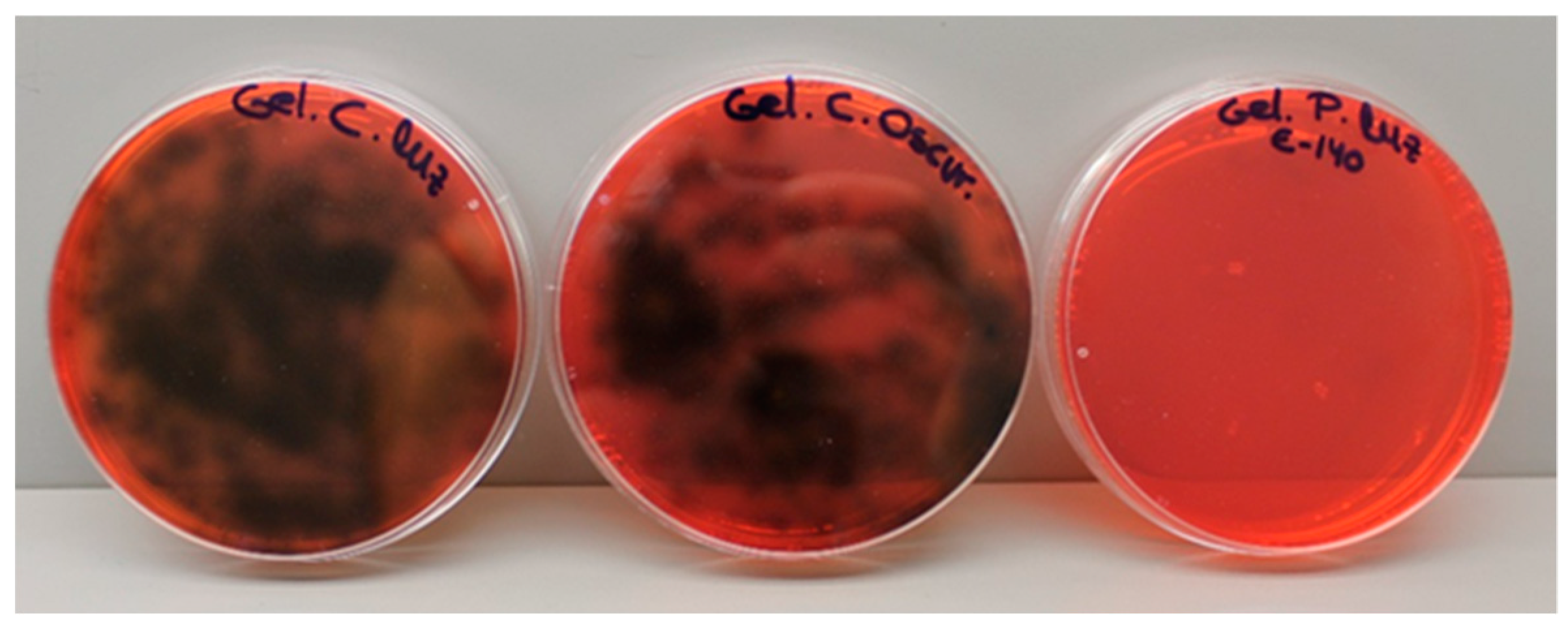
The coatings were inoculated on the surface and then illuminated in order to verify whether they could be self-sanitized by photoactivation. As an example, Figure 3 shows the results obtained with gelatin films contaminated with Listeria. As can be seen, there is growth both in the control plate that was exposed to light and in the plate with porphyrin films that was kept in darkness. By contrast, irradiated samples containing porphyrin E-140 showed no microbial growth.
Similar results were obtained for the other films: HPMC, PVOH, and PE. This shows that the films developed can be self-sanitized in case of possible contamination simply by subjecting them to visible light irradiation for 15 min.
However, no clear bactericidal effect against E. coli was observed with any of the coated films that contained E-140. These results are in agreement with other authors who obtained an antimicrobial effect against Gram-positive bacteria using porphyrins grafted onto nylon fibers but found no effect on E. coli bacteria at any light intensity [7]. As explained previously, this may be due to the extra protection of the outer membrane in Gram-negative bacteria. The outer membrane of Gram-negative bacteria plays an important role, which is related to their resistance to many active agents that are very effective against Gram-positive bacteria [17]. The resistance of Gram-negative bacteria to photosensitization has been widely reported [18,19]. In order to increase the permeability of E. coli, we added 40 mM EDTA (ethylenediaminetetraacetic acid) to the 100 μL dilution of E. coli. EDTA is a chelating agent that destabilizes the outer membrane and may facilitate access of free radicals generated with light to bacterial cells [20]. However, we did not observe significant differences between the controls and the treatment with light and porphyrin. Other authors have employed dimethyl sulfoxide (5%) to increase the permeability of E. coli to porphyrins without achieving the same results as with Gram-positive bacteria [14].
3.4. Migration of Chlorophyllins from Coatings to the Food Simulant
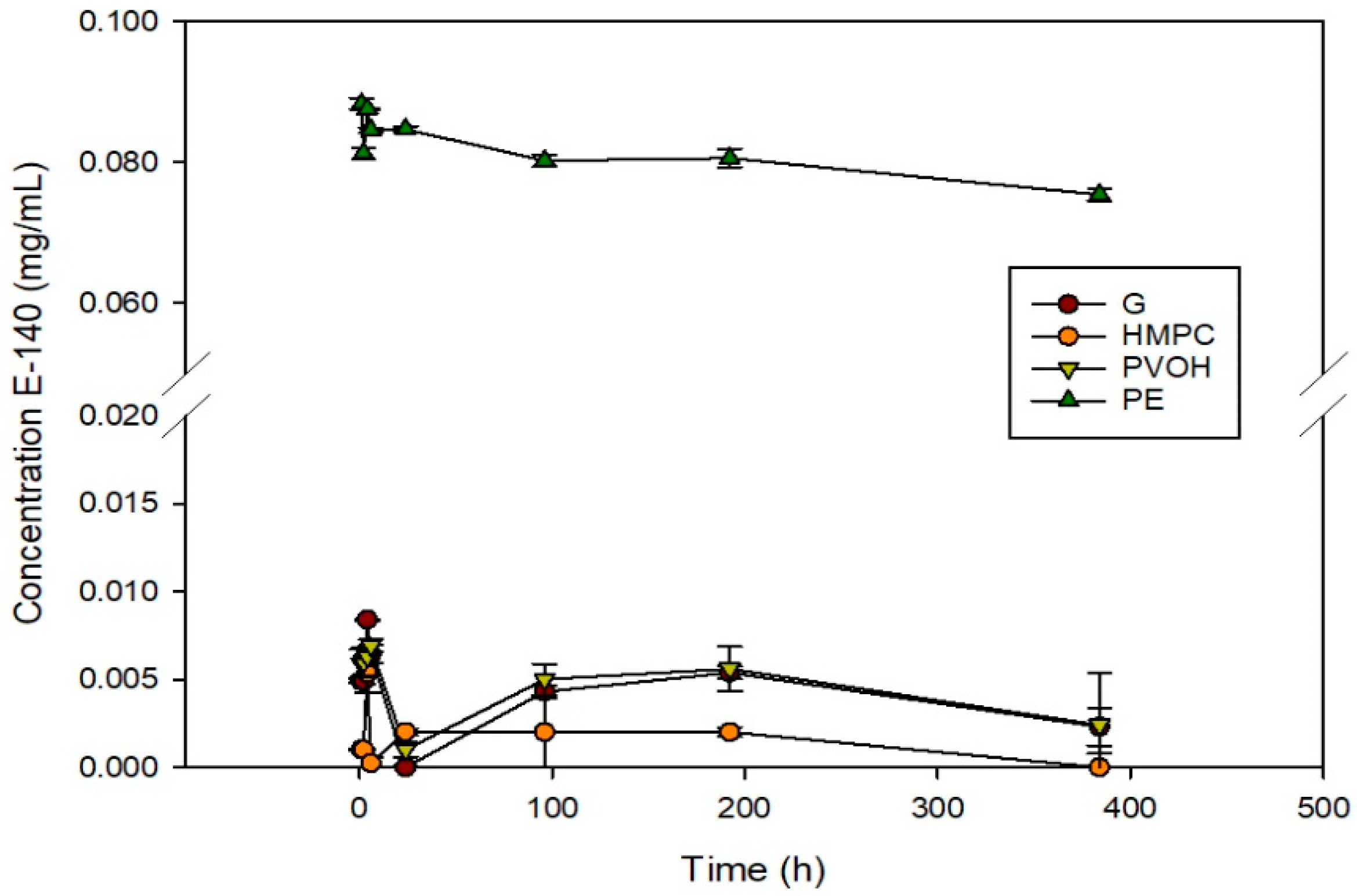
The release of chlorophyllin E-140 was studied in 50% ethanol as a fatty food simulant in accordance with Directive 85/572/EEC prior to application on bologna slices. The regulation stipulates that the results of overall migration obtained must be in accordance with the overall migration limit of 10 mg/dm2 established in Directive 2002/72/EC and in Royal Decree 866/2008, which are both related to materials and plastic objects in contact with food products. The results are shown in Figure 4.
The release of chlorophyllins from G, PVOH, and HPMC coatings on PET was very low—practically zero—indicating that there was no substantial migration. This means the antimicrobial effect was produced by the generation of free radicals and it was not necessary for the chlorophyllins to migrate to the simulating medium. On the other hand, the PE coatings presented a maximum release of 6.4 mg/dm2 (according to the surface of each coating, which was 0.125 dm2), which was less than the overall migration limit set by legislation (10 mg/dm2).
In the case of the other coatings, the low migration obtained makes it unnecessary to calculate migration limits, and therefore they can be used as packaging systems for fatty food. In this case, they were tested with bologna slices. The fact that the greatest release was observed in PE could be due to several factors. First, the thickness of the PE coating—well above 25 µm—results in a large amount of porphyrin in absolute values. Second, the poor affinity of LDPE for chlorophyllins might result in their separation, forming a two-phase matrix in which the agent is isolated in small regions dispersed in the pure LDPE matrix. When the material comes into contact with a solvent medium, the porphyrin located close to the surface is immediately released, as observed in Figure 4. In the other matrices there would be specific interactions between E-140 and the polymers, keeping the agent in the package where porphyrin molecules have a compatible chemical environment. Similar release results were observed for films containing E-141 (data not shown).
3.5. Application to Food
Once the antimicrobial and self-sanitizing capacity of the films had been determined, their effectiveness when applied to a real food was studied. The experiment was carried out with G, HPMC, PE, and PVOH coatings on PET films used as bologna slice separators.
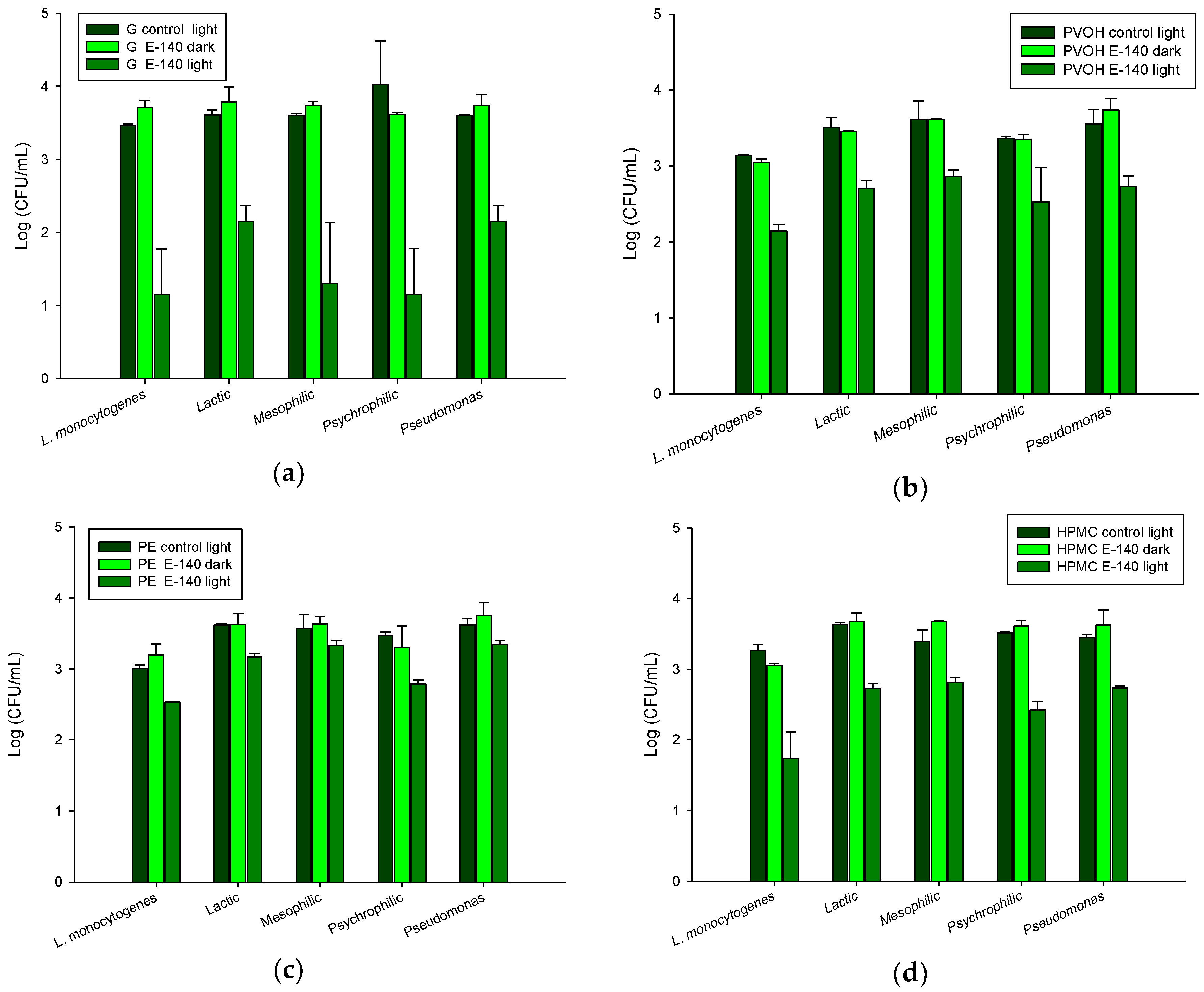
This food product was chosen to test the coatings developed because it has a high water activity (aw = 0.970) and is therefore very susceptible to spoilage by microorganisms. The antimicrobial effect of the films was studied against L. monocytogenes, which was inoculated on the bologna surface and against the usual microbial load of this meat product. The results are shown in Figure 5.
There was no microbial growth of Salmonella spp., enterobacteria, or coliform bacteria on any sample. Control coatings and coatings kept in darkness showed no antimicrobial effect, confirming the nontoxicity of the active agents without photoactivation. However, coatings with E-140 exposed to LED lights inhibited microbial growth of L. monocytogenes successfully. The greatest antimicrobial effect was observed with the G coating and the least with the PE coating. The HPMC and PVOH coatings were slightly less effective than G (Figure 5).
Growth of the microbial load was also inhibited in irradiated samples with chlorophyllin. Lactic, mesophilic, and psychrophilic bacteria and Pseudomonas were significantly reduced. Once again, the G coatings presented the greatest antimicrobial effect against all the microorganisms tested. The highest efficiency was observed against psychrophiles and mesophiles, with reductions of 2.87 and 2.30 log, respectively. In the case of lactic acid bacteria and Pseudomonas, although the degree of inhibition was lower, it was also considerable. The HPMC and PVOH coatings with chlorophyllin presented similar degrees of inhibition against all the microorganisms studied, ranging between 0.76 and 1 log. Finally, the antimicrobial effect of the PE coatings was lower, ranging between approximately 0.24 and 0.69 log.
Results showed that all the coatings developed in this study had antimicrobial activity, with gelatin being the most effective matrix. This is possibly due to its morphology that facilitates the release of free radicals produced by the porphyrin molecule, which is responsible for the antimicrobial action. It should also be taken into account that the G coatings were thicker, and therefore there was a greater amount of agent in absolute values than in the HPMC and PVOH coatings. The application of the gelatin coating would be very useful given gelatin is a component that is usually present in many foods and does not constitute a risk when it forms part of the surface of a package intended for food use.
To confirm the absence of effects on the organoleptic properties of the product and because the release of chlorophyllin could produce a green color, the color of the bologna slices was measured before and after the photoactivation treatment. The parameters L* [black (0) to white (100)], a* [green (−) to red (+)], and b* [blue (−) to yellow (+)] were obtained, and the polar coordinates, chroma C*, and hue angle h that were calculated are shown in Table 6.
The bologna slices were displaced towards the + a coordinate, i.e., towards red and slightly towards the + b coordinate (yellow), which is logical given their rosy hue.
In general, irradiation produced a slight decrease in the luminosity of the bologna slices and a slight shift to the left of the b* coordinates. However, these color modifications were not distinguishable to the naked eye. On the other hand, no color difference was observed between samples with and without porphyrin submitted to the same treatment (darkness or light). Therefore, from the data shown above, it can be confirmed that no substantial release of porphyrin to the bologna took place. Similar results were reported in a study of chlorophyllin gelatin films applied as wraps to frankfurter sausages, which found no substantial differences between uncoated and coated products [2].
Finally, it can be concluded that chlorophyllin-based photosensitization of coated films is an effective way of reducing the population of microorganisms naturally present in meat and poultry products and as a way of improving asepsis of packaging materials through their self-sanitizing function. Moreover, this efficiency has also been demonstrated when light is applied through the supporting material (in this case, PET) if the material is transparent to visible light. This aspect is of great importance because it would greatly facilitate carrying out its antimicrobial or self-sanitizing function in food products that have already been packaged. The process that has been developed in this study, which requires only white light, could become an alternative food process that is nonchemical, nonthermal, inexpensive, and environmentally friendly.
The films developed could be applied as part of a package intended for meat derivatives—either as an external protective cover or as separators of cold cuts—or in some dairy products, such as fresh cheese. In addition, the films developed could be used to wrap food that are sensitive to microbial contamination in daily use, such as pieces of cooked ham that are not immediately consumed completely but are rather manipulated (cut) on a number of occasions before being completely consumed, therefore making them potential vectors for the transmission of microorganisms. These films could be used to cover the cut surface and protect it from microbial spoilage thanks to the photoactivation induced by the white lights of refrigerated displays in grocery stores. Furthermore, this active packaging does not require any agent release, thereby increasing food shelf life without substantial changes in aroma, flavor, or color.
Author Contributions
Conceptualization, P.H.-M. and R.G.; Methodology, G.L.-C.; Validation, P.H.-M., R.G. and G.L.-C.; Formal Analysis, G.L.-C.; Investigation, P.H.-M., R.G. and G.L.-C.; Resources, P.H.-M. and R.G.; Writing, Original Draft Preparation, G.L.-C., R.G. and P.H.-M.
Funding
This research was funded by the Ministry of Economy and Competitiveness (No. AGL2015-64595-R).
Acknowledgments
The authors acknowledge the assistance of Karel Clapshaw (translation services) and the supply of materials by Paramelt B.V. (Heerhugowaard, The Netherlands) and the Nippon Synthetic Chemical Company (Osaka, Japan).
Conflicts of Interest
The authors declare no conflict of interest.
References
- Flint, J.A.; Van Duynhoven, Y.T.; Angulo, F.J.; DeLong, S.M.; Braun, P.; Kirk, M.; Scallan, E.; Fitzgerald, M.; Adak, G.K.; Sockett, P.; et al. Estimating the burden of acute gastroenteritis, foodborne disease, and pathogens commonly transmitted by food: An international review. Clin. Infect. Dis. 2005, 41, 698–704. [Google Scholar] [CrossRef] [PubMed]
- López-Carballo, G.; Hernández-Muñoz, P.; Gavara, R.; Ocio, M.J. Photoactivated chlorophyllin-based gelatin films and coatings to prevent microbial contamination of food products. Int. J. Food Microbiol. 2008, 126, 65–70. [Google Scholar] [CrossRef] [PubMed]
- Luksiene, Z.; Paskeviciute, E. Novel approach to decontaminate food-packaging from pathogens in non-thermal and not chemical way: Chlorophyllin-based photosensitization. J. Food Eng. 2011, 106, 152–158. [Google Scholar] [CrossRef]
- Evans, H.H.; DeMarini, D.M. Ionizing radiation-induced mutagenesis: Radiation studies in neurospora predictive for results in mammalian cells. Mutat. Res. Rev. Mutat. Res. 1999, 437, 135–150. [Google Scholar] [CrossRef]
- Martins, D.; Mesquita, M.Q.; Neves, M.G.P.M.S.; Faustino, M.A.F.; Reis, L.; Figueira, E.; Almeida, A. Photoinactivation of Pseudomonas syringae pv. Actinidiae in kiwifruit plants by cationic porphyrins. Planta 2018, 248, 409–421. [Google Scholar] [CrossRef] [PubMed]
- Paskeviciute, E.; Zudyte, B.; Luksiene, Z. Towards better microbial safety of fresh produce: Chlorophyllin-based photosensitization for microbial control of foodborne pathogens on cherry tomatoes. J. Photochem. Photobiol. B Biol. 2018, 182, 130–136. [Google Scholar] [CrossRef] [PubMed]
- Bozja, J.; Sherrill, J.; Michielsen, S.; Stojiljkovic, I. Porphyrin-based, light-activated antimicrobial materials. J. Polym. Sci. A Polym. Chem. 2003, 41, 2297–2303. [Google Scholar] [CrossRef]
- Krouit, M.; Granet, R.; Branland, P.; Verneuil, B.; Krausz, P. New photoantimicrobial films composed of porphyrinated lipophilic cellulose esters. Bioorg. Med. Chem. Lett. 2006, 16, 1651–1655. [Google Scholar] [CrossRef] [PubMed]
- George, L.; Hiltunen, A.; Santala, V.; Efimov, A. Photo-antimicrobial efficacy of zinc complexes of porphyrin and phthalocyanine activated by inexpensive consumer led lamp. J. Inorg. Biochem. 2018, 183, 94–100. [Google Scholar] [CrossRef] [PubMed]
- Smith-Palmer, A.; Stewart, J.; Fyfe, L. Antimicrobial properties of plant essential oils and essences against five important food-borne pathogens. Lett. Appl. Microbiol. 1998, 26, 118–122. [Google Scholar] [CrossRef] [PubMed]
- LED Lights—Detrimental to Our Health and Environment. Available online: http://www.sunkissedsolar.com.au/led-light-globes-detrimental-health-environment (accessed on 27 July 2018).
- Higueras, L.; López Carballo, G.; Hernández Muñoz, P.; Gavara, R.; Rollini, M. Development of a novel antimicrobial film based on chitosan with LAE (ethyl-Nα-dodecanoyl-l-arginate) and its application to fresh chicken. Int. J. Food Microbiol. 2013, 165, 339–345. [Google Scholar] [CrossRef] [PubMed]
- Delaquis, P.J.; Stanich, K.; Girard, B.; Mazza, G. Antimicrobial activity of individual and mixed fractions of dill, cilantro, coriander and eucalyptus essential oils. Int. J. Food Microbiol. 2002, 74, 101–109. [Google Scholar] [CrossRef]
- Caires, C.S.A.; Leal, C.R.B.; Ramos, C.A.N.; Bogo, D.; Lima, A.R.; Arruda, E.J.; Oliveira, S.L.; Caires, A.R.L.; Nascimento, V.A. Photoinactivation effect of eosin methylene blue and chlorophyllin sodium-copper against Staphylococcus aureus and Escherichia coli. Lasers Med. Sci. 2017, 32, 1081–1088. [Google Scholar] [CrossRef] [PubMed]
- Cavaleiro, J.A.S.; Görner, H.; Lacerda, P.S.S.; MacDonald, J.G.; Mark, G.; Neves, M.G.P.M.S.; Nohr, R.S.; Schuchmann, H.-P.; von Sonntag, C.; Tomé, A.C. Singlet oxygen formation and photostability of meso-tetraarylporphyrin derivatives and their copper complexes. J. Photochem. Photobiol. A Chem. 2001, 144, 131–140. [Google Scholar] [CrossRef]
- Levine, J.S. Biomass burning: The cycling of gases and particulates from the biosphere to the atmosphere. In Treatise on Geochemistry; Holland, H.D., Turekian, K.K., Eds.; Pergamon: Oxford, UK, 2003; pp. 143–158. [Google Scholar]
- Nikaido, H. Molecular basis of bacterial outer membrane permeability revisited. Microbiol. Mol. Biol. Rev. 2003, 67, 593–656. [Google Scholar] [CrossRef] [PubMed]
- Nitzan, Y.; Gozhansky, S.; Malik, Z. Effect of photoactivated hematoporphyrin derivative on the viability of Staphylococcus aureus. Curr. Microbiol. 1983, 8, 279–284. [Google Scholar] [CrossRef]
- Malik, Z.; Hanania, J.; Nitzan, Y. Bactercidal effects of photoactivated porphyrins-an alternative approach to antimicrobial drugs. J. Photochem. Photobiol. B Biol. 1990, 5, 281–293. [Google Scholar] [CrossRef]
- Malik, Z.; Ladan, H.; Nitzan, Y. Phtodynamic inactivation of gram-negative bacteria-problem and possible solutions. J. Photochem. Photobiol. B Biol. 1992, 14, 262–266. [Google Scholar] [CrossRef]
Figure 1.
Example of test: (a) from left to right: bologna slice without coating, sample with a control gelatin (G) coating on poly(ethylene terephthalate) (PET), and sample of G-coated PET containing E-140 porphyrin; (b) image of an experiment of treatment during the photoactivation process.
Figure 1.
Example of test: (a) from left to right: bologna slice without coating, sample with a control gelatin (G) coating on poly(ethylene terephthalate) (PET), and sample of G-coated PET containing E-140 porphyrin; (b) image of an experiment of treatment during the photoactivation process.

Figure 2.
Images of the PET films coated with G, PVOH, HPMC, and PE for the control and the films containing chlorophyllins (E-140 and E-141).
Figure 2.
Images of the PET films coated with G, PVOH, HPMC, and PE for the control and the films containing chlorophyllins (E-140 and E-141).

Figure 3.
Microbial growth of L. monocytogenes. Left to right: control G film which had been irradiated, G film with E-140 which had been kept in darkness, and G film with E-140 which had been irradiated.
Figure 3.
Microbial growth of L. monocytogenes. Left to right: control G film which had been irradiated, G film with E-140 which had been kept in darkness, and G film with E-140 which had been irradiated.

Figure 4.
Concentration of E-140 released from G, PVOH, HPMC, and PE coatings on PET in fatty simulant as a function of time.
Figure 4.
Concentration of E-140 released from G, PVOH, HPMC, and PE coatings on PET in fatty simulant as a function of time.

Figure 5.
Antimicrobial activity of (a) G, (b) PVOH, (c) PE, and (d) HPMC coatings on PET films applied to bologna slices.
Figure 5.
Antimicrobial activity of (a) G, (b) PVOH, (c) PE, and (d) HPMC coatings on PET films applied to bologna slices.


Table 1.
Minimum inhibitory concentration (MIC in mg/mL) and minimum bactericidal concentration (MBC in mg/mL) of chlorophyllins E-140 and E-141 against L. monocytogenes and E. coli activated by halogen light (15 min).
Table 1.
Minimum inhibitory concentration (MIC in mg/mL) and minimum bactericidal concentration (MBC in mg/mL) of chlorophyllins E-140 and E-141 against L. monocytogenes and E. coli activated by halogen light (15 min).
| Microorganism | E-140 | E-141 | ||
|---|---|---|---|---|
| MIC | MBC | MIC | MBC | |
| L. monocytogenes | 0.0031 | 0.10 | 2 | 10 |
| E. coli | 0.0031 | 0.63 | 3 | 11 |
Table 2.
Minimum inhibitory concentration (MIC in mg/mL) and minimum bactericidal concentration (MBC in mg/mL) of chlorophyllins E-140 and E-141 against L. monocytogenes and E. coli activated by LED light (15 min).
Table 2.
Minimum inhibitory concentration (MIC in mg/mL) and minimum bactericidal concentration (MBC in mg/mL) of chlorophyllins E-140 and E-141 against L. monocytogenes and E. coli activated by LED light (15 min).
| Microorganism | E-140 | E-141 | ||
|---|---|---|---|---|
| MIC | MBC | MIC | MBC | |
| L. monocytogenes | 0.025 | 0.400 | − | − |
| E. coli | 0.100 | 0.630 | − | − |
Table 3.
Thicknesses (µm) of films and coatings with and without chlorophyllin.
| Materials | Coatings | |
|---|---|---|
| Control | E-140 and E-141 | |
| G | 4.66 ± 1.63 | 5.16 ± 1.16 |
| PVOH | 2.28 ± 1.79 | 2.00 ± 1.03 |
| PE | 25.00 ± 1.87 | 25.17 ± 2.70 |
| HPMC | 2.14 ± 1.06 | 2.42 ± 1.61 |
Table 4.
Color coordinates L*, a*, b*, chromaticity (C*) and tone (h in °) of films coated with gelatin (G), polyvinyl alcohol (PVOH), polyethylene (PE), (hydroxypropyl)methyl cellulose (HPMC) without (control) and with active agents (E-140 and E-141).
Table 4.
Color coordinates L*, a*, b*, chromaticity (C*) and tone (h in °) of films coated with gelatin (G), polyvinyl alcohol (PVOH), polyethylene (PE), (hydroxypropyl)methyl cellulose (HPMC) without (control) and with active agents (E-140 and E-141).
| Coatings | a* | b* | L* | C* | h | |
|---|---|---|---|---|---|---|
| G | Control | −0.23 ± 0.02 c | 0.64 ± 0.02 a | 88.04 ± 0.01 c | 0.68 ± 0.05 b | 109.84 ± 0.02 c |
| E-140 | −2.50 ± 0.01 b | 8.18 ± 0.03 a | 85.77 ± 0.01 b | 8.55 ± 0.01 a | 106.99 ± 0.02 a | |
| E-141 | −3.02 ± 0.07 a | 9.31 ± 0.01 a | 83.61 ± 0.04 a | 9.78 ± 0.01 a | 107.97 ± 0.01 b | |
| PVOH | Control | 0.16 ± 0.04 a | 0.32 ± 0.02 b | 88.04 ± 0.02 c | 0.36 ± 0.05 a | 115.26 ± 0.01 b |
| E-140 | −2.89 ± 0.01 b | 9.76 ± 0.03 a | 85.26 ± 0.01 b | 10.18 ± 0.02 c | 106.54 ± 0.05 a | |
| E-141 | −5.02 ± 0.01 c | 7.89 ± 0.01 a | 83.12 ± 0.01 a | 9.35 ± 0.01 b | 122.47 ± 0.02 c | |
| PE | Control | −0.32 ± 0.01 c | -0.23 ± 0.02 a | 89.23 ± 0.01 c | 0.40 ± 0.06 a | 216.41 ± 0.02 c |
| E-140 | −4.73 ± 0.01 b | 38.06 ± 0.01 b | 69.79 ± 0.02 b | 38.35 ± 0.04 b | 97.09 ± 0.01 a | |
| E-141 | −26.24 ± 0.02 a | 28.12 ± 0.02 b | 67.51 ± 0.03 a | 38.46 ± 0.04 c | 133.01 ± 0.02 b | |
| HPMC | Control | −0.26 ± 0.03 c | 0.38 ± 0.04 a | 88.64 ± 0.05 c | 0.47 ± 0.01 a | 124.54 ± 0.04 b |
| E-140 | 1.11 ± 0.01 b | 3.15 ± 0.02 b | 87.69 ± 0.05 b | 3.34 ± 0.04 a | 109.33 ± 0.01 a | |
| E-141 | −3.02 ± 0.04 a | 3.41 ± 0.01 c | 85.72 ± 0.03 a | 4.56 ± 0.03 a | 131.53 ± 0.01 c | |
a–c Different letters in the same column for a coating indicate significant differences (Tukey’s adjusted analysis of variance, p < 0.05).
Table 5.
Microbiological counts (log CFU/mL) on the surface of the agar in the area under the coated films and antimicrobial effect expressed as log reduction values (LRV).
Table 5.
Microbiological counts (log CFU/mL) on the surface of the agar in the area under the coated films and antimicrobial effect expressed as log reduction values (LRV).
| Coatings | Dark Conditions | Light Conditions | |||
|---|---|---|---|---|---|
| Control | E-140 | Control | E-140 | LRV | |
| Listeria monocytogenes | |||||
| G | 6.90 ± 0.02 | 7.02 ± 0.01 | 7.09 ± 0.01 | no growth | 7.09 |
| PVOH | 7.13 ± 0.05 | 7.24 ± 0.04 | 7.25 ± 0.73 | no growth | 7.25 |
| PE | 7.05 ± 0.05 | 6.95 ± 0.01 | 7.38 ± 0.70 | 6.73 ± 0.05 | 0.65 |
| HPMC | 6.95 ± 0.01 | 6.90 ± 0.02 | 7.01± 0.02 | no growth | 7.01 |
| Escherichia coli | |||||
| G | 7.01 ± 0.02 | 7.02 ± 0.01 | 7.04 ± 0.01 | no growth | 7.04 |
| PVOH | 7.23 ± 0.02 | 7.01 ± 0.04 | 7.47 ± 0.67 | no growth | 7.47 |
| PE | 7.12 ± 0.03 | 6.95 ± 0.03 | 7.40 ± 0.70 | 6.79 ± 0.03 | 0.67 |
| HPMC | 7.01 ± 0.01 | 6.90 ± 0.02 | 7.04 ± 0.04 | no growth | 7.04 |
Table 6.
The color coordinates L*, a*, b*, the chromaticity (C*), and the tone (h in °) of bologna slices covered with G, PVOH, PE, and HPMC coatings on PET before and after photoactivation.
Table 6.
The color coordinates L*, a*, b*, the chromaticity (C*), and the tone (h in °) of bologna slices covered with G, PVOH, PE, and HPMC coatings on PET before and after photoactivation.
| Coatings | Activation | a* | b* | L* | C* | h |
|---|---|---|---|---|---|---|
| G control | before | 16.30 ± 0.04 a | 10.30 ± 0.04 a | 56.59 ± 0.04 b | 19.28 ± 0.04 c | 32.30 ± 0.17 b |
| after | 14.12 ± 0.02 b | 12.70 ± 0.02 b | 50.48 ± 0.02 a | 18.99 ± 0.02 c | 41.97 ± 0.06 b | |
| G E-140 | before | 15.95 ± 0.05 a | 9.24 ± 0.05 a | 56.62 ± 0.05 b | 18.44 ± 0.05 a | 30.09 ± 0.13 b |
| after | 14.10 ± 0.03 a | 14.07 ± 0.03 b | 50.29 ± 0.03 a | 19.92 ± 0.03 b | 44.94 ± 0.10 c | |
| PVOH control | before | 17.70 ± 0.05 a | 9.68 ± 0.05 a | 57.10 ± 0.05 b | 20.17 ± 0.05 a | 28.66 ± 0.21 b |
| after | 13.53 ± 0.03 b | 12.48 ± 0.03 b | 50.47 ± 0.03 a | 18.41 ± 0.03 b | 42.69 ± 0.17 a | |
| PVOH E-140 | before | 17.47 ± 0.03 a | 9.42 ± 0.03 a | 57.06 ± 0.03 b | 19.85 ± 0.03 a | 28.33 ± 0.10 b |
| after | 14.74 ± 0.01 b | 13.34 ± 0.01 b | 50.99 ± 0.01 a | 19.88 ± 0.01 a | 42.13 ± 0.09 a | |
| HPMC control | before | 16.20 ± 0.03 b | 10.38 ±0.03 a | 55.27 ± 0.03 b | 19.24 ± 0.03 b | 32.65 ± 0.07 b |
| after | 13.48 ± 0.02 a | 12.34 ± 0.02 b | 50.23 ± 0.02 a | 18.43 ± 0.02 a | 41.41 ± 0.29 a | |
| HPMC E-140 | before | 15.99 ± 0.04 b | 10.56 ± 0.04 a | 55.37 ± 0.04 b | 19.17 ± 0.04 b | 33.44 ± 0.21 b |
| after | 12.94 ± 0.07 a | 12.56 ± 0.07 b | 50.51 ± 0.07 a | 18.03 ± 0.07 a | 44.16 ± 0.07 c | |
| PE control | before | 17.59 ± 0.03 a | 10.65 ± 0.03 a | 54.36 ± 0.03 b | 20.57 ± 0.03 b | 31.20 ± 0.27 b |
| after | 13.26 ± 0.03 b | 12.04 ± 0.03 b | 50.78 ± 0.03 a | 17.91 ± 0.03 a | 42.26 ± 0.06 a | |
| PE E-140 | before | 16.61 ± 0.02 a | 10.53 ± 0.02 a | 55.87 ± 0.02 b | 19.67 ± 0.02 b | 32.37 ± 0.08 b |
| after | 14.80 ± 0.01 c | 12.39 ± 0.01 b | 50.54 ± 0.01 a | 19.30 ± 0.01 b | 39.95 ± 0.10 c |
a–c Different letters in the same column indicate significant differences (Tukey’s adjusted analysis of variance, p < 0.05).
© 2018 by the authors. Licensee MDPI, Basel, Switzerland. This article is an open access article distributed under the terms and conditions of the Creative Commons Attribution (CC BY) license (http://creativecommons.org/licenses/by/4.0/).
Share and Cite
MDPI and ACS Style
López-Carballo, G.; Hernández-Muñoz, P.; Gavara, R. Photoactivated Self-Sanitizing Chlorophyllin-Containing Coatings to Prevent Microbial Contamination in Packaged Food. Coatings 2018, 8, 328. https://doi.org/10.3390/coatings8090328
AMA Style
López-Carballo G, Hernández-Muñoz P, Gavara R. Photoactivated Self-Sanitizing Chlorophyllin-Containing Coatings to Prevent Microbial Contamination in Packaged Food. Coatings. 2018; 8(9):328. https://doi.org/10.3390/coatings8090328
Chicago/Turabian StyleLópez-Carballo, Gracia, Pilar Hernández-Muñoz, and Rafael Gavara. 2018. "Photoactivated Self-Sanitizing Chlorophyllin-Containing Coatings to Prevent Microbial Contamination in Packaged Food" Coatings 8, no. 9: 328. https://doi.org/10.3390/coatings8090328
Note that from the first issue of 2016, this journal uses article numbers instead of page numbers. See further details here.