Communication

The Potential Application of Spring Sargassum glaucescens Extracts in the Moisture-Retention of Keratinocytes and Dermal Fibroblast Regeneration after UVA-Irradiation

TCI Research and Design Center, TCI Co., Ltd., Taipei 11494, Taiwan
*
Author to whom correspondence should be addressed.
Cosmetics 2019, 6(1), 17; https://doi.org/10.3390/cosmetics6010017
Submission received: 22 January 2019 / Revised: 25 February 2019 / Accepted: 27 February 2019 / Published: 4 March 2019
(This article belongs to the Special Issue Anti-aging Properties of Natural Compounds)

Abstract

:
Sargassum glaucescens is a marine brown alga with high antioxidant activity. To evaluate the potential application of Sargassum glaucescens extracts (SGE) in skincare, we performed in vitro assays in dermal fibroblasts and epidermal keratinocytes. The antioxidant activity of SGE was confirmed by the suppression of H2O2-induced reactive oxygen species (ROS) production in dermal fibroblasts and in vitro 1,1-diphenyl-2-picrylhydrazyl (DPPH) scavenging activity. In the wound healing assay, application of 2 mg/ml SGE stimulated the wound closure of CCD-966SK fibroblasts by a 2.95-fold in comparison to the control. Furthermore, treatment with SGE of concentrations ranging from 0.25 to 1 mg/ml promoted CCD-966SK cell regeneration after UVA irradiation. At the molecular level, 1 mg/ml SGE induced expressions of anti-oxidative genes SOD1 (Superoxide dismutase 1) and GPX1 (Glutathione peroxidase 1), and DNA repair regulatory genes XRCC1 (X-ray repair cross-complementing protein 1) and ERCC6 (Excision repair cross-complementation Group 6) in CCD-966SK cells after UVA irradiation. Therefore, SGE displayed beneficial effects on cell regeneration and the protection of dermal cells against UVA irradiation. In epidermal cells, SGE stimulated the cell proliferation of human primary epidermal keratinocytes. Application of 0.03125 mg/ml SGE induced the expressions of skin barrier-related genes TGM1 (Transglutaminase 1), KRT10 (Keratin 10) and KRT14 in keratinocytes. Meanwhile, SGE induced the gene expression of FLG (Filaggrin), which promoted the production of natural moisturizing factor (NMF) for maintaining the moisture and barrier functions of skin.

1. Introduction

Marine algae are rich in metabolites that can be used to reduce oxidative stress and prevent skin aging. These metabolites include mycosporine-like amino acids (MAAs), polysaccharides, sulfated polysaccharides and polyphenols [1]. Brown seaweed Sargassum spp., which are marine macroalgae widely distributed in temperate and subtropical zones [2], have been found to contain high amounts of phenolic compounds and exhibit strong antioxidant activity [3]. Besides, Sargassum spp. are rich in the omega (ω)-3 fatty acids involving eicosapentaenoic acid (EPA) and docosahexaenoic acid (DHA) that contribute to the normal function of the heart [4].
Based on the contributions of algae nutrients on human health, we are interested in the application of S. glaucescens extracts in skincare. Skin aging is extrinsically induced by exposure to UV light and smoking [5,6]. UV irradiation results in photo-damage of the skin, with the aging signs of wrinkles, collagen degradation, coarseness, dryness, mottled pigmentation, increased epidermal thickness and so on [7]. UV irradiation induces the formation of intracellular reactive oxygen species (ROS), which triggers inflammation and causes dermal protein damage [8]. In UV-irradiated human dermal fibroblasts, Sargachromanol E isolated from Sargassum horneri was able to increase the scavenging activity against ROS in a dose-dependent manner, and protect cell membranes from oxidative modification of proteins and lipids [9,10]. Treatment of Sargachromanol E suppressed the expression levels of collagen-degrading matrix metalloproteinases (MMPs) and elevated the tissue inhibitor of metalloproteinases (TIMPs), indicating its potential in maintaining collagen fibers in skin. Another case of Sargassum fulvellum was reported showing that its ethylacetate fraction of ethanol extract inhibited UVB-induced cytotoxicity and proinflammatory protein expressions in HaCaT keratinocytes [11]. Besides antioxidant activity, polysaccharides isolated from Sargassum horneri exhibited strong in vitro moisture-absorption and –retention capacities [12]. These studies implied that Sargassum spp. were good sources of cosmeceutical molecules with efficient bioactivities in anti-photoaging and moisturization in skin.
Sargassum glaucescens is one of the most abundant species of algae on the rocky coasts of Taiwan. S. glaucescens has been considered a good source for fucoidan extraction [13]. As a sulfated polysaccharide, Fucoidan is used in dietary supplements for its potential antioxidant, antibacterial, anticancer, antiangiogenic and anti-inflammatory effects [14,15,16,17,18]. However, the seasonal variation will cause compositional changes in minerals, amino acids, fatty acids, alginate and fucoidan contents in Sargassum [19,20,21,22]. The thallus growth and the stand biomass of S. glaucescens peaked in spring, as well as the antioxidant activity. The spring S. glaucescens possessed five-fold higher DPPH scavenging activity than autumn-winter S. glaucescens (data unpublished). Up to now, limited research has been conducted on the applications of S. glaucescens in anti-photoaging or the improvement of skin properties.
In this study, we aimed to evaluate the probable use of S. glaucescens extracts in cosmeceutical industries. In order to unravel the role of S. glaucescens extracts in the anti-aging of skin, we chose spring algae as materials to obtain the highest antioxidant activity. Our investigation focused on the anti-UV irradiation and moisture-preserving potency of spring S. glaucescens extract. With these preliminary data, we provided useful information and further insights on the application of spring S. glaucescens extract in skincare.

2. Materials and Methods

2.1. Cell Lines and Chemicals

CCD-966SK (CRL-1881) cell line was derived from the American Type Culture Collection (Manassas, VA, USA). Human primary epidermal keratinocyte was purchased from CELLnTEC (CELLnTEC Advanced Cell Systems AG, Bern, Switzerland). All cell culture media and reagents including MTT [3-(4,5-dimethylthiazol-2-yl)-2,5-diphenyltetrazolium bromide] were purchased from Sigma-Aldrich (St. Louis, MO, USA) or Gibco (Thermo Fisher Scientific, Inc., Carlsbad, CA, USA) and were of reagent grade or cell-culture grade.

2.2. Preparation of Extracts

Fresh Sargassum glaucescens was collected from the coast of Penghu County, Taiwan in March. S. glaucescens was extracted following the hot-water extract method proposed by Hwang et al. [23] with modifications. Seaweed was washed and dried in a dryer at 40 °C for 90 min. The dried sample was ground into powder with a blender and then subjected to the dryer at 50 °C for 10 min. The dried seaweed was treated with distilled water in the proportion of 100 g powder in 5 L water, and boiled at 100 °C for 30 min. The extracts were centrifuged at 4500 rpm for 20 min, and the supernatant was freeze-dried at −20 °C under a reduced pressure of 2 mmHg. The S. glaucescens extract (SGE) was stored at −20 °C for further use. The working solution was prepared by dissolving the extract in distilled water.

2.3. Quantification of Intracellular Reactive Oxygen Species (ROS) Levels

The intracellular accumulation of ROS was analyzed by using 2’,7’-dichlorodihydrofluorescein diacetate (DCFH-DA, SI-D6883, Sigma-Aldrich, St. Louis, MO, USA) and measured by flow cytometry. DCFH-DA can easily diffuse into cells and be deacetylated by cellular esterases to form non-fluorescent 2’,7’-dichlorodihydrofluorescein (DCFH), which will be rapidly oxidized by ROS to form highly fluorescent 2’,7’-dichlorofluorescein (DCF). CCD-966SK fibroblasts were seeded in a 6-well plate at a density of 2 × 10⁵ cells with 2 ml culture medium per well and incubated at 37 °C for 24 hours. The MEM medium for the cell culture was renewed with mock reagent, 1 mM H2O2, 1 mM H2O2 + 0.125 mg/ml SGE or 1 mM H2O2 + 0.25 mg/ml SGE, and then the cells were incubated at 37 °C for 1 hour. 5 µg/ml DCFH-DA was added to each well and the culture plate was incubatet at 37 °C for 15 min. The cells were washed with 1×PBS, harvested and re-suspended in the dark. The fluorescence intensity was monitored by a CytoFLEX Flow Cytometer (Beckman Coulter, Inc., Indianapolis, IN, USA) using excitation wavelengths of 450–490 nm and emission wavelengths of 510–550 nm. The ROS production levels were expressed as a percentage considering the fluorescence value of 1 mM H2O2-treated group as 100%.

2.4. DPPH Radical Scavenging Activity

The DPPH (1,1-diphenyl-2-picrylhydrazyl) radical scavenging activity was determined according to the method of Shimada et al. (1992) with modifications [24]. One hundred microliters of sample was added to 100 µl of 0.2 mM DPPH solution (in methanol). The mixture was vortexed for 1 min and kept in the dark for 30 min at room temperature. The absorbance (Abs) of all sample solutions was measured at 517 nm using an ELISA reader. The scavenging activity of DPPH radicals was calculated as follows: Scavenging activity (%) = 100 × [(Abs Sample + DPPH)-(Abs Sample Blank)]/[(Abs DPPH) − (Abs Solvent)]. The IC50 values (concentration of SGE capable of scavenging 50% of DPPH) were calculated based on the calibration line generated by the DPPH scavenging activity at different concentrations.

2.5. Wound Healing Assay

In vitro wound scratch assay was performed as described in Yarrow et al. [25] with modifications. Briefly, CCD-966SK cells were grown to 80% confluence in 24-well plates. The scratching was performed by scraping with a sterile 1-ml pipette tip across the center of the well, followed by incubation with media only (mock) or with 2 mg/ml of SGE in culture media for 17 h. The wound closures were subsequently photographed under a microscope (Eclipse Ti-U, Nikon Corporation, Tokyo, Japan) using a CCD digital camera. Cell migration was analyzed by Image J (image processing and analysis in Java: https://imagej.nih.gov/ij/download.html) and expressed as percent wound coverage by cells moving into the scratched wound area. Cell migration data were expressed as the percent wound coverage.

2.6. Cell Viability Assay

For UVA resistance, human skin fibroblasts CCD-966SK (5 × 10³/well) were seeded in 96-well plates and incubated at 37 °C for 24 hours. Cells were irradiated before treatment of SGE with concentrations of 0.0625, 0.125, 0.25, 0.5, 1 mg/ml, and then incubated at 37 °C for 24 hours. Cells were irradiated with UVA for 1 hour in the Bio-Link 365 UV irradiation system (Vilber Lourmat, Marne-la-Vallée, France) at a dose of 15 J/cm2 [26] using UVA lamps with a peak wavelength of 365nm. This condition would cause LD50 (Lethal Dose, 50%), which presents the dose of ionizing radiation leading to the death of half the number (50%) of cells. Set up sample plates without UVA irradiation or SGE treatment as controls. Cell viability was assessed through MTT assay. Briefly, 15 µl of MTT (Sigma; 4 mg/ml) was added and the cells were incubated for an additional 4 hours. The medium was removed and 50 µl /well of DMSO was added to resolve formazan crystal. The plate was placed on the shaker and incubated for 10 min and the absorbance was measured at 570 nm. Cell viability in response to treatment was calculated as: Cell viability (%) = (OD sample/ OD control) × 100%.
For human primary epidermal keratinocytes, the cell viability assay was carried out using Cell Proliferation ELISA, BrdU Colorimetric kit (Roche, Roche Applied Science, Mannheim, Germany) at room temperature. Cells were seeded at a density of 3 × 10³ cells/well in a 96-well plate with incubation at 37 °C for 2 hours. Cells were treated with 0, 0.0625 or 0.03125 mg/ml of SGE and 10 µl of 100 µM BrdU in a volume of 100 µl medium per well for 24 hours. The medium was removed and cells were incubated with 200 μl of FixDenat per well (vial 2) for 30 min. The FixDenat solution was removed and cells were washed with 1×PBS once. Cells in each well were incubated with 100 μl of anti-BrdU-POD working solution (the dilution ratio of vial 3 to vial 4 equalled 1:100) for 90 min. The antibody conjugate was removed and cells were washed three times with 200–300 μl of washing solution. Cells were incubated with 100 μl of substrate solution (vial 6) for 5–30 min. 25 μl of 1M H2SO4 was added to each well and the plates were incubated on the shaker at 300 rpm for 1 min. The absorbance of the samples was measured in an ELISA reader at 450 nm.

2.7. Quantification of Gene Expressions by Real-Time PCR

The treated CCD-966SK fibroblasts or human primary epidermal keratinocytes were harvested, and total RNA was isolated from cells using an RNA purification kit (Geneaid, Taiwan). DNA-free total RNA was reversely transcribed to cDNA using a SuperScript™ Reverse Transcriptase kit (Invitrogen, Life Technologies Co., CA, USA). Quantitative real-time PCR was conducted using an ABI StepOnePlus™ Real-Time PCR System (Thermo Fisher Scientific, Inc., CA, USA) and the SYBR Green Master Mix (KAPA Biosystems, MA, USA) for transcript measurements. The reaction mixture was cycled as follows: One cycle at 95 °C for 20 s, then 40 cycles of 95 °C (1 s), 60 °C (20 s), and plate reading was conducted after each cycle. The melting curves of the PCR products were analyzed during the quantitative real-time PCR. The gene-specific primers used in this study are listed in Table 1. The GAPDH gene was used as a normalization control. Data were analyzed using the ABI StepOne™ Software v2.2.3 (Thermo Fisher Scientific, Inc., Carlsbad, CA, USA). All PCR assays were performed in duplicate three times.

2.8. Statistical Analysis

All values were expressed as mean ± SD. The statistical significance of the differences between two sample populations was determined by an unpaired two-tailed Student’s t-test. Statistical significance was considered at P value < 0.05.

3. Results and Discussion

3.1. Antioxidant and Wound Healing Activities of SGE

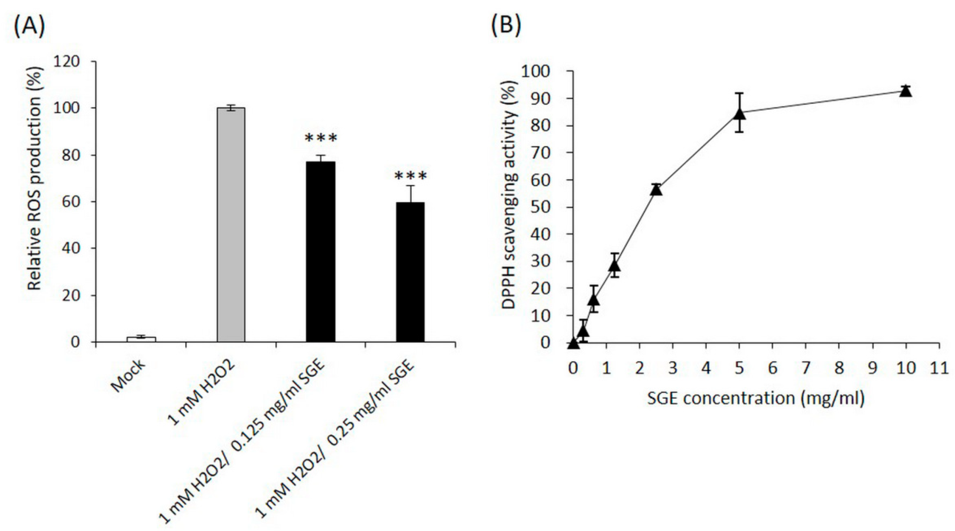
3.1.1. The Free Radical Scavenging Activities of SGE

Sargassum species are a rich source of natural antioxidants. Methods commonly used to determine the antioxidant activities of Sargassum species include DPPH (1,1-diphenyl-2-picryl hydrazyl) radical scavenging, NO scavenging, lipid peroxide inhibition and hydrogen peroxide (H2O2) radical scavenging assays [27,28,29,30,31]. The effect of SGE on intracellular ROS levels of CCD-966SK fibroblasts was shown in Fig. 1A. Treatment with 1 mM of H2O2 significantly increased intracellular ROS levels (defined as 100%). Pretreatment with 0.125 mg/ml of SGE blocked the increase of intracellular ROS generation induced by H2O2 stimulation (77%, P < 0.001). Furthermore, the increase of SGE concentration to 0.25 mg/ml reduced intracellular ROS levels to 60% compared with that induced by 1 mM H2O2 without SGE pretreatment (P < 0.001). Since treatment with 2 mg/ml SGE did not result in significant cytotoxicity in dermal fibroblasts in the following assays, we supposed that dosages higher than 0.25 mg/ml would confer a better suppression effect in the intracellular ROS generation. The scavenging effects of SGE on DPPH free radicals are shown in Fig. 1B by using 0, 0.3125, 0.625, 1.25, 2.5, 5 and 10 mg/ml of SGE. SGE exhibited DPPH scavenging properties in a dose-dependent manner. Use of 10 mg/ml of SGE reached 92.8% of DPPH scavenging activity. The IC50 value of SGE on DPPH radical scavenging activity was 2.31 mg/ml (Figure 1B).

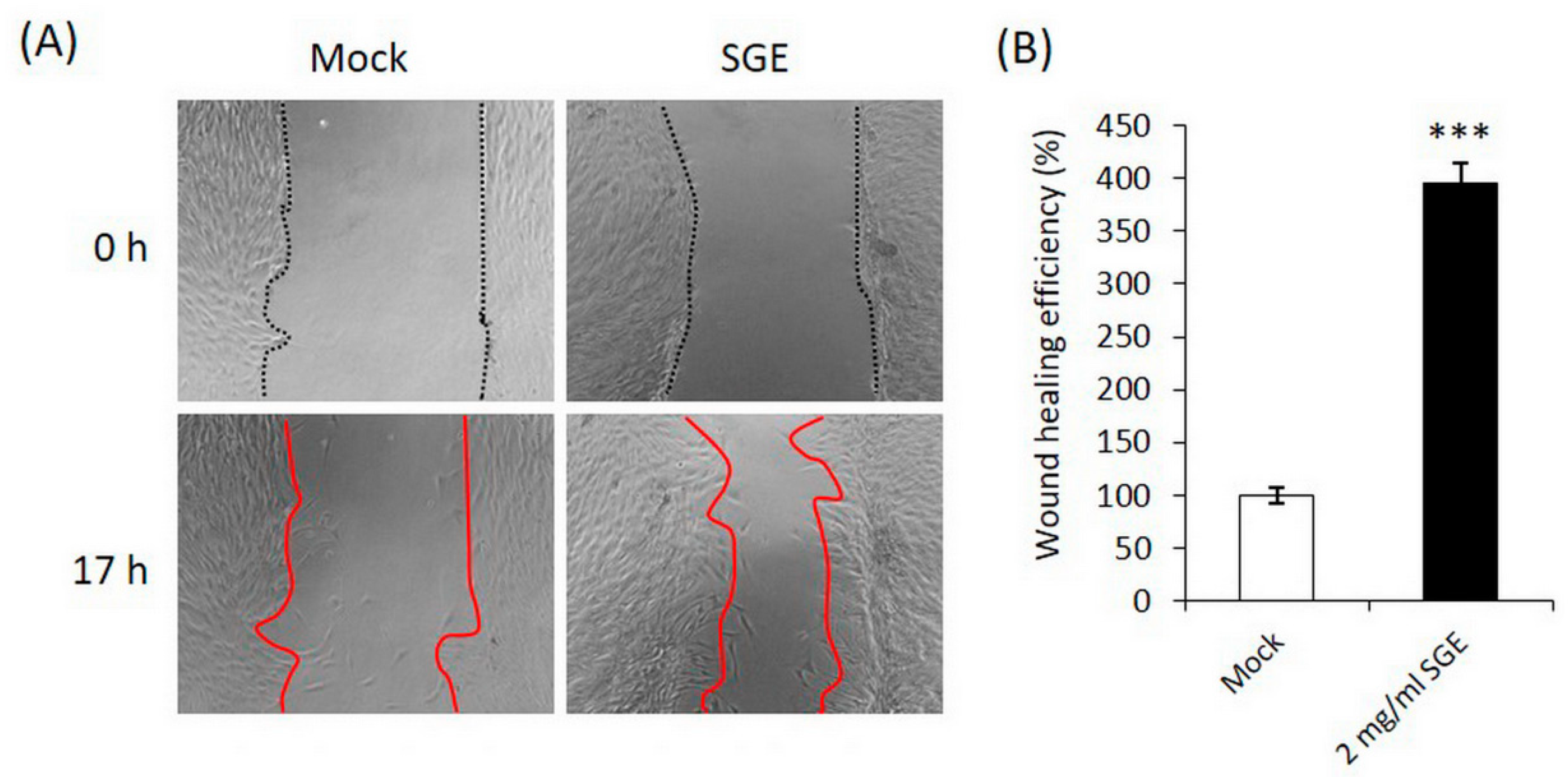
3.1.2. SGE Stimulated CCD-966SK Fibroblast Migration in the Wound Healing Assay

We next determined whether SGE stimulated dermal fibroblast migration. A wound healing assay showed that wound coverage was significantly increased in cells treated with 2 mg/ml SGE (Figure 2a,b). The wound healing efficiency enhanced 295% in SGE-treated CCD-966SK cells when compared to the control. The components of Sargassum species has been proven to be effective in anti-angiogenesis by inhibiting cancer cell migration and proliferation in the wound healing assay [17,32]. However, our data showed that SGE could be potentially used in repairing the wound of dermal fibroblasts.

3.2. Anti-Photoaging Activities Of SGE in Dermal Fibroblasts

3.2.1. SGE Recovered CCD-966SK Cell Regeneration after UVA Irradiation

Sargassum species contain functional ingredients such as fucoxanthin that protects the body against oxidative damage [33]. We examined the potential of SGE in the cell recovery ability of dermal fibroblasts against UVA irradiation. Under the treatment of 0.25, 0.5 and 1 mg/ml of SGE after UVA irradiation, the cell viability increased approximately 20% in CCD-966SK cells when compared with that of untreated irradiated cells (Figure 3). However, administration concentrations lower than 0.125 mg/ml did not recover the cell viability. Based on this result, we suggested that SGE with concentrations above 0.25 mg/ml possessed the ability to promote fibroblast regeneration and could be applied after UVA irradiation.

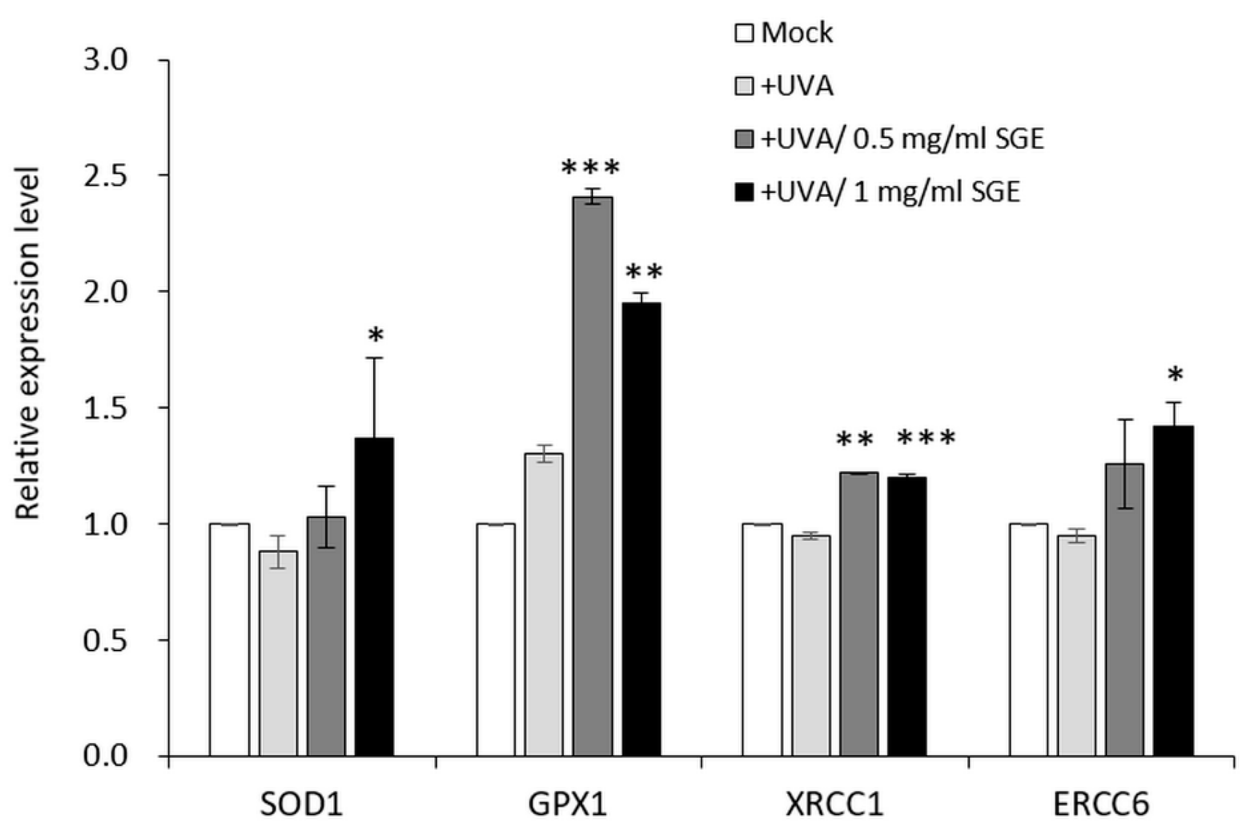
3.2.2. SGE Induced Antioxidant Gene Expressions in CCD-966SK Cell after UVA Irradiation

To alleviate and repair the damage caused by ROS, three primary antioxidant enzymes including superoxide dismutase (SOD), catalase (CAT), and glutathione peroxidase (GPX) would be activated by mammalian cells to develop an antioxidant defense system [34]. In the ROS-mediated oxidative stress, DNA damages are repaired by functionally important X-ray repair cross-complementing protein 1 (XRCC1) and excision repair cross-complementing complementation group 6 (ERCC6) [35,36,37]. To assess the effects of SGE on antioxidant activity, we analyzed anti-oxidative genes SOD1 and GPX1, and DNA repair regulatory genes XRCC1 and ERCC6 in CCD-966SK fibroblasts after UVA irradiation. As shown in Figure 4, the expression levels of SOD1 and ERCC6 were elevated only in the presence of 1 mg/ml SGE, whereas the expression of GPX1 and XRCC1 were significantly increased under the SGE treatment of both concentrations of 0.5 and 1 mg/ml. In summary, SGE exhibits protective effects against UVA irradiation-induced oxidative stress and DNA damage by upregulating the gene expression levels of SOD1, GPX1, XRCC1 and ERCC6.
Nowadays, photoprotection with natural product extracts may represent an effective strategy for skin protection without complicated synthesis or purification procedures. Most basically, to be one of the candidates, S. glaucescens can be extracted by simple, reactant-saving and low pollution procedures [13]. Although the production of brown algae is sufficient in Taiwan on a commercial scale, the biomass and functional component accumulations of S. glaucescens limitedly peak in spring. Among seaweeds, brown algae have revealed their superior antioxidant activity over red and green algae, and even greater than that of the synthetic antioxidant butylated hydroxytoluene (BHT) [38]. Compared to other brown seaweeds, SGE in this study showed a better IC50 value on DPPH radical scavenging activity (2.31 mg/ml), than that of fucoidan extracted from the brown algae Laminaria japonica (3.7 mg/ml) [39], and that of fucoidan extracted from S. glaucescens by the compressional-puffing-hydrothermal extraction (CPHE) process (4.27–5.15 mg/ml) [13]. Based on these facts, the synergistical work of bioactive compounds in SGE may confer higher antioxidant and anti-photoaging activities than a single compound.

3.3. Protective Effects of SGE in Keratinocytes and Skin Barrier

3.3.1. SGE Stimulated the Cell Proliferation of Human Primary Epidermal Keratinocytes

Epidermal keratinocytes serve as a barrier to retain water within the body and prevent the entry of infectious organisms or chemical agents [40]. To examine the SGE effects on keratinocytes proliferation, we performed a cell viability assay on human primary epithelial keratinocytes. By applying 0.0625 and 0.03125 mg/ml of SGE, keratinocytes revealed an approximately 20% increase in cell proliferation in comparison to the control (Figure 5).

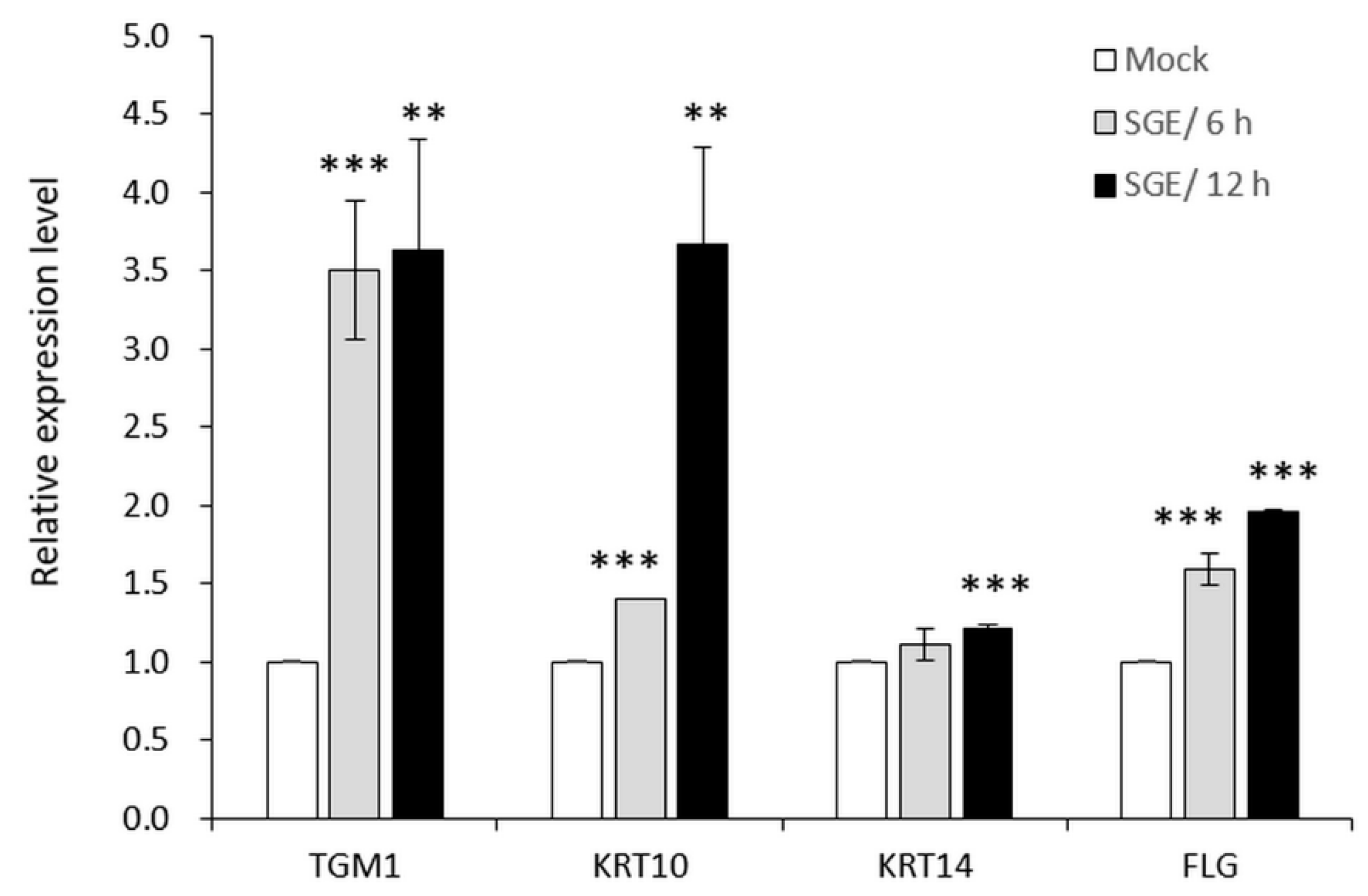
3.3.2. SGE Induced Expressions of Skin Barrier-Related Genes in Human Primary Epidermal Keratinocytes

Keratins are the major cytoskeletal scaffolding proteins of all epithelia, which are a diverse group of fibrous proteins. Distinct keratin pairs are responsible for the divergent epithelial functions for the protection of epithelial cells against mechanical stress [41,42]. Human KRT10 and KRT14 are type I keratins of epithelial keratinocytes. KRT14 paired with KRT5 forms bundled filaments in the basal layer, while KRT10/ KRT1 forms filaments in the suprabasal layers and KRT10/ KRT2 forms filaments in the uppermost layers [43,44]. The keratin fibers are cross-linked by the transglutaminase (TGM) enzyme, and bundled into macrofibrils through their association with filaggrin (FLG) to form a protective barrier between the body and its environment. Not only a key protein of the skin barrier, filaggrin is also essential for stratum corneum hydration. Processing of filaggrin proteins produces hygroscopic amino acids and their derivatives, known as natural moisturizing factor (NMF), which helps maintain hydration of the skin [45]. To determine the effects of SGE on the functions of the skin barrier, we analyzed the gene expressions of TGM1, KRT10, KRT14 and FLG. As shown in Figure 6, the expression levels of TGM1, KRT10 and FLG were all significantly elevated after the treatment of 0.03125 mg/ml SGE for 6 hours or 12 hours. Although KRT14 mRNAs were not increased after a 6-hour SGE treatment, extension of the treatment period to 12 hours conferred a mild elevation in expression. Based on these results, SGE is potentially involved in promoting the production of keratin fibers and the crosslinking enzyme, which strengthens the protective barrier. Additionally, SGE was able to stimulate the gene expression of FLG and therefore could produce abundant NMF for moisture-retention in skin.
In conclusion, we evaluated the antiaging activity and hydration-retention potential of Sargassum glaucescens extracts in skin cells. We demonstrated that SGE protected dermal fibroblasts against oxidative stress and the damages from UVA irradiation, probably through the regulation of antioxidant and DNA repair-related gene expressions. SGE repaired the scratch wound of dermal fibroblasts by promoting cell proliferation. In epidermal keratinocytes, SGE stimulated cell viability and cytoskeletal scaffolding-related gene expressions. We also observed the potential use of SGE in promoting NMF production in keratinocytes. Our preliminary studies demonstrated a novel potentiality of SGE for promoting moisture-retention and anti-photoaging of skin. The desire for natural agents in anti-photoaging and beauty has led to a growing trend in adding natural antioxidants to skincare products. Thus, in the present study, we discussed SGE for cosmetic applications. To optimize the efficacy and use of SGE in innovative formulations, we will engage in a safety assessment in human skin, for example, by patch testing, and conducting further human clinical trials. Meanwhile, the incorporation of other natural ingredients in the formulations for a synergetic work will be essential for an optimal photoprotecting and moisturizing effect.

Author Contributions

Conceptualization, Y.T.L.(Yu-Ting Lin) and H.L.S.; Methodology, Z.Y.L. and C.H.Y.; Validation, K.W.K., F.C.L., C.T.C., and Y.T.L.(Yi-Tsen Lin); Formal Analysis, Z.Y.L. and C.H.Y.; Investigation, Z.Y.L., K.W.K., F.C.L., C.T.C., and Y.T.L.(Yi-Tsen Lin); Resources, Y.H.L.; Data Curation, Z.Y.L. and C.H.Y.; Writing-Original Draft Preparation, H.F.H.; Writing-Review & Editing, H.F.H.; Visualization, H.F.H,; Supervision, Y.H.L. and H.L.S.; Project Administration, Y.T.L.(Yu-Ting Lin) and H.L.S.; Funding Acquisition, Y.H.L.

Funding

This research received no external funding.

Acknowledgments

This research was entirely funded by TCI Co., Ltd.

Conflicts of Interest

The authors declare no conflicts of interest.

References

  1. Berthon, J.Y.; Nachat-Kappes, R.; Bey, M.; Cadoret, J.P.; Renimel, I.; Filaire, E. Marine algae as attractive source to skin care. Free Radic. Res. 2017, 51, 555–567. [Google Scholar] [CrossRef] [PubMed]
  2. Komatsu, T.; Fukuda, M.; Mikami, A.; Mizuno, S.; Kantachumpoo, A.; Tanoue, H.; Kawamiya, M. Possible change in distribution of seaweed, Sargassum horneri, in northeast Asia under A2 scenario of global warming and consequent effect on some fish. Mar. Pollut. Bull. 2014, 85, 317–324. [Google Scholar] [CrossRef] [PubMed]
  3. Meenakshi, S.; Umayaparvathi, S.; Arumugam, M.; Balasubramanian, T. In vitro antioxidant properties and ftir analysis of two seaweeds of gulf of mannar. Asian Pac. J. Trop. Med. 2011, 1, S66–S70. [Google Scholar] [CrossRef]
  4. Manav, M.; Su, J.; Hughes, K.; Lee, H.P.; Ong, C.N. ω-3 fatty acids and selenium as coronary heart disease risk modifying factors in Asian Indian and Chinese males. Nutrition 2004, 20, 967–973. [Google Scholar] [CrossRef] [PubMed]
  5. Farage, M.A.; Miller, K.W.; Elsner, P.; Maibach, H.I. Intrinsic and extrinsic factors in skin ageing: A review. Int. J. Cosmet. Sci. 2008, 30, 87–95. [Google Scholar] [CrossRef] [PubMed]
  6. Han, A.; Chien, A.L.; Kang, S. Photoaging. Dermatol. Clin. 2014, 32, 291–299. [Google Scholar] [CrossRef] [PubMed]
  7. Polefka, T.G.; Meyer, T.A.; Agin, P.P.; Bianchini, R.J. Effects of solar radiation on the skin. J. Cosmet. Dermatol. 2012, 11, 134–143. [Google Scholar] [CrossRef] [PubMed]
  8. Fuentealba, D.; Galvez, M.; Alarcon, E.; Lissi, E.; Silva, E. Photosensitizing activity of advanced glycation endproducts on tryptophan, glucose 6-phosphate dehydrogenase, human serum albumin and ascorbic acid evaluated at low oxygen pressure. Photochem. Photobiol. 2007, 83, 563–569. [Google Scholar] [CrossRef] [PubMed]
  9. Kim, J.A.; Ahn, B.N.; Kong, C.S.; Kim, S.K. Protective effect of chromene isolated from Sargassum horneri against UV-A-induced damage in skin dermal fibroblasts. Exp. Dermatol. 2012, 21, 630–631. [Google Scholar] [CrossRef] [PubMed]
  10. Kim, J.A.; Ahn, B.N.; Kong, C.S.; Kim, S.K. The chromene sargachromanol E inhibits ultraviolet A-induced ageing of skin in human dermal fibroblasts. Br. J. Dermatol. 2013, 168, 968–976. [Google Scholar] [CrossRef] [PubMed]
  11. Lee, C.; Park, G.H.; Ahn, E.M.; Park, C.I.; Jang, J.H. Sargassum fulvellum protects HaCaT cells and BALB/c mice from UVB-induced proinflammatory responses. Evid. Based Complement. Alternat. Med. 2013, 2013, 747846. [Google Scholar] [CrossRef] [PubMed]
  12. Shao, P.; Chen, X.; Sun, P. Improvement of antioxidant and moisture-preserving activities of Sargassum horneri polysaccharide enzymatic hydrolyzates. Int. J. Biol. Macromol. 2015, 74, 420–427. [Google Scholar] [CrossRef] [PubMed]
  13. Huang, C.Y.; Wu, S.J.; Yang, W.N.; Kuan, A.W.; Chen, C.Y. Antioxidant activities of crude extracts of fucoidan extracted from Sargassum glaucescens by a compressional-puffing-hydrothermal extraction process. Food Chem. 2016, 197, 1121–1129. [Google Scholar] [CrossRef] [PubMed]
  14. Collins, K.G.; Fitzgerald, G.F.; Stanton, C.; Ross, R.P. Looking beyond the terrestrial: The potential of seaweed derived bioactives to treat non-communicable diseases. Mar. Drugs 2016, 14, 60. [Google Scholar] [CrossRef] [PubMed]
  15. Vijayabaskar, P.; Vaseela, N.; Thirumaran, G. Potential antibacterial and antioxidant properties of a sulfated polysaccharide from the brown marine algae Sargassum swartzii. Chin. J. Nat. Med. 2012, 10, 421–428. [Google Scholar] [CrossRef]
  16. Ale, M.T.; Maruyama, H.; Tamauchi, H.; Mikkelsen, J.D.; Meyer, A.S. Fucoidan from Sargassum sp. and fucus vesiculosus reduces cell viability of lung carcinoma and melanoma cells in vitro and activates natural killer cells in mice in vivo. Int. J. Biol. Macromol. 2011, 49, 331–336. [Google Scholar] [CrossRef] [PubMed]
  17. Cong, Q.; Chen, H.; Liao, W.; Xiao, F.; Wang, P.; Qin, Y.; Dong, Q.; Ding, K. Structural characterization and effect on anti-angiogenic activity of a fucoidan from Sargassum fusiforme. Carbohydr. Polym. 2016, 136, 899–907. [Google Scholar] [CrossRef] [PubMed]
  18. Dore, C.M.; das, C.F.A.M.G.; Will, L.S.; Costa, T.G.; Sabry, D.A.; de Souza Rego, L.A.; Accardo, C.M.; Rocha, H.A.; Filgueira, L.G.; Leite, E.L. A sulfated polysaccharide, fucans, isolated from brown algae Sargassum vulgare with anticoagulant, antithrombotic, antioxidant and anti-inflammatory effects. Carbohydr. Polym. 2013, 91, 467–475. [Google Scholar] [CrossRef] [PubMed]
  19. Morya, V.K.; Kim, J.; Kim, E.K. Algal fucoidan: Structural and size-dependent bioactivities and their perspectives. Appl. Microbiol. Biotechnol. 2012, 93, 71–82. [Google Scholar] [CrossRef] [PubMed]
  20. Wells, M.L.; Potin, P.; Craigie, J.S.; Raven, J.A.; Merchant, S.S.; Helliwell, K.E.; Smith, A.G.; Camire, M.E.; Brawley, S.H. Algae as nutritional and functional food sources: Revisiting our understanding. J. Appl. Phycol. 2017, 29, 949–982. [Google Scholar] [CrossRef] [PubMed]
  21. Marinho-Soriano, E.; Fonseca, P.C.; Carneiro, M.A.; Moreira, W.S. Seasonal variation in the chemical composition of two tropical seaweeds. Bioresour. Technol. 2006, 97, 2402–2406. [Google Scholar] [CrossRef] [PubMed]
  22. Murugaiyan, K.; Sivakumar, K. Seasonal variation in elemental composition of Stoechospermum marginatum (Ag.) Kutz and Sargassum wightii (Greville Mscr.) J.G. Agardh in relation to chemical composition of seawater. Colloids Surf. B Biointerfaces 2008, 64, 140–144. [Google Scholar] [CrossRef] [PubMed]
  23. Hwang, P.A.; Hung, Y.L.; Tsai, Y.K.; Chien, S.Y.; Kong, Z.L. The brown seaweed Sargassum hemiphyllum exhibits alpha-amylase and alpha-glucosidase inhibitory activity and enhances insulin release in vitro. Cytotechnology 2015, 67, 653–660. [Google Scholar] [CrossRef] [PubMed]
  24. Shimada, K.; Fujikawa, K.; Yahara, K.; Nakamura, T. Antioxidative properties of xanthan on the autoxidation of soybean oil in cyclodextrin emulsion. J. Agric. Food Chem. 1992, 40, 945–948. [Google Scholar] [CrossRef]
  25. Yarrow, J.C.; Perlman, Z.E.; Westwood, N.J.; Mitchison, T.J. A high-throughput cell migration assay using scratch wound healing, a comparison of image-based readout methods. BMC Biotechnol. 2004, 4, 21. [Google Scholar] [CrossRef] [PubMed]
  26. Shindo, Y.; Hashimoto, T. Antioxidant defence mechanism of the skin against uv irradiation: Study of the role of catalase using acatalasaemia fibroblasts. Arch. Dermatol. Res. 1995, 287, 747–753. [Google Scholar] [CrossRef] [PubMed]
  27. Yende, S.R.; Harle, U.N.; Chaugule, B.B. Therapeutic potential and health benefits of Sargassum species. Pharmacogn. Rev. 2014, 8, 1–7. [Google Scholar] [CrossRef] [PubMed]
  28. Hwang, P.A.; Wu, C.H.; Gau, S.Y.; Chien, S.Y.; Hwang, D.F. Antioxidant and immune-stimulating activities of hot-water extract from seaweed Sargassum hemiphyllum. J. Mar. Sci. Technol. 2010, 18, 41–46. [Google Scholar]
  29. Park, P.J.; Heo, S.J.; Park, E.J.; Kim, S.K.; Byun, H.G.; Jeon, B.T.; Jeon, Y.J. Reactive oxygen scavenging effect of enzymatic extracts from Sargassum thunbergii. J. Agric. Food Chem. 2005, 53, 6666–6672. [Google Scholar] [CrossRef] [PubMed]
  30. Lim, S.N.; Cheung, P.C.; Ooi, V.E.; Ang, P.O. Evaluation of antioxidative activity of extracts from a brown seaweed, Sargassum siliquastrum. J. Agric. Food Chem. 2002, 50, 3862–3866. [Google Scholar] [CrossRef] [PubMed]
  31. Wen, Z.S.; Liu, L.J.; OuYang, X.K.; Qu, Y.L.; Chen, Y.; Ding, G.F. Protective effect of polysaccharides from Sargassum horneri against oxidative stress in RAW264.7 cells. Int. J. Biol. Macromol. 2014, 68, 98–106. [Google Scholar] [CrossRef] [PubMed]
  32. Hulikere, M.M.; Joshi, C.G.; Ananda, D.; Poyya, J.; Nivya, T. Antiangiogenic, wound healing and antioxidant activity of Cladosporium cladosporioides (Endophytic fungus) isolated from seaweed (Sargassum wightii). Mycology 2016, 7, 203–211. [Google Scholar] [CrossRef] [PubMed]
  33. D’Orazio, N.; Gemello, E.; Gammone, M.A.; de Girolamo, M.; Ficoneri, C.; Riccioni, G. Fucoxantin: A treasure from the sea. Mar. Drugs 2012, 10, 604–616. [Google Scholar] [CrossRef]
  34. Weydert, C.J.; Cullen, J.J. Measurement of superoxide dismutase, catalase and glutathione peroxidase in cultured cells and tissue. Nat. Protoc. 2010, 5, 51–66. [Google Scholar] [CrossRef]
  35. Newman, J.C.; Bailey, A.D.; Weiner, A.M. Cockayne syndrome group B protein (CSB) plays a general role in chromatin maintenance and remodeling. Proc. Natl. Acad. Sci. USA 2006, 103, 9613–9618. [Google Scholar] [CrossRef]
  36. Lake, R.J.; Fan, H.-Y. Structure, function and regulation of CSB: A multi-talented gymnast. Mech. Ageing Dev. 2013, 134, 202–211. [Google Scholar] [CrossRef] [PubMed]
  37. Ramaniuk, V.P.; Nikitchenko, N.V.; Savina, N.V.; Kuzhir, T.D.; Rolevich, A.I.; Krasny, S.A.; Sushinsky, V.E.; Goncharova, R.I. Polymorphism of DNA repair genes OGG1, XRCC1, XPD and ERCC6 in bladder cancer in Belarus. Biomarkers 2014, 19, 509–516. [Google Scholar] [CrossRef]
  38. Kindleysides, S.; Quek, S.Y.; Miller, M.R. Inhibition of fish oil oxidation and the radical scavenging activity of New Zealand seaweed extracts. Food Chem. 2012, 133, 1624–1631. [Google Scholar] [CrossRef]
  39. Wang, J.; Zhang, Q.; Zhang, Z.; Song, H.; Li, P. Potential antioxidant and anticoagulant capacity of low molecular weight fucoidan fractions extracted from Laminaria japonica. Int. J. Biol. Macromol. 2010, 46, 6–12. [Google Scholar] [CrossRef] [PubMed]
  40. Segre, J.A. Epidermal barrier formation and recovery in skin disorders. J. Clin. Investig. 2006, 116, 1150–1158. [Google Scholar] [CrossRef]
  41. Chamcheu, J.C.; Siddiqui, I.A.; Syed, D.N.; Adhami, V.M.; Liovic, M.; Mukhtar, H. Keratin gene mutations in disorders of human skin and its appendages. Arch. Biochem. Biophys. 2011, 508, 123–137. [Google Scholar] [CrossRef] [PubMed]
  42. Magin, T.M.; Vijayaraj, P.; Leube, R.E. Structural and regulatory functions of keratins. Exp. Cell Res. 2007, 313, 2021–2032. [Google Scholar] [CrossRef] [PubMed]
  43. Schweizer, J.; Bowden, P.E.; Coulombe, P.A.; Langbein, L.; Lane, E.B.; Magin, T.M.; Maltais, L.; Omary, M.B.; Parry, D.A.; Rogers, M.A.; et al. New consensus nomenclature for mammalian keratins. J. Cell Biol. 2006, 174, 169–174. [Google Scholar] [CrossRef] [PubMed]
  44. Moll, R.; Divo, M.; Langbein, L. The human keratins: Biology and pathology. Histochem. Cell Biol. 2008, 129, 705–733. [Google Scholar] [CrossRef] [PubMed]
  45. Kezic, S.; Kemperman, P.M.; Koster, E.S.; de Jongh, C.M.; Thio, H.B.; Campbell, L.E.; Irvine, A.D.; McLean, W.H.; Puppels, G.J.; Caspers, P.J. Loss-of-function mutations in the filaggrin gene lead to reduced level of natural moisturizing factor in the stratum corneum. J. Investig. Dermatol. 2008, 128, 2117–2119. [Google Scholar] [CrossRef] [PubMed]
Figure 1. Antioxidant activities of SGE. (A) CCD-966SK cells were treated with 1 mM H2O2 to induce ROS production (grey bar). Cells were co-treated with 1 mM H2O2 and 0.125 or 0.25 mg/ml of SGE (black bars) to detect the antioxidant effect, or were left untreated as a negative control (Mock, white bar). Relative reactive oxygen species (ROS) production by different treatments was calculated in comparison with that of treatment with 1 mM H2O2 (100%). (B) The DPPH radical scavenging activity was evaluated by using 0, 0.3125, 0.625, 1.25, 2.5, 5 and 10 mg/ml of SGE. SGE exhibited DPPH scavenging properties in a dose-dependent manner. ***, P < 0.001 versus 1 mM H2O2. SGE, Sargassum glaucescens extracts.
Cosmetics 06 00017 g001
Figure 2. SGE stimulated CCD-966SK fibroblast migration in the wound healing assay. (A) Cells were grown to confluence and an in vitro wound was produced using a sterile pipette tip, followed by incubation with 2 mg/ml SGE or not (Mock) for 17 hours. Black dashed lines represent the initial scratch and red lines represent the migration front. (B) The coverage of scratch wound area in percent relative to initial wound area after a 17-hour SGE treatment (black bar) or not (Mock, white bar) was calculated. The percent of wound coverage was measured using a scan image. Histograms indicated the wound healing efficiency of 2 mg/ml SGE relative to Mock (100%). SGE, Sargassum glaucescens extracts. ***, P < 0.001 versus Mock. SGE, Sargassum glaucescens extracts.
Cosmetics 06 00017 g002
Figure 3. SGE recovered CCD-966SK cell regeneration after UVA irradiation. Cells were pretreated with UVA for 1 hour (+UVA, grey bar), and then treated with 0.0625, 0.125, 0.25, 0.5 or 1 mg/ml of SGE (black bars), or left untreated as negative control (−UVA, white bar, 100%). SGE was able to recover dermal fibroblasts from UVA irradiation by the treatment concentrations of 0.25, 0.5 and 1 mg/ml. **, P < 0.01 versus +UVA; ***, P < 0.001 versus +UVA. SGE, Sargassum glaucescens extracts.
Cosmetics 06 00017 g003
Figure 4. SGE extracts induced antioxidant gene expressions in CCD-966SK cells after UVA irradiation. Cells were pretreated with UVA for 1 hour (+UVA, light grey bar), and then treated with 0.5 or 1 mg/ml of SGE (dark grey and black bars), or left untreated as a negative control (Mock, white bar). Cells were harvested for total RNA isolation and expression detection for SOD1, GPX1, XRCC1 and ERCC6. *, P < 0.05 versus +UVA; **, P < 0.01 versus +UVA; ***, P < 0.001 versus +UVA. SGE, Sargassum glaucescens extracts.
Cosmetics 06 00017 g004
Figure 5. SGE stimulated the cell proliferation of human primary epidermal keratinocytes. Cells were treated with 0.03125, 0.0625 mg/ml of SGE, or left untreated as a control (Mock, white bar, 100%). Both 0.03125 mg/ml (grey bar) and 0.0625 mg/ml (black bar) of SGE were able to enhance cell viability after a 24-hour treatment. **, P < 0.01 versus Mock; ***, P < 0.001 versus Mock. SGE, Sargassum glaucescens extracts.
Cosmetics 06 00017 g005
Figure 6. SGE induced moisturizing-related gene expressions in human primary epidermal keratinocytes. Cells were treated with 0.03125 mg/ml SGE for 6 hours (grey bar) or 12 hours (black bar), or left untreated for 12 hours as a control (Mock, white bar). After treatment, cells were harvested for TGM1, KRT10, KRT14 and FLG gene expression analysis. **, P < 0.01 versus Mock; ***, P < 0.001 versus Mock. SGE, Sargassum glaucescens extracts.
Cosmetics 06 00017 g006
Table 1. Real-time quantitative PCR primers used in this study.
Gene NamePrimer NamePrimer SequenceGene Function
SOD1SOD1-F5′- GGTGGGCCAAAGGATGAAGAG -3′anti-oxidative gene
SOD1-R5′- CCACAAGCCAAACGACTTCC -3′
GPX1GPX1-F5′- CAGTCGGTGTATGCCTTCTCG -3′anti-oxidative gene
GPX1-R5′- GAGGGACGCCACATTCTCG -3′
XRCC1XRCC1-F5′- TTCTTCCAGGGCAAGCACTT -3′DNA repair regulatory gene
XRCC1-R5′- GATCCCATTCCTGTGCTGTGA -3′
ERCC6ERCC6-F5′- GTCCAAGATCACATAGTGCC -3′DNA repair regulatory gene
ERCC6-R5′- AAAAGGAGAAACTAATTCGA -3′
TGM1TGM1-F5′- GATCGCATCACCCTTGAGTTAC -3′epidermal barrier structural genes
TGM1-R5′- GCAGGTTCAGATTCTGCCC -3′
KRT10KRT10-F5′- TCCTACTTGGACAAAGTTCGGG -3′epidermal barrier structural genes
KRT10-R5′- CCCCTGATGTGAGTTGCCA -3′
KRT14KRT14-F5′- TTCTGAACGAGATGCGTGAC -3′epidermal barrier structural genes
KRT14-R5′- GCAGCTCAATCTCCAGGTTC -3′
FLGFLG-F5′- GGCAAATCCTGAAGAATCCA -3′epidermal barrier structural genes
FLG-R5′- TGCTTTCTGTGCTTGTGTCC -3′
GAPDHGAPDH-F5′- CTGGGCTACACTGAGCACC -3′housekeeping gene
GAPDH-R5′- AAGTGGTCGTTGAGGGCAATG -3′

