Article

Analytical Investigation of Forced Oxidized Anti-VEGF IgG Molecules: A Focus on the Alterations in Antigen and Receptor Binding Activities

1
Faculty of Engineering and Natural Sciences, Sabanci University, Istanbul 34956, Turkey
2
SUNUM Nanotechnology Research and Application Centre, Sabanci University, Istanbul 34956, Turkey
3
Kosuyolu High Specialization Education and Research Center, Surgical Gastroenterology Clinic, University of Health Sciences, Istanbul 34865, Turkey
*
Author to whom correspondence should be addressed.
Sci. Pharm. 2023, 91(3), 31; https://doi.org/10.3390/scipharm91030031
Submission received: 25 May 2023 / Revised: 8 June 2023 / Accepted: 16 June 2023 / Published: 28 June 2023
(This article belongs to the Special Issue Feature Papers in Scientia Pharmaceutica)

Abstract

:
Alterations in the biological activity of the molecules under stress conditions have not been documented as widely in the literature yet. This study was designed to reveal the functional impacts of various oxidation conditions on a model mAb, a commercial anti-VEGF IgG molecule. The responses to antigen binding, cell proliferation, FcRn receptors, and C1q binding, which rarely appear in the current literature, were investigated. The authors report peptide mapping data, post-translational modification (PTM) analysis, cell proliferation performance, and antigen (VEGF), C1q, and FcRn binding activities of the mAb under various stress conditions. The oxidation-prone site of the mAb was determined as Met252 in the DTLMISR peptide. The VEGF binding activity and anti-cell proliferation activity of the mAbs did not alter, while C1q and FcRn binding capacity significantly decreased under oxidative stress conditions. The full report is vital for many scientific and industrial processes about mAbs. The authors recommend performing functional analyses in addition to the structural studies while investigating the impacts of stress factors on therapeutic mAbs.

1. Introduction

Anti-VEGF Immunoglobulin G (IgG) antibody is a monoclonal antibody (mAb) explicitly developed for targeting the vascular endothelial growth factor (VEGF), which triggers the formation of new blood vessels by interacting with VEGF receptors on the surface of vascular endothelial cells [1]. Cells stimulate vascularisation by secreting VEGF at an elevated level to reach the nutrient and oxygen levels required for cell survival, which results in the growth and proliferation of cancer cells [2]. As a treatment strategy, the interaction of VEGF with its receptor can be inhibited to prevent vascularisation [3]. The mAbs developed against the VEGF inhibit this interaction; thus, tumour cells cannot receive nutrients and oxygen to grow. Because of this, in addition to their many advantages, the rapid development of biosimilar mAbs has gained attention worldwide [4]. The production of biotherapeutics, including mAbs, is known to be more challenging compared with the production of small molecular-weighted chemical drugs. The utilisation of mammalian cells as host organisms to produce monoclonal antibody drugs primarily leads to heterogeneities in post-translational modifications (PTMs) and glycosylation patterns of the mAbs [5]. In addition to the impact of the upstream process, even mild stress conditions during the downstream process or storage can lead to alterations in the effectiveness, quality, or half-life of the mAbs [6]. Hence, it is vital to reveal and report the structural and functional alterations caused by stress conditions before regulatory authorities approve them [7].
Monoclonal antibodies are exposed to many environmental factors such as temperature, light, mechanical stress, humidity, and pH during almost all production steps, from the upstream to post-development processes such as packaging, shipping, and storage. As a result of these environmental stresses, the biochemical structure of the antibodies can be severely affected. The products may undergo various degradation pathways, which causes them to lose their biological activity [8]. The degradation of recombinant mAbs is an important parameter affecting the product’s quality, biological efficacy, and safety. Forced degradation studies provide reliable data by imitating the potential stress factors via elevated conditions in a short time instead of the more prolonged exposure to mild stress conditions usually seen in production or post-production [9]. In addition, forced degradation studies help understand the molecule’s biophysical and biochemical properties, including the degradation mechanisms, in-depth [10].
Temperature is one of the crucial parameters in changing the critical quality attributes (CQA) of mAbs [11]. The mAbs can form aggregates by being exposed to many different temperatures (high and low values) during production, storage, and shipping. High temperatures especially lead to irreversible aggregate structures in mAbs [12]. Since low temperatures reduce hydrophobic interactions between solvents and proteins, a vital force in protein folding [13], intermolecular hydrophobic regions between proteins interact, increasing aggregates’ formation [14]. Another critical parameter that can change the stability and structure of mAbs is pH [15]. In mAb production, during purification steps such as protein A chromatography and virus inactivation, mAbs are exposed to low-pH solutions. Therefore, antibodies’ structural changes and biological activities in a low-pH environment should be determined. In addition, low-pH conditions during the purification stage can be a reference point for forced degradation studies [8]. At low pH, the formation of soluble [16] or insoluble aggregates [17,18] and fragmentation [19,20] is accelerated. Low pH leads to the fragmentation of antibodies, generally in the hinge region, and the formation of fragments of 40–50 kDa in non-reduced conditions and 20–30 kDa in reduced conditions [21,22]. Recombinant mAbs are also exposed to oxidising environmental conditions such as impurities from mAbs production, dissolved oxygen, oxygen in the air, and free radicals formed due to reactions with metals. Forced oxidation studies are also convenient for understanding how sensitive mAbs are to oxidation and observing how their activities change. In particular, the backbone of these studies is the investigation of antibodies’ potency and binding properties by oxidising residues in the regions. The most common approaches used for these studies are treating the antibody with hydrogen peroxide (H2O2) or tert-butyl hydrogen peroxide to investigate the oxidation of Met [23,24] and the incubation of antibodies with 2,2′-Azobis (2-amidinopropane) dihydrochloride for Trp oxidation [25].
Despite an enormous number of studies in the field, only a few studies are focused on the functional effects of stress conditions on mAbs, and many studies were limited to the effects of structural functions. In this study, the structural and functional characterisation of a commercial anti-VEGF IgG molecule that was exposed to a certain degree and duration of oxidation stress was reported. The essential structural characterisation, including peptide mapping and PTM analysis, was performed using the tandem mass spectrometry (UHPLC-MS/MS) technique. The functional characterisations that rarely appear in the current literature, such as proliferation activity, antigen-binding performance, response to FcRn receptors, and C1q binding, were also performed using different techniques. The surface plasmon resonance (SPR) method was used to determine the binding affinity of oxidative stress-exposed mAbs to VEGF and FcRn receptors. The enzyme-linked immunosorbent assay (ELISA) method was used to determine the binding affinity of anti-VEGF IgG molecules to the C1q protein. Moreover, the effects of oxidation stress on the anti-proliferative performance of anti-VEGF mAbs were investigated using in vitro assays. Although the effect of several stress conditions on the structure of anti-VEGF IgG molecules has been previously reported [26,27], potential alterations in the biological activity of the molecules under stress conditions have not been documented as widely and deeply yet. The EMA public assessment reports and FDA product quality reports of originator drugs (or biosimilars of anti-VEGF mAbs) do not present comprehensive information about the methodological details and receptor-binding behaviours of the mAbs upon stress exposure. This study was designed to reveal and document the effect of various oxidation conditions on mAbs, one of the most seen stress factors [28]. Therefore, the study’s full report is important for many scientific and industrial processes.

2. Material and Methods

2.1. Material

HEPES, NaCl, Polysorbate 20 (P20), and Ethanol were supplied from Sigma, Germany. EDTA was from AppliChem, Germany, and distilled water was from Merck Millipore, USA. Anti-VEGF IgG was obtained commercially (Avastin, 33808339) from Roche, Basel, Switzerland.

2.2. Generation of Oxidative Stress

Increased percentages of H2O2 (v/v) (Sigma, Darmstadt, Germany), 0.01, 0.05, 0.1, 0.3, 0.5, and 1, were used for 1 h at room temperature to examine the effect of H2O2 concentration on % oxidation of anti-VEGF IgG. In addition, the oxidation rate due to incubation time was determined via incubation of 1 mg/mL anti-VEGF IgG in 0.1% H2O2 for half an hour, 1 h, 2, and 4 h. After incubation, a 100 µL formulation buffer was exchanged using a single-use Amicon filter unit (Merck Millipore, Darmstadt, Germany).

2.3. Cell Culture Studies

The types of equipment used for cell culture experiments included a centrifuge (Eppendorf, 5415D, Darmstadt, Germany), CO2 incubator (Thermofisher Scientific, Waltham, CA, USA), microscope (Zeiss, Primo Vert, Germany), and Laminar flow (Heraeus, HeraSafe HS 12, Hanau, Germany). F.B.S. and 200 mM L-glutamine solutions were supplied from Thermo Fischer Scientific, USA. Cell counting was done with Trypan Blue (Sigma, Steinheim, Germany) and Hemocytometer (HBG Company, Giessen, Germany). Among the cell culture consumables, culture flasks and serological pipettes were from Sarstedt, Germany, and falcon tubes were from Eppendorf, Germany.
Primary Umbilical Vein Endothelial Cells (HUVEC) (ATCC PCS-100-013, ATCC, Manassas, VA, USA) were used for cell proliferation studies [29]. Cells were grown at 37 °C, 5% CO2 in Vascular Cell Basal Medium (ATCC® PCS-100-030, ATCC, Manassas, VA, USA) supplemented with Endothelial Cell Growth Kit-VEGF (ATCC® PCS-100-41, ATCC, Manassas, VA, USA). When the cells reached 80% confluency, subculture was performed. Cells were washed via centrifugation at 300× g for 5 min with D-PBS (Sigma, Steinheim, Germany) and resuspended with a complete medium. In total, 5 × 10³ HUVEC cells/well were inoculated into 96-well plates (Sarstedt, Nümbrecht, Germany) in 200 µL complete media and incubated at 37 °C overnight to allow the cells to adhere. Then, the cell medium was changed to a starvation medium, which was a vascular cell basal medium containing no growth factors: vascular endothelial growth factor (VEGF), endothelial growth factor (EGF), and fibroblast growth factor (FGF). Cells were inoculated within a starvation medium at 37 °C for 24 h to lose the proliferative effect of the VEGF growth factor. Then, control and oxidatively stressed anti-VEGF IgG were introduced into the cells at 5000 ng/mL concentrations. Then, VEGF was added at a 20 ng/mL concentration. The control well included only the VEGF growth factor and not the anti-VEGF IgG [30,31]. Cells treated with antibodies and VEGFs were incubated at 37 °C for 24 h.
CellTiter 96® Aqueous One Solution Cell Proliferation Assay (MTS) (Promega G3582, Madison, WI, USA) was used to determine the proliferation rates of cells after 24 h. The assay is a commonly used test based on metabolic activity assay [32]. A total of 20 µL/well of MTS reagent was added and incubated for 4 h at 37 °C. The blank was the reagent placed in an empty well for the measurements. At the end of the incubation period, absorbance values at 490 nm were measured using an ELISA microplate reader (Bio-Rad, Hercules, CA, USA).

2.4. Analyses

Target molecules were immobilised on CM5 chips (Cytiva, Marlborough, MA, USA) to perform binding analyses with a Biacore T200 SPR Instrument (Cytiva) [33]. The carboxylate groups on the chip surface were activated via a standard 1-ethyl-3-(3-dimethyl aminopropyl)carbodiimide/N-hydroxysuccinimide (EDC-NHS) (GE Healthcare, Uppsala, Sweden) reaction to induce the interaction between the carboxylate groups and the amine groups of the target molecules [34]. After all immobilisation processes, the remaining activated carboxyl groups were blocked with the injection of 1M ethanolamine-HCl (Cytiva). An ethanolamine-immobilised flow channel was considered the control surface in the experiments. For all analyses, 1X HBS-EP buffer (10 mM HEPES, 150 mM NaCl, 3 mM EDTA, 0.005% v/v polysorbate 20) at pH 7.4 was used as a running buffer. The data were presented as the mean value, calculated from at least three measurements per sample.

2.4.1. Antigen (VEGF) Binding Assays

The VEGF165A (Sigma, Steinheim, Germany) protein was prepared with a 5 ng/µL in pH 5.5 in 10 mM acetate buffer (GE Healthcare, Uppsala, Sweden) and immobilised onto the CM5 chip surface. The anti-VEGF IgG molecules exposed to specified stress factors at three concentrations (15 nM, 5 nM, and 1.66 nM) were prepared in a running buffer. Single-cycle kinetic analyses were conducted at a 30 µL/min flow rate at 22 °C. Analytes were injected for 120 s in the association phase and a dissociation phase of 1800 s with the running buffer. For all experiments, the final immobilisation level of the active flow cell was approximately 500 response units (RU). Blank measurements were performed on the active and control flow channels by running buffer injections under identical conditions. The chip surface was regenerated by injecting 10 mM glycine pH 1.5 buffer (GE Healthcare, Uppsala, Sweden) for 90 s. The results were found by subtracting responses from the blank flow cell and zero concentration analyte injection (running buffer) from the actual results. The equilibrium dissociation constants (KD) were calculated by Biacore Evaluation Software using a 1:1 Langmuir binding model [35,36].

2.4.2. Receptor (FcRn) Binding Assay

FcRn binding analyses of the samples were conducted on Anti-His IgG1 antibody (Cytiva, Marlborough, MA, USA) iimmobilised CM5 chips (Cytiva). His-Tagged FcRn (Sigma-Aldrich) anti-VEGF IgG was prepared in 1X HBS-EP (GE Healthcare, Uppsala, Sweden) pH 6.0 running buffer with three-fold dilutions (15 nM, 5 nM, 1.66 nM). Recombinant His-tagged FcRn molecule was captured on the active flow cell for 120 s with a 10 µL/min flow rate at 22 °C. A blocked flow cell was used as a blank reference during all measurements. Samples were injected over both flow cells (active and blank) at a 30 µL/min flow rate for 120 s, followed by a dissociation phase of 900 s with the running buffer. The chip surface was regenerated with a running buffer for 60 s. Blank buffer injections were also performed on both flow channels, later subtracted from the active surface data before the fitting. The results were evaluated with Biacore Evaluation Software using the steady-state and two-state binding models [37,38,39].

2.5. Peptide Mapping Using LC/MS-MS

An Acquity UHPLC-ESI-Xevo G2-XS QToF (Waters, Milford, MA, USA) system was used for peptide mapping analysis. All mobile phases (MS Grade water, formic acid, acetonitrile) were supplied from Merck, Germany. Mobile phases A, B, and C were MS-grade water, ACN, and 1% formic acid (FA). The instrument was calibrated using sodium cesium iodide (NaCsI) (Sigma-Aldrich, Steinheim, Germany), and Glu-1-fibrinopeptide B (Waters, Milford, MA, USA) was used as a lock-mass reference before running.
The 50 μg anti-VEGF IgG molecules exposed to oxidative stress were reduced through incubation at 56 °C for 15 min with 1% sodium dodecyl sulfate (SDS) (Sigma-Aldrich) and 0.1M Dithiothreitol (DTT) (Sigma-Aldrich, Steinheim, Germany) in 50 mM AMBIC solution. Then, samples were alkylated with 20 mM Iodoacetic acid (IAA) (Sigma-Aldrich, Steinheim, Germany) for 30 min in the dark at room temperature. After the alkylation process, all samples were mixed with 8M urea (Sigma, Steinheim, Germany) and purified using 30 kDa Molecular weight cut-off (MWCO) disposable filter units (Bedford, MA, USA) at 14,000 g for 10 min, twice. The purified samples were treated with 1 µg MS-grade trypsin (Sigma, Steinheim, Germany) in 75 µL AMBIC (1:50, w/w, enzyme to protein ratio) at 37 °C for 1.5 h. The tryptic peptides were collected by washing twice with 50 µL of 50 mM AMBIC at 14,000 g for 10 min. Finally, 1% formic acid was added to the samples before analysis [40].
The percentage of mobile phase C was fixed at 10%, and mobile phase B was increased from 1% to 80% over 85 s of a total run time to analyse collected peptides. Data-independent acquisition mode (DIA) was employed in sequential MS and MS/MS scans with a 0.5 s cycle time. The mass range was set to 50–2000 m/z, and all ions within the range were fragmented without any precursor ion selection in sensitivity mode. The raw data were processed by applying the UNIFI peptide mapping workflow parameters. The anti-VEGF IgG sequence as a reference was retrieved from the drug bank database. Trypsin was selected as a digesting reagent with one missed cleavage maximum. Carbamidomethyl-C was set as a fixed modification because of the alkylation step in the sample preparation, as opposed to the other modifications such as oxidation of methionine, deamidation of asparagine, and formation of succinimide intermediates and pyroglutamic acid from N-terminal glutamic acid. The mass tolerance window was set within 10 ppm. The components greater than 10% matched primary ions (b/y ions), and mass errors smaller than 10 ppm, but no in-source fragments were allowed for identification. The percentages of modifications were calculated using the following equation: “%peptide = (Response of modified peptide/Total response of the modified and unmodified peptide) × 100” [35].

2.6. C1q Binding Assays with ELISA

The direct ELISA method was used to evaluate the binding affinity of samples to the C1q molecule. An Antibody Pair Buffer kit containing coating buffer, blocking buffer (Assay buffer 5×), washing buffer (25×), chromogen, and stop solution was used (Invitrogen, Austria). Anti-VEGF IgG molecules (20–0.650 µg/mL) diluted in coating buffer (contained 50 mM carbonate buffer and 0.1% azide, pH 9.4) to Nunc-Immuno 96-well plate with high binding capacity (Thermo Scientific, Roskilde, Denmark) were placed in triplicate and incubated overnight at +4 °C. After the plate was washed once with 1× wash buffer, blocking was done with 1× assay buffer via agitation at 500 rpm for 1 h at room temperature (RT) to block the remaining binding sites. Then, human C1q protein (C1740-Sigma Aldrich) prepared in 1× assay buffer at a concentration of 2 µg/mL was added to the plate and incubated for 2 h at RT. After washing the plate five times with 1× wash buffer, it was treated with Horseradish Peroxidase (HRP)-conjugated anti C1q polyclonal antibody (Invitrogen, Waltham, MA, USA) diluted 1:1000 at R.T. for 2 h. After washing with 1× wash buffer five times, the substrate/chromogen mixture was added. The reaction was stopped with a stop solution, and absorbance was measured at 450 nm using a microplate reader.

2.7. Statistical Analyses

Data were analysed using one-way ANOVA for multiple comparisons using GraphPad Prism 9.0 (GraphPad Software, La Jolla, CA, USA) and Microsoft Excel (Microsoft). All the data were presented as mean ± S.D. p-values smaller than 0.05 were considered significant, and p-values smaller than 0.005 were considered highly significant.

3. Results

Forced degradation studies were performed to investigate the physicochemical and functional stability of mAb candidates under various environmental stress factors. The structural alterations in mAbs upon environmental stress conditions have been reported in the literature, but the majority are based on Multiple Sclerosis (M.S.) studies, constituting only a small portion of the required quality assessment criteria. The functional studies, such as antigen-binding capacity, receptor binding performance, and cell proliferation activity after stress exposure, have not been investigated as widely. The present study reveals anti-VEGF IgG molecules' structural and functional changes in response to various oxidation conditions.

3.1. Peptide Mapping of mAbs Exposed to Various Oxidation Conditions

The peptide mapping (pep map) method was used to characterise the peptide sequence and P.T.M.s on anti-VEGF IgG molecules under oxidative stress conditions. Considering the previous studies regarding the effect of oxidative stress on mAbs imposed with different percentages of H2O2, samples were incubated with various v/v percentages of H2O2 (0.01, 0.05, 0.1, 0.3, 0.5, and 1%) for 1 h at room temperature and buffer exchanged by formulation buffer before the analysis. First, the amino acid sequence of anti-VEGF IgG molecules under all oxidative conditions was identified by 100% coverage. In total ion chromatography (T.I.C.) of the samples exposed to different H2O2 percentages, there was no significant dissimilarity except for the peaks observed at the 25th min, identified as oxidised methionine located on the DTLMISR peptide. Met oxidation was not observed in the 0.01% H2O2 group, possibly due to low H2O2 availability for oxidation (Figure 1A).
The next question was whether there was a correlation between the oxidation level and exposure time at a certain amount of H2O2. For this aim, the samples were incubated in 0.1% H2O2 for 0.5, 1, 2, and 4 h at room temperature. Briefly, 0.1% H2O2 was chosen since it sufficiently oxidised methionine on DTLMISR at a measurable level. The TIC of samples exposed to different incubation times in 0.1% H2O2 showed similar profiles, except for the 1 h incubation period, which was insufficient to observe a detectable oxidation signal (Figure 1B).
On the other hand, 20% of the DTLMSIR peptides were Met-oxidised in the 0.05% H2O2 group, and the oxidation rate reached 99% at the maximum H2O2 concentration (Figure 2A). Approximately 23% of DTLMSIR peptide oxidation was observed in 0.5 h incubation, while it was 32% at 1 h, 42% at 2 h, and 64% at 4 h (Figure 2B). Interestingly, the oxidation at longer incubation time was observed only on the DTLMISR peptide and not on other methionine or tryptophane residues.
As stated in previous publications, the Met252 residue on the CH2 domain (DTLMISR) and the Met428 residue on the CH3 domain (WQQGNVFSCVMHEALHNHYTQK) are the most sensitive amino acids to oxidation [41,42]. High surface exposure is thought to be the reason for the oxidation of these Met residues. Furthermore, oxidation can also occur in the CDRs [43,44]. For example, it was shown that the Met34 residue in the CDR of the orthoclone OKT3 antibody was oxidised under oxidative stress [45], and the antibody’s biological activity was affected. All these studies showed that the most prone site to oxidation was the Met252 residue [46]. In this study, we found no oxidation in the CDR region of the anti-VEGF antibody, in contrast to the elevated oxidation observed in the Fc region under various oxidative conditions.

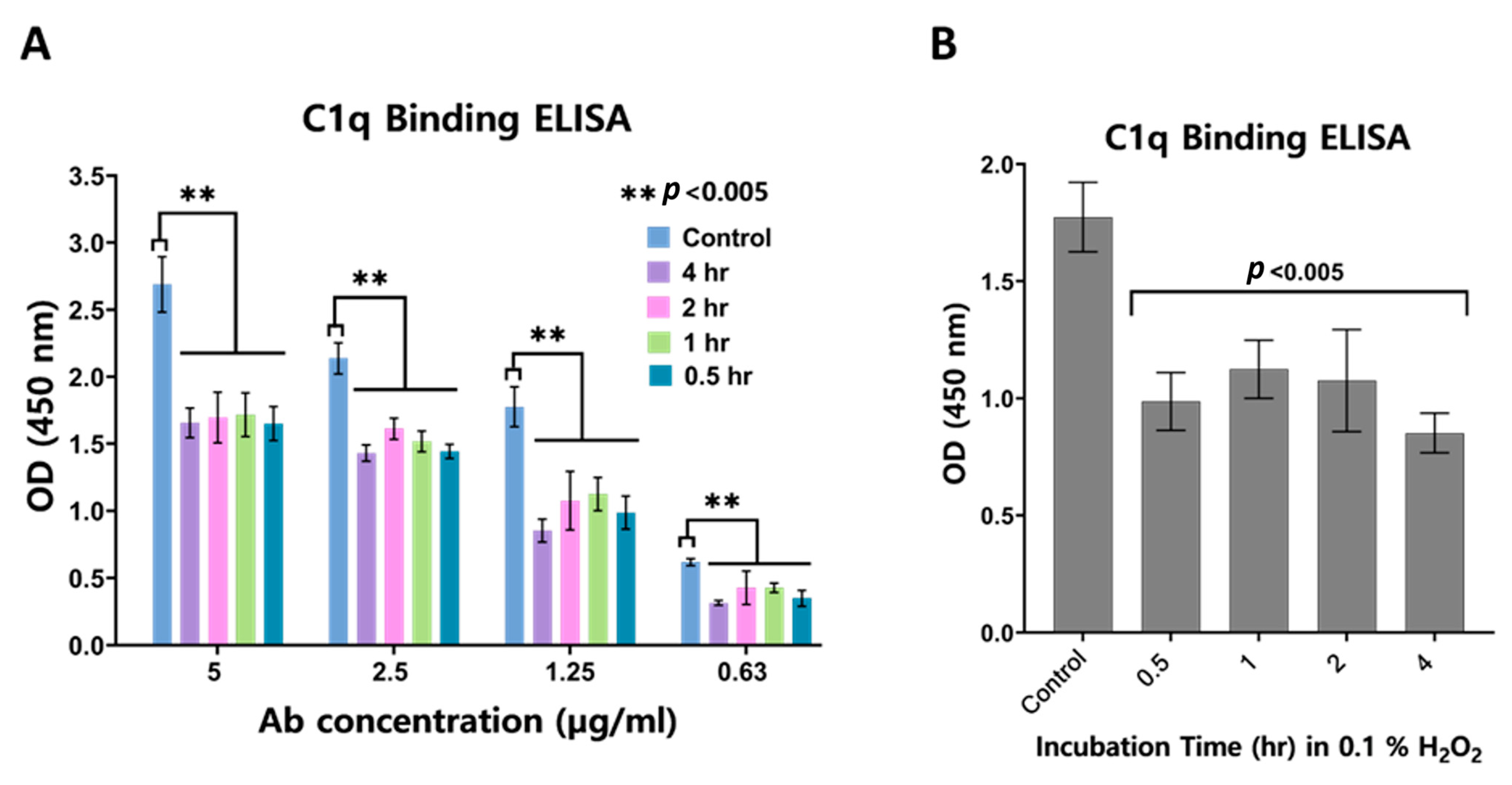
3.2. Impact of Oxidation on C1q Binding

The ELISA method was used to understand the binding interaction between anti-VEGF IgGs exposed to oxidative stress and C1q protein. The antibody concentration in ELISA varied between 0.6 and 5.0 µL/mL for determining the change in C1q binding capacity by measuring the absorbance values. Anti-VEGF IgG molecules not exposed to H2O2 were used as control samples, and the samples exposed to oxidative stress were evaluated according to this reference. When the antibodies treated with specific percentages of H2O2 for 1 h at room temperature were examined for their C1q binding affinities, it was observed that the amount of C1q binding decreased significantly with the decrease in antibody concentration (Figure 3A). Each percentage of H2O2 exposure resulted in decreased C1q binding affinities relative to the reference antibody at all antibody concentrations. Hence, the control sample showed the maximum C1q binding, while all other samples showed a statistically significant decrease in binding signal compared with the control. When the antibody concentration value of only 1.25 µg/mL was evaluated, it was seen that the affinity of antibodies exposed to all H2O2 percentages to C1q decreased significantly compared with that of the control (Figure 3B).
When samples incubated with 0.1% H2O2 at room temperature for specific incubation times were examined, it was observed that the amount of C1q binding decreased with decreasing antibody concentrations (Figure 4A). Furthermore, even half an hour of exposure to oxidative stress caused a significant decrease in the C1q binding affinity of the antibodies when compared with that of the control group. However, when the effects of exposure times were compared at a constant sample concentration, no direct relationship could be identified under the tested experimental conditions (Figure 4B).
Since C1q interacts with the CH2 domain in the Fc part of the antibody, the PTMs forming here may affect the C1q interaction [47]. Studies report that Met252 oxidation in the Fc part reduces C1q binding affinity [48]. With the oxidation of the CH2–CH3 interface region, a conformational change may occur here, affecting the binding of C1q. Furthermore, our pep map data also showed that Met252 in the DTLMSIR peptide was oxidised, which may partially explain the decrease in C1q binding upon H2O2 stress.

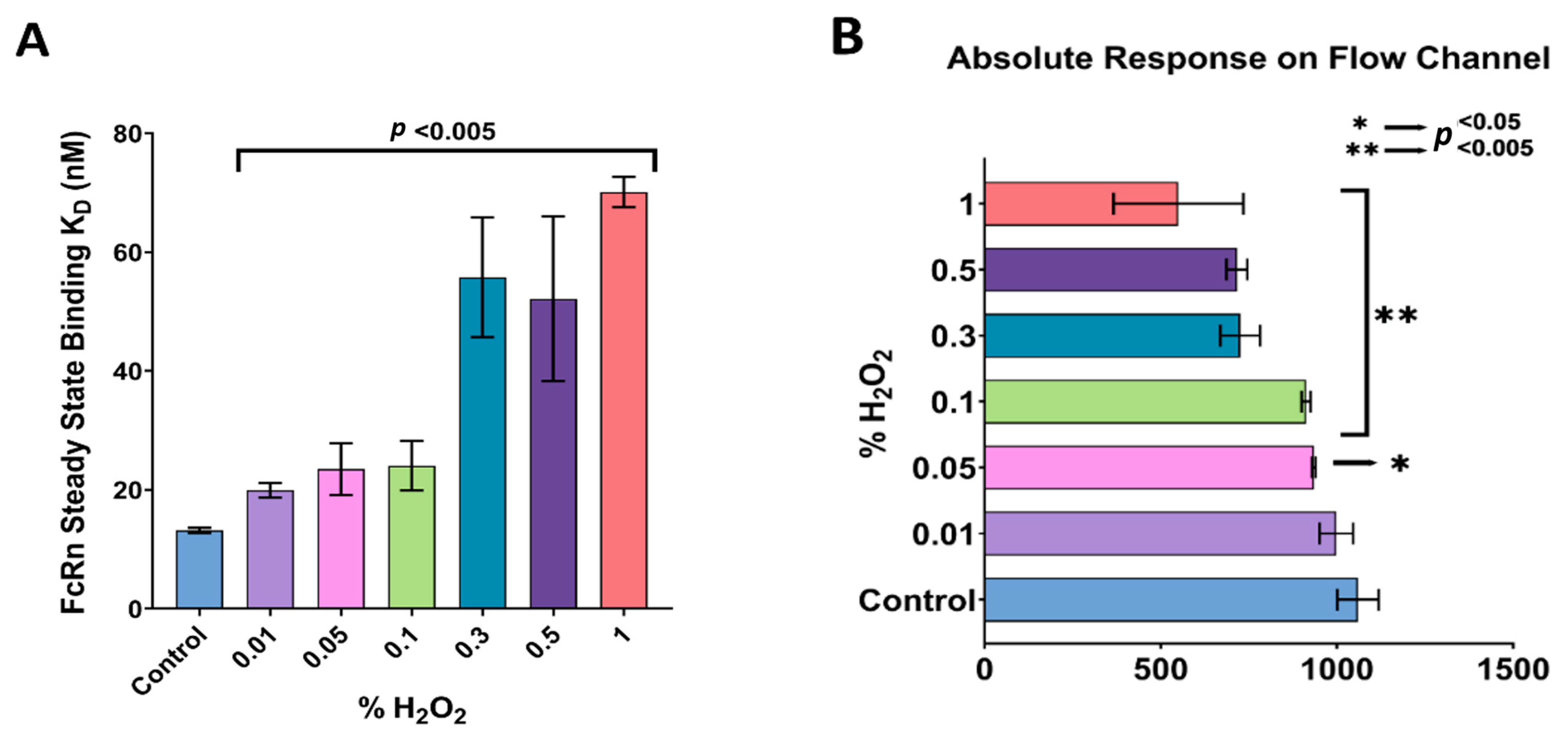
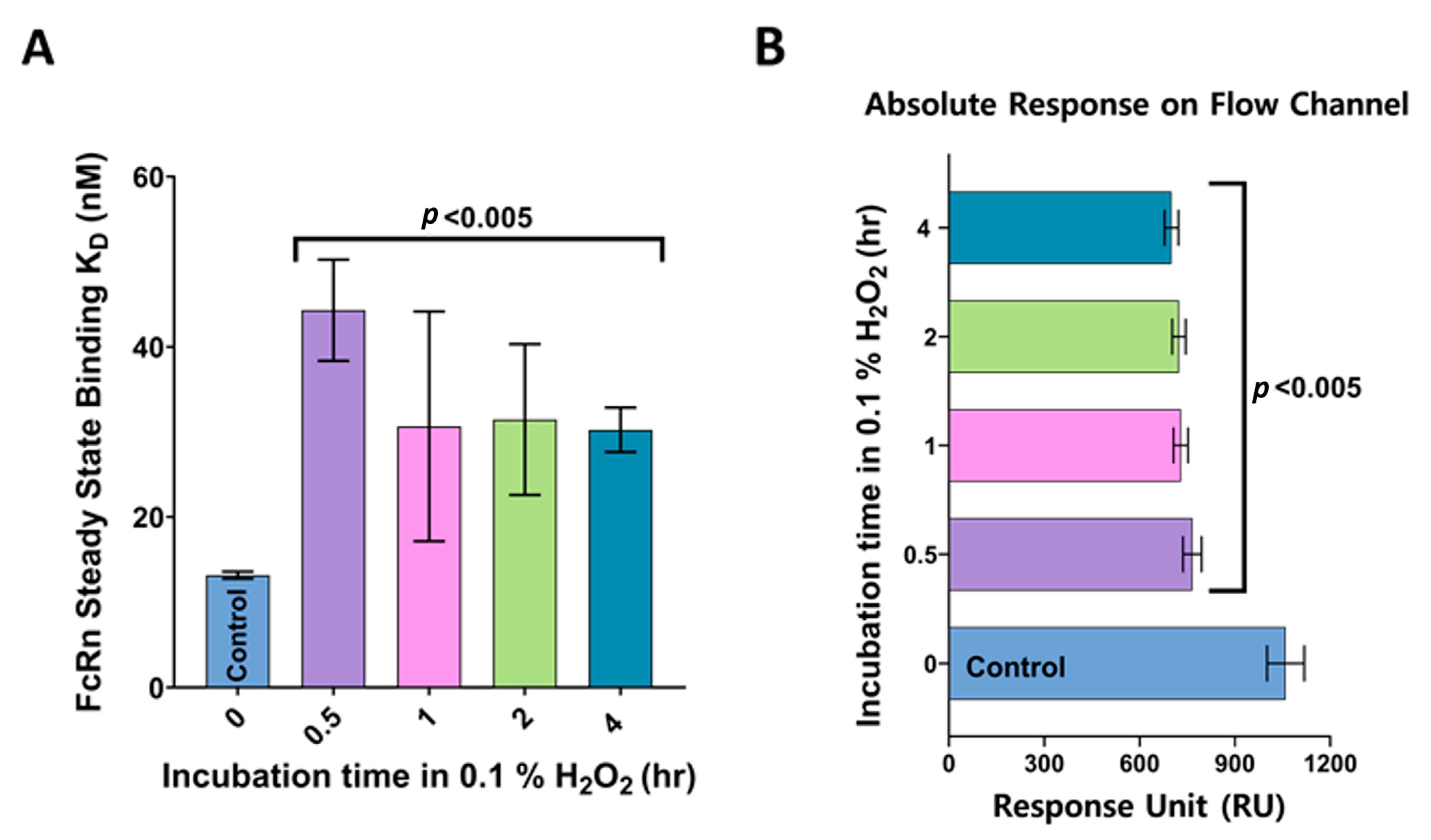
3.3. FcRn Binding Analysis of Anti-VEGF IgGs Exposed to Oxidation

The S.P.R. method was used to determine the binding affinity of FcRn molecules and the anti-VEGF IgG molecules exposed to varying oxidative stress conditions. KD values calculated using a steady-state affinity model are presented in Figure 5A. The KD value increased as the percentage of H2O2 increased. Therefore, the binding affinity of FcRn showed a statistically significant decrease with increased exposure to oxidative stress. The control anti-VEGF IgG sample (not exposed to stress) had the lowest and the best KD value, calculated as 13.15 ± 0.36 nM, indicating the disruptive effect of oxidative stress on the anti-VEGF IgG molecule's function. In addition to these results, the absolute binding response values of the samples were determined in the flow channel. When the absolute response values were examined at different amounts of H2O2 exposure, the control sample had the highest binding activity (Figure 5B), indicating an exceedingly high number of molecules bound on the chip surface due to high FcRn interaction. The absolute response rate decreased with the increase in oxidative stress exposure; therefore, the FcRn binding affinity also decreased.
Using the same steady-state binding model, the FcRn binding affinity of antibodies incubated in 0.1% H2O2 was separately investigated. The control sample showed the highest binding affinity with the lowest KD value, determined as 13.15 ± 0.36 mM (Figure 6A). The FcRn binding affinities of the oxidised samples showed a statistically significant decrease compared with the control sample's. Comparable results were obtained when the samples were exposed to a fixed oxidative stress level at varying exposure times. The absolute responses of the samples exposed to oxidative stress decreased as the exposure time increased compared with that of the control sample (Figure 6B), leading to a reduced FcRn binding affinity. As a result, the absolute response confirmed our results in the steady-state binding model.
The interaction between the crystal structure of the mAb Fc region and FcRn occurred in the CH2–CH3 interface region, where Met252 and Met428 residues are present [49]. It was previously shown that oxidative stress affected the function of mAbs, primarily due to oxidised Met and Trp residues [48,50]. Met252 and Met428 in the Fc part of the mAb were the two most oxidised residues, and their oxidation reduced the interaction of FcRn with IgG [51], which agrees with the current results. Likewise, the serum half-life of antibodies decreased by approximately 20% when 79% of Met252 residues and 57% of Met428 residues were oxidised in a previous study conducted in mouse models, showing the importance of combined structural and functional studies during stress evaluation [52].

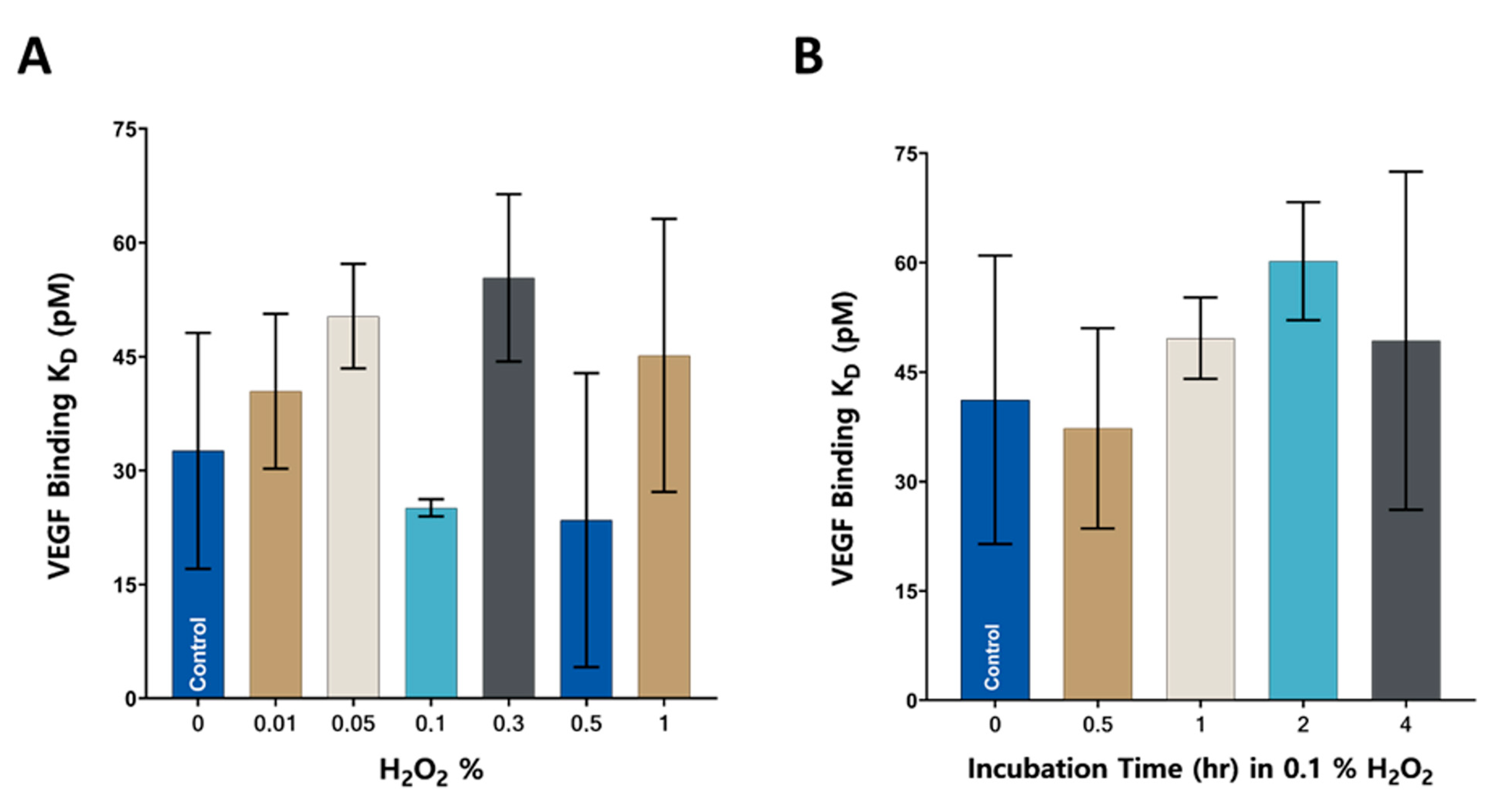
3.4. VEGF Binding Analysis of anti-VEGF IgGs Exposed to Oxidation

In this study, the SPR method was used to determine the affinity of VEGF towards the anti-VEGF-IgG molecules exposed to various oxidative stress conditions. First, VEGF binding analyses of antibodies exposed to different H2O2 percentages were performed (Figure 7A). The KD value of the control sample, which was not exposed to any oxidative stress condition, was determined as 32.60 ± 12.69 pM. There was no statistically significant increase or decrease in the exposed samples' binding affinities compared to that of the control sample; thus, the interaction of the mAbs with the target molecule was not impaired in our studies. The binding affinities of antibodies incubated in 0.1% H2O2 at different time intervals were also evaluated (Figure 7B). The KD value of the control sample was determined as 41.2 ± 17.2 pM. Similar to the trend we observed with antibodies exposed to different percentages of H2O2, the binding affinity of antibodies exposed to oxidative stress for different time intervals did not show any statistically significant change compared to the control sample.
Oxidative stress can have severe effects on the stability and function of mAbs. The impact of oxidation on the activity of the antibody may vary depending on the oxidation region. In particular, the oxidation that may occur in the CDRs can significantly affect the antibody’s activity since CDRs enable the antibody to recognise the target molecule. Few reports in the literature reveal the impact of various oxidation conditions on the antigen-binding capacity of mAbs. For example, it was observed that Met oxidation in the CDR3 decreased the target binding capacity [53]. In another study, the Met residue in the CDR was not oxidised in the presence of oxidative stress, and thus, the target binding activity did not decrease [44]. On the other hand, the oxidation of Trp in the CDR3 significantly reduced the binding of the antibody to the target molecule [54].
Similarly, Trp residue in the CDR1 of the recombinant IgG1 molecule was very sensitive to oxidation [55]. The peptide mapping analysis conducted in this study showed that the antibodies exposed to oxidative stress could only oxidise the Met252 residue in the Fc region, and no oxidation or other PTMs in the CDRs took place. There are limited studies about the oxidation of the peptides in CDRs; the antibody’s target molecule binding property may change due to this event [56,57]. Therefore, it can be assumed that mild oxidation conditions may not severely affect the antibody’s binding affinity to the target molecule unless there is oxidation in the CDRs, which still requires detailed structural studies to conclude.

3.5. Cell Proliferation Performances of mAbs Exposed to H2O2

The mechanism of HUVEC proliferation was used to assess the alterations in the biological activities of the mAbs. HUVECs carry several VEGF receptors on their surface, which allow them to generate a proliferation signal in the presence of VEGF. Anti-VEGF IgG molecules prevent the interaction of VEGF with HUVECs by binding to VEGF to inhibit cell division. Hence, changes in the proliferation of HUVECs were investigated to understand whether anti-VEGF IgG molecules were affected by exposure to oxidative stress. The growth rates of cells treated with VEGF and antibodies (exposed to 0.1% and 0.5% H2O2) were determined by measuring the absorbance values using the MTS method. The cells treated with no antibodies were taken as the positive control. Since there was no antibody binding to VEGF in the positive control sample, the proliferation effect of VEGF was at the maximum OD value, representing the maximum cell proliferation. The samples treated with naïve anti-VEGF IgG molecules were the negative control. The effect of anti-VEGF IgG molecules exposed to oxidative stress on cell proliferation was determined by comparing them with positive and negative control samples.
As presented in Figure 8, the negative control sample gave a low absorbance value, indicating inhibited cell proliferation. The treatment with antibodies exposed to 0.1% and 0.5% H2O2 reduced the optical density of HUVECs as much as the negative control sample. In other words, increased oxidative stress levels did not cause a statistically significant change in cell proliferation rates under these experimental conditions, indicating that the stressed anti-VEGF molecules continued to bind and neutralise VEGF molecules, which agrees with the existing literature [58].

4. Conclusions

The present study reveals anti-VEGF IgG molecules’ structural and functional changes in response to various oxidation conditions. The pep map analysis of the samples exposed to various oxidative stress conditions showed oxidation only in Met residue in the DTLMISR peptide. The Met oxidation increased with the increase in the percentage of H2O2 and the exposure time. Met252 and Met428 residues are the most prone sites to oxidation in the Fc region of mAbs [41,59]. In the current study, oxidation was only seen in Met252 but not in Met428 residue, and no other PTM types were observed. The C1q protein is responsible for the CDC activity, one of the antibody's vital effector functions [60]. When exposed to oxidative stress, the mAb samples showed decreased C1q binding affinity.
In addition, there was no correlation between the decreased C1q binding and the increased level of H2O2 or stress exposure time. Even at the lowest H2O2 exposure, the C1q binding reduced to a similar level for the highest H2O2 percentage compared to the control group. The interaction of therapeutic antibodies with FcRn is of immense importance in demonstrating the drug’s preclinic efficacy. The modifications on the CH2–CH3 domain of the Fc region may prevent FcRn binding. The binding capacity of the mAbs with FcRn decreased in the current study upon oxidation of the Met252 residue in the CH2 domain. The binding affinity of VEGF molecules with the oxidised antibodies was also examined using SPR, no significant change was observed in the target binding activity through the Fab region. As expected, it was found that HUVEC cell proliferation was also not significantly affected by the increased oxidative stress since Met252 was oxidised only in the Fc region instead of the CDR. In conclusion, the authors recommend performing functional analyses in addition to structural studies while investigating the impacts of the stress factors on therapeutic mAbs.

Author Contributions

Conceptualisation and methodology, M.Y.; data curation, A.P.; investigation, formal analysis, and validation, A.P. and B.G.; initial draft preparation, A.P. and M.Y.; review and editing, A.P., B.G., M.R.S. and M.Y.; supervision and project administration, M.Y. All authors have read and agreed to the published version of the manuscript.

Funding

This research was funded by The Scientific and Technological Research Council of Turkey (TUBITAK) KAMAG 1007 program grant number 115G016-115G074.

Institutional Review Board Statement

Not applicable.

Informed Consent Statement

Not applicable.

Data Availability Statement

The data presented in this study are available in this article.

Acknowledgments

The authors acknowledge Eda Çapkin (Ph.D. candidate at Sabanci University) for her help during S.P.R. studies. We also thank Defne Hız, an undergraduate student at Sabanci University, for her constructive comments and revisions.

Conflicts of Interest

The authors declare that they have no conflict of interest.

References

  1. Palmer, B.F.; Clegg, D.J. Oxygen sensing and metabolic homeostasis. Mol. Cell. Endocrinol. 2014, 397, 51–58. [Google Scholar] [CrossRef]
  2. Senger, D.R.; Galli, S.J.; Dvorak, A.M.; Perruzzi, C.A.; Harvey, V.S.; Dvorak, H.F. Tumor Cells Secrete a Vascular Permeability Factor That Promotes Accumulation of Ascites Fluid. Science 1983, 219, 983–985. [Google Scholar] [CrossRef] [PubMed]
  3. Karkkainen, M.J.; Petrova, T.V. Vascular endothelial growth factor receptors in the regulation of angiogenesis and lymphangiogenesis. Oncogene 2000, 19, 5598–5605. [Google Scholar] [CrossRef] [PubMed] [Green Version]
  4. Gupta, S.; Jiskoot, W.; Schöneich, C.; Rathore, A.S. Oxidation and Deamidation of Monoclonal Antibody Products: Potential Impact on Stability, Biological Activity, and Efficacy. J. Pharm. Sci. 2022, 111, 903–918. [Google Scholar] [CrossRef]
  5. Jenkins, N.; Murphy, L.; Tyther, R. Post-translational modifications of recombinant proteins: Significance for biopharmaceuticals. Mol. Biotechnol. 2008, 39, 113–118. [Google Scholar] [CrossRef] [PubMed]
  6. Li, X.; Xu, W.; Wang, Y.; Zhao, J.; Liu, Y.H.; Richardson, D.; Li, H. High throughput peptide mapping method for analysis of site-specific monoclonal antibody oxidation. J. Chromatogr. A 2016, 1460, 51–60. [Google Scholar] [CrossRef]
  7. Regl, C.; Wohlschlager, T.; Holzmann, J.; Huber, C.G. A Generic HPLC Method for Absolute Quantification of Oxidation in Monoclonal Antibodies and Fc-Fusion Proteins Using UV and MS Detection. Anal. Chem. 2017, 89, 8391–8398. [Google Scholar] [CrossRef]
  8. Nowak, C.; KCheung, J.; MDellatore, S.; Katiyar, A.; Bhat, R.; Sun, J.; Ponniah, G.; Neill, A.; Mason, B.; Beck, A.; et al. Forced degradation of recombinant monoclonal antibodies: A practical guide. mAbs 2017, 9, 1217–1230. [Google Scholar] [CrossRef] [Green Version]
  9. Chan, C.P. Forced degradation studies: Current trends and future perspectives for protein-based therapeutics. Expert Rev. Proteom. 2016, 13, 651–658. [Google Scholar] [CrossRef]
  10. Blessy, M.; Patel, R.D.; Prajapati, P.N.; Agrawal, Y.K. Development of forced degradation and stability indicating studies of drugs—A review. J. Pharm. Anal. 2014, 4, 159–165. [Google Scholar] [CrossRef] [Green Version]
  11. Hawe, A.; Wiggenhorn, M.; van de Weert, M.; Garbe, J.H.O.; Mahler, H.C.; Jiskoot, W. Forced degradation of therapeutic proteins. J. Pharm. Sci. 2012, 101, 895–913. [Google Scholar] [CrossRef]
  12. Vermeer, A.W.P.; Norde, W. The Thermal Stability of Immunoglobulin: Unfolding and Aggregation of a Multi-Domain Protein. Biophys. J. 2000, 78, 394–404. [Google Scholar] [CrossRef] [Green Version]
  13. Dias, C.L.; Ala-Nissila, T.; Karttunen, M.; Vattulainen, I.; Grant, M. Microscopic mechanism for cold denaturation. Phys. Rev. Lett. 2008, 100, 118101. [Google Scholar] [CrossRef] [Green Version]
  14. Luo, Q.; Joubert, M.K.; Stevenson, R.; Ketchem, R.R.; Narhi, L.O.; Wypych, J. Chemical Modifications in Therapeutic Protein Aggregates Generated under Different Stress Conditions. J. Biol. Chem. 2011, 286, 25134–25144. [Google Scholar] [CrossRef] [Green Version]
  15. Dyck, Y.F.K.; Rehm, D.; Joseph, J.F.; Winkler, K.; Sandig, V.; Jabs, W.; Parr, M.K. Forced Degradation Testing as Complementary Tool for Biosimilarity Assessment. Bioengineering 2019, 6, 62. [Google Scholar] [CrossRef] [Green Version]
  16. Jiskoot, W.; Beuvery, E.C.; de Koning, A.A.M.; Herron, J.N.; Crommelin, D.J.A. Analytical Approaches to the Study of Monoclonal Antibody Stability. Pharm. Res. Off. J. Am. Assoc. Pharm. Sci. 1990, 7, 1234–1241. [Google Scholar] [CrossRef]
  17. Sharma, D.K.; Oma, P.; Pollo, M.J.; Sukumar, M. Quantification and Characterization of Subvisible Proteinaceous Particles in Opalescent mAb Formulations Using Micro-Flow Imaging. J. Pharm. Sci. 2010, 99, 2628–2642. [Google Scholar] [CrossRef]
  18. Arosio, P.; Rima, S.; Morbidelli, M. Aggregation Mechanism of an IgG2 and two IgG1 Monoclonal Antibodies at low pH: From Oligomers to Larger Aggregates. Pharm. Res. 2013, 30, 641–654. [Google Scholar] [CrossRef]
  19. Gaza-Bulseco, G.; Liu, H. Fragmentation of a Recombinant Monoclonal Antibody at Various pH. Pharm. Res. 2008, 25, 1881–1890. [Google Scholar] [CrossRef]
  20. Cohen, S.L.; Price, C.; Vlasak, J. β-Elimination and Peptide Bond Hydrolysis: Two Distinct Mechanisms of Human IgG1 Hinge Fragmentation upon Storage. J. Am. Chem. Soc. 2007, 129, 6976–6977. [Google Scholar] [CrossRef]
  21. Cordoba, A.J.; Shyong, B.-J.; Breen, D.; Harris, R.J. Non-enzymatic hinge region fragmentation of antibodies in solution. J. Chromatogr. B 2005, 818, 115–121. [Google Scholar] [CrossRef] [PubMed]
  22. Xiang, T.; Lundell, E.; Sun, Z.; Liu, H. Structural effect of a recombinant monoclonal antibody on hinge region peptide bond hydrolysis. J. Chromatogr. B 2007, 858, 254–262. [Google Scholar] [CrossRef] [PubMed]
  23. Keck, R.G. The Use of t-Butyl Hydroperoxide as a Probe for Methionine Oxidation in Proteins. Anal. Biochem. 1996, 236, 56–62. [Google Scholar] [CrossRef] [PubMed]
  24. Folzer, E.; Diepold, K.; Bomans, K.; Finkler, C.; Schmidt, R.; Bulau, P.; Huwyler, J.; Mahler, H.C.; Koulov, A.V. Selective Oxidation of Methionine and Tryptophan Residues in a Therapeutic IgG1 Molecule. J. Pharm. Sci. 2015, 104, 2824–2831. [Google Scholar] [CrossRef] [PubMed]
  25. Bertolotti-Ciarlet, A.; Wang, W.; Lownes, R.; Pristatsky, P.; Fang, Y.; McKelvey, T.; Li, Y.; Li, Y.; Drummond, J.; Prueksaritanont, T.; et al. Impact of methionine oxidation on the binding of human IgG1 to FcRn and Fcγ receptors. Mol. Immunol. 2009, 46, 1878–1882. [Google Scholar] [CrossRef] [PubMed]
  26. Suárez, I.; Salmerón-García, A.; Cabeza, J.; Capitán-Vallvey, L.F.; Navas, N. Development and use of specific ELISA methods for quantifying the biological activity of bevacizumab, cetuximab and trastuzumab in stability studies. J. Chromatogr. B Anal. Technol. Biomed. Life Sci. 2016, 1032, 155–164. [Google Scholar] [CrossRef]
  27. Martínez-Ortega, A.; Herrera, A.; Salmerón-García, A.; Cabeza, J.; Cuadros-Rodríguez, L.; Navas, N. Validated reverse phase HPLC diode array method for the quantification of intact bevacizumab, infliximab and trastuzumab for long-term stability study. Int. J. Biol. Macromol. 2018, 116, 993–1003. [Google Scholar] [CrossRef]
  28. Burkitt, W.; Domann, P.; O’Connor, G. Conformational changes in oxidatively stressed monoclonal antibodies studied by hydrogen exchange mass spectrometry. Protein Sci. 2010, 19, 826–835. [Google Scholar] [CrossRef] [Green Version]
  29. Park, H.-J.; Zhang, Y.; Georgescu, S.P.; Johnson, K.L.; Kong, D.; Galper, J.B. Human Umbilical Vein Endothelial Cells and Human Dermal Microvascular Endothelial Cells Offer New Insights Into the Relationship Between Lipid Metabolism and Angiogenesis. Stem Cell Rev. 2006, 2, 93–102. [Google Scholar] [CrossRef]
  30. Kim, T.K.; Park, C.S.; Jang, J.; Kim, M.R.; Na, H.J.; Lee, K.; Kim, H.J.; Heo, K.; Yoo, B.C.; Kim, Y.M.; et al. Inhibition of VEGF-dependent angiogenesis and tumor angiogenesis by an optimized antibody targeting CLEC14a. Mol. Oncol. 2018, 12, 356–372. [Google Scholar] [CrossRef] [Green Version]
  31. Gürel, B.; Çapkın, E.; Parlar, A.; Özkan, A.; Çorbacıoğlu, M.; Dağlikoca, D.E.; Yüce, M. Optimized Methods for Analytical and Functional Comparison of Biosimilar mAb Drugs: A Case Study for Avastin, Mvasi, and Zirabev. Sci. Pharm. 2022, 90, 36. [Google Scholar] [CrossRef]
  32. Maghni, K.; Nicolescu, O.M.; Martin, J.G. Suitability of cell metabolic colorimetric assays for assessment of CD4+ T cell proliferation: Comparison to 5-bromo-2-deoxyuridine (BrdU) ELISA. J. Immunol. Methods 1999, 223, 185–194. [Google Scholar] [CrossRef] [PubMed]
  33. GE Healthcare Life Sciences. BiacoreTM T200 Instrument Handbook 28-9768-63 Edition AC; GE Healthcare Life Sciences: Uppsala, Sweden, 2013. [Google Scholar]
  34. de Mol, N.J.; Fischer, M.J.E. Surface Plasmon Resonance. In Methods in Molecular Biology; Humana Press: Totowa, NJ, USA, 2010; Volume 627. [Google Scholar] [CrossRef]
  35. Gurel, B.; Berksoz, M.; Capkin, E.; Parlar, A.; Pala, M.C.; Ozkan, A.; Capan, Y.; Daglikoca, D.E.; Yuce, M. Structural and Functional Analysis of CEX Fractions Collected from a Novel Avastin® Biosimilar Candidate and Its Innovator: A Comparative Study. Pharmaceutics 2022, 14, 1571. [Google Scholar] [CrossRef] [PubMed]
  36. GE Healthcare. Biacore T200—Getting Started (28-9840-98). cmi.hms.harvard.edu. 2013. Available online: https://cmi.hms.harvard.edu/files/cmi/files/biacoret200_gettingstarted_handbook.pdf (accessed on 30 April 2023).
  37. Abdiche, Y.N.; Yeung, Y.A.; Chaparro-Riggers, J.; Barman, I.; Strop, P.; Chin, S.M.; Pham, A.; Bolton, G.; McDonough, D.; Lindquist, K.; et al. The Neonatal Fc Receptor (FcRn) Binds Independently to Both Sites of the IgG Homodimer with Identical Affinity. MAbs 2015, 7, 331–343. [Google Scholar] [CrossRef] [Green Version]
  38. Neuber, T.; Frese, K.; Jaehrling, J.; Jäger, S.; Daubert, D.; Felderer, K.; Linnemann, M.; Höhne, A.; Kaden, S.; Kölln, J.; et al. Characterization and screening of IgG binding to the neonatal Fc receptor. mAbs 2014, 6, 928–942. [Google Scholar] [CrossRef] [Green Version]
  39. Wang, X.; McKay, P.; Yee, L.T.; Dutina, G.; Hass, P.E.; Nijem, I.; Allison, D.; Cowan, K.J.; Lin, K.; Quarmby, V.; et al. Impact of SPR biosensor assay configuration on antibody: Neonatal Fc receptor binding data. mAbs 2017, 9, 319–332. [Google Scholar] [CrossRef] [Green Version]
  40. Gundry, R.L.; White, M.Y.; Murray, C.I.; Kane, L.A.; Fu, Q.; Stanley, B.A.; Van Eyk, J.E. Preparation of Proteins and Peptides for Mass Spectrometry Analysis in a Bottom-Up Proteomics Workflow. In Current Protocols in Molecular Biology; John Wiley & Sons Inc.: Hoboken, NJ, USA, 2009; pp. 1–23. [Google Scholar] [CrossRef] [Green Version]
  41. Chumsae, C.; Gaza-Bulseco, G.; Sun, J.; Liu, H. Comparison of methionine oxidation in thermal stability and chemically stressed samples of a fully human monoclonal antibody. J. Chromatogr. B 2007, 850, 285–294. [Google Scholar] [CrossRef]
  42. Liu, H.; Gaza-Bulseco, G.; Xiang, T.; Chumsae, C. Structural effect of deglycosylation and methionine oxidation on a recombinant monoclonal antibody. Mol. Immunol. 2008, 45, 701–708. [Google Scholar] [CrossRef]
  43. Roberts, G.D.; Johnson, W.P.; Burman, S.; Anumula, K.; Carr, S.A. An Integrated Strategy for Structural Characterization of the Protein and Carbohydrate Components of Monoclonal Antibodies: Application to Anti-Respiratory Syncytial Virus MAb. Anal. Chem. 1995, 67, 3613–3625. [Google Scholar] [CrossRef]
  44. Shen, F.J.; Kwong, M.Y.; Keck, R.G.; Harris, R.J. The application of tert-butylhydroperoxide oxidation to study sites of potential methionine oxidation in a recombinant antibody. In Techniques in Protein Chemistry; Elsevier: Amsterdam, The Netherlands, 1996; pp. 275–284. [Google Scholar] [CrossRef]
  45. Kroon, D.J.; Baldwin-Ferro, A.; Lalan, P. Identification of Sites of Degradation in a Therapeutic Monoclonal Antibody by Peptide Mapping. Pharm. Res. Off. J. Am. Assoc. Pharm. Sci. 1992, 9, 1386–1393. [Google Scholar] [CrossRef]
  46. Lam, X.M.; Yang, J.Y.; Cleland, J.L. Antioxidants for prevention of methionine oxidation in recombinant monoclonal antibody HER2. J. Pharm. Sci. 1997, 86, 1250–1255. [Google Scholar] [CrossRef] [PubMed]
  47. Ambrogelly, A.; Gozo, S.; Katiyar, A.; Dellatore, S.; Kune, Y.; Bhat, R.; Sun, J.; Li, N.; Wang, D.; Nowak, C.; et al. Analytical comparability study of recombinant monoclonal antibody therapeutics. mAbs 2018, 10, 513–538. [Google Scholar] [CrossRef] [PubMed]
  48. Wang, S.; Ionescu, R.; Peekhaus, N.; Leung, J.; Ha, S.; Vlasak, J. Separation of post-translational modifications in monoclonal antibodies by exploiting subtle conformational changes under mildly acidic conditions. J. Chromatogr. A 2010, 1217, 6496–6502. [Google Scholar] [CrossRef]
  49. Martin, W.L.; West, A.P.; Gan, L.; Bjorkman, P.J. Crystal structure at 2.8 Å of an FcRn/heterodimeric Fc complex: Mechanism of pH-dependent binding. Mol. Cell 2001, 7, 867–877. [Google Scholar] [CrossRef]
  50. Lam, X.M.; Lai, W.G.; Chan, E.K.; Ling, V.; Hsu, C.C. Site-Specific Tryptophan Oxidation Induced by Autocatalytic Reaction of Polysorbate 20 in Protein Formulation. Pharm. Res. 2011, 28, 2543–2555. [Google Scholar] [CrossRef]
  51. Wang, W.; Lu, P.; Fang, Y.; Hamuro, L.; Pittman, T.; Carr, B.; Hochman, J.; Prueksaritanont, T. Monoclonal Antibodies with Identical Fc Sequences Can Bind to FcRn Differentially with Pharmacokinetic Consequences. Drug Metab. Dispos. 2011, 39, 1469–1477. [Google Scholar] [CrossRef] [Green Version]
  52. Wang, W.; Vlasak, J.; Li, Y.; Pristatsky, P.; Fang, Y.; Pittman, T.; Roman, J.; Wang, Y.; Prueksaritanont, T.; Ionescu, R. Impact of methionine oxidation in human IgG1 Fc on serum half-life of monoclonal antibodies. Mol. Immunol. 2011, 48, 860–866. [Google Scholar] [CrossRef]
  53. Haberger, M.; Bomans, K.; Diepold, K.; Hook, M.; Gassner, J.; Schlothauer, T.; Zwick, A.; Spick, C.; Kepert, J.F.; Hienz, B.; et al. Assessment of chemical modifications of sites in the CDRs of recombinant antibodies. mAbs 2014, 6, 327–339. [Google Scholar] [CrossRef] [PubMed] [Green Version]
  54. Wei, Z.; Feng, J.; Lin, H.Y.; Mullapudi, S.; Bishop, E.; Tous, G.I.; Casas-Finet, J.; Hakki, F.; Strouse, R.; Schenerman, M.A. Identification of a Single Tryptophan Residue as Critical for Binding Activity in a Humanized Monoclonal Antibody against Respiratory Syncytial Virus. Anal. Chem. 2007, 79, 2797–2805. [Google Scholar] [CrossRef] [PubMed]
  55. Hensel, M.; Steurer, R.; Fichtl, J.; Elger, C.; Wedekind, F.; Petzold, A.; Schlothauer, T.; Molhoj, M.; Reusch, D.; Bulau, P. Identification of Potential Sites for Tryptophan Oxidation in Recombinant Antibodies Using tert-Butylhydroperoxide and Quantitative LC-MS. PLoS ONE 2011, 6, e17708. [Google Scholar] [CrossRef] [Green Version]
  56. Boyd, D.; Kaschak, T.; Yan, B. HIC resolution of an IgG1 with an oxidized Trp in a complementarity determining region. J. Chromatogr. B 2011, 879, 955–960. [Google Scholar] [CrossRef]
  57. Yan, Y.; Wei, H.; Fu, Y.; Jusuf, S.; Zeng, M.; Ludwig, R.; Krystek, S.R., Jr.; Chen, G.; Tao, L.; Das, T.K. Isomerization and Oxidation in the Complementarity-Determining Regions of a Monoclonal Antibody: A Study of the Modification-Structure-Function Correlations by Hydrogen-Deuterium Exchange Mass Spectrometry. Anal. Chem. 2016, 88, 2041–2050. [Google Scholar] [CrossRef]
  58. Park, D.; Kim, J.; Yun, J.; Park, S.J. Evaluation of the Physico-Chemical and Biological Stability of SB8 (Aybintio), a Proposed Biosimilar to Bevacizumab, Under Ambient and In-Use Conditions. Adv. Ther. 2020, 37, 4308–4324. [Google Scholar] [CrossRef]
  59. Gaza-Bulseco, G.; Faldu, S.; Hurkmans, K.; Chumsae, C.; Liu, H. Effect of methionine oxidation of a recombinant monoclonal antibody on the binding affinity to protein A and protein G. J. Chromatogr. B 2008, 870, 55–62. [Google Scholar] [CrossRef]
  60. Hong, J.; Lee, Y.; Lee, C.; Eo, S.; Kim, S.; Lee, N.; Park, J.; Park, S.; Seo, D.; Jeong, M.; et al. Physicochemical and biological characterization of SB2, a biosimilar of Remicade®(infliximab). mAbs 2017, 9, 364–382. [Google Scholar] [CrossRef] [Green Version]
Figure 1. Peptide mapping analysis of mAbs exposed to oxidative stress using MS. (A) Total ion chromatograms of mAbs exposed to different amounts of H2O2. (B) Total ion chromatograms of mAbs exposed to 0.1% of H2O2 at different incubation times. The letters marked in red are the amino acids with modifications. The oxidation of the DTLMISR peptide is seen at a particular retention time.
Scipharm 91 00031 g001
Figure 2. Oxidation rate on DTLMISR peptide according to different percentages of H2O2 (A). Oxidation rate on DTLMISR peptide according to different incubation times in 0.1% H2O2 (B). The letters marked in red are the amino acids with the modifications. The percentage values were calculated by averaging their separate injections.
Scipharm 91 00031 g002
Figure 3. C1q ELISA binding analysis of the mAbs exposed to different H2O2 percentages. (A) The results of C1q ELISA binding of anti-VEGF IgG molecules exposed to oxidative stress at different Ab concentrations. (B) The results of C1q ELISA binding of oxidative-stress-exposed anti-VEGF IgG at 1.25 µg/mL Ab concentration. The data are represented as the mean of at least three independent measurements. For statistical analysis, a one-way ANOVA test was applied.
Scipharm 91 00031 g003
Figure 4. C1q ELISA binding analysis of the mAbs exposed to 0.1% H2O2 at different incubation times. (A) The results of C1q ELISA binding of anti-VEGF IgG exposed to oxidative stress at different Ab concentrations. (B) The results of C1q ELISA binding of anti-VEGF IgG exposed to oxidative stress at 1.25 µg/mL Ab concentrations. The data represent the mean of at least three independent measurements. For statistical analysis, one-way ANOVA was applied.
Scipharm 91 00031 g004
Figure 5. FcRn binding analysis of the oxidised samples using SPR: (A) Steady-state interaction of anti-VEGF IgG with immobilised FcRn is represented as the mean of at least three measurements. (B) Absolute response values of the samples on the flow channel. For statistical analysis, one-way ANOVA analysis was applied.
Scipharm 91 00031 g005
Figure 6. FcRn binding analysis of the mAbs exposed to 0.1% H2O2 at different incubation times using SPR: (A) Steady-state interaction of anti-VEGF IgG with immobilised FcRn is represented as the mean of at least three measurements. (B) Absolute response values of the samples on the flow channel. For statistical analysis, one-way ANOVA analysis was applied.
Scipharm 91 00031 g006
Figure 7. VEGF binding analysis of mAbs exposed to oxidative stress via S.P.R. (A) The VEGF binding affinity values of mAbs exposed to different amounts of H2O2. (B) The VEGF binding affinity values of mAbs exposed to 0.1% H2O2 at different incubation times. Experiments were conducted in triplicate. For statistical analysis, a one-way ANOVA test was applied. Langmuir 1:1 binding model was used.
Scipharm 91 00031 g007
Figure 8. Cell proliferation effects of mAbs exposed to H2O2 using MTS. Absorbance values were measured at a wavelength of 490 nm using a microplate reader. Experiments were conducted in triplicate. To interpret the results statistically, ANOVA was applied.
Scipharm 91 00031 g008
Disclaimer/Publisher’s Note: The statements, opinions and data contained in all publications are solely those of the individual author(s) and contributor(s) and not of MDPI and/or the editor(s). MDPI and/or the editor(s) disclaim responsibility for any injury to people or property resulting from any ideas, methods, instructions or products referred to in the content.

