Review

An Overview of Hox Genes in Lophotrochozoa: Evolution and Functionality

Dipartimento di Scienze della Vita e dell’Ambiente, Università Politecnica delle Marche, via Brecce Bianche, 60131 Ancona, Italy
*
Author to whom correspondence should be addressed.
J. Dev. Biol. 2016, 4(1), 12; https://doi.org/10.3390/jdb4010012
Submission received: 22 December 2015 / Revised: 2 March 2016 / Accepted: 12 March 2016 / Published: 19 March 2016
(This article belongs to the Special Issue Hox Genes and Development)

Abstract

:
Hox genes are regulators of animal embryonic development. Changes in the number and sequence of Hox genes as well as in their expression patterns have been related to the evolution of the body plan. Lophotrochozoa is a clade of Protostomia characterized by several phyla which show a wide morphological diversity. Despite that the works summarized in this review emphasize the fragmentary nature of the data available regarding the presence and expression of Hox genes, they also offer interesting insight into the evolution of the Hox cluster and the role played by Hox genes in several phyla. However, the number of genes involved in the cluster of the lophotrochozoan ancestor is still a question of debate. The data presented here suggest that at least nine genes were present while two other genes, Lox4 and Post-2, may either have been present in the ancestor or may have arisen as a result of duplication in the Brachiopoda-Mollusca-Annelida lineage. Spatial and temporal collinearity is a feature of Hox gene expression which was probably present in the ancestor of deuterostomes and protostomes. However, in Lophotrochozoa, it has been detected in only a few species belonging to Annelida and Mollusca.

1. Introduction

Hox genes, a subfamily of homeobox genes, encode transcription factors containing a highly conserved 60 amino acid homeodomain characterized by a helix-turn-helix motif [1]. The members of this gene subfamily are regulators of animal embryonic development and play a role in the patterning of the anterior-posterior body axis of Bilateria [2].
Hox genes were first discovered in Drosophila melanogaster where they are organized in the split Antennapedia-Bithorax complex located on chromosome 3 [3,4]. In some cases, these genes are arranged in clusters, and, hence, they are physically linked on the same chromosome. The number of clusters varies in agreement with the genome duplications that the organisms experienced during evolution, ranging from one in protostomes and invertebrate deuterostomes to four in sarcopterygians which experienced two rounds of whole genome duplication (WGD), with as many as seven in teleosts which experienced a third lineage specific event of WGD (Teleost Specific Genome Duplication, TSGD) [5].
Generally, one feature of the Hox cluster is spatial collinearity: the gene order on chromosomes reflects the order of gene expression and function. The genes at the 3′ end of the cluster are expressed in the anterior part of the body while those at the 5′ end in the posterior part. Collinearity may also be temporal, meaning that the genes at the 3′ end are expressed before those at the 5′ end [6]. This feature is more evident in bilaterian organisms displaying an unbroken cluster than in others which show dispersed or broken clusters [7,8,9,10].
On the basis of gene position and gene function, the Hox cluster can be subdivided into four classes [11]: anterior class, Paralog Group 3, central class, and posterior class. The composition of these classes vary across taxa due to duplication, inversion, or gene loss events that occurred during evolution [10,12,13,14]. Moreover, fragmented clusters may also be related to the presence of transposable elements that could promote chromosomal rearrangements [15].
Although Hox genes show a high sequence similarity, they play a remarkable role in the wide morphological diversity of animals [10]. One of the major groups of bilaterian organisms is Protostomia, which is subdivided into two clades: Ecdysozoa and Lophotrochozoa. The peculiarity of the former is the ability to undergo ecdysis under the hormonal control of ecdysteroids. The latter clade is characterized by the trochophore, the free-swimming ciliated larvae, and/or the lophophore, the feeding structure made up of tentacles surrounding the mouth of adults.
Regarding Hox cluster composition, besides the Hox genes belonging to the Paralog Group-1 (PG-1), PG-2, PG-3, PG-4, and PG-5, Ecdysozoa also exhibit ftz, Antp, Ubx, abd-A, and Abd-B, while Lophotrochozoa also include Lox5, Lox2, Lox4, Post-1 and Post-2 [16,17].
This paper focuses on Lophotrochozoa which are characterized by a high diversity of body architecture, and are, therefore, ideal for studying the evolution of development. This review presents an overview of the presence and expression patterns of Hox genes in 12 lophotrochozoan phyla. The data obtained allowed different hypotheses to be delineated regarding the evolution of the Hox gene subfamily within Lophotrochozoa and its implications on development.

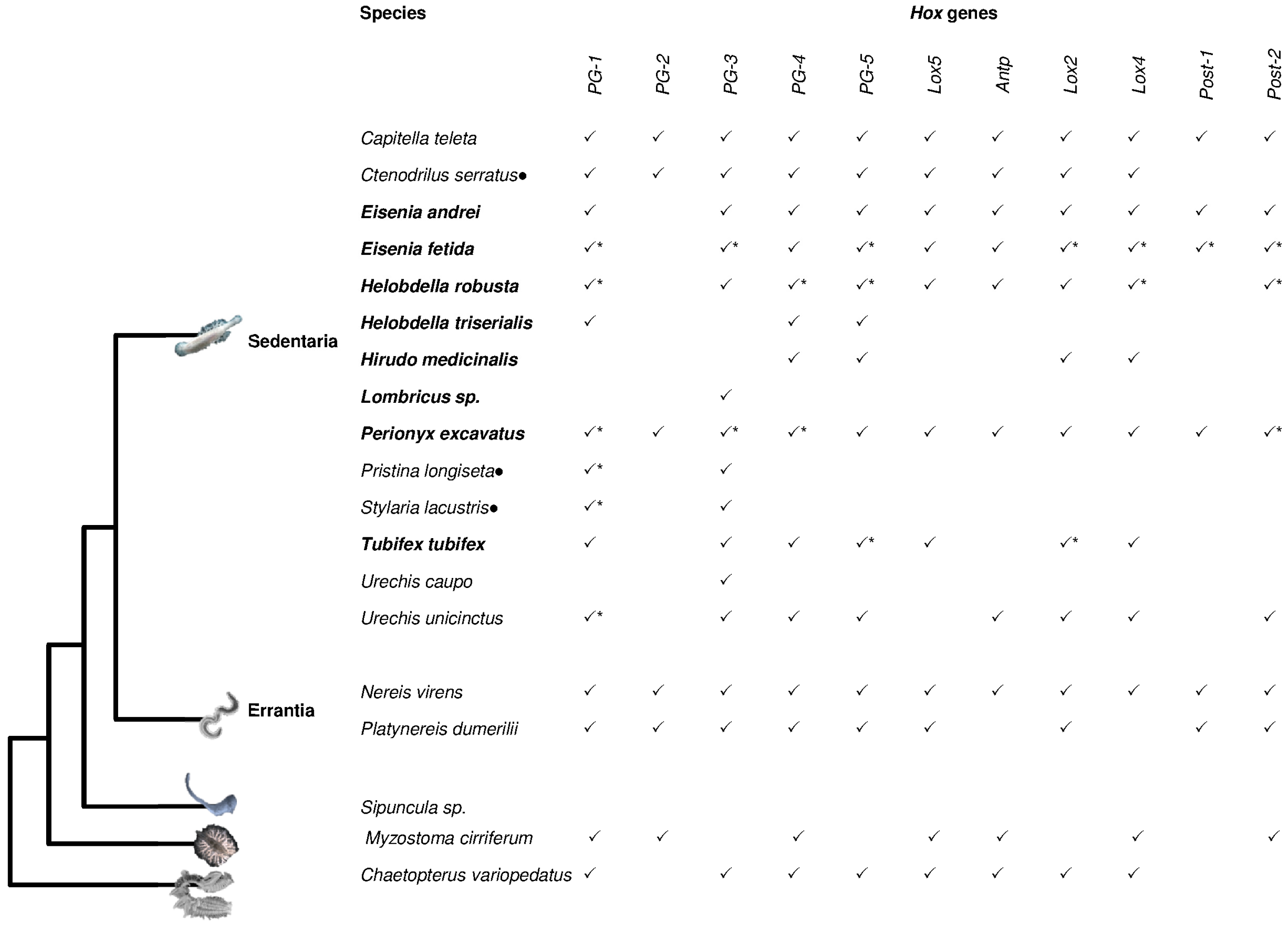
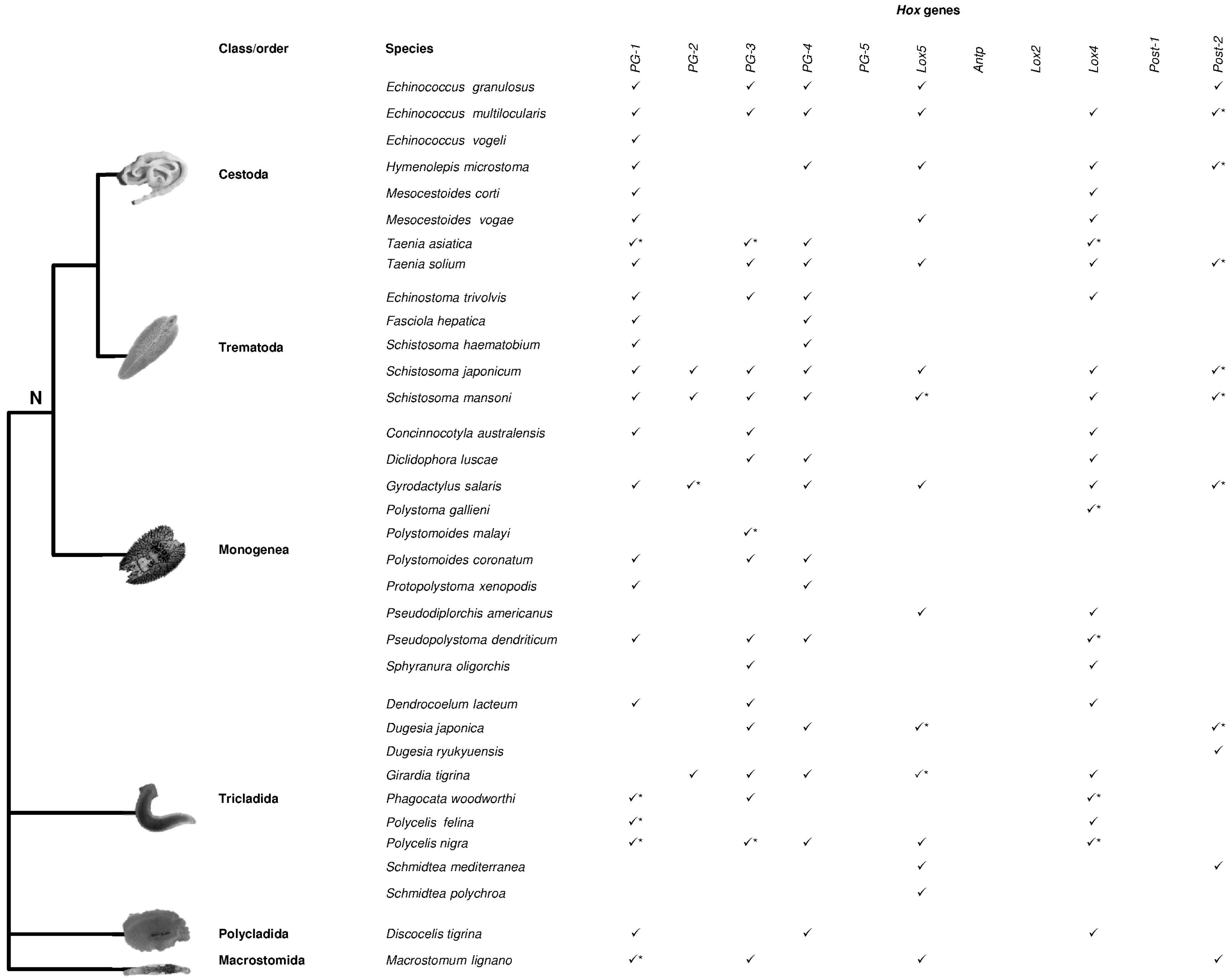
2. Hox Presence in Lophotrochozoa

Body plan evolution and diversification in metazoans have not only been related to changes in Hox cluster composition such as cluster and gene duplications, and gene loss, but also to gene expression and regulatory interactions [14]. An understanding of Hox gene cluster composition can provide insight into the evolutionary history that these genes have undergone within Lophotrochozoa.
Although internal relationships within the Lophotrochozoa clade are still controversial [18,19,20,21,22], we focused on works concerning the presence of Hox genes in 12 phyla, and the evolution of the genes composing the Hox cluster was discussed in relation to the evolutionary relationships between lophotrochozoan phyla. The works summarized here clearly indicate that data on Hox genes are rather scarce for Lophotrochozoa, and a limited number of works on complete genomes have provided insight into Hox gene cluster composition [20,22,23,24,25,26,27,28,29,30,31,32,33]. About half of the lophotrochozoan phyla have never been investigated while only one species has been analyzed in Brachiopoda and Bryozoa, two species in Rotifera, and three species in Nemertea (Table 1) [16,22,23,34,35,36,37]. More information is available for the three major lophotrochozoan phyla: Mollusca (about 30 species, Figure 1, Table S1) [16,20,24,26,38,39,40,41,42,43,44,45,46,47,48,49,50,51,52,53,54,55,56,57,58], Annelida (about 20 species, Figure 2, Table S2) [8,16,20,46,59,60,61,62,63,64,65,66,67,68,69,70,71,72,73] and Platyhelminthes (about 30 species, Figure 3, Table S3) [27,28,29,30,31,32,33,74,75,76,77,78,79,80,81,82,83,84,85,86,87,88,89,90].
The Mollusca phylum is divided into eight classes: Solenogastres, Caudofoveata, and Polyplacophora belonging to Aculifera and Bivalvia, Cephalopoda, Gastropoda, Monoplacophora, and Scaphopoda belonging to the sister taxa Conchifera [92,93]. Mollusks include organisms living in different ecological niches from marine to freshwater and terrestrial environments. Their morphology is extremely variable, ranging from Aplacophora with a wormlike appearance and no shell to Cephalopoda which have a well-developed cephalic region and have co-opted the mantle for locomotion. The muscular foot also presents different morphologies as it is adapted for a variety of functions. With the exception of Monoplacophora, for which no data are available, Hox genes have been identified in a single species of Scaphopoda [40], two species of Aplacophora [40], three species of Polyplacophora [40,57,58], five species of Cephalopoda [26,40,46,55,56], 10 species of Gastropoda [16,20,40,46,47,48,49,50,51,52,53,54], and 13 species of Bivalvia [24,38,39,40,41,42,43,44,45] (Figure 1, Table S1). Our previous review [94] provided evidence that all 11 genes of the Hox lophotrochozoan cluster are found within Bivalvia, Cephalopoda, and Gastropoda, which are the three most studied molluskan classes. Moreover, genome sequencing has revealed, for the first time in lophotrochozoans, that the gastropod Lottia gigantea presents an intact cluster [20]. In the genome assembly of the Pacific oyster, Crassostrea gigas, the Antp gene is clearly missing and Hox genes are located on four scaffolds [24,38]. In the pearl oyster Pinctada fucata, all 11 Hox genes are present and clustered on three scaffolds. Moreover, non-Hox genes are present in the Hox clusters of both the above-mentioned oysters, thereby suggesting that this is a feature which occurred in their common ancestor [25].
In the genome of the cephalopod, Octopus bimaculoides eight of the 11 Hox genes (with the exception of Hox2, Hox3 and PG-4) have been identified and located on eight separate scaffolds [26].
Annelida is another large and morphologically diverse taxon of Lophotrochozoa. Traditionally, this phylum includes segmented worms but currently Myzostomida [95], Echiura, and Sipuncula are also considered as Annelida. The two latter taxa show segmentation only at the larval stage [96,97].
A recent phylogenomic analysis divides Annelida into two main groups reflecting different lifestyles, Errantia and Sedentaria, with Sipuncula and Myzostomida which occupy a basal position (Figure 2) [95].
Within the Sedentaria group, the species belonging to Clitellata exhibit a highly dynamic Hox gene cluster which is broken into several genomic regions and characterized by gene duplication and loss events. In fact, extensive studies on the leech Helobdella robusta [20] and two earthworms, Eisenia fetida [61] and Perionyx excavatus [67], suggest that individual duplications, large segmental duplications and/or whole genome duplications could have played a pivotal role in determining the current Hox gene number and arrangement in these lineages. Moreover, the ortholog Post-1 gene has not been retrieved in leeches and the PG-2 gene seems be absent in Clitellata with the exception of P. excavates [67]. These organisms exhibit a different number of segments, internal anatomy, and shapes. Therefore, it has been suggested that differences in presence, absence, and the arrangement of Hox genes may be responsible for the evolution of the annelid body plan diversity [20].
The genomic survey performed in Capitella teleta indicates that Hox genes are located on three scaffolds although duplication events have not been detected [8,20].
In general, the common ancestor of Sedentaria and Errantia had all 11 genes of the Hox cluster. The species Myzostoma cirriferum and Chaetopterus variopedatus, which are basal in the annelid phylogeny, do not show a complete cluster although whole genome sequencing has not yet been performed for these species.
Within Lophotrochozoa, the phylogenetic relationships of Annelida and Mollusca with respect to Brachiopoda, Phoronida, and Nemertea are still a question of debate. Recently, three hypotheses have been proposed regarding the phylogenetic relationships between these taxa (Figure 4): according to the first hypothesis, Brachiozoa (Brachiopoda + Phoronida) is a sister group to Mollusca while Nemertea occupies a basal position in all the taxa considered [22,98]; the second hypothesis suggests that Brachiozoa is a sister group to Mollusca + Annelida [99]; the third hypothesis considers Annelida as a sister group to the clade including Brachiozoa and Nemertea [21].
Lox2 and Post-1 genes have not yet been recovered in three nemertean species, Lineus sanguineus [35], Micrura alaskensis [36], Pantinonemertes californiensis [37]. According to the evolutionary hypotheses 1 [22,98] and 2 [99], the duplication events leading to these two genes occurred after the separation of Nemertea and other lophotrochozoan phyla, thereby suggesting that the common ancestor of Nemertea and Annelida-Mollusca-Brachiopoda had nine genes [36,37,94]. Alternatively, considering evolutionary hypothesis 3, the absence of Lox2 and Post-1 might be due to a secondary loss.
The phylum Platyhelminthes is the fourth largest animal phylum after arthropods, mollusks and chordates. Members of the phylum Platyhelminthes have a simple bilateral body plan, characterized by the absence of traits found in most bilaterians (e.g., coelom, anus, circulatory and respiratory systems) and for this reason they were previously considered as basal bilaterians [100]. However, the presence in many flatworms of spiral embryonic cleavage, which is a mode of development present in other invertebrate phyla, such as annelids or mollusks, has made their classification very controversial. Platyhelminthes are currently considered as part of Lophotrochozoa, with the acoel and nemertodermatid flatworms separate from the Platyhelminthes and considered as the earliest branching extant bilaterians. However, some phylogenetic studies regard this taxon as separate from Lophotrochozoa [21,98]. The Platyhelminthes are divided into three classes, the free-living class “Turbellaria” and two parasitic classes, Cestoda and Trematoda. Subsequently, the parasitic species are subdivided into three classes, Trematoda, Cestoda, and Monogenea (together called Neodermata), while the class “Turbellaria”, a paraphyletic group, is divided into about 11 orders (depending on the authors) [100].
In Platyhelminthes, the Hox gene set is reduced (Figure 3, Table S3), with PG-5, Antp, Lox2, and Post-1 being absent in all the considered taxa [27,28,29,31,32]. The lack of these genes even in free-living flatworms suggests that their absence may not be related to the parasitic lifestyle of many species of this phylum. In Cestoda, the PG-2 gene seems to be absent and, among the other species examined, orthologs have only been reported in four species: Girardia tigrina, Schistosoma mansoni, S. japonicum, Gyrodactylus salaris (Figure 3). PG-1, PG-3, Lox5, Lox4, and Post-2 genes show duplications in several species belonging to all groups. A duplication has been detected for PG-2 in G. salaris although one of the two genes might belong to PG-3; indeed, in Platyhelminthes, PG-2 and PG-3 genes share several conserved amino acidic residues that might make correct gene attribution difficult [28,75].
Moreover, as regards Hox gene arrangement, genome surveys have shown that the cluster is disrupted. The developmental strategies of some of these organisms with a parasitic lifestyle [75] and/or the presence of transposable elements [79,101] have been proposed in order to explain this dynamic arrangement.
In Rotifera, the genome survey of Adineta vaga demonstrated that PG-3, Lox2 and posterior genes are absent while the other genes, with the exception of PG-2, present multiple copies. Moreover, not all the genes identified are arranged in a cluster [23]. The study in Philodina roseola, even if fragmentary, to date has identified only two central genes which present multiple copies (Table 1).

3. Expression Patterns

Insight into the presence of Hox genes and their expression patterns might be useful for better understanding the morphological innovations and the wide variety of lophotrochozoan species.
Most of the expression data on mollusks are limited to three species of gastropods [47,48,49,51,52], two species of cephalopods [46,56,102], one species of bivalves [24,38], and one species of chitons [57]. Data on the conchiferan species suggest the role of Hox genes in the central nervous system, their involvement in shell formation, tentacles, and the funnel; their expression has also been identified in sensory organs such as the apical organ and statocyst and in the light organ in the cephalopod Euprymna scolopes. Expression analysis in the bivalve C. gigas has shown that Hox genes are not activated according to temporal collinearity but that the peak in PG-4, PG-1, and Lox4 expression occurs before gastrulation, while Lox5 and Post-2 are expressed during the trochophore stage, and the other genes in late development [24,38]. Recent findings in the aculiferan Acanthochitona crinita reveal a collinearity of Hox gene expression [57]. This is the first evidence in mollusks of the ancestral role of Hox genes in patterning the structures along the anterior-posterior body axis in the same way as most other bilaterian animals. Therefore, after the aculiferan-conchiferan split, Hox genes were co-opted into the formation of novelties in gastropods and cephalopods and perhaps in all the other conchiferan classes [57].
A comparison of expression patterns during trochophore larvae development in mollusks and annelids reveals that chitons share comparable expression patterns with annelids along the anterior-posterior body axis [57].
Expression surveys in the annelid Sedentaria confirm the ancestral role of Hox genes in determining the structures along the anterior-posterior body axis [8,62,67,70,73,103]. In particular, the study performed in Capitella teleta has shown that Hox genes exhibit spatial and temporal collinearity in line with the ancestral role that these genes had in the deuterostome-protostome ancestor [8]. However, the absence of collinearity observed in H. robusta, that shows a disrupted Hox cluster, indicates that collinearity is not conserved within Sedentaria [62]. The duplicated genes present in P. excavatus also seem to be expressed from the anterior to the posterior body region [67].
In the Errantia N. virens and Platynereis dumerilii, the expression patterns are very similar: all the Hox genes are involved in vectorial regionalization with the exception of Post-1; PG-1, PG-4, PG-5, Lox5, and Post-2 are expressed in spatial collinearity while temporal collinearity is not respected since the Nereid body plan lacks unique segmental identities [70]. On the contrary, temporal collinearity has been detected in Chaetopterus variopedatus which occupies the basal position in Annelida [73].
The expression analysis of the nine Hox genes in the nemertean M. alaskensis clearly indicated that Hox genes are expressed not in the pilidial larva but in the juvenile stage of pilidiophorans [36]. The gene expression studies also performed on the hoplonemerteans P. californiensis revealed the homology between the imaginal discs of the pilidium and the paired larval invaginations in hoplonemerteans and showed that pilidial development evolved before the split between the two nemertean groups [37]. This sheds light on how Hox genes might be useful for understanding the evolution of embryonic development.
A high expression of Hox genes belonging to the paralog groups PG-2, PG-4, Lox5, Lox4 and, to a lesser extent, PG-1, has been identified inside the eggs and at the miracidium stage in Schistosoma and in monogenean parasites [31,79,104], thus suggesting that these genes could be involved in embryo development. Lox5 and PG-4 genes have also exhibited a higher expression in the schistosomulum stage of S. japonicum, thereby highlighting the possible involvement of these two genes in multiple ontogenetic development stages in schistosomes [31,79]. Moreover, high expression of PG-4, Lox5, and Lox4 has also been observed in sporocysts, another stage at which the determination of cell fate along the anterior-posterior axis may be important [79]. Lox4 could be involved in the process of development in the monogenean parasite, given its high expression in the early developmental stages of the branchial phenotype [104]. During embryogenesis and embryo patterning in the planarian Schmidtea polychroa, the Spol-hoxD (Lox5) gene transcripts started to be detected at early stage 6 (8–10 days), in a strip of cells on the side of the embryo containing the definitive pharynx; the pattern spreads from the definitive pharynx to the posterior end, as reported by Iglesias et al. [89] in the adult. These data suggest that the adult anterior-posterior axis is established after yolk ingestion and the proliferation of the blastomeres in the germ band (stage 5) [90].
Studies on Hox expression have also been performed in adults. The patterns in adult specimens of Girardia tigrina, Discocelis tigrina, and Dugesia japonica have shown two types of Hox genes, that either conserve or lose their typical differential spatial expression whereas duplicated genes may show both patterns of expression [84,105]. In the planarian D. japonica, Nogi and Watanabe [83] reported a similar expression pattern for the two Post-2 genes: DjAbd-Ba is expressed from the posterior pharyngeal region to the entire tail region suggesting that this gene is involved in the specification of the tail region, while DjAbd-Bb is expressed in several types of cells throughout the body. However, counter to the rule of spatial collinearity, the anterior boundary of the expression domain of the posterior gene DjAbd-Ba is anterior to the domains of the central genes PG-4 and Lox5. Different functions have also been reported for Hox genes, apart from their involvement in embryonic development; for example, it has been hypothesized that Lox4, being highly expressed in adult males and down-regulated in adult females, could be involved in tissue differentiation of the male reproductive tract [104]. Another feature is the permanent Hox expression in adult organisms; the high morphological plasticity of these organisms, related to the presence in adult organisms of neoblasts (undifferentiated and totipotent cell types, [106]), could explain their activity, suggesting that pattern formation in planarians may occur continuously [82,105].
As well as embryonic development, the anterior-posterior positional values of Hox genes are also involved in regeneration. After wounding, regenerative tissue is quickly formed [106], accompanied by cell proliferation and Hox expression, even if the two processes are not necessarily related [86]. It has been pointed out that during regeneration, only Lox5 and Post-2 genes have a differential axial nested expression, while the other genes are ubiquitously expressed. These genes are activated during the first day of tail regeneration and down-regulated during head regeneration [82,83,105,107,108]. In particular, the presence of a system that maintains anterior-posterior axial polarity and regulates the expression of DjAbd-Ba rapidly after amputation could be confirmed by the rapid expression of DjAbd-Ba (Post-2a) in the head piece after amputation, with the anterior boundary of the DjAbd-Ba expression domain shifting rapidly and dynamically toward the posterior in the tail pieces [83]. Moreover, Hox gene expression has also been detected during lateral regeneration underlining the importance of these genes in regenerative processes in order to specify positional information on any axis [86].
The extra copies of Hox genes identified in some Platyhelminthes may have lost their typical anterior-posterior axial patterning role as a result of independent duplication and may, on the contrary, have acquired a function in cell differentiation [79,82,86].
In general, flatworm Hox genes have been studied for many years, and, in some cases, distinct spatial domains of expression have been highlighted, although their specific functions have not yet been identified. Further research studies, especially on embryo development, are required in order to draw some conclusions on the role and the possible expression collinearity of Hox genes in Platyhelminthes.

4. Conclusions

This review provides clear evidence that insight into Hox cluster composition and expression patterns is limited to a few phyla and that information is completely lacking for about half the taxa belonging to Lophotrochozoa.
The overview of the literature considered in this work indicates that Lox2 and Post-1 genes have only been recovered in some taxa of Lophotrochozoa. In addition to the hypothesis of a common ancestor with 11 genes, a further suggestion is that these two genes originated as a result of secondary duplications and that the common ancestor had nine genes [36,37,94].
If the latter hypothesis is valid the duplications of the central Lox4 and Lox2 genes and the posterior Post-1 and Post-2 genes must have occurred in the ancestor of Mollusca-Annelida-Brachiopoda. As regards Nemerteans, if their position is within this clade, the lack of Lox2 and Post-1 genes (since they have not been identified to date) may be due to a secondary loss. Moreover, in Platyhelminthes, PG-5 and Antp have also been lost, thereby suggesting that additional gene losses have occurred compared with the common ancestor.
In Mollusca and Annelida, a complete spatial collinearity involving all the genes that make up the Hox cluster has only been detected in the polyplacophoran Acanthochitona and in the annelid Capitella, while in other species of annelids, such as Helobdella triserialis, Nereis virens, and Platynereis dumerilii, only some Hox genes maintain spatial collinearity [62,70].
This suggests that, during evolution, the collinearity of Hox gene expression has been maintained only in some taxa of these phyla. In flatworms, in which the breakage of the cluster and the loss of some genes occurred, spatial-temporal collinearity seems to have been lost. Since Hox genes are expressed in spatial collinearity in lophotrochozoans, ecdysozoans, and deuterostames, the most parsimonious conclusion is that the spatial collinearity of Hox genes was already a feature of the last common bilaterian ancestor.
Duplicated genes have also been identified in Clitellata and in Platyhelminthes, and the extra copies of Hox genes show different expression patterns suggesting neofunctionalizations.
The comparison of Hox cluster composition and expression patterns in various animal groups is a pivotal step toward understanding the mechanisms by which body plan modifications occurred determining animal radiation. Therefore, future studies should be focused on the identification and expression of Hox genes in phyla and classes which either have not yet been analyzed or which have received scarce attention (Micrognathozoa, Acanthocephala, Rotifera, Entoprocta, Cycliophora, and Phoronida). Furthermore, the information about some taxa is restricted to a single species and, therefore, more species need to be investigated so as to have a better overview.

Supplementary Materials

Supplementary are available online at www.mdpi.com/2221-3759/4/1/12/s1.

Acknowledgments

We apologize to colleagues whose work could not be cited because of space limitations. We thank Jean-François Flot and Andreas Hejnol for data on the rotifer Adineta vaga. We also wish to thank the anonymous reviewers whose comments and suggestions helped to improve the manuscript.

Author Contributions

All the authors have made contributions to the conception and design of the manuscript and have given final approval of the version to be published.

Conflicts of Interest

The authors declare no conflicts of interest.

References

  1. Gehring, W.J.; Affolter, M.; Burglin, T. Homeodomain proteins. Annu. Rev. Biochem. 1994, 63, 487–526. [Google Scholar] [CrossRef] [PubMed]
  2. McGinnis, W.; Krumlauf, R. Homeobox genes and axial patterning. Cell 1992, 68, 283–302. [Google Scholar] [CrossRef]
  3. Lewis, E.B. A gene complex controlling segmentation in Drosophila. Nature 1978, 276, 565–570. [Google Scholar] [CrossRef] [PubMed]
  4. Dessain, S.; Gross, C.T.; Kuziora, M.A.; McGinnis, W. Antp-type homeodomains have distinct DNA binding specificities that correlate with their different regulatory functions in embryos. EMBO J. 1992, 11, 991–1002. [Google Scholar] [PubMed]
  5. Meyer, A.; van de Peer, Y. From 2R to 3R: Evidence for a fish-specific genome duplication (FSGD). BioEssays 2005, 27, 937–945. [Google Scholar] [CrossRef] [PubMed]
  6. Duboule, D. Temporal colinearity and phylotypic progression: A basis for the stability of a vertebrate Bauplan and the evolution of morphologies through heterochrony. Dev. Suppl. 1994, 135–142. [Google Scholar]
  7. Ikuta, T.; Yoshida, N.; Satoh, N.; Saiga, H. Ciona intestinalis Hox gene cluster: Its dispersed structure and residual collinear expression in development. Proc. Natl. Acad. Sci. USA 2004, 101, 15118–15123. [Google Scholar] [CrossRef] [PubMed]
  8. Fröbius, A.C.; Matus, D.Q.; Seaver, E.C. Genomic organization and expression demonstrate spatial and temporal Hox gene colinearity in the lophotrochozoan Capitella sp I. PLoS ONE 2008, 3, e4004. [Google Scholar] [CrossRef] [PubMed]
  9. Moreno, E.; Permanyer, J.; Martinez, P. The origin of patterning systems in bilateria—Insights from the Hox and ParaHox genes in Acoelomorpha. Genom. Proteom. Bioinf. 2011, 9, 65–76. [Google Scholar] [CrossRef]
  10. Garcia-Fernàndez, J. The genesis and evolution of Homeobox gene clusters. Nat. Rev. Genet. 2005, 6, 881–892. [Google Scholar] [CrossRef] [PubMed]
  11. Kourakis, M.J.; Martindale, M.K. Hox gene duplication and deployment in the annelid leech Helobdella. Evol. Dev. 2001, 3, 145–153. [Google Scholar] [PubMed]
  12. Arthur, W. The Origin of Animal Body Plans: A Study in evolutionary Developmental Biology, 1st ed.; Cambridge University Press: Cambridge, UK, 1997. [Google Scholar]
  13. Garcia-Fernàndez, J. Hox, ParaHox, ProtoHox: Facts and guesses. Heredity 2005, 94, 145–152. [Google Scholar] [CrossRef] [PubMed]
  14. Holland, P.W. Evolution of homeobox genes. Wiley Interdiscip. Rev. Dev. Biol. 2013, 2, 31–45. [Google Scholar] [CrossRef] [PubMed]
  15. Moreno, E.; Nadal, M.; Baguñà, J.; Martínez, P. Tracking the origins of the bilaterian Hox patterning system: Insights from the acoel flatworm Symsagittifera roscoffensis. Evol. Dev. 2009, 11, 574–581. [Google Scholar] [CrossRef] [PubMed]
  16. De Rosa, R.; Grenier, J.K.; Andreeva, T.; Cook, C.E.; Adoutte, A.; Akam, M.; Carroll, S.B.; Balavoine, G. Hox genes in brachiopods and priapulids and protostome evolution. Nature 1999, 399, 772–776. [Google Scholar] [PubMed]
  17. Balavoine, G.; de Rosa, R.; Adoutte, A. Hox clusters and bilaterian phylogeny. Mol. Phylogenet. Evol. 2002, 24, 366–373. [Google Scholar] [CrossRef]
  18. Dunn, C.W.; Hejnol, A.; Matus, D.Q.; Pang, K.; Browne, W.E.; Smith, S.A.; Seaver, E.; Rouse, G.W.; Obst, M.; Edgecombe, G.D.; et al. Broad phylogenomic sampling improves resolution of the animal tree of life. Nature 2008, 452, 745–749. [Google Scholar] [CrossRef] [PubMed]
  19. Paps, J.; Baguñà, J.; Riutort, M. Lophotrochozoa internal phylogeny: New insights from an up-to-date analysis of nuclear ribosomal genes. Proc. Biol. Sci. 2009, 276, 1245–1254. [Google Scholar] [CrossRef] [PubMed]
  20. Simakov, O.; Marletaz, F.; Cho, S.-J.; Edsinger-Gonzales, E.; Havlak, P.; Hellsten, U.; Kuo, D.H.; Larsson, T.; Lv, J.; Arendt, D.; et al. Insights into bilaterian evolution from three spiralian genomes. Nature 2013, 493, 526–531. [Google Scholar] [CrossRef] [PubMed]
  21. Laumer, C.E.; Bekkouche, N.; Kerbl, A.; Goetz, F.; Neves, R.C.; Sørensen, M.V.; Kristensen, R.M.; Hejnol, A.; Dunn, C.W.; Giribet, G.; et al. Spiralian phylogeny informs the evolution of microscopic lineages. Curr. Biol. 2015, 25, 2000–2006. [Google Scholar] [CrossRef] [PubMed]
  22. Luo, Y.J.; Takeuchi, T.; Koyanagi, R.; Yamada, L.; Kanda, M.; Khalturina, M.; Fujie, M.; Yamasaki, S.; Endo, K.; Satoh, N. The Lingula genome provides insights into brachiopod evolution and the origin of phosphate biomineralization. Nat. Commun. 2015, 6. [Google Scholar] [CrossRef] [PubMed]
  23. Flot, J.F.; Hespeels, B.; Li, X.; Noel, B.; Arkhipova, I.; Danchin, E.G.; Hejnol, A.; Henrissat, B.; Koszul, R.; Aury, J.M.; et al. Genomic evidence for ameiotic evolution in the bdelloid rotifer Adineta vaga. Nature 2013, 500, 453–457. [Google Scholar] [CrossRef] [PubMed]
  24. Zhang, G.; Fang, X.; Guo, X.; Li, L.; Luo, R.; Xu, F.; Yang, P.; Zhang, L.; Wang, X.; Qi, H.; et al. The oyster genome reveals stress adaptation and complexity of shell formation. Nature 2012, 490, 49–54. [Google Scholar] [CrossRef] [PubMed]
  25. Takeuchi, T.; Koyanagi, R.; Gyoja, F.; Kanda, M.; Hisata, K.; Fujie, M.; Goto, H.; Yamasaki, S.; Nagai, K.; Morino, Y.; et al. Bivalve-specific gene expansion in the pearl oyster genome: Implications of adaptation to a sessile lifestyle. Zool. Lett. 2016, 2. [Google Scholar] [CrossRef] [PubMed]
  26. Albertin, C.B.; Simakov, O.; Mitros, T.; Wang, Z.Y.; Pungor, J.R.; Edsinger-Gonzales, E.; Brenner, S.; Ragsdale, C.W.; Rokhsar, D.S. The octopus genome and the evolution of cephalopod neural and morphological novelties. Nature 2015, 524, 220–224. [Google Scholar] [CrossRef] [PubMed]
  27. Zheng, H.; Zhang, W.; Zhang, L.; Zhang, Z.; Li, J.; Lu, G.; Zhu, Y.; Wang, Y.; Huang, Y.; Liu, J.; et al. The genome of the hydatid tapeworm Echinococcus granulosus. Nat. Genet. 2013, 45, 1168–1175. [Google Scholar] [CrossRef] [PubMed]
  28. Koziol, U.; Lalanne, A.I.; Castillo, E. Hox genes in the parasitic Platyhelminthes Mesocestoides corti, Echinococcus multilocularis, and Schistosoma mansoni: Evidence for a reduced Hox complement. Biochem. Genet. 2009, 47, 100–116. [Google Scholar] [CrossRef] [PubMed]
  29. Tsai, I.J.; Zarowiecki, M.; Holroyd, N.; Garciarrubio, A.; Sánchez-Flores, A.; Brooks, K.L.; Tracey, A.; Bobes, R.J.; Fragoso, G.; Sciutto, E.; et al. The genomes of four tapeworm species reveal adaptations to parasitism. Nature 2013, 496, 57–63. [Google Scholar] [CrossRef] [PubMed] [Green Version]
  30. Young, N.D.; Jex, A.R.; Li, B.; Liu, S.; Yang, L.; Xiong, Z.; Li, Y.; Cantacessi, C.; Hall, R.S.; Xu, X.; et al. Whole-genome sequence of Schistosoma haematobium. Nat. Genet. 2012, 44, 221–225. [Google Scholar] [CrossRef] [PubMed]
  31. Gu, J.L.; Chen, S.X.; Dou, T.H.; Xu, M.J.; Xu, J.X.; Zhang, L.; Hu, W.; Wang, S.J.; Zhou, Y. Hox genes from the parasitic flatworm Schistosoma japonicum. Genomics 2012, 99, 59–65. [Google Scholar] [CrossRef] [PubMed]
  32. Hahn, C.; Fromm, B.; Bachmann, L. Comparative genomics of flatworms (Platyhelminthes) reveals shared genomic features of ecto- and endoparastic Neodermata. Genome Biol. Evol. 2015, 6, 1105–1117. [Google Scholar] [CrossRef] [PubMed]
  33. Wasik, K.; Gurtowski, J.; Zhou, X.; Ramos, O.M.; Delás, M.J.; Battistoni, G.; El Demerdash, O.; Falciatori, I.; Vizoso, D.B.; Smith, A.D.; et al. Genome and transcriptome of the regeneration-competent flatworm, Macrostomum lignano. Proc. Natl. Acad. Sci. USA 2015, 112, 12462–12467. [Google Scholar] [CrossRef] [PubMed]
  34. Passamaneck, Y.J.; Halanych, K.M. Evidence from Hox genes that bryozoans are lophotrochozoans. Evol. Dev. 2004, 6, 275–281. [Google Scholar] [CrossRef] [PubMed]
  35. Kmita-Cunisse, M.; Loosli, F.; Bièrne, J.; Gehring, W.J. Homeobox genes in the ribbonworm Lineus sanguineus: Evolutionary implications. Proc. Natl. Acad. Sci. USA 1998, 95, 3030–3035. [Google Scholar] [CrossRef] [PubMed]
  36. Hiebert, L.S.; Maslakova, S.A. Hox genes pattern the anterior-posterior axis of the juvenile but not the larva in a maximally indirect developing invertebrate, Micrura alaskensis (Nemertea). BMC Biol. 2015, 13. [Google Scholar] [CrossRef] [PubMed]
  37. Hiebert, L.S.; Maslakova, S.A. Expression of Hox, Cdx, and Six3/6 genes in the hoplonemertean Pantinonemertes californiensis offers insight into the evolution of maximally indirect development in the phylum Nemertea. Evodevo 2015, 6. [Google Scholar] [CrossRef] [PubMed]
  38. Paps, J.; Xu, F.; Zhang, G.; Holland, P.W. Reinforcing the egg-timer: Recruitment of novel lophotrochozoa homeobox genes to early and late development in the pacific oyster. Genome Biol. Evol. 2015, 7, 677–688. [Google Scholar] [CrossRef] [PubMed]
  39. Barucca, M.; Olmo, E.; Canapa, A. Hox and ParaHox genes in bivalve molluscs. Gene 2003, 317, 97–102. [Google Scholar] [CrossRef]
  40. Iijima, M.; Akiba, N.; Sarashina, I.; Kuratani, S.; Endo, K. Evolution of Hox genes in molluscs: A comparison among seven morphologically diverse classes. J. Molluscan Stud. 2006, 72, 259–266. [Google Scholar] [CrossRef]
  41. Mesías-Gansbiller, C.; Sánchez, J.L.; Pazos, A.J.; Lozano, V.; Martínez-Escauriaza, R.; Luz Pérez-Parallé, M. Conservation of Gbx genes from EHG homeobox in bivalve molluscs. Mol. Phylogenet. Evol. 2012, 63, 213–217. [Google Scholar] [CrossRef] [PubMed]
  42. Peréz-Parallé, M.L.; Carpintero, P.; Pazos, A.J.; Abad, M.; Sánchez, J.L. The Hox cluster in the bivalve mollusc Mytilus galloprovincialis. Biochem. Genet. 2005, 43, 417–424. [Google Scholar] [CrossRef] [PubMed]
  43. Canapa, A.; Biscotti, M.A.; Olmo, E.; Barucca, M. Isolation of Hox and ParaHox genes in the bivalve Pecten maximus. Gene 2005, 348, 83–88. [Google Scholar] [CrossRef] [PubMed]
  44. Carpintero, P.; Pazos, A.J.; Abad, M.; Sánchez, J.L.; Pérez-Parallé, M.L. Presence Proboscipedia and Caudal gene homologues in a bivalve mollusc. J. Biochem. Mol. Biol. 2004, 37, 625–628. [Google Scholar] [CrossRef] [PubMed]
  45. Morino, Y.; Okada, K.; Niikura, M.; Honda, M.; Satoh, N.; Wada, H. A genome-wide survey of genes encoding transcription factors in the Japanese pearl oyster, Pinctada fucata: I. Homeobox genes. Zool. Sci. 2013, 30, 851–857. [Google Scholar] [CrossRef] [PubMed]
  46. Lee, S.E.; Gates, R.D.; Jacobs, D.K. Gene fishing: The use of a simple protocol to isolate multiple homeodomain classes from diverse invertebrate taxa. J. Mol. Evol. 2003, 56, 509–516. [Google Scholar] [PubMed]
  47. Samadi, L.; Steiner, G. Involvement of Hox genes in shell morphogenesis in the encapsulated development of a top shell gastropod (Gibbula varia L.). Dev. Genes Evol. 2009, 219, 523–530. [Google Scholar] [CrossRef] [PubMed]
  48. Samadi, L.; Steiner, G. Expression of Hox genes during the larval development of the snail, Gibbula varia [L]-further evidence of non-colinearity in molluscs. Dev. Genes Evol. 2010, 220, 161–172. [Google Scholar] [CrossRef] [PubMed]
  49. Giusti, A.F.; Hinman, V.F.; Degnam, S.M.; Degnan, B.M.; Morse, D.E. Expression of a Scr/Hox5 gene in the larval central nervous system of the gastropod Haliotis, a non-segmented spiralian lophotrochozoan. Evol. Dev. 2000, 2, 294–302. [Google Scholar] [CrossRef] [PubMed]
  50. Hinman, V.F.; Degnan, B.M. Mox homeobox expression in muscle lineage of the gastropod Haliotis asinina: Evidence for a conserved role in bilaterian myogenesis. Dev. Genes Evol. 2002, 212, 141–144. [Google Scholar] [PubMed]
  51. Hinman, V.F.; O’Brien, E.K.; Richards, G.S.; Degnan, B.M. Expression of anterior Hox genes during larval development of the gastropod Haliotis asinina. Evol. Dev. 2003, 5, 508–521. [Google Scholar] [CrossRef] [PubMed]
  52. Degnan, B.M.; Morse, D.E. Identification of eight homeobox-containing transcripts expressed during larval development and at metamorphosis in the gastropod mollusc Haliotis rufescens. Mol. Mar. Biol. Biotechnol. 1993, 2, 1–9. [Google Scholar] [PubMed]
  53. Lambert, J.D.; Chan, X.Y.; Spiecker, B.; Sweet, H.C. Characterizing the embryonic transcriptome of the snail Ilyanassa. Integr. Comp. Biol. 2010, 50, 768–777. [Google Scholar] [CrossRef] [PubMed]
  54. Kenny, N.J.; Namigai, E.K.; Marlétaz, F.; Hui, J.H.; Shimeld, S.M. Draft genome assemblies and predicted microRNA complements of the intertidal lophotrochozoans Patella vulgata (Mollusca, Patellogastropoda) and Spirobranchus (Pomatoceros) lamarcki (Annelida, Serpulida). Mar. Genom. 2015, 24, 139–146. [Google Scholar] [CrossRef] [PubMed]
  55. Callaerts, P.; Lee, P.N.; Hartmann, B.; Farfan, C.; Choy, D.W.; Ikeo, K.; Fischbach, K.-F.; Gehring, W.J.; Gert de Couet, H. Hox genes in the sepiolid squid Euprymna scolopes: Implications for the evolution of complex body plans. Proc. Natl. Acad. Sci. USA 2002, 99, 2088–2093. [Google Scholar] [CrossRef] [PubMed]
  56. Pernice, M.; Deutsch, J.S.; Andouche, A.; Boucher-Rodoni, R.; Bonnaud, L. Unexpected variation of Hox genes’ homeodomains in cephalopods. Mol. Phylogenet. Evol. 2006, 40, 872–879. [Google Scholar] [CrossRef] [PubMed]
  57. Fritsch, M.; Wollesen, T.; de Oliveira, A.L.; Wanninger, A. Unexpected co-linearity of Hox gene expression in an aculiferan mollusk. BMC Evol. Biol. 2015, 15. [Google Scholar] [CrossRef] [PubMed]
  58. Biscotti, M.A.; Canapa, A.; Olmo, E.; Barucca, M. Hox genes in the Antarctic polyplacophoran Nuttallochiton mirandus. J. Exp. Zool. Part B 2007, 308, 507–513. [Google Scholar] [CrossRef] [PubMed]
  59. Dick, M.H.; Buss, L.W. A PCR-based Survey of Homeobox Genes in Ctenodrilus serratus (Annelida: Polychaeta). Mol. Phylogenet. Evol. 1994, 3, 146–158. [Google Scholar] [CrossRef] [PubMed]
  60. Cho, P.Y.; Cho, S.J.; Lee, M.S.; Lee, J.A.; Tak, E.S.; Shin, C.; Choo, J.K.; Park, S.C.; Lee, K.-S.; Park, H.-Y.; et al. Note: A PCR-Based Analysis of Hox Genes in an Earthworm, Eisenia andrei (Annelida: Oligochaeta). Biochem. Genet. 2004, 42, 209–216. [Google Scholar] [CrossRef] [PubMed]
  61. Zwarycz, A.S.; Nossa, C.W.; Putnam, N.H.; Ryan, J. Timing and scope of genomic expansion within Annelida: Evidence from homeoboxes in the genome of the earthworm Eisenia fetida. Genome Biol. Evol. 2015. [Google Scholar] [CrossRef]
  62. Kourakis, M.J.; Master, V.A.; Lokhorst, D.K.; Nardelli-Haefliger, D.; Wedeen, C.J.; Martindale, M.Q.; Shankland, M. Conserved anterior boundaries of Hox gene expression in the central nervous system of the leech Helobdella. Dev. Biol. 1997, 190, 284–300. [Google Scholar] [CrossRef] [PubMed]
  63. Aisemberg, G.O.; Macagno, E.R. Lox1, an Antennapedia-class homeobox gene, is expressed during leech gangliogenesis in both transient and stable central neurons. Dev. Biol. 1994, 161, 455–465. [Google Scholar] [CrossRef] [PubMed]
  64. Wong, V.Y.; Aisemberg, G.O.; Gan, W.B.; Macagno, E.R. The leech homeobox gene Lox4 may determine segmental differentiation of identified neurons. J. Neurosci. 1995, 15, 5551–5559. [Google Scholar] [PubMed]
  65. Wong, V.Y.; Macagno, E.R. Lox6, a leech Dfd ortholog, is expressed in the central nervous system and in peripheral sensory structures. Dev. Genes Evol. 1998, 208, 51–55. [Google Scholar] [CrossRef] [PubMed]
  66. Wysocka-Diller, J.W.; Aisemberg, G.O.; Baumgarten, M.; Levine, M.; Macagno, E.R. Characterization of a homologue of bithorax-complex genes in the leech Hirudo medicinalis. Nature 1989, 341, 760–763. [Google Scholar] [CrossRef] [PubMed]
  67. Cho, S.J.; Vallès, Y.; Kim, K.M.; Ji, S.C.; Han, S.J.; Park, S.C. Additional duplicated Hox genes in the earthworm: Perionyx excavatus Hox genes consist of eleven paralog groups. Gene 2012, 493, 260–266. [Google Scholar] [CrossRef] [PubMed]
  68. Snow, P.; Buss, L.W. HOM/Hox-type homeoboxes from Stylaria lacustris (Annelida: Oligochaeta). Mol. Phylogenet. Evol. 1994, 3, 360–364. [Google Scholar] [CrossRef] [PubMed]
  69. Andreeva, T.F.; Cook, C.; Korchagina, N.M.; Akam, M.; Dondua, A.K. Cloning and analysis of structural organization of Hox genes in the polychaete Nereis virens. Ontogenez 2001, 32, 225–233. [Google Scholar] [PubMed]
  70. Kulakova, M.; Bakalenko, N.; Novikova, E.; Cook, C.E.; Eliseeva, E.; Steinmetz, P.R.; Kostyuchenko, R.P.; Dondua, A.; Arendt, D.; Akam, M.; et al. Hox gene expression in larval development of the polychaetes Nereis virens and Platynereis dumerilii (Annelida, Lophotrochozoa). Dev. Genes Evol. 2007, 217, 39–54. [Google Scholar] [CrossRef] [PubMed]
  71. Bleidorn, C.; Lanterbecq, D.; Eeckhaut, I.; Tiedemann, R. A PCR survey of Hox genes in the myzostomid Myzostoma cirriferum. Dev. Genes Evol. 2009, 219, 211–216. [Google Scholar] [CrossRef] [PubMed]
  72. Cho, S.J.; Lee, D.H.; Kwon, H.J.; Ahn, C.H.; Park, S.C.; Shin, K.S. Hox genes in the echiuroid Urechis unicinctus. Dev. Genes Evol. 2006, 216, 347–351. [Google Scholar] [CrossRef] [PubMed]
  73. Irvine, S.Q.; Martindale, M.Q. Expression patterns of anterior Hox genes in the polychaete Chaetopterus: Correlation with morphological boundaries. Dev. Biol. 2000, 217, 333–351. [Google Scholar] [CrossRef] [PubMed]
  74. Oliver, G.; Vispo, M.; Mailhos, A.; Martinez, C.; Sosa-Pineda, B.; Fielitz, W.; Ehrlich, R. Homeoboxes in flatworms. Gene 1992, 121, 337–342. [Google Scholar] [CrossRef]
  75. Olson, P.D. Hox genes and the parasitic flatworms: New opportunities, challenges and lessons from the free-living. Parasitol. Int. 2008, 57, 8–17. [Google Scholar] [CrossRef] [PubMed]
  76. Kim, K.H.; Lee, Y.S.; Jeon, H.K.; Park, J.K.; Kim, C.B.; Eom, K.S. Hox genes from the tapeworm Taenia asiatica (Platyhelminthes: Cestoda). Biochem. Genet. 2007, 45, 335–343. [Google Scholar] [CrossRef] [PubMed]
  77. Bartels, J.L.; Murtha, M.T.; Ruddle, F.H. Multiple Hox/HOM-class homeoboxes in Platyhelminthes. Mol. Phylogenet. Evol. 1993, 2, 143–151. [Google Scholar] [CrossRef] [PubMed]
  78. Webster, P.J.; Mansour, T.E. Conserved classes of homeodomains in Schistosoma mansoni, an early bilateral metazoan. Mech. Dev. 1992, 38, 25–32. [Google Scholar] [CrossRef]
  79. Pierce, R.; Wu, W.; Hirai, H.; Ivens, A.; Murphy, L.D.; Noël, C.; Johnston, D.A.; Artiguenave, F.; Adams, M.; Cornette, J.; et al. Evidence for a dispersed Hox gene cluster in the platyhelminth parasite Schistosoma mansoni. Mol. Biol. Evol. 2005, 22, 2491–2503. [Google Scholar] [CrossRef] [PubMed]
  80. Badets, M.; Verneau, O. Hox genes from Polystomatidae (Platyhelmintes, Monogenea). Int. J. Parassitol. 2009, 39, 1517–1523. [Google Scholar] [CrossRef] [PubMed]
  81. Balavoine, G.; Telford, M.J. Identification of planarian homeobox sequences indicates the antiquity of most Hox/homeotic gene subclasses. Proc. Natl. Acad. Sci. USA. 1995, 92, 7227–7231. [Google Scholar] [CrossRef] [PubMed]
  82. Orii, H.; Kato, K.; Umesono, Y.; Sakurai, T.; Agata, K.; Watanabe, K. The planarian HOM/HOX homeobox genes (Plox) expressed along the anteroposterior axis. Dev. Biol. 1999, 210, 456–468. [Google Scholar] [CrossRef] [PubMed]
  83. Nogi, T.; Watanabe, K. Position-specific and non-colinear expression of the planarian posterior (Abdominal-B-like) gene. Dev. Growth Differ. 2001, 43, 177–184. [Google Scholar] [CrossRef] [PubMed]
  84. Saló, E.; Tauler, J.; Jmenéz, E.; Bayascas, J.R.; González-Linares, J.; Garcia-Fernàndez, J.; Baguñà, J. Hox and ParaHox Genes in Flatworms. Characterization and Expression. Am. Zool. 2001, 41, 652–663. [Google Scholar]
  85. Tarabykin, V.S.; Lukyanov, K.A.; Potapov, V.K.; Lukyanov, S.A. Detection of planarian Antennapedialike, homeobox genes expressed during regeneration. Gene 1995, 158, 197–202. [Google Scholar] [CrossRef]
  86. Bayascas, J.R.; Castillo, E.; Muñoz-Mármol, A.M.; Saló, E. Planarian Hox genes: Novel patterns of expression during re generation. Development 1997, 124, 141–148. [Google Scholar] [PubMed]
  87. Bayascas, J.R.; Castillo, E.; Saló, A.M. Platyhelminthes have a Hox code differentially activated during regeneration, with genes closely related to those of spiralian protostomes. Dev. Genes Evol. 1998, 208, 467–473. [Google Scholar] [CrossRef] [PubMed]
  88. Balavoine, G. Identification of members of several homeobox genes in a planarian using a ligation-mediated polymerase chain reaction technique. Nucleic Acids. Res. 1996, 8, 1547–1553. [Google Scholar] [CrossRef]
  89. Iglesias, M.; Gomez-Skarmeta, J.L.; Saló, E.; Adell, T. Silencing of Smed-catenin1 generates radial-like hypercephalized planarians. Development 2008, 135, 1215–1221. [Google Scholar] [CrossRef] [PubMed] [Green Version]
  90. Martín-Durán, J.M.; Amaya, E.; Romero, R. Germ layer specification and axial patterning in the embryonic development of the freshwater planarian Schmidtea polychroa. Dev. Biol. 2010, 340, 145–158. [Google Scholar] [CrossRef] [PubMed]
  91. Welch, M.J.L.; Welch, M.D.B.; Center for Comparative Molecular Biology and Evolution, Woods Hole, MA, USA. Unpublished work. 2008.
  92. Smith, S.A.; Wilson, N.G.; Goetz, F.E.; Feehery, C.; Andrade, S.C.; Rouse, G.W.; Giribet, G.; Dunn, C.W. Resolving the evolutionary relationships of molluscs with phylogenomic tools. Nature 2011, 480, 364–367. [Google Scholar] [CrossRef] [PubMed]
  93. Smith, S.A.; Wilson, N.G.; Goetz, F.E.; Feehery, C.; Andrade, S.C.; Rouse, G.W.; Giribet, G.; Dunn, C.W. Corrigendum: Resolving the evolutionary relationships of molluscs with phylogenomic tools. Nature 2013, 493, 708. [Google Scholar] [CrossRef]
  94. Biscotti, M.A.; Canapa, A.; Forconi, M.; Barucca, M. Hox and ParaHox genes: A review on molluscs. Genesis 2014, 52, 935–945. [Google Scholar] [CrossRef] [PubMed]
  95. Struck, T.H.; Paul, C.; Hill, N.; Hartmann, S.; Hösel, C.; Kube, M.; Lieb, B.; Meyer, A.; Tiedemann, R.; Purschke, G.; et al. Phylogenomic analyses unravel annelid evolution. Nature 2011, 471, 95–98. [Google Scholar] [CrossRef] [PubMed]
  96. Kristof, A.; Wollesen, T.; Wanninger, A. Segmental mode of neural patterning in sipuncula. Curr. Biol. 2008, 18, 1129–1132. [Google Scholar] [CrossRef] [PubMed]
  97. Hessling, R. Metameric organisation of the nervous system in developmental stages of Urechis caupo (Echiura) and its phylogenetic implications. Zoomorphology 2002, 121, 221–234. [Google Scholar] [CrossRef]
  98. Struck, T.H.; Wey-Fabrizius, A.R.; Golombek, A.; Hering, L.; Weigert, A.; Bleidorn, C.; Klebow, S.; Iakovenko, N.; Hausdorf, B.; Petersen, M.; et al. Platyzoan paraphyly based on phylogenomic data supports a noncoelomate ancestry of spiralia. Mol. Biol. Evol. 2014, 31, 1833–1849. [Google Scholar] [CrossRef] [PubMed]
  99. Sperling, E.A.; Pisani, D.; Peterson, K.J. Molecular paleobiological insights into the origin of the Brachiopoda. Evol. Dev. 2011, 13, 290–303. [Google Scholar] [CrossRef] [PubMed]
  100. Riutort, M.; Alvarez-Presas, M.; Lazaro, E.; Solà, E.; Paps, J. Evolutionary history of the Tricladida and the Platyhelminthes: An up-to-date phylogenetic and systematic account. Int. J. Dev. Biol. 2012, 56, 5–17. [Google Scholar] [CrossRef] [PubMed]
  101. Ferrier, D.E.K.; Holland, P.W.H. Ciona intestinalis ParaHox genes: Evolution of Hox/ParaHox cluster integrity, developmental mode, and temporal colinearity. Mol. Phylogenet. Evol. 2002, 24, 412–417. [Google Scholar] [CrossRef]
  102. Focareta, L.; Sesso, S.; Cole, A.G. Characterization of homeobox genes reveals sophisticated regionalization of the central nervous system in the European cuttlefish Sepia officinalis. PLoS ONE 2014, 9, e109627. [Google Scholar] [CrossRef] [PubMed]
  103. Steinmetz, P.R.H.; Kostyuchenko, R.P.; Fischer, A.; Arendt, D. The segmental pattern of otx, gbx, and Hox genes in the annelid Platynereis dumerilii. Evol. Dev. 2011, 13, 72–79. [Google Scholar] [CrossRef] [PubMed]
  104. Badets, M.; Mitta, G.; Galinier, R.; Verneau, O. Expression patterns of Abd-A/Lox4 in a monogenean parasite with alternative developmental paths. Mol. Biochem. Parasit. 2010, 173, 154–157. [Google Scholar] [CrossRef] [PubMed]
  105. Bayascas, J.R.; Castillo, E.; Muñoz-Mármol, A.M.; Baguñà, J.; Saló, E. Synchronous and early activation of planarian Hox genes and re-specification of body axes during regeneration. Hidrobiologia 1998, 383, 125–130. [Google Scholar] [CrossRef]
  106. Baguñà, J.; Saló, E.; Romero, R.; Garcia-Fernàndez, J.; Bueno, D.; Muñoz-Mármol, A.M.; Bayascas-Ramírez, J.R.; Casali, A. Regeneration and pattern formation in planarians: Cells, molecules and genes . Zool. Sci. 1994, 11, 781–795. [Google Scholar]
  107. Saló, E.; Baguñà, J. Regeneration in planarians and other worms: New findings, new tools, and new perspectives. J. Exp. Zool. 2002, 292, 528–539. [Google Scholar] [CrossRef] [PubMed]
  108. Saló, E.; Abril, J.F.; Adell, T.; Cebrià, F.; Eckelt, K.; Fernandez-Taboada, E.; Handberg-Thorsager, M.; Iglesias, M.; Molina, D.; Rodriguez-Esteban, G. Planarian regeneration: Achievements and future directions after 20 years of research. Int. J. Dev. Biol. 2009, 53, 1317–1327. [Google Scholar] [CrossRef] [PubMed]
Figure 1. Hox genes in Mollusca. Hox genes identified in the Mollusca phylum are reported. Tree topology following Smith et al. [92,93]. C: Conchifera; A: Aculifera. * indicates duplicated genes probably due to erroneous attribution [94]. The graphics of the figure were modified from Biscotti et al. [94]. For references see Table S1.
Jdb 04 00012 g001
Figure 2. Hox genes in Annelida. Hox genes identified in the Annelida phylum are reported. Tree topology following Struck et al. [95]. * indicates duplicated genes. Black dots indicate species not included in the analysis carried out by Struck et al. [95]. Species in bold belong to Clitellata. For references see Table S2.
Jdb 04 00012 g002
Figure 3. Hox genes in Platyhelminthes. Hox genes identified in the Platyhelminthes phylum are reported. The tree is modified from Hahn et al. [32]. N: Neodermata. * indicates duplicated genes. The duplication reported for PG-2 gene in Gyrodactylus salaris might be due to an erroneous attribution of PG-3 gene. For references see Table S3.
Jdb 04 00012 g003
Figure 4. Hypotheses on phylogenetic relationships between the major lophotrochozoan phyla. In the trees displaying the three hypotheses [22], the species in which the largest numbers of Hox genes have been identified are reported for Nemertea and Brachiopoda, for Mollusca Lottia gigantea, the only species showing the intact cluster in Lophotrochozoa, for Annelida Capitella teleta, the species to date showing the least fragmented cluster. Lines underneath boxes indicate syntenic arrangement. Split lines indicate that the cluster is located on different chromosomes or scaffolds. The length of the depicted cluster is not proportional to the effective length in genomes. 1: in Lingula anatina, Lox4 and Lox2 genes have been identified in de Rosa et al. [16]. Dark boxes indicate genes belonging to the Anterior class; green boxes indicate genes belonging to the Paralog Group 3; grey boxes indicate genes belonging to the Central class; white boxes indicate genes belonging to the Posterior class. Gene abbreviations: Antp: Antennapedia; lab: labial; Lox2: Lophotrochozoa Hox2; Lox4: Lophotrochozoa Hox4; Lox5: Lophotrochozoa Hox5; pb: Proboscipedia; PG-1: paralog group 1; PG-2: paralog group 2; PG-3: paralog group 3; PG-4: paralog group 4; and PG-5: paralog group 5; Post-1: Posterior-1; Post-2: Posterior-2. The graphics of the figure were modified from Biscotti et al. [94]. For references see Table 1, Tables S1 and S2.
Jdb 04 00012 g004
Table 1. Hox genes identified to date in Brachiopoda, Bryozoa, Rotifera, and Nemertea.
Hox Genes
PhylumSpeciesPG-1PG-2PG-3PG-4PG-5Lox5AntpLox2Lox4Post-1Post-2References
BrachiopodaLingula anatina[16,22]
BryozoaBugula turrita √ * [34]
RotiferaAdineta vaga√ * √ *√ *√ *√ * √ * [23]
Philodina roseola √ *√ * [91]
NemerteaLineus sanguineus [35]
Micrura alaskensis [36]
Pantinonemertes californiensis [37]
* indicates duplicated genes.

