Article

Potential Anti-Infectious Activity of Essential Oil Chemotypes of Lippia origanoides Kunth on Antibiotic-Resistant Staphylococcus aureus Strains

by
Andrés Humberto Uc-Cachón
1,
Luz María Calvo-Irabien
2,*,
Angel de Jesús Dzul-Beh
1,
Haziel Eleazar Dzib-Baak
1,
Rosa Grijalva-Arango
2 and
Gloria María Molina-Salinas
1,*
1
Unidad de Investigación Médica Yucatán, Instituto Mexicano del Seguro Social, Mérida 97150, Yucatán, Mexico
2
Unidad de Recursos Naturales, Centro de Investigación Científica de Yucatán, Mérida 97204, Yucatán, Mexico
*
Authors to whom correspondence should be addressed.
Plants 2024, 13(9), 1172; https://doi.org/10.3390/plants13091172
Submission received: 26 March 2024 / Revised: 16 April 2024 / Accepted: 20 April 2024 / Published: 23 April 2024

Abstract

:
Staphylococcus aureus infections are prevalent in healthcare and community environments. Methicillin-resistant S. aureus is catalogued as a superbug of high priority among the pathogens. This Gram-positive coccus can form biofilms and produce toxins, leading to persistent infection and antibiotic resistance. Limited effective antibiotics have encouraged the development of innovative strategies, with a particular emphasis on resistance mechanisms and/or virulence factors. Medicinal aromatic plants have emerged as promising alternative sources. This study investigated the antimicrobial, antibiofilm, and antihemolysis properties of three different chemotypes of Lippia origanoides essential oil (EO) against susceptible and drug-resistant S. aureus strains. The chemical composition of the EO was analyzed using GC-MS, revealing high monoterpene concentrations, with carvacrol and thymol as the major components in two of the chemotypes. The third chemotype consisted mainly of the sesquiterpene β-caryophyllene. The MIC values for the two monoterpene chemotypes ranged from 62.5 to 500 µg/mL for all strains, whereas the sesquiterpene chemotype showed activity against seven strains at concentrations of 125–500 µg/mL, which is the first report of its anti-S. aureus activity. The phenolic chemotypes inhibited biofilm formation in seven S. aureus strains, whereas the sesquiterpene chemotype only inhibited biofilm formation in four strains. In addition, phenolic chemotypes displayed antihemolysis activity, with IC50 values ranging from 58.9 ± 3.8 to 128.3 ± 9.2 µg/mL. Our study highlights the importance of L. origanoides EO from the Yucatan Peninsula, which has the potential for the development of anti-S. aureus agents.

1. Introduction

The discovery of antibiotics is one of the most significant advances in medicine [1]. However, the overuse and misuse of antibiotics have become prevalent among outpatients, resulting in drug resistance and ending the gold era of antibiotics [2]. Antimicrobial resistance (AMR) is one of the principal public health problems of the 21 century and threatens the effective prevention and treatment of diverse infections caused by parasites, fungi, bacteria, and viruses that are not susceptible to the common drugs used to treat them [3]. AMR in bacteria is particularly urgent because, in recent decades, bacterial resistance to new antibiotics has developed in both community and healthcare-associated infections [4]. In 2017, the World Health Organization published a list of global priority microbes, including 12 species of bacteria with critical, high, and medium antibiotic resistance, and called upon academic and pharmaceutical scientific communities to conduct investigations to fight AMR [5]. One of these species is methicillin-resistant Staphylococcus aureus (MRSA), a Gram-positive coccus that spreads in the healthcare environment and community and causes a variety of infections such as suppurative or abscessed lesions, surgical wound infections, bacteremia, pneumonia, osteomyelitis, arthritis, and intravascular catheter-associated infection or urinary catheterization. The main serious complications of staphylococcal bacteremia are septic shock, endocarditis, myocarditis, meningitis, and pneumonia [6].
Numerous in vitro biological studies have been performed using commercially available reference strains, and only a few have included clinical isolates. It is important to perform a bioactivity assay using priority drug-resistant clinical isolates that involve mechanisms such as biofilm-formation and toxin production, which are associated with recalcitrant infections, to test a possible new antibiotic against wild/circulating bacteria [7,8]. Currently, the search for novel therapies for treating S. aureus infections is a prevalent area of research, with a particular emphasis on resistance mechanisms and/or virulence factors. Essential oils (EOs) obtained from aromatic plant species have emerged as a promising alternative [9,10,11,12].
Oregano is a spice commonly used in various culinary traditions and has been found to have medicinal properties. There are approximately 60 plant species in the oregano ethnobotanical complex, primarily from the genera Origanum and Lippia, which are characterized by the presence of carvacrol and thymol. Lippia origanoides (botanical synonyms L. graveolens and L. sidoides) is considered an American oregano species [13,14]. In traditional medicine, dried or fresh L. origanoides leaves have been used to treat respiratory and gastrointestinal diseases, such as colds, bronchitis, and diarrhea. It is also used to treat uterine inflammation, menstrual cramps, postpartum cramps, vaginal infections, headaches, wounds, mycoses, and pain [15,16,17,18]. Recently, EOs from L. origanoides have shown antimicrobial activity, including a reduction in biofilm-formation and antiquorum sensing (QS) activity [12,19,20].
EOs are obtained from aromatic plants via steam distillation and consist of lipophilic, low-molecular-weight volatile compounds, including monoterpenes, sesquiterpenes, phenylpropanes, and oxygenated derivatives [21]. The effectiveness of these compounds against pathogens is influenced by their EO composition and is primarily attributed to the most abundant metabolites present at high concentrations, which can exhibit simple, additive, or synergistic effects. The chemical complexity of EOs, which target multiple bacterial cell types simultaneously, reduces the likelihood of resistance in microorganisms [22,23,24,25,26].
The chemical composition of EOs depends on various factors, including the internal and external plants factors. Chemotypes, which are individuals from a plant species with an identical morphology that present EOs with different chemical compositions, are often found in aromatic species [27]. The Lippia origanoides EO has significant variability in its chemical composition, with two main phenolic chemotypes characterized by the oxygenated monoterpenes, carvacrol and thymol, and a non-phenolic chemotype with a higher diversity of metabolites [28,29,30].
To gain a more comprehensive understanding of the antimicrobial activity of the native L. origanoides Kunth EO from the Yucatan Peninsula, which is traditionally used to treat fever, asthma, cough, diarrhea, stomach cramps, and pain [31], and explore its potential against resistant strains of S. aureus, we evaluated three EO chemotypes against reference strains and clinical isolates of S. aureus including methicillin-resistant S. aureus (MRSA) and vancomycin-resistant S. aureus (VRSA). In this study, we analyzed the effects of these chemotypes on bacterial growth, drug resistance mechanisms, and virulence factors.

2. Results

2.1. Chemical Composition of the Three Chemotypes of the L. origanoides EO

The chemical composition of the L. origanoides EO varied considerably among the three chemotypes. The two phenolic chemotypes were dominated by monoterpenes and oxygenated monoterpenes (<80% chromatogram area), with carvacrol and thymol, respectively, as the major components, representing >55% of the total chromatogram area. In contrast, for the sesquiterpene chemotype, in which sesquiterpenes represented 80% of the total chromatogram area, β-caryophyllene, α-humulene, and α-selinene were the most abundant sesquiterpenes (Table 1).

2.2. Activity of the L. origanoides EO on Staphylococcal Growth

The antibacterial activity of the L. origanoides EO depends on the interaction between the bacterial strain and the chemotype. The antibacterial activities of the three chemotypes are summarized in Table 2. Clinical isolates (except SAU-UIMY-44) were found to be more susceptible to the three chemotype EOs than all the ATCC reference strains.
In total, 41.6% of the S. aureus strains (clinical isolates: SAU-UIMY-1, SAU-UIMY-10, SAU-UIMY-24, SAU-UIMY-26, and SAU-UIMY-39) were the most susceptible to the EO of the phenolic chemotypes (MIC = 62.5–125 µg/mL), while only in 16.6% (ATCC 25923 [500 µg/mL] and SAU-UIMY-5 [250 µg/mL]) of S. aureus strains was the activity of the three chemotypes equal. The SAU-UIMY-16 clinical isolate was the most susceptible to the three EO chemotypes (62.5–125 µg/mL). The carvacrol and thymol chemotypes showed MIC values between 62.5 and 500 µg/mL for all strains. In the case of the sesquiterpene chemotype, this EO only exhibited activity (125–500 µg/mL) on seven strains: SAU-UIMY-16, SAU-UIMY-31, SAU-UIMY-5, SAU-UIMY-10, SAU-UIMY-39, ATCC 25923, and SAU-UIMY-24 (Table 2). The EO of the three chemotypes showed bactericidal effects on various S. aureus strains, except for SAU-UIMY-44 (Table 2).

2.3. Activity of the L. origanoides EO on the Formation of the Biofilm of S. aureus

The effect of L. origanoides EO chemotypes on biofilm formation inhibition coincided with the results of the antibacterial assays. This response depends on the interaction between the S. aureus bacterial strain and EO chemotype. A two-way ANOVA showed a statistically significant interaction between the bacterial strain and EO chemotype (F(16,54) = 20.73, p = 0.001) on the inhibitory effect on biofilm formation. Phenolic chemotypes inhibited biofilm formation with IC50 values of less than 250 µg/mL in all S. aureus strains (except for SAU-UIMY-26 and SAU-UIMY-44). In contrast, the sesquiterpene chemotype presented an IC50 biofilm-inhibition value lower than 250 µg/mL for the strains SAU-UIMY-1, SAU-UIMY-31, SAU-UIMY-39, and S. aureus MRSA ATCC 4330. In these four strains, the effect of the sesquiterpene chemotype EO was statistically equal to that of phenolic chemotypes (Table 3). For the EO of the three chemotypes, a negative correlation was observed between the IC50 values of the EO on biofilm formation and the biofilm biomass (OD values) of the various S. aureus strains. The Spearman rank correlation coefficient (R) values were −0.73 p = 0.025; −0.85 p = 0.004; and −0.93 p = 0.0003 for the carvacrol, thymol, and sesquiterpene chemotypes, respectively. The clinical isolate SAU-UIMY-1 presented the highest biofilm biomass and the lowest IC50 values of the sesquiterpene (IC50 = 30.8 ± 1.5), thymol (IC50 = 60.6 ± 1.7), and carvacrol (IC50 = 77.3 ± 1.7) EOs. A similar trend was observed for the SAU-UIMY-31 and MRSA ATCC 43300 strains, with superior biofilm biomass values and generally lower IC50 values (Table 3).

2.4. Correlation of the Chemical Composition of the L. origanoides EO and Its Antibiofilm Activity

The nine S. aureus biofilm producing strains presented a significant association between the IC50 values for biofilm inhibition and the concentration (% peak area in chromatogram) of the major chemical components in the EO of L. origanoides chemotypes, as shown by the values of the Spearman rank correlation coefficient (Table 4).
The inhibition of biofilm formation varied according to the S. aureus strain. In two clinical isolates, SAU-UIMY-1 and SAU-UIMY-31, the concentration of the sesquiterpenes β-caryophyllene, α-humulene, α and β selinene, and γ and δ cadinene were significantly correlated with the IC50 values. Higher concentrations of these metabolites in the EO resulted in lower IC50 values. In the remaining seven clinical isolates, the percentages of the monoterpenes, p-cymene, carvacrol, and thymol showed a significant association with biofilm inhibition (Table 4).

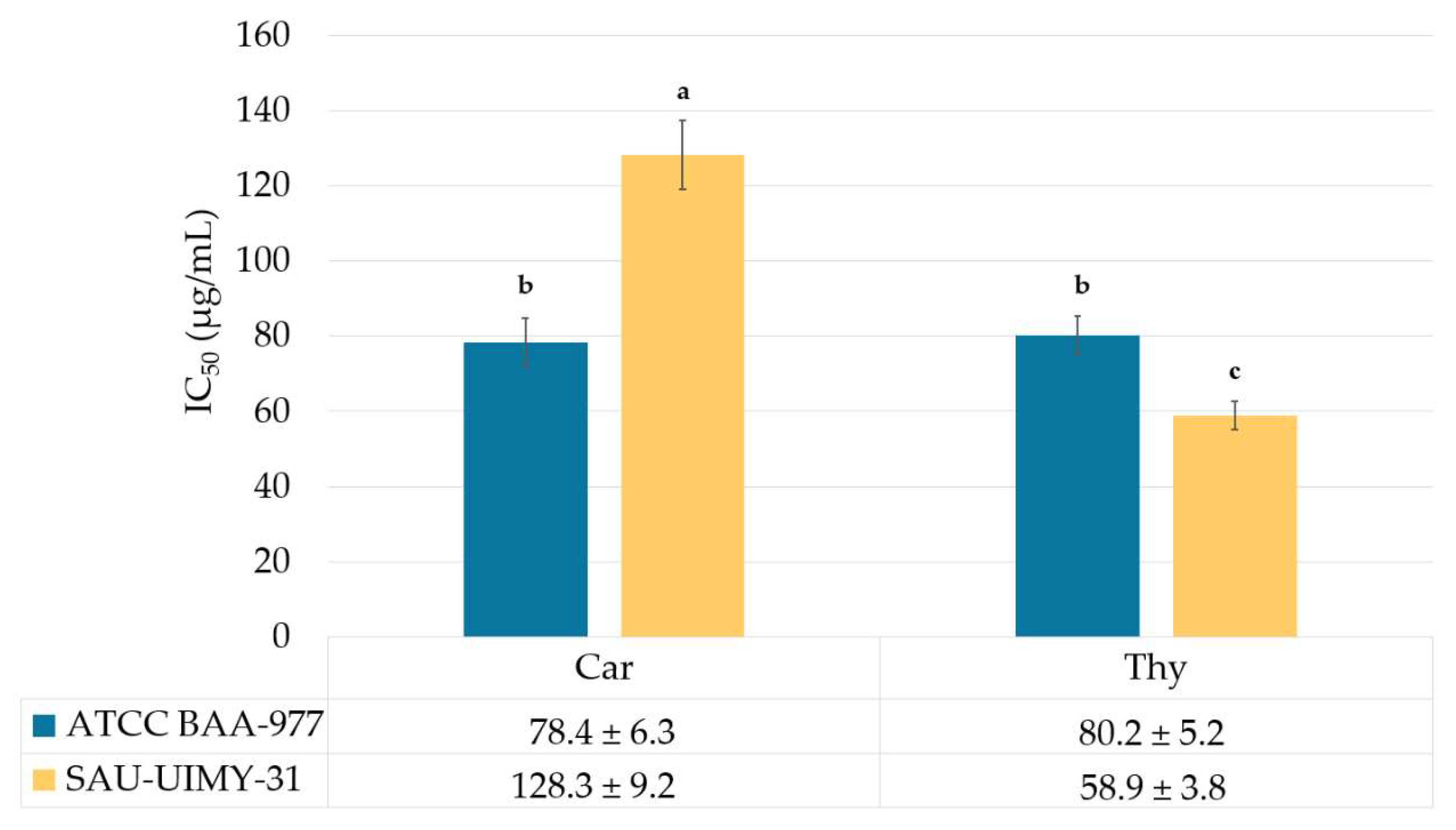
2.5. Antihemolysis Activity of the L. origanoides EO on S. aureus

In our study, the carvacrol and thymol chemotypes of the L. origanoides EO exhibited antihemolysis activity against S. aureus ATCC BAA-977 and SAU-UIMY-31. A two-way ANOVA showed a statistically significant interaction between the bacterial strain and the EO chemotype (F[1,8] = 339.9, p = 0.0001) on the IC50 average value. Among them, the thymol chemotype in strain SAU-UIMY-31 displayed the best activity (IC50 = 58.9 ± 3.8 µg/mL), while in the carvacrol chemotype, the lowest IC50 value was observed in the reference strain ATCC BAA-977 (IC50 = 78.4 ± 6.3 µg/mL; Figure 1). On the other hand, the sesquiterpene EO chemotype did not exhibit 50% hemolysis inhibition at its highest concentration (250 µg/mL).

3. Discussion

3.1. Chemical Composition of the Three Chemotypes of the L. origanoides EO

The three chemotypes identified in this study for the L. origanoides EO have been previously reported for this species [20,28,29,32]. These chemotypes were also observed in O. vulgare cultivars [33].

3.2. Activity of the L. origanoides EO on Staphylococcal Growth

Previous studies have reported the antibacterial activity of the L. origanoides EO against S. aureus strains. Gómez-Sequeda et al. 2020 evaluated the antibacterial activity of the L. origanoides EO against S. aureus ATCC MRSA and reported MIC50 values of 600, 1600, and >3000 µg/mL for carvacrol, thymol, and non-phenolic chemotypes, respectively [20]. Likewise, Martinez et al. 2021 reported antibacterial activity against S. aureus ATCC 29213 (MSSA) of the L origanoides thymol–carvacrol chemotype (major components: thymol 32.7%, carvacrol 18.8%, β-caryophyllene 6.4%, γ-terpinene 5.2%, and p-cymene 1.1%). The results indicated that L. origanoides EO exhibited an MIC50 value of 450 µg/mL [34]. The higher MIC values obtained in this study could be attributed to higher carvacrol and thymol concentrations, as these authors reported concentrations lower than 35% for both phenols.
Our research found that the majority of L. origanoides EOs were bactericidal agents. This information provides valuable insights into the mechanisms of action of the antibacterial agents. However, no extrapolations have been made to clinical therapeutics. Previous studies have shown that carvacrol and thymol, as phytoconstituents of EO, as well as pure compounds, exhibit bactericidal activity against S. aureus [20,33,34,35]. To the best of our knowledge, this is the first report of the bactericidal activity of the sesquiterpene chemotype of L. origanoides.

3.3. Activity of the L. origanoides EO on the Formation of the Biofilm of S. aureus

Gómez-Sequeda et al. (2020) reported that the carvacrol and thymol chemotypes of L. origanoides exhibited values of IC50 = 70 µg/mL and 1200 µg/mL, respectively, against S. aureus ATCC MRSA biofilms, whereas the non-phenolic chemotype showed an IC50 > 3000 µg/mL against the same strain [20]. Martinez et al. 2023 reported that the L. origanoides EO (thymol 32.7% and carvacrol 18.8%) exhibited a biofilm inhibition of 72% at 400 µg/mL against S. aureus ATCC 29213 (MSSA). In addition, these authors reported that EO alters the expression of genes related to biofilm formation and virulence factors associated with the QS system [35]. Our study found similar results for the carvacrol and thymol chemotypes. It is important to highlight that the non-phenolic chemotype, with a predominance of sesquiterpenes (β-caryophyllene, α-humulene, and α-selinene), also showed significant biofilm inhibition activity, like that of the phenolic chemotype, particularly against the S. aureus strains SAU-UIMY-1, SAU-UIMY-31, SAU-UIMY-39, and MRSA ATCC 4330.
Our study demonstrated that higher biofilm biomass producers were inhibited by lower concentrations of EO, but the antibiofilm activity of EO against S. aureus strains may not be directly related to the type of biofilm biomass producers. Rather, it may depend on the unique genetic characteristics of each strain [36]. The accessory gene regulator (agr) QS system in S. aureus has been found to correlate with biofilm biomass. Staphylococcus aureus strains can be grouped into four categories based on their agr system (agrI–IV). These groups often exhibit different phenotypes, such as varying biofilm formation capacities [37,38]. Certain chemotypes of L. origanoides with thymol (32.7%) and carvacrol (18.8%) have been found to reduce the expression of agr in S. aureus [35], suggesting that differences in the type of agr among the strains could affect the biofilm biomass production and IC50 antibiofilm activity of EO.
The Lippia origanoides EO suppresses the ica operon (ADBC), which is a key regulator of polysaccharide intercellular adhesin (PIA) synthesis in S. aureus biofilms [39]; icaA and icaD are responsible for PIA synthesis [40]. Previous research has demonstrated that the co-expression of icaA and icaD in S. aureus is required for strong biofilm production; however, the absence of these genes does not completely inhibit biofilm formation, implying the existence of ica-independent mechanisms [39]. These differences may also contribute to discrepancies in antibiofilm activity among EO chemotypes.

3.4. Correlation of the Chemical Composition of the L. origanoides EO and Its Antibiofilm Activity

Our results found a relationship between the presence of β-caryophyllene, α-humulene, α and β selinene, γ and δ cadinene p-cymene and carvacrol and the prevention of biofilm formation. Some EO components have been reported to exhibit antibiofilm properties. Studies have frequently reported the antibiofilm effects of the monoterpenes carvacrol and thymol, particularly against S. aureus [9,41]. Reichling (2020) discovered that carvacrol and thymol significantly reduced biofilm mass and inhibited the movement of bacteria [9]. Similarly, Kim et al. (2022) found that applying carvacrol and thymol at sublethal concentrations can effectively combat S. aureus by impacting its ability to form biofilm [41]. Our findings align with those of previous studies, as the L. origanoides EO from the carvacrol and thymol chemotypes showed a significant inhibitory effect on biofilm formation. Carvacrol and thymol have been shown to impede biofilm development by impacting membrane lipids, preventing protein accumulation, and halting the microcolony stage [42,43,44]. It also affects cell viability and interacts with transcriptional regulators of QS communication, biofilm formation, and virulence genes [35]. The activity of EO components can be influenced by their minor components in complex interactions, which could explain the variations in the anti-S. aureus activity of the L. origanoides EO depending on the bacterial strain and EO composition. The monoterpene p-cymene may enhance the activity of other antimicrobial compounds such as carvacrol and/or thymol, which cause membrane instability [34,45,46]
The sesquiterpene β-caryophyllene exhibited anti-S. aureus activity on fusidic acid resistant to the strain [47], which induces apoptosis and the disruption of mitochondrial membrane potential [48] and, using molecular docking analyses, interacts with bacterial DNA gyrase B, suggesting that it acts as an inhibitor [49]. The biological activity of β-selinene against S. aureus ATCC 25923 has also been reported [50]. It has been shown that two or three major components, constituting up to 85% of the chromatogram area, drive biological activity [25,26]; however, often, the minor components also play a role [22,24]. Because of the diverse chemical nature of EOs, their components could exert additive, antagonistic, or synergistic effects on each other; therefore, the specific modes of action could be a result of these interactions. Detailed studies with specific experimental designs are needed to deepen our understanding of the interactions between the different metabolites present in the EO of L origanoides and their anti-S aureus effect. Our results provide a basis for designing and conducting such experiments in future research.

3.5. Antihemolysis Activity of the L. origanoides EO on S. aureus

Previously, the antihemolytic effects of the three chemotypes of the L. origanoides EO from Colombia have been reported: 1 (thymol 32.7% and carvacrol 18.8%), 2 (thymol 22.1% and 10.7% carvacrol), and 3 (without thymol or carvacrol) were 54%, 28%, and inactive, respectively. These results highlight the importance of thymol and carvacrol in antihemolytic activity [34]. An EO from L. origanoides (thymol 32.7%, carvacrol 18.8%) inhibits the expression of QS genes, including the synthesis of a transcriptional regulator RNA III, responsible for several virulence factors, such as α and δ hemolysins [35]. In addition, sublethal concentrations of carvacrol and thymol reduced hemolytic activity [41].

4. Materials and Methods

This project was approved by the Scientific and Ethics Committees National of the Instituto Mexicano del Seguro Social (IMSS), with approval number R-785-2022-009. Figure 2 shows the flowchart of the methodology.

4.1. EO Extraction and Chemical Characterization

Lippia origanoides leaves from adult plants were collected from wild populations located in the Yucatan Peninsula [51]. The chemotype carvacrol was collected in Chicxulub Puerto Yucatán (21.253° N 89.573° W); the thymol chemotype was collected in San Felipe Yucatán (21.561° N, 88.189° W); and the sesquiterpene chemotype was collected in Sotuta Yucatán (20.514° N 88.946° W). The leaves were dried at 35 °C for 36 h in an air-flux drying oven (NOVATECH HS60, Tlaquepaque, Mexico) and stored at 4 °C until distillation, following the method described in [51]. Herbaria voucher specimens were deposited at the Centro de Investigación Científica de Yucatán, Mérida, Mexico (LM Calvo 240, 248, and 258); species identification was performed by the herbaria staff. EO was extracted from 250 g of dried leaves using hydrodistillation in a Clevenger-type apparatus for 1.5 h, with 25:1 mL/g water: plant material and 44 drops per minute as the average distillation rate, with hexane as the collector solvent. The oil–hexane mixture was dried with sodium sulfate, and the solvent was evaporated under a flow of nitrogen. The samples were stored in sealed amber vials at 4 °C until biological assays and chromatographic analysis.
To analyze the chemical composition of the EO in the three L. origanoides chemotypes, GC-MS analyses were conducted using an Agilent 6890N gas chromatograph (Agilent Technologies, Santa Clara, CA, USA), which was connected to an Agilent 5975 mass-selective detector and equipped with G1701DA GC-MSD ChemStation software v.D.03.00.552. The non-polar DB5 5% phenyl-metilpolisiloxane column (60 m, 0.25 mm i.d., film thickness 0.25 μm) was used, with a temperature program of 45 °C for 5 min, 45 to 150 °C at 4 °C/min, 150 °C for 2 min, 150 to 250 °C at 5 °C/min, 250 °C for 5 min, 250 to 275 °C at 10 °C/min, and 275 °C for 15 min. The injector temperature was 280 °C and the detector temperature was 290 °C. The carrier gas was He (1.5 mL/min); the injection volume was 1 μL, and the split ratio was 1:40. Mass spectra (MS) were obtained by electron impact at an energy of 70 eV. The temperatures of the ionization chamber and the transfer line were maintained at 230 °C and 285 °C, respectively. MS, total ionic currents, and extracted ions were obtained with a quadrupole analyzer using automatic radiofrequency scanning (full scan) in the mass range of m/z 35–300.
The arithmetic linear retention index (AI) of each peak was determined [52], relative to that of a homologous series of n-alkanes (C8–C40), which was injected in the CG-MS under the same conditions previously described. For a metabolite to be considered tentatively identified, its mass spectrum (MS) would have to match the corresponding mass spectra contained in the library ADAMS 2007 [52] and in the library of the National Institute of Standards and Technology (NIST 11). In addition, the corresponding AI value must coincide with the AI values reported in the literature [52,53,54]. When available, the definitive identification of individual metabolites was carried out by comparing their MS with those of commercial standards such as carvacrol, thymol, β-caryophyllene, and eucalyptol (98–99% purity, Sigma-Aldrich, St. Louis, MO, USA) or authentic samples previously purified and identified in our laboratories. The quantification of metabolites was performed using a Varian 430 GC (Varian BV, Santa Clara, CA, USA) equipped with an FID detector and N2 as the carrier gas, using the same column, temperature program, and conditions described previously.

4.2. Anti-Infectious Assays

4.2.1. Staphylococcus aureus Strains

Three reference strains and nine clinical isolates of susceptible and drug-resistant S. aureus were used in the bioassays (Table 5). All strains were maintained at −80 °C in tryptic soy broth (TSB; Becton Dickinson, Franklin Lakes, NJ, USA) supplemented with glycerol (J.T. Baker, Inc., Phillipsburg, NJ, USA).

4.2.2. Activity of EO on Staphylococcal Growth

The Minimal Inhibitory Concentration (MIC) of EO against the S. aureus strains was determined using the resazurin microtiter assay (REMA) broth dilution method. Briefly, bacterial cultures were cultured on Mueller–Hinton agar (MHA; Becton Dickinson Co., Ltd., Franklin Lakes, NJ, USA). Then, two to three bacterial colonies were suspended in 3 mL of Muller–Hinton broth (MHB; Becton Dickinson Co., Ltd., Franklin Lakes, NJ, USA) and incubated at 37 °C for 2–4 h until reaching growth comparable to the turbidity of the 0.5 McFarland standard (DEN-1; Biosan, Riga, Latvia). This suspension was further diluted at 1: 50 to obtain the working inoculum, and 100 μL of this suspension was incubated and cultured with 100 μL of MHB containing the EO at serial dilutions ranging from 500 to 15.62 μg/mL. All assays included a positive control (cultures with antibiotic specific) and negative controls (culture-free wells of the EO or antibiotic (Sigma-Aldrich, St. Louis, MO, USA) and a sterility control (culture broth alone). After incubation at 37 °C for 16 h, 30 μL of resazurin (Sigma-Aldrich, St. Louis, MO, USA) was added, and the microplates were incubated again at 37 °C for 2 h. A well-defined pink color indicated positive bacterial growth, whereas a blue color indicated the absence of growth. The results were expressed as the MIC, which corresponded to the greatest dilution of the EO in which a color shift from blue to pink was not observed. Each assay was performed three times independently in duplicate [55].
The minimal bactericidal concentration (MBC) of the EO was determined by reseeding the bacterial culture according to the method described by de Jesús Dzul-Beh et al. 2023 [56]. Succinctly, 5 μL of the EO-treated bacterial suspensions corresponding to MIC, 2× and ½ MIC in MHB in 96-well plates, were transferred to a new microplate containing 195 μL of fresh culture medium per well. In addition, antibiotic-treated bacterial suspensions (positive control), untreated bacterial suspensions (negative control), and culture medium alone (sterility control) were transferred to fresh broth [57]. After incubation at 37 °C for 16 h, resazurin was added to the wells, as previously described. MBC corresponded to the minimal EO concentration that did not cause a color shift in cultures re-incubated in the fresh medium. Each assay was performed three times in duplicate, and the MBC/MIC index for the EO was calculated.

4.2.3. Activity of the EO on the Formation of Biofilm

The inhibition of biofilm formation in all strains of S. aureus was evaluated using the crystal violet (CV) staining method in flat-bottom 96-well microplates, as previously described [55]. Briefly, the strains were activated and cultured on MHA, and two bacterial colonies were cultured in 3 mL of TSB (Beckton Dickinson Co., Ltd., Franklin Lakes, NJ, USA) at 37 °C for 24 h. Then, an aliquot was transferred into TBS supplemented with glucose 1% (w/v; TSB + G) to match the turbidity of 0.5 McFarland. This suspension was further diluted at 1:50, and 100 μL of this was incubated with 100 μL of TBS + G containing serial dilutions of 500 to 15.6 μg/mL concentrations of the EO. An ethylenediaminetetraacetic acid (Sigma-Aldrich, St. Louis, MO, USA) solution was used as the positive control, whereas the negative control was EO-free wells. The microplate was incubated for 24 h at 37 °C. After incubation, the culture broth was gently aspirated, and each well was washed thrice with sterile distilled water to remove non-adherent cells and dried at 60 °C for 45 min. The biofilm was stained by incubating for 30 min with 200 μL of 0.1% CV (Sigma-Aldrich, St. Louis, MO, USA) solution. Any excess CV was removed with sterile distilled water before adding 200 μL of 40% acetic acid (Fermont, Monterrey, Mexico), and the absorbance was measured at 490 nm using a microplate reader (IMark; Bio-Rad, Hercules, CA, USA), which was related to the amount of biofilm produced. Each assay was performed in triplicate, and the concentration of EO that inhibited 50% (IC50) biofilm formation was calculated using GraphPad Prism ver. 5 software (GraphPad Software Inc., La Jolla, CA, USA).

4.2.4. Antihemolytic Activity of EO

The antihemolytic activity of the EO was assessed using S. aureus ATCC BAA-977 and the clinical isolate SAU-UIMY-31, according to Loges et al., 2020, with minor modifications [58]. Briefly, an aliquot on an overnight culture of S. aureus was transferred into fresh TSB to match the turbidity of 0.5 McFarland. A total of 30 μL of the suspension was incubated with 2970 μL of TBS containing serial dilutions of 200 to 25 μg/mL EO and incubated with shaking at 200 rpm and 37 °C for 24 h. After incubation, the bacterial culture supernatant was collected via centrifugation. In total, 100 μL of the bacterial supernatant was incubated with 300 μL of diluted human red blood cells in PBS buffer (Sigma-Aldrich; 330 μL red blood cells/10 mL PBS buffer), with shaking at 200 rpm and 37 °C for 1 h. The mixture was centrifuged at 4000 rpm for 4 min to remove intact red blood cells, and the absorbance of the supernatant was measured at 430 nm using a microplate reader (IMark, Bio-Rad, Hercules, CA, USA). The percentage of hemolysis inhibition for each concentration of EO was calculated. Each assay was performed in triplicate, and the IC50 hemolysis was calculated using GraphPad Prism ver. 5 software (GraphPad Software, Inc., La Jolla, CA, USA).

4.3. Statistical Analysis

The Spearman rank correlation between the IC50 for biofilm formation inhibition and the biofilm producer index was estimated for the nine biofilm producer strains. Similarly, for each bacterial strain, a Spearman rank correlation was estimated between the IC50 for biofilm formation inhibition and the concentration of the major components (% area in the chromatogram) in the three chemotypes of the L. origanoides EO (N = 9). Two-way ANOVA was used to analyze the interaction of the bacterial strain and EO chemotype composition on the IC50 values for biofilm formation inhibition and antihemolytic activity. In all statistical analyses, results with p-values > 0.05 were considered non-significant.

5. Conclusions

In the present study, the chemical composition of the three chemotypes of the L. origanoides EO varied considerably. The two phenolic chemotypes mainly contained carvacrol and thymol, whereas the third chemotype contained sesquiterpenes, with β-caryophyllene as the major component. All chemotypes exhibited anti-staphylococcal and antibiofilm activities against the reference and clinical isolates of S. aureus strains. The activity in both assays varied depending on the interaction between the bacterial strain and the EO chemotype. However, the carvacrol and thymol chemotypes also showed antihemolytic activity against reference and clinical isolates of S. aureus strains. Our study highlights the potential of chemotypes of the L. origanoides EO from the Yucatan Peninsula as candidates for future research in developing anti-S. aureus agents, and they are even active against the drug-resistant strains MRSA and VRSA.

Author Contributions

Conceptualization, L.M.C.-I. and G.M.M.-S.; methodology, A.H.U.-C., L.M.C.-I., A.d.J.D.-B., H.E.D.-B. and G.M.M.-S.; validation, A.H.U.-C., L.M.C.-I. and G.M.M.-S.; formal analysis, A.H.U.-C., L.M.C.-I., A.d.J.D.-B., H.E.D.-B., R.G.-A. and G.M.M.-S.; investigation, A.H.U.-C., L.M.C.-I., A.d.J.D.-B., H.E.D.-B., R.G.-A. and G.M.M.-S.; resources, L.M.C.-I. and G.M.M.-S.; data curation, A.H.U.-C., L.M.C.-I., A.d.J.D.-B. and G.M.M.-S.; writing—original draft preparation, A.H.U.-C., L.M.C.-I. and G.M.M.-S.; writing—review and editing, A.H.U.-C., L.M.C.-I., A.d.J.D.-B., H.E.D.-B., R.G.-A. and G.M.M.-S.; supervision, L.M.C.-I. and G.M.M.-S.; project administration, L.M.C.-I. and G.M.M.-S.; funding acquisition, L.M.C.-I. and G.M.M.-S. All authors have read and agreed to the published version of the manuscript.

Funding

This research received funding from Centro de Investigación Científica de Yucatán (Project Semilla 2022) and Instituto Mexicano del Seguro Social (Project R-2022-785-009).

Data Availability Statement

Data are contained within the article.

Conflicts of Interest

The authors declare no conflicts of interest.

References

  1. Wohlleben, W.; Mast, Y.; Stegmann, E.; Ziemert, N. Antibiotic drug discovery. Microb. Biotechnol. 2016, 9, 541–548. [Google Scholar] [CrossRef]
  2. Lee Ventola, C. The antibiotic resistance crisis part 1: Causes and threats. Pharm. Ther. 2015, 40, 277–283. [Google Scholar]
  3. Prestinaci, F.; Pezzotti, P.; Pantosti, A. Antimicrobial resistance: A global multifaceted phenomenon. Pathog. Glob. Health 2015, 109, 309–318. [Google Scholar] [CrossRef]
  4. Chinemerem Nwobodo, D.; Ugwu, M.C.; Oliseloke Anie, C.; Al-Ouqaili, M.T.S.; Chinedu Ikem, J.; Victor Chigozie, U.; Saki, M. Antibiotic resistance: The challenges and some emerging strategies for tackling a global menace. J. Clin. Lab. Anal. 2022, 36, e24655. [Google Scholar] [CrossRef]
  5. Mancuso, G.; Midiri, A.; Gerace, E.; Biondo, C. Bacterial antibiotic resistance: The most critical pathogens. Pathogens 2021, 10, 1310. [Google Scholar] [CrossRef]
  6. Turner, N.A.; Sharma-Kuinkel, B.K.; Maskarinec, S.A.; Eichenberger, E.M.; Shah, P.P.; Carugati, M.; Holland, T.L.; Fowler, V.G. Methicillin-Resistant Staphylococcus aureus: An overview of basic and clinical research. Nat. Rev. Microbiol. 2019, 17, 203–218. [Google Scholar] [CrossRef]
  7. Yu, Z.; Tang, J.; Khare, T.; Kumar, V. The alarming antimicrobial resistance in ESKAPEE pathogens: Can essential oils come to the rescue? Fitoterapia 2020, 140, 104433. [Google Scholar] [CrossRef]
  8. Kumar, V.; Yasmeen, N.; Pandey, A.; Ahmad Chaudhary, A.; Alawam, A.S.; Ahmad Rudayni, H.; Islam, A.; Lakhawat, S.S.; Sharma, P.K.; Shahid, M. Antibiotic adjuvants: Synergistic tool to combat multi-drug resistant pathogens. Front. Cell. Infect. Microbiol. 2023, 13, 1293633. [Google Scholar] [CrossRef]
  9. Reichling, J. Anti-biofilm and virulence factor-reducing activities of essential oils and oil components as a possible option for bacterial infection control. Planta Med. 2020, 86, 520–537. [Google Scholar] [CrossRef]
  10. Álvarez-Martínez, F.J.; Barrajón-Catalán, E.; Herranz-López, M.; Micol, V. Antibacterial plant compounds, extracts and essential oils: An updated review on their effects and putative mechanisms of action. Phytomedicine 2021, 90, 153626. [Google Scholar] [CrossRef]
  11. Brożyna, M.; Paleczny, J.; Kozłowska, W.; Chodaczek, G.; Dudek-Wicher, R.; Felińczak, A.; Gołębiewska, J.; Górniak, A.; Junka, A. The antimicrobial and antibiofilm in vitro activity of liquid and vapour phases of selected essential oils against Staphylococcus aureus. Pathogens 2021, 10, 1207. [Google Scholar] [CrossRef]
  12. Romanescu, M.; Oprean, C.; Lombrea, A.; Badescu, B.; Teodor, A.; Constantin, G.D.; Andor, M.; Folescu, R.; Muntean, D.; Danciu, C.; et al. Current state of knowledge regarding who high priority pathogens—Resistance mechanisms and proposed solutions through candidates such as essential oils: A systematic review. Int. J. Mol. Sci. 2023, 24, 9727. [Google Scholar] [CrossRef]
  13. Kintzios, S.E. Profile of the multifaceted prince of the herbs. In Oregano the Genera Origanum and Lippia, 1st ed.; Kintzios, S.E., Ed.; Taylor & Francis Group: Oxfordshire, UK, 2002; pp. 1–6. [Google Scholar]
  14. O’Leary, N.; Denham, S.S.; Salimena, F.; Múlgura, M.E. Species delimitation in Lippia section Goniostachyum (verbenaceae) using the phylogenetic species concept. Bot. J. Linn. Soc. 2012, 170, 197–219. [Google Scholar] [CrossRef]
  15. Atlas de Las Plantas de La Medicina Tradicional Mexicana. Available online: http://www.medicinatradicionalmexicana.unam.mx/apmtm/index.html (accessed on 25 March 2024).
  16. Calvo-Irabien, L.M. Native mexican aromatic flora and essential oils: Current research status, gaps in knowledge and agro-industrial potential. Ind. Crops. Prod. 2018, 111, 807–822. [Google Scholar] [CrossRef]
  17. Siqueira-Lima, P.S.; Passos, F.R.S.; Lucchese, A.M.; Menezes, I.R.A.; Coutinho, H.D.M.; Lima, A.A.N.; Zengin, G.; Quintans, J.S.S.; Quintans-Júnior, L.J. Central nervous system and analgesic profiles of Lippia genus. Rev. Bras. Farm. 2019, 29, 125–135. [Google Scholar] [CrossRef]
  18. Candelaria-Dueñas, S.; Serrano-Parrales, R.; Ávila-Romero, M.; Meraz-Martínez, S.; Orozco-Martínez, J.; Ávila-Acevedo, J.G.; García-Bores, A.M.; Cespedes-Acuña, C.L.; Peñalosa-Castro, I.; Hernandez-Delgado, T. Evaluation of the antimicrobial activity of some components of the essential oils of plants used in the traditional medicine of the Tehuacán-Cuicatlán valley, Puebla, México. Antibiotics 2021, 10, 295. [Google Scholar] [CrossRef]
  19. Zapién-Chavarría, K.A.; Plascencia-Terrazas, A.; Venegas-Ortega, M.G.; Varillas-Torres, M.; Rivera-Chavira, B.E.; Adame-Gallegos, J.R.; González-Rangel, M.O.; Nevárez-Moorillón, G.V. Susceptibility of multidrug-resistant and biofilm-forming uropathogens to mexican oregano essential oil. Antibiotics 2019, 8, 186. [Google Scholar] [CrossRef]
  20. Gómez-Sequeda, N.; Cáceres, M.; Stashenko, E.E.; Hidalgo, W.; Ortiz, C. Antimicrobial and antibiofilm activities of essential oils against Escherichia coli o157: H7 and methicillin-resistant Staphylococcus aureus (MRSA). Antibiotics 2020, 9, 730. [Google Scholar] [CrossRef]
  21. Dhifi, W.; Bellili, S.; Jazi, S.; Bahloul, N.; Mnif, W. Essential oils’ chemical characterization and investigation of some biological activities: A critical review. Medicines 2016, 3, 25. [Google Scholar] [CrossRef]
  22. Feyaerts, A.F.; Mathé, L.; Luyten, W.; De Graeve, S.; Van Dyck, K.; Broekx, L.; Van Dijck, P. Essential oils and their components are a class of antifungals with potent vapour-phase-mediated anti-candida activity. Sci. Rep. 2018, 8, 3958. [Google Scholar] [CrossRef]
  23. El-Tarabily, K.A.; El-Saadony, M.T.; Alagawany, M.; Arif, M.; Batiha, G.E.; Khafaga, A.F.; Elwan, H.A.M.; Elnesr, S.S.; El-Hack, M.E.A. Using essential oils to overcome bacterial biofilm formation and their antimicrobial resistance. Saudi J. Biol. Sci. 2021, 28, 5145–5156. [Google Scholar] [CrossRef] [PubMed]
  24. Bassolé, I.H.N.; Juliani, H.R. Essential oils in combination and their antimicrobial properties. Molecules 2012, 17, 3989–4006. [Google Scholar] [CrossRef] [PubMed]
  25. Chouhan, S.; Sharma, K.; Guleria, S. Antimicrobial activity of some essential oils—Present status and future perspectives. Medicines 2017, 4, 58. [Google Scholar] [CrossRef] [PubMed]
  26. Chraibi, M.; Farah, A.; Elamin, O.; Iraqui, H.; Fikri-Benbrahim, K. Characterization, antioxidant, antimycobacterial, antimicrobial effects of Moroccan rosemary essential oil, and its synergistic antimicrobial potential with carvacrol. J. Adv. Pharm. Technol. Res. 2020, 11, 25–29. [Google Scholar] [PubMed]
  27. Trindade, H.; Pedro, L.G.; Figueiredo, A.C.; Barroso, J.G. Chemotypes and terpene synthase genes in Thymus genus: State of the art. Ind. Crops Prod. 2018, 124, 530–547. [Google Scholar] [CrossRef]
  28. Stashenko, E.; Martínez, J.; Ruiz, C.; Arias, G.; Duran, C.; Salgar, W.; Cala, M. Lippia origanoides chemotype differentiation based on essential oil GC-MS and principal components analysis. J. Sep. Sci. 2010, 33, 93–103. [Google Scholar] [CrossRef] [PubMed]
  29. Calvo-Irabién, L.M.; Parra-Tabla, V.; Acosta-Arriola, V.; Escalante-Erosa, F.; Díaz-Vera, L.; Dzib, G.R.; Peña-Rodríguez, L.M. Phytochemical diversity of the essential oils of mexican oregano (Lippia graveolens Kunth) populations along an edapho-climatic gradient. Chem. Biodivers. 2014, 11, 1010–1021. [Google Scholar] [CrossRef] [PubMed]
  30. Ribeiro, A.F.; Andrade, E.H.A.; Salimena, F.R.G.; Maia, J.G.S. Circadian and seasonal study of the cinnamate chemotype from Lippia origanoides Kunth. Biochem. Syst. Ecol. 2014, 55, 249–259. [Google Scholar] [CrossRef]
  31. Méndez González, M.E.; Durán García, R.; Borges Argáez, R.; Peraza Sánchez, S.; Dorantes Euan, A.; Tapia Muñoz, J.L. Flora Medicinal: De los Mayas Peninsulares, 1st ed.; Centro de Investigación Científica de Yucatán Pronatura Península de Yucatán: Mérida, Mexico, 2012; pp. 154–196. [Google Scholar]
  32. Fimbres-García, J.O.; Flores-Sauceda, M.; Othon-Díaz, E.D.; García-Galaz, A.; Tapia-Rodríguez, M.R.; Silva-Espinoza, B.A.; Ayala-Zavala, J.F. Facing resistant bacteria with plant essential oils: Reviewing the oregano case. Antibiotics 2022, 11, 1777. [Google Scholar] [CrossRef] [PubMed]
  33. Hao, Y.; Kang, J.; Yang, R.; Li, H.; Cui, H.; Bai, H.; Tsitsilin, A.; Li, J.; Shi, L. Multidimensional exploration of essential oils generated via eight oregano cultivars: Compositions, chemodiversities, and antibacterial capacities. Food Chem. 2022, 374, 131629. [Google Scholar] [CrossRef] [PubMed]
  34. Martínez, A.; Manrique-Moreno, M.; Klaiss-Luna, M.C.; Stashenko, E.; Zafra, G.; Ortiz, C. Effect of essential oils on growth inhibition, biofilm formation and membrane integrity of Escherichia coli and Staphylococcus aureus. Antibiotics 2021, 10, 1474. [Google Scholar] [CrossRef]
  35. Martínez, A.; Stashenko, E.E.; Sáez, R.T.; Zafra, G.; Ortiz, C. Effect of essential oil from Lippia origanoides on the transcriptional expression of genes related to quorum sensing, biofilm formation, and virulence of Escherichia coli and Staphylococcus aureus. Antibiotics 2023, 12, 845. [Google Scholar] [CrossRef] [PubMed]
  36. Ben Abdallah, F.; Lagha, R.; Gaber, A. Biofilm inhibition and eradication properties of medicinal plant essential oils against methicillin-resistant Staphylococcus aureus clinical isolates. Pharmaceuticals 2020, 13, 369. [Google Scholar] [CrossRef] [PubMed]
  37. Kwiecinski, J.M.; Jacobsson, G.; Horswill, A.R.; Josefsson, E.; Jin, T. Biofilm formation by Staphylococcus aureus clinical isolates correlates with the infection type. Infect. Dis. 2019, 51, 446–451. [Google Scholar] [CrossRef] [PubMed]
  38. Tan, L.; Huang, Y.; Shang, W.; Yang, Y.; Peng, H.; Hu, Z.; Wang, Y.; Rao, Y.; Hu, Q.; Rao, X.; et al. Accessory gene regulator (agr) allelic variants in cognate Staphylococcus aureus strain display similar phenotypes. Front. Microbiol. 2022, 13, 700894. [Google Scholar] [CrossRef] [PubMed]
  39. dos Santos Rodrigues, J.B.; de Carvalho, R.J.; de Souza, N.T.; de Sousa Oliveira, K.; Franco, O.L.; Schaffner, D.; de Souza, E.L.; Magnani, M. Effects of oregano essential oil and carvacrol on biofilms of Staphylococcus aureus from food-contact surfaces. Food Control. 2017, 73, 1237–1246. [Google Scholar] [CrossRef]
  40. Wu, X.; Wang, H.; Xiong, J.; Yang, G.-X.; Hu, J.-F.; Zhu, Q.; Chen, Z. Staphylococcus aureus biofilm: Formulation, regulatory, and emerging natural products-derived therapeutics. Biofilm 2024, 7, 100175. [Google Scholar] [CrossRef] [PubMed]
  41. Kim, Y.; Shin, M.; Kang, J.; Kang, D. Effect of sub-lethal treatment of carvacrol and thymol on virulence potential and resistance to several bactericidal treatments of Staphylococcus aureus. J. Food Saf. 2022, 42, e13004. [Google Scholar] [CrossRef]
  42. Knowles, J.R.; Roller, S.; Murray, D.B.; Naidu, A.S. Antimicrobial action of carvacrol at different stages of dual-species biofilm development by Staphylococcus aureus and Salmonella enterica serovar Typhimurium. Appl. Environ. Microbiol. 2005, 71, 797–803. [Google Scholar] [CrossRef] [PubMed]
  43. Nostro, A.; Roccaro, A.S.; Bisignano, G.; Marino, A.; Cannatelli, M.A.; Pizzimenti, F.C.; Cioni, P.L.; Procopio, F.; Blanco, A.R. Effects of oregano, carvacrol and thymol on Staphylococcus aureus and Staphylococcus epidermidis biofilms. J. Med. Microbiol. 2007, 56, 519–523. [Google Scholar] [CrossRef]
  44. Miladi, H.; Zmantar, T.; Chaabouni, Y.; Fedhila, K.; Bakhrouf, A.; Mahdouani, K.; Chaieb, K. Antibacterial and efflux pump inhibitors of thymol and carvacrol against food-borne pathogens. Microb. Pathog. 2016, 99, 95–100. [Google Scholar] [CrossRef] [PubMed]
  45. Ahmad, A.; Van Vuuren, S.; Viljoen, A. Unravelling the complex antimicrobial interactions of essential oils—The case of Thymus vulgaris (Thyme). Molecules 2014, 19, 2896–2910. [Google Scholar] [CrossRef] [PubMed]
  46. Cáceres, M.; Hidalgo, W.; Stashenko, E.; Torres, R.; Ortiz, C. Essential oils of aromatic plants with antibacterial, anti-biofilm and anti-quorum sensing activities against pathogenic bacteria. Antibiotics 2020, 9, 147. [Google Scholar] [CrossRef] [PubMed]
  47. Oliveira Ribeiro, S.; Fontaine, V.; Mathieu, V.; Abdesselam, Z.; Dominique, B.; Caroline, S.; Florence, S. Antibacterial activities of homemade matrices miming essential oils compared to commercial ones. Antibiotics 2021, 10, 584. [Google Scholar] [CrossRef] [PubMed]
  48. Dahham, S.S.; Tabana, Y.M.; Iqbal, M.A.; Ahamed, M.B.K.; Ezzat, M.O.; Majid, A.S.A.; Majid, A.M.S.A. The anticancer, antioxidant and antimicrobial properties of the sesquiterpene β-caryophyllene from the essential oil of Aquilaria crassna. Molecules 2015, 20, 11808–11829. [Google Scholar] [CrossRef] [PubMed]
  49. Thi Huong, L.; Chung, N.; Duc, D.; Dai, D.; Pham The, H.; Ninh The, S. Essential oil of Syzygium boisianum (Gagnep.) Merr. & L.M.Perry: Chemical compositions, antimicrobial activity, and molecular docking. Vietnam J. Chem. 2023, 61, 333–338. [Google Scholar]
  50. Huong, L.T.; Sam, L.N.; Dai, D.N.; Pham, T.V.; Son, N.T. Essential oils of two ginger plants Newmania orthostachys N.S. Lý & Škorničk. and N. Serpens N.S. Lý & Škorničk.: Chemical compositions and antimicrobial activity. J. Essent. Oil Bear. Plants 2022, 25, 1221–1228. [Google Scholar]
  51. Martínez-Natarén, D.A.; Parra-Tabla, V.; Ferrer-Ortega, M.M.; Calvo-Irabién, L.M. Genetic diversity and genetic structure in wild populations of mexican oregano (Lippia graveolens H.B.K.) and its relationship with the chemical composition of the essential oil. Plant Syst. Evol. 2014, 300, 535–547. [Google Scholar] [CrossRef]
  52. Adams, R.P. Identification of Essential Oil Components by Gas Chromatography/Mass Spectrometry, 4th ed.; Allured: Carol Stream, IL, USA, 2007; pp. 10–51. [Google Scholar]
  53. Babushok, V.I.; Linstrom, P.J.; Zenkevich, I.G. Retention indices for frequently reported compounds of plant essential oils. J. Phys. Chem. Ref. Data 2011, 40, 043101. [Google Scholar] [CrossRef]
  54. Zellner, B. d’Acampora; Bicchi, C.; Dugo, P.; Rubiolo, P.; Dugo, G.; Mondello, L. Linear retention indices in gas chromatographic analysis: A review. Flavour Fragr. J. 2008, 23, 297–314. [Google Scholar] [CrossRef]
  55. Uc-Cachón, A.H.; de Dzul-Beh, A.J.; Palma-Pech, G.A.; Jiménez-Delgadillo, B.; Flores-Guido, J.S.; Gracida-Osorno, C.; Molina-Salinas, G.M. Antibacterial and antibiofilm activities of mayan medicinal plants against methicillin-susceptible and -resistant strains of Staphylococcus aureus. J. Ethnopharmacol. 2021, 279, 114369. [Google Scholar] [CrossRef] [PubMed]
  56. de Jesús Dzul-Beh, A.; Uc-Cachón, A.H.; González-Sánchez, A.A.; Dzib-Baak, H.E.; Ortiz-Andrade, R.; Barrios-García, H.B.; Jiménez-Delgadillo, B.; Molina-Salinas, G.M. Antimicrobial potential of the mayan medicine plant Matayba oppositifolia (A. Rich.) britton against antibiotic-resistant priority pathogens. J. Ethnopharmacol. 2023, 300, 115738. [Google Scholar] [CrossRef] [PubMed]
  57. Molina-Salinas, G.M.; Ramos-Guerra, M.C.; Vargas-Villarreal, J.; Mata-Cárdenas, B.D.; Becerril-Montes, P.; Said-Fernández, S. Bactericidal activity of organic extracts from Flourensia cernua DC against strains of Mycobacterium tuberculosis. Arch. Med. Res. 2006, 37, 45–49. [Google Scholar] [CrossRef] [PubMed]
  58. Loges, L.A.; Silva, D.B.; Paulino, G.V.B.; Landell, M.F.; Macedo, A.J. Polyketides from marine-derived Aspergillus welwitschiae inhibit Staphylococcus aureus virulence factors and potentiate vancomycin antibacterial activity in vivo. Microb. Pathog. 2020, 143, 104066. [Google Scholar] [CrossRef] [PubMed]
Figure 1. Antihemolysis activity of the L. origanoides EO on S. aureus. Car: carvacrol; Thy: thymol; average IC50 values (±SD) with the same letter showed non-significant differences in the post hoc Tukey test (p < 0.05). The sesquiterpene chemotype IC50 is >250 µg/mL for both strains.
Plants 13 01172 g001
Figure 2. Flowchart of the methodology.
Plants 13 01172 g002
Table 1. Chemical composition of the EO chemotypes of L. origanoides.
Compound NameClassAI aAI ADAMS bChemotypeCompound
Identification d
CarThySes
% area c% area c% area c
myrceneM9929881.152.150.34RI, MS, Coi
p-cymeneM1029102018.1215.262.85RI, MS, Coi
limoneneM103310240.250.411.06RI, MS, Coi
eucalyptolOM103610260.742.632.77RI, MS, Coi
γ-terpineneM106110541.300.240.39RI, MS, Coi
thymol methyl etherOM123812321.313.330.07RI, MS
thymolOM129512892.7457.300.13RI, MS, Coi
carvacrolOM1309129868.510.200.19RI, MS, Coi
α-copaeneS13881374ndnd3.29RI, MS, Coi
β-caryophylleneS143814171.467.7533.00RI, MS, Coi
β-copaeneS144514300.040.081.00RI, MS
α-humuleneS147114520.803.8413.42RI, MS, Coi
aromadendrene-alloS14791458ndnd1.73RI, MS
γ-muuroleneS14911478ndnd4.23RI, MS
β-selineneS15021489nd0.135.05RI, MS
α-selineneS15101498nd0.217.91RI, MS
γ-cadineneS15291513ndnd1.36RI, MS
δ-cadineneS15391522nd0.087.21RI, MS
caryophyllene oxideOS160015820.791.331.32RI, MS, Coi
Total of compound class (%)
M 20.8218.064.64
OM 73.363.463.16
S 2.312.0978.20
OS 0.791.331.32
Total identified (%) 97.2194.9487.32
Only compounds with a >1% chromatogram area are shown. Car: carvacrol; Thy: thymol; Ses: sesquiterpene; M: monoterpenes; OM: oxygenated monoterpenes; S: sesquiterpenes; OS: oxygenated sesquiterpenes. a AI: Linear retention indices determined experimentally on the DB5 column relative to a series of n-alkanes (C8–C40). b Bibliographic linear retention indices. c Relative contents are given as GC-FID peak areas; for the three most abundant compounds of each chemotype, the contents (% area of chromatogram) are given in bold type; n.d. = not detected. d Identification method: RI = tentative identification based on AI; MS = tentative identification based on MS; Coi = co-injection with commercial standard GC-FID.
Table 2. Anti-S. aureus activity of the three chemotypes of the L. origanoides EO.
S. aaureus StrainDrug-Resistant ProfileL. origanoides EOPositive Control
CarThySes
MICMBCMICMBCMICMBCMIC
ATCC 43300 MRSA500500500500>500>500RIF = 0.04
ATCC 25923 MSSA500500500500500>500RIF = 0.08
ATCC BAA-977MSSA500500500500>500>500RIF = 0.08
SAU-UIMY-44 MDR, MRSA, VRSA500>500500>500>500>500TET = 0.5
SAU-UIMY-31 MSSA500500250500250250RIF = 0.08
SAU-UIMY-24 MDR, MRSA, VSSA250500250500500500TET = 0.5
SAU-UIMY-5MDR, MRSA250250250250250250RIF = 0.04
SAU-UIMY- 1XDR, MRSA, VISA125250125250>500>500AMK = 16
SAU-UIMY-26 MDR, MSSA, VISA125125125125>500>500TET = 0.5
SAU-UIMY-10 MDR, MRSA, VSSA125125125125250500TET = 0.5
SAU-UIMY-39 MDR, MSSA, VSSA125125125250250250TET = 0.5
SAU-UIMY-16 MDR, MRSA, VSSA12512562.5125125250TET = 0.5
Plants 13 01172 i001
Car: carvacrol; Thy: thymol; Ses: sesquiterpene; MIC: minimal inhibitory concentration and MBC: minimal bactericidal concentration; MSSA: Methicillin-Susceptible Staphylococcus aureus; MRSA: Methicillin-Resistant Staphylococcus aureus; MDR: MultiDrug-Resistant; VSSA: Vancomycin-Susceptible Staphylococcus aureus; VISA: Vancomycin-Intermediate Staphylococcus aureus; VRSA: Vancomycin-Resistant Staphylococcus aureus; XDR: Extensively Drug-Resistant; RIF: Rifampin; TET: Tetracycline; AMK: Amikacin. MIC and MBC values are expressed as μg/mL.
Table 3. Activity of the three chemotypes of the L. origanoides EO on S. aureus biofilm formation.
S. aureus StrainOriginIC50 (µg/mL)Biofilm Biomass (OD Values)
Lippia origanoides ChemotypesPositive Control
CarThySesqEDTA
ATCC 43300 -90.0 ± 1.9 a,b149.3 ± 1.9 a,b156.9 ± 2.1 a,b79.5 ± 2.90.728 ± 0.08
SAU-UIMY-26 Surgical wound379.0 ± 7.0 e568.3 ± 20.9 f706.6 ± 173.5 g406.9 ± 1.20.198 ± 0.01
SAU-UIMY-44 Blood 228.3 ± 7.5 b,c,d291.7 ± 14.9 b,c,d,e684.86 ± 90.4 f,g458.1 ± 3.30.324 ± 0.03
SAU-UIMY-16Bronchial secretion 213.8 ± 5.4 b,c,d192.8 ± 7.7 b,c,d659.3 ± 62.8 f,g123.9 ± 5.00.581 ± 0.06
SAU-UIMY-24Surgical wound 196.9 ± 6.8 b,c,d198.6 ± 3.3 b,c,d307.8 ± 12.8 d,e55.8 ± 2.60.407 ± 0.07
SAU-UIMY-10Urine 169.6 ± 4.5 b,c162.4 ± 7.4 b,c350.0 ± 2.0 d,e257.8 ± 4.60.420 ± 0.07
SAU-UIMY-31Breast abscess 99.3 ± 3.9 a,b82.4 ± 5.1 a,b79.1 ± 6.1 a,b199 ± 1.80.902 ± 0.2
SAU-UIMY-1Bronchial secretion77.3 ± 4.8 a,b60.6 ± 1.7 a,b30.8 ± 1.5 a84.8 ± 1.81.29 ± 0.27
SAU-UIMY-39Blood35.5 ± 1.8 a,b63.3 ± 4.1 a,b103.2 ± 5.2 a,b104.8 ± 3.10.482 ± 0.08
Plants 13 01172 i002
Car: carvacrol; Thy: thymol; Ses: sesquiterpene; EDTA: ethylendiamine tetraacetic acid; average IC50 values (± SD) with the same letter showed non-significant differences in the post hoc Tukey test (p < 0.05).
Table 4. Spearman rank correlation coefficient values between the IC50 of antibiofilm activity and the concentration of the major components in the EO of the three chemotypes of L. origanoides.
S. aureus Strainp-cymeneeucalyptolthymolcarvacrolβ-caryophylleneα-humuleneα+β-selineneγ+δ-cadinene
SAU-UIMY-10.95−0.950.470.95−0.95−0.95−0.95−0.95
SAU-UIMY-310.74−0.740.210.74−0.74−0.74−0.74−0.74
SAU-UIMY-16−0.530.53−0.90−0.530.530.530.530.53
SAU-UIMY-10−0.630.63−0.79−0.630.630.630.630.63
SAU-UIMY-24−0.740.74−0.69−0.740.740.740.740.74
SAU-UIMY-26−0.840.84−0.26−0.840.840.840.840.84
SAU-UIMY-39−0.950.95−0.47−0.950.950.950.950.95
SAU-UIMY-44−0.950.95−0.47−0.950.950.950.950.95
ATCC 43300−0.840.84−0.26−0.840.840.840.840.84
Non-significant correlation values > 0.05 are shown in italics for each of the nine analyzed S. aureus strains (N = 9). Highlighted in green are significant negative correlations, indicating that a lower IC50 is associated with a higher concentration of the component percentage in the EO.
Table 5. Origin and drug-resistant profile of S. aureus strains.
S. aureus Strain Drug-Resistant Profile Origin of Clinical Isolate Susceptible To Resistant To
ATCC 43300MDR, MRSADAP, LVX, LZD, MXF, RIF, SNC, SXT, TET, VANAMC, AMP, CLI, CRO, ERY, GEN, OXA, PEN, MET, SAM
ATCC 25923MSSAAMC, AMP, CLI, CRO, DAP, ERY, GEN, LVX, LZD, MET, MXF, PEN, OXA, RIF, SAM, SNC, SXT, TET, VAN
ATCC BAA-977MSSACHL, CIP, CLI, CPT, DAP, GEN, LZD, OXA, RIF, SXT, TGC, VANERY, PEN
SAU-UIMY-39MDR, MSSA, VSSAbloodAMC, CRO, DAP, GEN, LVX, LZD, MET, MXF, OXA, RIF, SAM, SNC, SXT, TET, VANAMP, CLI, ERY, PEN
SAU-UIMY-44MDR, MRSA, VRSAblood SXT, TETAMC, AMP, CIP, CLI, CRO, ERY, LVX, MET, MFX, OXA, PEN, RIF, SAM, SNC, VAN
SAU-UIMY-31MSSAbreast abscessAMC, CLI, CRO, DAP, ERY, GEN, LVX, LZD, MET, MXF, OXA, RIF, SAM, SNC, SXT, TET, VANAMP, PEN
SAU-UIMY-16MDR, MRSA, VSSAbronchial secretionDAP, GEN, LZD, SNC, SXT, TET, VANAMC, AMP, CLI, CRO, ERY, LVX, MET, MXF, OXA, PEN, RIF, SAM, SNC
SAU-UIMY-1XDR, MRSA, VISAbronchial secretionDAP, LZDAMC, AMP, CIP, CLI, CRO, ERY, LVX, MET, MXF, OXA, PEN, RIF, SAM, SXT, TET
SAU-UIMY-5MDR, MRSAsurgical woundDAP, GEN, LZD, RIF, SNC, SXT, TET, VANAMC, AMP, CIP, CLI, CRO, ERY, LVX, MET, MXF, OXA, PEN, SAM,
SAU-UIMY-24MDR, MRSA, VSSAsurgical woundCIP, DAP, ERY, GEN, LVX, LZD, MXF, RIF, SNC, SXT, TET, VANAMC, AMP, CLI, CRO, MET, OXA, PEN, SAM,
SAU-UIMY-26MDR, MSSA, VISAsurgical woundAMC, CLI, CRO, DAP, ERY, LVX, LZD, MET, MXF, OXA, RIF, SAM, SXT, TETAMP, GEN, PEN, SNC,
SAU-UIMY-10MDR, MRSA, VSSAurine DAP, SXT, TET, VANAMC, AMP, CIP, CLI, CRO, ERY, GEN, LVX, LZD, MET, MXF, OXA, PEN, RIF, SAM, SNC
Abbreviations: SAU-UIMY: clinical isolates from UIMY biobank, AMC: Amoxicillin/Acid clavulanic, AMP: Ampicillin, CHL: Chloramphenicol, CIP: Ciprofloxacin, CLI: Clindamycin, CPT: ceftaroline, CRO: Ceftriaxone, DAP: Daptomycin, ERY: Erythromycin, GEN: Gentamicin, Levofloxacin, LZD: Linezolid, MET: Methicillin, MXF: Moxifloxacin, OXA: Oxacillin, PEN: penicillin, RIF: Rifampicin, SAM: Ampicillin/Sulbactam, SNC: Synercid, SXT: Trimethoprim/Sulfamethoxazole, TET: Tetracycline, TGC: Tigeciclina, VAN: Vancomycin, MDR: multidrug-resistant, MRSA: Methicillin-resistant Staphylococcus aureus, MSSA: Methicillin-susceptible Staphylococcus aureus, VRSA: Vancomycin-resistant Staphylococcus aureus, VSSA: Vancomycin-susceptible Staphylococcus aureus, VISA: Vancomycin-intermediate Staphylococcus aureus.
Disclaimer/Publisher’s Note: The statements, opinions and data contained in all publications are solely those of the individual author(s) and contributor(s) and not of MDPI and/or the editor(s). MDPI and/or the editor(s) disclaim responsibility for any injury to people or property resulting from any ideas, methods, instructions or products referred to in the content.

