Article

Identification of Leaf Promoters for Use in Transgenic Wheat

by
Saqer S. Alotaibi
1,2,
Caroline A. Sparks
3,
Martin A. J. Parry
3,4,
Andrew J. Simkin
1,5,* and
Christine A. Raines
1,*
1
School of Biological Sciences, Wivenhoe Park, University of Essex, Colchester CO4 3SQ, UK
2
Biotechnology Department, Biological Sciences College, Taif University, At Taif 26571, Saudi Arabia
3
Rothamsted Research, West Common, Harpenden, Hertfordshire AL5 2JQ, UK
4
Lancaster Environment Centre, Lancaster University, Lancaster LA1 4YQ, UK
5
Genetics, Genomics and Breeding, NIAB EMR, New Road, East Malling ME19 6BJ, UK
*
Authors to whom correspondence should be addressed.
Plants 2018, 7(2), 27; https://doi.org/10.3390/plants7020027
Submission received: 2 December 2017 / Revised: 6 March 2018 / Accepted: 23 March 2018 / Published: 28 March 2018
(This article belongs to the Special Issue Crop Enhancement for Global Food Security)

Abstract

:
Wheat yields have plateaued in recent years and given the growing global population there is a pressing need to develop higher yielding varieties to meet future demand. Genetic manipulation of photosynthesis in elite wheat varieties offers the opportunity to significantly increase yields. However, the absence of a well-defined molecular tool-box of promoters to manipulate leaf processes in wheat hinders advancements in this area. Two promoters, one driving the expression of sedoheptulose-1,7-bisphosphatase (SBPase) and the other fructose-1,6-bisphosphate aldolase (FBPA) from Brachypodium distachyon were identified and cloned into a vector in front of the GUS reporter gene. Both promoters were shown to be functionally active in wheat in both transient assays and in stably transformed wheat plants. Analysis of the stable transformants of wheat (cv. Cadenza) showed that both promoters controlled gus expression throughout leaf development as well as in other green tissues. The availability of these promoters provides new tools for the expression of genes in transgenic wheat leaves and also paves the way for multigene manipulation of photosynthesis to improve yields.

1. Introduction

Over time, agricultural yields of our major crops, including wheat, have risen in line with demand due to improvements brought about through breeding and farming practices. World-wide, wheat is one of the most important agricultural crops providing in excess of 20% of the calories consumed on a daily basis [1]. However, yields of wheat have stagnated in recent years, while at the same time an estimated 70% increase in global supply will be needed by 2050 to meet the needs of the increasing global population [2,3,4,5]. Given that there is limited availability of land for cultivation, it will be necessary to develop higher yielding crop varieties in order to meet the predicted increases in demand [4,6]. This challenge is exacerbated further by the fact that improvements in yield will have to be realized with fewer resources and in a changing climate, where over the next forty years atmospheric CO2 levels are predicted to increase from the current level of 400 ppm to 550 ppm [7,8].
Recently, Driever et al. [9] generated transgenic wheat plants with increased yield through improvement of photosynthetic efficiency. These authors demonstrated that the over-expression of sedoheptulose-1,7-biphosphatase (SBPase) in wheat, an enzyme in the of the Calvin–Benson cycle, resulted in an increase in total biomass and dry seed yield (30–40% higher than wild type (WT)) in greenhouse-grown plants. This increase in yield was achieved either through a higher number of seeds being formed per ear (fewer tillers, at high plant density), or a larger number of ears being produced per plant (more tillers, at lower plant density) depending on the growing density [9]. Under field conditions, where environmental conditions can change rapidly, such a significant increase may be optimistic and manipulations involving multiple target genes of the Calvin–Benson cycle, photorespiration [10] and electron transport [11,12] may be required. Such multigene stacking approaches have been demonstrated in Arabidopsis [13] and tobacco [14] where a synergistic increase in biomass was observed. Due to the absence of a well-defined molecular tool-box in wheat, the simultaneous over-expression of multiple genes is more problematic.
Constitutive promoters are commonly used in plant studies to (over-)express genes of interest. Many of these promoters were identified in viruses such as the cauliflower mosaic virus (CaMV) 35S promoter [15,16] or the figwort mosaic virus (FMV) promoter [17,18]. Constitutive over-expression of transgenes can potentially result in gene silencing due to co-suppression [19,20,21] or alternatively have a negative impact on plant development, due to ectopic expression of the introduced gene. In response to this it was recognized that it would be important to identify and characterize promoters able to direct expression of the transgene in specific tissues. As early as 1989, the light-regulated and leaf-specific Solanum tuberosum ST-LS1 promoter was identified [22,23] as well as a number of Arabidopsis and tobacco photosynthetic tissue specific promoters [13,22], tomato fruit promoters [24,25,26], guard cell specific promoters [27,28,29] and promoters specific to seed endosperm [30,31,32,33,34]. Unlike many other species, for wheat there has been a dearth of promoters where the expression is restricted to green tissues. One reason for this is that it has proved to be difficult to use promoters from dicotyledonous species to drive transgene expression in wheat.
Currently, the maize ubiquitin 1 [35] and the rice actin 1 constitutive promoters [36] are frequently used to direct expression of transgenes in wheat. Additionally, the semi-constitutive rice tungro virus promoter [37,38] has been used to express genes in the aerial parts of the wheat plant [9]. More recently, the rubisco small subunit (Rubisco) gene promoter from wheat was shown to direct expression in immature wheat embryos and tobacco leaves in transient assays [39]. Although this demonstrates the possibility that wheat promoters can be used to target transcripts to photosynthetic tissues, further work is needed to demonstrate that the wheat Rubisco promoter can function in leaves in stable transgenic wheat plants. A study using the rice and maize rubisco small subunit promoters in wheat to down-regulate expression of genes involved in photorespiration produced disappointing results [40]. Additional promoters will also be needed if we are to undertake multigene approaches. The reason for this is that it has been shown that the use of repetitious elements in transgenic constructs can have a negative impact on the stability and expression of the introduced genes [41]. The establishment of a robust tool-kit containing a number of well characterized promoters to use in future studies is therefore essential to facilitate multigene modification of traits in wheat. As a step towards the development of these genetic tools the aim of this paper is to provide information on new promoters for expression of genes in wheat leaves. We focused on the genes encoding the Calvin–Benson cycle enzymes, sedoheptulose 1,7-bisphosphatase (SBPase) and fructose 1,6-bisphosphate aldolase (FBPA), known to be expressed in green tissues [9,42,43]. Given that it has not proved possible to use promoter sequences from dicots in wheat and that using wheat gene sequences has also not always been successful in leaf tissue, we chose to identify gene sequences from the monocot grass Brachypodium distachyon (B. distachyon). The rationale underlying this choice is that B. distachyon is (1) a close relative of wheat and (2) that a fully annotated genome is available [44,45]. The results presented in this paper provide a clear demonstration that the promoters of the SBPase and FBPA genes from B. distachyon are functional in stably transformed transgenic wheat and direct expression in green leaf tissues.

2. Materials and Methods

2.1. Construct Generation for Transient Expression Analysis in Nicotiana benthamiana Leaves

In order to isolate the DNA fragment of the SBPase promoter (Gene ID: Bradi2g55150) and FBPA promoter (Gene ID: Bradi4g24367) from the B. distachyon genome (see Phytozome website (https://phytozome.jgi.doe.gov/pz/portal.html)), specific primers (pSBPase fwd CACCTCGACGTCCATATGGCCCA; pSBPase rev TGCTGCGATGCGAGCTGC; pFBPA fwd CACCTCATTGGACGTGTTGATGTGC; pFBPA rev TGTTTCTGGCTCCAAAGG) designed from a region 2 kb upstream of the respective start codons were used (Supplementary Figures S4 and S5). The resulting 2 kb amplified products were cloned into pENTR/D as per manufacturer’s instructions (Invitrogen, Paisley, UK). The full-length promoter sequence was introduced into the pGWB3 gateway vector [46] (Supplementary Figure S1A AB289766) by recombination to make pGW:pSBPase::GUS (Supplementary Figure S1B) and pGW:pFBPA::GUS (Supplementary Figure S1C), respectively.

2.2. Construct Generation for Transient and Stable GUS Expression Analysis in Wheat

In order to analyze the expression of the SBPase and FBPaldolase promoters from B. distachyon in wheat tissue, the promoters were amplified using specifically designed primers (pSBPase fwd2 TTggcgcgccTCGACGTCCATATGGCCCA; pSBPase rev2 TTacgcgtTGCTGCGATGCGAGCTGC; pFBPA fwd2 TcccgggTCATTGGACGTGTTGATGTGC; pFBPA rev2 TTgaattcTGTTTCTGGCTCCAAAGG), with the addition of restriction enzyme sites (AscI and MluI sites in case of the isolation of SBPase promoter, and XmaI and EcoRI sites in the case of FBPA). The amplified sequences were cut with the relevant enzymes and then cloned separately into the pRRes14.041::GUS vector (supplied by Rothamsted Research, Harpenden, UK; Supplementary Figure S2A) at the equivalent sites to make pRRes:pSBPase::GUS Supplementary Figure S2B) and pRRes:pFBPA::GUS (Supplementary Figure S2C).

2.3. Construct Generation for Stable Expression of a Small FLAG-Tagged Protein in Wheat Leaves

To evaluate the variation in transcript levels and the level of associated protein accumulation driven by these two promoters, a small 11 kDa protein (codon optimized Porphyra umbilicalis cytochrome c6 protein (AFC39870) [12]) with a FLAG tag (DYKDDDDK) was used. The pRRes:pSBPase::GUS and pRRes:pFBPA::GUS constructs (Supplementary Figure S2B,C) were cut with restriction enzymes NcoI and EcoRV removing the intron and the gus reporter sequence. The FLAG-tagged cytochrome c6 protein was cloned into the respective sites generating plasmids pSBPase::FLAG (Supplementary Figure S3A) and pFBPA::FLAG (Supplementary Figure S3B).

2.4. Agrobacterium-Mediated Transient Expression in Nicotiana benthamiana Leaves

Transient expression was carried out using 4- to 5-week-old N. benthamiana leaves [47]. Agrobacterium tumefaciens cultures (strain GV1301) carrying the promoter::GUS binary vectors or P19 plasmid were grown overnight at 28 °C in Luria Broth media with the appropriate selection criteria. Cultures were centrifuged at 4500× g for 15 min at room temperature and gently re-suspended in infiltration buffer (5 mM MES, 5 mM MgSO4, pH 5.7, 100 mM acetosyringone) to an optical density of OD600 = 0.6. Prior to infiltration, suspensions of A. tumefaciens carrying the binary plasmids which included the gus gene were mixed in a 1:1 ratio with the A. tumefaciens suspension harboring the viral silencing suppressor (P19) in the binary plasmid pBIN19-p19 [48]. The final mixtures of A. tumefaciens cells were infiltrated into the underside of two leaves from 4- to 5-week-old wild-type leaves from N. benthamiana plants. For controls, leaves were infiltrated with A. tumefaciens carrying the P19 binary plasmid only. Treated plants were housed in a 24 °C plant growth room using the 12 h/12 h light–dark cycle for three days [49,50].

2.5. Transient Expression in Wheat Leaves

Immature inflorescence sheath leaves which surround the young inflorescence were used as target tissue as these are metabolically active and are known to demonstrate good transient expression [51]. Young tillers were collected from ~6-week-old wheat plants (cv. Cadenza) cutting below the base node. A section ~10 cm long which contained the immature inflorescence (ranging in length 0.3–1.0 cm) was cut from each tiller and the ends were sealed with Parafilm® (Sigma-Aldrich, Gillingham, UK). The trimmed stems were surface sterilized using 70% v/v ethanol for 3 min and 10% v/v domestic thin bleach (sodium hypochlorite content of 4–6%) for 3 min followed by several repeat washes with sterile, distilled water to remove the bleach. Sheath leaves were isolated by cutting away the outer leaf layers to expose the immature inflorescence and approximately 1–1.5 cm of the young sheath leaf closest to the inflorescence was removed. The isolated leaves were plated on L7 medium [52] supplemented with 3% w/v sucrose, 0.5 mg/L 2,4-D (Sigma-Aldrich, UK) and 10 mg/L AgNO3 (Sigma-Aldrich, UK), solidified with 5 g/L AgargelTM (Sigma-Aldrich, UK) in 9 cm petri dishes, placing sufficient leaf sections to cover the central 2 cm2. The prepared leaves were transformed on the same day as isolation. Particle bombardment was carried out according to Sparks and Jones [53]. Essentially, each promoter::GUS construct was precipitated onto 0.6 µm gold particles (Bio-Rad, Perth, UK). The coated particles were delivered into immature leaves using the Bio-Rad PDS-1000/HeTM particle gun with a rupture pressure of 650 psi and a vacuum of 28–29′′ Hg. Bombarded tissues were cultured at 22 °C with a 12 h photoperiod. After 48 h, bombarded tissue was stained to assess GUS activity as described below.

2.6. Histochemical GUS Assays

Histochemical localization of GUS activity in situ was assayed according to Jefferson et al. [54]. Infiltrated and wild type N. benthamiana leaves or bombarded wheat tissues were immersed in GUS reaction buffer (1 mM X-Gluc (5-bromo-4-chloro-3-indolyl-β-d-glucuronide), 100 mM phosphate buffer pH 7.0, 0.1% v/v Triton X-100, 0.5 mM K3Fe(CN)6, 0.5 mM K4Fe(CN)6. The wheat leaves were vacuum infiltrated for a few minutes to promote penetration of the GUS reaction buffer. Tissues were then incubated at 37 °C overnight. Chlorophyll was removed from the leaves by soaking in several changes of 70% v/v ethanol until the blue GUS staining was visible. Tissue from wild type plants (WT) and plants transformed with the bar only construct (CN) were used as a control in this analysis.

2.7. Production and Selection of Transgenic Wheat Plants

The recombinant plasmids pRRes:pSBPase::GUS, pRRes:FBPA::GUS (Supplementary Figure S2), pSBPase::FLAG and pFBPA::FLAG (Supplementary Figure S3) were introduced into wheat cv. Cadenza by particle bombardment of the scutellum of immature embryos, as described [53]. Essentially, each promoter::GUS construct was precipitated onto 0.6 µm gold particles (Bio-Rad, UK) alongside a selectable marker construct pRRes1.111 which contains the bar gene under control of the maize ubiquitin promoter with nos terminator to allow the selection of transformed material [35,55]. The coated particles were delivered into immature wheat scutella as described for transient expression analysis above. Bombarded tissues were cultured at 22 °C with a 12 h photoperiod following the subculture and selection regime as in Sparks and Jones [53]. A total of twenty-three independent transgenic lines were generated and confirmed to be carrying the pRRes:pSBPase::GUS construct and fifteen plants were confirmed to be carrying the pRRes:pFBPA::GUS construct using primers for gus: GusFwd: 5′ACTACGGGAAAGGACTGGAA′3 and GusRev: 5′GTCACAACCGAGATGTCCTC′3. The amplification reactions included 30 cycles of 1 min at 94 °C, 30 s at 57 °C and 45 s at 72 °C. Fifteen selected lines for construct pSBPase::FLAG (from a pool of 43 independent lines) and fifteen selected lines for construct pFBPA::FLAG (from a pool of 23 independent lines) were chosen and verified for the introduction of the cytochrome c6-FLAG tagged sequence using primers C6Fwd: 5′CCATGGCCTCCAACTCCCTCATGTCCTGC′3 and C6Rev: 5′CCAGCCCTTCTCGGACTGGGAGAGC′3. The presence of the selectable marker was verified using primers BarFwd: 5′GTCTGCACCATCGTCAACC′3′ and BarRev: 5′GAAGTCCAGCTGCCAGAAAC′3. The amplification protocol for the cytochrome c6-FLAG tagged sequence and selectable marker are as follows: reactions included 30 cycles of 1 min at 94 °C, 30 s at 57 °C and 30 s at 72 °C. These plants were self-pollinated and T1 and T2 plants used for further selection of stably transformed plants. Several lines were also retained as controls which had gone through transformation. These lines either contained no foreign DNA or had only the selectable marker bar construct present. Tissue was taken from transgenic lines, T1 and T2, at various stages of development to study gus expression; young leaves (~3 leaf stage), flag leaves, floral tissues and roots were assessed using a histochemical GUS assay as described above [54].

2.8. Plant Growth Conditions

Seeds of the T1 and T2 generations were germinated and seedlings were grown in compost (Levington F2S, Fisons, Ipswich, UK) in a climate controlled room for 3 weeks (22 °C, 12 h photoperiod). Selected seedlings were then transferred into 4 L pots (1 plant per pot), to a controlled environment greenhouse (25–32 °C day/18 °C night), with a 16 h photoperiod of natural irradiance (supplemented with high pressure sodium lamps, to a minimum light level of 600 μmol m−2 s−1). All plants were regularly watered and moved to minimize spatial variation of growth conditions [9].

2.9. RNA Isolation, cDNA Synthesis and qPCR

Leaf material (0.1 g fresh weight) was collected in 1.5 mL Eppendorf tubes and stored at −80 °C. Tissue was ground on dry ice in an Eppendorf tube and total RNA extracted from wheat leaf tissue as previously described [56] using the NucleoSpin® RNA Plant Kit (Macherey-Nagel, Fisher Scientific, Loughborough, UK) and cDNA generated using the Fermentas RevertAid Reverse Transcriptase kit as per manufacturer’s instructions (Fermentas Life Sciences, Paisley, UK). Expression of the construct was determined from cDNA by qPCR and expressed relative to the gene expression of the wheat endogenous SBPAse [9]. Cytochrome c6 was amplified using primers Fwd: 5′ATCGACGCCATCACCTACCAGG′3 and Rev: 5′TCCTCGATGTCCTCGTCCACG′3 and the endogenous wheat SBPase with primers Fwd: 5′TCCAAGAACGAGATCATCCG′3 and Rev: 5′TTAGGCGGTGGCGCCGACGGTGG′3. qPCR reactions were performed using SensiFast SYBR No-ROX mix (Bioline Reagents Ltd., London, UK) as specified by the manufacturer. The amplification reactions included 45 cycles of 5 s at 94 °C, 10 s at 60 °C and 5 s at 72 °C and determined from nine technical reps. Fold expression was determined according to Pfaffl [57]. The PCR reaction of 25 µL total volume contained the equivalent of 100 ng of RNA transcribed into cDNA.

2.10. Protein Extraction and Western Blot Analysis for FLAG Protein

Leaf samples (0.2 g fresh weight) were ground in liquid nitrogen, extracted and quantified essentially as described by Harrison and Willingham [58,59]. Total protein (8 µg) of each sample was loaded onto a 12% w/v SDS-PAGE gel, separated and transferred onto a nitrocellulose membrane. The resulting membranes were probed using FLAG antibodies (Sigma-Aldrich, Gillingham, UK) and detected by ECL chemiluminescence detection reagent (Thermo Scientific, Rockford, IL, USA) using horseradish peroxidase conjugated secondary antibody and visualized by chemiluminescence (PEQLAB Ltd., FUSION FX chemiluminescence detection system, Sarisbury Green, Fareham, UK).

3. Results

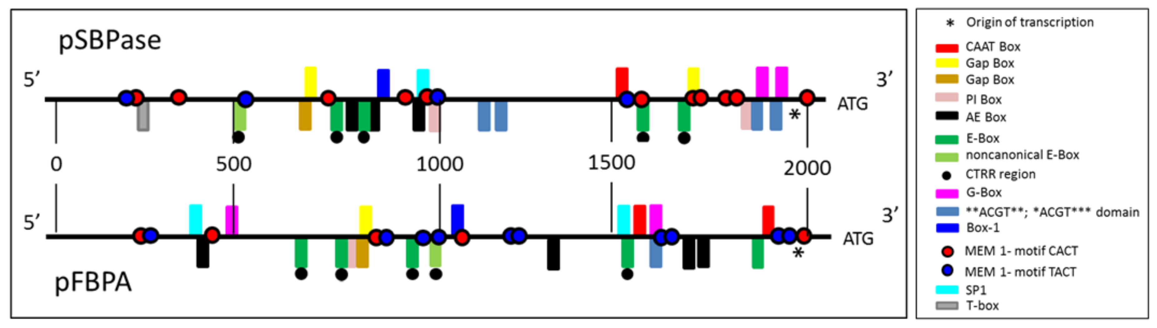
3.1. Identification and Analysis of Brachypodium distachyon SBPase and FBPA Promoters

To identify the promoter sequences from the B. distachyon SBPase (Bradi2g55150) and FBPA (Bradi4g24367) genes, the phytozome database (https://phytozome.jgi.doe.gov/pz/portal.html) was searched. A two-kilobase region upstream of the start codon (ATG) of both genes was isolated (Figure 1 and Supplementary Figures S4 and S5). The isolated promoter sequences were analyzed using PlantCare software (http://bioinformatics.psb.ugent.be/webtools/plantcare/html/) to identify and characterize the regulatory elements present in the promoters and these elements were compared with published material. Both promoters also contain a number of other cis-acting elements previously shown to be involved in light responsiveness (Figure 1 and Table 1).

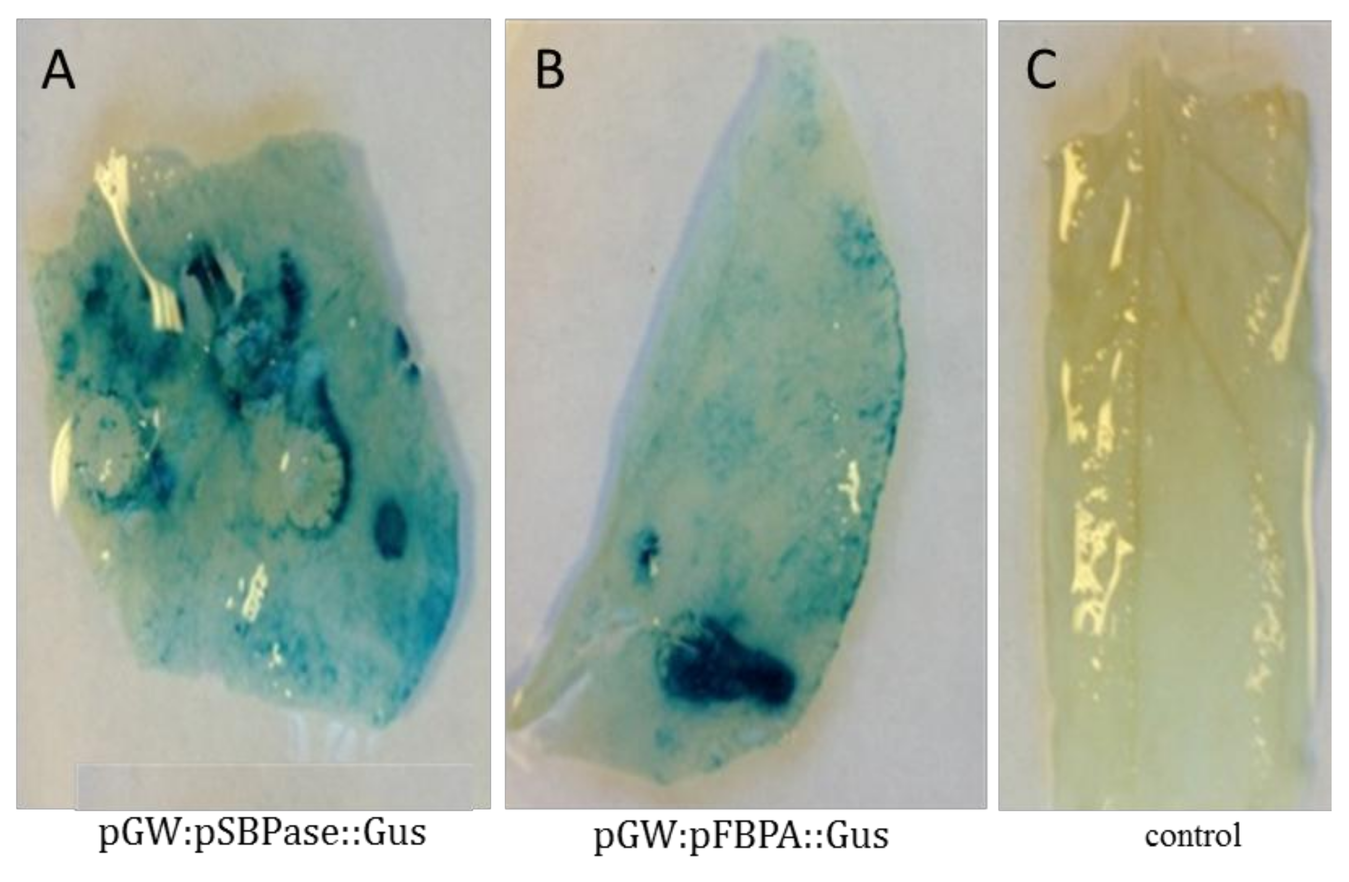
3.2. Agrobacterium-Mediated Transient Expression Analysis in Nicotiana benthamiana Leaves

To test the functionality of the SBPase and FBPA promoters in our chimearic gene constructs, the N. benthamiana transient assay system was used. Our results showed that both of these promoters were able to drive expression of the β-glucuronidase gene (gus) in young leaves of N. benthamiana using the transcriptional fusion constructs pGW:pSBPase::GUS and pGW:pFBPA::GUS (Supporting Figure S1). Leaves of Agrobacterium-infiltrated plants were subjected to GUS staining and a clear, strong blue product was observed in the infiltrated leaves, which indicated gus expression driven by the SBPase promoter (Figure 2A) and FBPA promoter (Figure 2B). In comparison, no GUS coloration was observed in WT control leaves (Figure 2C).

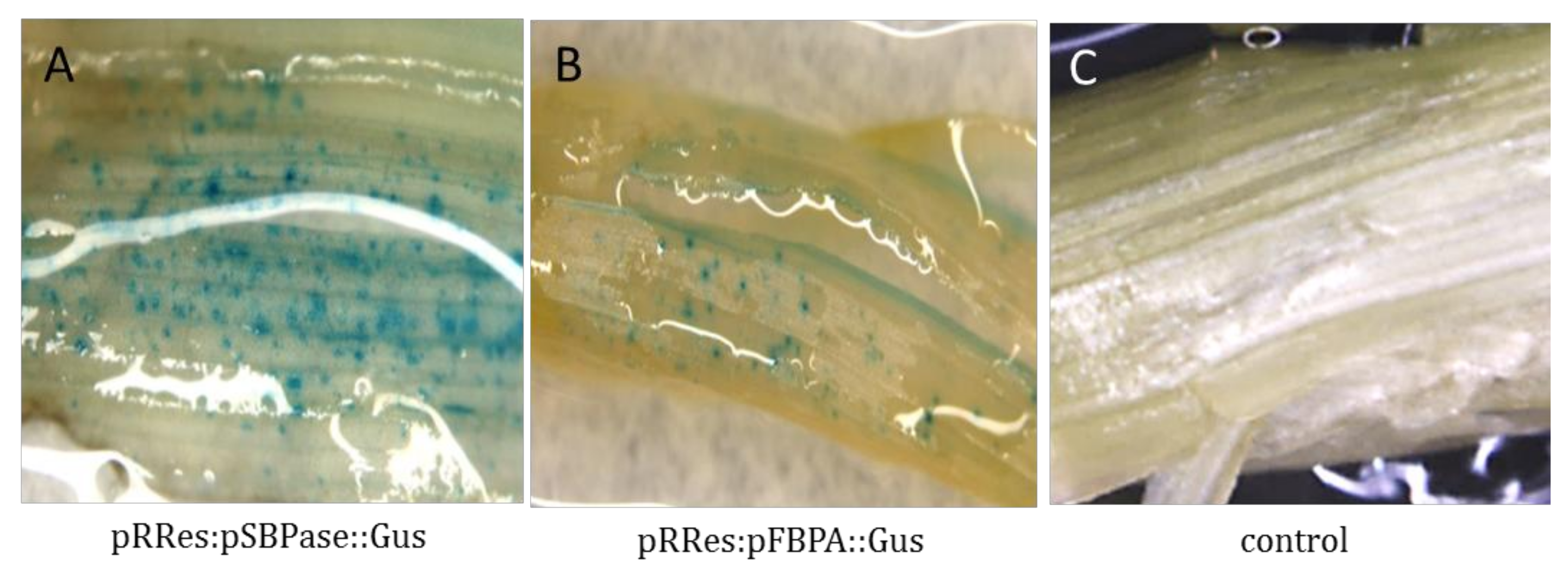
3.3. Transient Expression Analysis in Wheat

As a precursor to stable transformation in wheat, transient expression studies were carried out to test the functionality of both promoters to drive gus expression in wheat leaves using the constructs pRRes:pSBPase::GUS and pRRes:pFBPA::GUS (Supplementary Figure S2). Both promoters were shown to drive gus expression in wheat leaves. Many blue spots or patches were observed signifying that the gus transcript is being generated and translated into a functional protein (Figure 3).

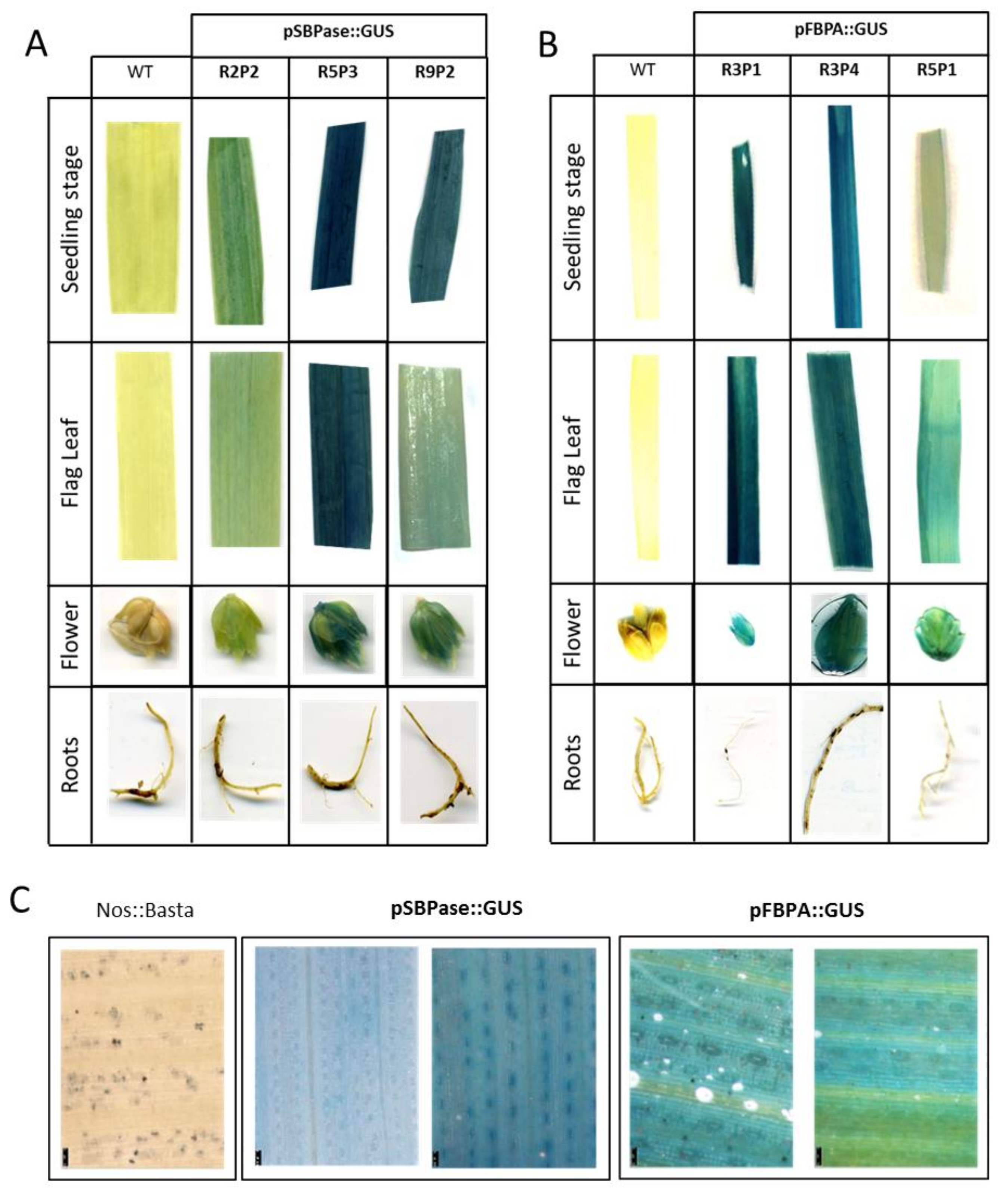
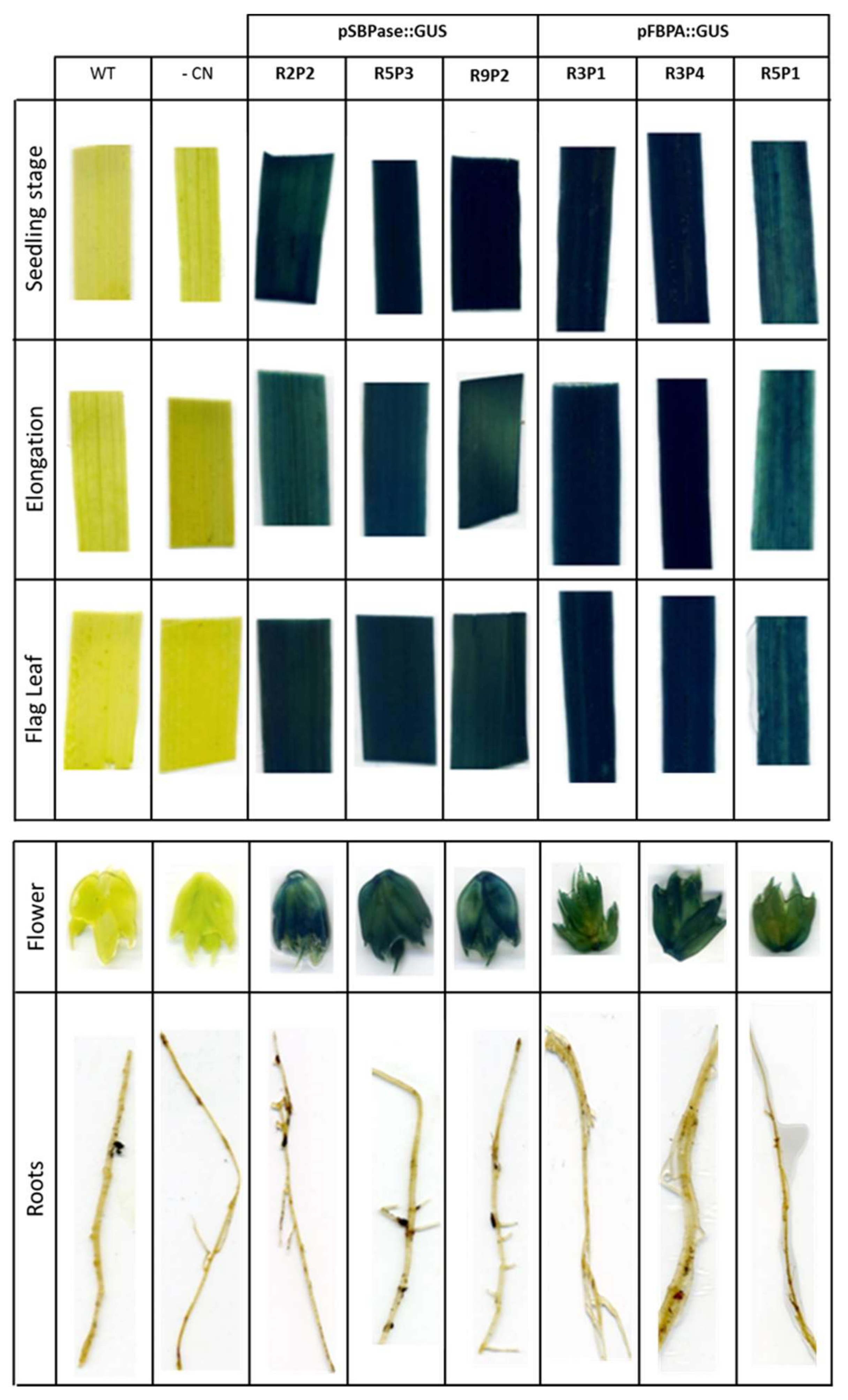
3.4. Stable Expression Analysis in Wheat

To study gus expression driven by the B. distachyon SBPase and FBPA promoters at different stages of wheat development and to confirm the suitability of these promoters for expressing transgenes in wheat, stably transformed lines were generated by particle bombardment of wheat scutella using constructs pRRes:pSBPase::GUS and pRRes:pFBPA::GUS.
Samples were taken for GUS staining from a range of tissues from the T1 transgenic plants, including leaves (at two different growth stages: seedlings and flag leaves), roots and flowers. Three independent SBPase promoter transgenic lines (R2P2, R5P3 and R9P2; Figure 4A) and three independent FBPA promoter lines (R3P1, R3P4 and R5P1; Figure 4B), were analyzed and intense GUS staining detected in leaves and flowers, but, as would have been expected no expression was seen in the roots. GUS stained leaves of two positive lines expressing either the pRRes:SBPase::GUS or pRRes:FBPA::GUS gene constructs were subject to microscopic investigation. GUS staining was evident in both mesophyll and guard cells, but not in the midrib or veins of these plants (Figure 4C).
To confirm the stability and suitability of the B. distachyon SBPase and FBPA promoters to drive the expression of gus in the next generation, GUS coloration was analyzed in T2 progeny arising from the lines studied above (Figure 5). Histochemical staining of leaves at three different growth stages (seedling, elongation and flag leaves), flowers and root tissues was carried out. GUS staining showed strong positive blue coloration in all three growth stages of leaves and in flowers of wheat plants, driven by either the B. distachyon SBPase promoter or FBPA promoter (Figure 5). Once again, no histochemical coloration of root tissue was observed with either promoter.

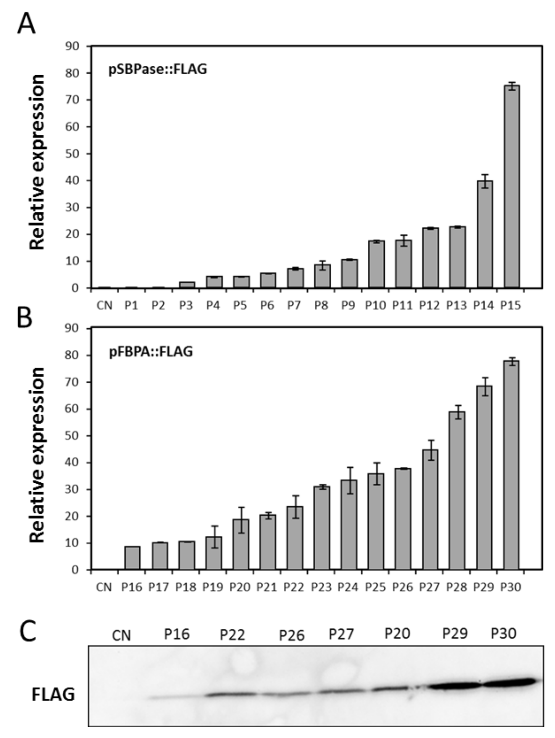
3.5. Evaluation of the SBPase or FBPA Promoters to Drive Expression of an Introduced Coding Sequence from Algae

To determine the range of expression levels that can be obtained using these promoters in stably transformed wheat, the SBPase or FBPA upstream sequences were fused to the coding sequence of a cytochrome c6-FLAG reporter (Supplementary Figure S3). The cytochrome c6-FLAG tagged construct was used to demonstrate successful expression of a potentially useful transgene other than gus in planta. Thirty confirmed primary transformed plants were analyzed by qPCR and a range of transcript levels obtained. Transcript levels were compared to the expression of the endogenous wheat SBPase gene. The SBPase promoter::cytochrome c6-FLAG transcript levels ranged from 0% to 76% of SBPase (Figure 6A) and the FBPA promoter::cytochrome c6-FLAG transcript levels ranged from 9% to 77% of SBPase (Figure 6B) in the first 30 transformed lines. Immunoblot analysis of seven selected FBPA promoter::CFLAG lines demonstrated that transcript levels also leads to an accumulation of protein (Figure 6C).

4. Discussion

In this study we have identified and tested two promoters from B. distachyon in wheat and shown that they can be used to drive the expression of the gus gene in mesophyll tissue and that expression is stable through subsequent generations. High levels of expression of introduced transgenes in wheat leaves have been difficult to achieve. Attempts to use dicot promoters, those from rice and even from wheat itself have often not been successful, with the promoters either failing or being ineffectual (author’s unpublished results). The work here has demonstrated that B. distachyon promoter sequences are functional in wheat, identifying a source of alternative promoters to those currently in use in wheat transformation, offering tissue-specificity to drive selected transgenes. We have also shown that N. benthamiana can be used to test promoters from B. distachyon prior to introduction into wheat. This is consistent with recent results which have shown that the rubisco small subunit gene promoter from wheat was functional in tobacco [39]. In plants expressing the FLAG-tagged cytochrome c6 protein under the control of either the SBPase or FBPA promoter, a range of transcript levels were seen between the individual independent primary transformants. This result is not unexpected and has been attributed to chromosome position, copy number or the presence of repeat sequences [60,61,62] and is known as “the position effect” [60,63,64,65]. The expression levels of this introduced gene can be as high as 80% of that of the endogenous SBPase transcript. The latter point is important as although GUS staining provides a convenient visual result for gene expression and tissue distribution, it did not provide information on the levels of expression relative to an endogenous gene. Although gus transcript levels could have been analyzed, it was important to demonstrate that these promoters could drive the expression of additional relevant transgenes and drive both transcript and protein accumulation.
In this study we isolated a two-kilobase genomic region, directly upstream of the initiating ATG for the B. distachyon SBPase and FBPA genes, with the view that this region would contain both the core promoter elements (required for basal expression) and the cis-acting regulatory elements found in the extended region upstream of the core. Comparison of these two-kilobase genomic sequences to published literature allowed the identification of a number of DNA regulatory sequences. The SBPase and FBPA promoters were found to contain putative TATA and CAAT box sequences which, although not found in all plant genes, when present form part of the core region of the promoter. In the SBPase gene a motif (TCTATCTTCT) located 41 bp upstream of the transcription initiation site (indicated by the asterisk Figure 1) is a probable candidate for the TATA-box sequence [66]. In the FBPA promoter two candidate TATA-box sequences (ATTCATATCC; TCATATCCTT) are found 61 bp and 26 bp upstream of the transcription initiation site respectively [66]. Both the SBPase and FBPA promoters identified in this study contain a putative CAAT-Box [67]. The CAAT-Box contains the sequence 5′-CCAAT-3/5′-CAAAT-3 at its core and is a cis-acting regulatory element that potentially binds a number of different transcription factors. CAAT-Boxes are generally found upstream of the TATA box and although their position can be variable they are often reported to be 70–80 bp upstream (Table 1).
Directly relevant to this study it is interesting to note that both the SBPase and FBPA promoters also contain a number of YACT Mesophyll Expression Module (MEM) 1-motifs which have been shown to direct expression in mesophyll cells [68]. The function of the MEM motif was demonstrated in genes functioning in the C4 photosynthetic pathway which requires expression of genes to be separated spatially between bundle sheath and mesophyll cells. There is evidence that the gene sequences are also found in the C3 species but functional analysis in these plants remains to be confirmed [69].
There is very little information available on the regulation of expression of genes encoding enzymes of the Calvin–Benson cycle in wheat, with the exception of SBPase and FBPase [70,71]. However, there is a large literature demonstrating the role of light in regulating the expression of a number of Calvin–Benson cycle genes, across a range of species and this has led to the identification of a number light regulatory motifs. For example analysis of the GapB promoter (B subunit of chloroplast glyceraldehyde 3-phosphate dehydrogenase) from Arabidopsis thaliana in transgenic tobacco revealed that the ATGAA(G/A)A consensus sequence is necessary for light induced expression of the GapB gene [72]. The SBPase promoter contains two copies of these repeat sequences (ATGAAAA) and the FBPA promoter contains one similar motif (ATGAAGC). The GapA promoter (A subunit of chloroplast glyceraldehyde 3-phosphate dehydrogenase) contains 3 copies of a similar sequences CAAATGAA(G/A)A, denoted as the Gap Box; the deletion of just one of these copies results in a six-fold decrease in light induction and deletion of all three copies resulted in total loss of light induction [73]. Very similar sequences are also found in both the SBPase (AAAATGAAAA) and FBPA (CATATGAAGC) promoters isolated in this study. The Gap Box is not sufficient alone to confer light induction but it acts together with the Activation Element (AE) Box [74], with the consensus AGAATTCT sequence found in both the GapA and GapB promoters [75]. Here we have identified similar sequences in the SBPase (AGAACTCT; AGAATTCT; AGAAATAT) and FBPA (AGAAACAC; AGAAACAG; AGAATTGT; AGAATGTC) promoters (Table 1) indicating a similar regulatory mechanism might exist in monocot species.
Both the SBPase and FBPA promoters were shown to contain a number of elements involved in directing light responsive gene expression (G-Box; CACGTG [76,77,78,79,80,81,82]; CACGTA). The G-box has been found in the promoters of circadian-regulated genes in plants [83,84] and is essential for phytochrome-regulated induction of transcription [77,85]. The related E-box core element (CANNTG), which has a significant overlap with the G-Box, is important in mammalian circadian promoters [86,87]. Interestingly in plants, the E-box CANNTG is involved in seed-specific expression of the French bean phaseolin [88], the 2S storage protein of Douglas-fir [89] and endosperm expression in coffee [34,90] was present in the B. distachyon SBPase promoter. A number of experiments have shown that the E-Box is an integral part of the circadian clock’s transcription-translation feedback loop [91,92]. Furthermore, a CT-region (CTRR) located approximately 23 bp upstream of the E-Box is considered to be essential for E-Box binding, transactivation and transcription of circadian genes in mice [91,92]. Each of the four E-Boxes identified here in the SBPase promoter have an identifiable CTRR domain 21, 9, 12 and 7 bp upstream of the E-Box. Corresponding E-box sequences were also identified in the FBPA promoter and were found to be in similar positions to those identified in the SBPase promoter. The first, second, third and fourth of these E-Boxes also followed identifiable CTRR domain at positions 13, 21, 14 and 14 bp upstream respectively [91]. Both the SBPase and FBPA promoter contain a single non-canonical E-Box previously shown to be essential for circadian expression in skeletal muscle [93] (Table 1).
Regulatory cis-acting elements previously shown to be involved in light responsiveness were also identified in both promoters (**ACGT** and *ACGT***). The motifs identified here are all similar to regulatory sequences previously identified in Arabidopsis [94]. These same sequences are also responsive to UV, drought and abscisic acid (ABA) and overlap with the G-Box motif CACGTC described in several plant promoters [65,66,67,68,69,70,71,72,73]. Other light responsive elements (Box-1 [95,96]) have also been identified in the SBPase and FBPA promoters as well as the light-responsive GAG-motif identified in the FBPA promoter [95]. Furthermore, sequences similar to the PI-box and T-Box identified in the GapB promoter [97] were present. The T-Box has also been identified in the geranylgeranyl pyrophosphate synthase promoter from Ginkgo biloba [98]. Mutations in the PI-Box and T-Box resulted in a reduction in light-activated gene transcription [97]. Furthermore, we also identified the SP1 motif in both the SBPase and FBPA promoters: SP1 is a light-responsive element previously identified in promoters from Zea mays [99] and G. biloba [98]. Although this analysis provides circumstantial evidence that the SBPase and FBPA promoters are regulated by light through a number of cis-acting motifs, further work is required to provide a functional demonstration that this aspect of the regulation is maintained in transgenic wheat.

5. Conclusions

Here we present clear evidence that the genomic sequences from the promoter and associated upstream regulatory regions of both the B. distachyon SBPase and FBPA genes are capable of driving expression of transgenes in wheat leaves. Importantly, we show successful expression of the GUS reporter gene in multiple independent lines in both the T1 and T2 generations of transgenic wheat. Moreover, our data indicate that the levels of transcript expression attained when the algal cytochrome c6 gene is introduced into transgenic wheat, under the control of either the SBPase or FBPA promoter, can be close to that of the endogenous SBPase gene. Recent research has demonstrated that manipulating photosynthesis using multigene approaches can significantly increase biomass and seed yields in Arabidopsis and tobacco. The results presented here provide strong evidence that the B. distachyon SBPase and FBPA promoter sequences can be used as tools for the production of transgenic wheat, including multigene manipulation, with the goal of improving yield to meet global demand.

Supplementary Materials

The following are available online at https://www.mdpi.com/2223-7747/7/2/27/s1. Figure S1. Schematic of transcriptional gene fusion construct of the B. distachyon SBPase and FBPA promoters with the β-glucuronidase (gus) reporter for expression analysis in N. benthamiana leaves. Genomic DNA of B. distachyon leaves was used to amplify the native promoters of SBPase and FBPA. They were cloned into the pENTR vector (Invitrogen). The resulting product was transferred into the (a) pGWB3 vector by LR recombination to make (b) pGW:pSBPase::GUS and (c) pGW:pFBPA::GUS, Figure S2. Schematic of transcriptional gene fusion constructs of the B. distachyon SBPase and FBPA promoters with the β-glucuronidase (gus) reporter for expression analysis in wheat leaves. Genomic DNA of B. distachyon leaves was used to amplify the native promoters of SBPase and FBPA and they were cloned into the corresponding restriction sites (pSBPase was cloned into the Mlul and AscI restriction sites and pFBPA was cloned into the EcoRI and XmaI restriction sites) of the (a) pRRes14.041::GUS vector to make (b) pRRes:pSBPase::GUS and (c) pRRES:pFBPA::GUS, Figure S3. Schematic of transcriptional gene fusion constructs of the B. distachyon SBPase and FBPA promoters and coding sequences fused to the cytochrome c6-FLAG sequence for expression analysis in wheat leaves. The cytochrome c6-FLAG-tagged protein was cloned into the NcoI and EcoRV restriction sites of vector pRRes:pSBPase::GUS and pRRes:pFBPA::GUS, simultaneously removing the intron and gus reporter gene to generate (a) pSBPase::FLAG and (b) pFBPA::FLAG respectively, Figure S4. Sequence and regulatory motifs in the 2 kb upstream region of the B. distachyon SBPase gene. The different coloured boxed sequences represent the promoter motifs (see Figure 1), Figure S5. Sequence and regulatory motifs in the 2 kb upstream region of the B. distachyon FBPA gene. The different coloured boxed sequences represent the promoter motifs (see Figure 1).

Acknowledgments

S.S.A. was funded by the Saudi Arabian Government and by the University of Essex Research Incentive Scheme to C.A.R. respectively. M.A.J.P. and C.A.R. were funded by the Biotechnology and Biological Sciences Research Council (BBSRC, grant BB/H01960X/1) awarded to C.A.R. and M.A.J.P., as part of the Crop Improvement Research Club (CIRC). A.J.S. was funded by Realizing Increased Photosynthetic Efficiency to Increase Wheat Yields (iWYP, BB/N021045/1) awarded to C.A.R. C.A.S. was funded by the Biotechnology and Biological Sciences Research Council (BBSRC) as part of the 20:20 wheat program. The full sequences of the SBPase (Gene ID: Bradi2g55150) and FBPA promoter (Gene ID: Bradi4g24367) are available from the Phytozome website (https://phytozome.jgi.doe.gov/pz/portal.html).

Author Contributions

S.S.A. performed T1 and T2 line selection, acquired, analyzed and interpreted data. A.J.S. acquired data on gene expression and drafted the manuscript. C.A.S. performed gene transformation and initial line selection (T0) and GUS staining. M.A.J.P. revised the manuscript. C.A.R. conceived the study, designed experiments and revised the manuscript. All authors approved the final version.

Conflicts of Interest

The authors declare no conflict of interest.

References

  1. Braun, H.J.; Atlin, G.; Payne, T. Multi-location testing as a tool to identify plant response to global climate change. In Climate Change and Crop Production; Climate Change Series; Reynolds, M.P., Ed.; CABI: Surrey, UK, 2010; pp. 115–138. [Google Scholar]
  2. Ray, D.K.; Mueller, N.D.; West, P.C.; Foley, J.A. Yield trends are insufficient to double global crop production by 2050. PLoS ONE 2013, 8, e66428. [Google Scholar] [CrossRef] [PubMed]
  3. Tilman, D.; Balzer, C.; Hill, J.; Befort, B.L. Global food demand and the sustainable intensification of agriculture. Proc. Natl. Acad. Sci. USA 2011, 108, 20260–20264. [Google Scholar] [CrossRef] [PubMed]
  4. Godfray, H.C.J. The challenge of feeding 9–10 billion people equitably and sustainably. J. Agric. Sci. 2014, 152, S2–S8. [Google Scholar] [CrossRef]
  5. RSOL. Royal Society of London, Reaping the Benefits: Science and the Sustainable Intensification of Global Agriculture; Royal Society: London, UK, 2009. [Google Scholar]
  6. Davies, W.J.; Ribaut, J.M. Stress resilience in crop plants: Strategic thinking to address local food production problems. Food Energy Secur. 2017, 6, 12–18. [Google Scholar] [CrossRef]
  7. Le Quere, C.; Raupach, M.R.; Canadell, J.G.; Marland, G.; Bopp, L.; Ciais, P.; Conway, T.J.; Doney, S.C.; Feely, R.A.; Foster, P.; et al. Trends in the sources and sinks of carbon dioxide. Nat. Geosci. 2009, 2, 831–836. [Google Scholar] [CrossRef]
  8. Solomon, S. Climate Change 2007—The Physical Science Basis: Working Group I Contribution to the Fourth Assessment Report of the IPCC; Cambridge University Press: Cambridge, UK, 2007; Volume 4. [Google Scholar]
  9. Driever, S.M.; Simkin, A.J.; Alotaibi, S.; Fisk, S.J.; Madgwick, P.J.; Sparks, C.A.; Jones, H.D.; Lawson, T.; Parry, M.A.J.; Raines, C.A. Increased SBPase activity improves photosynthesis and grain yield in wheat grown in greenhouse conditions. Philos. Trans. R. Soc. B 2017, 372, 1730. [Google Scholar] [CrossRef] [PubMed]
  10. Timm, S.; Florian, A.; Arrivault, S.; Stitt, M.; Fernie, A.R.; Bauwe, H. Glycine decarboxylase controls photosynthesis and plant growth. FEBS Lett. 2012, 586, 3692–3697. [Google Scholar] [CrossRef] [PubMed]
  11. Simkin, A.J.; McAusland, L.; Lawson, T.; Raines, C.A. Over-expression of the RieskeFeS protein increases electron transport rates and biomass yield. Plant Physiol. 2017, 175, 134–145. [Google Scholar] [CrossRef] [PubMed]
  12. Chida, H.; Nakazawa, A.; Akazaki, H.; Hirano, T.; Suruga, K.; Ogawa, M.; Satoh, T.; Kadokura, K.; Yamada, S.; Hakamata, W.; et al. Expression of the algal cytochrome c6 gene in Arabidopsis enhances photosynthesis and growth. Plant Cell Physiol. 2007, 48, 948–957. [Google Scholar] [CrossRef] [PubMed]
  13. Simkin, A.J.; Lopez-Calcagno, P.E.; Davey, P.A.; Headland, L.R.; Lawson, T.; Timm, S.; Bauwe, H.; Raines, C.A. Simultaneous stimulation of sedoheptulose 1,7-bisphosphatase, fructose 1,6-bisphophate aldolase and the photorespiratory glycine decarboxylase H-protein increases CO2 assimilation, vegetative biomass and seed yield in Arabidopsis. Plant Biotechnol. J. 2017, 15, 805–816. [Google Scholar] [CrossRef] [PubMed]
  14. Simkin, A.J.; McAusland, L.; Headland, L.R.; Lawson, T.; Raines, C.A. Multigene manipulation of photosynthetic carbon assimilation increases co2 fixation and biomass yield in tobacco. J. Exp. Bot. 2015, 66, 4075–4090. [Google Scholar] [CrossRef] [PubMed]
  15. Odell, J.T.; Nagy, F.; Chua, N.H. Identification of DNA sequences required for activity of the cauliflower mosaic virus 35S promoter. Nature 1985, 313, 810–812. [Google Scholar] [CrossRef] [PubMed]
  16. Exposito-Rodriguez, M.; Laissue, P.P.; Lopez-Calcagno, P.E.; Mullineaux, P.M.; Raines, C.A.; Simkin, A.J. Development of pGEMINI, a plant gateway destination vector allowing the simultaneous integration of two cDNA via a single LR-clonase reaction. Plants 2017, 6, 55. [Google Scholar] [CrossRef] [PubMed]
  17. Richins, R.D.; Scholthof, H.B.; Shepherd, R.J. Sequence of figwort mosaic-virus DNA (Caulimovirus group). Nucleic Acids Res. 1987, 15, 8451–8466. [Google Scholar] [CrossRef] [PubMed]
  18. Snowden, K.C.; Simkin, A.J.; Janssen, B.J.; Templeton, K.R.; Loucas, H.M.; Simons, J.L.; Karunairetnam, S.; Gleave, A.P.; Clark, D.G.; Klee, H.J. The decreased apical dominance1/petunia hybrida carotenoid cleavage dioxygenase8 gene affects branch production and plays a role in leaf senescence, root growth, and flower development. Plant Cell 2005, 17, 746–759. [Google Scholar] [CrossRef] [PubMed]
  19. Simkin, A.J.; Underwood, B.A.; Auldridge, M.; Loucas, H.M.; Shibuya, K.; Schmelz, E.; Clark, D.G.; Klee, H.J. Circadian regulation of the phccd1 carotenoid cleavage dioxygenase controls emission of beta-ionone, a fragrance volatile of petunia flowers. Plant Physiol. 2004, 136, 3504–3514. [Google Scholar] [CrossRef] [PubMed]
  20. De Borne Dorlhac, F.; Vincentz, M.; Chupeau, Y.; Vaucheret, H. Co-suppression of nitrate reductase host genes and transgenes in transgenic tobacco plants. Mol. Gen. Genet. 1994, 243, 613–621. [Google Scholar] [CrossRef]
  21. Palauqui, J.C.; Elmayan, T.; De Borne, F.D.; Crete, P.; Charles, C.; Vaucheret, H. Frequencies, timing, and spatial patterns of co-suppression of nitrate reductase and nitrite reductase in transgenic tobacco plants. Plant Physiol. 1996, 112, 1447–1456. [Google Scholar] [CrossRef] [PubMed]
  22. Dupree, P.; Pwee, K.H.; Gray, J.C. Expression of photosynthesis gene-promoter fusions in leaf epidermal-cells of transgenic tobacco plants. Plant J. 1991, 1, 115–120. [Google Scholar] [CrossRef]
  23. Xu, M.; Bernat, G.; Singh, A.; Mi, H.; Rogner, M.; Pakrasi, H.B.; Ogawa, T. Properties of mutants of Synechocystis sp. Strain PCC 6803 lacking inorganic carbon sequestration systems. Plant Cell Physiol. 2008, 49, 1672–1677. [Google Scholar] [CrossRef] [PubMed]
  24. Kuntz, M.; Chen, H.C.; Simkin, A.J.; Romer, S.; Shipton, C.A.; Drake, R.; Schuch, W.; Bramley, P.M. Upregulation of two ripening-related genes from a nonclimacteric plant (pepper) in a transgenic climacteric plant (tomato). Plant J. 1998, 13, 351–361. [Google Scholar] [CrossRef]
  25. Manac’h, N.; Kuntz, M. Stress induction of a nuclear gene encoding for a plastid protein is mediated by photo-oxidative events. Plant Physiol. Biochem. 1999, 37, 859–868. [Google Scholar] [CrossRef]
  26. Simkin, A.J.; Gaffe, J.; Alcaraz, J.P.; Carde, J.P.; Bramley, P.M.; Fraser, P.D.; Kuntz, M. Fibrillin influence on plastid ultrastructure and pigment content in tomato fruit. Phytochemistry 2007, 68, 1545–1556. [Google Scholar] [CrossRef] [PubMed]
  27. Muller-Rober, B.; Ellenberg, J.; Provart, N.; Willmitzer, L.; Busch, H.; Becker, D.; Dietrich, P.; Hoth, S.; Hedrich, R. Cloning and electrophysiological analysis of kst1, an inward rectifying k+ channel expressed in potato guard cells. EMBO J. 1995, 14, 2409–2416. [Google Scholar] [PubMed]
  28. Kelly, G.; Moshelion, M.; David-Schwartz, R.; Halperin, O.; Wallach, R.; Attia, Z.; Belausov, E.; Granot, D. Hexokinase mediates stomatal closure. Plant J. 2013, 75, 977–988. [Google Scholar] [CrossRef] [PubMed]
  29. Lawson, T.; Simkin, A.J.; Kelly, G.; Granot, D. Mesophyll photosynthesis and guard cell metabolism impacts on stomatal behaviour. New Phytol. 2014, 203, 1064–1081. [Google Scholar] [CrossRef] [PubMed]
  30. Baumlein, H.; Nagy, I.; Villarroel, R.; Inze, D.; Wobus, U. Cis-analysis of a seed protein gene promoter—The conservative RY repeat CATGCATG within the legumin box is essential for tissue-specific expression of a legumin gene. Plant J. 1992, 2, 233–239. [Google Scholar] [PubMed]
  31. Thilmony, R.; Guttman, M.E.; Lin, J.W.; Blechl, A.E. The wheat HMW-glutenin 1Dy10 gene promoter controls endosperm expression in Brachypodium distachyon. GM Crops Food 2014, 5, 36–43. [Google Scholar] [CrossRef] [PubMed]
  32. Song, F.; Cui, C.J.; Chen, L.; Sun, Y.L.; Wang, F.F.; Hussain, J.; Li, Y.; Wang, C.; Wang, C.; Chen, M.J.; et al. Isolation and characterization of an endosperm-specific promoter from wheat (Triticum aestivum L.). Z. Naturforsch. C 2012, 67, 611–619. [Google Scholar] [CrossRef] [PubMed]
  33. Lamacchia, C.; Shewry, P.R.; Di Fonzo, N.; Forsyth, J.L.; Harris, N.; Lazzeri, P.A.; Napier, J.A.; Halford, N.G.; Barcelo, P. Endosperm-specific activity of a storage protein gene promoter in transgenic wheat seed. J. Exp. Bot. 2001, 52, 243–250. [Google Scholar] [CrossRef] [PubMed]
  34. Simkin, A.J.; McCarthy, J.; Petiard, V.; Tanksley, S.; Lin, C. Oleosin Genes and Promoters from Coffee. U.S. Patent WO2007005928A2, 11 January 2007. [Google Scholar]
  35. Christensen, A.H.; Sharrock, R.A.; Quail, P.H. Maize polyubiquitin genes—Structure, thermal perturbation of expression and transcript splicing, and promoter activity following transfer to protoplasts by electroporation. Plant Mol. Biol. 1992, 18, 675–689. [Google Scholar] [CrossRef] [PubMed]
  36. Mcelroy, D.; Zhang, W.G.; Cao, J.; Wu, R. Isolation of an efficient actin promoter for use in rice transformation. Plant Cell 1990, 2, 163–171. [Google Scholar] [CrossRef] [PubMed]
  37. Mathur, S.; Dasgupta, I. Downstream promoter sequence of an Indian isolate of Rice tungro bacilliform virus alters tissue-specific expression in host rice and acts differentially in heterologous system. Plant Mol. Biol. 2007, 65, 259–275. [Google Scholar] [CrossRef] [PubMed]
  38. Bhattacharyyapakrasi, M.; Peng, J.Y.; Elmer, J.S.; Laco, G.; Shen, P.; Kaniewska, M.B.; Kononowicz, H.; Wen, F.J.; Hodges, T.K.; Beachy, R.N. Specificity of a promoter from the Rice tungro bacilliform virus for expression in phloem tissues. Plant J. 1993, 4, 71–79. [Google Scholar] [CrossRef]
  39. Mukherjee, S.; Stasolla, C.; Brule-Babel, A.; Ayele, B.T. Isolation and characterization of rubisco small subunit gene promoter from common wheat (Triticum aestivum L.). Plant Signal. Behav. 2015, 10, e989033. [Google Scholar] [CrossRef] [PubMed]
  40. Sparks, C.A.; Castleden, C.K.; West, J.; Habash, D.Z.; Madgwick, P.J.; Paul, M.J.; Noctor, G.; Harrison, J.; Wu, R.; Wilkinson, J.; et al. Potential for manipulating carbon metabolism in wheat. Ann. Appl. Biol. 2001, 138, 33–45. [Google Scholar] [CrossRef]
  41. Peremarti, A.; Twyman, R.M.; Gomez-Galera, S.; Naqvi, S.; Farre, G.; Sabalza, M.; Miralpeix, B.; Dashevskaya, S.; Yuan, D.; Ramessar, K.; et al. Promoter diversity in multigene transformation. Plant Mol. Biol. 2010, 73, 363–378. [Google Scholar] [CrossRef] [PubMed]
  42. Uematsu, K.; Suzuki, N.; Iwamae, T.; Inui, M.; Yukawa, H. Increased fructose 1,6-bisphosphate aldolase in plastids enhances growth and photosynthesis of tobacco plants. J. Exp. Bot. 2012, 63, 3001–3009. [Google Scholar] [CrossRef] [PubMed]
  43. Lefebvre, S.; Lawson, T.; Zakhleniuk, O.V.; Lloyd, J.C.; Raines, C.A.; Fryer, M. Increased sedoheptulose-1,7-bisphosphatase activity in transgenic tobacco plants stimulates photosynthesis and growth from an early stage in development. Plant Physiol. 2005, 138, 451–460. [Google Scholar] [CrossRef] [PubMed]
  44. The International Brachypodium Initiative. Genome sequencing and analysis of the model grass Brachypodium distachyon. Nature 2010, 463, 763–768. Available online: https://www.nature.com/articles/nature08747.pdf (accessed on 1 March 2018).
  45. Draper, J.; Mur, L.A.J.; Jenkins, G.; Ghosh-Biswas, G.C.; Bablak, P.; Hasterok, R.; Routledge, A.P.M. Brachypodium distachyon. A new model system for functional genomics in grasses. Plant Physiol. 2001, 127, 1539–1555. [Google Scholar] [CrossRef] [PubMed]
  46. Nakagawa, T.; Kurose, T.; Hino, T.; Tanaka, K.; Kawamukai, M.; Niwa, Y.; Toyooka, K.; Matsuoka, K.; Jinbo, T.; Kimura, T. Development of series of gateway binary vectors, pGWBs, for realizing efficient construction of fusion genes for plant transformation. J. Biosci. Bioeng. 2007, 104, 34–41. [Google Scholar] [CrossRef] [PubMed]
  47. Goodin, M.M.; Zaitlin, D.; Naidu, R.A.; Lommel, S.A. Nicotiana benthamiana: Its history and future as a model for plant–pathogen interactions. Mol. Plant Microbe Interact. 2008, 21, 1015–1026. [Google Scholar] [CrossRef] [PubMed]
  48. Lakatos, L.; Szittya, G.; Silhavy, D.; Burgyan, J. Molecular mechanism of RNA silencing suppression mediated by p19 protein of tombusviruses. EMBO J. 2004, 23, 876–884. [Google Scholar] [CrossRef] [PubMed]
  49. Kapila, J.; De Rycke, R.; van Montagui, M.; Angenon, G. An agrobacterium-mediated transient gene expression system for intact leaves. Plant Sci. 1997, 122, 101–108. [Google Scholar] [CrossRef]
  50. Wydro, M.; Kozubek, E.; Lehmann, P. Optimization of transient agrobacterium-mediated gene expression system in leaves of Nicotiana benthamiana. Acta Biochim. Pol. 2006, 53, 289–298. [Google Scholar] [PubMed]
  51. Jones, H.D.; Doherty, A.; Sparks, C.A. Transient transformation of plants. Methods Mol. Biol. 2009, 513, 131–152. [Google Scholar] [PubMed]
  52. Rasco-Gaunt, S.; Barcelo, P. Immature inflorescence culture of cereals. A highly responsive system for regeneration and transformation. Methods Mol. Biol. 1999, 111, 71–81. [Google Scholar] [PubMed]
  53. Sparks, C.A.; Jones, H.D. Genetic transformation of wheat via particle bombardment. Methods Mol. Biol. 2014, 1099, 201–218. [Google Scholar] [PubMed]
  54. Jefferson, R.A.; Kavanagh, T.A.; Bevan, M.W. GUS fusions—Beta-glucuronidase as a sensitive and versatile gene fusion marker in higher-plants. EMBO J. 1987, 6, 3901–3907. [Google Scholar] [PubMed]
  55. Christensen, A.H.; Quail, P.H. Ubiquitin promoter-based vectors for high-level expression of selectable and/or screenable marker genes in monocotyledonous plants. Transgenic Res. 1996, 5, 213–218. [Google Scholar] [CrossRef]
  56. Lopez-Calcagno, P.E.; Abuzaid, A.O.; Lawson, T.; Raines, C.A. Arabidopsis CP12 mutants have reduced levels of phosphoribulokinase and impaired function of the Calvin-Benson cycle. J. Exp. Bot. 2017, 68, 2285–2298. [Google Scholar] [CrossRef] [PubMed]
  57. Pfaffl, M.W. A new mathematical model for relative quantification in real-time RT-PCR. Nucleic Acids Res. 2001, 29, e45. [Google Scholar] [CrossRef] [PubMed]
  58. Harrison, E.P.; Willingham, N.M.; Lloyd, J.C.; Raines, C.A. Reduced sedoheptulose-1,7-bisphosphatase levels in transgenic tobacco lead to decreased photosynthetic capacity and altered carbohydrate accumulation. Planta 1998, 204, 27–36. [Google Scholar] [CrossRef]
  59. Willingham, N.M.; Lloyd, J.C.; Raines, C.A. Molecular cloning of the Arabidopsis thaliana sedoheptulose-1,7-biphosphatase gene and expression studies in wheat and Arabidopsis thaliana. Plant Mol. Biol. 1994, 26, 1191–1200. [Google Scholar] [CrossRef] [PubMed]
  60. Matzke, A.J.; Matzke, M.A. Position effects and epigenetic silencing of plant transgenes. Curr. Opin. Plant Biol. 1998, 1, 142–148. [Google Scholar] [CrossRef]
  61. Kooter, J.M.; Matzke, M.A.; Meyer, P. Listening to the silent genes: Transgene silencing, gene regulation and pathogen control. Trends Plant Sci. 1999, 4, 340–347. [Google Scholar] [CrossRef]
  62. Selker, E.U. Gene silencing: Repeats that count. Cell 1999, 97, 157–160. [Google Scholar] [CrossRef]
  63. Peach, C.; Velten, J. Transgene expression variability (position effect) of CAT and GUS reporter genes driven by linked divergent T-DNA promoters. Plant Mol. Biol. 1991, 17, 49–60. [Google Scholar] [CrossRef] [PubMed]
  64. Bhullar, S.; Chakravarthy, S.; Pental, D.; Burma, P.K. Analysis of promoter activity in transgenic plants by normalizing expression with a reference gene: Anomalies due to the influence of the test promoter on the reference promoter. J. Biosci. 2009, 34, 953–962. [Google Scholar] [CrossRef] [PubMed]
  65. Iglesias, V.A.; Moscone, E.A.; Papp, I.; Michalowski, S.; Phelan, T.; Spiker, S.; Neuhuber, F.; Matzke, M.; Matzke, A.J.M. Molecular and cytogenetic analysis of stably and unstably expressed transgene loci in tobacco. Plant Cell 1997, 9, 1251–1264. [Google Scholar] [CrossRef] [PubMed]
  66. Molina, C.; Grotewold, E. Genome wide analysis of Arabidopsis core promoters. BMC Genom. 2005, 6, 25. [Google Scholar] [CrossRef] [PubMed] [Green Version]
  67. Kusnetsov, V.; Landsberger, M.; Meurer, J.; Oelmuller, R. The assembly of the CAAT-box binding complex at a photosynthesis gene promoter is regulated by light, cytokinin, and the stage of the plastids. J. Biol. Chem. 1999, 274, 36009–36014. [Google Scholar] [CrossRef] [PubMed]
  68. Akyildiz, M.; Gowik, U.; Engelmann, S.; Koczor, M.; Streubel, M.; Westhoff, P. Evolution and function of a cis-regulatory module for mesophyll-specific gene expression in the C4 dicot Flaveria trinervia. Plant Cell 2007, 19, 3391–3402. [Google Scholar] [CrossRef] [PubMed]
  69. Gowik, U.; Schulze, S.; Saladie, M.; Rolland, V.; Tanz, S.K.; Westhoff, P.; Ludwig, M. A mem1-like motif directs mesophyll cell-specific expression of the gene encoding the C4 carbonic anhydrase in Flaveria. J. Exp. Bot. 2017, 68, 311–320. [Google Scholar] [CrossRef] [PubMed]
  70. Lloyd, J.C.; Raines, C.; John, U.P.; Dyer, T.A.A. The chloroplast FBPase gene of wheat: Structure and expression of the promoter in photosynthetic and meristematic cells of transgenic tobacco plants. Mol. Gen. Genet. 1991, 225, 209–216. [Google Scholar] [CrossRef] [PubMed]
  71. Raines, C.; Llloyd, J.C.; Willingham, N.M.; Potts, S.; Dyer, T.A.A. cDNA and gene sequences of wheat chloroplast sedoheptulose-1,7-bisphosphatase reveal homology with fructose-1,6-bisphosphatases. FEBS J. 1992, 205, 1053–1059. [Google Scholar] [CrossRef]
  72. Kwon, H.B.; Park, S.C.; Peng, H.P.; Goodman, H.M.; Dewdney, J.; Shih, M.C. Identification of a light-responsive region of the nuclear gene encoding the b subunit of chloroplast glyceraldehyde 3-phosphate dehydrogenase from Arabidopsis thaliana. Plant Physiol. 1994, 105, 357–367. [Google Scholar] [CrossRef] [PubMed]
  73. Conley, T.R.; Park, S.C.; Kwon, H.B.; Peng, H.P.; Shih, M.C. Characterization of cis-acting elements in light regulation of the nuclear gene encoding the A subunit of chloroplast isozymes of glyceraldehyde-3-phosphate dehydrogenase from Arabidopsis thaliana. Mol. Cell. Biol. 1994, 14, 2525–2533. [Google Scholar] [CrossRef] [PubMed]
  74. Park, S.C.; Kwon, H.B.; Shih, M.C. Cis-acting elements essential for light regulation of the nuclear gene encoding the A subunit of chloroplast glyceraldehyde 3-phosphate dehydrogenase in Arabidopsis thaliana. Plant Physiol. 1996, 112, 1563–1571. [Google Scholar] [CrossRef] [PubMed]
  75. Roy, S.; Choudhury, S.R.; Singh, S.K.; Das, K.P. Functional analysis of light-regulated promoter region of AtPolλ gene. Planta 2012, 235, 411–432. [Google Scholar] [CrossRef] [PubMed]
  76. Baum, K.; Wienand, U.; Meier, I. Reduction of G-box binding factor DNA binding activity, but not g-box binding factor abundance, causes the downregulation of RBCS2 expression during early tomato fruit development. FEBS Lett. 1999, 454, 95–99. [Google Scholar] [CrossRef]
  77. Menkens, A.E.; Schindler, U.; Cashmore, A.R. The G-box: A ubiquitous regulatory DNA element in plants bound by the GBF family of bZIP proteins. Trends Biochem. Sci. 1995, 20, 506–510. [Google Scholar] [CrossRef]
  78. Williams, M.E.; Foster, R.; Chua, N.H. Sequences flanking the hexameric G-box core CACGTG affect the specificity of protein binding. Plant Cell 1992, 4, 485–496. [Google Scholar] [CrossRef] [PubMed]
  79. Ishige, F.; Takaichi, M.; Foster, R.; Chua, N.H.; Oeda, K. A G-box motif (GCCACGTGCC) tetramer confers high-level constitutive expression in dicot and monocot plants. Plant J. 1999, 18, 443–448. [Google Scholar] [CrossRef]
  80. De Boer, K.; Tilleman, S.; Pauwels, L.; Vanden Bossche, R.; De Sutter, V.; Vanderhaeghen, R.; Hilson, P.; Hamill, J.D.; Goossens, A. APETALA2/ETHYLENE RESPONSE FACTOR and basic helix-loop-helix tobacco transcription factors cooperatively mediate jasmonate-elicited nicotine biosynthesis. Plant J. Cell Mol. Biol. 2011, 66, 1053–1065. [Google Scholar] [CrossRef] [PubMed]
  81. Toledo-Ortiz, G.; Huq, E.; Quail, P.H. The Arabidopsis basic/helix-loop-helix transcription factor family. Plant Cell 2003, 15, 1749–1770. [Google Scholar] [CrossRef] [PubMed]
  82. Ezer, D.; Shepherd, S.J.K.; Brestovitsky, A.; Dickinson, P.; Cortijo, S.; Charoensawan, V.; Box, M.S.; Biswas, S.; Jaeger, K.E.; Wigge, P.A. The g-box transcriptional regulatory code in Arabidopsis. Plant Physiol. 2017, 175, 628–640. [Google Scholar] [CrossRef] [PubMed]
  83. Michael, T.P.; McClung, C.R. Enhancer trapping reveals widespread circadian clock transcriptional control in Arabidopsis. Plant Physiol. 2003, 132, 629–639. [Google Scholar] [CrossRef] [PubMed]
  84. Hudson, M.E.; Quail, P.H. Identification of promoter motifs involved in the network of phytochrome a-regulated gene expression by combined analysis of genomic sequence and microarray data. Plant Physiol. 2003, 133, 1605–1616. [Google Scholar] [CrossRef] [PubMed]
  85. Martinez-Garcia, J.F.; Huq, E.; Quail, P.H. Direct targeting of light signals to a promoter element-bound transcription factor. Science 2000, 288, 859–863. [Google Scholar] [PubMed]
  86. Darlington, T.K.; Wager-Smith, K.; Ceriani, M.F.; Staknis, D.; Gekakis, N.; Steeves, T.D.; Weitz, C.J.; Takahashi, J.S.; Kay, S.A. Closing the circadian loop: CLOCK-induced transcription of its own inhibitors per and tim. Science 1998, 280, 1599–1603. [Google Scholar] [CrossRef] [PubMed]
  87. Gekakis, N.; Staknis, D.; Nguyen, H.B.; Davis, F.C.; Wilsbacher, L.D.; King, D.P.; Takahashi, J.S.; Weitz, C.J. Role of the clock protein in the mammalian circadian mechanism. Science 1998, 280, 1564–1569. [Google Scholar] [CrossRef] [PubMed]
  88. Kawagoe, Y.; Murai, N. Four distinct nuclear proteins recognize in vitro the proximal promoter of the bean seed storage protein beta-phaseolin gene conferring spatial and temporal control. Plant J. 1992, 2, 927–936. [Google Scholar] [PubMed]
  89. Chatthai, M.; Forward, B.S.; Yevtushenko, D.; Stefanov, I.; Osuska, L.; Osusky, M.; Misra, S. 2S storage protein gene of Douglas-fir: Characterization and activity of promoter in transgenic tobacco seeds. Plant Physiol. Biochem. 2004, 42, 417–423. [Google Scholar] [CrossRef] [PubMed]
  90. Simkin, A.J.; Qian, T.Z.; Caillet, V.; Michoux, F.; Ben Amor, M.; Lin, C.W.; Tanksley, S.; McCarthy, J. Oleosin gene family of Coffea canephora: Quantitative expression analysis of five oleosin genes in developing and germinating coffee grain. J. Plant Physiol. 2006, 163, 691–708. [Google Scholar] [CrossRef] [PubMed]
  91. Munoz, E.; Brewer, M.; Baler, R. Modulation of BMAL/CLOCK/E-BOX complex activity by a CT-rich cis-acting element. Mol. Cell. Endocrinol. 2006, 252, 74–81. [Google Scholar] [CrossRef] [PubMed]
  92. Liu, Y.W.; Li, X.; Li, K.W.; Liu, H.T.; Lin, C.T. Multiple bHLH proteins form heterodimers to mediate cry2-dependent regulation of flowering-time in Arabidopsis. PLoS Genet. 2013, 9, e1003861. [Google Scholar] [CrossRef] [PubMed]
  93. Zhang, X.P.; Patel, S.P.; McCarthy, J.J.; Rabchevsky, A.G.; Goldhamer, D.J.; Esser, K.A. A non-canonical E-box within the MyoD core enhancer is necessary for circadian expression in skeletal muscle. Nucleic Acids Res. 2012, 40, 3419–3430. [Google Scholar] [CrossRef] [PubMed]
  94. Yamamoto, Y.Y.; Ichida, H.; Matsui, M.; Obokata, J.; Sakurai, T.; Satou, M.; Seki, M.; Shinozaki, K.; Abe, T. Identification of plant promoter constituents by analysis of local distribution of short sequences. BMC Genom. 2007, 8, 67. [Google Scholar] [CrossRef] [PubMed]
  95. Wang, L.K.; Li, L.S.; Xu, L.N.; Zhou, J.; Zhuang, H.H.; Gong, X.D.; Wang, M.X.; Sun, S.S.M.; Zhuge, Q. Isolation and functional analysis of the poplar RBcS gene promoter. Plant Mol. Biol. Rep. 2013, 31, 120–127. [Google Scholar] [CrossRef]
  96. Tao, Y.; Wang, F.T.; Jia, D.M.; Li, J.T.; Zhang, Y.M.; Jia, C.G.; Wang, D.P.; Pan, H.Y. Cloning and functional analysis of the promoter of a stress-inducible gene (ZmRXO1) in maize. Plant Mol. Biol. Rep. 2015, 33, 200–208. [Google Scholar] [CrossRef]
  97. Chan, C.S.; Guo, L.; Shih, M.C. Promoter analysis of the nuclear gene encoding the chloroplast glyceraldehyde-3-phosphate dehydrogenase b subunit of Arabidopsis thaliana. Plant Mol. Biol. 2001, 46, 131–141. [Google Scholar] [CrossRef] [PubMed]
  98. Xu, F.; Huang, X.H.; Li, L.L.; Deng, G.; Cheng, H.; Rong, X.F.; Li, J.B.; Cheng, S.Y. Molecular cloning and characterization of GbDXS and GbGGPPS gene promoters from Ginkgo biloba. Genet. Mol. Res. 2013, 12, 293–301. [Google Scholar] [CrossRef] [PubMed]
  99. Morishima, A. Identification of preferred binding sites of a light-inducible DNA-binding factor (MNF1) within 5′-upstream sequence of C4-type phosphoenolpyruvate carboxylase gene in maize. Plant Mol. Biol. 1998, 38, 633–646. [Google Scholar] [CrossRef] [PubMed]
  100. Plesch, G.; Ehrhardt, T.; Mueller-Roeber, B. Involvement of TAAAG elements suggests a role for Dof transcription factors in guard cell-specific gene expression. Plant J. Cell Mol. Boil. 2001, 28, 455–464. [Google Scholar] [CrossRef]
  101. Lam, E.; Benfey, P.N.; Gilmartin, P.M.; Fang, R.X.; Chua, N.H. Site-specific mutations alter in vitro factor binding and change promoter expression pattern in transgenic plants. Proc. Natl. Acad. Sci. USA 1989, 86, 7890–7894. [Google Scholar] [CrossRef] [PubMed]
Figure 1. Schematic representation of a two-kilobase upstream region of the sedoheptulose-1,7-bisphosphatase (SBPase) and fructose-1,6-bisphosphate (FBPA) genes from B. distachyon showing potential regulatory motifs. ATG denotes the codon initiating translation of the SBPase and FBPA proteins.
Plants 07 00027 g001
Figure 2. Histochemical GUS assay of agroinfiltrated N. benthamiana leaves. The transient expression assays were performed on four- to five-week old N. benthamiana leaves, incubated at 24 °C in 16 h/8 h light/dark for 72 h after infiltration prior to GUS staining: (A) gus expression driven by the SBPase promoter; (B) gus expression driven by the FBPA promoter; and (C) control (wild type: non-infiltrated leaf).
Plants 07 00027 g002
Figure 3. Histochemical GUS assay for transient expression of B. distachyon SBPase and FBPA promoters in wheat leaves. The transient assays were performed by particle bombardment of young wheat leaves which were incubated at 22 °C in 12 h/12 h light/dark for 48 h prior to GUS staining: (A) gus expression driven by the SBPase promoter; (B) gus expression driven by the FBPA promoter; (C) control leaf bombarded without DNA.
Plants 07 00027 g003
Figure 4. Histochemical analysis of GUS activity in T1 wheat lines stably transformed with the B. distachyon SBPase or FBPA promoter constructs. gus expression driven by (A) SBPase promoter and (B) FBPA promoter in tissue from young seedlings, from flag leaves of mature plants and in flowers and roots. WT = wild type infiltrated tissue is used as a control. (C) Microscopic observation of localization of gus expression in T1 wheat leaves of two independent lines for each construct compared to plants transformed with the bar (Nos:Basta) selectable marker gene construct only (pRRes1.111).
Plants 07 00027 g004
Figure 5. Histochemical analysis of GUS activity in T2 wheat lines stably transformed with the B. distachyon SBPase or FBPA promoter constructs. Leaves were taken for GUS staining at three different growth stages (seedling, elongation and flag leaf) together with samples from different tissues: flowers and roots. Three different lines per promoter were analyzed and compared to WT (wild type) and CN (control plants transformed with bar gene only).
Plants 07 00027 g005
Figure 6. Analysis of transcript expression of the codon optimized P. umbilicalis cytochrome c6 [12]) with a FLAG tag (CytC6-FLAG) gene driven by the B. distachyon (A) SBPase or (B) FBPA promoters in stably transformed wheat T0 lines. Relative expression levels and range of expression in these 30 independent transgenic plants compared to the expression of the endogenous SBPase in the same plants. Data are presented as a % of the endogenous SBPase expression and standard error of nine technical reps are shown. (C) FLAG-tagged protein accumulation in a selection of FBPA promoter wheat lines.
Plants 07 00027 g006
Table 1. Cis-acting elements analysis of 5′-upstream regulatory elements and their positions in the B. distachyon SBPase and FBPA promoters identified by PLANTCARE and literature search.
Name of the ElementSignal SequencepSBPase PositionpFBPA PositionFunctional DescriptionReference
Activation Element (AE)-boxAGAAAC(A/T)(A/T)790, 859, 995419, 1323, 1698, 1734The AE Box and Gap Box act together and are essential components of light responsive elements[74,75]
ACGT-Box**ACGT**
*ACGT***
1214, 1289, 1852, 19341584Responsive to light, UV, drought and ABA[88]
Box-1TTTCAAA
TTTGAAA
8761163Light responsive element[89,90]
CATT motifGCATTC
CCAAT
CAAAT
15541559
1892
Part of a light responsive element. Evolutionarily conserved in eukaryotic organisms, including fungi, plants, and mammals[67]
MEM1-motif
YACT
CACT
TACT
241, 340, 755, 949, 1004, 1314, 1319, 1586, 1724, 1729, 1751, 1756, 2070
229, 593, 1082, 1569
205, 424, 857, 1176, 1260, 1988
226, 881, 982, 1046, 1243, 1250, 1600, 1666 1951, 1959
CACT key component of the Mesophyll Expression Module (MEM) 1 found in the cis-regulatory element in the phosphoenolpyruvate carboxylase promoter of the C4 dicot Flaveria trinervia[68]
E-BoxCANNTG769, 848, 1587, 1703687, 775, 954, 1536, 1840An integral part of the circadian clock’s transcription–translation feedback loop[86,87]
Non-canonical E-BoxCAGCTT5151006An integral part of the circadian clock’s transcription–translation feedback loop[93]
GAG-MotifAGAGAGT 146Light responsive element identified in the rbcS promoter from Poplar[95]
Gap-BoxATGAA(G/A)A724, 1709830Identified in the GapB promoter. Deletion of these repeats abolished light induction completely[73]
Gap-BoxCAAATGAA(G/A)A721827GapA promoter contains three sequences. Deletion of just one results in a six-fold decrease in light induction[74]
G-BoxCACGTA
CACGTG
CACGTC
GTACGTG
TACGTG
CACATGG
CCACGTAA
1852, 19351585
598
1584
The G-box has been identified in the promoters of circadian-regulated genes in plants and is important for phytochrome-regulated transcriptional induction. Confers high-level constitutive expression in dicot and monocot plants[76,77,78,79,80,81,82,83,84]
TAAAGSTKST1TAAAG941, 954129Target site for trans-acting StDof1 protein controlling guard cell-specific gene expression; KST1 gene encodes a K+ influx channel of guard cells[100]
PI-BoxGTGATCAC
GTGATCAG
GTGATCAA
TTGATCAC
1036804
1847
Identified in the GapB promoter. Mutation resulted in a reduction in light-activated gene transcription[97]
T-BoxACTTTG242 Identified in the GapB promoter. Mutation results in a reduction in light-activated gene transcription[97,98]
SP1CC(G/A)CCC10131510Light responsive element identified in Zea mays[98,99]
TGACG-motifTGACG1288, 1742 Cis-acting regulatory element involved in the MeJA-responsiveness. Mutation of the motif in the 35S promoter causes a 50% drop in expression in tobacco leaves[101]

