
Ammonia Sensor Based on Co2+/SCN− Modified Core-Shell MCM-41 for Rapid Naked-Eye Colorimetric Detection
1
School of Chemistry and Chemical Engineering, Beijing Institute of Technology, Beijing 102488, China
2
Shandong Institute of Nonmetallic Materials, Jinan 250031, China
*
Author to whom correspondence should be addressed.
Chemosensors 2023, 11(6), 336; https://doi.org/10.3390/chemosensors11060336
Submission received: 8 May 2023
/
Revised: 1 June 2023
/
Accepted: 5 June 2023
/
Published: 6 June 2023
(This article belongs to the Section Nanostructures for Chemical Sensing)
Abstract
:Mesoporous silica materials have been widely used as gas adsorbents due to their excellent adsorption ability, large specific surface area, simple preparation process, and convenient functionalization. In this study, a core–shell mesoporous material MCM-41@SiO2 was synthesized and functionalized by CoCl2, and subsequently KSCN, to prepare an ammonia adsorbent. The adsorbent was proved to possess high surface area, good sphericity, uniform size, good dispersibility, and high adsorption capability following DLS, SEM, TEM, and a static adsorption study. Moreover, the successful functionalization and thermal stability were confirmed by FT-IR, XPS, and TGA. The material was then used to fabricate a glass tube sensor for the rapid naked-eye detection of ammonia gas. The sensor showed good performance in terms of sensing speed, selectivity, accuracy, and reusability. Within 5 s, NH3 could be detected by the discoloration, and a wide NH3 concentration from 20 to 1000 ppm could be detected. It also showed a good linear relationship between discoloration length and ammonia concentration. A reversible color change from blue to yellow indicated the presence of NH3, which was attributed to the formation and disassembly of ammonia-containing complex [Co(NH3)6(NCS)2]. With fast adsorption, naked-eye sensing properties, and good selectivity, the sensor holds good promise for indoor NH3 sensing and other more complicated situations.
1. Introduction
NH3, with its pungent odor, is a noxious gas that irritates skin, eyeball mucosa, and mucous membranes of respiratory organs after being inhaled, despite the fact that it is widely used in various fields, such as in fertilizers, refrigerant, plastics, and the pharmaceuticals industry [1,2,3,4,5]. Moreover, inhalation of excessive NH3 gas could even cause lung swelling and death. Therefore, NH3 is classified as a Grade IV (mild hazard) gas according to the China National Standard TJ36-79, and the maximum allowable concentration of ammonia in an indoor workshop is 39.53 ppm. Moreover, according to the American Conference of Governmental Industrial Hygienists, NH3 concentration in ambient air should be less than 25 ppm for 8 h work and 35 ppm for a short period of work.
Generally, NH3 concentration detection is vital in many fields, such as methanol production and urea production. Typical detection methods, such as titration (which is based on the discoloration of an acid-base indicator to indicate the endpoint) and Nessler spectrophotometry (which depends on the light absorbance measurement), are commonly used. These methods are simple and easy to operate but tedious and inaccurate, with an unsatisfied limit of detection (LOD) around 0.02 mg/L. With the development and popularization of chromatography technology, gas chromatography (GC) has been used in gas detection, through which in situ ammonia quantification was obtained within 5 min, and the LOD was about 150 ppb (v/v, converted to 110 ng/L in gas phase) [6,7,8]. For example, ammonia in cigarette and cigar smoke was successfully identified by GC [9]. Although the accuracy was improved and online monitoring was realized by using these methods, large and expensive instruments and serious operation training are required for GC detection, resulting in inconvenience, untimely data acquisition, and high detection costs. An NH3 detection sensor was also developed due to its fast, simple, and convenient detection, easy operation, and low pollution [10,11,12,13,14,15]. Kanaparthi and co-workers introduced a NH3 sensor by using a solvent-free frictional deposition of polymer method. The sensor detected NH3 in the concentration range of 200 ppb to 3.15 ppm, and the sensitivity was 20.5%/ppm [16]. Electrochemical sensors aiming to detect NOx, which can indirectly indicate the presence of NH3, have also been reported. A metal-oxide-semiconductor sensor was developed based on the Pt-supported catalytic conversion of NH3 into NOx [17]. Similarly, a conductometric room temperature NOx sensor based a on metal-organic framework-derived Fe2O3/Co3O4 nanocomposite can sensitively respond to NH3 [18]. Additionally, other sensors detect ammonia directly [19,20]. Compared to traditional detection methods, sensor incontrovertibly has a broad prospect of application in NH3 detection. Additionally, colorimetric sensors have attracted a lot of attention due to their intuitive detection method, simplicity, and sufficient accuracy [21,22,23,24]. For instance, Kim and co-workers reported a colorimetric sensor for H2S and NH3 detection based on a composite nanofiber yarn functionalized with ionic liquid and lead iodide dyes, respectively [25]. Similarly, Al-Qahtani and co-workers developed a dye loaded microporous composite sensor to detect NH3, which showed an obvious bathochromic shift when the ammonia concentration increased [26].
Gas detection and naked-eye sensing can also be realized by colorimetric mesoporous silica materials based on dyeing or non-ferrous metal ion loading [27]. Mesoporous silica has emerged in recent years and been extensively explored in relation to gas sensing and adsorption, showing great promise as an adsorbent due to its evenly dispersed pores and easy functionalization of many functional groups, such as silicon hydroxyl group [28,29]. A series of mesoporous silica, for example, MCM-41, SBA-15, and KIT-6, was constructed based on different kinds of surfactants, such as CTAB or P123 [30,31,32,33]. Additionally, their adsorption capability on gases or contaminants and the remarkable modification strategies have been well documented [34,35,36]. The adsorption performance of MCM-41 and its modified substance were notably realized when applied to harmful gas due to their small pores, large surface area, and strong noncovalent binding interactions [37,38]. In order to improve their specificity, dyes, polymers, and ions were loaded in MCM-41 to fabricate functionalized materials, and they were applied in sensing different kinds of gases including CO2, methanol, and other volatile organic gas (VOC) [39]. Their binding capacity, adsorption rate, and selectivity were markedly enhanced since the target adsorbate gases combined both to the surfaces and internal channels of mesoporous MCM-41 through chemical and physical interactions. These materials provided new possibilities for the sensing and detection of target gases due to their high adsorption capacity and desirable selectivity.
Herein, a convenient, fast, and intuitive ammonia gas detection method was developed to dynamically monitor ammonia gas content in regular conditions based on a core–shell structure mesoporous silica MCM-41@SiO2. The spherical core–shell structure mean that the material had a better capacity and eased functionalization. Metal ions Co2+ and SCN− were introduced to enhance its adsorption capacity and selectivity, and they endowed the material with the ability to visually sense NH3 through a change in color due to the formation and disaggregation of an ammonia-containing complex. Furthermore, a detection tube sensor was fabricated by this material as the filler. Using this sensor, an accurate, fast, and convenient visual sensing method was established, with a linear relationship between the discoloration length of the sensor and the concentration of ammonia gas.
2. Materials and Methods
2.1. Materials
We purchased 3-aminopropyl trimethoxysilane (APTMS) and 3-mercaptopropyl trimethoxysilane (MPTMS) from TCI shanghai. Hexadecyl trimethyl ammonium bromide (CTAB) and ethyl orthosilicate were purchased from J&K Scientific Co., Ltd. (Guangzhou, China). Sodium hydroxide, acetone, ethanol, ammonia solution, NH3 gas, CoCl2, KSCN were purchased from Sinopharm Chemical Reagent Co., Ltd. (Shanghai, China). All the materials were used as received without further purification.
2.2. Synthesis of SiO2 and Core-Shell MCM-41@SiO2 Nanoparticles
SiO2 nanoparticles were synthesized using the emulsion polymerization method. Subsequently, 4.5 mL TEOS and 90 mL ethanol were added in the mixture of 10 mL aqua ammonia, 32.5 mL ethanol, and 47.5 mL deionized (DI) water, followed by stirring for 2 h. Then, the resulting nanoparticles were washed with ethanol and DI water and dried at 60 °C.
The core–shell structure MCM-41@SiO2 was fabricated with the obtained SiO2 nanoparticles as the substrate. A total of 0.3 g SiO2 nanoparticles and 0.4 g NaOH were added in the mixture of 200 mL DI water and 100 mL ethanol. Afterwards, the mixture was sonicated and stirred for 2 h before the addition of 2 g CTAB and 0.45 mL TEOS. Then, the reaction system was continuously stirred for 8 h and deposited overnight. After being centrifuged and washed with DI water and ethanol, the product was transferred to a muffle furnace and baked at 550 °C for 6 h to remove the surfactant CTAB. Finally, the white powdery solid core–shell mesoporous material (MCM-41@SiO2) was obtained.
2.3. Functionalization of MCM-41@SiO2
Specifically, 0.5 g MCM-41@SiO2, 0.8 g CoCl2, and 2.4 g KSCN were dispersed in 100 mL acetone solution and stirred continuously at room temperature for 48 h. Then, the solid was collected after filtration and subsequently dispersed in a small amount of acetone to remove the remanent inorganic salt. The blue powder functionalized MCM-41@SiO2 solid was finally obtained after being dried at 60 °C.
2.4. NH3 Detection Tube Sensor Fabrication and Performance Characterization
A certain amount of the obtained functionalized MCM-41@SiO2 was mixed with SiO2 nanoparticles and filled into a glass tube with scales, which was then end-capped with cotton and compacted to fabricate a NH3 detection tube sensor. NH3 gas mixture was diluted with air to a series concentration (20–3000 ppm), and a series of sensors were fabricated. One at a time, 100 mL of each concentrated gas mixture was injected into the detection sensors to verify their detection performance. Due to rapid adsorption, the color of the sensors would quickly change, and the discoloration lengths were recorded. The relationship between the discoloration lengths of the tube sensors and the NH3 gas concentrations was fitted to acquire a calibration curve.
2.5. Humidity Test and Long-Term Stability Test
The sensors consisting of 0.5 g functionalized materials were used to detect ammonia gas with different relative humidities (RHs). The RHs were set to 10%, 20%, 30%, and increased to 70% at intervals of 10%. The concentration of ammonia gas was fixed to 50 ppm, and the volume was fixed to 100 mL. Discoloration lengths were recorded.
The prepared materials that had been stored at room temperature for 1, 7, 14, 30, and 60 days, respectively, were used to fabricate sensors. The weight of the packing in the sensor was 0.5 g. Additionally, the ammonia concentration of the target gas mixture was 50 ppm, while the volume was fixed to 100 mL. The discoloration lengths were recorded.
2.6. Selectivity and Reusability Tests
A selectivity test was implemented by detecting the difference in quality of the functionalized MCM-41@SiO2 material before and after the adsorption of various target gases, including pure NH3, CO2, CO, MeOH, benzene, EtOH, and HCHO. The materials were put in a hermetically sealed container with different gases for a period of time to reach the adsorption equilibrium. Additionally, the functionalized materials were also purged with these gases to verify the response to target gas and interferences by color change. The quality of the sensing material was fixed to 0.5 g.
Similarly, 0.5 g of the material was used as the stuffing in sensor, and 100 mL ammonia gas with a concentration of 50 ppm was injected into the sensor. Afterwards, it was heated to completely desorb the gases. Then, the reusability test was conducted by detecting the change in discoloration length and capturing the optical photograph.
3. Results and Discussion
3.1. Characterizations of SiO2 and Core-Shell MCM-41@SiO2 Nanoparticles
As a typical mesoporous silica material, MCM-41 has been widely used due to its porous structure, large surface area, and good stability [40,41]. However, its adsorption capacity and application as a column packing material are generally limited due to its amorphousness. Therefore, a core–shell structural material with SiO2 nanoparticles and MCM-41 as the core and shell was prepared in order to obtain micro-spherical particles. Although SiO2 nanoparticles are commonly used in many research areas, their diameter and uniformity always cause a dilemma. Moreover, the physicochemical performance of the core–shell mesoporous silica material may be greatly influenced by the size, uniformity, and sphericity of the silica particles. Therefore, the preparation condition of SiO2 nanoparticles was systematically studied and optimized, and the obtained materials were characterized (Table 1).
It can be seen that stirring speed is one of the main factors influencing the nanoparticle size in the emulsion polymerization process. As shown in Table 1, the diameter of SiO2 nanoparticles decreased from 750 nm to 425 nm when the stirring speed increased from 100 r/min to 400 r/min since the higher stirring speed yielded the monomers evenly dispersed in the emulsion, lessening contact and the ability of micelles to assemble properly. Therefore, the growth rate of the micelles was slowed down, meaning that a smaller particle size was obtained. However, a dynamic light scattering (DLS) test showed that ill-distributed particles were obtained when the stirring speed exceeded 300 r/min because the excessive stirring speed may induce the breakage of free radical chains of unformed colloidal microspheres, and eventually, the nonuniform particle size distribution. Moreover, SiO2 nanoparticles were notably influenced by the polyreaction temperature. A clear reduction in the diameter of particles occurred when the reaction temperature was raised. This could be attributed to the thermodynamic characteristics of polymerization. Entropy increased and exotherm occurred in the polymerization process, which means a rise in reaction temperature could promote the reverse reaction, thus hindering the reaction to some extent. Additionally, with increasing temperature, the rate of free radical generation increased and the diffusion rate of free radicals from water phase to emulsion phase increased accordingly, which led to an enlarged latex particle number and decreased diameter. Resultingly, the diameter of particles deceased from 705 nm to 440 nm when the temperature increased from 0 °C to 50 °C. Moreover, it has been reported that the feed ratio of TEOS has an obvious influence on the size of colloidal particles [42,43]. Hence, we assumed that the feeding amount of aqua ammonia would also have an effect on the particle size. Thus, the volume ratio between ammonia and TEOS was considered. The results showed that the nanoparticle size remained nearly constant when the volume ratio of ammonia to TEOS raised from 1:1 to 4:1, which means there was no correlation between ammonia dosage and nanoparticle size. We assumed that the amount of TEOS rather than its feed ratio could influence the formation of SiO2 nanoparticles while providing an alkaline environment.
Considering the flow resistance and defects such as channel flow, SiO2 nanoparticles with a moderate size, uniform diameter, and good sphericity were recognized, and their morphology was shown in Figure 1a. The SiO2 nanoparticles that were 510 nm in diameter proved to have high sphericity and good dispersion, and they helped form a stable and uniform core in the following preparation.
The pore morphology of core–shell material MCM-41@SiO2 based on the prepared nanoparticles as seeds is shown in Figure 1b. It is clear that the spherical shape was maintained while the thickness of the shell was 195 nm. The thickness of the shell increased accordingly with the increase in the core size, which could be attributed to the larger contact surface brought on by a larger particle size. Specifically, TEOS would contact and be evenly coated (layer by layer) on the surface of SiO2 to form spherical core–shell nanoparticles. Additionally, with the increase in the size of the core, more cationic coagulants and surfactants were combined on the surface, leading to an equally increased amount of silicon source to be coated on the core. Therefore, the sizes of core–shell materials MCM-41@SiO2 could be modulated by modulating diameters of SiO2 nanoparticles. Moreover, the micro pores of 2~3 nm were observed through an insert graphic by TEM. The pores were arranged in order and formed a uniform spherical shell structure, providing the core–shell particles sufficient adsorption volume and area to be modified.
3.2. Functionalization of the Core-Shell MCM-41@SiO2 Mesoporous Material
Similar to water molecules, ammonia is a small molecular ligand, and for the transition elements of the periodic table of elements, the bonding ability of ammonia mostly exceeds that of water. An ammonia-containing complex can generally be formed between ammonia and many metals through a substitution reaction. Since many ammonia-containing complexes are colored, a colorimetric ammonia sensor can be developed because a ligand substitution reaction could occur, and a new complex with a different color is formed upon interaction with ammonia. A blue complex Co(SCN−4)2− formed by Co2+ and SCN− was used to indicate ammonia gas in this study based on the formation or dissociation of the colored ammonia-containing complex.
Therefore, Co2+ and SCN− were loaded in the core–shell MCM-41@SiO2 through a post-synthetic grafting technique. The obtained nanomaterials presented a blue color, which was attributed to the successfully grafted SCN− groups and Co2+. The Co(SCN)42− complexes were attached on the outer and inner surfaces of the mesoporous material, and that made the material appear bright blue in color.
The abundant hydroxy groups on the surfaces of pores in the mesoporous shell MCM-41 provided the binding sites with metal ions or other functional groups to fabricate a functionalized material. The chemical structure of MCM-41@SiO2 before and after modification was confirmed by FT-IR, as shown in Figure 2a. The broad diffraction peaks at 3428 cm−1 and 3285 cm−1 of both samples could be attributed to the stretching vibration of -OH and the existing hydrogen bond, which proved that the material was abundant with free hydroxy groups that could physically adsorb ammonia. Peaks at 457 cm−1, 802 cm−1, 962 cm−1, and 1082 cm−1 were attributed to bending vibration and the symmetrical and asymmetric telescopic vibration of Si-O-Si. After the material was functionalized with CoCl2 and KSCN, a sharp peak at 2077 cm−1 emerged, which was attributed to the symmetrical telescopic vibration of -SCN. Additionally, Peaks at 457 cm−1, 802 cm−1, 962 cm−1, and 1082 cm−1 showed a red-shift after being modified with CoCl2 and KSCN, proving successful functionalization.
The core–shell mesoporous silica material showed a typical Ⅳ isotherm and a hysteresis of H1 following a nitrogen adsorption and desorption test (Figure 2b). This indicated that the mesoporous MCM-41@SiO2 core–shell nanoparticles possessed evenly distributed pore size and good pore connectivity. The equilibrium adsorption capacity was 579.29 m2/g, the average pore size was 2.67 nm, and pore volume was 1.34 cm3/g. After functionalization, the adsorption capacity of the mesoporous material dropped to 43.07 m2/g, and the shape of isotherm stayed similar, with a hysteresis of the same type. This result showed that the functionalization did not destroy the original structure, but the function groups occupied part of the space in pores of the material, which led to a decrease in adsorption capacity.
The thermostability of MCM-41@SiO2 was tested via TGA, and the result is shown in Figure 2c. The core–shell material showed good thermal stability during the heating process when heated to 500 °C, which is consistent with the reported mesoporous silica. Additionally, the mass lost occurred at about 150 °C, which could be attributed to the condensation at the silicate mesopore walls and the vaporization of condensed water. Therefore, it could be confirmed the material possessed a good thermal stability when heated below 150 °C. Additionally, the XPS analysis is displayed in Figure 2d. The peak at 103.21 eV was attributed to Si 2p. The Gaussian fitted peaks around binding energy of 162.35 eV, 197.97 eV, 292.8 eV, and 781.28 eV proved the existence of S 2p, Cl 2p, K 2p, and Co 2p, respectively. Additionally, the peak at 284.32 eV, 398.02 eV, and 532.61 eV were attributed to the existence of C 1s, N 1s, and O 1s. Therefore, the introduction of KSCN and CoCl2 into MCM-41@SiO2 was confirmed both by XPS analysis and FT-IR spectra.
The functionalized core–shell MCM-41@SiO2 was then used as an ammonia-sensing material to indicate the presence of NH3 by color change.
Figure 3a shows the obvious color change of the mesoporous silica from sky blue to earthy yellow. During the adsorption process, ammonia was adsorbed on the surface of the MCM-41@SiO2 nanoparticle and then diffused into the pores. Ammonia combined with the particle both on the surfaces and mostly in the inner pores through covalent interaction and non-covalent interaction including electrostatic interactions, Van der Waal’s force, and hydrogen bonds. Subsequently, it replaced the thiocyanate ion in Co(SCN)42− and formed [Co(NH3)4]2+ complex and free SCN− groups, which caused the material to show a color change because of the yellow complex compounds. Then, the free thiocyanate ion interacted with NH3 through hydrogen bonds. Finally, an octahedral coordinated cobalt complex [Co(NH3)6(NCS)2] took shape, which gave the material an earthy yellow color (Supporting Information Video S1). This sensing material could quickly change color to indicate that ammonia gas remained in the air due to its high adsorption capacity. Moreover, the chemical modification strategy significantly improved its adsorption speed and selectivity. It has been proven that common VOCs or air pollutants such as ethanol, methanol, or benzene cannot cause the material to discolor (Figure 3b). As for the sensing speed, ammonia was adsorbed by the material, and within 5 s, the material showed obvious discoloration. The selectivity was further proved by a selective adsorption experiment. As shown in Figure 3c, pure gases, including NH3, CO2, CO, methanol, ethanol, and HCHO, were separately introduced into sealed air bags with functionalized MCM-41@SiO2 and adsorbed. Following adsorption, the sensing material showed a distinct adsorption selectivity for NH3 by an obvious mass difference. Additionally, by contract, other gases, such as CO2, were only adsorbed by less than 10% of the adsorption capacity of NH3. This could be attributed to the particular coordination between NH3 and Co(SCN)42− to form a stable coordination complex [Co(NH3)6(NCS)2], while the remaining gases could not coordinate with Co(SCN)42−, thus enhancing the selectivity of the functionalized MCM-41@SiO2. Based on their convenient and intuitive ability to sense NH3, the CoCl2/KSCN functionalized MCM-41@SiO2 mesoporous nanoparticles together with SiO2 nanoparticles were filled into blank glass detection tubes as fillers to fabricate innovative sensors of NH3.
3.3. The Fabrication of Colorimetric Detection Tube and Its Detection Performance
Colorimetrical tube sensors are widely used in the visual colorimetric analysis of gases and solutions, which can meet many practical situations that require relatively low measurement accuracy and realize testing on site. Based on the discoloration reaction, the existence and content of a certain component can be detected and quantified through a linear relationship between discoloration range and concentration. Currently, most of the colorimetric detection tubes used to roughly measure gas concentrations are disposable. A reusable and accurate gas detection colorimetric tube sensor deserves more attention and further research. In the present study, due to their uniform size and good sphericity, the functionalized mesoporous core–shell MCM-41@SiO2 materials together with a proportion of SiO2 particles with comparable diameter (d = 900 nm), which is known as colorlessness and inefficiency in ammonia adsorption, were used as the filler in graduated glass tubes to realize quick and naked-eye colorimetric ammonia detection. Given the significantly strong adsorption and high capacity of the functionalized MCM-41@SiO2 to NH3, only a small color change was generated in the entire tube when the prepared particles were employed as the stuffing individually, even though ammonia gas with high concentrations was present, which made it difficult to determine NH3 in different concentrations let alone to develop a calibration curve. Consequently, MCM-41@SiO2 particles together with SiO2 particles with little adsorption efficiency to NH3 were employed in different proportions to fabricate sensors that can quantitatively and accurately measure ammonia in a certain concentration range. A series of detection tube sensors were fabricated with a different mass ratio of the functionalized MCM-41@SiO2 and SiO2 particles. The sensors prompted a longer color change length with the increase in the SiO2 particles ratio since the increase in SiO2 particles diminished the overall adsorption capacity of the stuffing and led to a longer color change length when NH3 was injected into the tube sensors (Figure 4a). It reached a desirable sensitivity to NH3 when the ratio of mesoporous material to SiO2 particles was 1:40. Additional silica gel would lead to a color decline in the mixture, which damages the visual sensing property. To explore the sensing ability of the optimized sensor, NH3 at different concentrations (ppm) was introduced into the sensors to initiate adsorption and color change. After seconds-long adsorption to reach the balance, the sensors showed obvious discoloration due to the different ammonia gas concentrations.
The discoloration lengths were then curve-fitted as a function of the ammonia concentrations. In the low NH3 concentration range from 20 to 150 ppm, the sensor responded quickly, and the color of stuffing changed from blue to yellow in 5 s. It showed a good linear relationship between discoloration length and ammonia concentration, possessing a quadratic correlation coefficient of 0.991 (Figure 4b). Moreover, the well fitted linear relation between discolorations and NH3 concentrations from 20 to 50 ppm was obtained covering the concentration limitation of workshops and other indoor places, while the quadratic correlation coefficient R2 was 0.997. The LOD of the prepared sensor was 9.99 ppm. We inferred that the low-concentrated NH3 could be adsorbed fast and completely when it was injected into the sensor. The materials of the front end did not reach adsorption equilibrium when the ammonia gas concentration was low, so the sensor’s high adsorption capacity remained, which prompted it to discolor linearly. With an increase in NH3 concentration, the adsorption speed slowed down due to the reduced concentration gradient and increased flow resistance. Therefore, NH3 could not be adsorbed completely on the fore part and thus continued to flow into the depth of the sensor, and unoccupied materials adsorbed residual ammonia quickly and changed color. In this way, the detection sensor showed a different fitting curve in the NH3 concentration range from 150 to 1000 ppm. Additionally, the detection tube displayed good linear relationships in both concentration ranges, meaning that the material holds potential in different applications. Compared with some commercial ammonia detection tubes, our material showed advantages in terms of sensing speed and sensing range.
It is known that humidity may influence the adsorption and sensing performance of a sensor in some degree. Thus, the discoloration ability of the prepared sensor was tested by detecting NH3 with different RHs (Figure 4c). Eventually, the results showed that the RH (10–70%) rarely influenced the sensor and that the discoloration length stayed almost constant. Though the moisture in the air could possibly be adsorbed by the packing of the sensor, the better coordination ability of NH3 enabled the sensor to maintain high selectivity and adsorption capacity. The good anti-interference ability due to the good selectivity and adsorption ability of the functionalized material to ammonia could also be maintained because the coordination ability of NH3 was better than water molecules with Co(NCS)42−. In addition, recycling property is crucial in applications to protect the environment and save resources; hence, a recovery test was exerted on the sensor (Figure 4d). Briefly, 100 mL NH3 with a concentration of 50 ppm was injected into the sensor that consisted of 0.5 g mixture of MCM-41@SiO2 and SiO2 as the filler. Within 5 s, the sensor showed obvious discoloration and basically reached an equilibrium. Then, the sensor was heated to desorb the combined gas for 1 h. The discoloration length was recorded in both adsorption and desorption process. Specifically, after 20 cycles of adsorption and desorption, the sensing material exhibited a good reusability due to the fact that the color change from sky-blue to earth-yellow reversed. This meant that the formation of the coordination complex was reversible. When it was heated, the ammonia-combined coordination complex disintegrated and NH3 gas was released. In the meantime, coordination Co(NCS)42− was generated again, making the sensor experience discoloration (from back to blue). The good reusable property made the sensor more competitive compared with similar research and commercial ammonia sensors. Besides reusability, long-term stability is also crucial for the durability and robustness of a sensor. As shown in Figure 4e, the sensor in our study could accurately detect ammonia gas and change color after being stored two months. This stability came from the specific binding of materials and ammonia gas. Therefore, the practical application of the prepared sensor was further ensured by its anti-interference ability, durability, and long-term stability.
4. Conclusions
In this work, core–shell structured mesoporous silica MCM-41@SiO2 nanoparticles were fabricated and functionalized with CoCl2 and KSCN, which endowed the material with adsorption selectivity to ammonia gas and colorimetric sensing ability. The composite nanoparticles were proved uniform, and the core–shell structure was successfully prepared. A large adsorption capacity to NH3 as 113 mg/g of this material and a quick response time within 5 s was obtained via static adsorption study, respectively. The functionalized material reversibly changed from bright navy blue to earthy yellow before and after adsorbing ammonia gas because of the formation of an ammonia-containing complex. A NH3 detection tube sensor was fabricated based on this composite nanomaterial, and the dosage of MCM-41@SiO2 in the tube sensor was optimized. The sensor showed a good linear relationship between discoloration length and NH3 concentration, together with good selectivity to ammonia gas, stability, and reusability. Compared with similar ammonia gas sensors, the sensor in our study possesses a faster adsorbing rate, intuitive colorimetric detection ability, and accurate NH3 content indication, which could help expand the use of mesoporous silica in gas sensing. Additionally, the sensor holds good promise in practical ammonia detection applications.
Supplementary Materials
The following supporting information can be downloaded at: https://www.mdpi.com/article/10.3390/chemosensors11060336/s1, Video S1: Discoloration ability of MCM-41@SiO2 towards ammonia gas.
Author Contributions
Conceptualization, S.L. and M.X.; methodology, S.L., F.C. and Y.L.; validation, S.L. and K.W.; investigation, S.L., K.W. and F.C.; formal analysis, Y.L.; resources, M.X.; data curation, M.X.; writing original draft, S.L.; writing, reviewing & editing, M.X.; supervision, M.X.; funding acquisition, M.X. and All the authors were involved in the discussion of the results. All authors have read and agreed to the published version of the manuscript.
Funding
This work was supported by the National Natural Science Foundation of China, grant number 21874009.
Data Availability Statement
The data could be obtained if needed by contacting the corresponding authors.
Conflicts of Interest
The authors declare no conflict of interest.
References
- Zhang, X.; Gu, B.; van Grinsven, H.; Lam, S.K.; Liang, X.; Bai, M.; Chen, D. Societal benefits of halving agricultural ammonia emissions in China far exceed the abatement costs. Nat. Commun. 2020, 11, 4357. [Google Scholar] [CrossRef] [PubMed]
- Sienkiewicz, A.; Rusinek, I.; Siatecka, A.; Losada-Barreiro, S. Flower Color Change Demonstration as a Visualization of Potential Harmful Effects Associated with Ammonia Gas on Living Organisms. J. Chem. Educ. 2019, 96, 1982. [Google Scholar] [CrossRef]
- Ma, R.; Li, K.; Guo, Y.; Zhang, B.; Zhao, X.; Linder, S.; Guan, C.; Chen, G.; Gan, Y.; Meng, J. Mitigation potential of global ammonia emissions and related health impacts in the trade network. Nat. Commun. 2021, 12, 6308. [Google Scholar] [CrossRef]
- Claeys, W.; Van Hoecke, L.; Geerts, A.; Van Vlierberghe, H.; Lefere, S.; Van Imschoot, G.; Van Wonterghem, E.; Ghesquiere, B.; Vandenbroucke, R.E.; Van Steenkiste, C. A mouse model of hepatic encephalopathy: Bile duct ligation induces brain ammonia overload, glial cell activation and neuroinflammation. Sci. Rep. 2022, 12, 17558. [Google Scholar] [CrossRef] [PubMed]
- Zhao, H.; Liu, L.; Lin, X.; Dai, J.; Liu, S.; Fei, T.; Zhang, T. Proton-Conductive Gas Sensor: A New Way to Realize Highly Selective Ammonia Detection for Analysis of Exhaled Human Breath. ACS Sens. 2020, 5, 346. [Google Scholar] [CrossRef]
- Zheivot, V.I.; Nikoro, T.A.; Krivoruchko, V.N.; Panina, L.I.; Pinaeva, L.G.; Isupova, L.A. Potentials of gas chromatography in the determination of reaction products in the catalytic oxidation of ammonia to nitrogen(II) oxide. J. Anal. Chem. 2007, 62, 1170. [Google Scholar] [CrossRef]
- Trubyanov, M.M.; Mochalov, G.M.; Suvorov, S.S.; Puzanov, E.S.; Petukhov, A.N.; Vorotyntsev, I.V.; Vorotyntsev, V.M. Towards the interaction between calcium carbide and water during gas-chromatographic determination of trace moisture in ultra-high purity ammonia. J. Chromatogr. A 2018, 1560, 71. [Google Scholar] [CrossRef] [PubMed]
- Zaffaroni, R.; Ripepi, D.; Middelkoop, J.; Mulder, F.M. Gas Chromatographic Method for In Situ Ammonia Quantification at Parts per Billion Levels. ACS Energy Lett. 2020, 5, 3773. [Google Scholar] [CrossRef]
- Brunnemann, K.D.; Hoffmann, D. Chemical studies on tobacco smoke. XXXIV. Gas chromatographic determination of ammonia in cigarette and cigar smoke. J. Chromatogr. Sci. 1975, 13, 159. [Google Scholar] [CrossRef]
- Chakraborthy, A.; Nuthalapati, S.; Nag, A.; Afsarimanesh, N.; Alahi, M.E.E.; Altinsoy, M.E. A Critical Review of the Use of Graphene-Based Gas Sensors. Chemosensors 2022, 10, 355. [Google Scholar] [CrossRef]
- Ren, Z.; Shi, Y.; Song, T.; Wang, T.; Tang, B.; Niu, H.; Yu, X. Flexible Low-Temperature Ammonia Gas Sensor Based on Reduced Graphene Oxide and Molybdenum Disulfide. Chemosensors 2021, 9, 345. [Google Scholar] [CrossRef]
- Patial, P.; Deshwal, M. Selectivity and Sensitivity Property of Metal Oxide Semiconductor Based Gas Sensor with Dopants Variation: A Review. Trans. Electr. Electron. Mater. 2021, 23, 6. [Google Scholar] [CrossRef]
- Song, X.; Hu, R.; Xu, S.; Liu, Z.; Wang, J.; Shi, Y.; Xu, J.; Chen, K.; Yu, L. Highly Sensitive Ammonia Gas Detection at Room Temperature by Integratable Silicon Nanowire Field-Effect Sensors. ACS Appl. Mater. Interfaces 2021, 13, 14377. [Google Scholar] [CrossRef] [PubMed]
- Wang, P.; Wang, T.; Li, F.; Li, D.; Yang, Y.; Yu, H.; Dong, X. Enhanced sensing response of the first polyoxometalate electron acceptor modified MoS2 for NO2 gas detection at room temperature. Sens. Actuators B Chem. 2023, 382, 133495. [Google Scholar] [CrossRef]
- Wang, P.; Wang, T.; Pei, W.; Li, F.; Yang, Y.; Yu, H.; Dong, X. Bi-function of photocatalytic Cr(VI) removal and monitoring acetone gas by one-dimensional hierarchical TiO2 @polyoxometalates@MoS2 tandem heterojunctions. Sens. Actuators B Chem. 2023, 387, 133743. [Google Scholar] [CrossRef]
- Kanaparthi, S.; Singh, S.G. Solvent-free fabrication of a room temperature ammonia gas sensor by frictional deposition of a conducting polymer on paper. Org. Electron. 2019, 68, 108. [Google Scholar] [CrossRef]
- Ratan, S.; Kumar, C.; Kumar, A.; Jarwal, D.K.; Mishra, A.K.; Upadhyay, R.K.; Singh, A.P.; Jit, S. Room temperature high hydrogen gas response in Pd/TiO2/Si/Al capacitive sensor. Micro Nano Lett. 2020, 15, 632. [Google Scholar] [CrossRef]
- Zhang, C.; Zhang, S.; Zhang, D.; Yang, Y.; Zhao, J.; Yu, H.; Wang, T.; Wang, T.; Dong, X. Conductometric room temperature NOx sensor based on metal-organic framework-derived Fe2O3/Co3O4 nanocomposite. Sens. Actuators B Chem. 2023, 390, 133894. [Google Scholar] [CrossRef]
- Panes-Ruiz, L.A.; Shaygan, M.; Fu, Y.; Liu, Y.; Khavrus, V.; Oswald, S.; Gemming, T.; Baraban, L.; Bezugly, V.; Cuniberti, G. Toward Highly Sensitive and Energy Efficient Ammonia Gas Detection with Modified Single-Walled Carbon Nanotubes at Room Temperature. ACS Sens. 2018, 3, 79. [Google Scholar] [CrossRef] [Green Version]
- Tu, Y.; Kyle, C.; Luo, H.; Zhang, D.W.; Das, A.; Briscoe, J.; Dunn, S.; Titirici, M.M.; Krause, S. Ammonia Gas Sensor Response of a Vertical Zinc Oxide Nanorod-Gold Junction Diode at Room Temperature. ACS Sens. 2020, 5, 3568. [Google Scholar] [CrossRef]
- Bang, J.H.; Lim, S.H.; Park, E.; Suslick, K.S. Chemically responsive nanoporous pigments: Colorimetric sensor arrays and the identification of aliphatic amines. Langmuir 2008, 24, 13168. [Google Scholar] [CrossRef] [PubMed] [Green Version]
- Li, Z. Nanoporous Silica-Dye Microspheres for Enhanced Colorimetric Detection of Cyclohexanone. Chemosensors 2018, 6, 34. [Google Scholar] [CrossRef] [Green Version]
- Jaikang, P.; Paengnakorn, P.; Grudpan, K. Simple colorimetric ammonium assay employing well microplate with gas pervaporation and diffusion for natural indicator immobilized paper sensor via smartphone detection. Microchem. J. 2020, 152, 104283. [Google Scholar] [CrossRef]
- Hoang, A.T.; Cho, Y.B.; Kim, Y.S. A strip array of colorimetric sensors for visualizing a concentration level of gaseous analytes with basicity. Sens. Actuators B Chem. 2017, 251, 1089. [Google Scholar] [CrossRef]
- Kim, D.H.; Cha, J.H.; Lim, J.Y.; Bae, J.; Lee, W.; Yoon, K.R.; Kim, C.; Jang, J.S.; Hwang, W.; Kim, I.D. Colorimetric Dye-Loaded Nanofiber Yarn: Eye-Readable and Weavable Gas Sensing Platform. ACS Nano 2020, 14, 16907. [Google Scholar] [CrossRef] [PubMed]
- Al-Qahtani, S.D.; Habeebullah, T.M.; Alsoliemy, A.; Alzahrani, H.K.; Shah, R.; Alfi, A.A.; El-Metwaly, N.M. Preparation of polyvinyl alcohol reinforced with microcrystalline cellulose to function as test strips immobilized with a hydrazone chromophore for colorimetric identification of toxic ammonia. Mater. Chem. Phys. 2022, 275, 125218. [Google Scholar] [CrossRef]
- Esmaeili, S.; Zanjanchi, M.A.; Golmojdeh, H.; Shariati, S. Modification of MCM-410-Based Core-Shell for Construction of a Colorimetric Gas Sensor. IEEE Sens. J. 2021, 21, 17665. [Google Scholar] [CrossRef]
- Kruk, M.; Jaroniec, M.; Ko, C.H.; Ryoo, R. Characterization of the Porous Structure of SBA-15. Chem. Mater. 2000, 12, 1961. [Google Scholar] [CrossRef]
- Kishor, R.; Ghoshal, A.K. Amine-Modified Mesoporous Silica for CO2 Adsorption: The Role of Structural Parameters. Ind. Eng. Chem. Res. 2017, 56, 6078. [Google Scholar] [CrossRef]
- Li, C.; Wang, X.; Yang, A.; Chen, P.; Zhao, T.; Liu, F. Polyethyleneimine-Modified Amorphous Silica for the Selective Adsorption of CO(2)/N(2) at High Temperatures. ACS Omega 2021, 6, 35389. [Google Scholar] [CrossRef]
- He, S.; Han, C.; Wang, H.; Zhu, W.; He, S.; He, D.; Luo, Y. Uptake of Arsenic(V) Using Alumina Functionalized Highly Ordered Mesoporous SBA-15 (Alx-SBA-15) as an Effective Adsorbent. J. Chem. Eng. Data 2015, 60, 1300. [Google Scholar] [CrossRef]
- Pan, Q.; Ramanathan, A.; Snavely, W.K.; Chaudhari, R.V.; Subramaniam, B. Synthesis and Dehydration Activity of Novel Lewis Acidic Ordered Mesoporous Silicate: Zr-KIT-6. Ind. Eng. Chem. Res. 2013, 52, 15481. [Google Scholar] [CrossRef]
- Guillet-Nicolas, R.; Bérubé, F.; Thommes, M.; Janicke, M.T.; Kleitz, F. Selectively Tuned Pore Condensation and Hysteresis Behavior in Mesoporous SBA-15 Silica: Correlating Material Synthesis to Advanced Gas Adsorption Analysis. J. Phys. Chem. C 2017, 121, 24505. [Google Scholar] [CrossRef]
- Kishor, R.; Ghoshal, A.K. Polyethylenimine Functionalized As-Synthesized KIT-6 Adsorbent for Highly CO2/N2 Selective Separation. Energy Fuels 2016, 30, 9635. [Google Scholar] [CrossRef]
- Safaei Ghomi, J.; Akbarzadeh, Z.; Bakhtiari, A. Organic–inorganic hybrid material, dichloro N,N′-(1,2-phenylene)bis(2-aminobenzamide) cobalt(II)@Al-SBA-15: An environment friendly catalyst for the synthesis of 3-benzoxazol-2-yl-chromen-2-ones. J. Coord. Chem. 2019, 72, 826. [Google Scholar] [CrossRef]
- Meléndez-Ortiz, H.I.; Puente-Urbina, B.; Mercado-Silva, J.A.; García-Uriostegui, L. Adsorption performance of mesoporous silicas towards a cationic dye. Influence of mesostructure on adsorption capacity. Int. J. Appl. Ceram. Technol. 2019, 16, 1533. [Google Scholar] [CrossRef]
- Tillman, L.; Voskanyan, A.; Navrotsky, A. Synthesis of mesoporous silica using a mineral silica source. J. Am. Ceram. Soc. 2022, 106, 1993. [Google Scholar] [CrossRef]
- Ming, W.; Ma, J.; Wang, Y.; Li, R. Preparation and characterisation of MCM-41 materials with intra-wall cross-mesopores. Micro Nano Lett. 2015, 10, 363. [Google Scholar] [CrossRef]
- Zhao, X.S.; Ma, Q.; Lu, G.Q. VOC Removal: Comparison of MCM-41 with Hydrophobic Zeolites and Activated Carbon. Energy Fuels 1998, 12, 1051. [Google Scholar] [CrossRef]
- Yue, M.B.; Sun, L.B.; Cao, Y.; Wang, Y.; Wang, Z.J.; Zhu, J.H. Efficient CO2 capturer derived from as-synthesized MCM-41 modified with amine. Chemistry 2008, 14, 3442. [Google Scholar] [CrossRef]
- Zhao, X.S.; Lu, G.Q. Modification of MCM-41 by Surface Silylation with Trimethylchlorosilane and Adsorption Study. J. Phys. Chem. B 1998, 102, 1556. [Google Scholar] [CrossRef]
- Nguyen, H.T.T.; Habu, T.; Ohtani, M.; Kobiro, K. One-Step Direct Synthesis of SiO2–TiO2 Composite Nanoparticle Assemblies with Hollow Spherical Morphology. Eur. J. Inorg. Chem. 2017, 2017, 3017. [Google Scholar] [CrossRef]
- Potapov, V.V.; Cerdan, A.A.; Gorev, D.S. Silicic Acid Polymerization and SiO2 Nanoparticle Growth in Hydrothermal Solution. Polymers 2022, 14, 4044. [Google Scholar] [CrossRef] [PubMed]
Figure 1.
(a) SEM of the SiO2 nanoparticle with a diameter of 500 nm; (b) TEM of the core–shell materials MCM-41@SiO2 (The insert is an enlarged view of the local channel).
Figure 1.
(a) SEM of the SiO2 nanoparticle with a diameter of 500 nm; (b) TEM of the core–shell materials MCM-41@SiO2 (The insert is an enlarged view of the local channel).

Figure 2.
(a) FT-IR of the MCM-41@SiO2 material before and after functionalization; (b) Nitrogen adsorption and desorption curves (pore size distribution diagrams as the inset); (c) TGA curve of the functionalized material; (d) XPS of the functionalized material.
Figure 2.
(a) FT-IR of the MCM-41@SiO2 material before and after functionalization; (b) Nitrogen adsorption and desorption curves (pore size distribution diagrams as the inset); (c) TGA curve of the functionalized material; (d) XPS of the functionalized material.
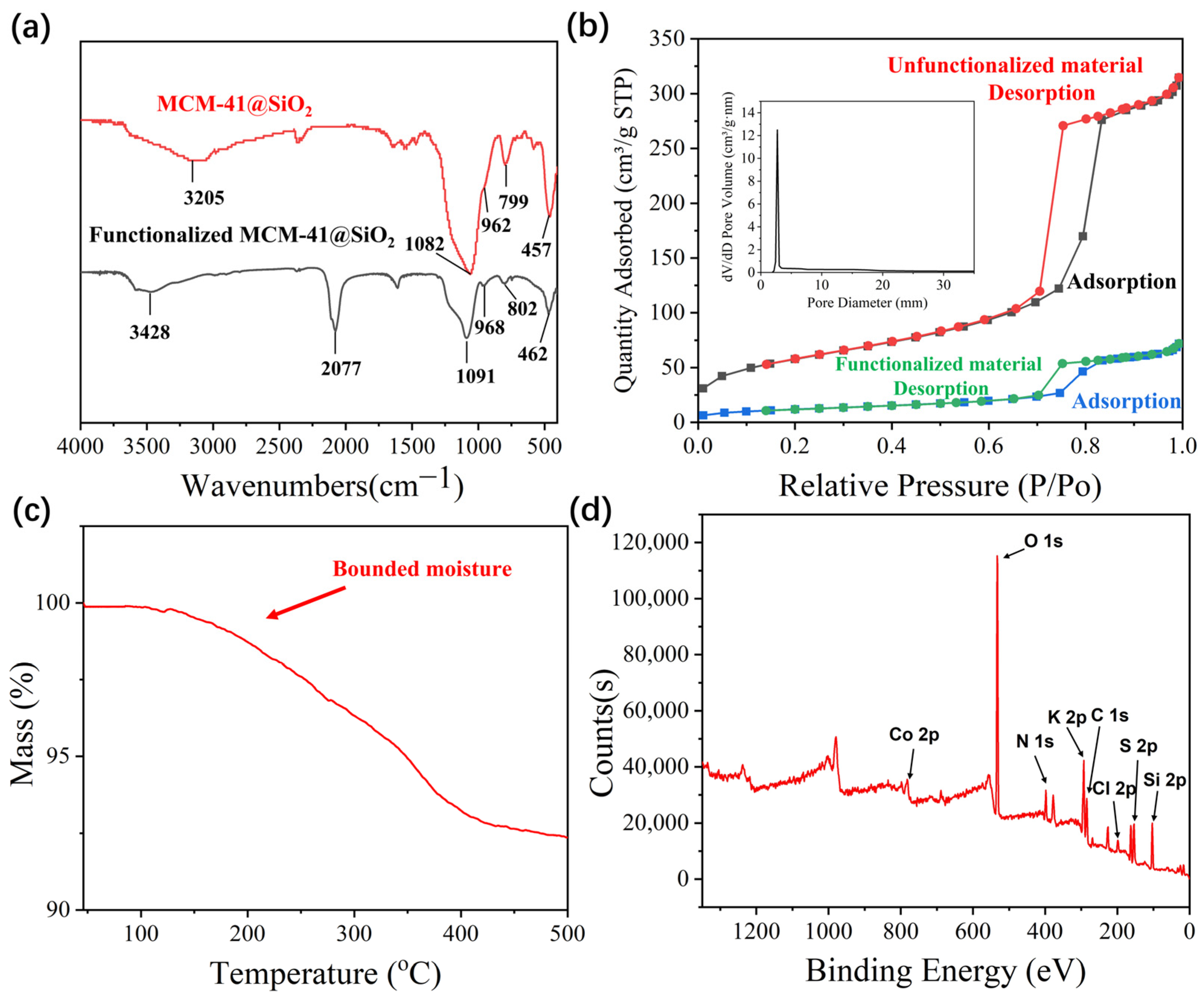
Figure 3.
(a) The scheme of the adsorption and coordination of ammonia on the functionalized material and the digital photos; (b) Anti-interference experiment of the materials; (c) Adsorption ability of the MCM-41@SiO2 nanoparticle in response to different gases (n = 3).
Figure 3.
(a) The scheme of the adsorption and coordination of ammonia on the functionalized material and the digital photos; (b) Anti-interference experiment of the materials; (c) Adsorption ability of the MCM-41@SiO2 nanoparticle in response to different gases (n = 3).
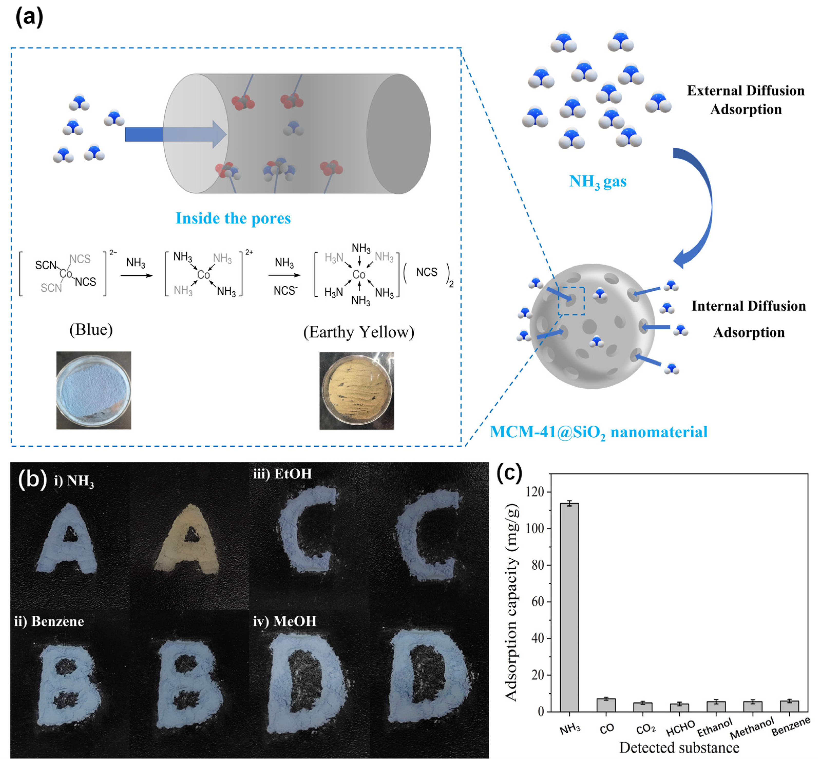
Figure 4.
(a) The discoloration ability of sensors with different SiO2 particles and functionalized MCM-41@SiO2 packing ratios; (b) Linear relations between discolorations and NH3 concentrations from 20 to 1000 ppm (inset graphs are the digital photos of the sensors and the sensing performance in the range from 20 to 50 ppm, n = 3); (c) Humidity test of the sensor (n = 3); (d) Reusability test of the sensor (n = 20); (e) Long-term stability test of the sensor (n = 3).


Table 1.
Preparation and characterization of SiO2 nanoparticles.
| Entry | Stirring Speed (r/min) | Temperature (°C) | Aqua Ammonia:TEOS (v/v) | Diameter of SiO2 Nanoparticles (nm) | PDI | Diameter of MCM-41@SiO2 |
|---|---|---|---|---|---|---|
| 1 | 100 | 20 | 2:1 | 750 | 0.103 | 1200 |
| 2 | 200 | 20 | 2:1 | 680 | 0.082 | 1150 |
| 3 | 300 | 20 | 2:1 | 510 | 0.071 | 900 |
| 4 | 400 | 20 | 2:1 | 425 | 0.174 | 630 |
| 5 | 300 | 0 | 2:1 | 705 | 0.113 | - |
| 6 | 300 | 30 | 2:1 | 490 | 0.089 | - |
| 7 | 300 | 50 | 2:1 | 440 | 0.095 | - |
| 8 | 300 | 20 | 1:1 | 500 | 0.072 | - |
| 9 | 300 | 20 | 3:1 | 495 | 0.091 | - |
| 10 | 300 | 20 | 4:1 | 510 | 0.057 | - |
Disclaimer/Publisher’s Note: The statements, opinions and data contained in all publications are solely those of the individual author(s) and contributor(s) and not of MDPI and/or the editor(s). MDPI and/or the editor(s) disclaim responsibility for any injury to people or property resulting from any ideas, methods, instructions or products referred to in the content. |
© 2023 by the authors. Licensee MDPI, Basel, Switzerland. This article is an open access article distributed under the terms and conditions of the Creative Commons Attribution (CC BY) license (https://creativecommons.org/licenses/by/4.0/).
Share and Cite
MDPI and ACS Style
Liu, S.; Wei, K.; Cheng, F.; Li, Y.; Xue, M. Ammonia Sensor Based on Co2+/SCN− Modified Core-Shell MCM-41 for Rapid Naked-Eye Colorimetric Detection. Chemosensors 2023, 11, 336. https://doi.org/10.3390/chemosensors11060336
AMA Style
Liu S, Wei K, Cheng F, Li Y, Xue M. Ammonia Sensor Based on Co2+/SCN− Modified Core-Shell MCM-41 for Rapid Naked-Eye Colorimetric Detection. Chemosensors. 2023; 11(6):336. https://doi.org/10.3390/chemosensors11060336
Chicago/Turabian StyleLiu, Songtao, Kaixin Wei, Fuqiang Cheng, Yongsheng Li, and Min Xue. 2023. "Ammonia Sensor Based on Co2+/SCN− Modified Core-Shell MCM-41 for Rapid Naked-Eye Colorimetric Detection" Chemosensors 11, no. 6: 336. https://doi.org/10.3390/chemosensors11060336
Note that from the first issue of 2016, this journal uses article numbers instead of page numbers. See further details here.