Review

Ca2+-Mediated Signaling Pathways: A Promising Target for the Successful Generation of Mature and Functional Stem Cell-Derived Pancreatic Beta Cells In Vitro

by
Razik Bin Abdul Mu-u-min
*,
Abdoulaye Diane
,
Asma Allouch
and
Heba H. Al-Siddiqi
Diabetes Research Center, Qatar Biomedical Research Institute (QBRI), Hamad Bin Khalifa University (HBKU), Qatar Foundation (QF), Doha P.O. Box 34110, Qatar
*
Author to whom correspondence should be addressed.
Biomedicines 2023, 11(6), 1577; https://doi.org/10.3390/biomedicines11061577
Submission received: 17 April 2023 / Revised: 18 May 2023 / Accepted: 23 May 2023 / Published: 29 May 2023
(This article belongs to the Special Issue The Promise and Challenge of Induced Pluripotent Stem Cells (iPSCs))

Abstract

:
Diabetes mellitus is a chronic disease affecting over 500 million adults globally and is mainly categorized as type 1 diabetes mellitus (T1DM), where pancreatic beta cells are destroyed, and type 2 diabetes mellitus (T2DM), characterized by beta cell dysfunction. This review highlights the importance of the divalent cation calcium (Ca2+) and its associated signaling pathways in the proper functioning of beta cells and underlines the effects of Ca2+ dysfunction on beta cell function and its implications for the onset of diabetes. Great interest and promise are held by human pluripotent stem cell (hPSC) technology to generate functional pancreatic beta cells from diabetic patient-derived stem cells to replace the dysfunctional cells, thereby compensating for insulin deficiency and reducing the comorbidities of the disease and its associated financial and social burden on the patient and society. Beta-like cells generated by most current differentiation protocols have blunted functionality compared to their adult human counterparts. The Ca2+ dynamics in stem cell-derived beta-like cells and adult beta cells are summarized in this review, revealing the importance of proper Ca2+ homeostasis in beta-cell function. Consequently, the importance of targeting Ca2+ function in differentiation protocols is suggested to improve current strategies to use hPSCs to generate mature and functional beta-like cells with a comparable glucose-stimulated insulin secretion (GSIS) profile to adult beta cells.

1. Introduction

Diabetes mellitus, commonly known as diabetes, is a collection of chronic and metabolic disorders typically observed with heightened blood glucose levels over an extended period. Insulin deficiency leading to impaired glucose metabolism is widely deemed to be the mechanism for the onset and effect of the disease [1,2]. The International Diabetes Federation (IDF) has been assessing the incidence of diabetes globally. Recent statistics have revealed that in 2021, 537 million adults live with diabetes, and the numbers are expected to increase to 783 million by 2045. Additionally, 6.7 million deaths were recorded due to the comorbidities of diabetes (www.idf.org, (accessed on 16 November 2022)). The American Diabetes Association (ADA) has classified diabetes into primarily two categories: type 1 diabetes (T1DM) and type 2 diabetes (T2DM) [3]. T1DM is a result of selective immune-mediated pancreatic beta-cell destruction, leading to a nearly complete deficiency of insulin production in the body. Although only 5–10% of all cases of diabetes are T1DM, it encompasses 80–90% of diabetes in children and adolescents [4,5,6]. T2DM, on the other hand, occurs when individuals have insulin resistance and a relative insulin deficiency due to beta cell dysfunction [2]. T2DM accounts for 90–95% of all cases of diabetes. Other types of diabetes include gestational diabetes mellitus (GDM), neonatal diabetes, and maturity-onset diabetes of the young (MODY) [7]. It is now well known that all forms of diabetes commonly share a dysfunction of the pancreatic beta cells that negatively impacts insulin secretion [4]. Unhealthy lifestyles, increasing global obesity in children and young adults, and an aging population are some of the risk factors for the increasing diabetes population of the world [2,8]. Diabetes is also associated with additional health burden through various macrovascular (stroke, peripheral artery disease, coronary heart diseases, and myocardial infarctions) and microvascular (neuropathy, retinopathy, and nephropathy) complications, which lead to diminished quality of life and premature death [2]. These diabetes-coupled complications also pose an immense financial, social, and medical challenge to both western and developing countries. The global healthcare expenses for managing diabetes were estimated to be USD 850 billion in 2017 [9,10,11,12,13].
T1DM treatment currently involves controlling glycemia through daily insulin supplementation through insulin injections or insulin pumps with integrated glucose monitors [14,15]. Although lifesaving, these invasive treatment strategies remain imperfect, as they could result in acute hypoglycemia and lead to heart and kidney failure. Cadaveric islet transplantation using the Edmonton protocol has been demonstrated as an ideal and effective treatment for T1DM, which could allow temporal exogenous insulin independence [16]. This treatment option is increasingly unrealistic for the growing number of patients with T1DM due to its high costs, lack of sufficient donors, and potential risk of tissue rejection resulting in lifelong immunosuppressive drugs [17,18]. Current T2DM treatment involves a combination of lifestyle and pharmacological interventions. Evidence suggests that regulated pharmacological interventions, along with dietary intake, physical exercise, and adequate sleep, are important to prevent aggravation of insulin resistance [19,20,21,22,23].
Recent progress in the field of regenerative medicine has focused on the generation of surrogate and transplantable functional pancreatic beta cells from human pluripotent stem cells (hPSCs) to treat diabetes. Patient-specific pluripotent stem cell (PSC)-derived beta cells, also known as autologous PSC-derived beta cells, would help circumvent the inadequate islet supply and/or allogeneic immune rejection, thereby being an unlimited source of beta cells for diabetes therapy. More importantly, patient-specific stem cell-derived beta cells can also be used in vitro to study diabetes-related mutations such as inherited monogenic diabetes as well as disease progression [24,25]. To that end, massive efforts are undertaken to differentiate hPSCs efficiently and reproducibly into insulin-expressing beta cells using multi-stage directed differentiation protocols that simulate the specific stages of pancreas development. However, differentiation attempts do not always result in functional insulin-expressing beta cells, and instead, the differentiated beta cells show an immature phenotype with impaired response to glucose-stimulated insulin secretion (GSIS) [18,26]. Both in vivo and in vitro studies consistently demonstrated that insulin release from electrically excitable pancreatic beta cells is a calcium (Ca2+)-dependent process. Therefore, defects in calcium signaling could be one of the underlying reasons for the immature and non-functional phenotypes observed in differentiated hPSC-derived pancreatic beta cells in vitro. In this review, we first describe the role of calcium signaling in pancreatic beta cell function and secondly highlight and summarize recent progress on the implications of impaired calcium signaling in defective insulin secretion in hPSC-derived β cells, and consequently, how targeting calcium signaling might be a new and interesting strategy to improve in vitro differentiation into functional hPSC-derived pancreatic beta cells for diabetes therapy. The methods used in this review are as follows: First, we carried out a comprehensive search of peer-reviewed original and review articles using the PubMed database and Google Scholar based on a wide range of key words linking the involvement of calcium signaling in insulin secretion, hPSCs, and diabetes. Secondly, the reference section for each selected article was searched to find additional articles to further develop this review.

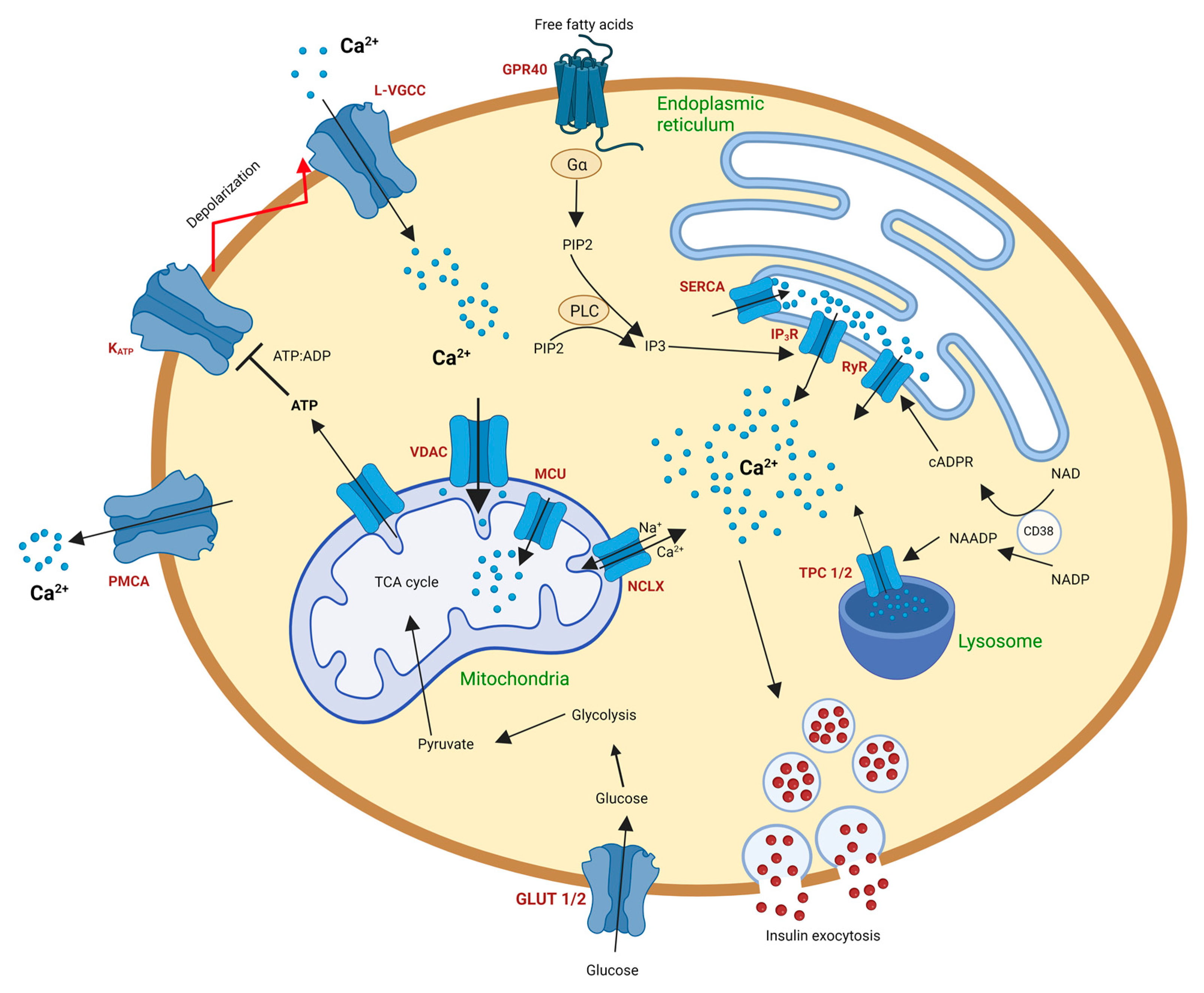
2. Calcium Signaling in Pancreatic Beta Cells (Basics)

Systemic glycemia is carefully regulated by the precise production and release of insulin by beta cells. The interplay between blood glucose and numerous other secretagogues such as neurotransmitters, hormones, and other compounds regulates this function [27,28]. As with most other cell types, Ca2+ has a critical function in the controlled release of insulin from beta cells.
In the postprandial state, an elevation of blood glucose is detected by beta cells, which in turn take glucose into the cell through glucose transporters (GLUT1 in humans and GLUT2 in rodents). Glycolysis is triggered as glucose is phosphorylated by glucokinase into pyruvate. Pyruvate thus formed is transported into the mitochondria and enters the tricarboxylic acid (TCA) cycle. A cascade of events in the TCA cycle enhances ATP production. This increase in the ATP/ADP ratio within the cell inhibits ATP-sensitive K+ channels, causing membrane depolarization. L-type calcium channels are opened because of this depolarization, leading to an influx of Ca2+ into the cell. This wave of Ca2+ triggers insulin exocytosis from secretory granules [29,30].
Although a major share of the intracellular Ca2+ is due to the influx of extracellular Ca2+, several intracellular organelles, including the endoplasmic reticulum (ER), nucleus, Golgi apparatus, and mitochondria, contribute to the regulation of Ca2+ within beta cells [31,32]. A summary of Ca2+ homeostasis in β-cells is shown in Figure 1.

2.1. Calcium Signaling in Beta Cell Endoplasmic Reticulum

The endoplasmic reticulum performs crucial functions such as protein folding and lipid synthesis, acts as a Ca2+ store, and effects its release [33]. Endoplasmic reticulum-mediated Ca2+ uptake and release are mainly dependent on inositol 1,4,5-triphosphate receptors (IP3R), ryanodine receptors (RyR), and sarco/endoplasmic reticulum Ca2+-ATPase (SERCA).
RyR receptors release Ca2+ from the ER through a positive feedback process named “calcium induced calcium release” (CICR). An increase in cytosolic Ca2+ triggers the opening of RyRs, which leads to an efflux of Ca2+ from the ER into the cytoplasm. The Ca2+ thus released by the ER facilitates the opening of more RyRs, hence amplifying the ER-dependent Ca2+ release [34]. Three isoforms of RyRs are known (RyR1, RyR2, and RyR3). RyR2 is believed to be the predominant isoform of RyR present in beta cells, although traces of RyR3 are also found [35,36]. Known activators of RyRs include cyclic AMP (cAMP), cyclic ADP ribose (cADPR), long-chain acyl CoA, and ATP [37,38]. Although the expression of RyRs in beta cells is relatively low, their high conductance capacity might be sufficient to trigger CICR in beta cells [39,40]. Additionally, drugs influencing RyR activity have been shown to have a slight effect on cytoplasmic Ca2+ in human beta cells without compromising GSIS activity [41]. RyR studies on islets have an additional complication wherein delta cells also express RyRs, hence distorting RyR effects on beta cells [42,43].
IP3R are IP3-sensitive receptors found on the ER membrane of many cell types, such as the cerebellum, smooth muscles, cardiac and skeletal muscle, as well as beta cells. IP3 is formed by the activation of phospholipase C, which hydrolyzes phosphatidylinositol bisphosphate (PIP2) into IP3 and diacylglycerol. IP3 is capable of mobilizing Ca2+ from Ca2+-stores and plays a significant role in the release of Ca2+ from the ER [44,45]. Glucose stimulation-coupled depolarizations have been indicated to promote IP3 production [46]. Studies have suggested numerous mechanisms for IP3R-mediated Ca2+ release from the ER. Heightened cytoplasmic Ca2+ levels achieved by the inhibition of SERCA have been shown to accelerate CICR due to the activation of IP3Rs [47]. CICR, independent of RyR activity, was observed when accompanied by increased levels of the second messenger cAMP in beta cells. These effects were attributed to IP3R activation [48]. In rodent islets, it was observed that ATP facilitated autocrine activation of the purinergic receptor P2Y1, which promoted ER Ca2+ release through IP3Rs, thereby increasing cytoplasmic Ca2+ levels [49]. Another mechanism of IP3R-mediated ER Ca2+ release is through the activation of the G-protein-coupled receptor GPR40 by free fatty acids. Fatty acid binding to GPR40 appears to activate the Gαq/11-protein complex, leading to the production of IP3 and subsequent ER Ca2+ release. However, studies have also attributed the production of diacylglycerol as a determining factor in the second phase of insulin secretion [50,51,52,53]. Under ER stress, upregulation of the chaperone ERp44 rearranges IP3R and thereby reduces IP3-mediated Ca2+ release from the ER [54]. Genetic variations of IP3Rs have been linked as a risk factor for T1DM [55].
Ca2+ released from the ER is sequestered back into the ER lumen through the activity of SERCA pumps in an ATP-dependent manner. This helps maintain a resting cytosolic Ca2+ concentration of 25–100 nM and a high ER Ca2+ concentration (200–500 µM). Alternative splicing of the genes encoding SERCA generates fourteen different isoforms of SERCA. In beta cells, the predominant isoforms are SERCA2b and SERCA3 [56,57,58]. The protein phosphatase calcineurin and the kinase Protein kinase R-like ER kinase (PERK) have been shown to regulate SERCA activity in beta cells. Studies demonstrated that PERK and calcineurin work in tandem to dephosphorylate calnexin (which binds to SERCA and limits activity), thereby increasing ER Ca2+ reuptake [59]. The importance of the role of SERCA in normal beta cell function is well supported by the observations that SERCA2b is lost in beta cells of db/db mice and cadaveric T2DM human islets [60]. SERCA dysfunction has also been implicated in diabetic cardiomyopathy [61].

2.2. Calcium Signaling in the Beta Cell Lysosomes

Nicotinic acid adenine dinucleotide phosphate (NAADP) is a Ca2+ mobilizing messenger produced by the ADP-ribosyl cyclase CD38 by a base exchange reaction between nicotinamide adenine dinucleotide phosphate (NADP) and nicotinic acid [62]. Elevated glucose levels have been shown to increase NAADP production in beta cells [63,64]. In postprandial conditions, glucagon-like peptide 1 (GLP-1) is produced by gut L-cells and stimulates the GLP-1 receptor (GLP1R). This activates adenylyl cyclases, leading to the sequential production of NAADP [65]. Inhibition of NAADP repressed membrane depolarization and Ca2+ spiking, proving NAADP to be an influential player in glucose-mediated insulin release [66,67]. The consensus mode of action for NAADP is Ca2+ release from acidic compartments such as lysosomes and insulin granules [39]. Lysosomes are acidic organelles that contain large concentrations of Ca2+. Their main function is the degradation of macromolecules, but they also play a role in Ca2+ regulation in cells. The nanomolecular concentration of NAADP triggers lysosomal Ca2+ release, while micromolecular NAADP concentrations inhibit Ca2+ release [68]. NAADP is believed to release Ca2+ from the lysosome through two-pore channels (TPCs), particularly TPC2. However, recent studies have questioned TPC2 as the major NAADP receptor and suggested TPC1 as the main NAADP receptor instead. Although TPC1 and TPC2 are both known to show high sensitivity to NAADP, studies on knockout mouse models have revealed conflicting results, confounding the identity of NAADP’s primary target receptor [68,69,70,71]. Alternatively, the transient receptor potential (TRP) cation channel, particularly TRPML1, has also been suggested as a potential NAADP target receptor [72].
Adenylyl cyclases such as CD38 are also responsible for the production of another Ca2+ second messenger, cADPR, by the cyclization of beta-nicotinamide adenine dinucleotide. Early studies have suggested that glucose stimulation generates cADPR in pancreatic islets. Follow-up studies by the same group observed that overexpression of CD38 in transgenic mice increased glucose and ketoisocaproate-mediated insulin release, while this effect was impaired in CD38 knockout mice [73,74,75]. cADPR potentiates Ca2+ release from the ER through RyRs. However, evidence suggests that cADPR does not directly interact with RyR, indicating the presence of accessory proteins that act as links between cADPR and RyR. FK506-binding protein 12.6 (FKBP12.6), calmodulin, and GAPDH have been proposed as potential accessory proteins that evoke Ca2+ release by cADPR [76,77,78,79]. The exact mechanism tying cADPR to RyR and the subsequent release of Ca2+ from the ER is currently unknown. cADPR has also been shown to trigger Ca2+ influx and subsequent insulin secretion by activating the plasma membrane channel TRMP2 [76]. Although mounting evidence has suggested the role of cADPR as a Ca2+-releasing messenger, its Ca2+ mobilizing effects are controversial since numerous studies have failed to find an action of cADPR in beta cells [80,81,82]. It has been suggested that these inconsistencies might be due to differences in species, cell types, and experimental designs in various labs [83]. Further studies need to be performed to better understand the role of cADPR in beta cells.

2.3. Calcium Signaling in the Beta Cell Mitochondria

Another organelle crucial for effective GSIS is the mitochondria. The major energy-producing mechanism of the mitochondria is the TCA cycle, where the electron transport chain produces ATP. ATP generated by mitochondria promotes plasma membrane depolarization, leading to Ca2+ influx and insulin exocytosis. A close association between Ca2+ homeostasis and beta cell function has been connected to mitochondrial function [84]. Studies that manipulated mitochondrial Ca2+ showed impaired insulin response to high glucose challenges, thereby linking the tight regulation of Ca2+ and mitochondrial energy metabolism [85,86]. Three dehydrogenases of the TCA cycle have been known to be modulated by Ca2+, namely, pyruvate dehydrogenase, NAD+-isocitrate dehydrogenase, and 2-oxoglutarate dehydrogenase, and serve as crucial factors for insulin release in beta cells. The influx of Ca2+ into the mitochondria is believed to be essential for the stimulation of Ca2+-dependent dehydrogenases of the TCA cycle to control ATP production under increased glucose conditions [27,87,88,89].
Ca2+ entry into the mitochondria occurs in the outer mitochondrial membrane and inner mitochondrial membrane through voltage-dependent anion channels (VDACs) and mitochondrial Ca2+ uniporters (MCU), respectively [90,91]. Experiments with MCU knockdown in murine beta cells and the Ins-1 cell line showed a decrease in mitochondrial Ca2+ uptake, leading to impaired ATP production and insulin secretion. Moreover, the expression of respiratory chain complexes, mitochondrial metabolic activity, and oxygen consumption were also lowered. These studies underlined the importance of MCU-mediated Ca2+ regulation for sustained ATP production and metabolism-secretion coupling in beta cells [92,93]. Similar observations were made in an MCU-null mouse model, where mitochondrial Ca2+ uptake and GSIS were impaired [94]. The uptake and buffering of intracellular Ca2+ by beta cell mitochondria have been implicated in the tone and frequency of cytosolic Ca2+ oscillations and might perhaps contribute to insulin release pulsatility [89,95]. Mitochondrial Ca2+ has also been linked to the activation of the inner membrane channel, the mitochondrial permeability transition pore (PTP). PTP is believed to perform two distinct roles in beta cells, depending on its activation or inhibition. The activation of PTP is required to promote glucose-dependent insulin secretion, while its inhibition is necessary to protect against glucotoxicity and hypoxia [31,96,97,98]. Ca2+ extrusion from mitochondria is also key to mitochondrial and cell function. Persistent accumulation of Ca2+ in the matrix space can cause Ca2+ overload in the mitochondria, leading to the opening of PTPs and, thereby, initiating cell death signals [99]. To maintain mitochondrial Ca2+ homeostasis, Ca2+ extrusion from the mitochondria occurs via the Na2+/Ca2+ exchanger (NCLX) [100]. Studies in primary beta cells and MIN6 cell lines have shown that NCLX not only regulates mitochondrial membrane potential and calcium levels after glucose challenges, but it also controls the rate and amplitude of cytosolic Ca2+ responses and the rate of GSIS during the first phase of insulin secretion [101].

3. Role of Ca2+ in Beta Cell Proliferation

Insulin-positive beta cells are first seen around embryonic day 13.5 in mice and weeks 8–9 in humans. During the earlier days of fetal growth, endocrine progenitor cells differentiate into beta cells. However, during the late neonatal stages, beta cells are formed by replication [102,103,104]. There is a reduction in the rate of replication after weaning, which subsequently drops considerably in adulthood. Additionally, beta cell preservation and repair are believed to primarily occur through beta cell replication rather than adult pluripotent stem cells [104].
A study revealed that a signaling pathway linking membrane depolarization was a key factor for beta cell replication in mice. Diazoxide, a KATP channel-opening drug, reduced the replicative capability of beta cells, thereby suggesting that the requirement for the closure of KATP channels and subsequent depolarization is critical for beta cell replication. Reciprocally, when mice were injected with the KATP channel-blocking drug glyburide, beta cell proliferation was rescued. Therefore, Ca2+ influx appears to play an important role in beta cell replication [105]. Another study showed that the L-type voltage-gated channel agonist Bay K 8644 induced beta cell proliferation, improving our understanding of the role of Ca2+ and calcium channels in beta cell replication.
A known mechanism of beta cell replication is via calcineurin/nuclear factor of activated T-cells (NFAT). Calcineurin/NFAT signaling is central to the regulation of neonatal pancreatic development in mice and human islets. NFATs are activated by the serine/threonine phosphatase, calcineurin. Increasing Ca2+ concentrations switches on calcineurin, which in turn dephosphorylates NFAT. This leads to the translocation of NFAT into the nucleus and the transcription of multiple genes responsible for beta cell function, glucose sensing, and proliferation [106,107,108]. In human islets, the NFAT isoforms NFATC1, NFATC2, NFATC3, and NFATC4 are present, with NFATC2 and NFATC3 being the most abundant [109]. In mouse islets, inactivation of calcineurin b1 (Cnb1) resulted in impaired cell mass and proliferation, among other dysfunctions [110]. Interestingly, inhibition of the NFAT by inhibitory kinases glycogen synthase kinase 3 beta (GSK3b) and dual tyrosine specificity kinase 1 (DYRK1A) has been shown to improve beta cell proliferation. Moreover, pharmacological interventions and genetic studies have deemed the two kinases critical for beta cell replication, and the inhibition of both has been shown to have an additive effect on the proliferation rate in primary rat beta cells [111,112]. Another study has demonstrated beta cell replication by the activation of the transcription factor c-Fos, which improves beta cell proliferation in an NKX6.1-dependent manner by the upregulation of the nuclear receptors Nr4a1, Nr4a3, and the hormone VGF. Interestingly, Nr4a1 has also been deemed critical for regulating NFATC2 in beta cell replication [113]. The calcineurin/NFAT signaling pathway may also be associated with the GLP-1 agonist receptor with respect to beta cell replication. Treatment of human islets with exendin-4, a GLP-1 receptor agonist, increases NFAT expression levels and beta cell proliferation rates. Exendin-4 has also been shown to promote beta cell proliferation by inhibiting the expression of Wnt5a [109,114]. Beta cell replication is also modulated by a Ca2+/calmodulin-dependent kinase (CaMK). A study observed that CaMK4 inhibition eliminated beta cell proliferation, whereas constitutively active CaMK4 expression increased beta cell proliferation via a cAMP response element binding protein (CREB) and insulin receptor substrate 2 (IRS-2) mechanism [115].

4. Role of Ca2+ in Beta Cell Survival

A multitude of studies have found a close association between Ca2+ handling and beta cell survival in pancreatic beta cells. High glucose enhances beta cell survival in a Ca2+-mediated manner. In cultured mouse islet beta cells and MIN6 insulinoma cells, a three-fold decrease in apoptosis was found in the presence of high glucose concentrations compared to low glucose concentrations. Blocking of the L-type Ca2+ channel with nifidepine reversed the protective effects of high glucose, suggesting the requirement of Ca2+ influx for beta cell survival at high glucose concentrations [116]. A recent publication studied the effects of prolonged hyperinsulinemia exposure on beta cell survival. Chronic high insulin exposure increased the intracellular Ca2+ in beta cells, along with an insulin dose-dependent decrease in SERCA2 expression. Prolonged high insulin exposure also triggered apoptosis in beta cells through Caspase-12 (a substrate to calcium-sensitive proteases (calpains) and a known mediator of ER stress) and correlated with an increase in intracellular Ca2+. This suggests that prolonged insulin exposure can disrupt ER function and induce apoptosis in beta cells. However, further studies need to be undertaken to deduce the mechanisms of these processes and the interplay between the protective and destructive effects of high glucose and insulin, respectively [117]. Ca2+ depletion-mediated ER stress has been associated with beta cell apoptosis in numerous studies. A study reported that Ca2+ depletion from the ER through dysfunctional SERCA2b, IP3Rs, and RyRs led to beta cell death and suggested a calpain-2-mediated pathway as the mechanism. The authors discussed the possibility of pharmacological modulations of the ER channels as a potential for beta cell survival [118]. Inflammatory cytokines, another potential ER stress inducer, cause ER Ca2+ depletion and increase cytosolic Ca2+ in beta cells. This led to beta cell death, marked by an increase in caspase-3 and caspase-7 activity levels. In INS-1 cells, cytokine-mediated beta cell apoptosis was inhibited by treating the cells with the calcium-modulating compounds dantrolene and sitagliptin, which restored the function of SERCA and the ER Ca2+ pump and decreased the levels of the pro-apoptotic protein thioredoxin-interacting protein (TXNIP) [119]. Free fatty acid-mediated beta cell apoptosis also centers around ER Ca2+ homeostasis. Heightened palmitate levels increase oxidative conditions, leading to Ca2+ depletion from the ER, which interferes with chaperones and results in misfolded proteins. This triggers a series of events where cytosolic and mitochondrial Ca2+ homeostasis is disrupted, thereby leading to beta cell apoptosis [120,121].
Calcineurin-dependent mechanisms have also been implicated in beta cell viability. Pharmacological inhibition of calcineurin is necessary to prevent organ transplant rejection and is associated with post-transplant beta cell failure. A study on human islets revealed that beta cell apoptosis was significantly increased in the presence of the calcineurin inhibitor tacrolimus. The mechanism of these effects linked calcineurin to IRS-2 and the phosphoinositide-3-kinase protein kinase B/Akt (PI3K-PKB/Akt) pathway. It was suggested that the effect of calcineurin on IRS-2 was also mediated partly through NFAT [122]. More evidence for the role of Ca2+ in beta cell survival was seen when CAMK4 was shown to regulate apoptosis. Constitutively active CAMK4 was shown to significantly improve beta cell survival as a reduction in caspase-3 and caspase-7 was observed. Moreover, increased apoptosis was observed in the presence of the dominant negative form of CAMK4. The authors proposed CAMK4 and its association with CREB as the primary mechanisms for improved beta cell survival. The activation of CAMK4 under intracellular Ca2+ elevations led to the upregulation of IRS-2 via CREB, leading to the regulation of beta cell survivability [115].

5. Calcium Signaling in Diabetes

As clearly highlighted in the earlier sections of this review, Ca2+ and its signaling pathways are critical to the proper function and survivability of pancreatic beta cells. As important as Ca2+ is for beta cell function, survival, and proliferation, it also plays an important role in insulin production and secretion. A plethora of studies have strongly linked Ca2+ dysfunction to diabetes in both human and animal models [123,124,125,126,127].
Studies have suggested the deleterious effects of long-term activation of calcium signaling pathways in beta cells. Calcineurin is a vital component of the calcineurin/NFAT pathway, which is critical to beta cell replication. However, chronic depolarization of beta cells increased calcineurin activity, which in turn reduced glucose tolerance, insulin secretion, and beta cell replication in mouse models [128]. Another publication demonstrated that mice lacking Abcc8 (Abcc8−/− mice), a key component of the beta cell KATP channel, showed sustained membrane depolarization, leading to continued elevation of Ca2+ associated with dysfunctional genes involved in Ca2+ transport, signaling, and beta cell maintenance. Additionally, beta cells were observed to dedifferentiate and transdifferentiate into pancreatic polypeptide cells, as seen through the increased expression of the Aldh1a3 gene (a marker for dedifferentiating beta cells). Interestingly, the expression of Aldh1a3 was reduced by treating the cells with Ca2+ channel blockers, solidifying the link between sustained depolarization and beta cell functional and identity loss [129]. Another study found the presence of the stomach hormone gastrin in the islets of diabetic rodents and humans. Normally, gastrin is expressed abundantly in pancreatic embryonic development and ceases expression after birth. The authors thus observed that gastrin overexpression following hyperglycemia in the mutant mouse models and T2DM human beta cells is mediated by membrane depolarization, Ca2+ influx, and calcineurin signaling that could also be reversed by the normalization of glycemia [130]. A more recent publication used stem cells with knocked out HNF1A gene, whose deficiency impaired a network of genes crucial to calcium signaling, GSIS, and beta cell fate. These cells were then target-differentiated into beta cells. Stem cell-derived beta cells from HNF1A knockouts showed a reduction in the SYT13 gene, which is responsible for calcium-regulated granule exocytosis. HNF1A deficiency also showed a developmental bias toward the generation of alpha cells, as an upregulation of glucagon was observed in the knockout cells. The observations from the knockout cells were comparable with those from the induced pluripotent stem cell (iPSC) line with HNF1A-MODY patient-specific mutations (R200Q) [131]. High-resolution microscopy experiments have revealed the existence of a subset of insulin granules in human beta cells and the INS-1 cell line whose exocytosis was triggered by localized Ca2+ influx. These pools of insulin granules were absent in T2DM donors and INS-1 cells mimicking diabetes, implicating the role of these Ca2+-triggered granules in the loss of rapid first-phase insulin secretion seen in T2DM patients [127]. Diabetic mice showed higher Cavβ3 (a voltage-gated Ca2+ channel subunit) expression, coupled with impaired calcium signaling and insulin secretion, and these effects were reduced in high-fat diet-induced diabetes in Cavβ3 deficient mice. Overexpression of Cavβ3 in human islets also showed impaired GSIS [132,133].
Glucolipotoxicity is an umbrella term to describe the detrimental effects of elevated glucose and fatty acid levels on beta cell function and survival. Glucolipotoxicity-induced beta cell failure and subsequently T2DM are associated with calcium dysregulation and ER stress, along with other non-calcium-related complications. A phenotypic screening study of compounds showed that glucolipotoxicity stress had a close association with calcium flux modulation and apoptosis. It was suggested that a Ca2+ overload was the main mechanism of glucolipotoxicity-mediated apoptosis and dysfunction, which was inhibited by compounds that decreased glucolipotoxicity-mediated Ca2+ influx into the cells [134]. The ubiquitously expressed redox regulator TXNIP has been found to be upregulated in diabetes. TXNIP overexpression led to beta cell apoptosis and glucotoxicity-induced beta cell death. It also induces inflammation activation and interleukin-1β production, which have implications for T1DM. Additionally, the lack of TXNIP promoted beta cell survival and prevented T1DM and T2DM [135,136,137,138]. The authors observed that various calcium channel blockers reduced the expression of TXNIP and deduced that the effects were due to the reduction of intracellular Ca2+ [139]. A phase 2 clinical study by the same group using the L-type calcium channel blocker verapamil showed improved beta cell function, lowered insulin requirements, fewer hypoglycemic events, and on-target glycemic control in patients with T1DM [140]. Free fatty acids and mediated beta cell loss via ER stress can also contribute to T2DM. Evidence for beta cell ER stress in diabetes includes enhanced expression of ER stress markers in islets of diabetic db/db mice and cultured human islets [141,142]. The free fatty acid palmitate upregulated ER stress markers and induced ER stress and apoptosis by depleting ER Ca2+ stores in INS-1 cells [143].

6. Calcium Signaling in Stem Cell-Derived Beta Cells

As discussed in the introduction section above, insulin-secreting beta cells from the pancreatic islet of Langerhans play a crucial role in the body by maintaining glucose homeostasis. It is well established that destruction or loss of beta cell mass leading to a negative impact on effective insulin secretion has emerged as the main pathogenic factor in both T1DM and T2DM [144]. Current treatment strategies, including exogenous insulin administration and islet transplantation, come with a myriad of complications, hindering them being an ideal end-point solution to treat diabetes. Recent advances in stem cell technologies have evoked great excitement and expectations for treating various disorders and dysfunctions, including diabetes. Reprogramming patient-derived somatic cells into iPSC lines followed by controlled differentiation into any cell kind makes them an attractive target for personalized treatment strategies, and since they are autologous, the risks of immune rejection are greatly reduced [145,146]. Stem cell-derived beta cells can also be used to study disease phenotypes and serve as an excellent model for drug screening studies [147,148]. Thus, beta cell replacement through iPSC technology has the potential to greatly assist in the treatment of T1DM and T2DM.
Directed differentiation protocols are created from an extensive study of the developmental phases of cell types. The conversion of pluripotent stem cells into target cells is achieved by the timely addition of small molecules and growth factors in a stagewise manner that mimics the embryonic development of the desired cell type. The success of the protocol is usually determined by the extent to which these cells mirror their in vivo counterparts with respect to functional maturity and efficiency. Indeed, over the past decade, these strategies have been applied to differentiate human iPSCs and ESCs into pancreatic beta cells and islets. Pioneering work from the Melton laboratory at Harvard University has paved the way for protocols that generate beta cells with moderate efficiency. However, multiple labs have published protocols with more optimized culture conditions for differentiation media and small molecules, with the resulting protocols applicable to multiple iPSC cell lines for improved differentiation efficiency and beta cell functionality [149,150,151]. Transplantation of such generated islet-like clusters into mice has also been shown to promote their maturation and functionality [152]. A general outline for pancreatic beta cell differentiation protocols is presented in Figure 2. A common pitfall for these differentiation strategies was that the resultant beta cells showed impaired GSIS functionality compared to human islets. The GSIS response in cadaveric islets was five times higher than that observed in stem cell-derived beta cells [153].
Beta cells facilitate GSIS through increased cytosolic Ca2+ concentration, indicating that increased Ca2+ influx is a cellular function required for GSIS. Since evidence has highlighted that Ca2+ dysregulation in beta cells has a key role in diabetes, it is possible that functionally defective Ca2+ dynamics play a key role in the generation of immature and non-functionally differentiated hPSC-derived beta cells in vitro. A study used the human ESC (hESC) cell line H1 to create a seven-stage protocol to generate beta cells. The cells thus obtained expressed key markers of mature beta cells and displayed GSIS capabilities. However, as compared to adult beta cells, the differentiated cells showed reduced amplitudes and a slower time to peak of the Ca2+ signals. Sustained Ca2+ transients were observed even after the glucose stimulation ceased, a marked difference from mature beta cells. Similar effects were seen with respect to insulin release on glucose challenges, in which insulin secretion did not return to pretreatment levels and KCl-mediated insulin release was blunted. Direct depolarization with high KCl concentrations revealed the presence of voltage-gated Ca2+ channels and normal Ca2+ efflux pumps, thereby suggesting that the generated beta-like cells had functional immaturity compared to adult beta cells [150]. Another study performed a genome-wide transcriptional analysis of enriched insulin-expressing cells derived from multiple human iPSCs, human fetal pancreata, and adult human islets and drew the conclusion that iPSC-derived insulin-expressing cells resemble human fetal beta cells more than adult beta cells [26]. The authors observed that iPSC-derived insulin-positive cells had similar elevated basal glucose secretions and fared poorly under higher glucose challenges as compared to adult beta cells. The transcription factor pancreatic duodenal homeobox 1 (PDX1), which plays critical roles in early pancreas formation and several important aspects of beta cell function, is also indispensable in the maintenance of ER health [155,156,157]. A lack of the PDX1 gene altered the expression of SERCA2b, the main isoform of SERCA in beta cells that pumps Ca2+ into the ER, thereby impairing ER function and thereby Ca2+ homeostasis in beta cells [158]. A three-fold lower expression of PDX1 was found in iPSC-derived insulin-positive cells compared to adult beta cells [26]. MafA, a gene exclusively expressed in adult beta cells that promotes beta cell maturation and function, is known to be regulated by calcium signaling pathways [159,160,161]. MafA overexpression has been shown to induce GSIS in immature rat beta cells [162]. A recent publication has linked MafA regulation with Cav subunit gamma 4 (Cavγ4) in a CaMKII-mediated manner in controlling beta cell glucose homeostasis [162]. Insulin-expressing iPSC-derived cells were observed to have much lower expression of MafA compared with adult beta cells [26]. Another transcription factor that regulates beta cell mass and function is Pax4, which has also been implicated in T2DM [163]. Studies have shown that Pax4 overexpression causes increased beta cell proliferation combined with a transactivation of the antiapoptotic gene Bcl-xL. Increased Bcl-xL activity altered mitochondrial Ca2+ levels and ATP production, leading to impaired GSIS in islets [164]. Interestingly, Pax4 expression was significantly increased in iPSC-derived insulin-positive cells compared to adult beta cells [26]. The altered expression levels of key transcription factors such as the ones described above might impair Ca2+ handling in the differentiating cells and might explain the difficulty in generating beta cells functionally similar to adult human beta cells. One cannot assume that impaired calcium signaling is the major contributor to the formation of immature insulin-producing cells from iPSCs. Impaired expression of genes responsible for potassium channel regulation and mitochondrial function was also found in immature iPSC-derived beta-like cells [19,26]. Perhaps the differentiation strategies are missing key ingredients to fully activate various important genes and transcription factors across different organelles to efficiently differentiate iPSCs into functional beta cells compared to their in vivo counterparts.
Nevertheless, more recent publications have generated beta cells with comparable functionality to human islets by incorporating enrichment and reaggregation of late-stage differentiated beta-like cells for better GSIS functionality [165,166]. Cells expressing C-peptide were isolated using fluorescence-activated cell sorting (FACS), and a week-long differentiation protocol was followed after reaggregation of these cells. The aggregated enriched beta cell clusters (eBCs) contained mainly C-peptide-positive cells, although a minute population of cells expressing C-peptide, glucagon, and somatostatin were also seen. These eBCs were studied to compare their properties to those of human islets and pre-aggregation or non-enriched yet aggregated beta-like immature cells. In the presence of high glucose, the pre-aggregation cells showed a slow rise in calcium flux, which was unresponsive to lower glucose concentrations, an observation consistent with other reported beta-like cells [150]. The eBCs, however, showed a significant increase and lowering of calcium flux dependent on the concentration of the glucose challenge, as seen in human islets, suggesting the proper activation of voltage-gated Ca2+ channels in the eBCs. Transcriptome analysis comparing non-enriched clusters and eBCs showed an upregulation of the SCGN gene, which is responsible for calcium sensing. Moreover, genes upregulated in neonatal mouse and human fetal beta cells were seen to be upregulated in the immature beta cells, providing further evidence that immature iPSC-derived beta cells resemble fetal cells more than adult beta cells [166].
Taken together, these studies that compare the differences between iPSC-derived immature beta-like cells and iPSC-derived beta cells functionally closer to human beta cells show the importance of appropriate Ca2+ handling in the maturation of beta cells during their development phase in in vitro conditions, suggesting that the calcium signaling pathway is an important puzzle piece in the generation of iPSC-derived pancreatic beta-like cells. Careful considerations need to be made in the differentiation protocols to include small molecules and growth factors in the differentiation media that target the calcium signaling pathways and their components in a timely manner to generate fully functional beta cells in vitro. Current strategies have been built upon accumulating knowledge over the past decade to produce functional beta cells, and more research needs to be performed to generate high-quality iPSC-derived beta cells that resemble human beta cells.
An important tool to better understand diabetes and its progression is the integration of gene editing technology (CRISPR/Cas9) with stem cell technology. Several studies have reported the use of CRISPR/Cas9 to create stem cell-based disease models of monogenetic diabetes, MODY, and neonatal diabetes, among others, to better understand the mechanisms of disease progression [167,168,169,170]. One such study highlighted the effect of mutations in the HNF1A gene on the onset of MODY. CRISPR/Cas9 was used to generate HNF1A mutations in the hESC Mel1, which was subsequently differentiated into beta cells. The resulting beta cells were observed to have impaired GSIS and intracellular calcium levels [131]. Moreover, studies have also used CRISPR/Cas9 to correct mutated variants in disease models. Different groups have used CRISPR/Cas9 techniques to correct mutations in patient-specific iPSC lines. These iPSCs were then targeted and differentiated into beta cells [169,171]. A study used CRISPR/Cas9 in a patient-specific iPSC to correct a mutation responsible for the inactivation of the sulfonylurea receptor 1 subunit of the KATP channel in congenital hyperinsulinism. These cells were target-differentiated into beta cells and used as controls to study the effect of the mutation on beta cell function [169]. Another study used CRISPR/Cas9 to correct a diabetes-causing pathogenic variant in Wolfram syndrome 1 (WFS1) in IPSCs generated from a patient with Wolfram syndrome. The IPSCs were differentiated into beta cells, and it was observed that the corrected WFS1 IPSC-derived beta cells showed strong dynamic insulin secretion and reversed streptozocin-induced diabetes following transplantation into mice [167]. Thus, CRISPR/Cas9 and its incorporation into the stem cell field promise an exciting future to better understand various diabetes disease models and device approaches to improve beta cell function and survivability. This also opens new avenues to better study the role of calcium signaling in these disease models and help formulate novel treatment strategies.

7. Conclusions

In this review, we emphasize the role of Ca2+ in the functioning of pancreatic beta cells. The role of several Ca2+-handling organelles have been discussed through recent studies, which highlight that the disruption of key Ca2+ pathways could lead to beta-cell dysfunction and thereby play a role in the onset of diabetes. We examined the key advances made in the field of stem cell technology in the study of diabetes. A comparative study of Ca2+ handling characteristics of stem cell-derived beta-like cells and their adult human counterparts was performed, showcasing the pitfalls of the current differentiation protocols and underlining key research strategies targeting Ca2+ dynamics to improve the identity and functionality of directed stem cell differentiation. Massive strides are being made to achieve the goal of generating functional beta-like cells from stem cells. Advances in the efficiency of iPSC differentiation protocols can close the gaps in the structural and functional differences between stem cell-derived pancreatic and human islets. This opens the door to fully using stem cell-derived beta cells for drug screening due to their enhanced kinetics, and the mechanisms of beta cell failure can also be studied, paving the way to greatly improve our knowledge of the intricate aspects of diabetes and its treatment, including transplantation in patients with diabetes [165,166,172].

Author Contributions

Manuscript—conceptualization, H.H.A.; performed literature search and manuscript—draft, R.B.A.M.; manuscript—review and editing, A.D. and H.H.A.; figure preparation, R.B.A.M. and A.A. All authors have read and agreed to the published version of the manuscript.

Funding

This work was supported by Qatar Biomedical Research Institute, Hamad Bin Khalifa University, Qatar foundation intramural grant (BR01) to Heba H. Al-Siddiqi.

Institutional Review Board Statement

Not applicable.

Informed Consent Statement

Not applicable.

Data Availability Statement

No data was used in this manuscript.

Conflicts of Interest

The authors declare no conflict of interest.

References

  1. Atkinson, M.A.; Bluestone, J.A.; Eisenbarth, G.S.; Hebrok, M.; Herold, K.C.; Accili, D.; Pietropaolo, M.; Arvan, P.R.; Von Herrath, M.; Markel, D.S.; et al. How does type 1 diabetes develop? The notion of homicide or beta-cell suicide revisited. Diabetes 2011, 60, 1370–1379. [Google Scholar] [CrossRef] [PubMed]
  2. Eizirik, D.L.; Pasquali, L.; Cnop, M. Pancreatic beta-cells in type 1 and type 2 diabetes mellitus: Different pathways to failure. Nat. Rev. Endocrinol. 2020, 16, 349–362. [Google Scholar] [CrossRef] [PubMed]
  3. American Diabetes Association. Diagnosis and classification of diabetes mellitus. Diabetes Care 2014, 37 (Suppl. 1), S81–S90. [Google Scholar] [CrossRef] [PubMed]
  4. Maahs, D.M.; West, N.A.; Lawrence, J.M.; Mayer-Davis, E.J. Epidemiology of type 1 diabetes. Endocrinol. Metab. Clin. N. Am. 2010, 39, 481–497. [Google Scholar] [CrossRef]
  5. Craig, M.E.; Hattersley, A.; Donaghue, K.C. Definition, epidemiology and classification of diabetes in children and adolescents. Pediatr. Diabetes 2009, 10 (Suppl. 12), 3–12. [Google Scholar] [CrossRef]
  6. Dabelea, D.; Mayer-Davis, E.J.; Saydah, S.; Imperatore, G.; Linder, B.; Divers, J.; Bell, R.; Badaru, A.; Talton, J.W.; Crume, T.; et al. Prevalence of type 1 and type 2 diabetes among children and adolescents from 2001 to 2009. JAMA 2014, 311, 1778–1786. [Google Scholar] [CrossRef]
  7. American Diabetes Association. 2. Classification and Diagnosis of Diabetes: Standards of Medical Care in Diabetes-2018. Diabetes Care 2018, 41 (Suppl. 1), S13–S27. [Google Scholar] [CrossRef]
  8. Maric-Bilkan, C. Sex differences in micro- and macro-vascular complications of diabetes mellitus. Clin. Sci. 2017, 131, 833–846. [Google Scholar] [CrossRef]
  9. Dal Canto, E.; Ceriello, A.; Ryden, L.; Ferrini, M.; Hansen, T.B.; Schnell, O.; Standl, E.; Beulens, J.W. Diabetes as a cardiovascular risk factor: An overview of global trends of macro and micro vascular complications. Eur. J. Prev. Cardiol. 2019, 26 (Suppl. 2), 25–32. [Google Scholar] [CrossRef]
  10. Bener, A.; Kim, E.J.; Mutlu, F.; Eliyan, A.; Delghan, H.; Nofal, E.; Shalabi, L.; Wadi, N. Burden of diabetes mellitus attributable to demographic levels in Qatar: An emerging public health problem. Diabetes Metab. Syndr. 2014, 8, 216–220. [Google Scholar] [CrossRef]
  11. Pradeepa, R.; Mohan, V. Prevalence of type 2 diabetes and its complications in India and economic costs to the nation. Eur. J. Clin. Nutr. 2017, 71, 816–824. [Google Scholar] [CrossRef]
  12. Bommer, C.; Heesemann, E.; Sagalova, V.; Manne-Goehler, J.; Atun, R.; Barnighausen, T.; Vollmer, S. The global economic burden of diabetes in adults aged 20–79 years: A cost-of-illness study. Lancet Diabetes Endocrinol. 2017, 5, 423–430. [Google Scholar] [CrossRef]
  13. Cho, N.H.; Shaw, J.E.; Karuranga, S.; Huang, Y.; da Rocha Fernandes, J.D.; Ohlrogge, A.W.; Malanda, B. IDF Diabetes Atlas: Global estimates of diabetes prevalence for 2017 and projections for 2045. Diabetes Res. Clin. Pract. 2018, 138, 271–281. [Google Scholar] [CrossRef]
  14. Katsarou, A.; Gudbjornsdottir, S.; Rawshani, A.; Dabelea, D.; Bonifacio, E.; Anderson, B.J.; Jacobsen, L.M.; Schatz, D.A.; Lernmark, A. Type 1 diabetes mellitus. Nat. Rev. Dis. Prim. 2017, 3, 17016. [Google Scholar] [CrossRef]
  15. Miller, K.M.; Foster, N.C.; Beck, R.W.; Bergenstal, R.M.; DuBose, S.N.; DiMeglio, L.A.; Maahs, D.M.; Tamborlane, W.V. Current state of type 1 diabetes treatment in the U.S.: Updated data from the T1D Exchange clinic registry. Diabetes Care 2015, 38, 971–978. [Google Scholar] [CrossRef]
  16. Shapiro, A.M.J.; Ricordi, C.; Hering, B.J.; Auchincloss, H.; Lindblad, R.; Robertson, R.P.; Secchi, A.; Brendel, M.D.; Berney, T.; Brennan, D.C.; et al. International trial of the Edmonton protocol for islet transplantation. N. Engl. J. Med. 2006, 355, 1318–1330. [Google Scholar] [CrossRef]
  17. Diane, A.; Al-Shukri, N.A.; Bin Abdul Mu, U.M.R.; Al-Siddiqi, H.H. β-cell mitochondria in diabetes mellitus: A missing puzzle piece in the generation of hPSC-derived pancreatic β-cells? J. Transl. Med. 2022, 20, 163. [Google Scholar] [CrossRef]
  18. Sun, Z.Y.; Yu, T.Y.; Jiang, F.X.; Wang, W. Functional maturation of immature beta cells: A roadblock for stem cell therapy for type 1 diabetes. World J. Stem Cells 2021, 13, 193–207. [Google Scholar] [CrossRef]
  19. McNeil, J.; Doucet, E.; Chaput, J.P. Inadequate sleep as a contributor to obesity and type 2 diabetes. Can. J. Diabetes 2013, 37, 103–108. [Google Scholar] [CrossRef]
  20. Marin-Penalver, J.J.; Martin-Timon, I.; Sevillano-Collantes, C.; Del Canizo-Gomez, F.J. Update on the treatment of type 2 diabetes mellitus. World J. Diabetes 2016, 7, 354–395. [Google Scholar] [CrossRef]
  21. Song, R. Mechanism of Metformin: A Tale of Two Sites. Diabetes Care 2016, 39, 187–189. [Google Scholar] [CrossRef] [PubMed]
  22. Eldor, R.; Raz, I. Diabetes therapy--focus on Asia: Second-line therapy debate: Insulin/secretagogues. Diabetes Metab. Res. Rev. 2012, 28 (Suppl. 2), 85–89. [Google Scholar] [CrossRef] [PubMed]
  23. Gerich, J.; Raskin, P.; Jean-Louis, L.; Purkayastha, D.; Baron, M.A. PRESERVE-beta: Two-year efficacy and safety of initial combination therapy with nateglinide or glyburide plus metformin. Diabetes Care 2005, 28, 2093–2099. [Google Scholar] [CrossRef] [PubMed]
  24. Maxwell, K.G.; Millman, J.R. Applications of iPSC-derived beta cells from patients with diabetes. Cell Rep. Med. 2021, 2, 100238. [Google Scholar] [CrossRef]
  25. Abdelalim, E.M.; Bonnefond, A.; Bennaceur-Griscelli, A.; Froguel, P. Pluripotent stem cells as a potential tool for disease modelling and cell therapy in diabetes. Stem Cell Rev. Rep. 2014, 10, 327–337. [Google Scholar] [CrossRef]
  26. Hrvatin, S.; O’donnell, C.W.; Deng, F.; Millman, J.R.; Pagliuca, F.W.; DiIorio, P.; Rezania, A.; Gifford, D.K.; Melton, D.A. Differentiated human stem cells resemble fetal, not adult, beta cells. Proc. Natl. Acad. Sci. USA 2014, 111, 3038–3043. [Google Scholar] [CrossRef]
  27. Klec, C.; Ziomek, G.; Pichler, M.; Malli, R.; Graier, W.F. Calcium Signaling in β-cell Physiology and Pathology: A Revisit. Int. J. Mol. Sci. 2019, 20, 6110. [Google Scholar] [CrossRef]
  28. Renstrom, E.; Ding, W.G.; Bokvist, K.; Rorsman, P. Neurotransmitter-induced inhibition of exocytosis in insulin-secreting beta cells by activation of calcineurin. Neuron 1996, 17, 513–522. [Google Scholar] [CrossRef]
  29. Lemaire, K.; Ravier, M.A.; Schraenen, A.; Creemers, J.W.M.; Van de Plas, R.; Granvik, M.; Van Lommel, L.; Waelkens, E.; Chimienti, F.; Rutter, G.A.; et al. Insulin crystallization depends on zinc transporter ZnT8 expression, but is not required for normal glucose homeostasis in mice. Proc. Natl. Acad. Sci. USA 2009, 106, 14872–14877. [Google Scholar] [CrossRef]
  30. Iynedjian, P.B. Molecular physiology of mammalian glucokinase. Cell. Mol. Life Sci. 2009, 66, 27–42. [Google Scholar] [CrossRef]
  31. Weiser, A.; Feige, J.N.; De Marchi, U. Mitochondrial Calcium Signaling in Pancreatic beta-Cell. Int. J. Mol. Sci. 2021, 22, 2515. [Google Scholar] [CrossRef]
  32. Prins, D.; Michalak, M. Organellar calcium buffers. Cold Spring Harb. Perspect. Biol. 2011, 3, a004069. [Google Scholar] [CrossRef]
  33. Berridge, M.J. The endoplasmic reticulum: A multifunctional signaling organelle. Cell Calcium 2002, 32, 235–249. [Google Scholar] [CrossRef]
  34. Graves, T.K.; Hinkle, P.M. Ca(2+)-induced Ca(2+) release in the pancreatic beta-cell: Direct evidence of endoplasmic reticulum Ca(2+) release. Endocrinology 2003, 144, 3565–3574. [Google Scholar] [CrossRef]
  35. Santulli, G.; Pagano, G.; Sardu, C.; Xie, W.; Reiken, S.; D’Ascia, S.L.; Cannone, M.; Marziliano, N.; Trimarco, B.; Guise, T.A.; et al. Calcium release channel RyR2 regulates insulin release and glucose homeostasis. J. Clin. Investig. 2015, 125, 1968–1978. [Google Scholar] [CrossRef]
  36. Zhang, I.X.; Raghavan, M.; Satin, L.S. The Endoplasmic Reticulum and Calcium Homeostasis in Pancreatic Beta Cells. Endocrinology 2020, 161, bqz028. [Google Scholar] [CrossRef]
  37. Islam, M.S. The ryanodine receptor calcium channel of beta-cells: Molecular regulation and physiological significance. Diabetes 2002, 51, 1299–1309. [Google Scholar] [CrossRef]
  38. Sabatini, P.V.; Speckmann, T.; Lynn, F.C. Friend and foe: Beta-cell Ca(2+) signaling and the development of diabetes. Mol. Metab. 2019, 21, 1–12. [Google Scholar] [CrossRef]
  39. Mitchell, K.J.; Lai, F.A.; Rutter, G.A. Ryanodine receptor type I and nicotinic acid adenine dinucleotide phosphate receptors mediate Ca2+ release from insulin-containing vesicles in living pancreatic beta-cells (MIN6). J. Biol.Chem. 2003, 278, 11057–11064. [Google Scholar] [CrossRef]
  40. Dixit, S.S.; Wang, T.; Manzano, E.J.; Yoo, S.; Lee, J.; Chiang, D.Y.; Ryan, N.; Respress, J.L.; Yechoor, V.K.; Wehrens, X.H. Effects of CaMKII-mediated phosphorylation of ryanodine receptor type 2 on islet calcium handling, insulin secretion, and glucose tolerance. PLoS ONE 2013, 8, e58655. [Google Scholar] [CrossRef]
  41. Johnson, J.D.; Kuang, S.; Misler, S.; Polonsky, K.S. Ryanodine receptors in human pancreatic beta cells: Localization and effects on insulin secretion. FASEB J. 2004, 18, 878–880. [Google Scholar] [CrossRef] [PubMed]
  42. Adriaenssens, A.E.; Svendsen, B.; Lam, B.Y.; Yeo, G.S.; Holst, J.J.; Reimann, F.; Gribble, F.M. Transcriptomic profiling of pancreatic alpha, beta and delta cell populations identifies delta cells as a principal target for ghrelin in mouse islets. Diabetologia 2016, 59, 2156–2165. [Google Scholar] [CrossRef] [PubMed]
  43. Zhang, Q.; Bengtsson, M.; Partridge, C.; Salehi, A.; Braun, M.; Cox, R.; Eliasson, L.; Johnson, P.R.; Renström, E.; Schneider, T.; et al. R-type Ca(2+)-channel-evoked CICR regulates glucose-induced somatostatin secretion. Nat. Cell Biol. 2007, 9, 453–460. [Google Scholar] [CrossRef] [PubMed]
  44. Berridge, M.J. Inositol trisphosphate and diacylglycerol as second messengers. Biochem. J. 1984, 220, 345–360. [Google Scholar] [CrossRef] [PubMed]
  45. Woll, K.A.; Van Petegem, F. Calcium-release channels: Structure and function of IP3 receptors and ryanodine receptors. Physiol. Rev. 2022, 102, 209–268. [Google Scholar] [CrossRef]
  46. Gromada, J.; Frokjaer-Jensen, J.; Dissing, S. Glucose stimulates voltage- and calcium-dependent inositol trisphosphate production and intracellular calcium mobilization in insulin-secreting beta TC3 cells. Biochem. J. 1996, 314 Pt 1, 339–345. [Google Scholar] [CrossRef]
  47. Dyachok, O.; Tufveson, G.; Gylfe, E. Ca2+-induced Ca2+ release by activation of inositol 1,4,5-trisphosphate receptors in primary pancreatic beta-cells. Cell Calcium 2004, 36, 1–9. [Google Scholar] [CrossRef]
  48. Dyachok, O.; Gylfe, E. Ca(2+)-induced Ca(2+) release via inositol 1,4,5-trisphosphate receptors is amplified by protein kinase A and triggers exocytosis in pancreatic beta-cells. J. Biol. Chem. 2004, 279, 45455–45461. [Google Scholar] [CrossRef]
  49. Khan, S.; Yan-Do, R.; Duong, E.; Wu, X.; Bautista, A.; Cheley, S.; Macdonald, P.E.; Braun, M. Autocrine activation of P2Y1 receptors couples Ca(2+) influx to Ca(2+) release in human pancreatic beta cells. Diabetologia 2014, 57, 2535–2545. [Google Scholar] [CrossRef]
  50. Burant, C.F. Activation of GPR40 as a therapeutic target for the treatment of type 2 diabetes. Diabetes Care 2013, 36 (Suppl. 2), S175–S179. [Google Scholar] [CrossRef]
  51. Schnell, S.; Schaefer, M.; Schofl, C. Free fatty acids increase cytosolic free calcium and stimulate insulin secretion from beta-cells through activation of GPR40. Mol. Cell. Endocrinol. 2007, 263, 173–180. [Google Scholar] [CrossRef]
  52. Ferdaoussi, M.; Bergeron, V.; Zarrouki, B.; Kolic, J.; Cantley, J.; Fielitz, J.; Olson, E.N.; Prentki, M.; Biden, T.; MacDonald, P.E.; et al. G protein-coupled receptor (GPR)40-dependent potentiation of insulin secretion in mouse islets is mediated by protein kinase D1. Diabetologia 2012, 55, 2682–2692. [Google Scholar] [CrossRef]
  53. Fujiwara, K.; Maekawa, F.; Yada, T. Oleic acid interacts with GPR40 to induce Ca2+ signaling in rat islet beta-cells: Mediation by PLC and L-type Ca2+ channel and link to insulin release. Am. J. Physiol. Endocrinol. Metab. 2005, 289, E670–E677. [Google Scholar] [CrossRef]
  54. Kiviluoto, S.; Vervliet, T.; Ivanova, H.; Decuypere, J.-P.; De Smedt, H.; Missiaen, L.; Bultynck, G.; Parys, J.B. Regulation of inositol 1,4,5-trisphosphate receptors during endoplasmic reticulum stress. Biochim. Biophys. Acta 2013, 1833, 1612–1624. [Google Scholar] [CrossRef]
  55. Roach, J.C.; Deutsch, K.; Li, S.; Siegel, A.F.; Bekris, L.M.; Einhaus, D.C.; Sheridan, C.M.; Glusman, G.; Hood, L.; Lernmark, Å.; et al. Genetic mapping at 3-kilobase resolution reveals inositol 1,4,5-triphosphate receptor 3 as a risk factor for type 1 diabetes in Sweden. Am. J. Hum. Genet. 2006, 79, 614–627. [Google Scholar] [CrossRef]
  56. Periasamy, M.; Kalyanasundaram, A. SERCA pump isoforms: Their role in calcium transport and disease. Muscle Nerve 2007, 35, 430–442. [Google Scholar] [CrossRef]
  57. Tong, X.; Kono, T.; Anderson-Baucum, E.K.; Yamamoto, W.; Gilon, P.; Lebeche, D.; Day, R.N.; Shull, G.E.; Evans-Molina, C. SERCA2 Deficiency Impairs Pancreatic beta-Cell Function in Response to Diet-Induced Obesity. Diabetes 2016, 65, 3039–3052. [Google Scholar] [CrossRef]
  58. Varadi, A.; Molnar, E.; Ashcroft, S.J. Characterisation of endoplasmic reticulum and plasma membrane Ca(2+)-ATPases in pancreatic beta-cells and in islets of Langerhans. Biochim. Biophys. Acta 1995, 1236, 119–127. [Google Scholar] [CrossRef]
  59. Wang, R.; McGrath, B.C.; Kopp, R.F.; Roe, M.W.; Tang, X.; Chen, G.; Cavener, D.R. Insulin secretion and Ca2+ dynamics in beta-cells are regulated by PERK (EIF2AK3) in concert with calcineurin. J. Biol. Chem. 2013, 288, 33824–33836. [Google Scholar] [CrossRef]
  60. Kono, T.; Ahn, G.; Moss, D.R.; Gann, L.; Zarain-Herzberg, A.; Nishiki, Y.; Fueger, P.T.; Ogihara, T.; Evans-Molina, C. PPAR-gamma activation restores pancreatic islet SERCA2 levels and prevents beta-cell dysfunction under conditions of hyperglycemic and cytokine stress. Mol. Endocrinol. 2012, 26, 257–271. [Google Scholar] [CrossRef]
  61. Zarain-Herzberg, A.; Garcia-Rivas, G.; Estrada-Aviles, R. Regulation of SERCA pumps expression in diabetes. Cell Calcium 2014, 56, 302–310. [Google Scholar] [CrossRef] [PubMed]
  62. Aley, P.K.; Singh, N.; Brailoiu, G.C.; Brailoiu, E.; Churchill, G.C. Nicotinic acid adenine dinucleotide phosphate (NAADP) is a second messenger in muscarinic receptor-induced contraction of guinea pig trachea. J. Biol. Chem. 2013, 288, 10986–10993. [Google Scholar] [CrossRef] [PubMed]
  63. Masgrau, R.; Churchill, G.C.; Morgan, A.J.; Ashcroft, S.J.; Galione, A. NAADP: A new second messenger for glucose-induced Ca2+ responses in clonal pancreatic beta cells. Curr. Biol. 2003, 13, 247–251. [Google Scholar] [CrossRef] [PubMed]
  64. Park, K.H.; Kim, B.J.; Shawl, A.I.; Han, M.K.; Lee, H.C.; Kim, U.H. Autocrine/paracrine function of nicotinic acid adenine dinucleotide phosphate (NAADP) for glucose homeostasis in pancreatic beta-cells and adipocytes. J. Biol. Chem. 2013, 288, 35548–35558. [Google Scholar] [CrossRef] [PubMed]
  65. Kim, B.-J.; Park, K.-H.; Yim, C.-Y.; Takasawa, S.; Okamoto, H.; Im, M.-J.; Kim, U.-H. Generation of nicotinic acid adenine dinucleotide phosphate and cyclic ADP-ribose by glucagon-like peptide-1 evokes Ca2+ signal that is essential for insulin secretion in mouse pancreatic islets. Diabetes 2008, 57, 868–878. [Google Scholar] [CrossRef]
  66. Naylor, E.; Arredouani, A.; Vasudevan, S.R.; Lewis, A.M.; Parkesh, R.; Mizote, A.; Rosen, D.; Thomas, J.M.; Izumi, M.; Ganesan, A.; et al. Identification of a chemical probe for NAADP by virtual screening. Nat. Chem. Biol. 2009, 5, 220–226. [Google Scholar] [CrossRef]
  67. Arredouani, A.; Ruas, M.; Collins, S.C.; Parkesh, R.; Clough, F.; Pillinger, T.; Coltart, G.; Rietdorf, K.; Royle, A.; Johnson, P.; et al. Nicotinic Acid Adenine Dinucleotide Phosphate (NAADP) and Endolysosomal Two-pore Channels Modulate Membrane Excitability and Stimulus-Secretion Coupling in Mouse Pancreatic beta Cells. J. Biol. Chem. 2015, 290, 21376–21392. [Google Scholar] [CrossRef]
  68. Calcraft, P.J.; Ruas, M.; Pan, Z.; Cheng, X.; Arredouani, A.; Hao, X.; Tang, J.; Rietdorf, K.; Teboul, L.; Chuang, K.-T.; et al. NAADP mobilizes calcium from acidic organelles through two-pore channels. Nature 2009, 459, 596–600. [Google Scholar] [CrossRef]
  69. Cane, M.C.; Parrington, J.; Rorsman, P.; Galione, A.; Rutter, G.A. The two pore channel TPC2 is dispensable in pancreatic beta-cells for normal Ca(2+) dynamics and insulin secretion. Cell Calcium 2016, 59, 32–40. [Google Scholar] [CrossRef]
  70. Brailoiu, E.; Churamani, D.; Cai, X.; Schrlau, M.G.; Brailoiu, G.C.; Gao, X.; Hooper, R.; Boulware, M.J.; Dun, N.J.; Marchant, J.S.; et al. Essential requirement for two-pore channel 1 in NAADP-mediated calcium signaling. J. Cell Biol. 2009, 186, 201–209. [Google Scholar] [CrossRef]
  71. Guse, A.H.; Diercks, B.P. Integration of nicotinic acid adenine dinucleotide phosphate (NAADP)-dependent calcium signalling. J. Physiol. 2018, 596, 2735–2743. [Google Scholar] [CrossRef]
  72. Zhang, F.; Jin, S.; Yi, F.; Li, P.L. TRP-ML1 functions as a lysosomal NAADP-sensitive Ca2+ release channel in coronary arterial myocytes. J. Cell. Mol. Med. 2009, 13, 3174–3185. [Google Scholar] [CrossRef]
  73. Takasawa, S.; Nata, K.; Yonekura, H.; Okamoto, H. Cyclic ADP-ribose in insulin secretion from pancreatic beta cells. Science 1993, 259, 370–373. [Google Scholar] [CrossRef]
  74. Kato, I.; Takasawa, S.; Akabane, A.; Tanaka, O.; Abe, H.; Takamura, T.; Suzuki, Y.; Nata, K.; Yonekura, H.; Yoshimoto, T.; et al. Regulatory role of CD38 (ADP-ribosyl cyclase/cyclic ADP-ribose hydrolase) in insulin secretion by glucose in pancreatic beta cells. Enhanced insulin secretion in CD38-expressing transgenic mice. J. Biol. Chem. 1995, 270, 30045–30050. [Google Scholar] [CrossRef]
  75. Kato, I.; Yamamoto, Y.; Fujimura, M.; Noguchi, N.; Takasawa, S.; Okamoto, H. CD38 disruption impairs glucose-induced increases in cyclic ADP-ribose, [Ca2+]i, and insulin secretion. J. Biol. Chem. 1999, 274, 1869–1872. [Google Scholar] [CrossRef]
  76. Togashi, K.; Hara, Y.; Tominaga, T.; Higashi, T.; Konishi, Y.; Mori, Y.; Tominaga, M. TRPM2 activation by cyclic ADP-ribose at body temperature is involved in insulin secretion. EMBO J. 2006, 25, 1804–1815. [Google Scholar] [CrossRef]
  77. Walseth, T.F.; Aarhus, R.; Kerr, J.A.; Lee, H.C. Identification of cyclic ADP-ribose-binding proteins by photoaffinity labeling. J. Biol. Chem. 1993, 268, 26686–26691. [Google Scholar] [CrossRef]
  78. Noguchi, N.; Takasawa, S.; Nata, K.; Tohgo, A.; Kato, I.; Ikehata, F.; Yonekura, H.; Okamoto, H. Cyclic ADP-ribose binds to FK506-binding protein 12.6 to release Ca2+ from islet microsomes. J. Biol. Chem. 1997, 272, 3133–3136. [Google Scholar] [CrossRef]
  79. Zhang, K.; Sun, W.; Huang, L.; Zhu, K.; Pei, F.; Zhu, L.; Wang, Q.; Lu, Y.; Zhang, H.; Jin, H.; et al. Identifying Glyceraldehyde 3-Phosphate Dehydrogenase as a Cyclic Adenosine Diphosphoribose Binding Protein by Photoaffinity Protein-Ligand Labeling Approach. J. Am. Chem. Soc. 2017, 139, 156–170. [Google Scholar] [CrossRef]
  80. Webb, D.L.; Islam, M.S.; Efanov, A.M.; Brown, G.; Kohler, M.; Larsson, O.; Berggren, P.O. Insulin exocytosis and glucose-mediated increase in cytoplasmic free Ca2+ concentration in the pancreatic beta-cell are independent of cyclic ADP-ribose. J. Biol. Chem. 1996, 271, 19074–19079. [Google Scholar] [CrossRef]
  81. Rutter, G.A.; Theler, J.M.; Li, G.; Wollheim, C.B. Ca2+ stores in insulin-secreting cells: Lack of effect of cADP ribose. Cell Calcium 1994, 16, 71–80. [Google Scholar] [CrossRef] [PubMed]
  82. Islam, M.S.; Berggren, P.O. Cyclic ADP-ribose and the pancreatic beta cell: Where do we stand? Diabetologia 1997, 40, 1480–1484. [Google Scholar] [CrossRef]
  83. Kim, U.H. Roles of cADPR and NAADP in pancreatic beta cell signaling. Cell Calcium 2022, 103, 102562. [Google Scholar] [CrossRef] [PubMed]
  84. Wiederkehr, A.; Wollheim, C.B. Mitochondrial signals drive insulin secretion in the pancreatic beta-cell. Mol. Cell. Endocrinol. 2012, 353, 128–137. [Google Scholar] [CrossRef] [PubMed]
  85. Wiederkehr, A.; Szanda, G.; Akhmedov, D.; Mataki, C.; Heizmann, C.W.; Schoonjans, K.; Pozzan, T.; Spat, A.; Wollheim, C.B. Mitochondrial matrix calcium is an activating signal for hormone secretion. Cell Metab. 2011, 13, 601–611. [Google Scholar] [CrossRef]
  86. Tanaka, T.; Nagashima, K.; Inagaki, N.; Kioka, H.; Takashima, S.; Fukuoka, H.; Noji, H.; Kakizuka, A.; Imamura, H. Glucose-stimulated single pancreatic islets sustain increased cytosolic ATP levels during initial Ca2+ influx and subsequent Ca2+ oscillations. J. Biol. Chem. 2014, 289, 2205–2216. [Google Scholar] [CrossRef]
  87. Hansford, R.G. Relation between mitochondrial calcium transport and control of energy metabolism. Rev. Physiol. Biochem. Pharmacol. 1985, 102, 1–72. [Google Scholar]
  88. Denton, R.M.; McCormack, J.G. On the role of the calcium transport cycle in heart and other mammalian mitochondria. FEBS Lett. 1980, 119, 1–8. [Google Scholar] [CrossRef]
  89. Wiederkehr, A.; Wollheim, C.B. Impact of mitochondrial calcium on the coupling of metabolism to insulin secretion in the pancreatic beta-cell. Cell Calcium 2008, 44, 64–76. [Google Scholar] [CrossRef]
  90. Raffaello, A.; Mammucari, C.; Gherardi, G.; Rizzuto, R. Calcium at the Center of Cell Signaling: Interplay between Endoplasmic Reticulum, Mitochondria, and Lysosomes. Trends Biochem. Sci. 2016, 41, 1035–1049. [Google Scholar] [CrossRef]
  91. Decuypere, J.P.; Monaco, G.; Bultynck, G.; Missiaen, L.; De Smedt, H.; Parys, J.B. The IP(3) receptor-mitochondria connection in apoptosis and autophagy. Biochim. Biophys. Acta 2011, 1813, 1003–1013. [Google Scholar] [CrossRef]
  92. Alam, M.R.; Groschner, L.N.; Parichatikanond, W.; Kuo, L.; Bondarenko, A.I.; Rost, R.; Waldeck-Weiermair, M.; Malli, R.; Graier, W.F. Mitochondrial Ca2+ uptake 1 (MICU1) and mitochondrial Ca2+ uniporter (MCU) contribute to metabolism-secretion coupling in clonal pancreatic beta-cells. J. Biol. Chem. 2012, 287, 34445–34454. [Google Scholar] [CrossRef]
  93. Quan, X.; Nguyen, T.T.; Choi, S.-K.; Xu, S.; Das, R.; Cha, S.-K.; Kim, N.; Han, J.; Wiederkehr, A.; Wollheim, C.B.; et al. Essential role of mitochondrial Ca2+ uniporter in the generation of mitochondrial pH gradient and metabolism-secretion coupling in insulin-releasing cells. J. Biol. Chem. 2015, 290, 4086–4096. [Google Scholar] [CrossRef]
  94. Georgiadou, E.; Haythorne, E.; Dickerson, M.T.; Lopez-Noriega, L.; Pullen, T.J.; da Silva Xavier, G.; Davis, S.P.X.; Martinez-Sanchez, A.; Semplici, F.; Rizzuto, R.; et al. The pore-forming subunit MCU of the mitochondrial Ca(2+) uniporter is required for normal glucose-stimulated insulin secretion in vitro and in vivo in mice. Diabetologia 2020, 63, 1368–1381. [Google Scholar] [CrossRef]
  95. Gilon, P.; Shepherd, R.M.; Henquin, J.C. Oscillations of secretion driven by oscillations of cytoplasmic Ca2+ as evidences in single pancreatic islets. J. Biol. Chem. 1993, 268, 22265–22268. [Google Scholar] [CrossRef]
  96. Dufer, M.; Krippeit-Drews, P.; Lembert, N.; Idahl, L.A.; Drews, G. Diabetogenic effect of cyclosporin A is mediated by interference with mitochondrial function of pancreatic B-cells. Mol. Pharmacol. 2001, 60, 873–879. [Google Scholar]
  97. Koshkin, V.; Bikopoulos, G.; Chan, C.B.; Wheeler, M.B. The characterization of mitochondrial permeability transition in clonal pancreatic beta-cells. Multiple modes and regulation. J. Biol. Chem. 2004, 279, 41368–41376. [Google Scholar] [CrossRef]
  98. Lablanche, S.; Cottet-Rousselle, C.; Lamarche, F.; Benhamou, P.Y.; Halimi, S.; Leverve, X.; Fontaine, E. Protection of pancreatic INS-1 beta-cells from glucose- and fructose-induced cell death by inhibiting mitochondrial permeability transition with cyclosporin A or metformin. Cell Death Dis. 2011, 2, e134. [Google Scholar] [CrossRef]
  99. Bonora, M.; Patergnani, S.; Ramaccini, D.; Morciano, G.; Pedriali, G.; Kahsay, A.E.; Bouhamida, E.; Giorgi, C.; Wieckowski, M.R.; Pinton, P. Physiopathology of the Permeability Transition Pore: Molecular Mechanisms in Human Pathology. Biomolecules 2020, 10, 998. [Google Scholar] [CrossRef]
  100. Palty, R.; Silverman, W.F.; Hershfinkel, M.; Caporale, T.; Sensi, S.L.; Parnis, J.; Nolte, C.; Fishman, D.; Shoshan-Barmatz, V.; Herrmann, S.; et al. NCLX is an essential component of mitochondrial Na+/Ca2+ exchange. Proc. Natl. Acad. Sci. USA 2010, 107, 436–441. [Google Scholar] [CrossRef]
  101. Nita, I.I.; Hershfinkel, M.; Fishman, D.; Ozeri, E.; Rutter, G.A.; Sensi, S.L.; Khananshvili, D.; Lewis, E.C.; Sekler, I. The mitochondrial Na+/Ca2+ exchanger upregulates glucose dependent Ca2+ signalling linked to insulin secretion. PLoS ONE 2012, 7, e46649. [Google Scholar] [CrossRef] [PubMed]
  102. Meier, J.J.; Butler, A.E.; Saisho, Y.; Monchamp, T.; Galasso, R.; Bhushan, A.; Rizza, R.A.; Butler, P.C. Beta-cell replication is the primary mechanism subserving the postnatal expansion of beta-cell mass in humans. Diabetes 2008, 57, 1584–1594. [Google Scholar] [CrossRef] [PubMed]
  103. Finegood, D.T.; Scaglia, L.; Bonner-Weir, S. Dynamics of beta-cell mass in the growing rat pancreas. Estimation with a simple mathematical model. Diabetes 1995, 44, 249–256. [Google Scholar] [CrossRef] [PubMed]
  104. Dor, Y.; Brown, J.; Martinez, O.I.; Melton, D.A. Adult pancreatic beta-cells are formed by self-duplication rather than stem-cell differentiation. Nature 2004, 429, 41–46. [Google Scholar] [CrossRef] [PubMed]
  105. Porat, S.; Weinberg-Corem, N.; Tornovsky-Babaey, S.; Schyr-Ben-Haroush, R.; Hija, A.; Stolovich-Rain, M.; Dadon, D.; Granot, Z.; Ben-Hur, V.; White, P.; et al. Control of pancreatic beta cell regeneration by glucose metabolism. Cell Metab. 2011, 13, 440–449. [Google Scholar] [CrossRef]
  106. Heit, J.J.; Apelqvist, A.A.; Gu, X.; Winslow, M.M.; Neilson, J.R.; Crabtree, G.R.; Kim, S.K. Calcineurin/NFAT signalling regulates pancreatic beta-cell growth and function. Nature 2006, 443, 345–349. [Google Scholar] [CrossRef]
  107. Lawrence, M.C.; Bhatt, H.S.; Watterson, J.M.; Easom, R.A. Regulation of insulin gene transcription by a Ca(2+)-responsive pathway involving calcineurin and nuclear factor of activated T cells. Mol. Endocrinol. 2001, 15, 1758–1767. [Google Scholar] [CrossRef]
  108. Miranda, J.G.; Schleicher, W.E.; Wells, K.L.; Ramirez, D.G.; Landgrave, S.P.; Benninger, R.K.P. Dynamic changes in beta-cell [Ca(2+)] regulate NFAT activation, gene transcription, and islet gap junction communication. Mol. Metab. 2022, 57, 101430. [Google Scholar] [CrossRef]
  109. Dai, C.; Hang, Y.; Shostak, A.; Poffenberger, G.; Hart, N.; Prasad, N.; Phillips, N.; Levy, S.E.; Greiner, D.L.; Shultz, L.D.; et al. Age-dependent human beta cell proliferation induced by glucagon-like peptide 1 and calcineurin signaling. J. Clin. Investig. 2017, 127, 3835–3844. [Google Scholar] [CrossRef]
  110. Goodyer, W.R.; Gu, X.; Liu, Y.; Bottino, R.; Crabtree, G.R.; Kim, S.K. Neonatal beta cell development in mice and humans is regulated by calcineurin/NFAT. Dev. Cell 2012, 23, 21–34. [Google Scholar] [CrossRef]
  111. Shen, W.; Taylor, B.; Jin, Q.; Nguyen-Tran, V.; Meeusen, S.; Zhang, Y.Q.; Kamireddy, A.; Swafford, A.; Powers, A.F.; Walker, J.; et al. Inhibition of DYRK1A and GSK3B induces human beta-cell proliferation. Nat. Commun. 2015, 6, 8372. [Google Scholar] [CrossRef]
  112. Abdolazimi, Y.; Zhao, Z.; Lee, S.; Xu, H.; Allegretti, P.; Horton, T.M.; Yeh, B.; Moeller, H.P.; Nichols, R.J.; McCutcheon, D.; et al. CC-401 Promotes beta-Cell Replication via Pleiotropic Consequences of DYRK1A/B Inhibition. Endocrinology 2018, 159, 3143–3157. [Google Scholar] [CrossRef]
  113. Simonett, S.P.; Shin, S.; Herring, J.A.; Bacher, R.; Smith, L.A.; Dong, C.; Rabaglia, M.E.; Stapleton, D.S.; Schueler, K.L.; Choi, J.; et al. Identification of direct transcriptional targets of NFATC2 that promote beta cell proliferation. J. Clin. Investig. 2021, 131, e144833. [Google Scholar] [CrossRef]
  114. Wu, X.; Liang, W.; Guan, H.; Liu, J.; Liu, L.; Li, H.; He, X.; Zheng, J.; Chen, J.; Cao, X.; et al. Exendin-4 promotes pancreatic beta-cell proliferation via inhibiting the expression of Wnt5a. Endocrine 2017, 55, 398–409. [Google Scholar] [CrossRef]
  115. Liu, B.; Barbosa-Sampaio, H.; Jones, P.M.; Persaud, S.J.; Muller, D.S. The CaMK4/CREB/IRS-2 cascade stimulates proliferation and inhibits apoptosis of beta-cells. PLoS ONE 2012, 7, e45711. [Google Scholar]
  116. Srinivasan, S.; Bernal-Mizrachi, E.; Ohsugi, M.; Permutt, M.A. Glucose promotes pancreatic islet beta-cell survival through a PI 3-kinase/Akt-signaling pathway. Am. J. Physiol. Endocrinol. Metab. 2002, 283, E784–E793. [Google Scholar] [CrossRef]
  117. Rachdaoui, N.; Polo-Parada, L.; Ismail-Beigi, F. Prolonged Exposure to Insulin Inactivates Akt and Erk(1/2) and Increases Pancreatic Islet and INS1E beta-Cell Apoptosis. J. Endocr. Soc. 2019, 3, 69–90. [Google Scholar] [CrossRef]
  118. Hara, T.; Mahadevan, J.; Kanekura, K.; Hara, M.; Lu, S.; Urano, F. Calcium efflux from the endoplasmic reticulum leads to beta-cell death. Endocrinology 2014, 155, 758–768. [Google Scholar] [CrossRef]
  119. Clark, A.L.; Kanekura, K.; Lavagnino, Z.; Spears, L.D.; Abreu, D.; Mahadevan, J.; Yagi, T.; Semenkovich, C.F.; Piston, D.W.; Urano, F. Targeting Cellular Calcium Homeostasis to Prevent Cytokine-Mediated Beta Cell Death. Sci. Rep. 2017, 7, 5611. [Google Scholar] [CrossRef]
  120. Ly, L.D.; Xu, S.; Choi, S.-K.; Ha, C.-M.; Thoudam, T.; Cha, S.-K.; Wiederkehr, A.; Wollheim, C.B.; Lee, I.-K.; Park, K.-S. Oxidative stress and calcium dysregulation by palmitate in type 2 diabetes. Exp. Mol. Med. 2017, 49, e291. [Google Scholar] [CrossRef]
  121. Gwiazda, K.S.; Yang, T.L.; Lin, Y.; Johnson, J.D. Effects of palmitate on ER and cytosolic Ca2+ homeostasis in beta-cells. Am. J. Physiol. Endocrinol. Metab. 2009, 296, E690–E701. [Google Scholar] [CrossRef] [PubMed]
  122. Soleimanpour, S.A.; Crutchlow, M.F.; Ferrari, A.M.; Raum, J.C.; Groff, D.N.; Rankin, M.M.; Liu, C.; De Leon, D.D.; Naji, A.; Kushner, J.A.; et al. Calcineurin signaling regulates human islet beta-cell survival. J. Biol. Chem. 2010, 285, 40050–40059. [Google Scholar] [CrossRef] [PubMed]
  123. Marie, J.C.; Bailbe, D.; Gylfe, E.; Portha, B. Defective glucose-dependent cytosolic Ca2+ handling in islets of GK and nSTZ rat models of type 2 diabetes. J. Endocrinol. 2001, 169, 169–176. [Google Scholar] [CrossRef] [PubMed]
  124. Chen, C.; Chmelova, H.; Cohrs, C.M.; Chouinard, J.A.; Jahn, S.R.; Stertmann, J.; Uphues, I.; Speier, S. Alterations in beta-Cell Calcium Dynamics and Efficacy Outweigh Islet Mass Adaptation in Compensation of Insulin Resistance and Prediabetes Onset. Diabetes 2016, 65, 2676–2685. [Google Scholar] [CrossRef]
  125. Rose, T.; Efendic, S.; Rupnik, M. Ca2+-secretion coupling is impaired in diabetic Goto Kakizaki rats. J. Gen. Physiol. 2007, 129, 493–508. [Google Scholar] [CrossRef]
  126. Li, L.; Trifunovic, A.; Kohler, M.; Wang, Y.; Petrovic Berglund, J.; Illies, C.; Juntti-Berggren, L.; Larsson, N.G.; Berggren, P.O. Defects in beta-cell Ca2+ dynamics in age-induced diabetes. Diabetes 2014, 63, 4100–4114. [Google Scholar] [CrossRef]
  127. Gandasi, N.; Yin, P.; Riz, M.; Chibalina, M.V.; Cortese, G.; Lund, P.-E.; Matveev, V.; Rorsman, P.; Sherman, A.; Pedersen, M.G.; et al. Ca2+ channel clustering with insulin-containing granules is disturbed in type 2 diabetes. J. Clin. Investig. 2017, 127, 2353–2364. [Google Scholar] [CrossRef]
  128. Bernal-Mizrachi, E.; Cras-Meneur, C.; Ye, B.R.; Johnson, J.D.; Permutt, M.A. Transgenic overexpression of active calcineurin in beta-cells results in decreased beta-cell mass and hyperglycemia. PLoS ONE 2010, 5, e11969. [Google Scholar] [CrossRef]
  129. Stancill, J.S.; Cartailler, J.P.; Clayton, H.W.; O’Connor, J.T.; Dickerson, M.T.; Dadi, P.K.; Osipovich, A.B.; Jacobson, D.A.; Magnuson, M.A. Chronic beta-Cell Depolarization Impairs beta-Cell Identity by Disrupting a Network of Ca(2+)-Regulated Genes. Diabetes 2017, 66, 2175–2187. [Google Scholar] [CrossRef]
  130. Dahan, T.; Ziv, O.; Horwitz, E.; Zemmour, H.; Lavi, J.; Swisa, A.; Leibowitz, G.; Ashcroft, F.M.; In’t Veld, P.; Glaser, B.; et al. Pancreatic beta-Cells Express the Fetal Islet Hormone Gastrin in Rodent and Human Diabetes. Diabetes 2017, 66, 426–436. [Google Scholar] [CrossRef]
  131. González, B.J.; Zhao, H.; Niu, J.; Williams, D.J.; Lee, J.; Goulbourne, C.N.; Xing, Y.; Wang, Y.; Oberholzer, J.; Blumenkrantz, M.H.; et al. Reduced calcium levels and accumulation of abnormal insulin granules in stem cell models of HNF1A deficiency. Commun. Biol. 2022, 5, 779. [Google Scholar] [CrossRef]
  132. Lee, K.; Kim, J.; Kohler, M.; Yu, J.; Shi, Y.; Yang, S.N.; Ryu, S.H.; Berggren, P.O. Blocking Ca(2+) Channel beta(3) Subunit Reverses Diabetes. Cell Rep. 2018, 24, 922–934. [Google Scholar] [CrossRef]
  133. Becker, A.; Wardas, B.; Salah, H.; Amini, M.; Fecher-Trost, C.; Sen, Q.; Martus, D.; Beck, A.; Philipp, S.E.; Flockerzi, V.; et al. Cavbeta3 Regulates Ca(2+) Signaling and Insulin Expression in Pancreatic beta-Cells in a Cell-Autonomous Manner. Diabetes 2021, 70, 2532–2544. [Google Scholar] [CrossRef]
  134. Vogel, J.; Yin, J.; Su, L.; Wang, S.X.; Zessis, R.; Fowler, S.; Chiu, C.H.; Wilson, A.C.; Chen, A.; Zecri, F.; et al. A Phenotypic Screen Identifies Calcium Overload as a Key Mechanism of beta-Cell Glucolipotoxicity. Diabetes 2020, 69, 1032–1041. [Google Scholar] [CrossRef]
  135. Minn, A.H.; Hafele, C.; Shalev, A. Thioredoxin-interacting protein is stimulated by glucose through a carbohydrate response element and induces beta-cell apoptosis. Endocrinology 2005, 146, 2397–2405. [Google Scholar] [CrossRef]
  136. Minn, A.H.; Pise-Masison, C.A.; Radonovich, M.; Brady, J.N.; Wang, P.; Kendziorski, C.; Shalev, A. Gene expression profiling in INS-1 cells overexpressing thioredoxin-interacting protein. Biochem. Biophys. Res. Commun. 2005, 336, 770–778. [Google Scholar] [CrossRef]
  137. Chen, J.; Saxena, G.; Mungrue, I.N.; Lusis, A.J.; Shalev, A. Thioredoxin-interacting protein: A critical link between glucose toxicity and beta-cell apoptosis. Diabetes 2008, 57, 938–944. [Google Scholar] [CrossRef]
  138. Zhou, R.; Tardivel, A.; Thorens, B.; Choi, I.; Tschopp, J. Thioredoxin-interacting protein links oxidative stress to inflammasome activation. Nat. Immunol. 2010, 11, 136–140. [Google Scholar] [CrossRef]
  139. Xu, G.; Chen, J.; Jing, G.; Shalev, A. Preventing beta-cell loss and diabetes with calcium channel blockers. Diabetes 2012, 61, 848–856. [Google Scholar] [CrossRef]
  140. Ovalle, F.; Grimes, T.; Xu, G.; Patel, A.J.; Grayson, T.B.; Thielen, L.A.; Li, P.; Shalev, A. Verapamil and beta cell function in adults with recent-onset type 1 diabetes. Nat. Med. 2018, 24, 1108–1112. [Google Scholar] [CrossRef]
  141. Laybutt, D.R.; Preston, A.M.; Åkerfeldt, M.C.; Kench, J.G.; Busch, A.K.; Biankin, A.V.; Biden, T.J. Endoplasmic reticulum stress contributes to beta cell apoptosis in type 2 diabetes. Diabetologia 2007, 50, 752–763. [Google Scholar] [CrossRef] [PubMed]
  142. Marchetti, P.; Bugliani, M.; Lupi, R.; Marselli, L.; Masini, M.; Boggi, U.; Filipponi, F.; Weir, G.C.; Eizirik, D.L.; Cnop, M. The endoplasmic reticulum in pancreatic beta cells of type 2 diabetes patients. Diabetologia 2007, 50, 2486–2494. [Google Scholar] [CrossRef] [PubMed]
  143. Cunha, D.A.; Hekerman, P.; Ladriere, L.; Bazarra-Castro, A.; Ortis, F.; Wakeham, M.C.; Moore, F.; Rasschaert, J.; Cardozo, A.K.; Bellomo, E.; et al. Initiation and execution of lipotoxic ER stress in pancreatic beta-cells. J. Cell Sci. 2008, 121 Pt 14, 2308–2318. [Google Scholar] [CrossRef] [PubMed]
  144. Rorsman, P.; Braun, M. Regulation of insulin secretion in human pancreatic islets. Annu. Rev. Physiol. 2013, 75, 155–179. [Google Scholar] [CrossRef]
  145. Zakrzewski, W.; Dobrzynski, M.; Szymonowicz, M.; Rybak, Z. Stem cells: Past, present, and future. Stem Cell Res. Ther. 2019, 10, 68. [Google Scholar] [CrossRef]
  146. Takahashi, K.; Yamanaka, S. Induction of pluripotent stem cells from mouse embryonic and adult fibroblast cultures by defined factors. Cell 2006, 126, 663–676. [Google Scholar] [CrossRef]
  147. Zeng, H.; Guo, M.; Zhou, T.; Tan, L.; Chong, C.N.; Zhang, T.; Dong, X.; Xiang, J.Z.; Yu, A.S.; Yue, L.; et al. An Isogenic Human ESC Platform for Functional Evaluation of Genome-wide-Association-Study-Identified Diabetes Genes and Drug Discovery. Cell Stem Cell 2016, 19, 326–340. [Google Scholar] [CrossRef]
  148. Shang, L.; Hua, H.; Foo, K.; Martinez, H.; Watanabe, K.; Zimmer, M.; Kahler, D.J.; Freeby, M.; Chung, W.; LeDuc, C.; et al. beta-cell dysfunction due to increased ER stress in a stem cell model of Wolfram syndrome. Diabetes 2014, 63, 923–933. [Google Scholar] [CrossRef]
  149. Hogrebe, N.J.; Maxwell, K.G.; Augsornworawat, P.; Millman, J.R. Generation of insulin-producing pancreatic beta cells from multiple human stem cell lines. Nat. Protoc. 2021, 16, 4109–4143. [Google Scholar] [CrossRef]
  150. Rezania, A.; Bruin, J.E.; Arora, P.; Rubin, A.; Batushansky, I.; Asadi, A.; O’Dwyer, S.; Quiskamp, N.; Mojibian, M.; Albrecht, T.; et al. Reversal of diabetes with insulin-producing cells derived in vitro from human pluripotent stem cells. Nat. Biotechnol. 2014, 32, 1121–1133. [Google Scholar] [CrossRef]
  151. Pagliuca, F.W.; Millman, J.R.; Gurtler, M.; Segel, M.; Van Dervort, A.; Ryu, J.H.; Peterson, Q.P.; Greiner, D.; Melton, D.A. Generation of functional human pancreatic beta cells in vitro. Cell 2014, 159, 428–439. [Google Scholar] [CrossRef]
  152. Sui, L.; Danzl, N.; Campbell, S.R.; Viola, R.; Williams, D.; Xing, Y.; Wang, Y.; Phillips, N.; Poffenberger, G.; Johannesson, B.; et al. beta-Cell Replacement in Mice Using Human Type 1 Diabetes Nuclear Transfer Embryonic Stem Cells. Diabetes 2018, 67, 26–35. [Google Scholar] [CrossRef]
  153. Davis, J.C.; Alves, T.C.; Helman, A.; Chen, J.C.; Kenty, J.H.; Cardone, R.L.; Liu, D.R.; Kibbey, R.G.; Melton, D.A. Glucose Response by Stem Cell-Derived beta Cells In Vitro Is Inhibited by a Bottleneck in Glycolysis. Cell Rep. 2020, 31, 107623. [Google Scholar] [CrossRef]
  154. Veres, A.; Faust, A.L.; Bushnell, H.L.; Engquist, E.N.; Kenty, J.H.; Harb, G.; Poh, Y.C.; Sintov, E.; Gurtler, M.; Pagliuca, F.W.; et al. Charting cellular identity during human in vitro beta-cell differentiation. Nature 2019, 569, 368–373. [Google Scholar] [CrossRef]
  155. Sachdeva, M.M.; Claiborn, K.C.; Khoo, C.; Yang, J.; Groff, D.N.; Mirmira, R.G.; Stoffers, D.A. Pdx1 (MODY4) regulates pancreatic beta cell susceptibility to ER stress. Proc. Natl. Acad. Sci. USA 2009, 106, 19090–19095. [Google Scholar] [CrossRef]
  156. Johnson, J.D.; Ahmed, N.T.; Luciani, D.S.; Han, Z.; Tran, H.; Fujita, J.; Misler, S.; Edlund, H.; Polonsky, K.S. Increased islet apoptosis in Pdx1+/− mice. J. Clin. Investig. 2003, 111, 1147–1160. [Google Scholar] [CrossRef]
  157. Brissova, M.; Shiota, M.; Nicholson, W.E.; Gannon, M.; Knobel, S.M.; Piston, D.W.; Wright, C.V.E.; Powers, A.C. Reduction in pancreatic transcription factor PDX-1 impairs glucose-stimulated insulin secretion. J. Biol. Chem. 2002, 277, 11225–11232. [Google Scholar] [CrossRef]
  158. Johnson, J.S.; Kono, T.; Tong, X.; Yamamoto, W.R.; Zarain-Herzberg, A.; Merrins, M.J.; Satin, L.S.; Gilon, P.; Evans-Molina, C. Pancreatic and duodenal homeobox protein 1 (Pdx-1) maintains endoplasmic reticulum calcium levels through transcriptional regulation of sarco-endoplasmic reticulum calcium ATPase 2b (SERCA2b) in the islet beta cell. J. Biol. Chem. 2014, 289, 32798–32810. [Google Scholar] [CrossRef]
  159. Artner, I.; Hang, Y.; Guo, M.; Gu, G.; Stein, R. MafA is a dedicated activator of the insulin gene in vivo. J. Endocrinol. 2008, 198, 271–279. [Google Scholar] [CrossRef]
  160. Vanderford, N.L.; Cantrell, J.E.; Popa, G.J.; Ozcan, S. Multiple kinases regulate mafA expression in the pancreatic beta cell line MIN6. Arch. Biochem. Biophys. 2008, 480, 138–142. [Google Scholar] [CrossRef]
  161. Santos, G.J.; Ferreira, S.M.; Ortis, F.; Rezende, L.F.; Li, C.; Naji, A.; Carneiro, E.M.; Kaestner, K.H.; Boschero, A.C. Metabolic memory of ß-cells controls insulin secretion is mediated by CaMKII. Mol. Metab. 2014, 3, 484–489. [Google Scholar] [CrossRef] [PubMed]
  162. Wu, R.; Karagiannopoulos, A.; Eliasson, L.; Renstrom, E.; Luan, C.; Zhang, E. The Calcium Channel Subunit Gamma-4 as a Novel Regulator of MafA in Pancreatic Beta-Cell Controls Glucose Homeostasis. Biomedicines 2022, 10, 770. [Google Scholar] [CrossRef] [PubMed]
  163. Shimajiri, Y.; Sanke, T.; Furuta, H.; Hanabusa, T.; Nakagawa, T.; Fujitani, Y.; Kajimoto, Y.; Takasu, N.; Nanjo, K. A missense mutation of Pax4 gene (R121W) is associated with type 2 diabetes in Japanese. Diabetes 2001, 50, 2864–2869. [Google Scholar] [CrossRef] [PubMed]
  164. Brun, T.; Franklin, I.; St-Onge, L.; Biason-Lauber, A.; Schoenle, E.J.; Wollheim, C.B.; Gauthier, B.R. The diabetes-linked transcription factor PAX4 promotes beta-cell proliferation and survival in rat and human islets. J. Cell Biol. 2004, 167, 1123–1135. [Google Scholar] [CrossRef]
  165. Velazco-Cruz, L.; Song, J.; Maxwell, K.G.; Goedegebuure, M.M.; Augsornworawat, P.; Hogrebe, N.J.; Millman, J.R. Acquisition of Dynamic Function in Human Stem Cell-Derived beta Cells. Stem Cell Rep. 2019, 12, 351–365. [Google Scholar] [CrossRef]
  166. Nair, G.G.; Liu, J.S.; Russ, H.A.; Tran, S.; Saxton, M.S.; Chen, R.; Juang, C.; Li, M.L.; Nguyen, V.Q.; Giacometti, S.; et al. Recapitulating endocrine cell clustering in culture promotes maturation of human stem-cell-derived beta cells. Nat. Cell Biol. 2019, 21, 263–274. [Google Scholar] [CrossRef]
  167. Maxwell, K.G.; Augsornworawat, P.; Velazco-Cruz, L.; Kim, M.H.; Asada, R.; Hogrebe, N.J.; Morikawa, S.; Urano, F.; Millman, J.R. Gene-edited human stem cell-derived beta cells from a patient with monogenic diabetes reverse preexisting diabetes in mice. Sci. Transl. Med. 2020, 12, eaax9106. [Google Scholar] [CrossRef]
  168. Cardenas-Diaz, F.L.; Osorio-Quintero, C.; Diaz-Miranda, M.A.; Kishore, S.; Leavens, K.; Jobaliya, C.; Stanescu, D.; Ortiz-Gonzalez, X.; Yoon, C.; Chen, C.S.; et al. Modeling Monogenic Diabetes using Human ESCs Reveals Developmental Metabolic Deficiencies Caused by Mutations in, H.N.F.1.A. Cell Stem Cell 2019, 25, 273–289.e5. [Google Scholar] [CrossRef]
  169. Lithovius, V.; Saarimäki-Vire, J.; Balboa, D.; Ibrahim, H.; Montaser, H.; Barsby, T.; Otonkoski, T. SUR1-mutant iPS cell-derived islets recapitulate the pathophysiology of congenital hyperinsulinism. Diabetologia 2021, 64, 630–640. [Google Scholar] [CrossRef]
  170. Wang, X.; Sterr, M.; Ansarullah Burtscher, I.; Bottcher, A.; Beckenbauer, J.; Siehler, J.; Meitinger, T.; Haring, H.U.; Staiger, H.; Cernilogar, F.M.; et al. Point mutations in the PDX1 transactivation domain impair human beta-cell development and function. Mol. Metab. 2019, 24, 80–97. [Google Scholar] [CrossRef]
  171. Balboa, D.; Saarimaki-Vire, J.; Borshagovski, D.; Survila, M.; Lindholm, P.; Galli, E.; Eurola, S.; Ustinov, J.; Grym, H.; Huopio, H.; et al. Insulin mutations impair beta-cell development in a patient-derived iPSC model of neonatal diabetes. Elife 2018, 7, e38519. [Google Scholar] [CrossRef]
  172. Balboa, D.; Barsby, T.; Lithovius, V.; Saarimäki-Vire, J.; Omar-Hmeadi, M.; Dyachok, O.; Montaser, H.; Lund, P.-E.; Yang, M.; Ibrahim, H.; et al. Functional, metabolic and transcriptional maturation of human pancreatic islets derived from stem cells. Nat. Biotechnol. 2022, 40, 1042–1055. [Google Scholar] [CrossRef]
Figure 1. Illustration of Ca2+ homeostasis in pancreatic beta cells. Glucose enters beta cells via glucose transporters 1 or 2 (GLUT1 in humans and GLUT2 in rodents) and is converted to pyruvate by glycolysis. Pyruvate enters the mitochondria and fuels the TCA cycle, producing ATP, which shifts the ATP:ADP ratio, resulting in the closure of the ATP-sensitive potassium channel (KATP). Voltage-gated Ca2+ channels (L-VGCC) are opened, leading to an influx of extracellular Ca2+ and a cascade of events resulting in insulin exocytosis. An increase in intracellular Ca2+ induces Ca2+ release from ryanodine receptors (RyR) on the endoplasmic reticulum (ER). Ca2+ can also be released from the ER by activation of IP3 receptors (IP3R) by IP3, which is produced from phosphatidylinositol 4,5-bisphosphate (PIP2) by the activation of phospholipase C by intracellular Ca2+ or fatty acid-mediated stimulation of G-protein-coupled receptors such as GPR40. Ca2+ reentry into the ER occurs through sarco/endoplasmic reticulum Ca2+-ATPase (SERCA). Ca2+ release from acidic organelles such as lysosomes occurs by activation of two-pore channels 1 or 2 (TPC1/TPC2) by nicotinic acid adenine dinucleotide phosphate (NAADP), which is produced by CD38 from nicotinamide adenine dinucleotide phosphate (NADP). CD38 also generates cADPR from nicotinamide adenine dinucleotide (NAD), which may elicit Ca2+ release from the ER through RyR. Mitochondrial Ca2+ entry occurs through the voltage-dependent anion channel (VDAC) and mitochondrial Ca2+ uniporter (MCU), and Ca2+ extrusion occurs through the Na+/Ca2+ exchanger (NCLX). Intracellular Ca2+ is extruded from the cell via plasma membrane Ca2+-ATPase (PMCA). Organelles are labeled in green, Ca2+ channels in red, Ca2+ is represented as blue spheres, and insulin is represented as red spheres. This illustration was created with “BioRender.com (accessed on 17 April 2023)”.
Biomedicines 11 01577 g001
Figure 2. Schematic of the generation of stem cell-derived pancreatic beta cell islets. (A) Embryonic stem cells (ESC) are extracted from the inner cell mass of the blastocyst stage of the developing embryo and cultured under appropriate conditions to proliferate indefinitely. Induced pluripotent cells are generated by the direct reprogramming of somatic cells from a donor into pluripotent stem cells. (B) A general protocol outlining the stages of differentiation for the generation of pancreatic beta cell islets from stem cells (adapted from [149,154]). The resulting cells at each stage are labeled in black, and the quality control markers to track the efficiency (by flow cytometry or immunocytochemistry) of differentiation at each stage are labeled in red. This illustration was created with “BioRender.com (accessed on 17 April 2023)”.
Biomedicines 11 01577 g002
Disclaimer/Publisher’s Note: The statements, opinions and data contained in all publications are solely those of the individual author(s) and contributor(s) and not of MDPI and/or the editor(s). MDPI and/or the editor(s) disclaim responsibility for any injury to people or property resulting from any ideas, methods, instructions or products referred to in the content.

