Article

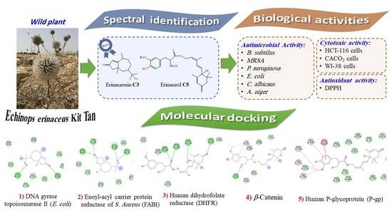
Phytochemical, Antimicrobial, Antioxidant, and In Vitro Cytotoxicity Evaluation of Echinops erinaceus Kit Tan

by
Sherouk Hussein Sweilam
1,2,
Fatma M. Abdel Bar
1,3,
Ahmed I. Foudah
1,
Mohammed H. Alqarni
1,
Nouran A. Elattal
4,
Omayma D. El-Gindi
2,
Moshera M. El-Sherei
5 and
Essam Abdel-Sattar
5,*
1
Department of Pharmacognosy, College of Pharmacy, Prince Sattam Bin Abdulaziz University, Al-Kharj 11942, Saudi Arabia
2
Department of Pharmacognosy, Faculty of Pharmacy, Egyptian Russian University, Cairo-Suez Road, Badr City, Cairo 11829, Egypt
3
Department of Pharmacognosy, Faculty of Pharmacy, Mansoura University, Mansoura 35516, Egypt
4
Department of Chemistry of Natural and Microbial Products, National Research Center, Dokki, Giza 12622, Egypt
5
Department of Pharmacognosy, Faculty of Pharmacy, Cairo University, Kasr El-Aini Street, Cairo 11562, Egypt
*
Author to whom correspondence should be addressed.
Separations 2022, 9(12), 447; https://doi.org/10.3390/separations9120447
Submission received: 1 December 2022 / Revised: 9 December 2022 / Accepted: 12 December 2022 / Published: 16 December 2022
(This article belongs to the Section Analysis of Natural Products and Pharmaceuticals)

Abstract

:
Wild plants are used by many cultures for the treatment of diverse ailments. However, they are formed from mixtures of many wanted and unwanted phytochemicals. Thus, there is a necessity to separate the bioactive compounds responsible for their biological activity. In this study, the chemical composition as well as antimicrobial and cytotoxic activities of Echinops erinaceus Kit Tan (Asteraceae) were investigated. This led to the isolation and identification of seven compounds, two of which are new (erinaceosin C3 and erinaceol C5), in addition to methyl oleate (C1) and ethyl oleate (C2), loliolide (C4), (E)-p-coumaric acid (C6), and 5,7,3`,5`-tetrahydroxy flavanone (C7). The structures of the isolated compounds were elucidated by 1D, 2D NMR, and HR-ESI-MS. The methanol extract showed the highest antimicrobial activity among the tested extracts and fractions. The n-hexane and EtOAc extracts showed remarkable antimicrobial activity against B. subtilus, P. aeruginosa, E. coli, and C. albicans. A cytotoxicity-guided fractionation of the most bioactive chloroform extract resulted in the isolation of bioactive compounds C1/C2, which showed significant cytotoxicity against HCT-116 and CACO2 cell lines (IC50 24.95 and 19.74 µg/mL, respectively), followed by compounds C3 (IC50 82.82 and 76.70 µg/mL) and C5 (IC50 99.09 and 87.27 µg/mL), respectively. The antioxidant activity of the bioactive chloroform fractions was screened. Molecular docking was used to explain the results of the antimicrobial and anticancer activities against five protein targets, including DNA gyrase topoisomerase II, enoyl-acyl carrier protein reductase of S. aureus (FabI), dihydrofolate reductase (DHFR), β-catenin, and human P-glycoprotein (P-gp).

Graphical Abstract

1. Introduction

Plants of the family Asteraceae demonstrate significant therapeutic applications because of their unique and diverse pool of secondary metabolites. The reported biological activities include antioxidant, antiproliferative, anti-ulcer, and anti-inflammatory activities. They are mainly due to their wide range of phytochemicals, such as phenolics [1], sesquiterpene lactones [2], alkaloids [3,4], and triterpenes [5,6]. Traditionally, the plants of the genus Echinops (Asteraceae) are used to relieve gastrointestinal disturbances [7], kidney inflammation [8], microbial infections, and pain [9,10]. In addition, other reported biological properties include hepatoprotective [11], antifertility [12], analgesic, antipyretic, wound-healing, anthelmintic [13,14], and insecticidal properties [9,15]. Echinops spp. contains thiophenes, terpenoids (such as sesqui- and triterpenoids), phenolics (such as flavonoids, coumarins, phenylpropanoids, and lignans), alkaloids, and essential oils [9,16]. Echinops erinaceus Kit Tan is an annual herbaceous plant strictly distributed in the Arabian Peninsula and is well-known in Saudi Arabia and Yemen under the name of “Alkana’a, Kanab, and Hawa elghool”. Reviewing the literature data, nothing was reported regarding the traditional uses of the plant. Our research group previously reported the phytochemical screening, antioxidant, and in vitro anti-inflammatory activities of E. erinaceus [17]. However, research studies that describe the phytochemical composition of this plant are still scarce.
Cancer is one of the most common diseases that cause death worldwide with an increased number of new cases every year. The common treatment for cancers is chemotherapy. Nowadays, patients are suffering from multidrug resistance (MDR), a phenomenon whereby cells confer drug resistance to structurally and functionally unrelated compounds. MDR may be a consequence of reduced drug influx, increased drug efflux, activation of detoxifying systems, activation of DNA repair mechanisms, evasion of drug-induced apoptosis, etc. [18]. One of the most common mechanisms in MDR is often associated with decrease in cellular drug accumulation mediated by MRP1 (ABCC1) and/or P-glycoprotein (P-gp, ABCB1). To overcome MDR, MRP1 and/or P-gp proteins inhibitors are used to block the transport function might be potentially used [19]. There is an urgent need for discovering new natural products against life-threatening multidrug-resistant microbial pathogens [20] and chemotherapy.
The current study aimed at bioguided isolation, structure elucidation, and in vitro biological evaluation of E. erinaceus. The in vitro biological evaluation targeted the cytotoxic activity against HCT-116 (colon carcinoma), CACO2 (human colorectal intestinal carcinoma) cell lines, and its selectivity was assessed using the normal mammalian cell line, WI-38 (human lung fibroblast) using crystal violet assay. The antimicrobial activities of the fractions and the isolated compounds were performed on six pathogens, including two Gram-positive (viz., B. subtilus and MRSA), two Gram-negative bacteria (viz., E. coli, P. aeruginosa), a fungus (viz., A. niger), and a yeast-like pathogen (viz., C. albicans). Moreover, to reveal the potential molecular mechanisms responsible for the multi-biological activities of the isolated compounds, molecular docking experiments were conducted against P-glycoprotein (P-gp), a key protein in MDR of anticancer drugs [21], and human dihydrofolate reductase (DHFR) involved in malignancies and multidrug-resistance of microbial pathogens [22], in addition to DNA gyrase topoisomerase II and enoyl-acyl carrier protein reductase of S. aureus (FabI) as targets for bacteria and β-catenin as a target for cancer.

2. Materials and Methods

2.1. Plant Material and Extraction

The flowering aerial parts of E. erinaceus Kit Tan were collected from Riyadh region, Saudi Arabia, in March 2018 [17]. The plant material was authenticated by Mohamed Abdel-Fattah, a taxonomist and botanist, the botanical garden of the Department of Botany and Microbiology, College of Science, King Saud University. A verifier specimen (ID: 23.6.19.1-5) was placed at the local herbarium of the Pharmacognosy Department, Faculty of Pharmacy, Cairo University. The powdered shade-dried plant material was extracted and fractionated according to the method reported by Sweilam et al. (2021). The crude MeOH extract, its fractions, and the isolated compounds (C1C7) from CHCl3 fraction were subjected to in vitro cytotoxic and antimicrobial investigations.

2.2. Isolation and Purification of Compounds from the CHCl3 Fraction

The powdered plant material (4 kg) was extracted by cold maceration with MeOH (5 × 10 L). The obtained extract was evaporated by a rotary evaporator (Büchi, Lugano, Switzerland) to a semisolid consistency (650 g) which was suspended in water and fractionated successively with solvents viz. n-hexane (Hex), chloroform (CHCl3), and ethyl acetate (EtOAc), to give 150 g, 50 g, and 60 g, respectively [17]. The CHCl3 fraction (38 g) was chromatographed on a Si gel column CC (170 × 5 cm2) and eluted with a mixture of CHCl3/MeOH (100/0 to 70/30). The effluent was monitored using TLC on Si gel GF245 plates and visualized by spraying with 10% vanillin-H2SO4 reagent (Flowchart S1). Fraction-1 (1.0 g) eluted with CHCl3 (100%) was further purified on a Si gel CC (50 × 1 cm2) with gradient elution (EtOAc in n-hexane; 1–10%) to afford subfraction-1-II (500 mg). The latter was purified on an MPLC RP-18 column (isopropanol–water, 6:4 to 10:0) to obtain compounds C1/C2 as an unresolved mixture (methyl oleate C1/ethyl oleate C2, 9 mg). Fraction-3 (1.0 g) was subjected to a Si gel CC (65 × 2 cm2) and eluted with EtOAc in n-hexane (10 to 50%) to obtain subfraction-3 I-IX. Subfraction-3-II (120 mg) and subfraction-3-III (200 mg) were purified by chromatography onto an MPLC RP-18 column (MeOH-H2O, 1:1 and 3:7, respectively) to give compounds C3 (erinaceosin, 8 mg) and C4 (loliolide, 4.5 mg), respectively. Fraction-4 was subjected to purification on an MPLC RP-18 column (MeOH-H2O, 3:7) followed by a Sephadex LH-20 column to afford compound C5 (erinaceol, 3 mg). In addition, Fraction-5 (1.5 g) gave subfraction-5-I (65 mg) and subfraction-5-II (36 mg) upon chromatography on a Si gel column (EtOAc in n-hexane; 10 to 100). Purification of subfraction-5-I and subfraction-5-II on Sephadex LH-20 columns (MeOH-CH2Cl2, 1 to 10) resulted in the isolation of compounds C6 (E-p-coumaric acid, 4 mg) and C7 (5,7,3`,5`-tetrahydroxy flavanone, 3 mg), respectively (Flowchart S1).

2.3. In Vitro Cytotoxicity Assay

2.3.1. Materials and Cell Lines

Mammalian cell lines HCT-116 cells (human colon cancer cell line), CACO2 cells (human colorectal intestinal carcinoma), and WI-38 cells (human lung fibroblast normal cells) were obtained from the American Type Culture Collection (ATCC, Rockville, MD, USA). Dimethyl sulfoxide (DMSO), crystal violet, and trypan blue dye were purchased from Sigma (St. Louis, MO, USA). Fetal Bovine serum, RPMI-1640, HEPES buffer solution, L-glutamine, gentamycin, and 0.25% Trypsin-EDTA were purchased from Lonza (Verviers, Belgium). Crystal violet stain (1%) made from 0.5% (w/v) crystal violet and 50% MeOH was then brought to volume with dd.H2O and filtered through a Whatmann No.1 filter paper.

2.3.2. Cell Culture Condition and Propagation

The HCT-116 and CACO2 cells were propagated in RPMI-1640 medium, and WI-38 cells were propagated in Dulbecco’s modified Eagle’s medium (DMEM), which was supplemented with 10% heat-inactivated fetal bovine serum, 1% L-glutamine, HEPES buffer, and 50 µg/mL gentamycin. All cells were maintained at 37 °C in a humidified atmosphere with 5% CO2 and were subcultured two times a week [23,24,25].

2.3.3. Cytotoxicity Evaluation Using Viability Assay

The cells were seeded in a 96-well plate at a cell concentration of 1 × 10⁴ cells per well in 100 µL of growth medium. Fresh medium containing different concentrations of the test sample was added after 24 h of seeding. Two-fold serial dilutions of the tested sample were added to confluent cell monolayers dispensed into 96-well, flat-bottomed microtiter plates (Falcon, NJ, USA) using a multichannel pipette. The microtiter plates were incubated at 37 °C in a humidified incubator with 5% CO2 for a period of 48 h. Three wells were used for each concentration of the test sample. Control cells were incubated without a test sample and with or without DMSO. The low percentage of DMSO present in the wells (max 0.1%) was found not to affect the experiment. After incubation of the cells at 37 °C, various concentrations of samples were added, and the incubation was continued for 24 h, and viable cells’ yield was determined by a colorimetric method. All experiments were carried out in triplicate [23,24,25].
The 50% inhibitory concentration (IC50, the concentration required to cause toxic effects in 50% of cancer cells) and the cytotoxic concentration (CC50, the concentration required to cause toxic effects in 50% of normal cells) were determined from graphic plots of the dose–response curve for each concentration using Graphpad Prism software (San Diego, CA, USA).

2.4. In Vitro Antimicrobial Activity

The antimicrobial activity of E. erinaceus extracts was tested by the agar well diffusion method [26] against six micro-organisms (Bacillus subtilus, MRSA, Pseudomonas aeruginosa, and Escherichia coli on nutrient agar, and Candida albicans, and Asperigllus niger on potato dextrose agar, PDA). In this experiment, all extracts were dissolved in MeOH (200 μg/mL), and 50 µL of the prepared sample solution was added to each well, separately in each case. The diameter of inhibition zone (DIZ) was measured.

2.5. In Vitro Antioxidant Effect

The in vitro antioxidant activity of fractions 3, 4, and fraction 5 of the CHCl3 extract (Flowchart S1) was established in accordance with Burits and Bucar [27]. In brief, 50 µL of the sample solution (200 μg/mL) was added to 100 µL of 2,2-diphenyl-1-picrylhydrazyl (DPPH) methanolic solution (0.1%). After a period of 50 min incubation in the dark, absorbance was measured at 517 nm against the blank. The inhibition percentage of DPPH free radicals (I) was as follows:
I (%) = (A blank − B sample/A blank) × 100
where A (blank) is the absorbance of control (containing all reagents, except the test compound), and B (sample) is the absorbance of the test sample; ascorbic acid is used as a standard drug.

2.6. In Silico Studies of the Isolated Compounds

2.6.1. PASS and ADME Predictions

The isolated compounds (Figure 1), namely, methyl oleate (C1), ethyl oleate (C2), erinaceosin (C3), loliolide (C4), erinaceol (C5), (E)-p-coumaric acid (C6), and 5,7,3`,5`-tetrahydroxy flavanone (C7), were drawn by MarvinSketch program and simulated by the Prediction of Activity Spectra for Substances (PASS) and Absorption, Distribution, Metabolism, and Elimination (ADME) prediction web tools. The isolated compounds were analyzed by SwissADME online free site for prediction of physicochemical properties, drug likeness, solubility, and pharmacokinetics [28,29,30].

2.6.2. Molecular Docking Analysis

Docking analyses of the isolated compounds were accomplished to understand the antibacterial and anticancer activities. For antibacterial activity, DNA gyrase topoisomerase II (E. coli) enzyme (PDB ID: 1KZN) [31] and enoyl-acyl carrier protein reductase of S. aureus, FabI (PDB ID: 3GNS) [32] were used as the target proteins. For anticancer activity, β-catenin in complex with compound 6 (PDB ID: 7AFW) [33] was used. For both activities, dihydrofolate reductase (DHFR) (PDB ID: 4M6J) [34] has been chosen as the target protein. The study of the MDR of the anticancer and antimicrobial candidates, the human P-gp (PDB ID: 6C0V) [21], were downloaded from PDB (https://www.rcsb.org (accessed on 1 December 2022)). The downloaded proteins were prepared by removing water and any unwanted residual matter and adding non-hydrogen atoms by PyMOL 2.3. The PyRx Autodock Vina (Scripps Research, La Jolla, CA, USA) was utilized for these in silico studies. The co-crystalized ligands, including clorobiocin, triclosan, 3-[(24-methyl-5-oxidanylidene-2,3-dihydro-1,4-benzoxazepin-2-yl]benzenecarbonitrile (R9Q), ciprofloxacin, and methotrexate for studying the comparative binding affinity to the target proteins, were downloaded and saved in 3D SDF format.
Figure 1. Structures of isolated compounds from Echinops erinaceus (C1, C2, C3C7).
Separations 09 00447 g001

3. Results and Discussion

3.1. Identification of the Isolated Compounds

The spectral data of the known compounds (C1, C2, C4, C6, and C7) are recorded in Tables S1–S4, while the spectra of all isolated compounds (C1C7) are displayed in Figures S1–S37.

3.1.1. Identification of Compounds C1 and C2

The mixture of compounds C1 and C2 was identified as a mixture of two esterified mono-unsaturated long-chain fatty acids using 1D, 2D NMR, and ESI-MS data (Figure 1, Table S1) [35,36]. It is worth noting that methyl oleate (C1) has been isolated before from the Echinops genus [37]; however, this is the first report on the identification of ethyl oleate (C2) from the Echinops genus, although it was reported before in the Asteraceae family [38].

3.1.2. Identification of Compound C3

According to HR-ESI-MS, compound C3 had a molecular formula of C15H24O2 based on the ion peak at m/z 237.1859 [M + H, 1.5%] (calcd. 237.1855) and 219.1753 [M+ − OH, 85%] (calcd. 219.1749). The NMR spectral data of compound C3 (Table 1) were consistent with the basic skeleton of pseudoguaiane sesquiterpenes, except for the presence of the α, β-unsaturated ketone of a cyclopentenone ring observed at positions 1, 2, and 3 [39]. The 1H-NMR spectrum of C3 exhibited four methyl signals at δH 1.09 (s, C-13), 1.07 (s, C-12), 1.04 (s, C-15), and 0.96 (d, J = 6.7 Hz, C-14), which were correlated in the HSQC spectrum with the carbon signals at δC 28.1, 26.1, 20.1, and 16.2, respectively. The 13C-NMR spectrum displayed fifteen carbon signals, categorized as four methyls (δC 26.1, 28.1, 20.1, and 16.2 assigned to C-12, C-13, C-15, and C-14, respectively), four methylenes (δC 43.5, 35.9, 30.6, and 27.6 assigned to C-4, C-6, C-9, and C-8, respectively), three methines (δC 125.9, 43.4, and 36.5 assigned to C-2, C-7, and C-10, respectively), and four quaternary carbon atoms at δC 202.5 (C-3), 179.8 (C-1), 73.4 (C-11), and 41.9 (C-5). These data, along with additional information provided by 1H-1H spin interactions between the coupled protons for H-6/H-7, H-7/H-8, H-8/H-9, and H-9/H-10, were observed in the COSY spectrum (Table 1 and Figure S11) establishing a heptocyclic moiety of the sesquiterpene skeleton. The HMBC correlations (Figure 2 and Figure S12) between the protons at δH 1.07 (s, H-12) and 1.09 (s, H-13) with the carbon signals at δC 73.4 (C-11) and 43.4 (C-7) indicated the presence of a hydroxyisopropyl group attached to C-7. In addition, the presence of HMBC correlations of H-2/C-5 and H-2/C-9 and H-4/C-1, H-4/C-2, H-4/C-3, H-4/C-5, and H-4/C-6 confirmed the presence of a cyclopentanone ring system. From the aforementioned data, compound C3 was identified as a new natural compound named erinaceosin (Figure 1).
Table 1. NMR spectral data of compound C3 in CD3OD (500 MHz for 1H- and 125 MHz for 13C-NMR).
HSQC HMBC (HC) COSY
TypeδH (J in Hz) δC 2JCH3JCH4JCH1H-1H
1C 179.8
2CH5.76, s125.9 C-5C-9
3C=O 202.5
4CH22.27, m; 2.21, m, overlapped43.5C-3, C-5C-1, C-2, C-6
5C 41.9
6CH21.23, m; 1.88, m35.9C-5C-1, C-8, C-11C-10, C-13H-7
7CH1.36, m43.4C-6, C-11C-9C-4H-6, H-8
8CH21.67, m27.6C-9C-11C-1H-7, H-9
9CH22.21, m, overlapped; 2.50, m30.6 C-1, C-2, C-5, C-6H-10, H-8
10CH2.22, overlapped 36.6C-1C-2, C-5C-3, C-7H-14
11C 73.4
12CH31.07, s26.1C-11 C-6, C-8
13CH31.09, s28.1C-11C-12
14CH30.96, d (6.7) 16.2C-10 C-5H-10
15CH31.04, s20.1C-5C-1, C-4, C-6
Figure 2. (a) Selected COSY correlations and (b) HMBC correlations of the new compounds (C3 and C5).
Separations 09 00447 g002

3.1.3. Identification of Compound C4

The HR-ESI-MS of compound C4 (Figure S21) indicated a molecular formula of C11H16O3 based on the ion peak at m/z 197.1156 [M + H]+, 100%; 219.0973 [M + Na] +, 70%. The detailed study of MS, 1D, and 2D NMR data (Table S2 and Figures S14–S20) identified the structure of C4 as loliolide, which was further confirmed by comparison of its spectral data with those reported in the literature. This compound was reported from genus Echinops for the first time and previously isolated from Codium tomentosum and Xanthium spinosum [40,41].

3.1.4. Identification of Compound C5

Compound C5 displayed an m/z of 575.1975 [M + 3K] + (100%, cal. 575.1216) in the positive mode of the HR-ESI-MS spectrum (Figure S29) coincident with a molecular formula of C26H34O7K3. One- and two-dimensional NMR spectral data of C5 (Table 2 and Figures S22–S27) showed the possible presence of an abscisic alcohol moiety [42,43,44,45], which was confirmed by the presence of four tertiary methyl singlets at δH 1.85 (H3-12; δC 20.2), 1.95 (H3-13; δC 21.8), 0.98 (H3-14; δC 24.1), and 0.94 (H3-15; δC 25.2); two trans-coupled olefinic proton doublets at δH 7.66 (J = 16.1 Hz, H-8; δC 129.9) and 6.13 (J = 16.1 Hz, H-7; δC 138.2); and two proton signals at δH 5.67 (1H, br.s, H-10; δC 120.4) and 5.85 (1H, s, H-3; δC 128.0). The latter two double bonds, 2(3) and 9(10), bear two methyl substituents (i.e., H3-13 and H3-12) attached to C-2 (δC 152.2) and C-9 (δC 131.0), respectively. The presence of a hydroxy methylene group was assigned based on the proton multiplet at δH 3.45 (H-11; δC 62.6). The triene system (Δ2(3), Δ7(8), and Δ9(10)) is connected to a carbonyl carbon at δC 203.0 (C-4), and the connection was confirmed by the HMBC spectrum. This was evident from the HMBC correlations of the two methylene proton signals at δH 2.11 and 2.45 (Ha-5 and Hb-5) with the ketonic group at C-4. Other significant HMBC correlations, including Ha-5/C-1, H-14/C-1, H-15/C-1, H-13/C-2, H-12/C-8, H-3/C-13, H-3/C-1, H-7/C-1, H-8/C-1, and H-11/C-8, were also used to confirm the presence of an abscisic alcohol moiety (Table 2 and Figure S27).
In addition to the abscisic alcohol moiety, a set of aromatic ABX systems were revealed from the doublet signal at δH 6.70 (d, J = 7.8 Hz, H-5`), a broad doublet at δH 6.45 (br.d, J = 7.8 Hz, H-6`), and a broad singlet at δH 6.49 (1H, br.s, H-2`), which are directly correlated to carbon signals at δC 116.2, 123.2, and 113.7, respectively (HSQC). The singlet proton signal at δH 3.64 (3H, s) correlated with the carbon at δC 56.6 (HSQC) and was assigned to a m-methoxy substituent at C-3`of the aromatic ring. A third moiety formed from a 3-hydroxy-2-methylpropanoic acid chain (HMPA) was attached to C-1` of the aromatic ring and esterified the abscisic alcohol moiety at C-11. The HMPA moiety was deduced from the presence of a terminal hydroxy methylene group revealed from a proton signal that appeared as a doublet of doublet (J = 8.8, 4.5 Hz; H-10`) overlapped with the proton multiplet of H2-11 of the abscisic alcohol moiety at δH 3.45 (4H). This proton signal is directly correlated with the carbon at δC 62.6 (i.e., coincident signals of C-10`/C-11). Additionally, the NMR data revealed the presence of two proton multiplets of a methylene group of H2-7` (δH 2.49 and 2.65; δC 36.5), which is correlated in the HMBC spectrum with C-8, C-1`, C-2`, and C-6`, confirming its attachment to the aromatic ring. Finally, the proton signal at δH 1.84 (1H, m, H-8`, δC 44.5), forming the branching point of the side chain formed by C7`-C-8`-C-10`, showed an HMBC correlation with the ester carbonyl group at δC 169.4 (C-9`). The above-mentioned data suggested a 4-(3-hydroxypropyl)-2-methoxyphenol moiety that is closely related to the previously published data of 5-(3-hydroxypropyl)-2-methoxyphenol derivative [46]. The analysis of the COSY and HMBC spectra of compound C5 (Table 2) established the connections of the assigned protons and carbons. From the aforementioned discussion, the structure of C5 was confirmed to be a new derivative composed of an abscisic alcohol moiety esterified with a modified phenylpropane carboxylic acid moiety and was named erinaceol, which is recorded herein for the first time from nature.
Table 2. NMR spectral data of compound C5 in CD3OD (500 MHz for 1H- and 125 MHz for 13C-NMR).
C/H#HSQCHMBC (HC) COSY
TypeδH (J in Hz) δC 2JCH3JCH4JCH1H-1H
Abscisic alcohol moiety
1C 82.1
2C 152.2
3CH5.85, 1H, s128.0 C-1, C-13
4C 203.0
5CH22.11, m; 2.45, m51.1C-4, C-6C-1, C-14, C-15
6C 43.4
7CH6.13, d (16.1)138.2C-1, C-8C-2, C-9 H-7
8CH7.66, d (16.1)129.9C-7C-1, C-10, C-12 C-2H-8
9C 131.0
10CH5.67, br.s120.4C-9C-12
11CH23.45, m a62.6 bC-8C-8`
12CH31.85, s 20.2 C-8, C-10C-9`
13CH31.95, s21.7C-2C-1, C-3
14CH30.98, s24.1C-6C-1, C-5, C-15
15CH30.94, s25.1C-6C-1, C-5, C-14C-4
4-(3-Hydroxypropyl)-2-methoxyphenol moiety
1`C 134.4
2`CH6.49, br. s113.7C-3`C-4`, C-7`, C-6`
3`C 149.3
4`C 146.4
5`CH6.70, d (7.8) 116.2C-4`C-3`, C-1` H-6`
6`CH6.45, br. d (7.9)123.2 C-4`, C-2` H-5`
7`CH22.49, m; 2.65, m36.5C-1`,
C-8`
C-2`, C-6` H-8`
8`CH1.84, m44.5C-9` H-7`,
H-10`
9`C 169.4
10`CH23.45, dd (8.8, 4.5) a62.6 bC-8`
O-CH33.64, s56.6 C-3`
a, b Similar letters indicate coincident signals.

3.1.5. Identification of Compound C6

Compound C6 was identified as (E)-p-coumaric acid from the 1H-NMR and APT spectra (Table S3 and Figures S30 and S31) and by co-chromatography with an authentic sample of (E)-p-coumaric acid, which was isolated for the first time from E. erinaceus [47].

3.1.6. Identification of Compound C7

The spectral analysis of C7 (Table S4 and Figures S32–S37) confirmed its structure as 5,7,3`,5`-tetrahydroxy flavanone [48], and it is worth mentioning that this is the first report of C7 from Echinops spp.

3.2. Biological Activities of Main Fractions and Isolates from E. erinaceus

3.2.1. In Vitro Cytotoxic Activity

The results of bio-guided cytotoxic activity against HCT-116 and CACO2 cells of the different extracts showed that the CHCl3 extract showed the highest activity among the tested extracts (Table 3). However, it exhibited moderate cytotoxic activity with IC50 of 67.30 ± 4.87 and 81.95 ± 4.63 µg/mL with a selectivity index (SI) > 1 (1.73 and 1.42) against HCT-116 and CACO2 cells, respectively. Further bio-guided fractionation of the CHCl3 extract revealed that fractions Fr.1, Fr.3, and Fr.4 were the most active among the tested fractions. Considering Fr.1, it showed the strongest activity with IC50 of 14.93 ± 1.28 and 10.50 ± 0.61 µg/mL and SI of 3.37 and 4.80 against HCT-116 and CACO2 cells, respectively. Compound C1/C2 showed significant antiproliferative activity with IC50 of 24.95 ± 1.23 and 19.74 ± 1.94 µg/mL and good SI (1.95 and 2.47) against the tested cells, respectively. The new compound (C3) purified from Fr.3 showed a weak cytotoxic activity (IC50 82.82 ± 3.94 and 99.09 ± 5.84 µg/mL) with good SI (2.15 and 1.80), respectively. Similarly, compound C5 obtained from Fr.4 showed weak cytotoxicity (IC50 76.70 ± 3.71 and 87.27 ± 4.67 µg/mL, respectively) and good SI (2.12 and 1.87, respectively) (Table 3).

3.2.2. In Vitro Antimicrobial Activity

The different extracts and fractions of E. erinaceus were tested for their antimicrobial activity against a panel of pathogenic micro-organisms, including two strains of Gram-positive bacteria (Bacillus subtilus and methicillin-resistant Staphylococcus aureus, MRSA), two strains of Gram-negative bacteria (Pseudomonas aeruginosa and Escherichia coli), and two fungus and yeast-like micro-organisms (Asperigllus niger and Candida albicans), using the agar well diffusion assay by measuring the diameter of inhibition zone (DIZ). The results of the antibacterial properties of the plant extracts (Table 4) demonstrated that the total MeOH extract had the highest antimicrobial activity against all the tested strains, except against MRSA. It showed significant antibacterial activity against B. subtilus (27.5 ± 0.7 mm), which is more active than the reference drug, streptomycin (18 ± 1.41 mm). It also showed a pronounced antifungal activity against C. albicans (26 ± 1.41 mm), which is almost comparable to the reference drug, clotrimazole (28 ± 2.82 mm). This was followed by the n-hexane and EtOAc extracts, which showed strong antibacterial and antifungal activities. However, no antimicrobial activity against MRSA was detected in any of the investigated E. erinaceus samples. The CHCl3 extract showed good activity against B. subtillis (20.5 ± 1.41 mm), P. aeruginosa (17.5 ± 1.41 mm), and E. coli (18 ± 1.41 mm) test strains. Fr.3 of the CHCl3 extract showed the highest antimicrobial effect compared to other fractions against the same bacterial strains as its main extract. However, the CHCl3 extract and its fractions (Fr.3, Fr.4, and Fr.5) showed no activity against the tested fungal strains, C. albicans and A. niger. The aqueous extract showed good activity against all strains, except MRSA and A. niger (Table 4).

3.2.3. Antioxidant Activity

DPPH free radical scavenging activities of Fr.3, Fr.4, and Fr.5 of the CHCl3 fraction were assessed. Fr.3 exhibited remarkable free radical scavenging activity (55%), whereas the lowest activity was observed for sample Fr.4 (32%) (Figure 3).

3.3. PASS and ADME Predictions of the Isolated Compounds

The isolated compounds were sketched using the Marvinsketch program and simulated by the Prediction of Activity Spectra for Substances (PASS) and Absorption, Distribution, Metabolism, and Elimination (ADME) prediction web tools [28,29,30]. The PASS tool predicted several biological activities. The isolated compounds (C1/C2, C3C7) displayed significant probable activity “Pa” ranges, including antioxidant (0.828–0.297), anticancer (0.746–0.343), anti-inflammatory (0.717–0.325), antifungal (0.593–0.364), and antibacterial (0.455–0.176) activities (Table S5). The ADME prediction results showed that all compounds showed a great bioavailability score ranging from 0.85 to 0.55 and fulfilled all drug-likeness rules and synthetic accessibility (1.61–4.14), which showed an explicit synthetic route. Additionally, all compounds were predicted to be moderate to very soluble in water. In addition, skin permeation and ADME properties were analyzed by the Swiss-ADME software, as recorded in Table S5. The BOILED-Egg method [50] showed that compounds C3C6 have high GI absorption properties with high predicted diffusion through the BBB and good skin permeability (log Kp). However, compound C7 showed high predictable GI absorption and skin permeability properties but may diffuse poorly through the BBB [51]. Compounds C1C6 showed no predictable binding to P-gp, except for C7, which may suffer from cellular efflux. Regarding inhibition of metabolic enzymes, compound C1 may inhibit CYP1A2, whereas C7 may inhibit CYP3A4, resulting in potential drug–drug interactions and adverse effects [28]. The findings revealed that five out of the seven compounds fulfilled the oral drug ability of Lipinski’s rule of five (RO5), while two slightly met the criteria of RO5.

3.4. In Silico Docking Study of the Isolated Compounds

A docking study was executed for the isolated compounds from E. erinaceus against five explored molecular targets, including DNA gyrase topoisomerase II (PDB ID: 1KZN) [31], enoyl-acyl carrier protein reductase of S. aureus, FabI (PDB ID: 3GNS) [32], and dihydrofolate reductase (DHFR) (PDB ID: 4M6J) [34] as targets for bacteria, and β-catenin (PDB ID: 7AFW) [33] in addition to human P-glycoprotein (P-gp) (PDB ID: 6C0V) [21] as targets for cancer. The results showed that the isolated phenolics and sesquiterpenes were predicted to have remarkable in silico binding affinities against the investigated molecular targets.

3.4.1. Docking against Antimicrobial Molecular Targets

Interactions with DNA Gyrase Topoisomerase II

DNA gyrase topoisomerase II is a bacterial enzyme that regulates the topological properties of bacterial DNA, especially of E. coli. Compounds (C1C6) demonstrated moderate–weak binding affinities toward this protein, as depicted by their binding free energy (BFE) values ranging from −4.1 to −5.7 kcal/mol (Table S6) compared to the reference ligand, clorobiocin (CBN, −7.4 kcal/mol). Of these, compound C3 (BFE, −5.7 kcal/mol) showed H-bonding interactions with Ala96 and the crucial amino acid, Gly117 (Figure 4a), while compound C5 (BFE, −5.7 kcal/mol) showed H-bonding interactions with Ala96 and Ser121 (Figure 4b) [52]. On the other hand, compound C7 exhibited better BFE (−7.7 kcal/mol) than CBN, which can be explained by the presence of several binding interactions with the amino acid residues (Table S6), including strong H-bondings with Glu42, Ser121, and the crucial Val120 residue (Figure 4c) [52]. Therefore, C7 could contribute to the antibacterial activity of MeOH and CHCl3 extracts.

Interactions with Enoyl-Acyl Carrier Protein Reductase (FabI)

FabI protein is one of the critical targets for discovering antimicrobial compounds. It has been found in several bacteria, especially in E. coli and S. aureus. From the docking results (Table S6), compounds C4, C-5, and C6 have comparable BFE values (−5.9, −6.0, and −5.8 kcal/mol, respectively) to the reference FabI inhibitor, Triclosan (BFE, −5.8 kcal/mol) [32], while C3 and C7 have greater BFE (−6.3 and −6.9 kcal/mol, respectively). It was observed that the new compound C3 showed strong H-bonding interactions with Tyr39, Arg45, Gln64, and Glu72 (Figure 5a), whereas compound C5 (Figure 5b) shared Triclosan in binding with the amino acids Ile20 and Leu196 through the π-alkyl/alky interaction and formed strong H-bondings with Gly13, Ala21, Ser93, and Ser121 [52]. Finally, C7 was able to interact with several residues, such as Ala15, Ile20, Gly13, and Ser93 (Table S6 and Figure 5c). Consequently, it could be concluded that compounds C3C5 and C7 may be participating in the observed antimicrobial activities of the investigated E. erinaceus extracts.

3.4.2. Docking against Cytotoxic Molecular Targets

Interactions with β-Catenin

In carcinogenesis, the Wnt/β-catenin signal pathway regulates cell proliferation, differentiation, and embryonic development [53]. β-catenin is a signaling molecule in the Wnt pathway, which plays a central role in carcinogenicity [33,53]. Any impairment or activation of the Wnt pathway leads to cancerous diseases, such as breast, intestine, and prostate cancers [33]. The isolated compounds (C1C7) were docked into the active site of β-catenin (PDB ID: 7AFW). They were found to bind to 205–210 and 243–251 amino acid residues with BFE between −4.2 and −6.0 kcal/mol (Table S6) [33]. The ligand C5 showed a BFE of −5.1 kcal/mol compared to the co-crystallized inhibitor R9Q (−6.2 kcal/mol) and formed a distinct complement to the binding site while orienting the hydroxyl group toward the Thr205, and the benzene ring formed π-alkyl interactions with the backbone CH of Lys242 (Figure 6c). This moderate interaction was in full agreement with the obtained cytotoxic activity of C5 (Table 3) and suggested the Wnt/β-catenin signal pathway as a potential mechanism for its cytotoxicity. Regarding compounds C1 and C2, they showed low BFE values (−4.3 and −4.2 kcal/mol), although C1 shared the co-crystallized inhibitor in binding with Asn206. Both compounds (C1 and C2) interacted hydrophobically with Lys242 and Pro247 residues through π-alkyl interactions (Figure 6a,b) [33]. However, these results contradicted the obtained high cytotoxic activities of C1/C2, which suggested that they may act through another mechanism.

Interactions with Human Dihydrofolate Reductase (DHFR)

The human dihydrofolate reductase (DHFR) protein has a major role in DNA synthesis in the human and bacterial cell development process. Therefore, it could be considered a common target for both cytotoxicity and antimicrobial activities [34]. The tested compounds (C1C7) displayed variable binding affinity to the DHFR protein with BFE values ranging from −3.8 to −6.3 kcal/mol in comparison with ciprofloxacin (−5.5 kcal/mol) and methotrexate (−7.1 kcal/mol) (Table S6). Compound C5 showed good BFE (−5.3 kcal/mol), which is comparable to ciprofloxacin, and showed H-bonding interactions with the amino acids Gly20, Ser118, Asp145, and Thr146 (Figure 7a), which is in full agreement with the result of the cytotoxic activity of this compound [33,34]. Notably, compound C7 exhibited the highest BFE to DHFR enzyme (−6.3 kcal/mol) among the other isolated compounds and shared ciprofloxacin in binding with Arg77, Ser118, and Ser119 residues (Figure 7b) [34]. Although compounds C1 and C2 showed low binding affinities, they exhibited binding to Ser119 and Lys55 residues and shared methotrexate in the interaction with Gly20 and Thr146 residues (Table S6) [34].

Interactions with Human P-gp

The P-gp protein is a vital protein in MDR to anticancer drugs and other therapeutics by causing cellular efflux. Therefore, there is a necessity to discover safe MDR inhibitors [21]. Natural products represent a major source of safely used chemotherapeutic drugs [54]. The isolated compounds from E. erinaceus were virtually investigated for their binding to P-gp protein. In general, the in silico docking experiments predicted quite strong affinities of the investigated compounds toward P-gp, with BFE values ranging between −6.1 and −8.4 kcal/mol. Based on published data by Marques et al., 2021 [21], the preferred binding positions of the P-gp macromolecule are M or H sites. The inhibitors showed strong hydrophobic interactions at the M site of the macromolecule, the human P-gp (PDB ID: 6C0V), provided mainly by the multiple isoleucine, leucine, phenylalanine, serine, and tyrosine residues in the binding site (namely Ile340, 731, 735; Leu332, 339; Phe72, 314, 335, 336, 728, 732, 759, 983; Ser733, 979; and Tyr307, 310). Compound C3 (BFE, −7.7 kcal/mol) exhibited hydrophobic interactions with Phe335, Leu339, Phe728, and Phe759 residues (Figure 8a). This was also observed in the case of C5 (BFE, −8.4 kcal/mol), which showed several hydrophobic interactions with Phe335, Leu339, Phe759, Ile731, and Ile735 (Figure 8b). In some cases, such as in C1, C6, and C7, interactions with the hydroxyl or carbonyl groups of these ligands with Ser979, Ile736, and Ala729, respectively, were observed (Figure S42a,g and Figure 8c). On the other hand, the hydroxyl or carbonyl groups present in other ligands, including C2, C3, C4, and C5, did not contribute much to any hydrophilic binding affinity (Table S6). Thus, the investigated compounds may act as potential MDR inhibitors via binding to P-gp protein.

4. Conclusions

The current study includes the chemical and biological evaluations of the Saudi wild plant E. erinaceus. Cytotoxic, antioxidant, and antimicrobial activities were investigated. The phytochemical investigation led to the isolation and identification of seven bioactive phytochemicals, including two unsaturated fatty acid esters, a pseudoguaiane sesquiterpene, a sesquiterpene lactone, two phenolics, and an abscisic alcohol derivative. Among the isolated compounds, two compounds were identified for the first time from nature viz., erinaceosin (a pseudoguaiane) and erinaceol (an abscisic alcohol derivative). In general, the results of this study demonstrated that the title plant has weak to moderate cytotoxic activity, however, it showed promising antibacterial, antifungal, and antioxidant properties. The results of the antimicrobial properties of the plant extracts and/or the fractions demonstrated that the total MeOH extract had the highest antimicrobial activity against all the tested strains except against MRSA. Compounds C1/C2 showed the highest cytotoxic activity against HCT-116 and CACO2 cell lines. This research also demonstrated that the secondary metabolites isolated from E. erinaceus have variable degrees of binding affinity towards the active sites of selected target proteins, including DNA gyrase topoisomerase II, FabI, β-catenin, DHFR, and P-gp, which may contribute to their antimicrobial, anticancer, and multidrug-resistance inhibition mechanisms.

Supplementary Materials

The following supporting information can be downloaded at: https://www.mdpi.com/article/10.3390/separations9120447/s1, Figure S1–S37: Spectra of C1C7; Tables S1–S4: NMR spectral data of C1, C2, C4, C6, and C7; Table S5: In silico physicochemical and pharmacokinetics of C1C7; Table S6: Docking results of C1C7 against PDB ID:1KZN, 3GNS, 7AFW, 4M6J, and 6C0V; Flowchart S1: Fractionation and purification of the CHCl3 extract of E. erinaceus; Figure S38–S42: 2D molecular docking interactions of C1C7 against PDB ID:1KZN, 3GNS, 7AFW, 4M6J, and 6C0V; Figure S43: Bioavailability radar representation of C1C7; and Figure S44: Predicted BOILED-Egg diagram of C1C7. Refs. [55,56] is cited in Supplementary Materials.

Author Contributions

Conceptualization, E.A.-S., M.M.E.-S. and O.D.E.-G.; methodology, S.H.S., F.M.A.B., A.I.F., M.H.A. and N.A.E.; compounds analysis and validation, S.H.S., F.M.A.B. and E.A.-S.; molecular docking software, S.H.S.; in vitro biological investigations, A.I.F., M.H.A. and N.A.E.; resources, A.I.F. and M.H.A.; writing-original draft, S.H.S., F.M.A.B. and E.A-S.; review & editing, all authors; supervision, M.M.E.-S., O.D.E.-G. and E.A.-S. All authors have read and agreed to the published version of the manuscript.

Funding

This research received no external funding.

Data Availability Statement

The following supporting information can be downloaded at: https://www.mdpi.com/article/10.3390/separations9120447/s1, Figures S1–S37: Spectra of C1C7; Tables S1–S4: NMR spectral data of C1, C2, C4, C6, and C7; Table S5: In silico physicochemical and pharmacokinetics of C1C7; Table S6: Docking results of C1C7 against PDB ID:1KZN, 3GNS, 7AFW, 4M6J, and 6C0V; Flowchart S1: Fractionation and purification of the CHCl3 extract of E. erinaceus; Figures S38–S42: 2D molecular docking interactions of C1C7 against PDB ID:1KZN, 3GNS, 7AFW, 4M6J, and 6C0V; Figure S43: Bioavailability radar representation of C1C7; and Figure S44: Predicted BOILED-Egg diagram of C1C7.

Acknowledgments

The authors would like to express a special thanks of gratitude to members of the Department of Pharmacognosy, Faculty of Pharmacy, Prince Sattam Bin Abdulaziz University, for their kind help and the use of lab facilities.

Conflicts of Interest

The authors declare no conflict of interest.

References

  1. Senejoux, F.; Demougeot, C.; Karimov, U.; Muyard, F.; Kerram, P.; Aisa, H.A.; Girard-Thernier, C. Chemical constituents from Echinops integrifolius. Biochem. Syst. Ecol. 2013, 47, 42–44. [Google Scholar] [CrossRef]
  2. Dong, M.; Cong, B.; Yu, S.-H.; Sauriol, F.; Huo, C.-H.; Shi, Q.-W.; Gu, Y.-C.; Zamir, L.O.; Kiyota, H. Echinopines A and B: Sesquiterpenoids Possessing an Unprecedented Skeleton from Echinops spinosus. Org. Lett. 2008, 10, 701–704. [Google Scholar] [CrossRef] [PubMed]
  3. Khadim, E.J.; Abdulrasool, A.A.; Awad, Z.J. Phytochemical Investigation of Alkaloids in the Iraqi Echinops heterophyllus (Compositae). Iraqi J. Pharm. Sci. 2014, 23, 26–34. [Google Scholar] [CrossRef]
  4. Kiyekbayeva, L.; Mohamed, N.M.; Yerkebulan, O.; Mohamed, E.I.; Ubaidilla, D.; Nursulu, A.; Assem, M.; Srivedavyasasri, R.; Ross, S.A. Phytochemical constituents and antioxidant activity of Echinops albicaulis. Nat. Prod. Res. 2018, 32, 1203–1207. [Google Scholar] [CrossRef] [PubMed]
  5. Lan, H.; Qi-Rong, C.; Rong, L.; Guo-Qiang, L.; Hao, H. A new pentacyclic triterpene, gmeliniin A, from Echinops gmelinii Turcz. Chin. J. Chem. 2000, 18, 112–114. [Google Scholar] [CrossRef]
  6. Rolnik, A.; Olas, B. The Plants of the Asteraceae Family as Agents in the Protection of Human Health. Int. J. Mol. Sci. 2021, 22, 3009. [Google Scholar] [CrossRef]
  7. Bulut, G.; Haznedaroğlu, M.Z.; Doğan, A.; Koyu, H.; Tuzlacı, E. An ethnobotanical study of medicinal plants in Acipayam (Denizli-Turkey). J. Herb. Med. 2017, 10, 64–81. [Google Scholar] [CrossRef]
  8. Menut, C.; Lamaty, G.; Weyerstahl, P.; Marschall, H.; Seelmann, I.; Amvam Zollo, P.H. Aromatic plants of tropical Central Africa. Part XXXI. Tricyclic sesquiterpenes from the root essential oil of Echinops giganteus var. lelyi C. D. Adams. Flavour Fragr. J. 1997, 12, 415–421. [Google Scholar] [CrossRef]
  9. Bitew, H.; Hymete, A. The Genus Echinops: Phytochemistry and Biological Activities: A Review. Front. Pharmacol. 2019, 10, 1234. [Google Scholar] [CrossRef] [Green Version]
  10. Mustafa, B.; Hajdari, A.; Krasniqi, F.; Hoxha, E.; Ademi, H.; Quave, C.L.; Pieroni, A. Medical ethnobotany of the Albanian Alps in Kosovo. J. Ethnobiol. Ethnomed. 2012, 8, 6. [Google Scholar] [CrossRef]
  11. Abdallah, H.M.; Ezzat, S.M.; El Dine, R.S.; Abdel-Sattar, E.; Abdel-Naim, A.B. Protective effect of Echinops galalensis against CCl4-induced injury on the human hepatoma cell line (Huh7). Phytochem. Lett. 2013, 6, 73–78. [Google Scholar] [CrossRef]
  12. Sharma, K.S.; Mishra, S.; Mehta, B.K. Antifertility activity of Echinops echinatus in albino rats. Indian J. Med. Sci. 1988, 42, 23–26. [Google Scholar]
  13. Jin, Q.; Lee, J.W.; Jang, H.; Choi, J.E.; Kim, H.S.; Lee, D.; Hong, J.T.; Lee, M.K.; Hwang, B.Y. Dimeric sesquiterpene and thiophenes from the roots of Echinops latifolius. Bioorg. Med. Chem. Lett. 2016, 26, 5995–5998. [Google Scholar] [CrossRef]
  14. Alam, M.K.; Ahmed, S.; Anjum, S.; Akram, M.; Shah, S.M.; Wariss, H.M.; Usmanghani, K. Evaluation of antipyretic activity of some medicinal plants from Cholistan desert Pakistan. Pak. J. Pharm. Sci. 2016, 29, 529–533. [Google Scholar]
  15. Nakano, H.; Ali, A.; Ur Rehman, J.; Mamonov, L.K.; Cantrell, C.L.; Khan, I.A. Toxicity of thiophenes from Echinops transiliensis (Asteraceae) against Aedes aegypti (Diptera: Culicidae) larvae. Chem. Biodivers. 2014, 11, 1001–1009. [Google Scholar] [CrossRef]
  16. Radulović, N.S.; Denić, M.S. Essential oils from the roots of Echinops bannaticus Rochel ex Schrad. and Echinops sphaerocephalus L. (Asteraceae): Chemotaxonomic and biosynthetic aspects. Chem. Biodivers. 2013, 10, 658–676. [Google Scholar] [CrossRef]
  17. Sweilam, S.H.; Abdel Bar, F.M.; ElGindi, O.D.; El- Sherei, M.M.; Abdel-Sattar, E.A. Chemical and In Vitro Anti-inflammatory Assessment of Echinops erinaceus. Trop. J. Nat. Prod. Res. 2021, 5, 715–719. [Google Scholar]
  18. Gillet, J.-P.; Gottesman, M.M. Mechanisms of multidrug resistance in cancer. In Multi-Drug Resistance in Cancer; Springer: Berlin/Heidelberg, Germany, 2010; pp. 47–76. [Google Scholar]
  19. Wróbel, A.; Eklund, P.; Bobrowska-Hägerstrand, M.; Hägerstrand, H. Lignans and norlignans inhibit multidrug resistance protein 1 (MRP1/ABCC1)-mediated transport. Anticancer Res. 2010, 30, 4423–4428. [Google Scholar]
  20. Luo, L.; Yang, J.; Wang, C.; Wu, J.; Li, Y.; Zhang, X.; Li, H.; Zhang, H.; Zhou, Y.; Lu, A.; et al. Natural products for infectious microbes and diseases: An overview of sources, compounds, and chemical diversities. Sci. China Life Sci. 2022, 65, 1123–1145. [Google Scholar] [CrossRef]
  21. Marques, S.M.; Šupolíková, L.; Molčanová, L.; Šmejkal, K.; Bednar, D.; Slaninová, I. Screening of Natural Compounds as P-Glycoprotein Inhibitors against Multidrug Resistance. Biomedicines 2021, 9, 357. [Google Scholar] [CrossRef]
  22. Bhabha, G.; Ekiert, D.C.; Jennewein, M.; Zmasek, C.M.; Tuttle, L.M.; Kroon, G.; Dyson, H.J.; Godzik, A.; Wilson, I.A.; Wright, P.E. Divergent evolution of protein conformational dynamics in dihydrofolate reductase. Nat. Struct. Mol. Biol 2013, 20, 1243–1249. [Google Scholar] [CrossRef] [PubMed] [Green Version]
  23. Mosmann, T. Rapid colorimetric assay for cellular growth and survival: Application to proliferation and cytotoxicity assays. J. Immunol. Methods 1983, 65, 55–63. [Google Scholar] [CrossRef] [PubMed]
  24. Abd-El-Aziz, A.S.; El-Ghezlani, E.G.; Elaasser, M.M.; Afifi, T.H.; Okasha, R.M. First example of cationic cyclopentadienyliron based chromene complexes and polymers: Synthesis, characterization, and biological applications. J. Inorg. Organomet. Polym. Mater. 2020, 30, 131–146. [Google Scholar] [CrossRef]
  25. Gomha, S.M.; Riyadh, S.M.; Mahmmoud, E.A.; Elaasser, M.M. Synthesis and Anticancer Activities of Thiazoles, 1,3-Thiazines, and Thiazolidine Using Chitosan-Grafted-Poly(vinylpyridine) as Basic Catalyst. Heterocycles 2015, 91, 1227–1243. [Google Scholar]
  26. Mishra, V.; Prasad, D.N. Application of in vitro methods for selection of Lactobacillus casei strains as potential probiotics. Int. J. Food Microbiol. 2005, 103, 109–115. [Google Scholar] [CrossRef]
  27. Burits, M.; Bucar, F. Antioxidant activity of Nigella sativa essential oil. Phytother. Res. 2000, 14, 323–328. [Google Scholar] [CrossRef]
  28. Foudah, A.I.; Alqarni, M.H.; Alam, A.; Salkini, M.A.; Ross, S.A.; Yusufoglu, H.S. Phytochemical Screening, In Vitro and In Silico Studies of Volatile Compounds from Petroselinum crispum (Mill) Leaves Grown in Saudi Arabia. Molecules 2022, 27, 934. [Google Scholar] [CrossRef]
  29. Filimonov, D.A.; Lagunin, A.A.; Gloriozova, T.A.; Rudik, A.V.; Druzhilovskii, D.S.; Pogodin, P.V.; Poroikov, V.V. Prediction of the Biological Activity Spectra of Organic Compounds Using the Pass Online Web Resource. Chem. Heterocycl. Compd. 2014, 50, 444–457. [Google Scholar] [CrossRef]
  30. Daina, A.; Michielin, O.; Zoete, V. SwissADME: A free web tool to evaluate pharmacokinetics, drug-likeness and medicinal chemistry friendliness of small molecules. Sci. Rep. 2017, 7, 42717. [Google Scholar] [CrossRef] [Green Version]
  31. Lafitte, D.; Lamour, V.; Tsvetkov, P.O.; Makarov, A.A.; Klich, M.; Deprez, P.; Moras, D.; Briand, C.; Gilli, R. DNA gyrase interaction with coumarin-based inhibitors: The role of the hydroxybenzoate isopentenyl moiety and the 5′-methyl group of the noviose. Biochemistry 2002, 41, 7217–7223. [Google Scholar] [CrossRef]
  32. Priyadarshi, A.; Kim, E.E.; Hwang, K.Y. Structural insights into Staphylococcus aureus enoyl-ACP reductase (FabI), in complex with NADP and triclosan. Proteins 2010, 78, 480–486. [Google Scholar] [CrossRef]
  33. Kessler, D.; Mayer, M.; Zahn, S.K.; Zeeb, M.; Wöhrle, S.; Bergner, A.; Bruchhaus, J.; Ciftci, T.; Dahmann, G.; Dettling, M.; et al. Getting a Grip on the Undrugged: Targeting β-Catenin with Fragment-Based Methods. ChemMedChem 2021, 16, 1420–1424. [Google Scholar] [CrossRef]
  34. Khatun, M.; Muhit, M.; Hossain, M.J.; Al-Mansur, M.; Rahman, S.M. Isolation of phytochemical constituents from Stevia rebaudiana (Bert.) and evaluation of their anticancer, antimicrobial and antioxidant properties via in vitro and in silico approaches. Heliyon 2021, 7, e08475. [Google Scholar] [CrossRef]
  35. Chira, N.; Nicolescu, A.; Raluca, S.; Rosca, S. Fatty Acid Composition of Vegetable Oils Determined from 13C-NMR Spectra. Rev. Chim. (Bucharest) 2016, 67, 1257–1263. [Google Scholar]
  36. Di Pietro, M.E.; Mannu, A.; Mele, A. NMR Determination of Free Fatty Acids in Vegetable Oils. Processes 2020, 8, 410. [Google Scholar] [CrossRef] [Green Version]
  37. Hymete, A.; Rohloff, J.; Kjøsen, H.; Iversen, T.-H. Acetylenic thiophenes from the roots of Echinops ellenbeckii from Ethiopia. Nat. Prod. Res. 2005, 19, 755–761. [Google Scholar] [CrossRef]
  38. Lao, A.; Fujimoto, Y.; Tatsuno, T. Studies on the Constituents of Artemisia argyi LEVL et VANT. Chem. Pharm. Bull. 1984, 32, 723–727. [Google Scholar] [CrossRef] [Green Version]
  39. Atta-Ur-Rahman; Ahmad, V.U. 13C-NMR of Natural Prodacts: Volume 1 Monoterpenes and Sesquiterpenes, 1st ed.; Springer: Boston, MA, USA, 1992; Volume 1, pp. X–966. [Google Scholar]
  40. Yuan, Z.; Zheng, X.; Zhao, Y.; Liu, Y.; Zhou, S.; Wei, C.; Hu, Y.; Shao, H. Phytotoxic Compounds Isolated from Leaves of the Invasive Weed Xanthium spinosum. Molecules 2018, 23, 2840. [Google Scholar] [CrossRef] [Green Version]
  41. Silva, J.; Alves, C.; Martins, A.; Susano, P.; Simões, M.; Guedes, M.; Rehfeldt, S.; Pinteus, S.; Gaspar, H.; Rodrigues, A.; et al. Loliolide, a New Therapeutic Option for Neurological Diseases? In Vitro Neuroprotective and Anti-Inflammatory Activities of a Monoterpenoid Lactone Isolated from Codium tomentosum. Int. J. Mol. Sci. 2021, 22, 1888. [Google Scholar] [CrossRef]
  42. Milborrow, B.V. The conformation of abscisic acid by NMR. and a revision of the proposed mechanism for cyclization during its biosynthesis. Biochem. J. 1984, 220, 325–332. [Google Scholar] [CrossRef] [PubMed] [Green Version]
  43. Ferreres, F.; Andrade, P.; Tomás-Barberán, F.A. Natural Occurrence of Abscisic Acid in Heather Honey and Floral Nectar. J. Agric. Food Chem. 1996, 44, 2053–2056. [Google Scholar] [CrossRef]
  44. Frackenpohl, J.; Grill, E.; Bojack, G.; Baltz, R.; Busch, M.; Dittgen, J.; Franke, J.; Freigang, J.; Gonzalez, S.; Heinemann, I.; et al. Insights into the in Vitro and in Vivo SAR of Abscisic Acid—Exploring Unprecedented Variations of the Side Chain via Cross-Coupling-Mediated Syntheses. Eur. J. Org. Chem. 2018, 2018, 1403–1415. [Google Scholar] [CrossRef]
  45. Jo, M.S.; Lee, S.; Yu, J.S.; Baek, S.C.; Cho, Y.-C.; Kim, K.H. Megastigmane Derivatives from the Cladodes of Opuntia humifusa and Their Nitric Oxide Inhibitory Activities in Macrophages. J. Nat. Prod. 2020, 83, 684–692. [Google Scholar] [CrossRef] [PubMed]
  46. Ng, V.A.; Agoo, E.M.; Shen, C.-C.; Ragasa, C. Chemical Constituents of Cycas aenigma. J. Appl. Pharm. Sci 2015, 5, 32–36. [Google Scholar] [CrossRef] [Green Version]
  47. Sytar, O.; Hemmerich, I.; Zivcak, M.; Rauh, C.; Brestic, M. Comparative analysis of bioactive phenolic compounds composition from 26 medicinal plants. Saudi J. Biol. Sci. 2016, 25, 631–641. [Google Scholar] [CrossRef] [Green Version]
  48. Nessa, F.; Ismail, Z.; Mohamed, N. Xanthine oxidase inhibitory activities of extracts and flavonoids of the leaves of Blumea balsamifera. Pharm. Biol. 2010, 48, 1405–1412. [Google Scholar] [CrossRef]
  49. Ayyad, S.E.; Abdel-Lateff, A.; Alarif, W.M.; Patacchioli, F.R.; Badria, F.A.; Ezmirly, S.T. In vitro and in vivo study of cucurbitacins-type triterpene glucoside from Citrullus colocynthis growing in Saudi Arabia against hepatocellular carcinoma. Environ. Toxicol. Pharmacol. 2012, 33, 245–251. [Google Scholar] [CrossRef]
  50. Daina, A.; Zoete, V. A BOILED-Egg To Predict Gastrointestinal Absorption and Brain Penetration of Small Molecules. ChemMedChem 2016, 11, 1117–1121. [Google Scholar] [CrossRef] [Green Version]
  51. Bojarska, J.; Remko, M.; Breza, M.; Madura, I.D.; Kaczmarek, K.; Zabrocki, J.; Wolf, W.M. A Supramolecular Approach to Structure-Based Design with A Focus on Synthons Hierarchy in Ornithine-Derived Ligands: Review, Synthesis, Experimental and in Silico Studies. Molecules 2020, 25, 1135. [Google Scholar] [CrossRef] [Green Version]
  52. Kandsi, F.; Elbouzidi, A.; Lafdil, F.Z.; Meskali, N.; Azghar, A.; Addi, M.; Hano, C.; Maleb, A.; Gseyra, N. Antibacterial and Antioxidant Activity of Dysphania ambrosioides (L.) Mosyakin and Clemants Essential Oils: Experimental and Computational Approaches. Antibiotics 2022, 11, 482. [Google Scholar] [CrossRef]
  53. He, S.; Tang, S. WNT/β-catenin signaling in the development of liver cancers. Biomed. Pharmacother. 2020, 132, 110851. [Google Scholar] [CrossRef]
  54. Li, W.; Zhang, H.; Assaraf, Y.G.; Zhao, K.; Xu, X.; Xie, J.; Yang, D.H.; Chen, Z.S. Overcoming ABC transporter-mediated multidrug resistance: Molecular mechanisms and novel therapeutic drug strategies. Drug Resist. Updates 2016, 27, 14–29. [Google Scholar] [CrossRef]
  55. Guzel, A.; Aksit, H.; Elmastas, M.; Erenler, R. Bioassay-guided isolation and identification of antioxidant flavonoids from Cyclotrichium origanifolium (Labill.) Manden and Scheng. Pharmacogn. Mag. 2017, 13, 316–320. [Google Scholar] [CrossRef] [Green Version]
  56. Hussein, N.; Amen, Y.; Abdel Bar, F.; Halim, A.; Saad, H.-E. Antioxidants and α-Glucosidase Inhibitors from Lactuca serriola L. Rec. Nat. Prod. 2020, 14, 410–415. [Google Scholar] [CrossRef]
Figure 3. DPPH free radical scavenging activity (%). The results are the average of two replicate experiments, and the error bars show standard deviations.
Separations 09 00447 g003
Figure 4. Two-dimensional (2D) molecular interactions of (a) Compound C3; (b) Compound C5; and (c) Compound C7 with the active site of DNA gyrase topoisomerase II (E. coli) enzyme (PDB ID:1KZN), (dimensions X:21.0176, Y: 30.3575, Z:27.6357), (root mean square deviation) RMSD < 2.
Separations 09 00447 g004
Figure 5. Two-dimensional (2D) molecular interactions of (a) Compound C3; (b) Compound C5; and (c) Compound C7 with enoyl-acyl carrier protein reductase of S. aureus (FabI) (PDB ID: 3GNS), (dimensions (Å); X:43.7680, Y: 51.7046, Z:49.0095), (root mean square deviation) RMSD < 2.
Separations 09 00447 g005
Figure 6. Two-dimensional (2D) molecular interactions of (a) Compound C1 and (b) C2, and (c) C5 with β-catenin (PDB ID:7AFW), (dimensions (Å); X:20.4162, Y: 21.1198, Z:25.0), (root mean square deviation) RMSD < 2.
Separations 09 00447 g006
Figure 7. Two-dimensional (2D) molecular interactions of (a) compound C5; and (b) compound C7 with the crystal structure of human dihydrofolate reductase (DHFR) bound to NADPH (PDB ID:4M6J), (dimensions (Å); X:20.8191, Y:24.1576, Z:27.1117), (root mean square deviation) RMSD < 2.
Separations 09 00447 g007
Figure 8. Two-dimensional (2D) molecular interactions of (a) Compound C3; (b) Compound C5; and (c) Compound C7 with the crystal structure of human P-gp (PDB ID: 6C0V), (dimensions (Å) X:20.8191, Y:24.1576, Z:27.1117), (root mean square deviation) RMSD < 2. Width 995, (root mean square deviation) RMSD < 2.
Separations 09 00447 g008
Table 3. In vitro cytotoxic activity (IC50, µg/mL), selectivity index (SI) of the different extracts, fractions, and compounds C1C7 from E. erinaceus.
Test SampleIC50 (µg/mL)CC50 (µg/mL)Selectivity Index (SI)
HCT-116 aCACO2 aWI-38 aHCT-116CACO2
Extract
Total MeOH165.92 ± 9.82192.82 ± 12.86226.14 ± 11.821.361.17
n-Hex88.91 ± 5.4287.93 ± 4.89110.79 ± 7.431.251.26
CHCl367.30 ± 4.87 ʺ81.95 ± 4.63 ʺ116.53 ± 9.271.731.42
EtOAc170.84 ± 10.29218.72 ± 11.04246.41 ± 14.231.441.13
Re. Aq323.25 ± 15.83361.08 ± 18.24449.72 ± 21.341.391.25
* CHCl3 Fractions
* Fr.114.93 ± 1.28 ʺ10.50 ± 0.61 ʺ50.36 ± 3.803.374.80
* Fr.330.94 ± 1.78 ʺ38.9 ± 1.89 ʺ60.62 ± 3.421.961.56
* Fr.424.93 ± 1.29 ʺ12.95 ± 0.61 ʺ53.24 ± 3.082.144.11
* Fr.583.41 ± 4.03101.78 ± 4.08117.64 ± 6.721.411.16
* Fr.654.43 ± 2.1959.85 ± 2.73105.31 ± 4.931.931.76
Compounds
C1/C224.95 ± 1.23 ʺ19.74 ± 1.94 ʺ48.75 ± 3.911.952.47
C382.82 ± 3.94 ʺ99.09 ± 5.84 ʺ178.02 ± 8.742.151.80
C4173.12± 9.74217.25 ± 8.73272.93 ± 16.251.581.26
C576.70 ± 3.71 ʺ87.27 ± 4.67 ʺ162.84 ± 7.082.121.87
C6179.81 ± 14.08425.48 ± 16.71416.52 ± 18.962.320.98
C7219.35 ± 9.76284.73 ± 14.93382.53 ± 17.211.741.34
Vin b2.35 ± 0.412.62 ± 0.4413.98 ± 1.345.955.34
a Samples were analyzed in triplicate (n = 3) and expressed as mean ± standard deviation; b Vin: Vinblastine Sulfate (Reference standard drug); MeOH = methanol extract; n-Hex = n-hexane extract; CHCl3 = chloroform extract; EtOAc = ethyl acetate extract; Re.Aq = remaining aqueous extract; * Chloroform fractions; HCT-116 = human colon cancer cell line; CACO2 = human colorectal intestinal carcinoma cells; WI-38 = human lung fibroblast normal cells. ʺ IC50 (µg/mL): 1–10 = very strong, 11–20 = strong, 21–50 = moderate, 51–100 = weak, and above 100 = non-cytotoxic [49].
Table 4. Antimicrobial activity of the extracts and selected fractions of E. erinaceus against a selected group of bacterial and fungal pathogens.
Bacterial IsolatesGram-PositiveGram-NegativeFungi and Yeast
B. subtilus (a)MRSA(a)P. aeruginosa (a)E. coli (a)C. albicans (b)A. niger (b)
ATCC6633ATCC25923ATCC27953ATCC25922NRRLY477NRRL599
MeOH ext.27.5 ± 0.7-23.5± 0.724 ± 1.4126 ± 1.4116 ± 1.41
n-hex ext.22.5 ± 0.7-22 ± 2.8222.25 ± 1.7622.5 ± 2.82-
CHCl3 ext.20.5 ± 1.41-17.5 ± 1.4118 ± 1.41--
EtOAc ext.20.0 ± 1.41-22 ± 1.4121.5 ± 0.722 ± 1.4112.5 ± 0.7
Re. Aq. ext.18.5 ± 2.12-17 ± 1.4120.5 ± 2.1218 ± 1.41-
* Fr.317.5± 0.7-16 ± 1.4117.25 ± 2.82 --
* Fr.4 *14.5 ± 2.12-14 ± 0.7116 ± 0.7--
* Fr.5 *17 ± 1.41-14 ± 1.4114.5 ± 0.7---
Streptomycin a18 ± 1.4120 ± 1.4127 ± 1.4125 ± 2.82--
Clotrimazole b----28 ± 2.8226 ± 1.41
Antimicrobial activity of bacterial isolates by agar diffusion method. (a) Grown on nutrient medium agar; (b) on potato dextrose agar (PDA); diameter of inhibition zone (IZD) measured in mm. Each value is expressed as mean ± SD, pore size 5 mm; -: negative; MeOH = methanol extract; n-hex = n-hexane extract; CHCl3 = chloroform extract; EtOAc = ethyl acetate extract; and Re. Aq = remaining aqueous extract, tested at a concentration of 200 μg/mL (50 µL/well); * CHCl3 fractions. a: Streptomycin (10 µg/mL) and b: Clotrimazole (15 µg/mL).
Publisher’s Note: MDPI stays neutral with regard to jurisdictional claims in published maps and institutional affiliations.

