Article

Photo-Modulation of Single-Molecule Magnetic Dynamics of a Dysprosium Dinuclear Complex via a Diarylethene Bridge

by
Goulven Cosquer
1,2,*,
Mritunjoy Kamila
2,
Zhao-Yang Li
2,4,*,
Brian K. Breedlove
2 and
Masahiro Yamashita
1,2,3,4,*
1
CREST, JST, 4-1-8 Honcho, Kawaguchi, Saitama 332-0012, Japan
2
Department of Chemistry, Graduate School of Science, Tohoku University, 6-3 Aramaki-Aza-Aoba, Sendai 980-8578, Japan
3
WPI Research Center, Advanced Institute for Materials Research, Tohoku University, 2-1-1 Katahira, Aoba-ku, Sendai 980-8577, Japan
4
School of Materials Science and Engineering, Nankai University, Tianjin 300350, China
*
Authors to whom correspondence should be addressed.
Inorganics 2018, 6(1), 9; https://doi.org/10.3390/inorganics6010009
Submission received: 30 November 2017 / Revised: 11 December 2017 / Accepted: 28 December 2017 / Published: 2 January 2018
(This article belongs to the Special Issue Single-Molecule Magnets)

Abstract

:
A photo-switchable single molecule-magnet (SMM) with two Dy ions bridged by a bis-bipyridine-dithienylethene ligand was synthesised. Isomerisation effects on the magnetic properties were investigated. Particular attention was paid to the slow relaxation of the magnetisation in order to determine precisely the role played by the ligand. Photo-isomerization of the ligand induced a geometric rearrangement of the complex and an electronic reconfiguration. The changes were studied by solid and solution state magnetic measurements.

Graphical Abstract

1. Introduction

Single molecule-magnets (SMMs) are a special class of compounds exhibiting slow magnetic relaxation and magnetic hysteresis at the molecular level. They show great promise for the construction of molecular scale devices with the ability to store or process information. SMMs that can be controlled by applying external stimuli, such as light, electric current, pressure, etc. will be useful for such applications. This can be accomplished by including multifunctional ligands [1,2,3,4]. Photo-switchable magnetic materials have become quite important in the field of high-density information storage media [5,6,7]. Photoactive dithienylethene derivatives (DTE) reversibly isomerizes between open and closed forms when irradiated with UV or visible light, respectively. The closed form is fully conjugated, whereas the open form is not.
Recently, we have reported a complex in which a photoisomerisable dicarboxylato DTE ligand bridges two Mn(salen) units [8]. The reversible photocyclization of the complex by irradiation with UV or visible light induces an on/off switching of the slow relaxation. In addition, complexes of 4f ions or 3d4f cores bridged by the dicarboxylato DTE have been reported [9,10,11,12]. Lanthanoid ions are more sensitive to the surrounding crystal field than 3d ions are, and so, isomerization of the ligand should enhance the switching of the SMM behaviour. However, due to the high coordination degree of 4f ions and their oxophilicity, only coordination polymers with weak modification of the magnetic properties after irradiation have been obtained.
To avoid this problem, we synthesised a new DTE ligand functionalised with two bipyridine coordination sites, instead of two carboxylato groups (Figure 1). This ligand was obtained in both open (1open) and closes forms (1close). Coordination to neutral Dy(hfac)3 moieties (hfac = hexafluoroacetylacetonate) afforded a dinuclear complex with open (2open) and closed forms (2close). The effects of the isomerisation of the ligand on the magnetic properties of the ion were investigated in both solid and solution states.

2. Results

A DTE-bridged bisbipyridine ligand was synthesized in the open form (1open) and coordinated to Dy(hfac)3 units to form the dinuclear complex 2open. By irradiating 1open with UV light, its closed form (1close), and the corresponding complex (2close) were obtained. The DTE core acts as a switch between the two Dy ions, turning on and off the electronic communication between ions of the complex.

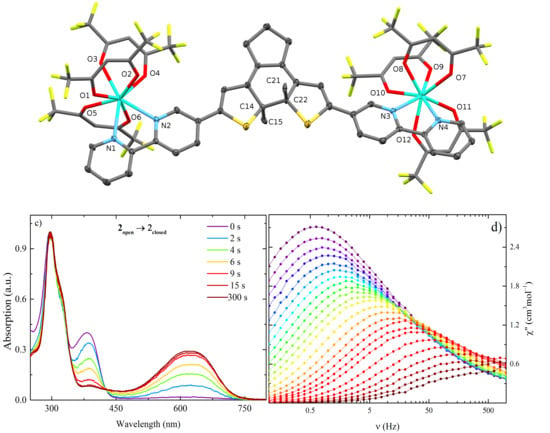
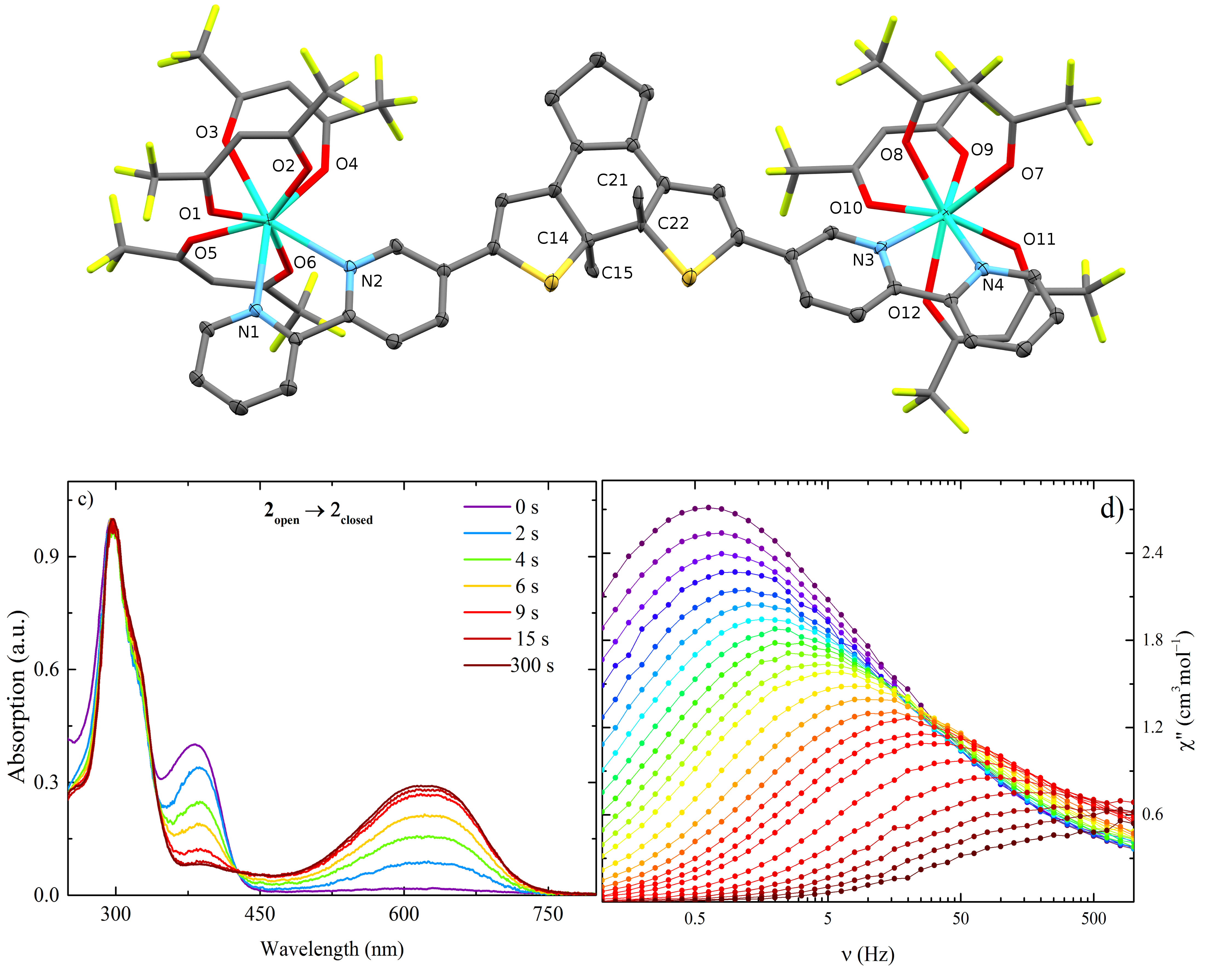
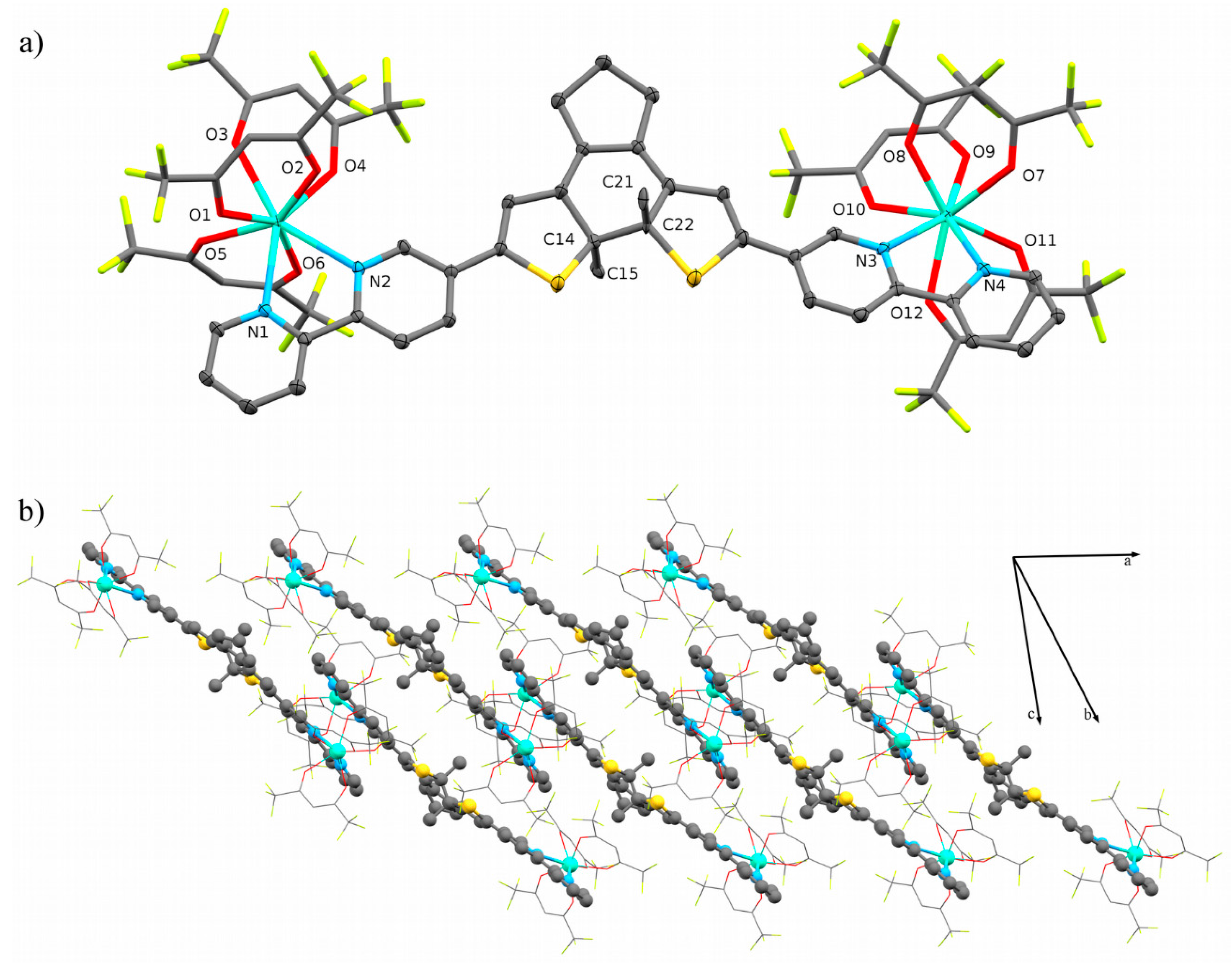
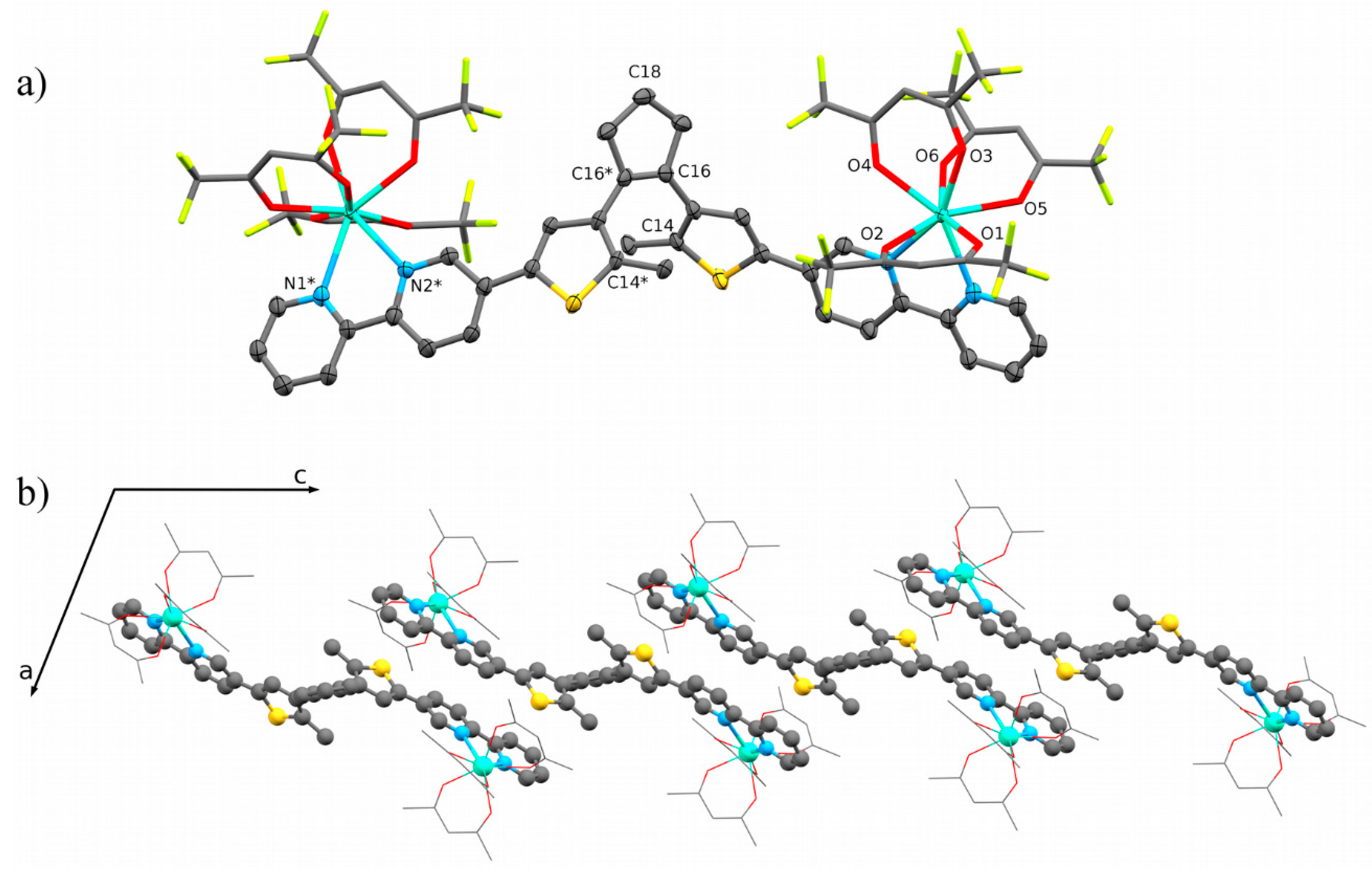
2.1. Structural Description

The 2open crystallized in the monoclinic C2/c space group (Table S1). The complex, generated from the asymmetric unit by applying C2 symmetry, was composed of two Dy(hfac)3 units bridged by one DTE ligand and one heptane solvent molecule (Figure 2). The two Dy ions are equivalent by a C2 rotation with the rotation axis passing through C18 and the C16–C16* double bond. The Dy ions are disordered on two positions (0.692 and 0.308 occupancies). The disorder was attributed to the steric hindrance of the solvent molecule. The Dy ions are coordinated to two nitrogen atoms from the bipyridine moiety of the DTE ligand and six oxygen atoms from three hfac units with a distorted D2d tetragonal dodecahedron coordination geometry (Table S2). The intramolecular distance between the two Dy ions was determined to be 17.021 Å. The C14–C14* distance of 3.569 Å is short enough to photoisomerise the complex to its closed form [13]. Nevertheless, the presence of the solvent molecule prevent this isomerization (Figure S1).
The thiobipyridine moiety is almost planar with dihedral angles between the central and the two lateral rings of 11.26° and 8.69°. This planarity allows π–π interactions between neighbouring ligands with an average distance of 3.527 Å and form a one-dimensional structure along the c direction. The shortest intermolecular Dy–Dy distance was 9.047 Å.
The 2close crystallized in the triclinic P 1 ¯ space group. The asymmetric unit is composed of two Dy(hfac)3 bridged by a DTE ligand and one heptane molecule as crystal solvent (Figure 3). Each Dy ion was crystallographically independent and was coordinated by two nitrogen atoms from the bipyridine group and six oxygen atoms from three hfac moieties. The coordination geometry of both Dy ions was a distorted D4d square antiprism, as determine by SHAPE 2.1 [14,15], and the intramolecular distance between the two Dy ion was 17.705 Å. The four carbon atoms C14, C15, C21 and C22 were disordered (0.51 and 0.49 occupancies). The closed form causes the delocalization of the π-electrons over the entire ligand, making the ligand planar with maximum dihedral angle between two adjacent aromatic rings of 14.29°.
A dimer was formed through π–π interactions between bipyridine rings of two neighbouring molecules with an average distance of 3.442 Å without formation of extended 1D structure. The shortest intermolecular Dy–Dy distance was 8.606 Å. The photo-isomerisation from closed to open forms was not possible in the solid state due to the presence of heptane as crystal solvent (Figure S2) and/or π–π interactions between the neighbouring ligands.

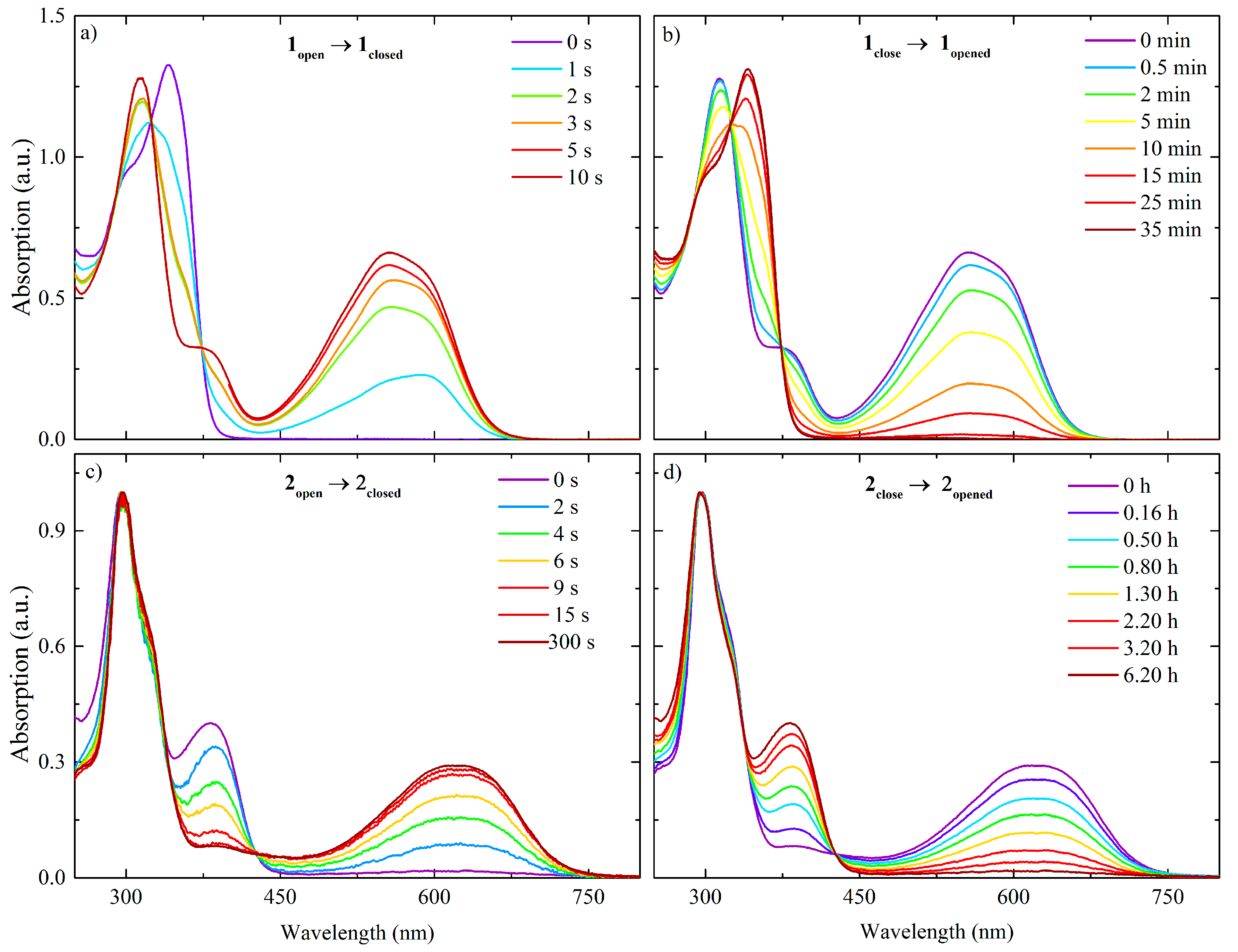
2.2. Optical Properties

Optical properties of the ligands and complexes were measured in CH2Cl2 solution (Figure 4). Photo-isomerisation of the compounds was followed using UV–Vis absorption spectroscopy in the solution state as a function of UV or visible light irradiation times. Before UV light irradiation, in the spectrum for 1open, a strong π–π* transition band, which corresponded to an S0→S1 singlet intraligand (IL) transition, was observed around 340 nm [16]. Upon UV light irradiation, two new absorption bands appeared around 380 and 560 nm within 10 s, and they were assigned to π–π* transitions of the “closed” form [11]. Upon irradiation with visible light, the initial spectrum was recovered within 35 min. This longer photo-isomerisation time was explained by the larger delocalization of the π-electrons across the DTE core and the bipyridine rings due to the full conjugation of the ligand in its close form. Thus, the close form is more stable, and photo-isomerization is slower.
The photo-isomerization from 2open to 2close took place within 10 s, whereas the reverse ring-opening reaction needed more than 4 h to complete. The electron-withdrawing effects of the Dy(hfac)3 units when the ligand is fully conjugated is thought to be the reason for the longer time needed for the complex to return to its open form, as compared to the free ligand.
Photoisomerization in the solid state was not observed.

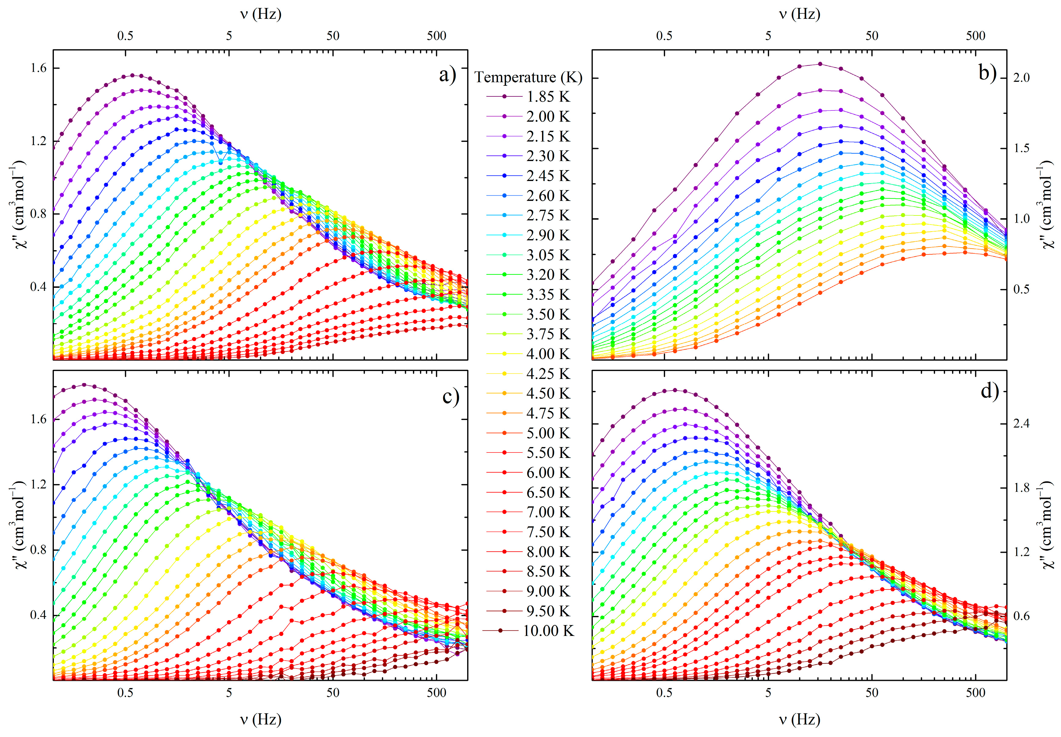
2.3. Magnetic Properties

The magnetic susceptibilities (χ) of polycrystalline samples of 2open and 2close were measured (Figure S3). The χT values at room temperature of 28.25 and 27.98 cm3·K·mol−1 for 2open and 2close, respectively, agree with the expected value for two free Dy ions (28.34 cm3·K·mol−1) [17], and decreased continuously with a decrease in T, becoming 22.45 and 21.5 cm3·K·mol−1 for 2open and 2close, respectively, at 2 K. For both complexes, this decrease in the χT values was attributed to the depopulation of the mJ sub-levels of the ground state. The difference in the depopulation rate can be explain by different energy splittings of the mJ sub-levels for the two complexes due to the slightly different crystal fields around the Dy ions (D2d for 2open and D4d for 2close). Anti-ferromagnetic interactions between the ions is not a suitable explanation for this decrease in χT due to the long intra- (≈17 Å) and intermolecular (≈9 Å) distances.
M vs. H curves measured for 2open and 2close at 1.82 K are slightly different. Both showed pseudo saturation from 1 T with a slope of 0.276 and 0.199 NβT−1 for 2open and 2close, respectively. M values of 10.48 and 9.62 Nβ at 5 T for 2open and 2close, respectively, was obtained. The difference in magnetization for both complexes confirms the presence of different ground states for the two complexes. Hysteresis loops were not observed for both complexes.
The dynamic χ measurement in the absence of external magnetic field shows an out of range, out-of-phase signal (χ) in the high frequency region for both complexes (Figure S4a,b). Analyses by using Cole–Cole model [18] (Figure S5) and the complete model equation [19] revealed that 2close underwent an Orbach/QTM relaxation process (Table S3). 2open only underwent two Orbach processes. Nevertheless, since the maximum of χ was out of range, the extracted relaxation time will not be accurate, and the actual relaxation mechanism may be unclear. In order to confirm the existence of QTM and to suppress it, an optimum static field of 1000 Oe was applied (Figure S6a,b). In this field, both complexes show a single relaxation process with the maxima of χ shifted to a lower frequency region compared to that without static field (Figure 5a,b), and the relaxation process was determined to be a combination of a direct/Raman for 2open and a direct/Orbach for 2close.
In order to determine more precisely the role of the DTE ligand in the relaxation mechanism of the complexes, the magnetic properties were measured in a frozen dichloromethane solution (Figures S4c,d, S5, S6c,d and 5c,d). In the frozen solution, each complex molecule was considered to be isolated without any packing effects on the coordination geometry of the Dy ions. Without an external magnetic field, the maximum of χ of 2open shifted to a lower frequency as compared to that in the solid state, whereas the χ of 2close did not change. The magnetisations of both complexes undergo an Orbach/QTM relaxation mechanism with a small energy barrier (<10 cm−1) (Table S3). In a 1000 Oe magnetic field, the maximum of χ of both complexes shifted to a lower frequency as compared to that of the solid state, meaning that a direct/Raman relaxation process occurred for both complexes.
The relaxation times in the solution state were determined using the Havriliak–Negami model [20], instead of the classical Cole–Cole model. Due to the asymmetry of the relaxation signal, the Cole–Cole model was not able to fit correctly the data and the Havriliak–Negami have been used to solve this problem. The asymmetry can also be observed on the Cole–Cole plot (Figures S7 and S8). A similar behaviour have been observed in our previous solution measurements [19].

3. Discussion

As we previously reported [21], the crystal structure and constraints on the coordination sphere play significant roles in the photo-isomerisation of the complexes. In this study, we show that crystal packing also play a role in the photo-isomerisation. The presence of a crystal solvent molecule close to the DTE core in 2open prevents the cyclization of the ligand due to steric hindrance. For 2close, the crystal solvent and π–π interaction block ring opening. Furthermore, isomerization of the complexes induces large molecular rearrangements, which are restricted by the crystal packing. Single-crystal to single-crystal transformation is not possible for these complexes. This was confirmed since we were able to follow the isomerization by UV–Visible spectroscopy in solution but not in the solid state. In solution, previously mentioned restraints are removed, and photo-isomerisation can be observed.
Although isomerisation does not occur in the solid state, the form of the DTE ligand seems to play a role in the magnetic relaxation process of the Dy ions. Both in the solid and solution states, the complex with closed form of the DTE ligand underwent faster magnetic relaxation than the complex with the open form of the DTE ligand. Similar behaviour has been reported in our previous work involving an Mn(salen) SMM bridged by a DTE ligand [8]. The closed form of the ligand is fully conjugated from one coordination site to the other one, whereas the open form does not have such conjugation. Therefore, conjugation increases the electronic interactions between the Dy ions in the closed form. Although the form of the ligand plays an important role in the magnetic properties, how the change in conjugation of the ligand and the change in the electronic interactions affect the magnetic relaxation mechanism pathway (i.e., direct, Raman, Orbach) remain unclear.

4. Materials and Methods

All chemicals and solvents used in this study were obtained from Sigma Aldrich, Tokyo Chemical Industry, Strem or Wako Chemicals GmbH. Synthesis of 1,2-bis(5-chloro-2-methylthiophen-3-yl)cyclopent-1-ene and Dy(hfac)3·2H2O was performed following a previously reported procedure [22,23].
All synthesis and characterizations of the closed form of the ligand and complex was performed under dark conditions to avoid isomerisation to the open form.

4.1. Synthesis of 1open

To a solution of 1,2-bis(5-chloro-2-methylthiophen-3-yl)cyclopent-1-ene (988 mg, 3 mmol) and dry tetrahydrofuran (15 mL) under a nitrogen atmosphere cooled in an ice bath, 2.6 M n-BuLi (2.96 mL, 6.6 mmol) was slowly added over 1 h to give a dark-pink mixture. Then tris-n-butylborate (2.07 g, 2.42 mL, 9 mmol) was added, and the resulting orange solution was allowed to warm up to room temperature and stirred for 1 h. Next, Pd(PPh3)4 (200 mg, 5 mol %,) was suspended in tetrahydrofuran (15 mL) and heated to 70 °C. After 30 min, aqueous 2.5 M K2CO3 (30 mL), ethylene glycol (10 drops), and 5-bromo-2,2′-bipyridine (1.55 g, 6.6 mmol) were added. This reaction mixture was slowly added to the first solution. The combined solution was heated at 70 °C for 24 h and then cooled to room temperature, followed by addition of water (20 mL). The crude product was extracted with diethyl ether, the combined organic phases were dried over anhydrous Na2SO4, and the solvent was removed. Pure violet solid 1open was obtained by using column chromatography on SiO2 (4:1 v/v hexane:ethyl-acetate), followed by a recrystallization from the same solvents, in 35% yield. 1H NMR (500 MHz, CDCl3): δ = 2.07 (6H, s, ArCH3), 2.12 (2H, m, ArCH2CH2), 2.88 (4H, t, J = 7.5 Hz, ArCH2CH2), 7.15 (2H, s, ArH), 7.29 (2H, ddd, J = 7.4, 4.8, 1.0 Hz, ArH), 7.80 (2H, td, J = 7.8, 1.7 Hz, ArH), 7.88 (2H, dd, J = 8.3, 2.4 Hz, ArH), 8.38 (4H, dd, J = 7.8, 6.6 Hz, ArH), 8.67 (2H, d, J = 4.8 Hz, ArH), 8.82 (2H, d, J = 1.9 Hz, ArH).

4.2. Synthesis of 2open

Dy(hfac)3·2H2O (24.5 mg, 0.03 mmol) was dissolved in boiling heptane (10 mL). After cooling down to 60 °C, a dichloromethane solution (5 mL) of 1open (8.5 mg, 0.015 mmol) was added, and the solution was stirred at room temperature for 10 min. Slow evaporation of the solvent at room temperature for 24 h afforded green sheet-like crystals of 2open, suitable for single crystal X-ray diffraction. Yield: 60% (20 mg).

4.3. Synthesis of 2close

1open (8.5 mg, 0.015 mmol) was first dissolved in dichloromethane (7 mL) and irradiated with 365 nm light for an hour to form 1close. Dy(hfac)3·2H2O (24.5 mg, 0.03mmol) was dissolved in boiling heptane (10 mL). After cooling down to 60 °C, the solution of 1close was added and stirred at room temperature for 10 min. Slow evaporation of the solvent at room temperature for a few days gave black block like crystals of 2close, suitable for single crystal X-ray diffraction. Yield: 35% (10 mg).

4.4. Single-Crystal X-ray Diffraction

Single-crystal X-ray diffraction data of 2open was collected on a Bruker APEX-II diffractometer with an Bruker Photon 100 CMOS detector and a Cryo system DX-CS190LD (JAPAN thermal Engineering Co., Ltd., Tokyo, Japan). Single-crystal crystallographic data of 2close was collected on a Rigaku Saturn70 CCD diffractometer (Rigaku, Tokyo, Japan) with graphite-monochromated Mo Kα radiation (λ = 0.71075 Å) produced by a VariMax micro-focus X-Ray rotating anode source at 90 K. Data processing was performed using the CrystalClear crystallographic software package [24]. For both complexes, the structures were solved by using direct methods via SIR [25], and refinement were carried out using SHELXL-2014 [26]. The non-H atoms were refined anisotropically as possible using weighted full-matrix least-squares on F2. H atoms attached to the C atoms were positioned using idealized geometries and refined using a riding model.
CCDC Nos. 1586166–1586167 contains the supplementary crystallographic data for complexes (See Supplementary Materials). These data can be obtained free of charge via http://www.ccdc.cam.ac.uk/conts/retrieving.html, or from the Cambridge Crystallographic Data Centre, 12 Union Road, Cambridge CB2 1EZ, UK; fax: (+44) 1223-336-033; or e-mail: [email protected].

4.5. Physical Measurements

UV–Vis absorption spectra were acquired in CH2Cl2 solution on a Shimadzu UV-3150 spectrophotometer (Shimadzu, Tokyo, Japan). Compounds were irradiated for different amounts of time with a UV light at 365 nm to isomerise from open to close forms and with a visible light at 490 nm to isomerise from close to open forms. Photo-isomerization was performed with a 300 W Xenon Light Source Asahi Spectra MAX-303 (Asahi, Tokyo, Japan).
Magnetic susceptibility measurements were performed on solid polycrystalline samples on a Quantum Design MPMS-XL SQUID magnetometer (Quantum Design, San Diego, CA, USA). The experimental data were corrected for the diamagnetism of the sample holder, and the intrinsic diamagnetism of the materials was evaluated using Pascal’s tables [27]. Ac measurements were performed in a 3 Oe oscillating magnetic field with and without a static dc field. For the solution state magnetic measurements, the masses of the sample and the solvent were measured after the measurement and confirmed with the values by the MH curve in solid and solution states [28].

5. Conclusions

A new DTE bridged bipyridine ligand was synthesized in its open and close forms. Dy ions were coordinated to this ligand to form a photosensitive dinuclear complex, which have been structurally, optically and magnetically characterized by using single X-ray diffraction, UV–Visible absorption spectroscopy and static and dynamic magnetometry. Although photo-isomerisation of complexes in the solid state was not possible due to presence of strong π–π interactions and a crystal solvent molecule, in the solution state, photo-isomerisation was observed. The ring-opening process was around 1500 times slower than the closing one was. Furthermore, the closed form of the ligand, which is fully conjugated, induced faster relaxation of the magnetization than the open form did in both solution and solid states. We are currently studying the effects of the ligand form on the relaxation mechanism pathway.

Supplementary Materials

The following are available online at www.mdpi.com/2304-6740/6/1/9/s1. Cif and cif-checked files, Table S1: Summary of the crystal parameters for 2open and 2close; Table S2: deviation from the idealized coordination geometries obtained by using SHAPE 2.1. Lowest value is better; Figure S1: (a) top, (b) front and (c) side views of the interactions between the DTE ligand and the heptane solvent molecule in 2open; Figure S2: (a) top, (b) front and (c) side views of the interactions between the DTE ligand and the heptane solvent molecule in 2close; Figure S3: T dependence of χT for 2open and 2close and the field dependence of the magnetization at 1.82 K in inset; Figure S4: T and ν dependences of χ″ in (a,b) the solid state and (c,d) solution state, for 2open and 2close, respectively, in a 0 Oe magnetic field; Figure S5: Arrhenius plots with best-fit curves (lines); Figure S6: H and ν dependences of χ″ in (a,b) the solid state and (c,d) solution state for 2open and 2close, respectively, at 1.85 K; Table S3: Summary of the fitting parameters for the complete model; Table S4: Fitting parameters for the Cole–Cole model for the frequency dependence of χ of 2open in the solid state in a 0 Oe field. χadia was fixed at 0; Table S5: Fitting parameters for the two relaxation Cole-Cole model for the frequency dependence of χ of 2open in the solid state in a 1000 Oe field. χadia was fixed at 0; Table S6: Fitting parameters for the Cole-Cole model for the frequency dependence of χ″ of 2close in the solid state in a 0 Oe field; Table S7: Fitting parameters for the Havriliak–Negami model for the frequency dependence of χ″ of 2close in the solid state in a 1000 Oe field; Table S8: Fitting parameters for the Havriliak–Negami model for the frequency dependence of χ″ of 2open in the solution state in a 1000 Oe field. Χadia was fixed at 0; Table S9: Fitting parameters for the Havriliak–Negami model for the frequency dependence of χ″ of 2open in the solution state in a 1000 Oe field. Table S10: Fitting parameters for the Cole-Cole model for the frequency dependence of χ″ of 2close in the solution state in a 0 Oe field; Table S11: Fitting parameters for the Havriliak-Negami model for the frequency dependence of χ″ of 2close in the solution state in a 1000 Oe field.
Supplementary File 1

Acknowledgments

The authors wish to thank the Japan International Cooperation Agency (JICA) for the scholarship of Mritunjoy Kamila.

Author Contributions

Goulven Cosquer and Mritunjoy Kamila conceived and designed the experiments; Mritunjoy Kamila performed the experiments and Zhao-Yang Li performed the DTE ligand synthesis; Mritunjoy Kamila and Zhao-Yang Li performed the optical property study; Goulven Cosquer analyzed the data; Goulven Cosquer and Brian K. Breedlove wrote the paper. Masahiro Yamashita supervised and managed the project.

Conflicts of Interest

The authors declare no conflict of interest.

References

  1. Komeda, T.; Isshiki, H.; Liu, J.; Zhang, Y.-F.; Lorente, N.; Katoh, K.; Breedlove, B.K.; Yamashita, M. Observation and Electric Current Control of a Local Spin in a Single-Molecule Magnet. Nat. Commun. 2011, 2, 217. [Google Scholar] [CrossRef] [PubMed] [Green Version]
  2. Zhou, Q.; Yang, F.; Xin, B.; Zeng, G.; Zhou, X.; Liu, K.; Ma, D.; Li, G.; Shi, Z.; Feng, S. Reversible Switching of Slow Magnetic Relaxation in a Classic Lanthanide Metal–Organic Framework System. Chem. Commun. 2013, 49, 8244–8246. [Google Scholar] [CrossRef] [PubMed]
  3. Liu, J.-L.; Chen, Y.-C.; Zheng, Y.-Z.; Lin, W.-Q.; Ungur, L.; Wernsdorfer, W.; Chibotaru, L.F.; Tong, M.-L. Switching the Anisotropy Barrier of a Single-Ion Magnet by Symmetry Change from Quasi-D5h to Quasi-Oh. Chem. Sci. 2013, 4, 3310–3316. [Google Scholar] [CrossRef]
  4. Feng, X.; Mathonière, C.; Jeon, I.-R.; Rouzières, M.; Ozarowski, A.; Aubrey, M.L.; Gonzalez, M.I.; Clérac, R.; Long, J.R. Tristability in a Light-Actuated Single-Molecule Magnet. J. Am. Chem. Soc. 2013, 135, 15880–15884. [Google Scholar] [CrossRef] [PubMed]
  5. Kuch, W. Magnetic Nanostructures: Edge Atoms Do All the Work. Nat. Mater. 2003, 2, 505–506. [Google Scholar] [CrossRef] [PubMed]
  6. Thirion, C.; Wernsdorfer, W.; Mailly, D. Switching of Magnetization by Nonlinear Resonance Studied in Single Nanoparticles. Nat. Mater. 2003, 2, 524–527. [Google Scholar] [CrossRef] [PubMed]
  7. Gütlich, P.; Garcia, Y.; Woike, T. Photoswitchable Coordination Compounds. Coord. Chem. Rev. 2001, 219, 839–879. [Google Scholar] [CrossRef]
  8. Fetoh, A.; Cosquer, G.; Morimoto, M.; Irie, M.; El-Gammal, O.; El-Reash, G.A.; Breedlove, B.K.; Yamashita, M. Photo-Activation of Single Molecule Magnet Behavior in a Manganese-Based Complex. Sci. Rep. 2016, 6, 23785. [Google Scholar] [CrossRef] [PubMed]
  9. Morimoto, M.; Miyasaka, H.; Yamashita, M.; Irie, M. Coordination Assemblies of [Mn4] Single-Molecule Magnets Linked by Photochromic Ligands: Photochemical Control of the Magnetic Properties. J. Am. Chem. Soc. 2009, 131, 9823–9835. [Google Scholar] [CrossRef] [PubMed]
  10. Shiga, T.; Miyasaka, H.; Yamashita, M.; Morimoto, M.; Irie, M. Copper(II)-terbium(III) Single-Molecule Magnets Linked by Photochromic Ligands. Dalt. Trans. 2011, 40, 2275–2282. [Google Scholar] [CrossRef] [PubMed]
  11. Pinkowicz, D.; Ren, M.; Zheng, L.-M.; Sato, S.; Hasegawa, M.; Morimoto, M.; Irie, M.; Breedlove, B.K.; Cosquer, G.; Katoh, K.; et al. Control of the Single-Molecule Magnet Behavior of Lanthanide-Diarylethene Photochromic Assemblies by Irradiation with Light. Chem. Eur. J. 2014, 20, 12502–12513. [Google Scholar] [CrossRef] [PubMed]
  12. Cosquer, G.; Morimoto, M.; Irie, M.; Fetoh, A.; Breedlove, B.K.; Yamashita, M. Photo-Control of the Magnetic Properties of Dy(III) and Ho(III) Homometal Coordination Polymers Bridged by a Diarylethene Ligand. Dalt. Trans. 2015, 44, 5996–6002. [Google Scholar] [CrossRef] [PubMed]
  13. Kobatake, S.; Uchida, K.; Tsuchida, E.; Irie, M. Single-Crystalline Photochromism of Diarylethenes: Reactivity—Structure Relationship. Chem. Commun. 2002, 2, 2804–2805. [Google Scholar] [CrossRef]
  14. Alvarez, S.; Alemany, P.; Casanova, D.; Cirera, J.; Llunell, M.; Avnir, D. Shape Maps and Polyhedral Interconversion Paths in Transition Metal Chemistry. Coord. Chem. Rev. 2005, 249, 1693–1708. [Google Scholar] [CrossRef]
  15. Ruiz-Martínez, A.; Casanova, D.; Alvarez, S. Polyhedral Structures with an Odd Number of Vertices: Nine-Coordinate Metal Compounds. Chem. Eur. J. 2008, 14, 1291–1303. [Google Scholar] [CrossRef] [PubMed]
  16. Guerchais, V.; Ordronneau, L.; Le Bozec, H. Recent Developments in the Field of Metal Complexes Containing Photochromic Ligands: Modulation of Linear and Nonlinear Optical Properties. Coord. Chem. Rev. 2010, 254, 2533–2545. [Google Scholar] [CrossRef]
  17. Kahn, O. Molecular Magnetism; Wiley-VCH: New York, NY, USA, 1993. [Google Scholar]
  18. Cole, K.S.; Cole, R.H. Dispersion and Absorption in Dielectrics I. Alternating Current Characteristics. J. Chem. Phys. 1941, 9, 341. [Google Scholar] [CrossRef]
  19. Pedersen, K.S.; Ungur, L.; Sigrist, M.; Sundt, A.; Schau-magnussen, M.; Vieru, V.; Mutka, H.; Rols, S.; Weihe, H.; Waldmann, O.; et al. Modifying the Properties of 4f Single-Ion Magnets by Peripheral Ligand Functionalisation. Chem. Sci. 2014, 5, 1650–1660. [Google Scholar] [CrossRef]
  20. Havriliak, S.; Negami, S. A Complex Plane Representation of Dielectric and Mechanical Relaxation Processes in Some Polymers. Polymer 1967, 8, 161–210. [Google Scholar] [CrossRef]
  21. Kamila, M.; Cosquer, G.; Breedlove, B.K.; Yamashita, M. Packing Structure Effects on the Slow Magnetic Relaxation Pathways of Dysprosium (III) Complexes. Bull. Chem. Soc. Jpn. 2017, 90, 595–603. [Google Scholar] [CrossRef]
  22. Morimoto, M.; Irie, M. Photochromic Reactions of Diarylethenes in Single Crystals with Intermolecular O–H···N Hydrogen-Bonding Networks. Chem. Eur. J. 2006, 12, 4275–4282. [Google Scholar] [CrossRef] [PubMed]
  23. Richardson, M.F.; Wagner, W.F.; Sands, D.E. Rare-Earth Trishexafluoroacetylacetonates and Related Compounds. J. Inorg. Nucl. Chem. 1968, 30, 1275–1289. [Google Scholar] [CrossRef]
  24. CrystalClear-SM, 1.4.0 SP1; Rigaku and Rigaku/MSC: The Woodlands, TX, USA, 2008.
  25. Altomare, A.; Burla, M.C.; Camalli, M.; Cascarano, G.L.; Giacovazzo, C.; Guagliardi, A.; Moliterni, A.G.; Polidori, G.; Spagna, R. SIR97: A New Tool for Crystal Structure Determination and Refinement. J. Appl. Crystallogr. 1999, 32, 115. [Google Scholar] [CrossRef]
  26. Sheldrick, G. A Short History of SHELX. Acta Crystallogr. Sect. A 2008, 64, 112–122. [Google Scholar] [CrossRef] [PubMed]
  27. Bain, G.A.; Berry, J.F. Diamagnetic Corrections and Pascal’s Constants. J. Chem. Educ. 2008, 85, 532–536. [Google Scholar] [CrossRef]
  28. Liang, Z.; Damjanović, M.; Kamila, M.; Cosquer, G.; Breedlove, B.K.; Enders, M.; Yamashita, M. Proton Control of the Lanthanoid Single-Ion Magnet Behavior of a Double-Decker Complex with an Indolenine-Substituted Annulene Ligand. Inorg. Chem. 2017, 56, 6512–6521. [Google Scholar] [CrossRef] [PubMed]
Figure 1. Synthesis diagram for 1open, 1close, 2open, and 2close.
Inorganics 06 00009 g001
Figure 2. (a) Diagram of 2open. Thermal ellipsoids for 1open are drawn at 30% probability. H atoms and solvent molecules are omitted for clarity; (b) π–π chain structure of 2open along c direction.
Inorganics 06 00009 g002
Figure 3. (a) ORTEP diagram of 2close. Thermal ellipsoids are drawn at 30% probability. H atoms and solvent molecules are omitted for clarity; (b) π–π chain structure of 2close along the a direction.
Inorganics 06 00009 g003
Figure 4. UV–Vis spectra for the photo-isomerisation (a) from 1open to 1close, (b) from 1close to 1open, (c) from 2open to 2close and (d) from 2close to 2open isomerization.
Inorganics 06 00009 g004
Figure 5. T and ν dependences of χ″ in (a,b) the solid state and (c,d) solution state for 2open and 2close, respectively in a 1000 Oe magnetic field.
Inorganics 06 00009 g005

