Concise Review of Nickel Human Health Toxicology and Ecotoxicology
by
 , ,
, ,
Samuel Buxton
* ,
,
Emily Garman
,
Katherine E. Heim
,
Tara Lyons-Darden
,
Christian E. Schlekat
,
Michael D. Taylor
and
Adriana R. Oller
* NiPERA Inc., 2525 Meridian Pkwy Ste 240, Durham, NC 27713, USA
*
Authors to whom correspondence should be addressed.
Inorganics 2019, 7(7), 89; https://doi.org/10.3390/inorganics7070089
Submission received: 24 May 2019
/
Revised: 17 June 2019
/
Accepted: 4 July 2019
/
Published: 12 July 2019
(This article belongs to the Special Issue Bioinorganic Chemistry of Nickel)
Abstract
:Nickel (Ni) metal and Ni compounds are widely used in applications like stainless steel, alloys, and batteries. Nickel is a naturally occurring element in water, soil, air, and living organisms, and is essential to microorganisms and plants. Thus, human and environmental nickel exposures are ubiquitous. Production and use of nickel and its compounds can, however, result in additional exposures to humans and the environment. Notable human health toxicity effects identified from human and/or animal studies include respiratory cancer, non-cancer toxicity effects following inhalation, dermatitis, and reproductive effects. These effects have thresholds, with indirect genotoxic and epigenetic events underlying the threshold mode of action for nickel carcinogenicity. Differences in human toxicity potencies/potentials of different nickel chemical forms are correlated with the bioavailability of the Ni2+ ion at target sites. Likewise, Ni2+ has been demonstrated to be the toxic chemical species in the environment, and models have been developed that account for the influence of abiotic factors on the bioavailability and toxicity of Ni2+ in different habitats. Emerging issues regarding the toxicity of nickel nanoforms and metal mixtures are briefly discussed. This review is unique in its covering of both human and environmental nickel toxicity data.
Keywords:
nickel; bioavailability; carcinogenicity; genotoxicity; allergy; reproductive; asthma; nanoparticles; ecotoxicity; environment1. Nickel Occurrence and Uses
Nickel (Ni) is a naturally occurring element and is found in abundance in the earth’s crust and core. Nickel occurs in air, water, sediments, and soil from various natural sources and anthropogenic processes. Nickel is introduced into the environment and is circulated through the system by chemical and physical processes and through biological transport mechanisms of living organisms [1]. Nickel is essential for the normal growth of many species of microorganisms and plants [1].
Nickel exists in nature mainly in the form of sulfide, oxide, and silicate minerals, and is an important commercial element in industrialized societies. Thus, human and environmental Ni exposures are ubiquitous. Anthropogenic nickel releases to the environment occurs locally from emissions of metal mining, smelting, and refining operations; from industrial activities, such as nickel plating and alloy manufacturing; from land disposal of sludges, solids, and slags; and from disposal as effluents. Other diffuse sources may arise from combustion of fossil fuels, waste incineration, and wood combustion.
Nickel compounds can be water-insoluble, like oxidic (such as black nickel oxide) and sulfidic (such as nickel subsulfide), with the latter being sparingly soluble in some media. A third group of nickel compounds are water-soluble (such as nickel sulfate). Metallic nickel (nickel metal and alloys) are nickel substances with very low or no water solubility.
The important uses of nickel substances in transportation products, aerospace equipment, paints, ceramics, medical applications, electronics, food and beverage production, batteries, chemicals, and many other uses indicate that potential exposure to nickel metal, nickel compounds, and nickel-containing alloys is wide ranging. Although many sources and types of exposure exist, potential toxicity is dependent on the physico-chemical characteristics of the nickel substance, as well as the amount, duration, and route of exposure.
2. Nickel Exposure to Humans and Toxicokinetics
2.1. Nickel Exposures
2.1.1. Occupational
Occupational exposure to Ni is primarily associated with workers in the nickel-producing and nickel-using industry sectors. Industries associated with nickel production include mines, mills, refineries, and smelters, whereas, nickel using/processing industries include alloy and stainless-steel production, catalysts, pigments, batteries, and electroplating. In addition, many industrial processes can generate Ni exposures such as welding and grinding of Ni-containing alloys [2,3,4]. Workplace exposure is primarily to airborne nickel; inhalation is therefore the major route of exposure of toxicological importance in occupational settings. To a lesser extent, skin contact can occur during certain processes or physical handling of nickel and/or nickel containing-products [3,4]. Oral exposure also occurs as a consequence of swallowing inhaled coarse particles or through hand to mouth contact.
2.1.2. General Public
Nickel is naturally present in the air, soil, water, plants, and various foods. Nickel in the air can result from forest fires, volcanoes, and anthropogenic activities [4,5]. Anthropogenic activities can also contribute to nickel levels in the soil, water and plants; their Ni levels are generally higher near industrial sites involved with the mining and processing of nickel. The primary source of nickel exposure for the general public is via dietary intake from foods like chocolate, coffee, teas, legumes, and nuts that tend to have naturally higher Ni levels and to a lesser extent from drinking water [2,5]. Other nickel exposure sources for the general public include commonly used products like cooking pots and pans, jewelry, and medical devices like dental appliances and joint prostheses [4,6]. Exposure of the public to nickel and its compounds is generally low [7].
2.2. Toxicokinetics and Bioavailability of Nickel
The absorption, distribution and elimination of nickel is affected by factors like route of exposure, physical form of the material (massive or powder), metal release and in the case of dusts or powders, the aerodynamic size of the nickel particles. While most of the historically available information relates to micron-size particles of nickel-containing substances, recent studies have looked at the toxicokinetics of nickel nanoparticles to characterize how they differ from those of the corresponding micron size. This is further discussed under Section 5.2.
2.2.1. Gastrointestinal
Gastrointestinal absorption of nickel comes from nickel present in ingested beverages, drinking water and foods. Nickel is naturally present in foodstuff as it is essential to plants. Ingestion of soil is also possible, particularly in small children. For the general public, oral ingestion of nickel is the most relevant exposure pathway for systemic absorption and toxicity. In occupational settings, mucociliary clearance of inhaled nickel dust that is swallowed can contribute an appreciable amount to absorption via the gastrointestinal tract [2]. The gastrointestinal absorption and bioavailability of ingested nickel is affected by the type of matrix (food, soil) ingested and the prior presence of food in the stomach. For example, the absorption of nickel in fasted subjects given water soluble nickel sulfate in drinking water was higher (up to 27%) than in subjects given nickel sulfate with food (0.7–5%) [8,9]. Other factors like the chemical form of nickel also affect absorption. Generally, water soluble nickel compounds have a greater oral absorption than poorly soluble nickel substances.
2.2.2. Respiratory
In toxicological terms, inhalation is the most important exposure route for nickel particles in occupational settings. Absorption of nickel particles deposited in the nasopharyngeal, tracheobronchial, or alveolar regions of the respiratory tract is dictated by several factors, such as the particles aerodynamic diameter (dae), solubility, surface area, amount deposited, ventilation rate, clearance, and retention rates [10,11,12]. Only particles sufficiently small (<100 µm dae) can enter the respiratory tract and be inhaled. An important first step in inhalation absorption is particle deposition. Particle size dictates both the depth of deposition along the respiratory tract and the subsequent absorption. Particles deposited in the lower regions of the respiratory tract (alveolar) are predominantly ≤4 µm dae; particles deposited in the tracheobronchial region are 4–10 µm dae and particles deposited in the nasopharyngeal region are between 10 µm and 100 µm. Less than or equal to 10% of inhaled respirable size aerosol are deposited in the pulmonary region of the human respiratory tract [13]; the fraction is even lower for workplace aerosols. While this small percentage is expected to be cleared via dissolution, macrophages, or lymph nodes, the mucociliary clearance of the majority of deposited undissolved particles leads to their expectoration or swallowing, contributing to the gastrointestinal absorption. Similar to gastrointestinal absorption, the water-soluble nickel compounds are more readily absorbed in the respiratory tract than the poorly soluble compounds [5]. Animal studies with respirable size aerosols have shown that the poorly soluble nickel particles have long lung retention times and slow clearance, and thus accumulate over time.
2.2.3. Dermal
A very small fraction of nickel that is dermally exposed is absorbed. Following dermal exposure, nickel ions (Ni2+) and particles can penetrate the skin, especially at sweat ducts and hair follicles. Here too, particle size is a limiting factor for absorption; smaller sized particles are absorbed more readily than larger sized particles. Additionally, dermal absorption of nickel is affected by solubilizing agents like detergents, solvents, and the presence of barriers to the skin, such as clothes or gloves [14,15,16,17]. During exposure to metallic nickel in massive forms (e.g., jewelry) corrosion and Ni2+ ion release must occur prior to absorption. Research has shown that approximately <2% of soluble compounds [18,19] and <0.2% of metallic and insoluble nickel [20] is absorbed.
2.3. Distribution, Metabolism and Excretion of Nickel
The distribution and elimination of nickel is influenced by the route of administration and binding to proteins. Nickel in the bloodstream is bound to albumin and metalloproteins, which modulates their tissue distribution and elimination. Postmortem analysis of nickel in human tissues shows that the highest amounts of absorbed nickel is distributed to the lungs, thyroid glands, and adrenal glands, with lesser amounts to brain, kidneys, heart, liver, spleen, and pancreas [2,21]. Inhaled nickel is predominantly distributed in the respiratory tract (lungs, nasal sinus), followed by the kidneys [22]. Inhaled soluble nickel is eliminated primarily in the urine, while mucociliary clearance leads to a fraction of the inhaled poorly soluble nickel particles being eliminated in the feces. Orally absorbed nickel is distributed to the kidneys, followed by the liver, brain, and heart [23]. Nickel absorbed via the gastrointestinal tract is excreted predominantly in urine; unabsorbed nickel is eliminated with the feces. Hair is another distribution and elimination tissue for absorbed nickel. Nickel can also be eliminated via sweat and human breast milk [24,25,26]. The majority of dermally exposed nickel is not absorbed and thus not available for distribution.
3. Toxicity of Nickel
3.1. Toxicity and Nickel Ion
As with other metals, the toxicity of nickel-containing substances is considered to be related to the bioavailability of the metal ion (Ni2+) at systemic or local target sites [10]. The main human health effects of concern associated with Ni exposure include nickel allergic contact dermatitis, respiratory carcinogenicity, reproductive toxicity and non-cancer respiratory effects.
3.2. Nickel Allergic Contact Dermatitis (NACD)
3.2.1. Prevalence in General and Clinical Populations
Nickel is one of the most common causes of allergic contact dermatitis (ACD). An estimated 12–19% of females and 3–6% of males in the general population are allergic to nickel (i.e., nickel-sensitized) [27]. Higher percentages are recorded in dermaotology clinics [28]. The reason for the relatively high prevalence of nickel sensitization is due to the use of nickel-releasing consumer items that come in direct and continuous prolonged contact with the skin. Although exposure may occur in some occupational settings (generally associated with soluble nickel salts), the marked prevalence of nickel sensitization in the general population is primarily due to consumer dermal exposure to nickel released from articles (e.g., in jewelry, watches, eyeglasses) that are made of nickel-plated materials or high nickel-releasing alloys.
3.2.2. Induction vs. Elicitation
Many chemical agents, including nickel, can cause allergic contact dermatitis (ACD) which results in inflammation of areas of the skin in sensitized (i.e., allergic) individuals. While nickel ACD can cause pain, inflammation and discomfort, it is not life threatening because it causes a delayed-type allergy (type 4), which cannot trigger anaphylactic shock, contrary to some other types of allergies (type 1, 2, or 3). The development of nickel ACD requires that an individual become immunologically sensitized to nickel. This is termed the induction phase or sensitization phase and the length of this phase varies between individuals. It can range from 1–3 weeks to develop, following days to weeks of prolonged intimate contact in a piercing or on the skin with a nickel-containing article that has released a sufficient amount of solubilized Ni2+ onto the skin. The quantity of Ni2+ that is sufficient to induce sensitivity varies with the individual. If the skin is already damaged, sensitization may be induced more quickly and by lower amounts of the solubilized Ni2+. Temperature, the presence of other allergic conditions, gender, and age may also be determining factors for (1) susceptibility, (2) the amount of Ni2+ required for a reaction, and (3) the time to develop sensitization to nickel. Induction of nickel sensitization most commonly originates from body piercing but is also more likely if skin exposure to Ni is combined with irritants and/or moisture that could also compromise the skin barrier.
A nickel-sensitized individual, when re-exposed to Ni2+ on the skin in sufficient amounts, may have an allergic response within several hours. This is termed the elicitation phase, which often occurs at a lower concentration of Ni2+ than required for inducing sensitization in the first place. The elicitation of nickel ACD usually only occurs at the site of exposure but it can also occur in skin remote from the current site of contact with nickel, (e.g., at the location where previous nickel sensitization reactions have occurred) [29].
While oral systemic elicitation of ACD in individuals previously sensitized by direct and continuous prolonged skin contact is well documented to occur in a small proportion of nickel-sensitized individuals (e.g., hypersensitive people), there exists some controversy about the ability to sensitize individuals when nickel exposure is oral, intravenous, or inhaled [30].
3.2.3. Mechanisms of Nickel ACD
Nickel ions released from nickel compounds, nickel metal, and various alloys may trigger skin reactions when they are absorbed into the skin. These Ni2+ ions can then bind to and activate epithelial cells such as Langerhans or dendritic cells in the basal layer of the epidermis (see Figure 1). These cells produce cytokines or chemokines, triggering complex immune reactions that activate antigen-presenting cells and T cells [31,32,33]. As part of this process, migration of activated antigen-presenting cells to the draining lymph nodes occurs, where the bound nickel, as a hapten, is presented to the naive CD4-positive T cells [34]. Nickel differs from classical haptens by its ability to form coordinative bonds with proteins and to directly activate human innate immune cells via the toll-like receptor (TLR) 4 [35].
Future exposure to nickel in sufficiently high amounts (above threshold) would lead to the activation of the nickel-specific T-cells. Migration of these cells into the bloodstream triggers visible signs of allergic reactivity after hours of Ni2+ exposure [36]. The exact sequence of events and interactions between antigen presenting and immune cells involved in nickel allergy are still being elucidated.
3.2.4. Sources of Exposure: Nickel Release versus Content
Nickel ACD was first noticed in occupational settings where soluble forms of nickel came into contact with worker’s skin [37]. Individuals working in electroplating shops, in battery manufacturing, and with nickel catalysts were the most susceptible to nickel ACD. However, workplace-related nickel dermatitis is now relatively rare due to improved production processes and occupational hygiene measures that limit exposure.
Non-occupational nickel sensitization is well documented. It was first observed in individuals who had skin contact with suspenders in the 1950s–1960s, followed by jean buttons and zippers, then nickel-releasing ear-piercing items and nickel-plated jewelry [38]. The significant differences in prevalence between females and males is sometimes correlated with the much higher prevalence of ear-piercing among women, but other factors such as hormone differences and the tendency for young women to wear more and/or low-quality jewelry than males may also play a role [39].
The release of Ni2+ is necessary for causing nickel sensitization and nickel ACD, which are threshold effects (requiring release of ions above a specific amount to cause a reaction). Alloys such as many stainless steels contain nickel but do not release a sufficient amount of Ni2+ to cause an individual to become nickel sensitized or elicit a nickel ACD reaction if they are nickel-sensitized.
To have nickel release from metallic nickel or nickel alloys, the nickel metal must be corroded and the corrosion product dissolved into Ni2+. For this reason, sweat or other wet conditions can increase the release rate compared to dry conditions.
The risk of nickel sensitization or elicitation of nickel ACD can be managed and minimized through reduced exposure to nickel-releasing items. In the workplace, exposure reduction includes personal protective equipment and other risk management measures. For consumers, exposure can be reduced through avoidance of direct and continuous prolonged exposure to items releasing nickel in amounts greater than the threshold for nickel ACD, and switching to items made from surgical stainless steel (AISI 316L) and other low nickel-releasing alloys, or non-nickel-containing materials.
Accordingly, the European Union (EU) nickel restriction (REACH, Entry 27 of Annex XVII) [40] is based on nickel release, rather than nickel content. Articles intended to come into direct and continuous prolonged contact with the skin must release less than 0.5 μg Ni/cm2 surface area of the item per week using the standardized methodology to assess conformity and compliance with this specific regulation (EN1811:2011+A1) [41]. Items known to be associated with nickel ACD that are included under the EU nickel restriction include necklaces, bracelets and chains, anklets, finger rings, wrist-watch cases, watch straps, rivet buttons, tighteners, rivets, earrings, and zippers.
3.2.5. Susceptible Populations
Nickel sensitization is not an inherited condition. It is related to direct and continuous prolonged skin contact (i.e., exposure) to materials releasing an amount of nickel sufficiently high to cause sensitization reactions, be that nickel metal, nickel alloys, or soluble nickel salts.
A common cause of nickel sensitization and nickel ACD is body piercing, which involves inserting high nickel-releasing studs into the wound to prevent closure during healing and bypassing the skin barrier. Once healed, with the stud removed, additional contact with nickel in the pierced area may occur by wearing jewelry or posts in piercings that release a significant amount of Ni2+.
Individuals who have reactions to other allergens, have overly sensitive skin or other skin diseases, and individuals who sweat excessively have been considered to be more susceptible to nickel allergy. This susceptibility is not unique to nickel but is rather a function of increased immunological reactivity, decreased skin barrier function, or increased corrosion due to sweating. These individuals would be more likely to attend dermatology clinics for treatment of nickel allergy and other skin problems, and would have to avoid not only high nickel-releasing items but also other skin allergens and irritants.
A very small part of the nickel-allergic population is hypersensitive to nickel. These individuals react to lower concentrations of nickel on the skin than most nickel-sensitive individuals. A small fraction of these people also react to oral nickel exposure. Prevention of elicitation in these individuals is important and is done on a case-by-case basis. Regulation and prevention of nickel sensitization and nickel ACD of the general population is not intended to protect these hypersensitive individuals [42], as they are a small subset of the general population and may need more specific medical advice. While a low-nickel diet is helpful for some of these individuals who react to ingestion of nickel [43], oral hyposensitization, using gradually increasing low doses of nickel has also been shown to increase the amount of nickel needed to cause a nickel allergic reaction [44].
3.3. Nickel Carcinogenicity
Nickel carcinogenicity is an occupational concern due to the required inhalation route of exposure and high exposure levels. The evidence for carcinogenicity, or lack thereof, of nickel metal and nickel compounds come from epidemiological and animal (rats and mice) studies. These studies indicate that the inhalation route is the exposure route of concern and the respiratory system (lungs and nasal sinus) the target organ for carcinogenicity of nickel compounds. The human and animal evidence supports the respiratory carcinogenicity of nickel compounds but do not identify nickel metal as a respiratory carcinogen. The hazard classifications reflect this difference in carcinogenicity between nickel metal and nickel compounds.
Under the European Union Classification, Labeling and Packaging (CLP) legislation, many soluble and insoluble nickel compounds are classified as Carc 1A, stating that these compounds are known to have carcinogenic potential for humans, based largely on human evidence. This classification specifies inhalation as the only route of concern [45]. Nickel metal is classified as Carc 2, suspected human carcinogen based on insufficient evidence from human studies with suggestive evidence from animal studies via non-relevant routes of exposure. Likewise, the International Agency for Research on Cancer (IARC) classified soluble and insoluble nickel compounds under Group 1, carcinogenic to humans, and nickel metal and alloys under Group 2B, possibly carcinogenic to humans [46].
3.3.1. Human and Animal Evidence for Nickel Carcinogenicity
High exposures to mixtures of water-soluble and complex-insoluble nickel compounds in workers involved with mining, refining, and processing of sulfidic nickel ores have been associated with excess respiratory cancer risks. No excess respiratory cancer risks in workers at lateritic ore refineries, alloy manufacturing, or electroplating have been observed. A seminal comprehensive study by the International Committee on Nickel Carcinogenesis in Man (ICNCM) examining cancer risks in 10 cohorts of about 80,000 nickel processing and nickel alloy production workers reported an association between exposure to certain sulfidic, oxidic and water-soluble nickel compounds, and respiratory cancer of the lungs and nasal sinus; no association with exposure to metallic nickel was identified [47]. Among the nickel compounds, different chemical forms appear to have different carcinogenic potentials and potencies in the human studies.
Animal studies are useful in elucidating mechanisms of carcinogenesis and determining the source of the carcinogenicity observed in humans (mixed exposures) to specific nickel substances. There are eight relevant lifetime inhalation and oral carcinogenicity studies in rats and mice [48,49,50,51,52]. The animal studies support the conclusions from human studies that the inhalation route and the respiratory tract are the relevant exposure route and target organ, respectively, for nickel compounds carcinogenicity. No carcinogenicity is associated with the oral exposure route. A recent review of the human and animal evidence for the respiratory carcinogenicity of nickel metal and nickel compounds is provided in the European Chemicals Agency (ECHA) background document in support of occupational exposure limit values [2].
In interpreting nickel carcinogenicity studies, it is important to realize that exposures in animal studies are to a “pure” nickel compound (a single nickel compound), while exposures in human epidemiological studies are to mixtures of nickel compounds (plus other inorganic compounds). Any potential co-carcinogenic or promoting effect of the different nickel compounds and other inorganic compounds (e.g., arsenic, acid mists) in the human studies will not exist in the single exposure animal studies.
There is generally a good correlation between the human occupational exposure studies and animal studies on the carcinogenicity of nickel and nickel compounds. The evidence from both human and animal studies point to the absence of carcinogenic effects of nickel metal but the presence of carcinogenic effects for sulfidic and oxidic nickel compounds. The only inconsistency between the human and animal evidence relates to the carcinogenicity of soluble nickel compounds [53]. The animal studies have failed to show carcinogenic effect of pure soluble nickel compounds following inhalation and oral exposures. In the human studies, an association between inhalation exposure to soluble nickel (with additional exposures to insoluble nickel compounds) and/or smoking and lung cancer was observed in some groups of workers.
3.3.2. Inhalation Exposure Route
The 1990 ICNCM report [47] concluded that inhalation exposure to mixtures of water-soluble nickel compounds (e.g., nickel sulfate, nickel chloride) and water insoluble nickel compounds (e.g., nickel subsulfide, nickel oxide, complex Ni-Cu oxides) were associated with excess respiratory cancer risk in workers. Much of the excess respiratory cancer risk was associated with exposure to high concentrations (≥1 mg Ni/m3) of soluble compounds or (≥10 mg Ni/m3) of a mixture of sulfidic and oxidic nickel compounds. Excess nasal and/or lung cancer risks have been observed in different cohorts of workers. More recently, analyses of dose-responses for the main chemical forms of nickel (soluble, oxidic and sulfidic compounds) that included 13 cohorts of nickel workers (~100,000 workers), indicated that no excess cancer risk were observed in these studies when exposures to nickel in the inhalable aerosol fraction were kept ≤0.1 mg Ni/m3 [13].
The ICNCM report and subsequent studies found no association between metallic nickel and excess risks of lung or nasal cancer [54]. In two reports where hints of possible correlations between excess cancer risk and nickel metal exposure have been indicated, failure of cross-validation of the test model and non-significant odds ratio after adjusting for confounding exposures suggest that the risks for metallic nickel may have been overestimated [55,56]. No association has been found between increased respiratory cancer risk and inhalation exposure to metallic nickel outside the nickel refineries, when local populations were used as controls [57,58]. More recent cohort studies support the lack of association between exposure to metallic nickel and excess respiratory cancer [59,60,61,62,63].
In rats and mice, seven lifetime carcinogenicity studies via the inhalation route have been conducted in which the maximum tolerated doses were reached [48,49,50,52,64]. A 30-month inhalation study with nickel metal powder in Wistar rats did not increase the incidence of lung tumors [52] consistent with an earlier study in rats and mice, that, although compromised by high mortality, also suggested that metallic nickel did not cause cancer [65]. While positive results have been found in one intratracheal instillation study of nickel metal in rats [66], the doses in that study were shown to not be achievable via the normal inhalation route; intratracheal instillation is a non-physiological and non-relevant exposure route for workers and the public. Other inhalation studies in guinea pigs and hamsters have buttressed the negative carcinogenicity of metallic nickel [65,67]. Thus, there has been a consistent lack of increased respiratory cancer risk associated with metallic nickel exposures in animals and humans.
The ability of nickel substances to induce respiratory tumors after inhalation may be related to the bioavailability of the Ni2+ ions at target sites within epithelial cells (Figure 2). The bioavailability of Ni2+ ions in the nucleus of target respiratory epithelial cells is not dictated by just the water solubility of the nickel particle but by the interplay of factors like respiratory toxicity, extracellular and intracellular dissolution, and lung clearance [10]. According to the bioavailability model, it is the delivery of sufficient (above threshold) Ni2+ ions to the nucleus of respiratory epithelial cells, which is influenced by other factors, that governs the respiratory carcinogenicity of nickel.
Nickel subsulfide exposure induced lung tumors in male and female rats at levels ≥0.1 mg Ni/m3 respirable aerosol, but not in mice. At the lowest concentration level at which increased tumors were detected, the incidence and severity of chronic lung inflammation in rats was similar between nickel sulfate and subsulfide, even though the tumor outcome was different. According to the Ni2+ bioavailability model, nickel subsulfide has a high carcinogenic potential due to its low extracellular, but high intracellular dissolution, resulting in the highest dose of bioavailable Ni2+ in the nucleus of respiratory epithelial cells.
Green nickel oxide induced some tumors in rats, but only at higher exposures (≥1.0 mg Ni/m3 respirable aerosol) and with no clear dose-response [49]. The low extracellular but medium intracellular dissolution of nickel oxide results in the delivery of above-threshold doses Ni2+ ions to the nucleus of respiratory epithelial cells, causing the tumors observed in rats. Nickel oxide had equivocal evidence of tumors in female mice. Perhaps mice are not very susceptible to the carcinogenic effects of nickel even tough other metals, such as cobalt, have been able to induce excess lung tumors in mice [68,69]. Another possibility is that mice have higher thresholds for the nickel cancer-causing mechanisms.
Nickel sulfate inhalation exposures in rats have not induced tumors at exposure levels up to 0.11 mg Ni/m3 respirable aerosol, and 0.22 mg Ni/m3 respirable aerosol in mice. A plausible explanation for this is that nickel sulfate is not carcinogenic by itself at the exposure levels that can be tolerated by rats without overt toxicity; in human studies exposures are mixed and soluble Ni can enhance the lung carcinogenicity of more insoluble Ni compounds through inflammatory and proliferative effects. However, the lack of tumors in rats is likely due to the high extracellular but low intracellular dissolution of nickel sulfate that results in the delivery of below threshold or insufficient dose of Ni2+ ions to the nuclei of respiratory epithelial cells.
Likewise, metallic nickel may have failed to induce lung tumors in rats following inhalation exposure due to its high extracellular but low intracellular dissolution, resulting in low bioavailability of Ni2+ ions in the nucleus of target respiratory epithelial cells.
3.3.3. Oral Exposure Route
Besides respiratory cancer, there are currently no consistent and reliable epidemiological data to suggest that nickel causes excess cancer risks at other organ sites. Although excess cancer of the buccal cavity and pharynx, or stomach, or prostate have been reported in some workers exposed to nickel, these findings have been rare and have not been consistently reproduced [46,70].
Exposure to soluble nickel compounds, by themselves, via the oral route has not produced tumors in rats [51] or mice [71]. No robust animal studies exist for oral administration of metallic or insoluble nickel compounds. However, the negative results with the most bioavailable of the nickel compounds via the oral route are also relevant for these less bioavailable substances since absorption is needed for any systemic effects.
3.3.4. Dermal Exposure Route
There is presently no human epidemiological study linking dermal exposure to metallic nickel and/or nickel compounds with excess local or systemic cancer risks. For the dermal route, no skin cancers have been reported with exposure to nickel compounds or alloys, and the systemic absorption of nickel through the skin is very low (≤2% for soluble Ni substances and 0.2% for nickel metal). Thus, the epidemiological data do not suggest an association between cancer and dermal nickel exposure.
No cancer studies in animals using dermal administration of soluble, insoluble nickel compounds, or metallic nickel have been conducted. However, based on the low dermal bioavailability of Ni2+ and other physiological considerations, it is not expected that dermal exposure to metallic nickel and/or nickel compounds will be associated with cancer.
3.3.5. Other Exposure Routes
No consistent epidemiological data currently exists linking nickel exposure via other routes (e.g., implants) to cancer. Given the specificity of nickel cancer on the respiratory system, it is not expected that nickel cancer will be associated with routes other than inhalation.
There are various animal studies assessing the carcinogenicity of the different nickel chemical forms following parenteral, intratracheal instillation, intraperitoneal and injection administrations [2]. However, these routes of exposure are not appropriate nor physiologically relevant for metallic nickel and nickel compounds. For example, while local sarcomas at sites of injection of nickel metal powder were found, the relevance of these findings for the assessment of nickel metal respiratory carcinogenicity or other carcinogenicity in humans is highly questionable [54].
3.3.6. Nickel Compounds’ Genotoxic and Carcinogenic Mode of Action
Generally, carcinogenic substances can be direct-acting genotoxicants, indirect-acting genotoxicants, or substances that cause cancer via non-genotoxic modes of action. For direct-acting genotoxicants, all exposure levels are assumed to be associated with some degree of excess cancer risk. Direct-acting genotoxic agents are positive in bacterial, germ cell, and mammalian cell mutagenicity tests and interact directly with DNA. Nickel compounds have been consistently negative or showed weak effects in bacterial, in vitro and in vivo mutagenicity tests. Nickel ions have a weak interaction with DNA but a preferentially stronger interaction with proteins. For example, Ni2+ has binding constants of 6.7 × 10⁻¹ M−1 and 4.37 × 10⁹ M−1 for adenosine (nucleic acid) and cysteine (amino acid), respectively [72,73]. Nickel and nickel compounds are therefore not considered to have a direct genotoxic mode of action (MOA) [2].
For indirect-acting genotoxic agents and non-genotoxic agents, threshold exposure levels can be identified below which cancer risks are expected to be negligible. Indirect genotoxicants can damage DNA via secondary mechanisms like generation of reactive oxygen species (ROS) or inhibition of repair. Table 1 summarizes the genotoxicity evidence for nickel and provides examples of relevant references. In vitro, nickel compounds have been shown to cause DNA damage (such as fragmentation, single-strand breaks) indirectly through increased formation of oxidative radicals. Nickel compounds enhanced the induction of sister chromatid exchange, chromosomal aberrations and micronucleus formation in vitro. Nickel compounds have also been shown to increase formation of DNA-Protein crosslinks.
Additionally, nickel compounds are known to inhibit DNA repair enzymes in vitro; Ni2+ ions competitively inhibit the repair enzyme ABH2 by binding to the same site as Fe2+ [74]. DNA repair inhibition may also occur via inhibition of DNA ligation and post replication repair. Non-genotoxic modes of action include the induction of epigenetic effects that can affect gene expression. Nickel compounds can increase histone phosphorylation (H3S10), methylation (H3K4), ubiquitination (H2B and H2A) and decrease histone acetylation (H4) through decreased histone acetyltransferase activity (Table 1). Nickel compounds can induce selective fragmentation or decondensation of heterochromatic long arms of X-chromosomes (Table 1). Recent reports also suggest that non-coding RNAs play a role in nickel respiratory carcinogenesis [75,76,77]; see the review “The role of Non-coding RNAs Involved in Nickel Induced Lung Carcinogenic Mechanisms” by Zhu et al. in this issue.
The in vivo genotoxic effects of nickel compounds in the lung are observed following inflammation and macrophage activation that results in indirect oxidative DNA damage. Preventing inflammation would therefore prevent the indirect genotoxic effects and prevent tumor formation. The indirect genotoxic and non-genotoxic effects of nickel compounds have thresholds below which these effects are not observed. This furthers support the bioavailability model wherein nickel carcinogenicity depends on the delivery of sufficient amounts of Ni2+ ions to the nucleus of respiratory epithelial cells [10]. Taken together, the totality of the evidence supports an indirect genotoxic and/or non-genotoxic MOA with thresholds for nickel carcinogenesis.
3.4. Reproductive and Developmental Toxicity
3.4.1. Human Epidemiological Studies
In 1994, Chaschschin et al. [131] published a preliminary qualitative report of an apparent increase in spontaneous abortions and structural malformations in newborn babies whose mothers were employed in the Russian nickel refinery at Monchegorsk. These workers were exposed via inhalation to a mixture of water-soluble and water-insoluble nickel compounds, as well as metallic nickel. Due to this concern, a large epidemiological study of the Russian cohort was launched in 1995 to determine whether the observed effects were due to their workplace nickel exposures or to other factors in the workplace and/or ambient environment. Researchers set up a birth registry for all births occurring in the region during the period of the study, which included information on 22,836 newborns and 2793 pregnancy outcomes surveyed for spontaneous abortions [132,133]. Exposures were reconstructed (using air and urinary nickel measurements) for the female workers at the refineries to be able to link specific pregnancy outcomes with occupational exposures via inhalation and systemically bioavailable doses of nickel (i.e., urinary nickel levels). A series of reports by Vaktskjold et al. [132,133,134,135] based on results from this study indicated that exposure of female refinery workers to soluble nickel was not associated with adverse pregnancy outcomes for (1) male newborns with genital malformations, (2) spontaneous abortions, (3) small-for-gestational-age newborns, or (4) musculoskeletal effects in newborns.
Background urinary nickel levels in the female Monchegorsk population had a geometric mean of 5.9 µg/L, the low-exposure refinery workers had urinary levels up to 70 µg/L (~12-fold increase in urinary levels) and the high-exposure workers had urinary levels between 70 and 179 µg/L (up to 30-fold increase in urinary levels). Urinary nickel levels are better indicators of fetal exposure, as they account for systemically absorbed Ni2+ from occupational and non-occupational sources (e.g., diet) by all routes of exposure. The data from this study showed no correlation between nickel exposures (urinary levels as high as 30-fold over background) and observed reproductive impairment. Further evidence that nickel exposure was not adversely affecting the reproduction of these women was provided by the lack of a “small-for-gestational-age” finding and the lack of an association of male genital malformations with nickel exposure. Both endpoints are considered “sentinel” effects (i.e., sensitive endpoints) for developmental toxicity in humans.
Other epidemiological studies have been carried out to examine potential associations between nickel exposure and reproductive and developmental outcomes in both occupational and environmental exposure. While there are population-based studies that examined potential associations between nickel exposure and reproductive and developmental outcomes; these studies suffer from limitations derived from their reliance on single pollutant models to assess risks in multi-pollutant studies. These studies are useful to generate hypotheses; yet, they are not robust enough to establish reliable evidence of causality. For example, a comparison of the low birth weight results from Ebisu and Bell [136] to those in the refinery workers’ study of Vaktskjold et al. [132] demonstrates that the risks predicted from the population study were not realized in workers with daily air nickel intake levels 60 to 376-fold higher than the minimal needed to detect these effects with sufficient statistical power.
3.4.2. Studies in Animals
Potential fertility impairment due to exposure to nickel compounds (including the most bioavailable forms) has been extensively studied, and no effects on fertility have been found. There are several reliable 13 week and one- and two-generation studies utilizing inhalation or oral administration of water-soluble nickel compounds in rats that have not indicated adverse effects on fertility, estrous cycling, sperm parameters, vaginal cytology, copulation and fertility indices, precoital intervals, gestation lengths, gross necropsy findings and histopathology [137,138,139,140,141,142]. Because reproductive toxicity effects are related to the bioavailable Ni2+ at target sites, the lack of fertility effects following exposure to water-soluble nickel compounds (which have the highest bioavailability) is also relevant to water-insoluble nickel compounds and nickel metal, which have lower bioavailability [143]. Therefore, based on reliable studies, neither water-soluble nickel compounds, nickel metal, nor insoluble nickel compounds carry harmonized classifications for fertility effects in the EU (see Part 3 of Annex VI; CLP Regulation) [144].
While no fertility effects were reported in these studies, adverse developmental effects have been consistently observed following exposure to water-soluble nickel compounds such as nickel sulfate and nickel chloride in rats and mice. The most sensitive effect observed in rat studies was perinatal mortality, or an increased death rate of the offspring around parturition [138,141,142,145]. While no other developmental effects, including malformations (i.e., teratogenesis), were identified in a rat prenatal developmental toxicity study with water-soluble nickel chloride at the maximum tolerated dose of 42 mg Ni/kg bw/day [139,140], nickel chloride was shown to cause malformations (e.g., microphthalmia) in a prenatal developmental toxicity study in mice at 46 mg/kg bw/day and other teratogenic effects at higher doses [146]. Based on these studies, soluble nickel compounds are classified as Repro 1B for developmental effects, with the most sensitive effect being perinatal mortality in rats. For comparison, toxicokinetic data indicates that oral exposure to insoluble nickel compounds or metallic nickel (micron-size powder or massive form) would not allow enough absorption of Ni2+ to exceed the threshold for developmental effects in rodents with soluble nickel compounds (i.e., there is 100-fold lower oral absorption of nickel from nickel metal compared to soluble nickel compounds in rats [143]).
3.4.3. Conclusions on Reproductive Toxicity
While developmental toxicity was observed in high-dose oral animal studies with water-soluble nickel compounds, an epidemiological study of female workers exposed to nickel metal and nickel compounds at exposure levels higher than present in current operations [132,133,134,135] has not indicated adverse developmental effects in humans. Thus, the threshold-mediated developmental effects observed in rodents (1) may not be relevant to humans or (2) may not have been observed in the exposed human population because even the highest measured for female workers’ exposure (e.g., ~179 µg Ni/L in urine) was lower than the levels achieved in rats at the most sensitive LOAEL for reproductive effects (2300 µg Ni/L in urine). In either case, the relevance of the positive results in rats with soluble nickel compounds (at what seem to be concentrations not relevant to human exposures) needs to be considered together with the negative results for the highest exposed human population in a weight of evidence approach [145]. At the very least, the data indicates that humans do not appear to be more sensitive to developmental toxicity effects of Ni2+ than rodents.
3.5. Non-Cancer Lung Effects
Inhalation of nickel-containing aerosols can result in acute as well as chronic effects in the respiratory tract. Acute effects, such as upper respiratory irritation, pneumonitis and even death have been described in workers exposed to very high levels (≥0.5 mg Ni/m3) of very fine nickel-containing particles (e.g., welding fumes <0.5 µm particle diameter and spraying of nanosize powders <25 nm particle diameter) [147,148]. Some cases of acute exposures to water-soluble nickel substance aerosols have been associated with asthma attacks in sensitive individuals. Potential non-cancer effects associated with repeated exposure to nickel-containing aerosols include chronic bronchitis and lung fibrosis [5,149]. While inhalation animal studies with Ni sulfate, Ni subsulfide, Ni oxide and Ni metal have reported the presence of inflammation at the histopathological level after chronic exposure, fibrosis has also been noted after repeated exposure to Ni sulfate and Ni subsulfide.
Studies of respiratory disease in nickel-exposed workers are limited [150,151] but have not indicated that nickel exposed workers experience pneumoconiosis to any significant extent. The overall incidence of irregular opacities (ILO ≥ 1/0) in X-rays taken at a nickel refinery (4.5%) was not significantly different from the incidence among “normal” X-rays from a hospital (4.2%) and was lower than for X-rays from quarry workers (13.6%) [151]. It should be noted that an X-ray finding does not necessarily correspond with a functional diagnosis of lung fibrosis e.g., [152]. More information on respiratory disease can be obtained from mortality studies. Studies of tens of thousands of workers (many of whom would have experienced very high nickel exposures have not indicated increased mortality from non-cancer respiratory disease) [57,153,154,155,156]. A comparison of the animal (histopathology) and workers (X-ray) findings suggest that humans are not more sensitive to lung toxicity effects than rats.
Regarding occupational asthma, even though there are tens of thousands of workers exposed to metallic nickel and water-insoluble nickel compounds in primary and secondary nickel production facilities, grinding applications, catalyst manufacturing etc., only a few anecdotal reports of nickel-related asthma exist e.g., [157,158]. Although nickel is a weak to moderate skin sensitizer, it is not necessarily a respiratory sensitization, since the type of immunological reaction is different for the skin and respiratory sensitizations [158,159,160]. While soluble nickel compounds are classified as respiratory sensitizers in the EU, the evidence linking soluble nickel exposures and hypersensitivity reactions of the respiratory tract indicate low sensitization potential. Some case reports have shown positive responses of subjects to inhalation challenges; yet, the presence of IgE (indicative of Type I immune response), the appearance of early or late responses (indicative of Type I or IV immune responses, respectively), the correlation with positive skin tests and other signs of immunological reactions have been quite inconsistent among studies [160].
4. Nickel Exposure in the Environment
4.1. Exposure Sources in the Environment
As a naturally occurring element, nickel substances are present in all compartments of the environment at background concentrations. Chemical and physical degradation of rocks, and soils and atmospheric deposition of nickel-containing particulates release nickel into ambient waters [1,161]. Natural sources of airborne nickel include soil dust, sea salt, volcanoes, forest fires, and vegetation exudates, accounting for about 16% of the atmospheric nickel burden [1,162].
Human activities that contribute to nickel loading in aquatic and terrestrial ecosystems include mining, smelting, refining, alloy processing, scrap metal reprocessing, fossil fuel combustion, and waste incineration. The primary human sources of nickel to soils are emissions from smelting and refining operations and disposal of sewage sludge or application of sludge as a fertilizer. Secondary sources include automobile emissions and emissions from electric power utilities [161]. Weathering and erosion of geological materials release nickel into the environment [162], and acid rain may leach nickel from plants into soils as well [1].
Emissions of nickel can be influenced by many factors, including specific production methods, fuel sources, and pollution control measures. The variability of these factors spatially and over time make it difficult to quantify emissions with accuracy.
4.2. Interactions between Nickel and Natural Chemical Parameters and the Concept of Bioavailability
Nickel substances present in the environment interacts with ecosystems and the organisms that live there, with the type and degree of reaction depending on the speciation of the nickel substance. When nickel in the environment reaches sufficiently high concentrations, it can be toxic to plants and animals. It has been well documented that the amount of nickel that will cause toxicity is highly dependent on a range of factors, including the intrinsic sensitivity of the organism, the geochemical conditions of the environmental media, the presence of other stressors in the environment, and even the route of exposure [163,164,165]. This concept, known as “metal bioavailability”, is a measure that reflects the exposures that organisms actually “experience” and is driven by a combination of the physico-chemical factors governing metal behavior and the specific physiology of the organisms. A substance is bioavailable if it can be adsorbed or absorbed by an organism with the potential for distribution, metabolism, elimination and/or bioaccumulation [166].
Research has demonstrated that the free Ni2+ ion is the most bioavailable and toxic form of nickel in the environment [167,168,169,170,171]. Soluble compounds, like soluble nickel salts, are associated with high bioavailability, while sparingly soluble compounds, like some nickel oxides, are less bioavailable. Understanding the chemical characteristics of the media in which exposure occurs is as important as characteristics of the organism (e.g., physiology) when assessing potential bioavailability of nickel. These geochemical characteristics influence the chemical species of nickel in the media, as well as the distribution of nickel between different environmental components, like complexes with organic matter or other inorganic species.
4.2.1. Water
Research has demonstrated that the most important water chemistry parameters affecting bioavailability and toxicity of nickel to freshwater organisms are hardness, pH, and the amount of dissolved organic carbon (DOC) [167,168,169,171,172,173]. These parameters drive the two fundamental chemical processes that occur in natural waters when nickel is present. The first is complexation, where DOC forms complexes with dissolved free ionic Ni2+, thereby reducing the quantity of Ni2+ that is available to bind to the site of biological activity on the organism, sometimes referred to as the biotic ligand. The term ‘biotic ligand’ is used to conceptually describe the fact that binding sites within the organism are subject to the same chemical processes as abiotic ligands (e.g., HCO3−, Cl−, etc.). From this conceptual perspective, the amount of metal that is bound to the biotic ligand is proportional to adverse effects. The second process affecting bioavailability and toxicity is competition, which describes the interaction between similarly charged ions, such as calcium (Ca2+), magnesium (Mg2+), and protons (H+, expressed as pH). These cations will compete with nickel for binding sites on the biotic ligand. For nickel, it was observed that these interactions result in the following trends: as pH increases, toxicity increases; as hardness increases, toxicity decreases; as DOC increases, toxicity decreases [167,168,169,171,172,173].
This means that nickel bioavailability and toxicity can vary considerably among different freshwater systems with different pH, hardness, and DOC conditions. Also, it means that toxicity tests with the same aquatic species that are performed under different water quality conditions can result in different toxicity conclusions. To remove the influence of chemical conditions on the outcome of toxicity tests, tests should be conducted in the same type of water or should be normalized to similar water quality conditions.
One way to normalize a dataset is to “correct” the data to a common water quality condition using a bioavailability-based toxicity model, like the Biotic Ligand Model (BLM) [164,174]. The BLM is a mechanistically based model that was developed to describe metal bioavailability and toxicity to freshwater organisms [175]. For nickel, chronic BLMs have been developed and validated for the invertebrates Ceriodaphnia dubia and Daphnia magna, the fish Oncorhynchus mykiss, and the green alga Pseudokirchneriella subcapitata) [167,168,169]. Acute BLMs are available for invertebrates and fish [176]. For more information on data normalization, see Section 4.3.1.
Currently there are no bioavailability models for assessing the impacts of nickel on marine organisms although recent research suggests that nickel toxicity to marine organisms is highly dependent on quantity and chemical composition of DOC in the ecosystem [177,178]. Toxicity tests with shrimp (Americamysis bahia), sea urchin (Strongylocentrotus purpuratus, Evechinus chloroticus) and mussels (Mytilus edulis) have been used to investigate nickel bioavailability relationships in marine waters [177,178]. Research demonstrated that toxicity is controlled by organic carbon content, but DOC quantity alone may not always be the best predictor of toxicity [178,179]. Efforts are currently underway to develop marine BLMs using the data developed in these experiments.
In recent years, the BLM has increasingly been used in the regulatory setting, for instance to develop new water quality standards that ensure the protection of aquatic plants and animals [180,181,182]. The Environmental Quality Standard for nickel under the European Water Framework Directive accounts for bioavailability [183,184].
4.2.2. Sediment
Like the aquatic compartment, nickel toxicity from contaminated sediments can rarely be predicted from total metal concentrations because of the influence of the sediment geochemistry and the biochemistry, physiology, and behavior of benthic organisms [185]. Sediment parameters known to affect metal bioavailability include total organic carbon (TOC), iron (Fe) and manganese (Mn) oxides, and the relationship between acid volatile sulfides (AVS) and simultaneously extracted metals (SEM) [186,187]. AVS and SEM can be used to predict bioavailability in anoxic sediments containing sulfides that react to form insoluble metal complexes, while Fe and Mn oxides control bioavailability in oxic sediments. In anoxic sediments, AVS is the amount of amorphous iron sulfide in sediments available for binding to the metals, while SEM is the amount of metals in sediment that could be available to biotic receptors. When the amount of SEM exceeds AVS content, the metal in the sediment may be bioavailable [188,189]. Di Toro et al. [190] demonstrated that normalizing the molar difference between SEM and AVS by the fraction of sediment organic carbon is an even more precise estimate of metal bioavailability.
Nickel sediment bioavailability was investigated in a research program examining chronic toxicity of several nickel-spiked sediments containing a wide range of AVS concentrations on ten test species. Based on the results from these studies, bioavailability models were developed [186,187,191]. For all species tested, the sediment parameter showing the strongest linear relationship with toxicity was AVS, indicating that as AVS concentration increased, nickel toxicity decreased [186,187,192]. The empirical relationships developed between sediment toxicity endpoints and AVS concentration allow nickel ecotoxicity data to be normalized to different sediment conditions. Although the inverse relationship between toxicity and AVS was consistently observed for all species, the magnitude of the effect was not similar among species, and these differences appear to be linked with organism behavior. The strongest mitigating effects of AVS are observed for those species with an epibenthic lifestyle, such as H. azteca, S. corneum, and G. pseudolimnaeus [187].
4.2.3. Soil
Research has demonstrated that when considering the bioavailability of nickel in soils, the most important factors in determining the ecotoxicity to soil organisms are metal form (chemical species), ageing, and soil characteristics [193,194]. Nickel can enter the soil environment in different forms, such as soluble or sparingly soluble compounds. Additionally, laboratory soils spiked with nickel (or other metals) often show greater toxicity than field contaminated soils with the same nickel concentration. The greater toxicity of nickel in spiked soils compared to corresponding field contaminated soils is associated with the time between the addition of nickel to soils and the measurement of toxicity [194]. These concerns can be addressed by leaching and ageing the soil after spiking. It has been demonstrated that the bioavailability and toxicity of nickel in spiked soils tend to decrease with time after spiking and is dependent on soil pH [194].
As with the aquatic and sediment compartments, the toxicity of nickel in soil is highly dependent on the characteristics of the environment. Specifically, nickel toxicity to plants, invertebrates, and microbial processes decreases as the effective cation exchange capacity (eCEC) of the soil increases [194,195]. The eCEC is a measure for the sum of exchangeable cations plus extractable acidity held on or near the surface of negatively charged material, such as clay or organic matter, at native soil pH (measured at the native pH of the soil). Chronic regression bioavailability models for nickel have been developed using laboratory data. These studies show that accounting for ageing and leaching (via a leaching-ageing factor) and accounting for differences in soil properties significantly explains variation in nickel toxicity for all endpoints tested. It was observed that chronic Ni toxicity was best correlated with the eCEC of the soils and the same trends were observed for all the species tested: as soil eCEC increased, chronic toxicity decreased [194]. The application of bioavailability models to normalize nickel soil toxicity data can be found in Section 4.3.4.
4.3. Ecosystem-Specific Nickel Ecotoxicity
4.3.1. Identification, Screening and Aggregation of Nickel Ecotoxicity Data
Ecotoxicity data for nickel are widely available. The data described in this paper were obtained from existing critical reviews of the literature, which are available for temperate [196] and tropical [197,198] ecosystems. These reviews followed data screening approaches consistent with those reported previously [181,196,199,200].
Abundant acute ecotoxicity data are available for nickel for a wide range of organisms; however, given the current preference for chronic ecotoxicity data in global regulatory processes, acute data will not be discussed [181,196]. The current global regulatory focus is toward ecosystem-level protection, with environmental guidelines and standards being based on chronic, rather than acute toxicity endpoints. To this end, chronic laboratory ecotoxicity data based on ecologically relevant endpoints (e.g., growth, reproduction, mortality) are the focus, where “chronic” refers to adverse effects caused by exposure to nickel for a substantial (>10%) portion of the lifespan of the test organism, or effects experienced during the most sensitive life stage [201].
To be classified as reliable chronic nickel ecotoxicity data, several data quality criteria need to be satisfied, including the use of soluble nickel salts (e.g., NiCl2, NiSO4, etc.), full reporting of methods employed during toxicity tests, satisfying minimum performance in control treatments, and full reporting of analytical chemistry data (including the quantification of nickel exposure during the test). Toxicity threshold values calculated as L(E)C10 (the concentration that causes 10% effect during a specified time interval) values are preferred; however, NOEC values (No Observed Effect Concentration) are seen as equivalent.
Nickel ecotoxicity varies as a function of the chemistry of the relevant environmental matrix (i.e., water, sediment, soil). Modern ecological risk assessment of metals relies on the ability to normalize ecotoxicity data to a common set of chemistry parameters to remove the influence of inter-test chemistry differences on test organisms. The ecotoxicity data described in this paper have been normalized for bioavailability using established approaches for freshwater [172], soil [194], and sediments [202]. To perform bioavailability normalizations, ecotoxicity data must be accompanied by the appropriate environmental chemistry data, i.e., pH, dissolved organic carbon (DOC), and hardness (Ca2+ and Mg2+) for freshwater data, effective cation exchange capacity (eCEC) and pH for soils, and acid volatile sulfides (AVS) for sediments.
Statistical extrapolation using Species Sensitivity Distributions (SSD) are increasingly used in ecological risk assessments when large ecotoxicity datasets are available. SSDs are appropriate when the ecotoxicity datasets are representative of the ecosystem to which they are applied. For example, for freshwater systems, the ecotoxicity dataset should include data for algae, invertebrates, and fish that include different life history strategies and feeding behaviors. SSDs are constructed by applying an appropriate curve fitting distribution (usually a log-normal distribution) to the normalized high-quality aggregated chronic toxicity data [203]. The concentration associated with the 5th percentile cumulative probability is calculated from the SSD. This value, referred to as the HC5 (hazard concentration at the 5th percentile) represents the concentration that is protective of 95% of organisms in the SSD, and corresponds to protection goals expressed by many regulatory jurisdictions.
4.3.2. Freshwater
High-quality chronic ecotoxicity data are available for a wide range of freshwater aquatic organisms, including unicellular algae, vascular plants, invertebrates, fish, and amphibians. Nys et al. [172] normalized Ni ecotoxicity data (n = 31 species) for a high bioavailability scenario (pH = 8.1, hardness = 165 mg CaCO3/L, and DOC = 3.8 mg/L), and calculated an HC5 of 8.1 μg Ni/L. When normalized to this condition, the most sensitive species include invertebrates, unicellular algae, and vascular plants. Among invertebrates, the most sensitive species is the snail Lymnaea stagnalis, which has consistently exhibited sensitivity to Ni exposure in different studies [171,172,204]. Cladocera have also consistently demonstrated sensitivity, with the most sensitive species within this group being Ceriodaphnia dubia. Evidence supporting cladoceran sensitivity is broad, with studies from Europe [172], North America [171] and Australia [165] all demonstrating that C. dubia is among the most sensitive species in normalized SSDs.
The majority of nickel ecotoxicity data have been generated for temperate species. Application of these data to tropical ecosystems, which are increasingly important in terms of global nickel production, carries uncertainty because of differences in taxonomic groups (e.g., corals live only in tropical systems) and the distribution of chemistry parameters known to influence nickel toxicity. Binet et al. [197] critically reviewed the literature for available nickel ecotoxicity data for tropical species, and only identified high-quality data for four species, including one unicellular alga, two vascular plants, and one invertebrate. The low number of available tropical data suggests that additional testing of key species (crustaceans, gastropods, and fish) may be necessary to develop a robust tropical dataset. On the other hand, Peters et al. [205] demonstrated that normalized sensitivities of tropical and temperate species to nickel overlap, despite differences in species, temperature, and geochemistry. This means that tropical ecotoxicity data, which are relatively few in number, can be pooled with the broader temperate ecotoxicity data to develop a comprehensive and robust database that can be used in ecological nickel risk assessments regardless of location.
While the freshwater ecotoxicity dataset for nickel is among the most extensive for any chemical substance, uncertainty exists when applying data from single species laboratory toxicity tests to field conditions. Mesocosm and field studies can address this uncertainty. Hommen et al. [206] performed a four-month exposure to a freshwater community that included species known to be sensitive to nickel, including L. stagnalis and several species of cladocerans. The water used in this study (median pH, DOC, and hardness = 8.6, 3.9 mg/L, and 107 mg CaCO3/L, respectively) represented a high nickel bioavailability condition. The most sensitive species was L. stagnalis, which is consistent with the ranking of sensitivities for single species tests. A study-specific No Effects Concentration of 12 µg Ni/L was reported, which is greater than the HC5 from an SSD comprised of laboratory ecotoxicity endpoints normalized to the water chemistry conditions of the mesocosm. Peters et al. [207] determined bioavailable nickel concentrations above which benthic macroinvertebrate communities were affected. They showed that the most sensitive benthic organisms were snails, and that the bioavailability-normalized HC5 based on laboratory results was protective of (i.e., lower than) the effects based on field data. Together, results of Hommen et al. [206] and Peters et al. [207] show consistency in terms of the sensitivity of snails to nickel exposure, and they demonstrate that the bioavailability-normalized HC5 can be used confidently in ecological risk assessments of Ni without the application of additional uncertainty factors.
Several regulatory agencies have used nickel ecotoxicity data to generate threshold concentrations that are meant to be protective of aquatic life. In 2013, the European Union announced an Environmental Quality Standard (EQS) based on chronic ecotoxicity data for 31 species [208]. The EQS is bioavailability-based and uses a reference value of 4 µg Ni/L that represents conditions of high bioavailability in Europe. The EQS is implemented using a tiered approach. In cases where dissolved Ni concentrations exceed the reference value of 4 µg Ni/L, local or regional water chemistry data (pH, DOC, and hardness) are used to calculate a bioavailability-based EQS for that area [164]. If dissolved Ni concentrations are below the site-specific bioavailability-based EQS, then a conclusion of no risk is assessed.
4.3.3. Marine
Chronic nickel sensitivities among marine organisms vary greatly. DeForest and Schlekat [209] compiled available high-quality chronic ecotoxicity data for temperate marine organisms and demonstrated a 7 × 10³ difference in EC10s among 17 species. The most sensitive species in this review was the early life stage of the sea urchin Diadema antillarum, with an EC10 based on developmental success of 2.9 µg Ni/L. On the other hand, fish are among the least sensitive organisms, with an EC10 for the sheepshead minnow, Cyprinodon variegatus, of 2.1 × 10⁴ µg Ni/L. Wide ranges of sensitivities are observed within specific marine phyla. For example, DeForest and Schlekat [209] report additional EC10s for the sea urchins Paracentrotus lividus and Strongylocentrotus purpuratus ranging from 89 to 335 µg Ni/L. More recently, Blewett et al. [179] reported a developmental EC50 for the sea urchin Evechinus chloroticus of 14 µg Ni/L, which is equivalent to an EC10 of approximately 2.8 µg Ni/L, again illustrating the wide range of sensitivities within a given taxonomic groups.
Coastal marine and open ocean environments are highly consistent in terms of pH and ionic composition (e.g., Ca and Mg concentrations). The one water chemistry parameter that does vary is DOC, and while Ni toxicity has been demonstrated to vary as a function of DOC quality [179], the relationship is non-linear, and shows a less than two-fold difference among natural waters varying in DOC concentration from 1 to 5 mg/L [177]. Therefore, the differences observed among closely related species cannot be attributed to differences in water chemistry, and instead reflect true differences in intrinsic sensitivity.
No bioavailability normalization approaches are available for Ni in marine systems, and as a consequence, all of the data reviewed here are expressed as dissolved concentrations. A marine EQS under the EU WFD of 8.6 µg Ni/L was established in 2013 [208], -based largely on the chronic ecotoxicity data reported by DeForest and Schlekat [209]. The US EPA ambient water quality criterion for dissolved Ni in saltwater is 8.2 µg Ni/L and was developed using a completely different ecotoxicity dataset and derivation approach.
Recently, ecotoxicity data have been generated on tropical marine species to address questions about the relative sensitivity of nickel in temperate and tropical ecosystems, and on the sensitivity of obligate tropical species like corals. Gissi et al. [210] tested tropical marine species including a gastropod, a copepod, and a barnacle. While the gastropod (EC10 = 64 µg Ni/L) and barnacle (EC10 = 67 µg Ni/L) were within the range of similar temperate species, the marine copepod was highly sensitive to Ni exposure, with an EC10 of 5.5 µg Ni/L. A range of endpoints have been measured for coral, including fertilization (Acropora aspera, NOEC < 280 µg Ni/L; Acropora digitifera, EC10 = 2000 µg Ni/L; Platygyra daedalea, EC10 > 4610 µg Ni/L; [211]), and the integrity of the coral microbiome associated with A. muricata (NOEC > 9050 µg Ni/L; [212]). These studies showed that corals are not especially sensitive to Ni exposure. High-quality chronic ecotoxicity data aggregated in an SSD yielded an HC5 value of 8.2 μg/L Ni, which is in the range of existing threshold concentrations for temperate species.
4.3.4. Sediment
Sediments represent a sink for many contaminants entering aquatic ecosystems, and this includes metals like nickel. The EU REACH legislation includes an assessment of effects to sediment organisms [213], and chronic nickel ecotoxicity data were recently generated to satisfy this obligation. Fewer standardized sediment toxicity test methods are available for sediment organisms compared with water column organisms, so non-standard tests were also used. The generated dataset met the requirements for using refined risk assessment approaches like SSDs and bioavailability normalization.
An important methodological step when testing the toxicity of metals like Ni in sediment phases is controlling the diffusion of Ni to overlying water during testing. Earlier testing efforts showed that Ni concentrations in overlying water reached toxic levels, e.g., [214] creating difficulty in attributing adverse effects to nickel in sediment phases. Brumbaugh et al. [215] developed a methodology that limits diffusion of nickel from sediment phases to the overlying water. The approach includes neutralizing sediment pH to relevant levels (adding soluble metal salts to sediments decreases sediment pH) and exchanging overlying water to maintain non-toxic overlying nickel concentrations. The resulting distribution of nickel among pore water and sediment phases is similar to that of field-contaminated sediment and is therefore relevant for estimating Ni toxicity in real world situations.
High-quality chronic ecotoxicity data exist for eight species of freshwater benthic invertebrates, including amphipods, oligochaetes, insects, and molluscs. In low-bioavailability sediments, the amphipod Hyalella Azteca is the most sensitive organism, with an EC10 based on biomass of 149 mg Ni/kg [186]. The least sensitive species is the oligochaete Tubifex, with a biomass EC10 of 1100 mg Ni/kg [202]. Using empirical bioavailability relationships to predict nickel toxicity based on sediment chemistry parameters, Schlekat et al. [202] reported ranges of HC5 values from 136 to 437 mg Ni/kg for sediments ranging in AVS concentrations from 0.77 to 38.4 µM/g dry weight (dw), which represents the 10th to 90th percentile distribution of European freshwater sediments.
Few data are available for marine or estuarine sediment organisms. Chandler et al. [216] showed that the benthic copepod Amphiascus tenuiremis was more sensitive to nickel exposure than the amphipod Leptocheirus plumulosus in water-only exposures. In sediment exposures, however, no adverse effects were observed to A. tenuiremis in sediment nickel concentrations as high as 676 mg Ni/kg, whereas L. plumulosus experienced toxicity when sediment nickel concentrations exceeded 334 mg Ni/kg. This demonstrates the need to collect sediment toxicity data from appropriate testing as opposed to extrapolating water-only data using approaches like equilibrium partitioning.
Extensive field information is available on effects of nickel to sediment ecosystems [192,217,218,219]. Costello et al. [192] demonstrated that nickel toxicity decreases over time, and that no effects associated with nickel exposure are detectable after 58 days, despite sediment nickel concentrations exceeding 4500 mg Ni/kg. Costello et al. [220] demonstrated that this decrease in toxicity can be explained by a transition in partitioning and mineralogy from more labile sediment phases like sulfides to more stable phases like iron oxides. Mendonca et al. [221] demonstrated that nickel bioavailability is controlled by iron oxides in a field contaminated site. Together, this information suggests that the bioavailability-based HC5 using laboratory ecotoxicity data is highly protective of effects that are observed in the field.
4.3.5. Soil
Ecotoxicological data are available for a range of soil organisms, including plants and soil invertebrates. Microbial processes and enzymatic activity are also considered relevant for ecological risk assessments of chemical substances on soil ecosystems. When adjusted for leaching and ageing and after normalization to soil eCEC, the most sensitive endpoints-organisms have been shown to be enzymatic activity like dehydrogenase, growth of microbes such as Aspergillus clavatus, and growth of plants like Lolium perenne [194,222]. The variability within these groups is high, and examples of insensitive endpoints can be found for each of these groups of soil organisms. Invertebrates like earthworms and collembola are the least sensitive groups.
When aggregated in an SSD, a European ecotoxicity soil dataset showed HC5 values ranging from 8.6 to 194.3 mg Ni/kg, for soils ranging in eCEC from 10.4 to 36.0 cmol/kg (Figure 3). This range represents the 10th to 90th percentile of eCEC for European soils.
Hale et al. [222] compared soil laboratory toxicity data to endpoints measured under field conditions. The field data included a study in which soluble NiCl2 had been added to Chinese soils varying in eCEC (15.2 to 37.1 cmol/kg) and pH (5.3 to 8.9). Laboratory data normalized to the field soil chemistry were protective of the same endpoints measured in field studies with the exception of a soil with low pH (5.2) and relatively low eCEC (15.2 cmol/kg). This suggests that situations of high bioavailability, i.e., low soil pH (<6.5) and low eCEC (<15 cmol/kg), require special attention in terms of risk assessment.
4.4. Mechanisms of Toxicity
Our understanding of the mechanisms of Ni toxicity to aquatic organisms is limited, despite many studies that have explored potential mechanisms of action for nickel. Evidence exists in the literature to support several possible mechanisms, including disruption of trace element and ion homeostasis (e.g., Ca, Mg, and Fe), allergic reactions of the respiratory epithelia, disruption of energy metabolism, and oxidative stress [223].
A negative relationship between increasing water hardness and nickel toxicity to aquatic organisms has been well documented [173]. Studies have shown that increasing Ca2+ reduces nickel toxicity to a variety of aquatic and terrestrial animals [173]. In contrast, studies indicate Ca2+ has no effect on nickel toxicity to aquatic or terrestrial plants, suggesting nickel uptake occurs via different mechanisms in plants [223]. Additionally, increasing Mg2+ concentration reduces Ni accumulation and toxicity in a variety of organisms (daphnids, oligochaetes, rainbow trout, and frog embryos) [167,173,224,225]. Increasing Mg2+ also reduces nickel toxicity in terrestrial [170] but not aquatic plants [226]. Competitive interactions between nickel and iron observed in several biological systems in both aquatic and terrestrial environments suggest that disruption of iron homeostasis by nickel may be an important mechanism of action [223].
Nickel is a well-known contact allergen in humans; however, while T cell activation by nickel in fish gills is a potentially viable mechanism for an allergic response, it is not considered a likely mechanism of action for nickel toxicity in fish or invertebrates [223].
Alternatively, formation of reactive oxygen species (ROS) is a potential mechanism for nickel toxicity in all organisms. While there have been few studies investigating the effects of nickel on excessive ROS generation in aquatic animals, there is extensive literature in mammalian and terrestrial plant systems, showing that at elevated levels, ROS will damage various cell structures including lipids, membranes, proteins and nucleic acids (oxidative stress) [227]. Overall, it appears that nickel-induced oxidative stress may be a viable mechanism of action in aquatic and terrestrial systems, although it is not clear if oxidative stress occurs at environmentally relevant concentrations and if so, whether this stress at the cellular level translates into significant effects at higher levels of biological organization for multi-cellular organisms [223].
4.5. Bioaccumulation and Trophic Transfer Potential
The bioaccumulation of chemical substances is addressed in risk assessment because of concerns related to the direct toxicity of a bioaccumulated substance and the potential for trophic transfer of the substance via biomagnification. In general, inorganic metals are subject to homeostatic control, and do not exhibit biomagnification [228]. Bioconcentration factors (BCF), which represent the ratio between tissue concentrations and dissolved concentrations, are relatively low for nickel. For example, a median nickel BCF for bivalves of log 2.43 was reported by the European Commission [196]; this value is below the threshold of log 3.0 that triggers evaluation of secondary poisoning effects. Furthermore, the median soil nickel bioaccumulation factor for earthworms was 0.3, indicating low risk of exposure to small mammals and birds that feed on earthworms [229]. An exception to the general conclusion of low bioaccumulation of Ni by soil organisms occurs in serpentine soils, where natural nickel concentrations can exceed 10,000 mg Ni/kg. Plant species that have developed specific adaptions to tolerate these conditions are referred to as hyperaccumulators, and tissue Ni concentrations in these plants can exceed 24,000 mg/kg [230]. Generic assessments of nickel bioaccumulation intentionally exclude data from hyperaccumulators. In general, the conclusion of the European Commission [196] was that nickel exhibits biodilution, i.e., a decrease in tissue concentration within increasing trophic level. Therefore, effects associated with the trophic transfer of nickel are of low concern.
5. Emerging Issues
5.1. Mixtures
The fate and effects of nickel in the environment are well understood; however, in the environment, metals do not often occur singly, but as mixtures with other metals and contaminants. When metals occur together, they can interact with each other or with other ligands (e.g., DOC) and potentially affect the bioavailability and toxicity of each individual component [231]. To address these complexities, metals mixture models are under varying stages of development [232,233,234].
Two general modelling approaches can be applied to understanding the toxicity of chemical mixtures to the environment: concentration addition (CA) and independent action (IA) [231]. In CA, substances have a similar mode of action, and in IA (also known as response addition), substances have different modes of action. Nickel in the aquatic compartment, when combined in binary or ternary mixtures with other metals, does not necessarily exhibit a strictly response-additive toxicity in invertebrates or fish [235,236,237] and responses depend on the other metals present. For instance, Traudt et al. [236] found that acute Ni + Cd mixtures resulted in less-than-additive toxicity to D. magna neonates, while Ni + Cu mixtures resulted in greater-than-additive toxicity and Ni + Zn mixtures resulted in response-additive toxicity. Results from chronic studies with L. stagnalis similarly showed that trends from binary metal mixtures (including nickel) were highly dependent on the metals combinations [238]. Cremazy et al. [238] found that for chronic binary studies with L. stagnalis, the IA model was the most accurate and the CA model was the most conservative. They concluded that for binary combinations, the simple CA model may provide adequate protection from the chronic metal toxicity of metal mixtures to aquatic invertebrates [239]. There have been significant advances in the last ten years on metal mixture toxicity in the environment and though some bioavailability models have begun the effort to accurately predict the resulting toxicity of multi-metal mixtures, more questions remain to be answered before metal–metal interactions are fully understood and metals mixtures can be considered in regulatory frameworks.
Similarly, human health effects in epidemiological studies are associated with exposure to mixtures of substances, making determination of the toxicity relationship (e.g., additivity) of nickel substances with each other and with other substances difficult. Recent rat intraperitoneal toxicity studies and mathematical modeling of binary and/or ternary combinations of manganese, chromium (VI) and nickel demonstrated additivity and/or subadditivity of combined subchronic toxicity effects [240,241]; these effects were influenced by dose, effect level, and type of effect assessed. In another study, with oral gavage administration of soluble forms of zinc, copper, manganese, chromium, cadmium, lead, mercury and nickel to rats, systemic toxicity, mortality and effects on neurobehavioral function were observed dose-dependently at the two highest mixture exposure levels of 464 and 1000 mg/kg bw [242]. Here, the interactions (such as antagonism, synergism, additivity) were not accounted for.
Welding fumes provide a good example of how integrating human and animal toxicology can be used to understand the toxicity of mixtures of nano materials. Exposure to welding fumes has been demonstrated to increase lung cancer risk among welders of stainless or mild steel [243]. During welding, particles in the submicron, nanometer range are generated [244]. These particles are made of oxides of iron and manganese (when welding mild steel) or iron, manganese, chromium and nickel (when welding stainless steel); these particles are generated from the electrode or flux material [243]. In A/J mice studies using a two-stage model for lung carcinogenicity, oropharyngeal aspiration of freshly generated stainless steel and mild steel welding fumes could act as a lung tumor promoter [245,246]. When individual metal oxides were tested in the two-stage mice model it was surprising to find that inhalation of iron oxide (Fe2O3) significantly promoted lung tumors, while neither mixed chromium oxides nor nickel oxide did [246]. Combining human studies with exposures to complex mixtures and animal studies with exposures to individual components can help understand risks associated with metal mixtures [247].
The above studies demonstrate the necessity of, but also the unique challenges with, conducting studies on metal mixtures to appropriately mimic human exposures occupationally and in the environment. However, emerging technologies and alternative animal models (such as organotypic cultures or organ-on-a-chip) makes the testing of metal mixtures easier. A case-by-case approach for risk assessment of metal mixtures may be more prudent.
5.2. Nanoparticles
The small size and unique properties of nanoparticles have increased demand for their use in many consumer products today. Manufactured and engineered nickel nanoparticles have primarily commercial and industrial uses, including conductive coatings, lithium ion batteries, fuel cells, electrodes, catalysts, ceramics, additives, etc. [248]. Nanoparticles are generally tightly bound or well-integrated into most end-products for consumer use [249], thus greatly reducing the potential for inhalation or dermal exposure on the part of the general public.
Occupational exposure in manufacturing and research laboratories could be a concern for workers during production and handling of nickel nanoparticles. Additionally, hot processes such as welding can generate exposures in the nano size range. In vitro and in vivo studies suggest that these nickel nanoparticles are primarily associated with lung toxicity, inflammation, oxidative stress, and apoptosis, e.g., [250,251,252,253,254,255,256,257]. Recent inhalation studies indicate possible differences in toxicokinetics between NiO nanoparticle and larger micron particles [48,252,253,256,257].
Although there are many recent studies evaluating both the health and environmental effects of nickel nanoparticles [254,255] there is still uncertainty with regards to the toxicity of nanoparticles in general. For instance, some intratracheal instillation or pharyngeal aspiration studies suggest that nickel oxide nanoparticles are more toxic (e.g., lung inflammation, allergic response) than larger nickel oxide micron particles [254,256]. However, an in vitro study with nickel metal nanoparticles indicated that the nanoparticles were not more toxic than larger nickel metal micron particles [257]. Additionally, reliable studies are needed to increase our knowledge of the reproductive and carcinogenic effects of nickel-containing nano-powders. For example, there is a study indicating that oral exposure to nickel metal nanoparticles at relatively high levels (5–45 mg/kg bw) caused perinatal mortality in rats [258]. However, because of some statistical and methodological limitations of the study, further research is needed to evaluate the accuracy of this report.
In the environment, nickel nanoparticles appear to pose a low ecological risk to aquatic organisms [258,259,260,261,262]. In the Ispas et al. [255] study, zebrafish were treated with three different nickel nanoparticle sizes resulting in less toxicity than the larger soluble nickel. Another aquatic study showed that soluble micron nickel appeared to result in more toxicity compared to nickel nanoparticles in marine calanoid copepods [261]. Unfortunately, very little reliable toxicity and exposure information is available regarding terrestrial organisms. With continued research and increasing knowledge in this growing field, it will be easier to clarify the health and environmental toxicological profiles of nickel nanoparticles.
As discussed in Section 5.1, above, the combination of nickel nanoparticles with other metal nanoparticles can have unexpected outcomes, such as reduced or additive effects, depending on the specific toxicity endpoint, the metal nanoparticles involved, and the percentage of each component in the mixture; this is shown in intraperitoneal injection and in vitro studies comparing and combining nickel oxide and manganese oxide or cobalt oxide nanoparticles [263,264,265,266]. The results from these studies suggest that the toxicokinetics and toxicity profile for combined exposures to metal nanoparticles (e.g., occupational exposure) may differ from what could be predicted from each of the individual nanoparticle components alone.
6. Conclusions
The main human and environmental health effects associated with exposure to nickel and its compounds have been summarized in the previous sections. The bioavailable Ni2+ ion dictates the human health toxicity and environmental toxicity of metallic nickel and nickel compounds. Thresholds for the toxicity of Ni2+ have been identified, below which the toxicity associated with exposure to metallic nickel and nickel compounds may not be observed. Table 2 provides a summary of key threshold values for the main human and environmental toxicity effects of concern.
This review is the first to integrate the human health toxicology and ecotoxicology of nickel, emphasizing the presence of threshold effects and the central role of the bioavailable Ni2+ ion in driving toxicity.
Funding
This research received no external funding.
Acknowledgments
The authors acknowledge the support of NiPERA Inc. colleague, Connie Lawson, with formatting and reviewing the references in this review article. The authors also thank Mike Dutton and Violaine Verougstraete for their expert review of this article.
Conflicts of Interest
All the authors are Toxicologists at NiPERA Inc., the science division of the Nickel Institute, a global association of leading nickel producers.
References
- World Health Organization (WHO). Environmental Health Criteria 108. Nickel; Prepared as a Part of WHO’s International Programme on Chemical Safety; World Health Organization: Geneva, Switzerland, 1991; p. 383. [Google Scholar]
- European Chemicals Agency (ECHA). Annex 1–Background Document in Support of the Committee for Risk Assessment (RAC) for Evaluation of Limit Values for Nickel and Its Compounds in the Workplace; ECHA/RAC/A77-0-0000001412-86-189/F; European Chemicals Agency: Helsinki, Finland, 2018; pp. 1–211. [Google Scholar]
- Nickel Institute. Safe Use of Nickel in the Workplace: A Guide for Health Maintenance of Workers Exposed to Nickel, Its Compounds and Alloys, 3rd ed.; Nickel Institute: Brussels, Belgium, 2008. [Google Scholar]
- International Agency for Research on Cancer (IARC). IARC Monographs on the Evaluation of Carcinogenic Risks to Humans: Nickel and Nickel Compounds Monograph; WHO Press: Geneva, Switzerland, 2017; Volume 100C, pp. 169–218. [Google Scholar]
- Agency for Toxic Substances and Disease Registry (ATSDR). Toxicological Profile for Nickel; Department of Health and Human Services, Public Health Service: Atlanta, GA, USA, 2005; pp. 1–397.
- National Toxicology Program (NTP). Final Report on Carcinogens Background Document for Metallic Nickel and Certain Nickel Alloys, Proceedings of theMeeting of the NTP Board of Scientific Counselors Report on Carcinogens Subcommittee, Durham, NC, USA, 13–14 December 2000; Technology Planning and Management Corporation: Durham, NC, USA, 2000; pp. 1–102. [Google Scholar]
- De Brouwere, K.; Buekers, J.; Cornelis, C.; Schlekat, C.E.; Oller, A.R. Assessment of indirect human exposure to environmental sources of nickel: Oral exposure and risk characterization for systemic effects. Sci. Total Environ. 2012, 419, 25–36. [Google Scholar] [CrossRef] [PubMed]
- Sunderman, F.W., Jr.; Hopfer, S.M.; Sweeney, K.R.; Marcus, A.H.; Most, B.M.; Creason, J. Nickel absorption and kinetics in human volunteers. Proc. Soc. Exp. Biol. Med. 1989, 191, 5–11. [Google Scholar] [CrossRef] [PubMed]
- Nielsen, G.D.; Søderberg, U.; Jørgensen, P.J.; Templeton, D.M.; Rasmussen, S.N.; Andersen, K.E.; Grandjean, P. Absorption and retention of nickel from drinking water in relation to food intake and nickel sensitivity. Toxicol. Appl. Pharmacol. 1999, 154, 67–75. [Google Scholar] [CrossRef] [PubMed]
- Goodman, J.E.; Prueitt, R.L.; Thakali, S.; Oller, A.R. The nickel ion bioavailability model of the carcinogenic potential of nickel-containing substances in the lung. Crit. Rev. Toxicol. 2011, 41, 142–174. [Google Scholar] [CrossRef] [PubMed]
- Oller, A.R.; Oberdörster, G. Incorporation of particle size differences between animal studies and human workplace aerosols for deriving exposure limit values. Regul. Toxicol. Pharmacol. 2010, 57, 181–194. [Google Scholar] [CrossRef] [PubMed]
- Oller, A.R.; Oberdörster, G. Incorporation of dosimetry in the derivation of reference concentrations for ambient or workplace air: A conceptual approach. J. Aerosol. Sci. 2016, 99, 40–45. [Google Scholar] [CrossRef] [PubMed] [Green Version]
- Oller, A.R.; Oberdörster, G.; Seilkop, S.K. Derivation of PM10 size-selected human equivalent concentrations of inhaled nickel based on cancer and non-cancer effects on the respiratory tract. Inhal. Toxicol. 2014, 26, 559–578. [Google Scholar] [CrossRef]
- Fullerton, A.; Menné, T. In vitro and in vivo evaluation of the effect of barrier gels in nickel contact allergy. Contact Dermat. 1995, 32, 100–106. [Google Scholar] [CrossRef]
- Fullerton, A.; Andersen, J.R.; Hoelgaard, A. Permeation of nickel through human skin in vitro—Effect of vehicles. Br. J. Dermatol. 1988, 118, 509–516. [Google Scholar] [CrossRef]
- Fischer, T. Occupational nickel dermatitis. In Nickel and the Skin: Immunology and Toxicology, 1st ed.; Maibach, H.I., Menné, T., Eds.; CRC Press: Baton Rouge, FL, USA, 1989; pp. 117–132. [Google Scholar]
- Wilkinson, D.S.; Wilkinson, J.D. Nickel allergy and hand eczema. In Nickel and the Skin: Immunology and Toxicology, 1st ed.; Maibach, H.I., Menné, T., Eds.; CRC Press: Baton Rouge, FL, USA, 1989; pp. 133–163. [Google Scholar]
- Fullerton, A.; Andersen, J.R.; Hoelgaard, A.; Menné, T. Permeation of nickel salts through human skin in vitro. Contact Dermat. 1986, 15, 173–177. [Google Scholar] [CrossRef]
- Tanojo, H.; Hostýnek, J.J.; Mountford, H.S.; Maibach, H.I. In vitro permeation of nickel salts through human stratum corneum. Acta Derm. Venereol. 2001, 212, 19–23. [Google Scholar] [CrossRef] [PubMed]
- Hostynek, J.J.; Dreher, F.; Pelosi, A.; Anigbogu, A.; Maibach, H.I. Human Stratum corneum penetration by nickel: In vivo study of depth distribution after occlusive application of the metal as powder. Acta Derm. Venereol. 2001, 212, 5–10. [Google Scholar] [CrossRef] [PubMed]
- Rezuke, W.N.; Knight, J.A.; Sunderman, F.W., Jr. Reference values for nickel concentrations in human tissues and bile. Am. J. Ind. Med. 1987, 11, 419–426. [Google Scholar] [CrossRef] [PubMed]
- Dunnick, J.K.; Elwell, M.R.; Benson, J.M.; Hobbs, C.H.; Hahn, F.F.; Haly, P.J.; Cheng, Y.S.; Eidson, A.F. Lung toxicity after 13-week inhalation exposure to nickel oxide, nickel subsulfide, or nickel sulfate hexahydrate in F344/N rats and B6C3F1 mice. Fundam. Appl. Toxicol. 1989, 12, 584–594. [Google Scholar] [CrossRef]
- EFSA (European Food Safety Agency). Scientific opinion on the risks to public health related to the presence of nickel in food and drinking water, EFSA Panel on Contaminants in the Food Chain (CONTAM). EFSA J. 2015, 13, 4002. [Google Scholar]
- Christensen, O.B.; Möller, H.; Andrasko, L.; Lagesson, V. Nickel concentration of blood, urine and sweat after oral administration. Contact Dermat. 1979, 5, 312–316. [Google Scholar] [CrossRef]
- Sunderman, F.W., Jr.; Aitio, A.; Morgan, L.G.; Norseth, T. Biological monitoring of nickel. Toxicol. Ind. Health 1986, 2, 17–78. [Google Scholar] [CrossRef] [PubMed]
- Grandjean, P. Human exposure to nickel. IARC Sci. Publ. 1984, 53, 469–485. [Google Scholar]
- Alinaghi, F.; Bennike, N.H.; Egeberg, A.; Thyssen, J.P.; Johansen, J.D. Prevalence of contact allergy in the general population: A systematic review and meta-analysis. Contact Dermat. 2019, 80, 77–85. [Google Scholar] [CrossRef]
- Warshaw, E.M.; Aschenbeck, K.A.; DeKoven, J.G.; Maibach, H.I.; Taylor, J.S.; Sasseville, D.; Belsito, D.V.; Fowler, J.F., Jr.; Zug, K.A.; Zirwas, M.J.; et al. Epidemiology of pediatric nickel sensitivity: Retrospective review of North American Contact Dermatitis Group (NACDG) data 1994–2014. J. Am. Acad. Dermatol. 2018, 79, 664–671. [Google Scholar] [CrossRef]
- Zenz, C.; Dickerson, O.B.; Horvath, E.P. Occupational Medicine, 3rd ed.; Mosby: St. Louis, MO, USA, 1994. [Google Scholar]
- Menné, T.; Veien, N.; Sjolin, K.-E.; Maibach, H.I. Systemic contact dermatitis. Dermatitis 1994, 5, 1–12. [Google Scholar] [CrossRef]
- Curtis, A.; Morton, J.; Balafa, C.; MacNeil, S.; Gawkrodger, D.J.; Warren, N.D.; Evans, G.S. The effects of nickel and chromium on human keratinocytes: Differences in viability, cell associated metal and IL-1α release. Toxicol. Vitr. 2007, 21, 809–819. [Google Scholar] [CrossRef] [PubMed]
- Larsen, J.M.; Bonefeld, C.M.; Poulsen, S.S.; Geisler, C.; Skov, L. IL-23 and T(H)17-mediated inflammation in human allergic contact dermatitis. J. Allergy Clin. Immunol. 2009, 123, 486–492. [Google Scholar] [CrossRef] [PubMed]
- Sebastiani, S.; Albanesi, C.; Nasorri, F.; Girolomoni, G.; Cavani, A. Nickel-specific CD4(+) and CD8(+) T cells display distinct migratory responses to chemokines produced during allergic contact dermatitis. J. Investig. Dermatol. 2002, 118, 1052–1058. [Google Scholar] [CrossRef] [PubMed]
- Saito, M.; Arakaki, R.; Yamada, A.; Tsunematsu, T.; Kudo, Y.; Ishimaru, N. Molecular Mechanisms of Nickel Allergy. Int. J. Mol. Sci. 2016, 17, 202. [Google Scholar] [CrossRef] [PubMed]
- Schmidt, M.; Raghavan, B.; Müller, V.; Vogl, T.; Fejer, G.; Tchaptchet, S.; Keck, S.; Kalis, C.; Nielsen, P.J.; Galanos, C.; et al. Crucial role for human Toll-like receptor 4 in the development of contact allergy to nickel. Nat. Immunol. 2010, 11, 814–819. [Google Scholar] [CrossRef]
- Steinman, R.M.; Pack, M.; Inaba, K. Dendritic cells in the T-cell areas of lymphoid organs. Immunol. Rev. 1997, 156, 25–37. [Google Scholar] [CrossRef]
- Thyssen, J.P.; Johansen, J.D.; Menné, T. Contact allergy epidemics and their controls. Contact Dermat. 2007, 56, 185–195. [Google Scholar] [CrossRef]
- Thyssen, J.P.; Menné, T. Metal allergy—A review on exposures, penetration, genetics, prevalence, and clinical implications. Chem. Res. Toxicol. 2010, 23, 309–318. [Google Scholar] [CrossRef]
- Johansen, J.D.; Menné, T.; Christophersen, J.; Kaaber, K.; Veien, N. Changes in the pattern of sensitization to common contact allergens in Denmark between 1985–1986 and 1997–1998, with a special view to the effect of preventive strategies. Br. J. Dermatol. 2000, 142, 490–495. [Google Scholar] [CrossRef]
- European Commission (EC). EC Regulation No 552/2009 of 22 June 2009, Entry 27 in Annex XVII; European Union: Brussels, Belgium, 2009. [Google Scholar]
- European Committee for Standardisation (CEN). Reference Test Method for Release of Nickel from All Post Assemblies Which Are Inserted into Pierced Parts of the Human Body and Articles Intended to Come into Direct and Prolonged Contact with the Skin; EN 1811:2011+A1; European Union: Brussels, Belgium, 2015. [Google Scholar]
- Menné, T.; Rasmussen, K. Regulation of nickel exposure in Denmark. Contact Dermat. 1990, 23, 57–58. [Google Scholar] [CrossRef]
- Antico, A.; Soana, R. Chronic allergic-like dermatopathies in nickel sensitive patients. Results of dietary restrictions and challenge with nickel salts. Allergy Asthma Proc. 1999, 20, 235–242. [Google Scholar] [CrossRef] [PubMed]
- Di Gioacchino, M.; Ricciardi, L.; De Pità, O.; Minelli, M.; Patella, V.; Voltolini, S.; Di Rienzo, V.; Braga, M.; Ballone, E.; Mangifesta, R.; et al. Nickel oral hyposensitization in patients with systemic nickel allergy syndrome. Ann. Med. 2014, 46, 31. [Google Scholar] [CrossRef] [PubMed]
- European Chemicals Agency (ECHA). Classification, Labelling and Packaging. Table of Harmonized Entries in Annex VI to CLP; European Chemicals Agency: Helsinki, Finland, 2018. [Google Scholar]
- International Agency for Research on Cancer (IARC). Nickel and nickel compounds. IARC Monogr. Eval. Carcinog. Risks Hum. 2012, 100C, 169–218. [Google Scholar]
- International Committee on Nickel Carcinogenesis in Man (ICNCM). Report of the International Committee on Nickel Carcinogenesis in Man. Scand. J. Work Environ. Health 1990, 16, 1–82. [Google Scholar] [CrossRef]
- National Toxicology Program (NTP). NTP toxicological and carcinogenesis studies of nickel oxide (CAS No. 1313-99-1) in F344/N rats and B6C3F1 mice (inhalation studies). Natl. Toxicol. Progr. Tech. Rep. Ser. 1996, 451, 1–381. [Google Scholar]
- National Toxicology Program (NTP). NTP toxicological and carcinogenesis studies of nickel subsulfide (CAS No. 12035-72-2) in F344/N rats and B6C3F1 mice (inhalation studies). Natl. Toxicol. Progr. Tech. Rep. Ser. 1996, 453, 1–365. [Google Scholar]
- NTP (National Toxicology Program). NTP toxicological and carcinogenesis studies of nickel sulphate hexahydrate (CAS No. 10101-97-0) in F344/N rats and B6C3F1 mice (inhalation studies). Natl. Toxicol. Progr. Tech. Rep. Ser. 1996, 454, 1–380. [Google Scholar]
- Heim, K.E.; Bates, H.K.; Rush, R.E.; Oller, A.R. Oral carcinogenicity study with nickel sulfate hexahydrate in Fischer 344 rats. Toxicol. Appl. Pharmacol. 2007, 224, 126–137. [Google Scholar] [CrossRef]
- Oller, A.R.; Kirkpatrick, D.T.; Radovsky, A.; Bates, H.K. Inhalation carcinogenicity study with nickel metal powder in Wistar rats. Toxicol. Appl. Pharmacol. 2008, 233, 262–275. [Google Scholar] [CrossRef]
- Oller, A.R. Respiratory carcinogenicity assessment of soluble nickel compounds. Environ. Health Perspect. 2002, 110, 841–844. [Google Scholar] [CrossRef]
- Sivulka, D.J. Assessment of respiratory carcinogenicity associated with exposure to metallic nickel: A review. Regul. Toxicol. Pharmacol. 2005, 43, 117–133. [Google Scholar] [CrossRef] [PubMed]
- Easton, D.F.; Peto, J.; Morgan, L.G.; Metcalfe, L.P.; Usher, V.; Doll, R. Respiratory cancer mortality in Welsh nickel refiners: Which nickel compounds are responsible. In Nickel and Human Health: Current Perspectives; Nieboer, E., Nriagu, J.O., Eds.; John Wiley & Sons, Inc.: New York, NY, USA, 1992; pp. 603–619. [Google Scholar]
- Grimsrud, T.K.; Berge, S.R.; Haldorsen, T.; Andersen, A. Exposure to different forms of nickel and risk of lung cancer. Am. J. Epidemiol. 2002, 156, 1123–1132. [Google Scholar] [CrossRef] [PubMed]
- Arena, V.C.; Sussman, N.B.; Redmond, C.K.; Costantino, J.P.; Trauth, J.M. Using alternative comparison populations to assess occupation-related mortality risk. Results for the high nickel alloys workers cohort. J. Occup. Environ. Med. 1998, 40, 907–916. [Google Scholar] [CrossRef] [PubMed]
- Arena, V.C.; Costantino, J.P.; Sussman, N.B.; Redmond, C.K. Issues and findings in the evaluation of occupational risk among women high nickel alloys workers. Am. J. Ind. Med. 1999, 36, 114–121. [Google Scholar] [CrossRef]
- Morfeld, P.; Groϐ, J.V.; Erren, T.C.; Noll, B.; Yong, M.; Kennedy, K.J.; Esmen, N.A.; Zimmerman, S.D.; Buchanich, J.M.; Marsh, G.M. Mortality among hardmetal production workers: German historical cohort study. J. Occup. Environ. Med. 2017, 59, e288–e296. [Google Scholar] [CrossRef] [PubMed]
- Marsh, G.M.; Buchanich, J.M.; Zimmerman, S.; Liu, Y.; Balmert, L.C.; Graves, J.; Kennedy, K.J.; Esmen, N.A.; Moshammer, H.; Morfeld, P.; et al. Mortality among hardmetal production workers: Pooled analysis of cohort data from an international investigation. J. Occup. Environ. Med. 2017, 59, e342–e364. [Google Scholar] [CrossRef] [PubMed]
- Marsh, G.M.; Buchanich, J.M.; Zimmerman, S.; Liu, Y.; Balmert, L.C.; Esmen, N.A.; Kennedy, K.J. Mortality among hardmetal production workers: US cohort and nested case-control studies. J. Occup. Environ. Med. 2017, 59, e306–e326. [Google Scholar] [CrossRef]
- Westberg, H.; Bryngelsson, I.-L.; Marsh, G.; Buchanich, J.; Zimmerman, S.; Kennedy, K.; Esmen, N.; Svartengren, M. Mortality among hardmetal production workers: The Swedish cohort. J. Occup. Environ. Med. 2017, 59, e263–e274. [Google Scholar] [CrossRef]
- Westberg, H.; Bryngelsson, I.-L.; Marsh, G.; Kennedy, K.; Buchanich, J.; Zimmerman, S.; Esmen, N.; Svartengren, M. Mortality among hardmetal production workers: Swedish measurement data and exposure assessment. J. Occup. Environ. Med. 2017, 59, e327–e341. [Google Scholar] [CrossRef]
- Dunnick, J.K.; Elwell, M.R.; Radovsky, A.E.; Benson, J.M.; Hahn, F.F.; Nikula, K.J.; Barr, E.B.; Hobbs, C.H. Comparative carcinogenic effects of nickel subsulfide, nickel oxide, or nickel sulfate hexahydrate chronic exposures in the lung. Cancer Res. 1995, 55, 5251–5256. [Google Scholar]
- Hueper, W.C. Experimental studies in metal cancerigenesis. IX. Pulmonary lesions in guinea pigs and rats exposed to prolonged inhalation of powdered metallic nickel. Arch. Pathol. 1958, 65, 600–607. [Google Scholar]
- Pott, F.; Ziem, U.; Reiffer, F.J.; Huth, F.; Ernst, H.; Mohr, U. Carcinogenicity studies of fibres, metal compounds, and some other dusts in rats. Exp. Pathol. 1987, 32, 129–152. [Google Scholar] [CrossRef]
- Wehner, A.P.; Dagle, G.E.; Milliman, E.M. Chronic inhalation exposure of hamsters to nickel-enriched fly ash. Environ. Res. 1981, 26, 195–216. [Google Scholar] [CrossRef]
- National Toxicology Program (NTP). NTP toxicology studies of cobalt metal (CAS No. 7440-48-4) in F344/N rats and B6C3F1/N mice and toxicology and carcinogenesis studies of cobalt metal in F344/NTac rats and B6C3F1/N mice (inhalation studies). Natl. Toxicol. Progr. Am. Tech. Rep. Ser. 2014, 581, 1–308. [Google Scholar]
- National Toxicology Program (NTP). NTP toxicology and carcinogenesis studies of cobalt sulfate heptahydrate (CAS No. 10026-24-1) in F344/N rats and B6C3F1/N mice (inhalation studies). Natl. Toxicol. Progr. Am. Tech. Rep. Ser. 1998, 471, 1–268. [Google Scholar]
- International Agency for Research on Cancer (IARC). Chromium, nickel and welding. IARC Monogr. Eval. Carcinog. Risks Hum. 1990, 49, 1–648. [Google Scholar]
- Uddin, A.N.; Burns, F.J.; Rossman, T.G.; Chen, H.; Kluz, T.; Costa, M. Dietary chromium and nickel enhance UV-carcinogenesis in skin of hairless mice. Toxicol. Appl. Pharmacol. 2007, 221, 329–338. [Google Scholar] [CrossRef]
- Lee, J.E.; Ciccarelli, R.B.; Jennette, K.W. Solubilization of the carcinogen nickel subsulfide and its interaction with deoxyribonucleic acid and protein. Biochemistry 1982, 21, 771–778. [Google Scholar] [CrossRef]
- Biggart, N.W.; Costa, M. Assessment of the uptake and mutagenicity of nickel chloride in Salmonella tester strains. Mutat. Res. 1986, 175, 209–215. [Google Scholar] [CrossRef]
- Chen, H.; Giri, N.C.; Zhang, R.; Yamane, K.; Zhang, Y.; Maroney, M.; Costa, M. Nickel ions inhibit histone demethylase JMJD1A and DNA repair enzyme ABH2 by replacing the ferrous iron in the catalytic centers. J. Biol. Chem. 2010, 285, 7374–7383. [Google Scholar] [CrossRef]
- Zhang, J.; Zhou, Y.; Ma, L.; Huang, S.; Wang, R.; Gao, R.; Wu, Y.; Shi, H.; Zhang, J. The alteration of miR-222 and its target genes in nickel-induced tumor. Biol. Trace Elem. Res. 2013, 152, 267–274. [Google Scholar] [CrossRef] [PubMed]
- Zhang, J.; Zhou, Y.; Wu, Y.J.; Li, M.J.; Wang, R.J.; Huang, S.Q.; Gao, R.R.; Ma, L.; Shi, H.J.; Zhang, J. Hyper-methylated miR-203 dysregulates ABL1 and contributes to the nickel-induced tumorigenesis. Toxicol. Lett. 2013, 223, 42–51. [Google Scholar] [CrossRef] [PubMed]
- Ji, W.; Yang, L.; Yuan, J.; Yang, L.; Zhang, M.; Qi, D.; Duan, X.; Xuan, A.; Zhang, W.; Lu, J.; et al. MicroRNA-152 targets DNA methyltransferase 1 in NiS-transformed cells via a feedback mechanism. Carcinogenesis 2013, 34, 446–453. [Google Scholar] [CrossRef]
- Chen, C.-Y.; Wang, Y.-F.; Huang, W.-R.; Huang, Y.-T. Nickel induces oxidative stress and genotoxicity in human lymphocytes. Toxicol. Appl. Pharmacol. 2003, 189, 153–159. [Google Scholar] [CrossRef]
- Åkerlund, E.; Cappellini, F.; Di Bucchianico, S.; Islam, S.; Skoglund, S.; Derr, R.; Wallinder, I.O.; Hendriks, G.; Karlsson, H.L. Genotoxic and mutagenic properties of Ni and NiO nanoparticles investigated by comet assay, γ-H2AX staining, Hprt mutation assay and ToxTracker reporter cell lines. Environ. Mol. Mutagen. 2018, 59, 211–222. [Google Scholar] [CrossRef] [PubMed]
- Jia, J.; Chen, J. Chronic nickel-induced DNA damage and cell death: The protection role of ascorbic acid. Environ. Toxicol. 2008, 23, 401–406. [Google Scholar] [CrossRef] [PubMed]
- Benson, J.M.; March, T.H.; Hahn, F.F.; Seagrave, J.C.; Divine, K.K.; Belinsky, S.A. Short-Term Inhalation Study with Nickel Compounds; Final Report to NiPERA, Inc.; Lovelace Respiratory Research Institute: Albuquerque, NM, USA, 2002. [Google Scholar]
- Kawanishi, S.; Oikawa, S.; Inoue, S.; Nishino, K. Distinct mechanisms of oxidative DNA damage induced by carcinogenic nickel subsulfide and nickel oxides. Environ. Health Perspect. 2002, 110, 789–791. [Google Scholar] [CrossRef]
- Saplakoglu, U.; Iscan, M.; Iscan, M. DNA single-strand breakage in rat lung, liver and kidney after single and combined treatments of nickel and cadmium. Mutagen. Res. 1997, 394, 133–140. [Google Scholar]
- Werfel, U.; Langen, V.; Eickhoff, I.; Schoonbrood, J.; Vahrenholz, C.; Brauksiepe, A.; Popp, W.; Norpoth, K. Elevated DNA single-strand breakage frequencies in lymphocytes of welders exposed to chromium and nickel. Carcinogenesis 1998, 19, 413–418. [Google Scholar] [CrossRef] [Green Version]
- Danadevi, K.; Rozati, R.; Banu, B.S.; Grover, P. Genotoxic evaluation of welders occupationally exposed to chromium and nickel using the comet and micronucleus assays. Mutagenesis 2004, 19, 35–41. [Google Scholar] [CrossRef]
- Faccioni, F.; Franceschetti, P.; Cerpelloni, M.; Fracasso, M.E. In vivo study on metal release from fixed orthodontic appliances and DNA damage in oral mucosa cells. Am. J. Orthod. Dentofac. Orthop. 2003, 124, 687–694. [Google Scholar] [CrossRef]
- Hafez, H.S.; Selim, E.M.N.; Kamel Eid, F.H.; Tawfik, W.A.; Al-Ashkar, E.A.; Mostafa, Y.A. Cytotoxity, genotoxicity, and metal release in patients with fixed orthodontic appliances: A longitudinal in vivo study. Am. J. Orthod. Dentofac. Orthop. 2011, 140, 298–308. [Google Scholar] [CrossRef] [PubMed]
- Fernández-Miñano, E.; Ortiz, C.; Vicente, A.; Calvo, J.L.; Ortiz, A.J. Metallic ion content and damage to the DNA in oral mucosa cells of children with fixed orthodontic appliances. Biometals 2011, 24, 935–941. [Google Scholar] [CrossRef] [PubMed]
- Hartwig, A.; Mullenders, L.H.F.; Schlepegrell, R.; Kasten, U.; Beyersmann, D. Nickel(II) interferes with the incision step in nucleotide excision repair in mammalian cells. Cancer Res. 1994, 54, 4045–4051. [Google Scholar] [PubMed]
- Hartwig, A.; Schlepegrell, R.; Dally, H.; Hartmann, M. Interaction of carcinogenic metal compounds with deoxyribonucleic acid repair processes. Ann. Clin. Lab. Sci. 1996, 26, 31–38. [Google Scholar] [PubMed]
- Hartwig, A.; Beyersmann, D. Enhancement of UV-induced mutagenesis and sister-chromatid exchanges by nickel ions in V79 cells: Evidence for inhibition of DNA repair. Mutat. Res. 1989, 217, 65–73. [Google Scholar] [CrossRef]
- Mayer, C.; Klein, R.G.; Wesch, H.; Schmezer, P. Nickel subsulfide is genotoxic in vitro but shows no mutagenic potential in respiratory tract tissues of BigBlueTM rats and MutaTM Mouse mice in vivo after inhalation. Mutat. Res. 1998, 420, 85–98. [Google Scholar] [CrossRef]
- Arrouijal, F.Z.; Hildebrand, H.F.; Vophi, H.; Marzin, D. Genotoxic activity of nickel subsulphide α-Ni3S2. Mutagenesis 1990, 5, 583–589. [Google Scholar] [CrossRef]
- Fletcher, G.G.; Rossetto, F.E.; Turnbull, J.D.; Nieboer, E. Toxicity, uptake, and mutagenicity of particulate and soluble nickel compounds. Environ. Health Perspect. 1994, 102 (Suppl. 3), 69–79. [Google Scholar]
- Kargacin, B.; Klein, C.B.; Costa, M. Mutagenic responses of nickel oxides and nickel sulfides in Chinese Hamster V79 cell lines at the xanthine-guanine phosphoribosyl transferase locus. Mutat. Res. 1993, 300, 63–72. [Google Scholar] [CrossRef]
- Danish Environmental Protection Agency (Danish EPA). Nickel and Nickel Compounds: Background Document in Support of Individual Risk Assessment Reports of Nickel Compounds Prepared in Relation to Council Regulation (EEC) 793/93; Danish Environmental Protection Agency: Copenhagen, Denmark, 2008. [Google Scholar]
- Nishimura, M.; Umeda, M. Induction of chromosomal aberrations in cultured mammalian cells by nickel compounds. Mutat. Res. 1979, 68, 337–349. [Google Scholar] [CrossRef]
- Sen, P.; Costa, M. Incidence and localization of sister chromatid exchanges induced by nickel and chromium compounds. Carcinogenesis 1986, 7, 1527–1533. [Google Scholar] [CrossRef] [PubMed]
- Oller, A.R.; Erexson, G. Lack of micronuclei formation in bone marrow of rats after repeated oral exposure to nickel sulfate hexahydrate. Mutat. Res. 2007, 626, 102–110. [Google Scholar] [CrossRef] [PubMed]
- Dhir, H.; Agarwal, K.; Sharma, A.; Talukder, G. Modifying role of Phyllanthus emblica and ascorbic acid against nickel clastogenicity in mice. Cancer Lett. 1991, 59, 9–18. [Google Scholar] [CrossRef]
- Morita, T.; Asano, N.; Awogi, T.; Sasaki, Y.F.; Sato, S.; Shimada, H.; Sutou, S.; Suzuki, T.; Wakata, A.; Sofuni, T.; et al. Evaluation of the rodent micronucleus assay in the screening of IARC carcinogens (Groups 1, 2A and 2B), The summary report of the 6th collaborative study by CSGMT/JEMS.MMS. Mutat. Res. 1997, 389, 3–122. [Google Scholar] [CrossRef]
- Waksvik, H.; Boysen, M.; Brogger, A.; Klepp, O. Chromosome aberrations and sister chromatid exchanges in persons occupationally exposed to mutagens/carcinogens. In Chromosome Damage Repair; NATO Advanced Study Institutes Series (Series A: Life Sciences); Seeberg, E., Kleppe, K., Eds.; Springer: New York, NY, USA, 1981; Volume 40, pp. 563–566. [Google Scholar]
- Waksvik, H.; Boysen, M.; Hogetveit, C. Increased incidence of chromosomal aberrations in peripheral lymphocytes of retired nickel workers. Carcinogenesis 1984, 5, 1525–1527. [Google Scholar] [CrossRef]
- Morán-Martínez, J.; Monreal-de Luna, K.D.; Betancourt-Martínez, N.D.; Carranza-Rosales, P.; Contreras-Martínez, J.G.; López-Meza, M.C.; Rodríguez-Villareal, O. Genotoxicity in oral epithelial cells in children caused by nickel in metal crowns. Genet. Mol. Res. 2013, 12, 3178–3185. [Google Scholar] [CrossRef]
- Natarajan, M.; Padmanabhan, S.; Chiharajan, A.; Narasimhan, M. Evaluation of the genotoxic effects of fixed appliances on oral mucosal cells and the relationship to nickel and chromium concentrations: An in vivo study. Am. J. Orthod. Dentofac. Orthop. 2011, 140, 383–388. [Google Scholar] [CrossRef]
- Heravi, F.; Abbaszadegan, M.R.; Merati, M.; Hasanzadeh, N.; Dadkhah, E.; Ahrari, F. DNA damage in oral mucosa cells of patients with fixed orthodontic appliances. J. Dent. 2013, 10, 494–500. [Google Scholar]
- Westphalen, G.H.; Menezes, L.M.; Prá, D.; Garcia, G.G.; Schmitt, V.M.; Henriques, J.A.; Medina-Silva, R. In vivo determination of genotoxicity induced by metals from orthodontic appliances using micronucleus and comet assays. Genet. Mol. Res. 2008, 7, 1259–1266. [Google Scholar] [CrossRef]
- Zhou, D.; Salnikow, K.; Costa, M. Cap43, a novel gene specifically induced by Ni2+ compounds. Cancer Res. 1998, 58, 2182–2189. [Google Scholar] [PubMed]
- Salnikow, K.; An, W.G.; Melillo, G.; Blagosklonny, M.V.; Costa, M. Nickel-induced transformation shifts the balance between HIF-1 and p53 transcription factors. Carcinogenesis 1999, 20, 1819–1823. [Google Scholar] [CrossRef] [PubMed]
- Salnikow, K.; Blagosklonny, M.V.; Ryan, H.; Johnson, R.; Costa, M. Carcinogenic nickel induces genes involved with hypoxic stress. Cancer Res. 2000, 60, 38–41. [Google Scholar] [PubMed]
- Salnikow, K.; Davidson, T.; Costa, M. The role of hypoxia-inducible signaling pathway in nickel carcinogenesis. Health Perspect. 2002, 110 (Suppl. 5), 831–834. [Google Scholar] [CrossRef]
- Kang, Y.-T.; Hsu, W.-C.; Wu, C.-H.; Hsin, I.-L.; Wu, P.-R.; Yeh, K.-T.; Ko, J.-L. Metformin alleviates nickel-induced autophagy and apoptosis via inhibition of hexokinase-2, activating lipocalin-2, in human bronchial epithelial cells. Oncotarget 2017, 8, 105536–105552. [Google Scholar] [CrossRef] [PubMed] [Green Version]
- Wong, V.C.; Morse, J.L.; Zhitkovich, A. p53 activation by Ni(II) is a HIF-1α independent response causing caspases 9/3-mediated apoptosis in human lung cells. Toxicol. Appl. Pharmacol. 2013, 269, 233–239. [Google Scholar] [CrossRef] [PubMed]
- Green, S.E.; Luczak, W.W.; Morse, J.L.; DeLoughery, Z.; Zhitkovich, A. Uptake, p53 pathway activation, and cytotoxic responses for Co(II) and Ni(II) in human lung cells: Implications for carcinogenicity. Toxicol. Sci. 2013, 136, 467–477. [Google Scholar] [CrossRef] [PubMed]
- Di Bucchianico, S.; Gliga, A.R.; Åkerlund, E.; Skoglund, S.; Wallinder, I.O.; Fadeel, B.; Karlsson, H.L. Calcium-dependent cyto-and genotoxicity of nickel metal and nickel oxide nanoparticles in human lung cells. Part. Fibre Toxicol. 2018, 15, 32. [Google Scholar] [CrossRef] [PubMed]
- Bonin, S.; Larese, F.F.; Trevisan, G.; Avian, A.; Rui, F.; Stanta, G.; Bovenzi, M. Gene expression changes in peripheral blood mononuclear cells in occupational exposure to nickel. Exp. Dermatol. 2011, 20, 147–148. [Google Scholar] [CrossRef]
- Chen, H.; Ke, Q.; Kluz, T.; Yan, Y.; Costa, M. Nickel ions increase histone H3 lysine 9 dimethylation and induce transgene silencing. Mol. Cell. Biol. 2006, 26, 3728–3737. [Google Scholar] [CrossRef]
- Chen, H.; Kluz, T.; Zhang, R.; Costa, M. Hypoxia and nickel inhibit histone demethylase JMJD1A and repress Spry2 expression in human bronchial epithelial BEAS-2B cells. Carcinogenesis 2010, 31, 2136–2144. [Google Scholar] [CrossRef] [PubMed]
- Ma, L.; Bai, Y.; Pu, H.; Gou, F.; Dai, M.; Wang, H.; He, J.; Zheng, T.; Cheng, N. Histone methylation in nickel-smelting industrial workers. PLoS ONE 2015, 10, e0140339. [Google Scholar] [CrossRef] [PubMed]
- Arita, A.; Niu, J.; Qu, Q.; Zhao, N.; Ruan, Y.; Nadas, A.; Chervona, Y.; Wu, F.; Sun, H.; Hayes, R.B.; et al. Global levels of histone modifications in peripheral blood mononuclear cells of subjects with exposure to nickel. Environ. Health Perspect. 2012, 120, 198–203. [Google Scholar] [CrossRef] [PubMed]
- Cantone, L.; Nordio, F.; Hou, L.; Apostoli, P.; Bonzini, M.; Tarantini, L.; Angelici, L.; Bollati, V.; Zanobetti, A.; Schwartz, J.; et al. Inhalable metal-rich air particles and histone H3K4 dimethylation and H3K9 acetylation in a cross-sectional study of steel workers. Environ. Health Perspect. 2011, 119, 964–969. [Google Scholar] [CrossRef] [PubMed]
- Ke, Q.; Li, Q.; Ellen, T.P.; Sun, H.; Costa, M. Nickel compounds induce phosphorylation of histone H3 at serine 10 by activating JNK-MAPK pathway. Carcinogenesis 2008, 29, 1276–1281. [Google Scholar] [CrossRef] [Green Version]
- Ke, Q.; Ellen, T.P.; Costa, M. Nickel compounds induce histone ubiquitination by inhibiting histone deubiquitinating enzyme activity. Toxicol. Appl. Pharmacol. 2008, 228, 190–199. [Google Scholar] [CrossRef] [PubMed] [Green Version]
- Kang, J.; Zhang, Y.; Chen, J.; Chen, H.; Lin, C.; Wang, Q.; Ou, Y. Nickel-induced histone hypoacetylation: The role of reactive oxygen species. Toxicol. Sci. 2003, 74, 279–286. [Google Scholar] [CrossRef]
- Kang, J.; Zhang, D.; Chen, J.; Lin, C.; Liu, Q. Involvement of histone hypoacetylation in Ni2+-induced bcl-2 down-regulation and human hepatoma cell apoptosis. J. Biol. Inorg. Chem. 2004, 9, 713–723. [Google Scholar] [CrossRef]
- Huang, X.; Frenkel, K.; Klein, C.B.; Costa, M. Nickel induces increased oxidants in intact cultured mammalian cells as detected by dichlorofluorescein fluorescence. Toxicol. Appl. Pharmacol. 1993, 120, 29–36. [Google Scholar]
- Huang, X.; Klein, C.B.; Costa, M. Crystalline Ni3S2 specifically enhances the formation of oxidants in the nuclei of CHO cells as detected by dichlorofluorescein. Carcinogenesis 1994, 15, 545–548. [Google Scholar] [CrossRef]
- Jadhav, S.H.; Sarkar, S.N.; Aggarwal, M.; Tripathi, H.C. Induction of oxidative stress in erythrocytes of male rats subchronically exposed to a mixture of eight metals found as groundwater contaminants in different parts of India. Arch. Environ. Contam. Am. Toxicol. 2007, 52, 145–151. [Google Scholar] [CrossRef] [PubMed]
- El-Habit, O.H.; Moneim, A.E. Testing the genotoxicity, cytotoxicity, and oxidative stress of cadmium and nickel and their additive effect in male mice. Biol. Trace Elem. Res. 2014, 159, 364–372. [Google Scholar] [CrossRef] [PubMed]
- Merzenich, H.; Hartwig, A.; Ahrens, W.; Beyersmann, D.; Schlepegrell, R.; Scholze, M.; Timm, J.; Jöckel, K.-H. Biomonitoring on carcinogenic metals and oxidative DNA damage in a cross-sectional study. Cancer Epidemiol. Prev. Biomark. 2001, 10, 515–522. [Google Scholar]
- Chaschschin, V.P.; Artunina, G.P.; Norseth, T. Congenital defects, abortion and other health effects in nickel refinery workers. Sci. Total Environ. 1994, 148, 287–291. [Google Scholar] [CrossRef]
- Vaktskjold, A.; Talykova, L.; Chashchin, V.; Odland, J.; Nieboer, E. Small-for-gestational age newborns of female refinery workers exposed to nickel. Int. J. Occup. Med. Environ. Health 2007, 20, 327–338. [Google Scholar] [CrossRef] [PubMed]
- Vaktskjold, A.; Talykova, L.; Chashchin, V.; Odland, J.; Nieboer, E. Spontaneous abortions among nickel-exposed female refinery workers. Int. J. Environ. Health Res. 2008, 18, 99–115. [Google Scholar] [CrossRef] [PubMed]
- Vaktskjold, A.; Talykova, L.; Chashchin, V.; Nieboer, E.; Thomassen, Y.; Odland, J. Genital malformations in newborns of female nickel-refinery workers. Scand. J. Work Environ. Health 2006, 32, 41–50. [Google Scholar] [CrossRef] [Green Version]
- Vaktskjold, A.; Talykova, L.; Chashchin, V.; Odland, J.; Nieboer, E. Maternal nickel exposure and congenital musculoskeletal defects. Am. J. Ind. Med. 2008, 51, 825–833. [Google Scholar] [CrossRef]
- Ebisu, K.; Bell, M.L. Airborne PM2.5 chemical components and low birth weight in the Northeastern and Mid-Atlantic regions of the United States. Environ. Health Perspect. 2012, 120, 1746–1752. [Google Scholar] [CrossRef]
- Ambrose, A.M.; Larson, P.S.; Borzelleca, J.F.; Hennigar, G.R., Jr. Long-term toxicologic assessment of nickel in rats and dogs. J. Food Sci. Technol. 1976, 13, 181–187. [Google Scholar]
- Smith, M.K.; George, E.L.; Stober, J.A.; Feng, H.A.; Kimmel, G.A. Perinatal toxicity associated with nickel chloride. Environ. Res. 1993, 61, 200–211. [Google Scholar] [CrossRef] [PubMed]
- Research Triangle Institute (RTI). Fertility and Reproductive Performance of the P(0) Generation: Two-Generation Reproduction and Fertility Study of Nickel Chloride Administered to CD Rats in the Drinking Water; Final Report, RTI Master Protocol No. 182, Study Code No. RT85-NICL.REPRO, Project No. 472U-3228-07, EPA No. 68-01-7075; Research Triangle Institute: Research Triangle Park, NC, USA, 1988. [Google Scholar]
- Research Triangle Institute (RTI). Fertility and Reproductive Performance of the F(1) Generation: Two-Generation Reproduction and Fertility Study of Nickel Chloride Administered to CD Rats in the Drinking Water; Final Report; Research Triangle Institute: Research Triangle Park, NC, USA, 1988. [Google Scholar]
- Springborn Laboratories, Inc. (SLI). An Oral (Gavage) 1-Generation Reproduction Study of Nickel Sulfate Hexahydrate in Rats; Study No. 3472.3; Springborn Laboratories, Inc.: Spencerville, OH, USA, 2000. [Google Scholar]
- Springborn Laboratories, Inc. An Oral (Gavage) Two-Generation Reproduction Toxicity Study in Sprague-Dawley Rats with Nickel Sulfate Hexahydrate; Study No. 3472.4; Springborn Laboratories, Inc.: Spencerville, OH, USA, 2000. [Google Scholar]
- Ishimatsu, S.; Kawamoto, T.; Matsuno, K.; Kodama, Y. Distribution of various nickel compounds in rat organs after oral administration. Biol. Trace Elem. Res. 1995, 49, 43–52. [Google Scholar] [CrossRef] [PubMed]
- Classification, Labelling and Packaging (CLP). Regulation (EC) No 1272/2008 of the European Parliament and of the Council of 16 December 2008 on Classification, Labelling and Packaging of Substances and Mixtures; Amending and Repealing Directives 67/548/EEC and 1999/45/EC, and Amending Regulation (EC) No 1907/2006; European Union: Brussels, Belgium, 2008. [Google Scholar]
- Haber, L.T.; Bates, H.K.; Allen, B.C.; Vincent, M.J.; Oller, A.R. Derivation of an oral toxicity reference value for nickel. Regul. Toxicol. Pharmacol. 2017, 87 (Suppl. 1), S1–S18. [Google Scholar] [CrossRef]
- Saini, S.; Nair, N.; Saini, M.R. Embryotoxic and teratogenic effects of nickel in Swiss albino mice during organogenetic period. BioMed. Res. Int. 2013, 2013, 701439. [Google Scholar] [CrossRef] [PubMed]
- Akesson, B.; Skerfving, S. Exposure in welding of high nickel alloy. Int. Arch. Occup. Environ. Health 1985, 56, 111–117. [Google Scholar] [CrossRef] [PubMed]
- Phillips, J.I.; Green, F.Y.; Davies, J.C.; Murray, J. Pulmonary and systemic toxicity following exposure to nickel nanoparticles. Am. J. Ind. Med. 2010, 53, 763–767. [Google Scholar] [CrossRef]
- California Environmental Protection Agency (Cal EPA). Nickel Reference Exposure Levels; Office of Environmental Health Hazard Assessment: Sacramento, CA, USA, 2012.
- Muir, D.C.F.; Julian, J.; Roberts, R.; Roos, J.; Chan, J.; Machle, W.; Morgan, W.K.C. Prevalence of small opacities in chest radiographs of nickel sinter plant workers. Br. J. Ind. Med. 1993, 50, 428–431. [Google Scholar] [CrossRef]
- Berge, S.R.; Skyberg, K. Radiographic evidence of pulmonary fibrosis and possible etiologic factors at a nickel refinery in Norway. J. Environ. Monit. 2003, 5, 681–688. [Google Scholar] [CrossRef]
- Miller, A.; Warshaw, R.; Nezamis, J. Diffusing capacity and forced vital capacity in 5003 asbestos-exposed workers: relationships to interstitial fibrosis (ILO profusion score) and pleural thickening. Am. J. Ind. Med. 2013, 56, 1383–1393. [Google Scholar] [CrossRef]
- Sorahan, T. Mortality of workers at a plant manufacturing nickel alloys, 1958–2000. Occup. Med. 2004, 54, 28–34. [Google Scholar] [CrossRef]
- Sorahan, T.; Williams, S.P. Mortality of workers at a nickel carbonyl refinery, 1958–2000. Occup. Environ. Med. 2005, 62, 80–85. [Google Scholar] [CrossRef] [PubMed]
- Moulin, J.J.; Clavel, T.; Roy, D.; Danaché, B.; Marquis, N.; Févotte, J.; Fontana, J.M. Risk of lung cancer in workers producing stainless steel and metallic alloys. Int. Arch. Occup. Environ. Health 2000, 73, 171–180. [Google Scholar] [CrossRef] [PubMed]
- Cragle, D.L.; Hollis, D.R.; Newport, T.H.; Shy, C.M. A Retrospective Cohort Mortality StudyAamon Workers Occupationally Exposed to Metallic Nickel Powder at the Oak Ridge Gaseous Diffusion Plant. In Nickel in the Human Environment, Proceedings of a JoInt. Symposium, IARC Scientific Publications No. 53, Lyon, France, 6–8 October March 1983; Sunderman, F.W., Jr., Ed.; International Agency for Research on Cancer: Lyon, France, 1984; pp. 57–63. [Google Scholar]
- Block, G.T.; Yeung, M. Asthma induced by nickel. JAMA 1982, 247, 1600–1602. [Google Scholar] [CrossRef] [PubMed]
- Estlander, T.; Kanerva, L.; Tupasela, O.; Keskinen, H.; Jolanki, R. Immediate and delayed allergy to nickel with contact urticaria, rhinitis, asthma and contact dermatitis. Clin. Exp. Allergy 1993, 23, 306–310. [Google Scholar] [CrossRef] [PubMed]
- Bright, P.; Burge, P.S.; O’Hickey, S.P.; Gannon, P.F.G.; Robertson, A.S.; Boran, A. Occupational asthma due to chrome and nickel electroplating. Thorax 1997, 52, 28–32. [Google Scholar] [CrossRef] [PubMed] [Green Version]
- Fernandez-Nieto, M.; Quirce, S.; Carnes, J.; Sastre, J. Occupational asthma due to chromium and nickel salts. Int. Arch. Occup. Environ. Health 2006, 79, 483–486. [Google Scholar] [CrossRef] [PubMed]
- United States Environmental Protection Agency (US EPA). Guidelines for Deriving Numerical National Water Quality Criteria for the Protection of Aquatic Organisms and Their Uses; Office of Research and Development, Environmental Research Laboratories: Duluth, MN, USA; Narragansett, RI, USA; Corvallis, OR, USA, 1985; pp. 1–59.
- Chau, Y.K.; Kulikovsky-Cordeiro, O.T.R. Occurrence of nickel in the Canadian environment. Environ. Rev. 1995, 3, 95–120. [Google Scholar] [CrossRef]
- Ankley, G.T.; Di Toro, D.M.; Hansen, D.J.; Berry, W.J. Technical basis and proposal for deriving sediment quality criteria for metals. Environ. Toxicol. Chem. 1996, 15, 2056–2066. [Google Scholar] [CrossRef]
- Merrington, G.; Peters, A.; Schlekat, C. Accounting for metal bioavailability in assessing water quality: A step change? Environ. Toxicol. Chem. 2016, 35, 257–265. [Google Scholar] [CrossRef]
- Peters, A.; Merrington, G.; Schlekat, C.; de Schamphelaere, K.; Stauber, J.; Batley, G.; Harford, A.; van Dam, R.; Pease, C.; Mooney, T.; et al. Validation of the nickel biotic ligand model for locally relevant species in Australian freshwaters. Environ. Toxicol. Chem. 2018, 37, 2566–2574. [Google Scholar] [CrossRef]
- Chapman, P.M. Environmental risks of inorganic metals and metalloids: A continuing, evolving scientific odyssey. Hum. Ecol. Risk Assess. 2008, 14, 5–40. [Google Scholar] [CrossRef]
- Deleebeeck, N.; De Schamphelaere, K.; Janssen, C. A bioavailability model predicting the toxicity of nickel to rainbow trout (Oncorhynchus mykiss) and fathead minnow (Pimephales promelas) in synthetic and natural waters. Ecotoxicol. Toxicol. Environ. Saf. 2007, 67, 1–13. [Google Scholar] [CrossRef] [PubMed]
- Deleebeeck, N.; De Schamphelaere, K.; Janssen, C. A novel method for predicting chronic nickel bioavailability and toxicity to Daphnia magna in artificial and natural waters. Environ. Toxicol. Chem. 2008, 27, 2097–2107. [Google Scholar] [CrossRef]
- Deleebeeck, N.; De Schamphelaere, K.; Janssen, C. Effects of Mg2+ and H+ on the toxicity of Ni2+ to the uni-cellular green alga Pseudokirchneriella subcapitata: Model development and validation with surface waters. Sci. Total Environ. 2009, 407, 1901–1914. [Google Scholar] [CrossRef] [PubMed]
- Lock, K.; Van Eeckhout, H.; De Schamphelaere, K.; Criel, P.; Janssen, C. Development of a Biotic Ligand Model (BLM) predicting nickel toxicity to barley (Hordeum vulgare). Chemosphere 2007, 66, 1346–1352. [Google Scholar] [CrossRef] [PubMed]
- Schlekat, C.; DeSchamphelaere, K.; Van Genderen, E.; Antunes, P.; Stubblefield, W.; Rogevich, E. Cross-species extrapolation of chronic nickel Biotic Ligand Models. Sci. Total Environ. 2010, 408, 6148–6157. [Google Scholar] [CrossRef] [PubMed]
- Nys, C.; Janssen, C.; van Sprang, P.; de Schamphelaere, K. The effect of pH on chronic aquatic Ni toxicity is dependent on the pH itself: Extending the chronic Ni bioavailability models. Environ. Toxicol. Chem. 2016, 35, 1097–1106. [Google Scholar] [CrossRef] [PubMed]
- Deleebeeck, N.M.E.; Muyssen, B.; De Lander, F.; De Schamphelaere, K.A.C.; Janssen, C.R. Comparison of nickel toxicity to cladocerans in soft versus hard surface waters. Aquat. Toxicol. 2007, 84, 223–235. [Google Scholar] [CrossRef]
- Santore, R.C.; Di Toro, D.M.; Paquin, P.R.; Allen, H.E.; Meyer, J.S. Biotic ligand model of the acute toxicity of metals. 2. Application to acute copper toxicity in freshwater fish and Daphnia. Environ. Toxicol. Chem. 2001, 20, 2397–2402. [Google Scholar] [CrossRef]
- Paquin, P.R.; Gorsuch, J.W.; Apte, S.; Batley, G.E.; Bowles, K.C.; Campbell, P.G.C.; Delos, C.G.; Di Toro, D.M.; Dwyer, R.L.; Galvez, F.; et al. The biotic ligand model: A historical overview. Comp. BioChem. Physiol. C Toxicol. Pharmacol. 2002, 133, 3–35. [Google Scholar] [CrossRef]
- Nys, C.; Janssen, C.; De Schamphelaere, K. Evaluation of Acute Ni Bioavailability Models for Model and Non-Model Species; Faculty of Bioscience Engineering, Laboratory of Environmental Toxicology and Aquatic Ecology, Ghent University: Ghent, Belgium, 2015. [Google Scholar]
- Blewett, T.A.; Dow, E.; Wood, C.M.; McGeer, J.C.; Smith, D.S. The role of dissolved organic carbon concentration and composition in ameliorating nickel toxicity to early life-stages of the blue mussel Mytilus edulis. Ecotoxicol. Toxicol. Environ. Saf. 2018, 160, 162–170. [Google Scholar] [CrossRef] [PubMed]
- Sherman, S. Influence of Ligand Complexation on Nickel Toxicity, Speciation and Bioavailability in Marine Waters. Master’s Thesis, Wilfrid Laurier University, Waterloo, ON, Canada, 2019. [Google Scholar]
- Blewett, T.A.; Smith, D.S.; Wood, C.M.; Glover, C.N. Mechanisms of nickel toxicity in the highly sensitive embryos of the sea urchin Evechinus chloroticus, and the modifying effects of dissolved organic carbon. Environ. Sci. Technol. 2016, 2, 1595–1603. [Google Scholar] [CrossRef] [PubMed]
- US EPA (United States Environmentmental Protection Agency). The Aquatic Life Ambient Freshwater Criteria–Copper; 2007 Revision (CAS Registry Number 7440-50-8); US Environmental Protection Agency, Office of Water, Office of Science and Technology: Washington, DC, USA, 2007.
- Warne, M.; Batley, G.E.; van Dam, R.A.; Chapman, J.C.; Fox, D.R.; Hickey, C.W.; Stauber, J.L. Revised Method for Deriving Australian and New Zealand Water Quality Guideline Values for Toxicants–Update of 2015 Version, Prepared for the Revision of the Australian and New Zealand Guidelines for Fresh and Marine Water Quality; Australian and New Zealand Governments and Australian State and Territory Governments: Canberra, Australia, 2018.
- Canadian Council of Ministers of the Environment (CCME). A Protocol for the Derivation of Water Quality Guidelines for the Protection of Aquatic Life 2007. In Canadian Environmental Quality Guidelines, 1999; Canadian Council of Ministers of the Environment: Winnipeg, MB, Canada, 2007. [Google Scholar]
- European Commission (EC). Nickel and Its Compounds (Final Revision Oct 12 2010) EQS Sheet; Prepared by Denmark, Danish Environmental Protection Agency on behalf of the European Union; European Union: Brussels, Belgium, 2010. [Google Scholar]
- European Commission (EC). Common Implementation Strategy for the Water Framework Directive (2000/60/EC) Guidance Document No. 27 Technical Guidance for Deriving Environmental Quality Standards; Technical Report for Approval by Water Directors-WD 2018-1-1; Office for Official Publications in the European Communities: Luxembourg, 2018. [Google Scholar]
- Paller, M.H.; Knox, A.S. Bioavailability of metals in contaminated sediments. EDP Sci. 2013, 1, 2001. [Google Scholar] [CrossRef]
- Besser, J.M.; Brumbaugh, W.G.; Ingersoll, C.G.; Ivey, C.D.; Kunz, J.L.; Kemble, N.E.; Schlekat, C.E.; Garman, E.R. Chronic Toxicity of Nickel-Spiked Freshwater Sediment: Variation in Toxicity Among Eight Invertebrate Taxa and Eight Sediments. Environ. Toxicol. Chem. 2013, 32, 2495–2506. [Google Scholar] [CrossRef] [PubMed]
- Vangheluwe, M.; Verdonck, F.; Besser, J.M.; Brumbaugh, W.G.; Ingersoll, C.G.; Schlekat, C.E.; Garman, E.R. Improving sediment quality guidelines for nickel: Development and application of predictive bioavailability models to assess chronic toxicity of nickel in freshwater sediments. Environ. Toxicol. Chem. 2013, 32, 2507–2519. [Google Scholar] [CrossRef] [PubMed]
- Di Toro, D.M.; Mahony, J.D.; Hansen, D.J.; Scott, K.J.; Carlson, A.R.; Ankley, G.T. Acid volatile sulfide predicts the acute toxicity of cadmium and nickel in sediments. Environ. Sci. Technol. 1992, 26, 96–101. [Google Scholar] [CrossRef]
- Hansen, D.J.; Berry, W.J.; Mahony, J.D.; Boothman, W.S.; Di Toro, D.M.; Robson, D.L.; Ankley, G.T.; Ma, D.; Yan, Q.; Pesch, C.E. Predicting the toxicity of metal-contaminated field sediments using interstitial concentrations of metals and acid-volatile sulfide normalizations. Environ. Toxicol. Chem. 1996, 15, 2080–2094. [Google Scholar] [CrossRef]
- Di Toro, D.M.; McGrath, J.A.; Hansen, D.J.; Berry, W.J.; Paquin, P.R.; Mathew, R.; Wu, K.B.; Santore, R.C. Predicting sediment metal toxicity using a sediment biotic ligand model: Methodology and initial application. Environ. Toxicol. Chem. 2005, 24, 2410–2427. [Google Scholar] [CrossRef]
- Vangheluwe, M.; Nguyen, L. Advanced Research on Nickel Toxicity in Sediments: Results Additional Species and Modelling; Final Report to NiPERA; Nickel Producers Environmental Research Association, Inc.: Durham, NC, USA, 2015. [Google Scholar]
- Costello, D.M.; Burton, G.A.; Hammerschmidt, C.R.; Rogevich, E.C.; Schlekat, C.E. Nickel phase partitioning and toxicity in field-deployed sediments. Environ. Sci. Technol. 2011, 45, 5798–5805. [Google Scholar] [CrossRef]
- Lock, K.; Janssen, C. Ecotoxicity of nickel to Eisenia fetida, Enchytraeus albidus, and Folsomia candida. Chemosphere 2002, 46, 197–200. [Google Scholar] [CrossRef]
- Smolders, E.; Oorts, K.; Van Sprang, P.; Schoeters, I.; Janssen, C.; McGrath, S.; McLaughlin, M. Toxicity of trace metals in soil as affected by soil type and aging after contamination: Using calibrated bioavailability models to set ecological soil standards. Environ. Toxicol. Chem. 2009, 28, 1633–1642. [Google Scholar] [CrossRef] [PubMed]
- Rooney, C.; Zhao, F.J.; McGrath, S. Phytotoxicity of nickel in a range of European soils: Influence of soil properties, nickel solubility, and speciation. Environ. Pollut. 2007, 145, 596–605. [Google Scholar] [CrossRef] [PubMed]
- European Commission (EC). European Union Risk Assessment Report (EU RAR) of Nickel and Nickel Compounds; Prepared by the Danish Environmental Protection Agency (DEPA); European Union: Brussels, Belgium, 2008. [Google Scholar]
- Binet, M.T.; Adams, M.S.; Gissi, F.; Golding, L.A.; Schlekat, C.E.; Garman, E.R.; Merrington, G.; Stauber, J.L. Toxicity of nickel to tropical freshwater and sediment biota: A critical literature review and gap analysis. Environ. Toxicol. Chem. 2017, 37, 293–317. [Google Scholar] [CrossRef] [PubMed]
- Gissi, F.; Stauber, J.L.; Binet, M.T.; Golding, L.A.; Adams, M.S.; Schlekat, C.E.; Garman, E.R.; Jolley, D.F. A review of nickel toxicity to marine and estuarine tropical biota with particular reference to the South East Asian and Melanesian region. Environ. Pollut. 2016, 218, 1308–1323. [Google Scholar] [CrossRef] [PubMed] [Green Version]
- Klimisch, H.-J.; Andreae, M.; Tillmann, U. A systematic approach for evaluating the quality of experimental toxicological and ecotoxicological data. Regul. Toxicol. Pharmacol. 1997, 25, 1–5. [Google Scholar] [CrossRef] [PubMed]
- Moermond, C.; Kase, R.; Korkaric, M.; Ågerstrand, M. CRED-Criteria for reporting and evaluating ecotoxicity data. Environ. Toxicol. Chem. 2015, 35, 1297–1309. [Google Scholar] [CrossRef] [PubMed]
- Batley, G.E.; van Dam, R.; Warne, M.S.J.; Chapman, J.C.; Fox, D.R.; Hickey, C.W.; Stauber, J.L. Technical Rationale for Changes to the Method for Deriving Australian and New Zealand Water Quality Guideline Values for Toxicants–Update of 2014 Version; Prepared for the revision of the Australian and New Zealand Guidelines for Fresh and Marine Water Quality; Australian and New Zealand Governments and Australian State and Territory Governments: Canberra, Australia, 2018.
- Schlekat, C.E.; Garman, E.R.; Vangheluwe, M.L.U.; Burton, G.A., Jr. Development of a bioavailability-based risk assessment approach for Ni in sediments. Integr. Environ. Assess. Manag. 2016, 12, 735–746. [Google Scholar] [CrossRef]
- Aldenberg, T.; Jaworska, J.S. Uncertainty of the hazardous concentration and fraction affected for normal species sensitivity distributions. Ecotoxicol. Toxicol. Environ. Saf. 2000, 46, 1–18. [Google Scholar] [CrossRef]
- Niyogi, S.; Brix, K.V.; Grosell, M. Effects of chronic waterborne nickel exposure on growth, ion homeostasis, acid-base balance, and nickel uptake in the freshwater pulmonate snail, Lymnaea stagnalis. Aquat. Toxicol. 2014, 150, 36–44. [Google Scholar] [CrossRef]
- Peters, A.; Merrington, G.; Leverett, D.; Wilson, I.; Schlekat, C.; Garman, E. Comparison of the chronic toxicity of nickel to temperate and tropical freshwater species. Environ. Toxicol. Chem. 2019. [Google Scholar] [CrossRef]
- Hommen, U.; Knopf, B.; Rüdel, H.; Schäfers, C.; De Schamphelaere, K.; Schlekat, C.; Garman, E.R. A microcosm study to support aquatic risk assessment of nickel: Community-level effects and comparison with bioavailability-normalized species sensitivity distributions. Environ. Toxicol. Chem. 2016, 35, 1172–1182. [Google Scholar] [CrossRef] [PubMed]
- Peters, A.; Simpson, P.; Merrington, G.; Schlekat, C.; Rogevich-Garman, E. Assessment of the effects of nickel on benthic macroinvertebrates in the field. Environ. Sci. Pollut. Res. Int. 2014, 21, 193–204. [Google Scholar] [CrossRef] [PubMed]
- European Union (EU). Directive 2013/39/EU of the European Parliament and of the Council. Off. J. Eur. Union 2013, 226, 338–436. [Google Scholar]
- Deforest, D.K.; Schlekat, C.E. Species sensitivity distribution evaluation for chronic nickel toxicity to marine organisms. Integr. Environ. Assess. Manag. 2013, 9, 580–589. [Google Scholar] [CrossRef] [PubMed]
- Gissi, F.; Stauber, J.L.; Binet, M.T.; Trenfield, M.A.; Van Dam, J.W.; Jolley, D.F. Assessing the chronic toxicity of nickel to a tropical marine gastropod and two crustaceans. Ecotoxicol. Toxicol. Environ. Saf. 2018, 159, 284–292. [Google Scholar] [CrossRef] [PubMed]
- Gissi, F.; Stauber, J.; Reichelt-Brushett, A.; Harrison, P.L.; Jolley, D.F. Inhibition in fertilisation of coral gametes following exposure to nickel and copper. Ecotoxicol. Toxicol. Environ. Saf. 2017, 145, 32–41. [Google Scholar] [CrossRef] [Green Version]
- Gissi, F.; Reichelt-Brushett, A.J.; Chariton, A.A.; Stauber, J.L.; Greenfield, P.; Humphrey, C.; Salmon, M.; Stephenson, S.A.; Cresswell, T.; Jolley, D.F. The effect of dissolved nickel and copper on the adult coral Acropora muricate and its microbiome. Environ. Pollut. 2019, 250, 792–806. [Google Scholar] [CrossRef]
- European Chemicals Agency (ECHA). Guidance on Information Requirements and Chemical Safety Assessment, Chapter R.10: Characterisation of Dose [Concentration]-Response for Environment; European Chemicals Agency: Helsinki, Finland, 2008. [Google Scholar]
- Vandegehuchte, M.B.; Roman, Y.E.; Nguyen, L.T.; Janssen, C.R.; De Schamphelaere, K.A. Toxicological availability of nickel to the benthic oligochaete Lumbriculus variegatus. Environ. Int. 2007, 33, 736–742. [Google Scholar] [CrossRef] [Green Version]
- Brumbaugh, W.G.; Besser, J.M.; Ingersoll, C.G.; May, T.W.; Ivey, C.D.; Schlekat, C.E.; Garman, E.R. Preparation and characterization of nickel-spiked freshwater sediments for toxicity tests: Toward more environmentally realistic testing conditions. Environ. Toxicol. Chem. 2013, 32, 2482–2494. [Google Scholar]
- Chandler, G.T.; Schlekat, C.E.; Garman, E.R.; He, L.; Washburn, K.M.; Stewart, E.R.; Ferry, J.L. Sediment nickel bioavailability and toxicity to estuarine crustaceans of contrasting bioturbative behaviors—An evaluation of the SEM-AVS paradigm. Environ. Sci. Technol. 2014, 48, 12893–12901. [Google Scholar] [CrossRef]
- Costello, D.M.; Burton, G.A.; Hammerschmidt, C.R.; Taulbee, W.K. Evaluating the performance of diffusive gradients in thin films (DGTs) for predicting Ni sediment toxicity. Environ. Sci. Technol. 2012, 46, 10239–10246. [Google Scholar] [PubMed]
- Nguyen, L.T.; Burton, G.A., Jr.; Schlekat, C.E.; Janssen, C.R. Field measurement of nickel sediment toxicity: Role of acid volatile sulfide. Environ. Toxicol. Chem. 2011, 30, 162–172. [Google Scholar] [CrossRef] [PubMed]
- Costello, D.M.; Burton, G.A. Response of stream ecosystem function and structure to sediment metal: Context dependency and variation among endpoints. Elem. Sci. Anth. 2014, 2, 30. [Google Scholar] [CrossRef]
- Costello, D.M.; Hammerschmidt, C.R.; Burton, G.A. Nickel partitioning and toxicity in sediment during aging: Variation in toxicity related to stability of metal partitioning. Environ. Sci. Technol. 2016, 50, 11337–11345. [Google Scholar] [CrossRef] [PubMed]
- Mendonca, R.M.; Daley, J.M.; Hudson, M.L.; Schlekat, C.E.; Burton, G.A.; Costello, D.M. Metal oxides in surface sediment control nickel bioavailability to benthic macroinvertebrates. Environ. Sci. Technol. 2017, 51, 13407–13416. [Google Scholar] [CrossRef] [PubMed]
- Hale, B.; Gopalapillai, Y.; Pellegrino, A.; Jennett, T.; Kikkert, J.; Lau, W.; Schlekat, C.; McLaughlin, M.J. Validation of site-specific soil Ni toxicity thresholds with independent ecotoxicity and biogeochemistry data for elevated soil ni. Environ. Pollut. 2017, 231 Pt 1, 165–172. [Google Scholar] [CrossRef]
- Brix, K.; Schlekat, C.; Garman, E. The mechanisms of nickel toxicity in aquatic environments: An adverse outcome pathway (AOP) analysis. Environ. Toxicol. Chem. 2017, 36, 1128–1137. [Google Scholar] [CrossRef]
- He, E.; Qiu, H.; Dimitrova, K.; Van Gestel, C.A. A generic biotic ligand model quantifying the development in time of Ni toxicity to Enchytraeus crypticus. Chemosphere 2015, 124, 170–176. [Google Scholar] [CrossRef]
- Luo, S.Q.; Plowman, M.C.; Hopfer, S.M.; Sunderman, F.W. Mg(2+)-deprivation enhances and Mg(2+)-supplementation diminishes the embryotoxic and teratogenic effects of Ni2+, Co2+, Zn2+ and Cd2+ for frog embryos in the FETAX assay. Ann. Clin. Lab. Sci. 1993, 23, 121–129. [Google Scholar]
- Gopalapillai, Y.; Hale, B.; Vigneault, B. Effect of major cations (Ca2+, Mg2+, Na+, K+) and anions (SO42−, Cl−, NO3−) on Ni accumulation and toxicity in aquatic plant (Lemna minor L.): Implications for Ni risk assessment. Environ. Toxicol. Chem. 2013, 32, 810–821. [Google Scholar] [CrossRef]
- Shahzad, B.; Tanveer, M.; Rehman, A.; Cheema, S.A.; Fahad, S.; Rehman, S.; Sharma, A. Nickel; whether toxic or essential for plants and environment—A review. Plant Physiol. Biochem. 2018, 132, 641–651. [Google Scholar] [CrossRef] [PubMed]
- Schlekat, C.E.; McGeer, J.C.; Blust, R.; Borgmann, U.; Brix, K.; Bury, N.; Couillard, Y.; Dwyer, R.L.; Luoma, S.N.; Robertson, S.; et al. Bioaccumulation: Hazard identification of metals and inorganic metal substances. In Assessing the Hazard of Metals and Inorganic Metal Substances in Aquatic and Terrestrial Systems; Adams, W.J., Chapman, P.M., Eds.; SETAC Press: Pensacola, FL, USA, 2006; pp. 55–89. [Google Scholar]
- DeForest, D.; Schlekat, C.; Brix, K.; Fairbother, A. Secondary poisoning of nickel. Integr. Environ. Assess. Manag. 2011, 8, 107–119. [Google Scholar] [CrossRef] [PubMed]
- Van der Ent, A.; Baker, A.J.M.; Reeves, R.D.; Pollard, A.J.; Schat, H. Hyperaccumulators of metal and metalloid trace elements: Facts and fiction. Plant Soil 2013, 362, 319–334. [Google Scholar] [CrossRef]
- Meyer, J.S.; Farley, K.J.; Garman, E.R. Metal Mixtures Modeling Evaluation project: 1. Background. Environ. Toxicol. Chem. 2015, 34, 726–740. [Google Scholar] [CrossRef] [PubMed]
- Farley, K.J.; Meyer, J.S. Metal Mixture Modeling Evaluation project: 3. Lessons learned and steps forward. Environ. Toxicol. Chem. 2015, 34, 821–832. [Google Scholar] [CrossRef] [PubMed]
- Nys, C.; Janssen, C.R.; De Schamphelaere, K.A.C. Development and validation of a metal mixture bioavailability model (MMBM) to predict chronic toxicity of Ni–Zn–Pb mixtures to Ceriodaphnia dubia. Environ. Pollut. 2017, 220 Pt B, 1271–1281. [Google Scholar] [CrossRef]
- Nys, C.; Van Regenmortel, T.; Janssen, C.R.; Oorts, K.; Smolders, E.; De Schamphelaere, K.A.C. A framework for ecological risk assessment of metal mixtures in aquatic systems. Environ. Toxicol. Chem. 2018, 37, 623–642. [Google Scholar] [CrossRef] [PubMed]
- Lynch, N.R.; Hoang, T.C.; O’Brien, T.E. Acute toxicity of binary-metal mixtures of copper, zinc, and nickel to Pimephales promelas: Evidence of more-than-additive effect. Environ. Toxicol. Chem. 2015, 35, 446–457. [Google Scholar] [CrossRef]
- Traudt, E.M.; Ranville, J.F.; Meyer, J.S. Effect of age on acute toxicity of cadmium, copper, nickel, and zinc in individual-metal exposures to Daphnia magna neonates. Environ. Toxicol. Chem. 2016, 36, 113–119. [Google Scholar] [CrossRef]
- Traudt, E.M.; Ranville, J.F.; Meyer, J.S. Acute toxicity of ternary Cd–Cu–Ni and Cd–Ni–Zn mixtures to Daphnia magna: Dominant metal pairs change along a concentration gradient. Environ. Sci. Technol. 2017, 51, 4471–4481. [Google Scholar] [CrossRef]
- Crémazy, A.; Brix, K.V.; Wood, C.M. Chronic toxicity of binary mixtures of six metals (Ag, Cu, Cd, Pb, Ni and Zn) to the great pond snail Lymnaea stagnalis. Environ. Sci. Technol. 2018, 52, 5979–5988. [Google Scholar] [CrossRef] [PubMed]
- Crémazy, A.; Brix, K.V.; Wood, C.M. Using the Biotic Ligand Model framework to investigate binary metal interactions on the uptake of Ag, Cd, Cu, Ni, Pb and Zn in the freshwater snail Lymnaea stagnalis. Sci. Total Environ. 2019, 647, 1611–1625. [Google Scholar] [CrossRef] [PubMed]
- Katsnelson, B.A.; Panov, V.G.; Minigaliyeva, I.A.; Varaksin, A.N.; Privalova, L.I.; Slyshkina, T.V.; Grebenkina, S.V. Further development of the theory and mathematical description of combined toxicity: An approach to classifying types of action of three-factorial combinations (a case study of manganese-chromium-nickel subchronic intoxication). Toxicology 2015, 334, 33–44. [Google Scholar] [CrossRef] [PubMed]
- Minigaliyeva, I.A.; Katsnelson, B.A.; Privalova, L.I.; Gurvich, V.B.; Panov, V.G.; Varaksin, A.N.; Makeyev, O.H.; Sutunkova, M.P.; Loginova, N.V.; Kireyeva, E.P.; et al. Toxicodynamic and Toxicokinetic descriptors of combined Chromium (VI) and Nickel toxicity. Int. J. Toxicol. 2014, 33, 498–505. [Google Scholar] [CrossRef] [PubMed]
- Kenston, S.S.F.; Su, H.; Li, Z.; Kong, L.; Wang, Y.; Song, X.; Gu, Y.; Barber, T.; Aldinger, J.; Hua, Q.; et al. The systemic toxicity of heavy metal mixtures in rats. Toxicol. Res. 2018, 7, 396–407. [Google Scholar] [CrossRef] [PubMed] [Green Version]
- International Agency for Research on Cancer (IARC). Welding, Molybdenum Trioxide, and Indium Tin Oxide. IARC Monogr. Eval. Carcinog. Risks Hum. 2018, 118, 36–265. [Google Scholar]
- Zimmer, A.T. The influence of metallurgy on the formation of welding aerosols. J. Environ. Monit. 2002, 4, 628–632. [Google Scholar] [CrossRef]
- Zeidler-Erdely, P.C.; Meighan, T.G.; Erdely, A.; Battelli, L.A.; Kashon, M.L.; Keane, M.; Antonini, J.M. Lung tumor promotion by chromium-containing welding particulate matter in a mouse model. Part. Fibre Toxicol. 2013, 10, 45. [Google Scholar] [CrossRef]
- Falcone, L.M.; Erdely, A.; Meighan, T.G.; Battelli, L.A.; Salmen, R.; McKinney, W.; Stone, S.; Cumpston, A.; Cumpston, J.; Andrews, R.N.; et al. Inhalation of gas metal arc-stainless steel welding fume promotes lung tumorigenesis in A/J mice. Arch. Toxicol. 2017, 91, 2953–2962. [Google Scholar] [CrossRef]
- Zeidler-Erdely, P.C.; Falcone, L.M.; Antonini, J.M. Influence of welding fume metal composition on lung toxicity and tumor formation in experimental animal models. J. Occup. Environ. Hyg. 2019, 16, 372–377. [Google Scholar] [CrossRef]
- Future Markets, Inc. The Global Market for Metal and Metal Oxide Nanoparticles, 2010–2027, 4th ed.; Future Markets, Inc.: Edinburgh, UK, 2017. [Google Scholar]
- Biskos, G.; Schmidt-Ott, A. Airborne engineered nanoparticles: Potential risks and monitoring challenges for assessing their impacts on children. Paediatr. Respir. Rev. 2012, 13, 79–83. [Google Scholar] [CrossRef] [PubMed]
- Munoz, A.; Costa, M. Elucidating the mechanisms of nickel compound uptake: A review of particulate and nano-nickel endocytosis and toxicity. Toxicol. Appl. Pharmacol. 2012, 260, 1–16. [Google Scholar] [CrossRef] [PubMed] [Green Version]
- Magaye, R.; Zhao, J. Recent progress in studies of metallic nickel and nickel-based nanoparticles’ genotoxicity and carcinogenicity. Environ. Toxicol. Pharmacol. 2012, 34, 644–650. [Google Scholar] [CrossRef] [PubMed]
- Sutunkova, M.P.; Privalova, L.I.; Minigalieva, I.A.; Gurvich, V.B.; Panov, V.G.; Katsnelson, B.A. The most important inferences from the Ekaterinburg nanotoxicology team’s animal experiments assessing adverse health effects of metallic and metal oxide nanoparticles. Toxicol. Rep. 2018, 5, 363–376. [Google Scholar] [CrossRef] [PubMed]
- Sutunkova, M.P.; Solovyeva, S.N.; Minigalieva, I.A.; Gurvich, V.B.; Valamina, I.E.; Makeyev, O.H.; Shur, V.Y.; Shishkina, E.V.; Zubarev, I.V.; Saatkhudinova, R.R.; et al. Toxic effects of low-level long-term inhalation exposures of rats to nickel oxide nanoparticles. Int. J. Mol. Sci. 2019, 20, 1778. [Google Scholar] [CrossRef] [PubMed]
- Horie, M.; Stowe, M.; Tabei, M.; Kuroda, E. Metal ion release of manufactured metal oxide nanoparticles is involved in the allergic response to inhaled ovalbumin in mice. Occup. Dis. Environ. 2016, 4, 17–26. [Google Scholar] [CrossRef]
- Ispas, C.; Andreescu, D.; Patel, A.; Goia, D.V.; Andreescu, S.; Wallace, K.N. Toxicity and developmental defects of different sizes and shape nickel nanoparticles in zebrafish. Environ. Sci. Technol. 2009, 43, 6349–6356. [Google Scholar] [CrossRef]
- Ogami, A.; Morimoto, Y.; Myojo, T.; Oyabu, T.; Murakami, M.; Todoroki, M.; Nishi, K.; Kadoya, C.; Yamamoto, M.; Tanaka, I. Pathological features of different sizes of nickel oxide following intratracheal instillation in rats. Inhal. Toxicol. 2009, 21, 812–818. [Google Scholar] [CrossRef]
- Latvala, S.; Hedberg, J.; Di Bucchianico, S.; Möller, L.; Odnevall Wallinder, I.; Elihn, K.; Karlsson, H.L. Nickel release, ROS generation and toxicity of Ni and NiO micro- and nanoparticles. PLoS ONE 2016, 11, e0159684. [Google Scholar] [CrossRef]
- Kong, L.; Tang, M.; Zhang, T.; Wang, D.; Hu, K.; Lu, W.; Wei, C.; Liang, G.; Pu, Y. Nickel nanoparticles exposure and reproductive toxicity in healthy adult rats. Int. J. Mol. Sci. 2014, 15, 21253–21269. [Google Scholar] [CrossRef]
- Gallo, A.; Boni, R.; Buttino, I.; Tosti, E. Spermiotoxicity of nickel nanoparticles in the marine invertebrate Ciona intestinalis (Ascidians). Nanotoxicology 2016, 10, 1096–1104. [Google Scholar] [CrossRef] [PubMed]
- Griffitt, R.J.; Luo, J.; Gao, J.; Bonzongo, J.-C.; Barber, D.S. Effects of particle composition and species on toxicity of metallic nanomaterials in aquatic organisms. Environ. Toxicol. Chem. 2008, 27, 1972–1978. [Google Scholar] [CrossRef] [PubMed]
- Zhou, C.; Vitiello, V.; Casals, E.; Puntes, V.F.; Iamunno, F.; Pellegrini, D.; Changwen, W.; Benvenuto, G.; Buttino, I. Toxicity of nickel in the marine calanoid copepod Acartia tonsa: Nickel chloride versus nanoparticles. Aquat. Toxicol. 2016, 170, 1–12. [Google Scholar] [CrossRef] [PubMed]
- Oukarroum, A.; Barhoumi, L.; Samadani, M.; Dewez, D. Toxic effects of nickel oxide bulk and nanoparticles on the aquatic plant Lemna gibba L. BioMed. Res. Int. 2015, 2015, 501326. [Google Scholar] [CrossRef]
- Minigalieva, I.A.; Katsnelson, B.A.; Privalova, L.I.; Sutunkova, M.P.; Gurvich, V.B.; Shur, V.Y.; Shishkina, E.V.; Valamina, I.E.; Makeyev, O.H.; Panov, V.G.; et al. Attenuation of combined nickel(II) oxide and manganese(II, III) oxide nanoparticles’ adverse effects with a complex of bioprotectors. Int. J. Mol. Sci. 2015, 16, 22555–22583. [Google Scholar] [CrossRef] [PubMed]
- Minigalieva, I.; Bushueva, T.; Fröhlich, E.; Meindl, C.; Öhlinger, K.; Panov, V.; Varaksin, A.; Shur, V.; Shishkina, E.; Gurvich, V.; et al. Are in vivo and in vitro assessments of comparative and combined toxicity of the same metallic nanoparticles compatible, or contradictory, or both? A juxtaposition of data obtained in respective experiments with NiO and Mn3O4 nanoparticles. Food Chem. Toxicol. 2017, 109 Pt 1, 393–404. [Google Scholar] [CrossRef]
- Katsnelson, B.A.; Minigaliyeva, I.A.; Panov, V.G.; Privalova, L.I.; Varaksin, A.N.; Gurvich, V.B.; Sutunkova, M.P.; Shur, V.Y.; Shishkina, E.V.; Valamina, I.E.; et al. Some patterns of metallic nanoparticles’ combined subchronic toxicity as exemplified by a combination of nickel and manganese oxide nanoparticles. Food Chem. Toxicol. 2015, 86, 351–364. [Google Scholar] [CrossRef] [PubMed]
- Ali, A.A. Evaluation of some biological, biochemical, and hematological aspects in male albino rats after acute exposure to the nano-structured oxides of nickel and cobalt. Environ. Sci. Pollut. Res. Int. 2019, 26, 17407–17417. [Google Scholar] [CrossRef]
- Fischer, L.A.; Menné, T.; Johansen, J.D. Experimental nickel elicitation thresholds—A review focusing on occluded nickel exposure. Contact Dermat. 2005, 52, 57–64. [Google Scholar] [CrossRef]
- Buekers, J.; De Brouwere, K.; Lefebvre, W.; Willems, H.; Vendenbroele, M.; Van Sprang, P.; Eliat-Eliat, M.; Hicks, K.; Schlekat, C.E.; Oller, A.R. Assessment of human exposure to environmental sources of nickel in Europe: Inhalation exposure. Sci. Total Environ. 2015, 521–522, 359–371. [Google Scholar] [CrossRef]
Figure 1.
Nickel ions from exposure to solubilized nickel compounds or released from corrosion of nickel metal or alloys must cross the skin barrier and reach the basal layer of the epidermis to cause an allergic reaction. Modified with permission from original image: Designua/Shutterstock.com.
Figure 1.
Nickel ions from exposure to solubilized nickel compounds or released from corrosion of nickel metal or alloys must cross the skin barrier and reach the basal layer of the epidermis to cause an allergic reaction. Modified with permission from original image: Designua/Shutterstock.com.

Figure 2.
Summary of the bioavailability model for nickel carcinogenicity potential in humans and animals. According to this model, the interplay of factors like respiratory toxicity, maximum tolerated dose in animals, lung clearance, and delivery of sufficient nickel ions to the nuclei of respiratory epithelial cells influence the carcinogenicity observed with nickel in animal inhalation studies. DMT—Divalent Metal Transporter. Reproduced with permission from [10], Taylor & Francis Ltd. www.tandfonline.com.

Figure 3.
Species sensitivity distributions (SSDs) of soil organisms mean EC10 values (n = 42) for soils with different effective cation exchange capacities (eCEC). The 5th percentile of the hazard concentration (HC5) was calculated for each SSD yielding a range of HC5s from 8.6 to 194.3 mg Ni/kg. The “Agricultural peaty soil Netherlands” overlaps with the “Natural clay soil Greece” in the graph, with similar eCEC values of 35.0 cmol/kg and 36.0 cmol/kg, respectively.


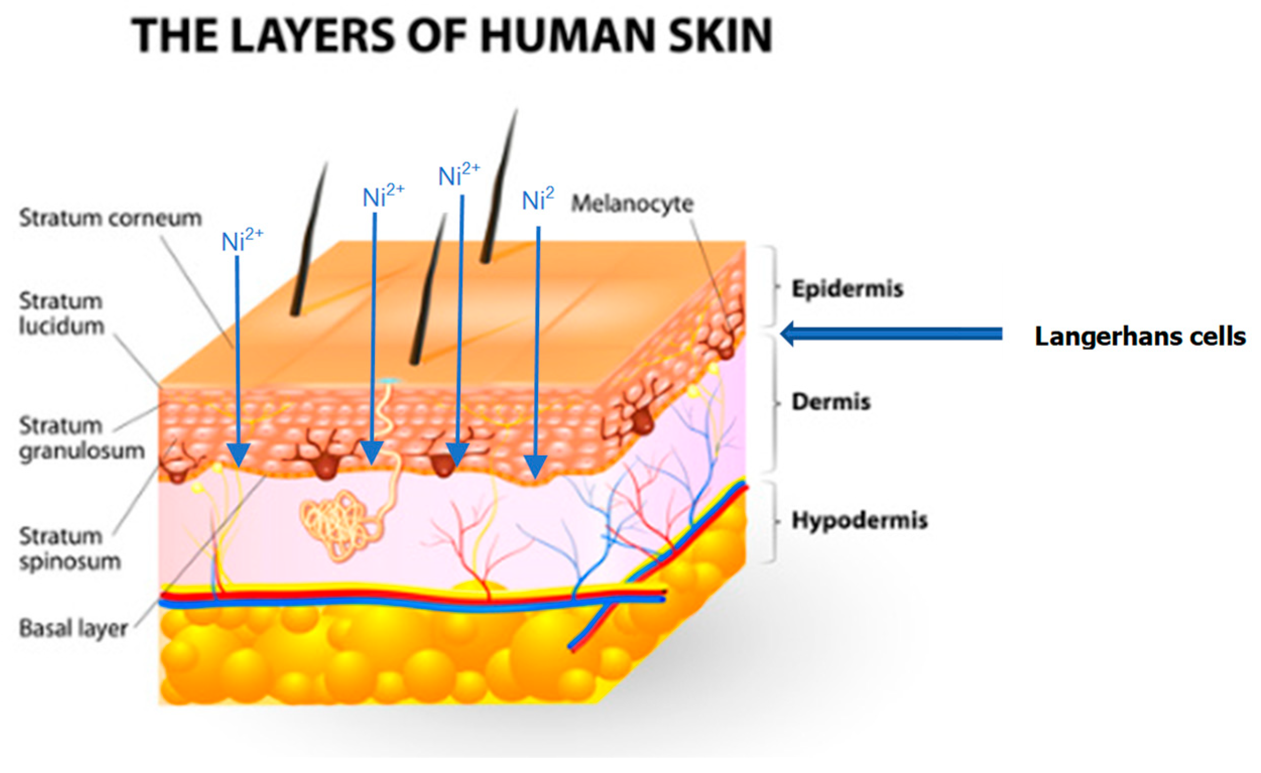
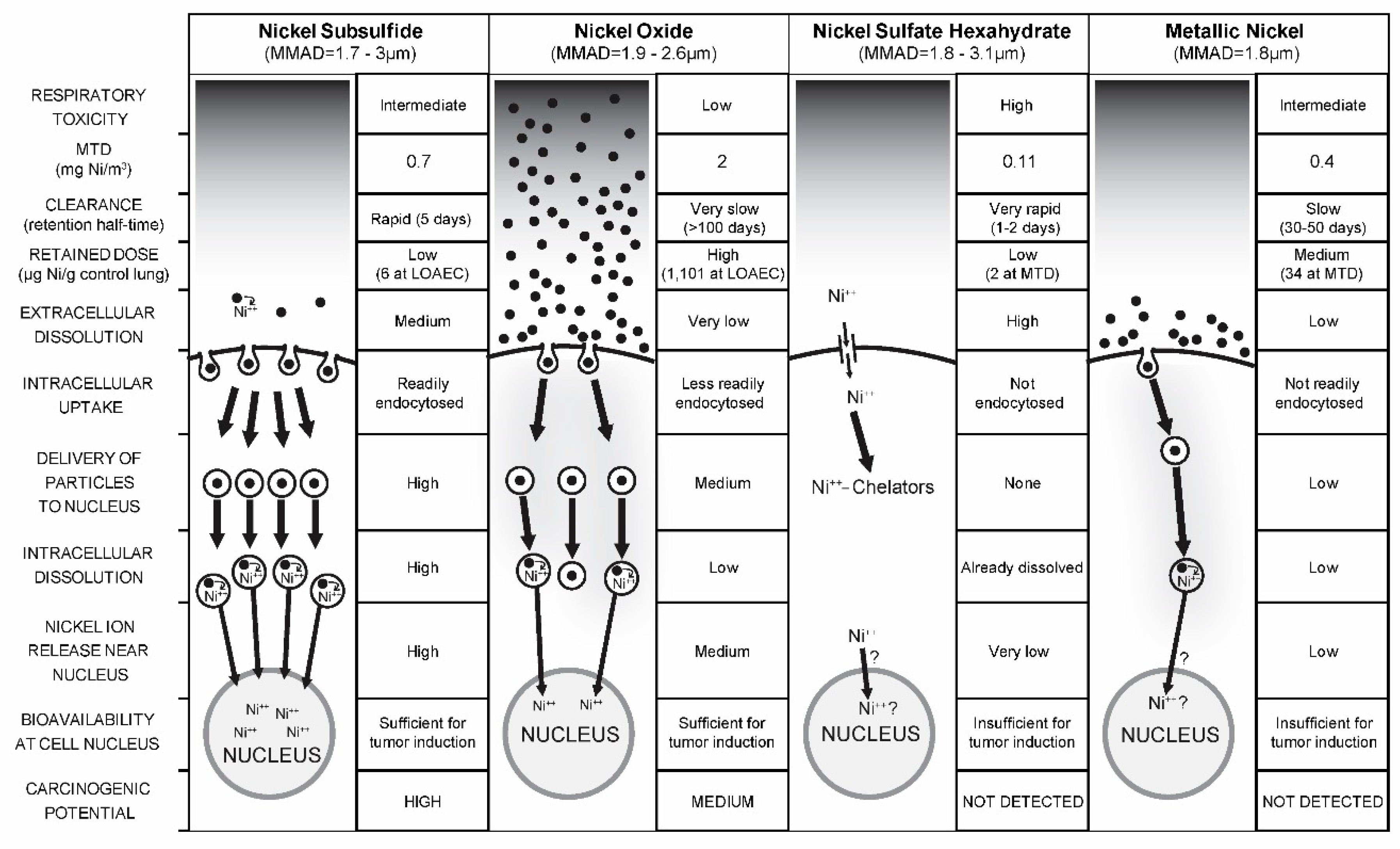
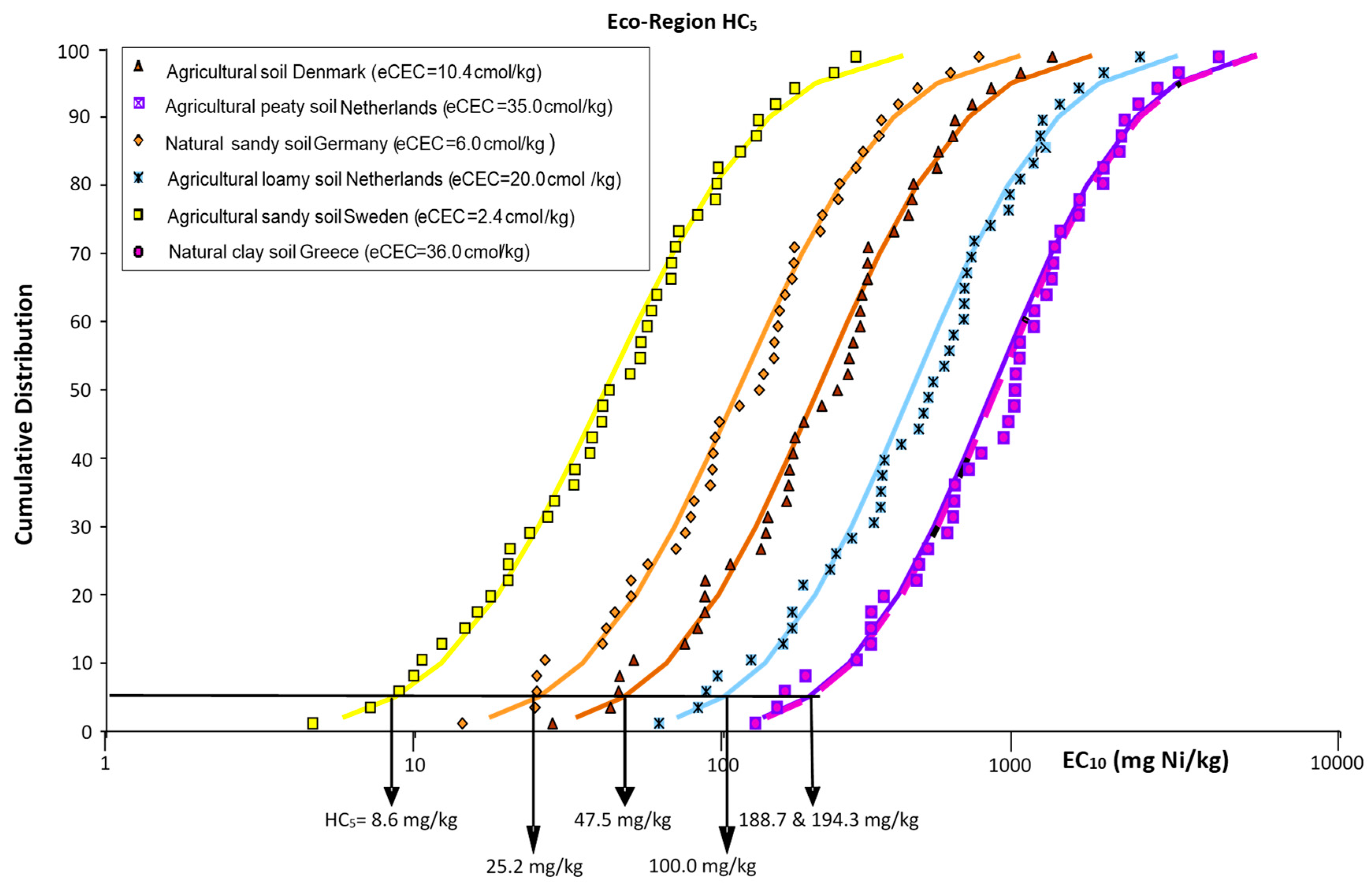
Table 1.
Summary of genotoxicity evidence of nickel and nickel compounds in vitro, in vivo and in humans.
Table 1.
Summary of genotoxicity evidence of nickel and nickel compounds in vitro, in vivo and in humans.
| Genotoxicity/Carcinogenicity | In Vitro Studies | In Vivo Animal Studies | Human Studies |
|---|---|---|---|
| DNA damage | Positive, indirect damage; e.g., single strand breaks, DNA fragmentation [78,79,80] | Positive [81,82,83] | Positive in welders but unassignable to Ni [84,85]; positive in mucosa of orthodontic patients but unassignable to Ni [86,87,88] |
| Impair DNA repair | Positive [74,89,90,91] | ||
| Gene Mutations | Low-to-none [92,93,94,95] | Low mutations [23,92,96] | |
| Chromosomal alterations | a Mixed results [23,96,97,98] | a Mixed results [99,100,101] | a Mixed results [102,103,104,105,106,107] |
| HIF-1α-dependent changes | Stabilization of HIF-1α, increased HIF-1α-dependent gene expressions, e.g., Cap43 [108,109,110,111] | Increased HIF-1α-dependent Cap43 expression [108], hematocrit and/or hemoglobin [51] | |
| Apoptosis/Autophagy | Increase [109,112,113,114,115] | Increase via increased expression of caspase 8 in workers [116] | |
| Histone modifications | |||
| *Hypermethylation | Positive at H3K9 [117,118] | In workers at H3K4 [119,120,121] | |
| *Hyperphosphorylation | Positive at H3S10 [122] | ||
| *Hyperubiquitination | Positive, inhibiting deubiquitinating enzyme activity [123] | ||
| *Hypo/hyperacetylation | Hypoacetylation via inhibiting histone acetyltransferase activity [124,125] | Increased H3K9 acetylation in steel workers [121] | |
| Oxidative stress, ROS production | Positive [78,124,126,127] | Positive in rats and mice [80,128,129] | Oxidative DNA lesions linked to nickel levels [130] |
a Mixed results indicates that both positive and negative effects were observed.
Table 2.
Thresholds for nickel toxicity corresponding to the main routes and health effects of concern to human health and for the main environmental compartments.
Table 2.
Thresholds for nickel toxicity corresponding to the main routes and health effects of concern to human health and for the main environmental compartments.
| Human Health Effects | ||
| Endpoint (Underlying Studies and Data) | Threshold Values a | Reference |
| NACD (human) | Dermal-NOAEL = 0.44 µg Ni/cm2 skin/day a | [267] |
| Oral-BMDL0.1 = 4.3 µg Ni/kg bw/day adult (in addition to diet) | [145] | |
| Respiratory cancer (human) | Inhalation—practical threshold = 0.1 mg Ni/m3 inhalable aerosol | [13] |
| Inhalation—practical threshold = 0.5 µg Ni/m3 ng/m3 PM10 aerosol | [268] | |
| Respiratory non cancer (animal) | Inhalation-NOAEC = 0.03 mg/m3 (Ni sulfate) respirable aerosol | [13] |
| Inhalation-NOAEC = 9.4 µg/m3 (Ni sulfate) PM10 aerosol | [268] | |
| Reproductive (animal) | Oral-BMDL0.05 = 1.8 mg Ni/kg bw/day adult | [145] |
| Environmental Health Effects | ||
| Compartments | Threshold Values a | Reference |
| Freshwater | Bioavailability-based EQS = 4 µg Ni/L | [208] |
| HC5 = 7.1 to 43.6 µg Ni/L for EU waters representing the 10th to 90th percentile of bioavailability conditions | [196] | |
| Marine water | Temperate HC5 = 8.6 µg Ni/L | [209] |
| Tropical aggregated HC5 = 8.2 µg Ni/L | [212] | |
| Sediment | HC5 = 136–437 mg Ni/kg (AVS: 0.77–38.4 µM/g dw) | [202] |
| Soil | HC5 = 8.6–194.3 mg Ni/kg (eCEC: 10.4–36.0 cmol/kg) | [194] |
a Values are based on, or derived from, the most bioavailable chemical form of nickel (i.e., water soluble nickel compounds) but are generally applied to “total nickel”.
© 2019 by the authors. Licensee MDPI, Basel, Switzerland. This article is an open access article distributed under the terms and conditions of the Creative Commons Attribution (CC BY) license (http://creativecommons.org/licenses/by/4.0/).
Share and Cite
MDPI and ACS Style
Buxton, S.; Garman, E.; Heim, K.E.; Lyons-Darden, T.; Schlekat, C.E.; Taylor, M.D.; Oller, A.R. Concise Review of Nickel Human Health Toxicology and Ecotoxicology. Inorganics 2019, 7, 89. https://doi.org/10.3390/inorganics7070089
AMA Style
Buxton S, Garman E, Heim KE, Lyons-Darden T, Schlekat CE, Taylor MD, Oller AR. Concise Review of Nickel Human Health Toxicology and Ecotoxicology. Inorganics. 2019; 7(7):89. https://doi.org/10.3390/inorganics7070089
Chicago/Turabian StyleBuxton, Samuel, Emily Garman, Katherine E. Heim, Tara Lyons-Darden, Christian E. Schlekat, Michael D. Taylor, and Adriana R. Oller. 2019. "Concise Review of Nickel Human Health Toxicology and Ecotoxicology" Inorganics 7, no. 7: 89. https://doi.org/10.3390/inorganics7070089
Note that from the first issue of 2016, this journal uses article numbers instead of page numbers. See further details here.