Next Issue
Volume 10, April
Previous Issue
Volume 10, February


Dent. J., Volume 10, Issue 3 (March 2022) – 18 articles

Cover Story (view full-size image): 
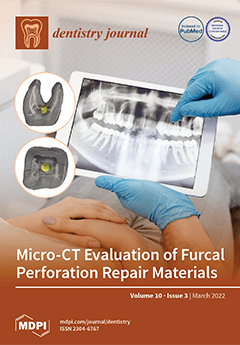
Furcal perforations are inconvenient procedural accidents that can create a pathological path between the root canal system and the periodontium. This can lead to an adverse scenario, with uncertain prognosis. The repair material needs to provide good filling ability by avoiding the development of gaps between the dentin walls and the material, and voids within the material content, minimizing bacterial microleakage. Calcium-silicate-based materials promote a tight seal and lead the regeneration of periodontal and bone tissues, becoming the standard choice to repair these accidents. Due to the lack of studies evaluating gaps and voids in cases of furcal perforation, this study evaluated the volume of gaps and voids of calcium-silicate based materials in simulated furcal perforations. View this paper
  • Issues are regarded as officially published after their release is announced to the table of contents alert mailing list.
  • You may sign up for e-mail alerts to receive table of contents of newly released issues.
  • PDF is the official format for papers published in both, html and pdf forms. To view the papers in pdf format, click on the "PDF Full-text" link, and use the free Adobe Reader to open them.
Order results
Result details
Section
Select all
Export citation of selected articles as:
12 pages, 665 KiB  
Review
Chronic Stress and Depression in Periodontitis and Peri-Implantitis: A Narrative Review on Neurobiological, Neurobehavioral and Immune–Microbiome Interplays and Clinical Management Implications
by Francesco D’Ambrosio, Mario Caggiano, Luigi Schiavo, Giulia Savarese, Luna Carpinelli, Alessandra Amato and Alfredo Iandolo
Dent. J. 2022, 10(3), 49; https://doi.org/10.3390/dj10030049 - 18 Mar 2022
Cited by 31 | Viewed by 6473
Abstract
Besides the well-known systemic factors for periodontal and peri-implant diseases, additional co-factors, such as chronic stress and depression, may also affect disease onset and progression as well as treatment responsiveness. Neurobiological and neurobehavioral pathogenic links between chronic stress and depression, on the one [...] Read more.
Besides the well-known systemic factors for periodontal and peri-implant diseases, additional co-factors, such as chronic stress and depression, may also affect disease onset and progression as well as treatment responsiveness. Neurobiological and neurobehavioral pathogenic links between chronic stress and depression, on the one side, and periodontitis and peri-implantitis, on the other side, which have been little investigated and principally related to necrotizing periodontal disease, have been reviewed, along with their putative interconnections with periodontal immune–microbiome balance. Rising evidence suggest that dysregulated neurobiological and neurobehavioral factors, as well as periodontal immune–microbiome unbalance, all related to chronic stress and depression, may crucially interact and thus represent contributing factors in the genesis and worsening not only of necrotizing periodontal lesions, but also of chronic periodontitis and peri-implantitis. Such potential interconnections may be even more relevant in recurrent and aggressive cases of periodontal and peri-implant disease, which are frequently refractory to therapy, and may, if corroborated, coherently pave the way for personalized prevention and treatment strategies, possibly targeting immune–microbiome unbalance and neurobehavioral factors and focusing on neurobiological ones, especially in chronically stressed and depressed subjects with periodontitis and peri-implantitis. Full article
(This article belongs to the Section Oral Hygiene, Periodontology and Peri-implant Diseases)
Show Figures

Figure 1

17 pages, 564 KiB  
Review
Deep Margin Elevation: A Literature Review
by Theodora Kalliopi Samartzi, Dimokritos Papalexopoulos, Panagiotis Ntovas, Christos Rahiotis and Markus B. Blatz
Dent. J. 2022, 10(3), 48; https://doi.org/10.3390/dj10030048 - 14 Mar 2022
Cited by 16 | Viewed by 14258
Abstract
A conservative approach for restoring deep proximal lesions is to apply an increment of composite resin over the preexisting cervical margin to relocate it coronally, the so-called “deep margin elevation” (DME). A literature search for research articles referring to DME published from January [...] Read more.
A conservative approach for restoring deep proximal lesions is to apply an increment of composite resin over the preexisting cervical margin to relocate it coronally, the so-called “deep margin elevation” (DME). A literature search for research articles referring to DME published from January 1998 until November 2021 was conducted using MEDLINE (PubMed), Ovid, Scopus, Cochrane Library and Semantic Scholar databases applying preset inclusion and exclusion criteria. Elevation material and adhesive system employed for luting seem to be significant factors concerning the marginal adaptation of the restoration. This technique does not affect bond strength, fatigue behavior, fracture resistance, failure pattern or repairability. DME and subgingival restorations are compatible with periodontal health, given that they are well-polished and refined. The available literature is limited mainly to in vitro studies. Therefore, randomized clinical trials with extended follow-up periods are necessary to clarify all aspects of the technique and ascertain its validity in clinical practice. For the time being, DME should be applied with caution respecting three criteria: capability of field isolation, the perfect seal of the cervical margin provided by the matrix, and no invasion of the connective compartment of biological width. Full article
Show Figures

Figure 1

21 pages, 978 KiB  
Systematic Review
Temporal and Permanent Changes Induced by Maxillary Sinus Lifting with Bone Grafts and Maxillary Functional Endoscopic Sinus Surgery in the Voice Characteristics—Systematic Review
by Rafael Delgado-Ruiz, Daniele Botticelli and Georgios Romanos
Dent. J. 2022, 10(3), 47; https://doi.org/10.3390/dj10030047 - 11 Mar 2022
Cited by 3 | Viewed by 3137
Abstract
Sinus surgery procedures such as sinus lifting with bone grafting or maxillary functional endoscopy surgery (FESS) can present different complications. The aims of this systematic review are to compile the post-operatory complications of sinus elevation with bone grafting and FESS including voice changes, [...] Read more.
Sinus surgery procedures such as sinus lifting with bone grafting or maxillary functional endoscopy surgery (FESS) can present different complications. The aims of this systematic review are to compile the post-operatory complications of sinus elevation with bone grafting and FESS including voice changes, and to elucidate if those changes are either permanent or temporary. The Preferred Reporting Items for Systematic Reviews and Meta-Analyses (PRISMA) were used, and the literature was exhaustively searched without time restrictions for randomized and non-randomized clinical studies, cohort studies (prospective and retrospective), and clinical case reports with ≥4 cases focused on sinus lift procedures with bone grafts and functional endoscopic maxillary sinus surgery. A total of 435 manuscripts were identified. After reading the abstracts, 101 articles were selected to be read in full. Twenty articles that fulfilled the inclusion criteria were included for analysis. Within the limitations of this systematic review, complications are frequent after sinus lifting with bone grafts and after FEES. Voice parameters are scarcely evaluated after sinus lifting with bone grafts and no voice changes are reported. The voice changes that occur after FESS include a decreased fundamental frequency, increased nasality, and nasalance, all of which are transitory. Full article
Show Figures

Figure 1

11 pages, 2923 KiB  
Article
Abrasion Behavior of Different Charcoal Toothpastes on Human Dentin When Using Electric Toothbrushes
by Nadin Osmanaj, Svea Petersen, Michael Eisenburger and Andreas Greuling
Dent. J. 2022, 10(3), 46; https://doi.org/10.3390/dj10030046 - 11 Mar 2022
Cited by 3 | Viewed by 3191
Abstract
The aim of this study was to investigate abrasion on human dentin after brushing with activated charcoal toothpastes. A self-designed brushing machine was used to brush five groups (Group A: Water, Group B: Sensodyne Pro Schmelz, Group C: Splat Blackwood, Group D: Curaprox [...] Read more.
The aim of this study was to investigate abrasion on human dentin after brushing with activated charcoal toothpastes. A self-designed brushing machine was used to brush five groups (Group A: Water, Group B: Sensodyne Pro Schmelz, Group C: Splat Blackwood, Group D: Curaprox Black is White, and Group E: Prokudent Black Brilliant) with electrically powered toothbrushes for 4 h. The abrasive dentin wear was calculated using profilometry data. Furthermore, thermogravimetric analyses and scanning electron microscopy were used to analyze the composition of the toothpastes. Mean dentin loss by brushing were (71 ± 28) µm (Splat Blackwood), (44 ± 16) µm (Curaprox Black is White), (38 ± 13) µm (Prokudent Black Brilliant), (28 ± 14) µm (Sensodyne Pro Schmelz), and (28 ± 13) µm (Water). Groups A/B/D/E and group C each lie in one subset, which is statistically different from the other subset according to a post hoc Tukey test (p = 0.05). Within the limitations, it can be concluded that the content of activated charcoal in charcoal toothpastes had little influence on the observed abrasive behavior, although one of the charcoal toothpastes showed the highest abrasion on dentin. Full article
(This article belongs to the Section Oral Hygiene, Periodontology and Peri-implant Diseases)
Show Figures

Figure 1

10 pages, 245 KiB  
Article
Association of Dental Fear with Caries Status and Self-Reported Dentition-Related Well-Being in Finnish Conscripts
by Antti Kämppi, Tarja Tanner, Olavi Viitanen, Vesa Pohjola, Jari Päkkilä, Leo Tjäderhane, Vuokko Anttonen and Pertti Patinen
Dent. J. 2022, 10(3), 45; https://doi.org/10.3390/dj10030045 - 11 Mar 2022
Cited by 2 | Viewed by 2352
Abstract
The main aim of this cross-sectional study was to examine the prevalence of dental fear among Finnish conscripts. Other aims were to study the association between dental fear and cariological status as well as their self-reported, dentition-related well-being. The study material consisted of [...] Read more.
The main aim of this cross-sectional study was to examine the prevalence of dental fear among Finnish conscripts. Other aims were to study the association between dental fear and cariological status as well as their self-reported, dentition-related well-being. The study material consisted of 13,564 men and 255 women conscripts who underwent oral examinations. Of those, 8713 responded to a computer-based questionnaire. The mean number of decayed teeth (DT) was used in analyses for cariological status. Self-reported dental fear, dentition-related well-being and regular check-ups were analysed. Data were analysed with cross tables, Pearson Chi-Square tests, Fisher’s exact test and binary logistic regressive analysis. High dental fear or finding dental visits very scary was associated with DT > 2 both among women (14.6%, when DT = 0; 33.3%, when DT > 2) and men conscripts (2.3% and 10.8%, respectively). In addition, those reporting that dental health had a negative impact on their well-being and had no regular check-ups were more likely to need cariological treatment than the rest. A high education level, both one’s own and parental, was a protective factor for restorative treatment need in male conscripts. The findings of this study support the concept of a vicious cycle of dental fear and dental caries. A preventive, interactive way of work by dental teams would most likely be beneficial for dental health, avoiding the development of dental fear, and dentition-related well-being. Full article
12 pages, 914 KiB  
Article
Burning Mouth Syndrome (BMS)—Treatment with Verbal and Written Information, B Vitamins, Probiotics, and Low-Level Laser Therapy: A Randomized Clinical Trial
by Božana Lončar-Brzak, Ivana Škrinjar, Vlaho Brailo, Danica Vidović-Juras, Lada Šumilin and Ana Andabak-Rogulj
Dent. J. 2022, 10(3), 44; https://doi.org/10.3390/dj10030044 - 10 Mar 2022
Cited by 3 | Viewed by 4188
Abstract
Background: The objective of this study was to determine the most effective treatment option for burning mouth syndrome. Methods: Informative treatment alone, B vitamin injections, oral cavity probiotics, and low-level laser therapy were evaluated and compared. The study included new patients diagnosed with [...] Read more.
Background: The objective of this study was to determine the most effective treatment option for burning mouth syndrome. Methods: Informative treatment alone, B vitamin injections, oral cavity probiotics, and low-level laser therapy were evaluated and compared. The study included new patients diagnosed with burning mouth syndrome, who were randomly allocated into one of four treatment groups. The primary outcome was improvement in patient’s quality of life as determined by a self-perceived Oral Health Impact Profile-14 (OHIP-14) quality of life questionnaire before and after therapy. The secondary outcome was determination of mucosal symptom intensity according to visual analog scale (VAS) grading from 0 to 10. Data were submitted to statistical analysis. Results: A total of 62 patients completed the study. Oral cavity probiotics and LLLT scores for OHIP-14 resulted in a statistically significant difference before and after therapy. Standardized effect sizes between OHIP scores before and after treatment were the greatest for patients who had received oral cavity probiotics. Conclusions: Oral cavity probiotics and LLLT were the most effective treatment for improvement in quality of life. Further investigation on a larger group of patients is required. Full article
Show Figures

Figure 1

14 pages, 4998 KiB  
Article
Thermal Load and Heat Transfer in Dental Titanium Implants: An Ex Vivo-Based Exact Analytical/Numerical Solution to the ‘Heat Equation’
by Grigorios P. Panotopoulos and Ziyad S. Haidar
Dent. J. 2022, 10(3), 43; https://doi.org/10.3390/dj10030043 - 10 Mar 2022
Cited by 1 | Viewed by 3421
Abstract
Introduction: Heat is a kinetic process whereby energy flows from between two systems, hot-to-cold objects. In oro-dental implantology, conductive heat transfer/(or thermal stress) is a complex physical phenomenon to analyze and consider in treatment planning. Hence, ample research has attempted to measure heat-production [...] Read more.
Introduction: Heat is a kinetic process whereby energy flows from between two systems, hot-to-cold objects. In oro-dental implantology, conductive heat transfer/(or thermal stress) is a complex physical phenomenon to analyze and consider in treatment planning. Hence, ample research has attempted to measure heat-production to avoid over-heating during bone-cutting and drilling for titanium (Ti) implant-site preparation and insertion, thereby preventing/minimizing early (as well as delayed) implant-related complications and failure. Objective: Given the low bone–thermal conductivity whereby heat generated by osteotomies is not effectively dissipated and tends to remain within the surrounding tissue (peri-implant), increasing the possibility of thermal-injury, this work attempts to obtain an exact analytical solution of the heat equation under exponential thermal-stress, modeling transient heat transfer and temperature changes in Ti implants (fixtures) upon hot-liquid oral intake. Materials and Methods: We, via an ex vivo-based model, investigated the impact of the (a) material, (b) location point along implant length, and (c) exposure time of the thermal load on localized temperature changes. Results: Despite its simplicity, the presented solution contains all the physics and reproduces the key features obtained in previous numerical analyses studies. To the best of our knowledge, this is the first introduction of the intrinsic time, a “proper” time that characterizes the geometry of the dental implant fixture, where we show, mathematically and graphically, how the interplay between “proper” time and exposure time influences temperature changes in Ti implants, under the suitable initial and boundary conditions. This fills the current gap in the literature by obtaining a simplified yet exact analytical solution, assuming an exponential thermal load model relevant to cold/hot beverage or food intake. Conclusions: This work aspires to accurately complement the overall clinical diagnostic and treatment plan for enhanced bone–implant interface, implant stability, and success rates, whether for immediate or delayed loading strategies. Full article
Show Figures

Figure 1

13 pages, 2873 KiB  
Article
Water Sorption, Solubility, and Translucency of 3D-Printed Denture Base Resins
by Mohammed M. Gad, Saleh Z. Alshehri, Shahad A. Alhamid, Alanoud Albarrak, Soban Q. Khan, Faris A. Alshahrani and Firas K. Alqarawi
Dent. J. 2022, 10(3), 42; https://doi.org/10.3390/dj10030042 - 9 Mar 2022
Cited by 29 | Viewed by 3724
Abstract
This study aimed to evaluate the water sorption, solubility, and translucency of 3D-printed denture base resins (NextDent, FormLabs, and Asiga), compare them to heat-polymerized acrylic denture base resins, and assess their performance under the effects of thermal cycling. A total of 80 acrylic [...] Read more.
This study aimed to evaluate the water sorption, solubility, and translucency of 3D-printed denture base resins (NextDent, FormLabs, and Asiga), compare them to heat-polymerized acrylic denture base resins, and assess their performance under the effects of thermal cycling. A total of 80 acrylic disc specimens were used in the current study, categorized into four groups (n = 10); in one group, the samples were fabricated conventionally with a heat-polymerizing process (control), while the other three groups were fabricated digitally from different 3D-printed reins (NextDent, FormLabs, and Asiga). Specimens were fabricated according to the manufacturers’ recommendations and immersed in distilled water for 48 h at 37 °C. Data on water sorption, solubility, and translucency measurements (T1) were obtained. All the specimens were subjected to 5000 thermal cycles, and then the measures were repeated using the same method (T2). Data analysis was attained via ANOVA and the post hoc Tukey test (α = 0.05). The type of resin significantly affected the values of water sorption, solubility, and translucency (p < 0.001). The water sorption of 3D-printed resins was increased significantly in comparison to control with or without a thermal cycling effect. In terms of solubility, a significant increase in 3D-printed resins before thermocycling was observed; however, after thermocycling, Asiga had a significantly low value compared to the other groups (p < 0.001). Thermal cycling increased the water sorption and solubility of all tested materials. In comparison to control, the translucency of the 3D-printed resins was significantly decreased (p < 0.001). The translucency was significantly decreased per material in terms of the thermal cycling effect (before and after). NextDent showed significantly low translucency values (p < 0.001) compared to the other groups. All 3D-printed resin groups had higher water sorption and solubility and lower translucency values in comparison to the heat-polymerized resin group. Regardless of resin types, thermal cycling adversely affected all tested properties. Full article
(This article belongs to the Section Digital Technologies)
Show Figures

Figure 1

10 pages, 2125 KiB  
Article
Volumetric Evaluation of Voids and Gaps of Different Calcium-Silicate Based Materials Used in Furcal Perforations: A Micro-CT Study
by Cassia Cestari Toia, Fabricio Batista Teixeira, Carolina Cucco, Marcia Carneiro Valera and Bruno Neves Cavalcanti
Dent. J. 2022, 10(3), 41; https://doi.org/10.3390/dj10030041 - 9 Mar 2022
Cited by 2 | Viewed by 2497
Abstract
This study aimed at evaluating volumetrically gaps and voids of calcium-silicate based materials of different generations and handling properties (BC—Endosequence BC RRM-Fast Set Condensable Putty, MTA—ProRoot MTA, and BIO—Biodentine) in simulated furcal perforations in an ex vivo setup by microcomputed tomography (Micro-CT) analysis. [...] Read more.
This study aimed at evaluating volumetrically gaps and voids of calcium-silicate based materials of different generations and handling properties (BC—Endosequence BC RRM-Fast Set Condensable Putty, MTA—ProRoot MTA, and BIO—Biodentine) in simulated furcal perforations in an ex vivo setup by microcomputed tomography (Micro-CT) analysis. Thirty-six extracted human mandibular molars with sound furcation areas were selected. Standardized perforations were created in the furcation area of the pulp chamber using #4 diamond burs. The specimens were randomly assigned to three groups (BC, MTA and BIO; n = 12). Samples were then scanned (SkyScan 1172; Bruker-microCT, Kontich, Belgium), and three-dimensional (3D) images reconstructed. The relative volume of gaps (VG%) and voids (VV%) present on each material was calculated. Data were analyzed using one-way analysis of variance (ANOVA) and Tukey’s HSD test (p < 0.05). Mean VG% for BC, MTA, and BIO groups were, respectively, 0.513%, 1.128%, 1.460%, with BC presenting statistically (p < 0.05) fewer gaps formation than the other groups. Mean VV% were, respectively, 0.018%, 0.037%, and 0.065%. The was no statistical difference regarding VV%. There were no gap-free and void-free samples. BC group had the lowest VG% among the groups with a significant statistical difference (p < 0.05). Full article
Show Figures

Figure 1

14 pages, 551 KiB  
Article
Intention of Collaboration among Dental Students during the COVID-19 Pandemic
by Kyriaki Hadjichambi, Evie Georgiadou, Vasileios Margaritis and Maria Antoniadou
Dent. J. 2022, 10(3), 40; https://doi.org/10.3390/dj10030040 - 8 Mar 2022
Cited by 3 | Viewed by 3251
Abstract
Interpersonal communication skills (ICS) are crucial for effective dental practice and interprofessional collaboration. The current study aimed to assess the attitudes of Greek dental undergraduate students towards team working and their cooperation abilities during the COVID-19 pandemic. One-hundred and twenty-seven fourth-semester dental students [...] Read more.
Interpersonal communication skills (ICS) are crucial for effective dental practice and interprofessional collaboration. The current study aimed to assess the attitudes of Greek dental undergraduate students towards team working and their cooperation abilities during the COVID-19 pandemic. One-hundred and twenty-seven fourth-semester dental students (N1 = 127) out of 145 (N0) filled in the online survey placed on Google forms. The “Dental Students Cooperation Questionnaire” (DSC) consisted of 49 questions and was available for completion for one week during April 2020. Bivariate (ANOVA) and linear regression analysis of data revealed that mean scores of the questionnaire increased as the parents’ educational level also increased. Data analysis showed that dental students had the required ICS and the intention to collaborate with each other. Many participants managed to achieve group goals, were willing to support other members to fulfill the project’s goals, and there was no competition among them. They acknowledged the importance of feedback, the reward at the end of a group project and social media as a tool for teamworking communication. The students reported that the most important characteristics of an academic teacher were patience, willingness to cooperate, friendliness, politeness, willingness to help, accessibility and availability. It is suggested that group work should be included in the curriculum of dental schools to enhance the integration and evolution of students’ ICS, and the DSC questionnaire can be an effective tool to assess these skills. Full article
Show Figures

Figure 1

13 pages, 644 KiB  
Article
Short Narrow Dental Implants versus Long Narrow Dental Implants in Fixed Prostheses: A Prospective Clinical Study
by Eduardo Antiua, Virginia Escuer and Mohammad H. Alkhraisat
Dent. J. 2022, 10(3), 39; https://doi.org/10.3390/dj10030039 - 4 Mar 2022
Cited by 3 | Viewed by 3480
Abstract
There is a paucity of studies that assess short and narrow dental implants. This prospective study aimed to evaluate the performance of both short (≤8 mm) and narrow (≤3.5 mm width) dental implants supporting fixed prostheses in the atrophic maxilla or mandible. Towards [...] Read more.
There is a paucity of studies that assess short and narrow dental implants. This prospective study aimed to evaluate the performance of both short (≤8 mm) and narrow (≤3.5 mm width) dental implants supporting fixed prostheses in the atrophic maxilla or mandible. Towards that aim, patients with short implants were included in the study. The control group was those with long and narrow dental implants (length > 8 mm and diameter ≤ 3.5 mm). Clinical and demographic variables were extracted from clinical records. During the follow-up, implant survival and marginal bone loss were evaluated and statistically analysed. Forty-one implants were included (18 and 23 implants in the test and control groups, respectively). The median follow-up time was 26 months since insertion in both groups. The results revealed that there was no implant failure and no statistically significant differences in terms of marginal bone loss. Only one screw-loosening effect occurred in the short implants group. Short, narrow dental implants could be an alternative for the restoration of severely resorbed jaws. Full article
(This article belongs to the Special Issue Oral Implantology and Bone Regeneration)
Show Figures

Figure 1

19 pages, 1943 KiB  
Article
Investigation of the Molecular Profile of Granular Cell Tumours and Schwannomas of the Oral Cavity
by Benjamin Rogala, Zia Ali Khan, Linda Jackson-Boeters and Mark Roger Darling
Dent. J. 2022, 10(3), 38; https://doi.org/10.3390/dj10030038 - 4 Mar 2022
Cited by 2 | Viewed by 2652
Abstract
Granular cell tumours (GCTs) are rare submucosal lesions, thought to develop from Schwann cells, characterised by large polygonal cells with abundant lysosomes. The objectives of this study are to investigate whether GCTs have an antigen-presenting cell (APC) phenotype or a neural crest phenotype [...] Read more.
Granular cell tumours (GCTs) are rare submucosal lesions, thought to develop from Schwann cells, characterised by large polygonal cells with abundant lysosomes. The objectives of this study are to investigate whether GCTs have an antigen-presenting cell (APC) phenotype or a neural crest phenotype using immunohistochemistry and to compare expression profiles with Schwannomas. Immunoreactivity to CD68, HLA-DR, CD163, CD40 and CD11c (APC phenotype) and markers of neural crest cell (NCC) origin S100, SOX10, NSE and GAP43 in 23 cases of GCTs and 10 cases of Schwannomas were evaluated. RT-qPCR was used to identify a possible NCC developmental phenotype in 6 cases of GCTs. GAP43 was identified as a new NCC marker for GCTs, and some evidence was found for an APC phenotype from CD68 and HLA-DR immunoreactivity. RT-qPCR failed to identify an NCC developmental phenotype of GCTs, likely due to technical issues. Full article
(This article belongs to the Special Issue Molecular Diagnostics in Oral Diseases)
Show Figures

Figure 1

11 pages, 2969 KiB  
Article
Intrasulcular Restorations of Anterior Teeth According to the BAIR Technique: Evaluation of Periodontal Parameters
by Luca Giachetti, Francesca Cinelli and Michele Nieri
Dent. J. 2022, 10(3), 37; https://doi.org/10.3390/dj10030037 - 2 Mar 2022
Cited by 2 | Viewed by 2968
Abstract
Some clinical situations, such as the closure of pronounced diastemas, and the transformation of malformed, small, or peg-shaped teeth, require a rebalancing of dental proportions accompanied by a modification of the gingival contour. A traditional treatment plan can include surgical, prosthetic, and/or orthodontic [...] Read more.
Some clinical situations, such as the closure of pronounced diastemas, and the transformation of malformed, small, or peg-shaped teeth, require a rebalancing of dental proportions accompanied by a modification of the gingival contour. A traditional treatment plan can include surgical, prosthetic, and/or orthodontic treatments. In some cases, it can be considered too invasive, and not all patients are willing to undertake long therapies. To overcome these limitations, a possible solution could be the application of the Biologically Active Intrasulcular Restoration (BAIR) technique, which allows us to modify the natural emergence tooth profile using simple intrasulcular direct restorations. The aims of this paper are to investigate possible effects on gingival health, and to assess the patient satisfaction about the aesthetic intervention performed. Periodontal data were collected, and patient satisfaction was registered using the VAS questionnaire. All sites healed without complications, no adverse events were registered or reported by the patients, and no signs of periodontal morbidity were visible. The results show that the patients evaluated the final aesthetics in a positive way, and they perceived a good condition of periodontal health as well. The intervention is felt as almost painless, and patients do not report significant post-operative distress. In conclusion, the BAIR technique can provide a valid therapeutic alternative for patients for whom traditional treatments are not indicated. It is a minimally invasive intervention where both the operating times and the biologic and economic costs are reduced. Full article
(This article belongs to the Special Issue Dental Materials and Their Clinical Applications)
Show Figures

Figure 1

18 pages, 4926 KiB  
Article
The Effects of Ultrasonic Scaling and Air-Abrasive Powders on the Decontamination of 9 Implant-Abutment Surfaces: Scanning Electron Analysis and In Vitro Study
by Francesco Gianfreda, Patrizio Bollero, Maurizio Muzzi, Andrea Di Giulio, Eleonora Nicolai and Luigi Canullo
Dent. J. 2022, 10(3), 36; https://doi.org/10.3390/dj10030036 - 1 Mar 2022
Cited by 5 | Viewed by 3093
Abstract
(1) Background: The aim of this study is to understand from a microscopic point of view whether bicarbonate air-abrasive powders associated with ultrasonic instruments can decontaminate nine different surfaces used for the abutment/implant junction. Fibroblast growth was carried out on decontaminated surface in [...] Read more.
(1) Background: The aim of this study is to understand from a microscopic point of view whether bicarbonate air-abrasive powders associated with ultrasonic instruments can decontaminate nine different surfaces used for the abutment/implant junction. Fibroblast growth was carried out on decontaminated surface in order to understand if there are significative differences in terms of biocompatibility. (2) Methods: After taking samples of patient plaque, nine different surfaces were contaminated and analyzed by SEM, then their wettability was evaluated. Fibroblasts were cultured on the decontaminated surfaces to understand their ability to establish a connective tissue seal after decontamination. The results were analyzed from a statistical point of view to hypothesize a mathematical model capable of explaining the properties of the surfaces. (3) Results: A negative correlation between roughness and contamination has been demonstrated, whereas a weak correlation was observed between wettability and decontamination capacity. All surfaces were topographically damaged after the decontamination treatment. Grade 5 titanium surfaces appear tougher, whereas anodized surfaces tend to lose the anodizing layer. (4) Conclusions: further studies will be needed to fully understand how these decontaminated surfaces affect the adhesion, proliferation and differentiation of fibroblasts and osteoblasts. Full article
(This article belongs to the Collection Bio-Logic Approaches to Implant Dentistry)
Show Figures

Figure 1

12 pages, 2325 KiB  
Article
Duplicating Complete Dentures with Conventional and Digital Methods: Comparisons of Trueness and Efficiency
by Li Chen, Deli Li, Jianfeng Zhou, Wei-Shao Lin and Jianguo Tan
Dent. J. 2022, 10(3), 35; https://doi.org/10.3390/dj10030035 - 1 Mar 2022
Cited by 8 | Viewed by 3352
Abstract
Background: A complete denture (CD) can be duplicated with a conventional or digital protocol. However, there are no comparative studies of these methods. This study aimed to compare the trueness and efficiency of conventional and digital CD duplication methods. Methods: A mandibular CD [...] Read more.
Background: A complete denture (CD) can be duplicated with a conventional or digital protocol. However, there are no comparative studies of these methods. This study aimed to compare the trueness and efficiency of conventional and digital CD duplication methods. Methods: A mandibular CD was digitized as the virtual reference model and duplicated using five methods (n = 10). The trueness (root mean square (RMS)) was calculated for the whole denture and across the dentition, cameo denture extension, and intaglio portions. The manual labor time spent during denture duplication was also recorded at different steps. The trueness and labor time comparisons were statistically analyzed among the five groups (α = 0.05). Results: The conventional group was the least true with the largest RMS (mean, 95% CI) in all of the comparisons. The four digital groups yielded similar trueness values across the dentition, cameo denture extension, and intaglio areas, yet they had a significant difference in the whole denture comparison between the Digital-CBCT-SLA printer (0.17, 0.15–0.19 mm) and Digital-Laboratory Scanner-SLA printer (0.13, 0.11–0.15 mm). The conventional protocol required longer trimming and finishing time (7.55 ± 1.02 min), as well as total labor time (27.64 ± 1.72 min) than the other four digital techniques. Conclusions: The conventional CD duplication method was less true and efficient than digital techniques. Full article
Show Figures

Figure 1

12 pages, 4529 KiB  
Case Report
Clear Cell Odontogenic Carcinoma: A Series of Three Cases
by Asad Ullah, Christian Cullen, Samantha N. Mattox, Diana Kozman, Nikhil Patel, Suash Sharma and Rafik Abdelsayed
Dent. J. 2022, 10(3), 34; https://doi.org/10.3390/dj10030034 - 25 Feb 2022
Cited by 1 | Viewed by 5273
Abstract
Background: Clear cell odontogenic carcinoma (CCOC) is a rare malignant odontogenic epithelial neoplasm of the jaws. It is composed of irregular nests of clear to faintly eosinophilic cells resembling clear cell rests of primitive dental lamina and an intermixed hyalinized fibrous stroma. Most [...] Read more.
Background: Clear cell odontogenic carcinoma (CCOC) is a rare malignant odontogenic epithelial neoplasm of the jaws. It is composed of irregular nests of clear to faintly eosinophilic cells resembling clear cell rests of primitive dental lamina and an intermixed hyalinized fibrous stroma. Most cases occur in the 5th and 6th decades of life, with a female predominance. The mandible is affected more than the maxilla. Clinical features vary from asymptomatic to non-specific pain, ill-defined radiolucency, root resorption, and sometimes soft tissue extension. Histology varies from bland to high grade. CCOC demonstrated a significant tendency to recur. Metastasis typically involves regional lymph nodes, which haves been reported in 20–25% of cases. Pulmonary metastasis rarely occurs. Differential diagnoses are broad and include odontogenic, salivary, melanocytic, and metastatic neoplasia. CCOCs are positive for cytokeratins, mainly AE1/AE3 and CK19. Most cases show EWSR1 rearrangement and rarely, the BRAFV600E mutation. Design: Patient charts were reviewed at our institution. A total of three cases were found in electronic medical records, which were diagnosed as clear cell odontogenic carcinoma over a period of six years (2014–2019). Patient charts were reviewed for medical history and radiology data. The pathology slides were reviewed by one or more faculty members. Results: We present three cases of CCOC, ranging in age from 40 to 69 years (two women and one man). Two cases involved the maxilla and one involved the mandible. Two presented with painful swelling and one with mass recurrence. Radiography results show that two had poorly defined radiolucent lesions, and one was heterogeneous with a small nodule projecting into the maxillary sinus. Histological examination revealed an epithelial neoplasm composed of irregular sheets, cords, and nests of polygonal cells with central hyperchromatic, mildly pleomorphic nuclei surrounded by clear to pale eosinophilic cytoplasm, with occasional mitotic figures. The tumor had infiltrated the bone and soft tissues. Two cases were immunopositive for CK5/6 and one case was positive for p63 and CK19. Interestingly, the eosinophilic dentinoid matrix interspersed among tumor cells in one case was consistent with its odontogenic origin. Histochemical staining showed PAS-positive and diastase-labile intracytoplasmic material consistent with glycogen. Conclusion: Our study highlights the potential diagnostic significance of dentinoid (although reportedly seen in only 7% of cases), along with CK5/6 immunopositivity, in supporting the histologic diagnosis of CCOC among a variety of neoplasia in its differential diagnosis. Full article
Show Figures

Figure 1

10 pages, 1979 KiB  
Article
Interfacial Biomaterial–Dentin Bacterial Biofilm Proliferation and Viability Is Affected by the Material, Aging Media and Period
by Muna Q. Marashdeh, Celine Lévesque, Shimon Friedman, Cameron A. Stewart and Yoav Finer
Dent. J. 2022, 10(3), 33; https://doi.org/10.3390/dj10030033 - 24 Feb 2022
Viewed by 2541
Abstract
Biomaterial–dentin interfaces undergo degradation over time, allowing salivary, tissue fluid, and bacterial movement between the root filling or restoration and dentin. This study aims to investigate the effect of aging in simulated human salivary/bacterial/blood esterases (SHSE) on proliferation and viability of Enterococcus faecalis [...] Read more.
Biomaterial–dentin interfaces undergo degradation over time, allowing salivary, tissue fluid, and bacterial movement between the root filling or restoration and dentin. This study aims to investigate the effect of aging in simulated human salivary/bacterial/blood esterases (SHSE) on proliferation and viability of Enterococcus faecalis biofilm within the dentin interface with four materials used to fill/restore the endodontic space. Root canals of human anterior teeth were prepared and filled with gutta-percha and one of the following: self-cured resin composite (BisfilTM 2B, Bisco, Schaumburg, IL, USA) with either self-etch (SE) (EasyBond) or total-etch (TE) (ScotchbondTM, 3M, Saint Paul, MN, USA) methacrylate-based adhesives, epoxy-resin sealer (AH Plus®, Dentsply Sirona, York, PA, USA), or bioceramic sealer (EndoSequence® BC Sealer™, Brasseler USA, Savannah, GA, USA). Specimens were aged in SHSE or phosphate-buffered saline (PBS) for up to 360 days, followed by cultivation of steady-state E. faecalis biofilm. Depth and viability of interfacial bacterial biofilm proliferation were assessed by confocal laser scanning microscopy and live/dead staining. Data were analyzed using three-way ANOVA and Scheffe’s post hoc analyses. Initial depths of biofilm proliferation were similar among material groups (p > 0.05). All groups showed significantly deeper biofilm proliferation with increased aging period (p < 0.05). SHSE aging increased interfacial biofilm depth for TE, SE and BC (p < 0.05) but not AH. For unaged interfaces, BC exhibited the lowest ratio of live bacteria, followed by AH, TE, and SE (p < 0.05). Interfacial bacterial biofilm proliferation and viability were dependent on the biomaterial, aging media, and period. Full article
(This article belongs to the Section Dental Materials)
Show Figures

Figure 1

10 pages, 717 KiB  
Article
Absence of Oral Opportunistic Infections in Patients with Inflammatory Bowel Disease Receiving Anti-TNF-α and Anti-Integrin-α4β7 Therapy
by Ema Saltović, Brankica Mijandrušić-Sinčić, Alen Braut, Ivana Škrobonja, Ella Sever, Irena Glažar, Sonja Pezelj-Ribarić and Miranda Muhvić-Urek
Dent. J. 2022, 10(3), 32; https://doi.org/10.3390/dj10030032 - 23 Feb 2022
Viewed by 2424
Abstract
Biological therapy of inflammatory bowel disease (IBD) carries an increased risk for the development of opportunistic infections due to immunomodulation. The aim of this study was to determine the prevalence and types of oral infections in IBD patients treated with biological (anti-TNF-α and [...] Read more.
Biological therapy of inflammatory bowel disease (IBD) carries an increased risk for the development of opportunistic infections due to immunomodulation. The aim of this study was to determine the prevalence and types of oral infections in IBD patients treated with biological (anti-TNF-α and anti-integrin-α4β7) and conventional medication protocols. The study included 20 IBD patients receiving anti-TNF-α therapy, 20 IBD patients receiving anti-integrin-α4β7 therapy and 20 IBD patients without immunomodulatory therapy. Participants completed questionnaires on medical information, oral lesions and symptoms. For each patient, clinical examination and a salivary flow rate test were performed, followed by a swab of the oral mucosa. The swab samples were cultured to identify Candida spp. and oral bacteria. No bacterial opportunistic infections were detected. Candidiasis was detected in four participants, with no significant difference between groups (p = 0.765). Hyposalivation was most common in the anti-TNF-α group, with a significant difference between groups (p = 0.036). There were no significant differences between groups in self-reported oral mucosal lesions and symptoms (p > 0.05), or in the distribution of oral mucosal lesions (p > 0.05). This study suggests that IBD patients receiving biological therapy are at no greater risk of developing oral opportunistic infections than IBD patients not receiving immunomodulatory therapy. Full article
Show Figures

Figure 1

Previous Issue
Next Issue
Back to TopTop