


Abstract
(1) Background: For non-growing patients with marked transverse maxillary deficiency, bone-borne surgically assisted rapid maxillary expansion (SARME) has been proposed as an effective treatment option. Objective: To evaluate the dental, skeletal, and soft tissue changes following bone-borne SARME. (2) Methods: An unrestricted systematic electronic search of six databases, supplemented by manual searches, was performed up to April 2023. The eligibility criteria included prospective/retrospective clinical studies with outcomes pertaining to objective measurements of dental/skeletal/soft tissue effects of bone-borne SARME in healthy patients. (3) Results: Overall, 27 studies satisfied the inclusion criteria. The risk of bias of the non-randomized trials ranged between moderate (20) and serious (4). For the two RCTs, there were some concerns of bias. Trials with outcomes measured at the same landmarks within the scope of the prespecified timeframe were deemed eligible for quantitative synthesis. Eventually, five trials were included in the meta-analysis. SARME was associated with a statistically significant lengthening of the dental arch perimeter immediately after expansion, along with a marginally significant decrease in palatal depth during the post-SARME retention period. Post-treatment SNA values exhibited no statistically significant change. (4) Conclusion: Current evidence indicates that bone-borne SARME constitutes an effective treatment option for adult patients with maxillary transverse deficiency. Further long-term randomized clinical trials with robust methodology, large sample sizes, and 3D evaluation of the outcomes are needed.
1. Introduction
Transverse maxillary discrepancies are a significant contributor to the emergence of orthodontic problems and represent a rather frequent issue faced during orthodontic treatment planning. The irregularities observed in these patients include lateral cross bites, narrow palatal vaults, increased buccal corridors, and dental crowding [1].
A variety of orthopedic and orthodontic techniques are available for transverse correction, particularly in young, growing patients. Nevertheless, in non-growing adolescents and adults, this particular challenge is more difficult to overcome, owing to the increased skeletal resistance observed post-ossification of the midpalatal suture [2]. Surgically assisted rapid maxillary expansion (SARME) has been introduced as a procedure aiming to address the treatment of marked transverse maxillary deficiencies in skeletally mature patients, with the goal of providing skeletal expansion with minimal dental effects [3]. Treatment with SARME entails a multi-disciplinary approach, which requires close collaboration between the orthodontic specialist and the oral-maxillofacial surgeon who will perform the orthognathic procedure [3].
SARME usually involves osteotomies akin to the Le Fort I technique, along with a mid-palatal suture disjunction, with or without pterygomaxillary disjunction (PMD). Subsequently, a palatal distractor is inserted in the patient’s oral cavity. This device can be attached to the posterior teeth of the patient (tooth-borne distractor) or directly to the maxilla (bone-borne distractor), or alternatively using a combination of both anchorage options (hybrid distractor) [4,5]. Previous studies have reported an array of approaches with regards to the surgical technique of choice used for the maxillary expander, the necessity for overcorrection, as well as the risk of relapse [5,6,7].
The effects of rapid maxillary expansion on hard and soft facial tissues have been investigated in individual studies via dental models, radiographic imaging, and, more recently, 3D facial scanners [8,9,10]. Several systematic reviews have assessed the differences between the outcomes of rapid maxillary expansion with tooth-borne and bone-borne devices, with results varying from no difference to the superiority of either method as far as different parameters are concerned [11,12,13]. However, limited information from individual studies is available on a meta-analytical level with regards to the evidence-based effects of bone-borne SARME, in terms of the objective measurements of the treatment-induced dental, skeletal, and soft tissue changes. A systematic review by Vilani et al. (2012) [14] examined the long-term dental and skeletal changes in SARME patients without differentiating between tooth-borne and bone-borne devices in the syntheses and found moderate evidence of an increase in alveolar and interdental widths. Similarly, Bortolotti et al. (2020) [15], in their recent review of RCTs investigating the skeletal and dental SARME effects, pooled data regardless of anchorage type and reported a successful expansion in the transverse maxillary dimension. Evidence focusing exclusively on bone-borne SARME would provide a multi-faceted understanding of the expected outcomes of the modality, thus assisting orthodontists in treatment plaining and enhancing the patient selection process. The aim of this systematic review was to assess evidence from clinical studies on the dental, skeletal, and soft tissue changes following bone-borne surgically assisted rapid maxillary expansion (SARME) on orthodontic patients.
2. Materials and Methods
2.1. Protocol, Eligibility Criteria, and Literature Search
The protocol of the present systematic review and meta-analysis was a priori developed in adherence with the Preferred Reporting Items for Systematic Reviews and Meta-Analysis Protocols guidelines (PRISMA-P) and was registered in PROSPERO (CRD42021250742). The study was conducted and reported according to the Cochrane Handbook for Systematic Reviews of Interventions [16] and the Preferred Reporting Items for Systematic Reviews and Meta-Analysis statement (PRISMA) [17]. The eligibility criteria, based on the PICOS schema of the review, included randomized clinical trials (RCTs), as well as prospective and retrospective clinical studies focusing on bone-borne SARME, with outcomes pertaining to objective measurements of dental, skeletal, and soft tissue effects of bone-borne SARME in otherwise healthy patients with transverse maxillary deficiency. Participants with a history of systemic disease or previous orthodontic treatment were excluded. No exclusions based on age, type of malocclusion, or the specific type of skeletal anchorage appliance used were applied. The outcomes comprised differences between pre-intervention and post-intervention measurements of the dental, skeletal, and soft-tissue characteristics reported either immediately after expansion (short-term effects) or during the retention period (long-term effects: 3 or more months post-expansion, in accordance with another meta-analysis) [12].
An unrestricted systematic electronic search of six databases (MEDLINE via PubMed, Cochrane Library/CENTRAL, Web of Science, CRD/DARE, Scopus, and Virtual Health Library) was performed by two independent researchers (C.S. and M.M.K.) up to April 2023. The search strategy was adapted for each database (available in the Appendix A, Table A1). The electronic search was supplemented by manual search of trial registries (Clinicaltrials.gov, WHO ICTRP, and EU Clinical Trials Register), grey literature sources (Open Grey, ProQuest Dissertations, and Theses), reference lists of the eligible studies, and relevant reviews. No restrictions regarding status, language, or year of publication were imposed.
2.2. Study Selection, Data Extraction, and Risk of Bias Assessment
Titles and abstracts were screened for relevance against the pre-specified inclusion/exclusion criteria. Full texts of potentially eligible articles were acquired and further assessed.
For the collection of all relevant information from the selected studies, a customized data extraction form was developed and piloted prior to data extraction, so as to maximize its efficiency and amend possible errors or ambiguity. All procedures were conducted in duplicate by two independent reviewers (C.S. and M.M.K.) to minimize errors and potential bias. A few minor conflicts were resolved via arbitration by the two senior reviewers (A.C. and O.E.K.) serving as content experts. All reasons for the exclusion of full-text articles were recorded.
Data concerning all objective dental, skeletal, and soft tissue changes following bone-borne surgically assisted rapid maxillary expansion (SARME) on orthodontic patients were collected from studies measuring the relevant outcomes immediately after expansion (IA Exp (T2)) and/or in retention (3 months or more after expansion (Τ3)). Studies that presented only comparisons of mean or median measures were excluded from the analysis owing to our inability to define the absolute number of patients with changes in outcomes in the groups. In cases of studies investigating the effects of different orthodontic modalities, only the data pertaining to the SARME arm were extracted.
Risk of bias assessment was performed at study level, using the revised Cochrane risk-of-bias tool for randomized trials (ROB 2) [18] and the ROBINS-I tool (risk of bias in non-randomized studies of interventions) [19], for the eligible randomized and non-randomized studies, respectively. The bias judgements were made taking into account the lack of a separate comparator group (patients’ values post-intervention served as comparators). The risk of bias was assessed in duplicate (C.S. and M.M.K.), with any disagreements resolved via the above-mentioned process.
2.3. Data Synthesis
Data were considered eligible for pooling on the condition that an outcome was similarly measured and reported in at least two studies. A two-step multivariate random-effects meta-analysis for longitudinal clinical trials using mixed-effects models was performed. Owing to the discrepancy of the data, to match time margins of follow-up between controlled and non-controlled clinical trials, raw mean change was considered a more appropriate effect size in statistical analysis, instead of standardized mean difference (as initially planned in the PROSPERO protocol). In the case of eligible studies with missing or incomplete outcome data, those were either calculated from existing data, if possible, or a request was extended to the original investigators via email. For the primary outcomes, the effect sizes were reported as raw mean changes with their 95% confidence intervals (CIs). The data were synthesized and analyzed using the review writing software RevMan 5.3 (Nordic Cochrane Center, Copenhagen, Denmark). Meta-regression analysis was performed with R version 3.5.0., using the package “metafor” and the random effects model. p-values were two-tailed and were considered significant at a 5% significance level. Statistical heterogeneity between studies reporting the same outcomes was investigated via visual inspection of forest plots, the chi-squared test, and the inconsistency index (I2) with 95% UI (I2 > 75% indicating considerable heterogeneity, demanding further investigation) [20]. The level of significance was set at α = 0.05, except for the test of heterogeneity, where a value of 0.10 was implemented owing to low power.
Publication bias was investigated through visual inspection of funnel plots for asymmetry. According to the study protocol, several sensitivity and subgroup analyses were planned in order to test the robustness of the results and to investigate possible sources of heterogeneity, respectively.
The overall strength of evidence pertaining to each primary outcome was assessed in accordance with the GRADE (Grades of Recommendation, Assessment, Development, and Evaluation) process and the corresponding Summary of Findings tables was constructed [21].
3. Results
3.1. Study Selection
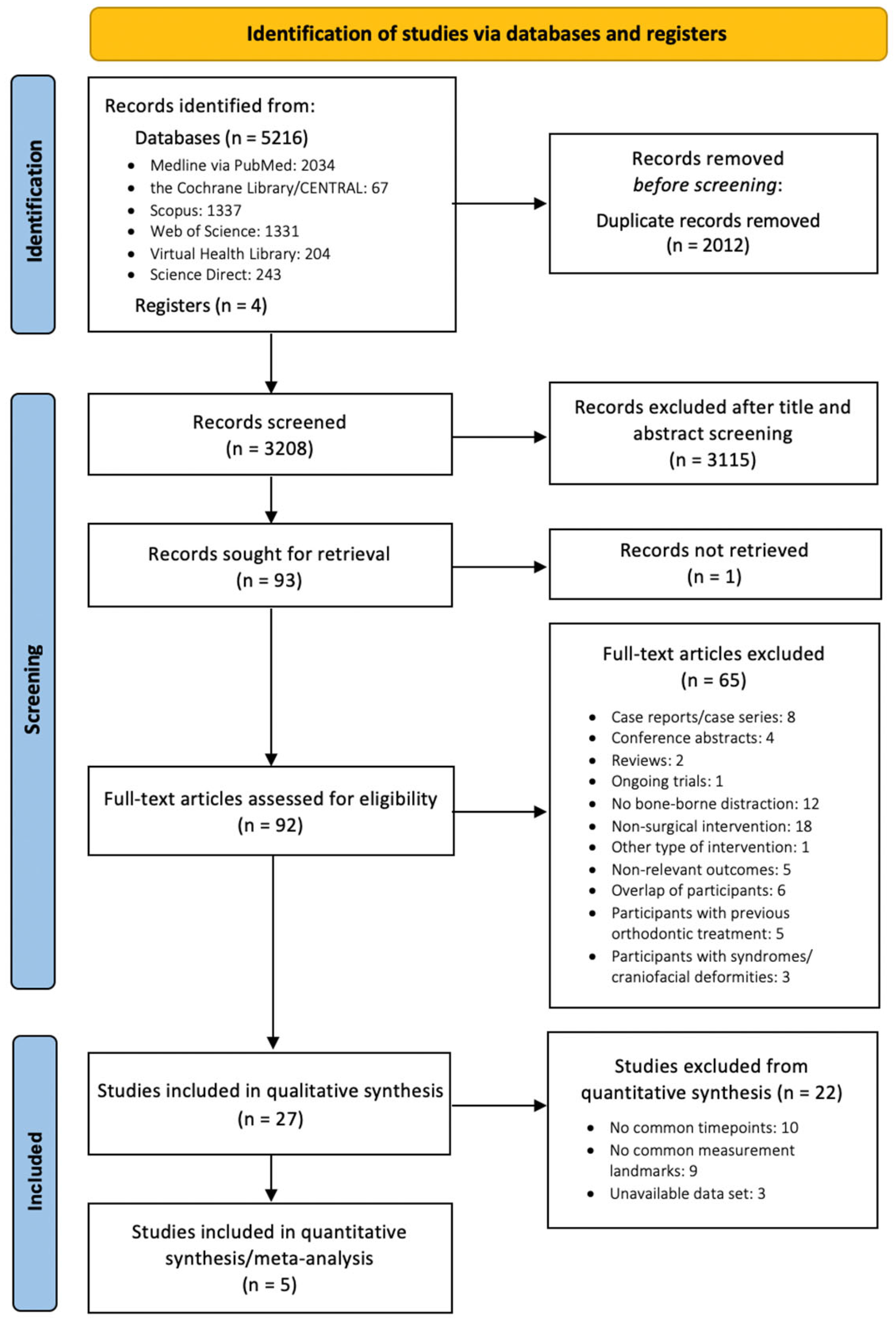
A total of 5216 articles were retrieved from the electronic databases, while 4 additional articles were identified manually (Figure 1). With the removal of the duplicates, 3208 records remained, and after screening of titles and abstracts, the full-text manuscripts of 92 articles were reviewed against the inclusion criteria. Eventually, a total of 27 publications with 621 patients were eligible for qualitative synthesis [4,22,23,24,25,26,27,28,29,30,31,32,33,34,35,36,37,38,39,40,41,42,43,44,45,46,47]. For the quantitative synthesis, we utilized a predefined timeframe for the short-term (immediately after expansion) and long-term outcomes (retention period: at least 3 months post expansion). Only trials that reported outcomes measured at the same landmarks and within the scope of the predefined timeframes were considered eligible for the meta-analytic process. Ultimately, eight trials satisfied these criteria [4,24,25,27,29,42,44,45], but four of them did not report their outcomes in variables suitable for data synthesis. Out of the four trial authors who were contacted for further access to the original dataset [4,27,42,44], only one responded [42]. Thus, five studies [24,25,29,42,45] with 233 patients were successfully included in the quantitative meta-analyses.
Figure 1.
PRISMA 2020 flow diagram for study selection.
3.2. Study Characteristics
Out of the 27 included studies (Table 1), 2 were RCTs, 11 were prospective studies, 10 were retrospective studies, and 4 included both prospective and retrospective cohorts. All trials had been published as journal papers between 2001 and 2022. In some studies, pterygomaxillary disjunction (PMD) was also performed as part of the SARME intervention. Regarding the different designs of appliances used (distractor type), most studies used the (1) Transpalatal Distractor (TPD) (SurgiTec NV, Bruges, Belgium), followed by the (2) RPE® expander (KLS Martin, Tuttligen, Germany) and the (3) MWD (Normed, Tuttlingen, Germany) distractor. The trials used different RME activation protocols, which ranged from 0.33 mm/day up to 1 mm/day, with the most common among them being the activation protocol of 1 mm/day. The mean age of patients varied between 16.9 and 30.2 years. As far as time points and follow ups are concerned, the measurements were obtained throughout different time periods, starting from immediately post-expansion, during the post-consolidation period, and lasting up to approximately 29 months post-operatively. The varied outcomes were measured via dental casts, CTs, lateral and posteroanterior (PA) cephalograms, and cone beam computerized tomography (CBCT), while in some studies, they were digitized and analyzed through 3D formats (list of types of outcomes available in the Appendix A, Table A2). The detailed tables of the characteris-tics and results of the eligible studies are available in the Supplementary Materials.
Table 1.
Characteristics and representative results of studies included in the qualitative synthesis.
3.3. Risk of Bias within Studies
The risk of bias of the non-randomized trials included in the qualitative synthesis ranged between moderate (20 trials) and serious (4 trials), while for one trial, there was no adequate information for a bias judgment. The most serious concerns of bias were introduced by the probable existence of confounding factors, followed by the retrospective selection of participants, the lack of blinding of the outcome assessors, and missing data issues that were inadequately addressed in the data analysis (Table 2). For the included RCTs, there were some concerns of bias, mainly owing to the lack of blinding and issues with the randomization process (Table 3).
Table 2.
Risk of bias assessment for the eligible non-randomized trials (ROBINS-I).
Table 3.
Risk of bias assessment for the eligible randomized trials (RoB 2).
3.4. Results from Individual Studies
The majority of the included trials reported favorable results after bone-borne SARME, including a substantial increase in the arch perimeter [24,28,41] and a significant maxillary expansion at the intercanine, interpremolar, and intermolar level, as measured at the alveolar process, the gingival margin, between dental cups, or interapically [4,24,26,28,29,41,45]. In greater detail, one of the earliest studies investigating maxillary outcomes of bone-borne SARME by Pinto et al. (2001) found a post-operative relative increase in the arch perimeter of 10.5 ± 4.6% [41]. According to measurements derived from the selected cast landmarks (i.e., the most lingual points at the gingival margin), the corresponding expansion observed at the canine, first premolar, and first molar areas amounted to 35.7 ± 17%, 31.7 ± 14%, and 20.4 ± 8.7%, respectively [41]. Asscherickx et al. (2016), at 10 weeks post-expansion, reported a 6.1 ± 0.74 (SE) mm increase in arch perimeter, as well as increases of 5.7 ± 0.48 (SE) mm, 5.7 ± 0.38 (SE) mm, and 5.4 ± 0.42 (SE) mm in the distances between maxillary canines, premolars, and molars, respectively [24]. All distances were measured at the most lingual point of the gingival margin of the teeth [24]. A different study by Kunz et al. (2016), studying the outcomes of bone-borne distraction in 16 patients, used the distance between the palatal sulcus points of canines, premolars, and molars to report a progressive anteroposterior increase in the observed expansion, in both absolute and relative values (canines: 4.43 ± 2.21 mm, 19.72 ± 9.84%; first premolars: 4.56 ± 2.27 mm, 18.35 ± 9.13%; first molars: 3.53 ± 1.83 mm, 10.93 ± 5.67%) [29]. Barone et al. (2020) provided measurements from dental impressions at a 1-year postoperative follow-up [4]. In the bone-borne group, both interdental and intergingival distances were documented for canines (interdental: 2.18 ± 1.30 mm, intergingival: 2.21 ± 1.60 mm), premolars (interdental: 4.89 ± 3.05 mm, intergingival: 4.35 ± 2.86 mm), and molars (interdental: 3.22 ± 3.56 mm, intergingival: 2.66 ± 2.73 mm) [4], indicating an increase in all six variables. As far as the mandible was concerned, only one eligible article reported relevant outcomes, pointing to an increase in the dental show (2.5 ± 2.1 mm) and an inferoposterior repositioning of the chin (1.5 ± 2.2 mm posterior displacement) [46].
Transpalatal distraction was likely to cause a V-shaped pattern of expansion (larger in the anterior area and bilaterally asymmetrical on occasion) along with a slight downward maxilla movement [27,29,44]. This may be attributed to the new center of resistance being placed more posteriorly, which, in order to achieve a more parallel expansion, could be counteracted by the surgical release of the pterygoid plates [24]. SARME with bone-borne distractors could lead to the dropping and protrusion of the maxilla and its anterior movement may reflect the surgical procedure followed [25].
In some cases, the angulation of canines, premolars, and molars was also slightly altered, through buccal tipping [24]. However, Laudemann et al. (2011) [34] observed the presence of inward tipping in patients with no PMD and explained it as a compensation mechanism to the buccal tipping of the maxillary segments. Generally, with bone-borne appliances, tipping was observed to a limited extent. The effect on bone remodeling was minimal and mainly recorded in the area of the molars [30,31]. Petrick et al. (2011) [40] concluded that the midpalatal suture achieved up to ¾ of its pre-treatment density 7 months after SARME. A retention period of 6 months or more was suggested to avoid relapse [24,40].
The reported soft tissue changes included a posterior repositioning and decreased thickness of the upper lip and an increased projection of the cheek area [36]. In the nose area, Wallner et al. (2022) [45], using CBCT superimposed images of 91 patients, measuring a significant increase in both alar nasal width (1.2 ± 1.1 mm) and alar nasal base width (2.1 ± 1.2 mm), while the nose tip height remained unaffected. Conversely, Aras et al. (2017) [23] found that bone-borne SARME did not affect the soft tissue profile of the patients.
The main advantages of the procedure comprise minimal intraoperative complications, less vestibular bone resorption, and limited dental tipping [29,32]. Furthermore, bone-borne appliances allow for the possibility of an ongoing orthodontic treatment alongside the retention period, which may result in a reduced total treatment time [43]. However, the procedure involves increased expenses accompanied by an additional operation under local anesthesia for the removal of the appliance [43], while one study [42] also reported frequent mechanical failure.
With regards to the immediate outcomes post-expansion, the RCT of Koudstaal et al. (2009) [28] indicated an increase in the intercanine (6 ± 3.4 mm), interpremolar (7 ± 3.1 mm), and intermolar width (5.2 ± 3.4 mm). The different maxillary widths were measured on dental casts from the buccal cusps of the corresponding teeth. In addition, even though a slight relapse was observed after the 12-month follow-up, the net gain remained significant for all three measurements (intercanine: 4.7 ± 3.2 mm; interpremolar: 7.0 ± 3.5 mm; intermolar: 4.6 ± 3.1 mm). The overall increase in the arch perimeter was significant (6.0 ± 5.8). The palatal depth at premolar level decreased after 12 months at the premolar (–0.1 ± 2.1 mm) and molar level (–0.1 ± 2.1 mm). The palatal width at premolar level showed an increase of 2.9 mm (± 2.2) and, at molar level, an increase of 2.6 mm (± 2.5). It is worth noting that, in the large retrospective cohort of Wallner et al. (2022) [45], no significant decrease in the vertical palate height at first molars could be observed (−0.1 ± 1, p = 0.3082).
In the RCT of Zandi et al. (2014) [47], the largest percentage of expansion was observed in the dental arch, followed by the palatal bone, while the pattern of expansion was parallel posteroanterior. Furthermore, in both the first premolar and molar regions, the mean expansion gain at the interapical area was the same as that of the palatal bone area. Specifically, the palatal bone width showed an increase of 4.53 ± 2.02 mm at premolar level and an increase of 4.33 ± 1.23 mm at molar level. On the other hand, the interdental cusp distance increased by 6.73 ± 2.15 mm at premolar level and 6.53 ± 2.67 mm at molar level. Finally, the interdental root distance showed an increase of 4.40 ± 1.68 mm at premolar level and of 4.50 ± 1.83 mm at molar level.
3.5. Quantitative Synthesis
Trials with outcomes measured at the same landmarks within the scope of the prespecified timeframe for short-term and/or long-term effects were deemed eligible for quantitative synthesis. Nevertheless, out of the eight trials initially included, only four reported their outcomes via suitable variables [24,25,28,45] and, out of the other four, only one group of researchers [41] provided relevant data upon request. The additional dataset, however, was incomplete and could only be incorporated with regards to one quantitative variable (arch perimeter). Thus, five trials were eventually included in the meta-analyses (Table 4).
Table 4.
Results of random effects meta-analyses performed for relevant outcomes after SARME.
3.5.1. Arch Perimeter
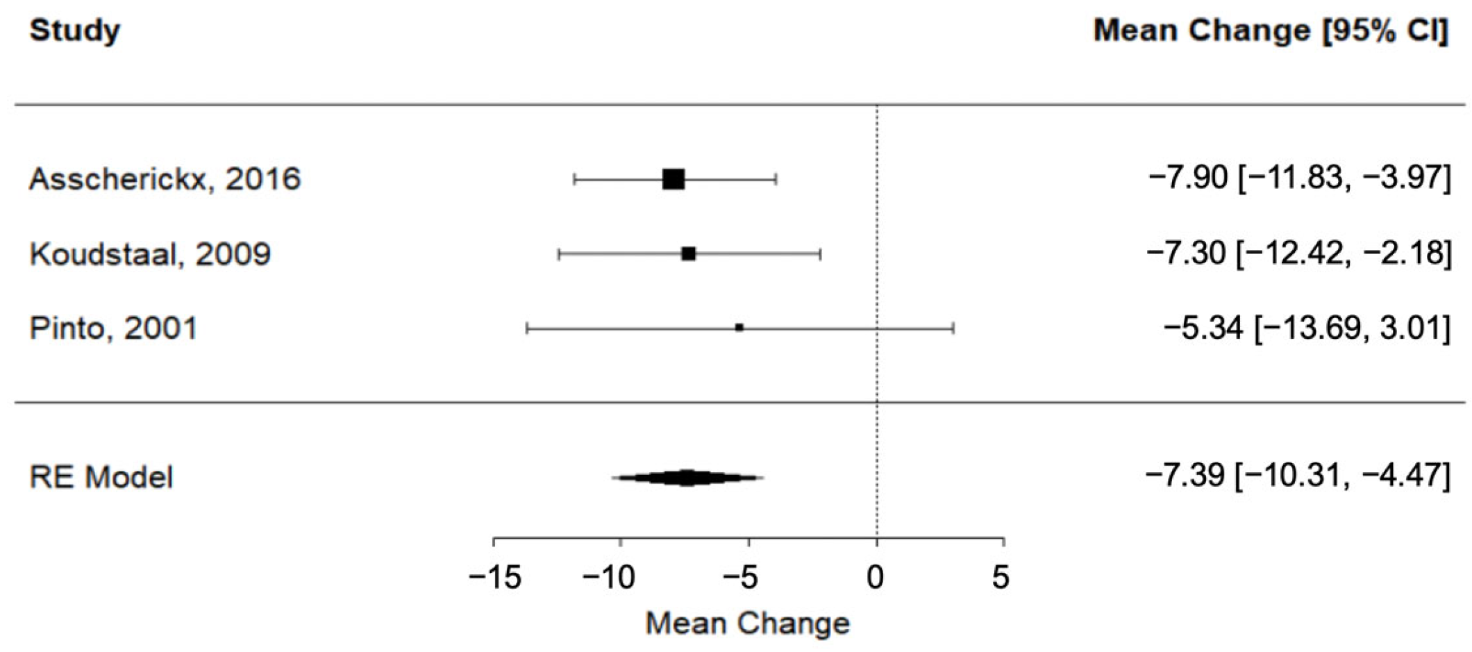
According to evidence from three different trials [24,28,41], SARME was associated with a significant lengthening of the dental arch perimeter immediately after expansion (RC = −7.39; 95% CI = lower: −10.31, upper: −4.47; p-value < 0.001; I2 = 0%) (Figure 2).
Figure 2.
Forest plot for meta-analysis of arch perimeter from three trials [24,28,41].
3.5.2. Palatal Depth
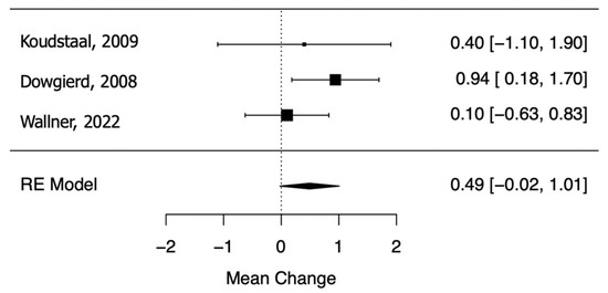
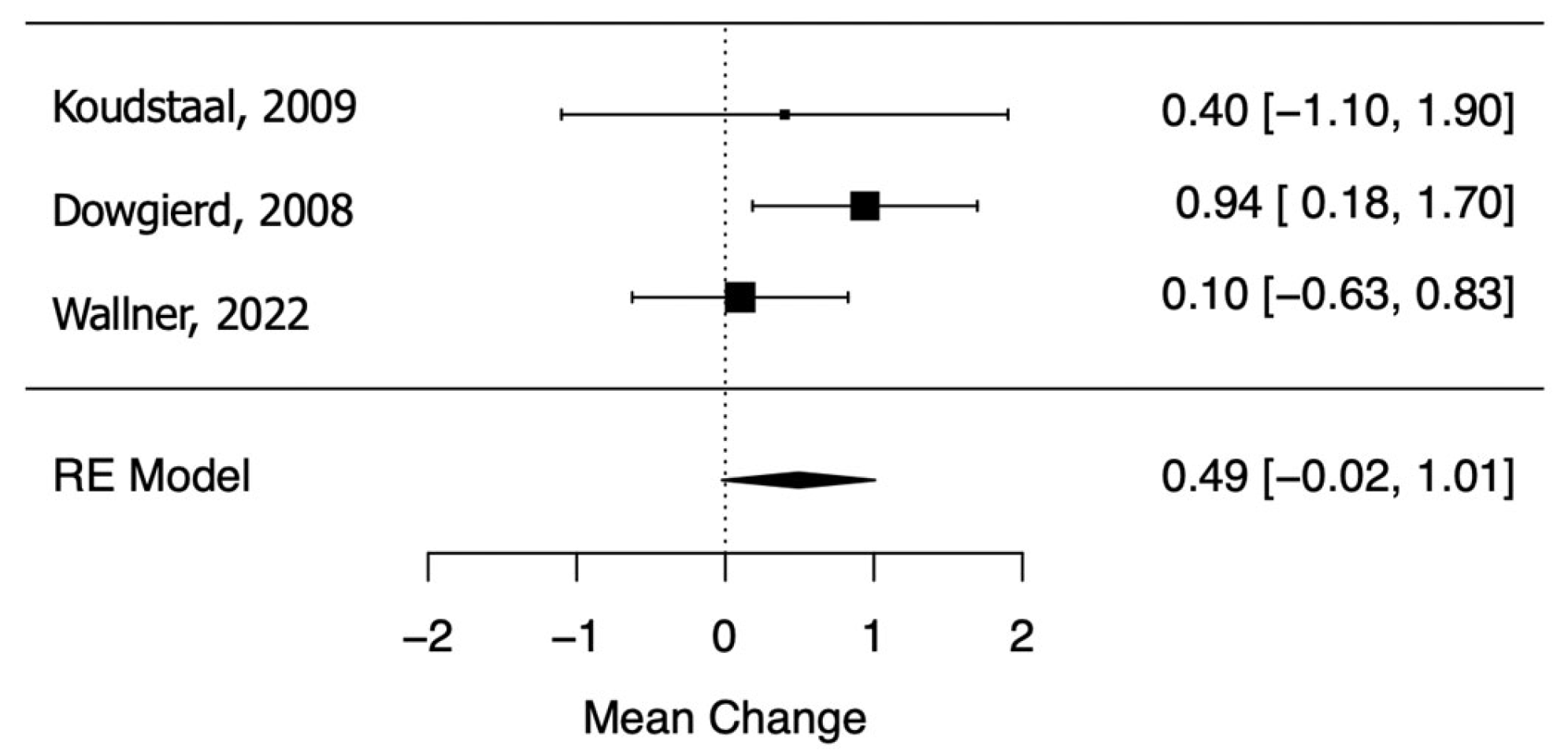
Dowgierd et al. (2018) [25], Koudstaal et al. (2009) [28], and Wallner et al. (2022) [45] provided evidence of a marginally significant decrease in palatal depth during the retention period after SARME (RC = 0.49; 95% CI = lower: −0.02, upper: 1.01; p-value = 0.06; I2 = 5.61%) (Figure 3).
Figure 3.
Forest plot for meta-analysis of palatal depth from three trials [25,28,45].
3.5.3. Radiograph Outcomes
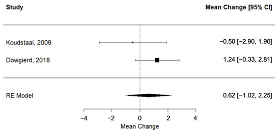
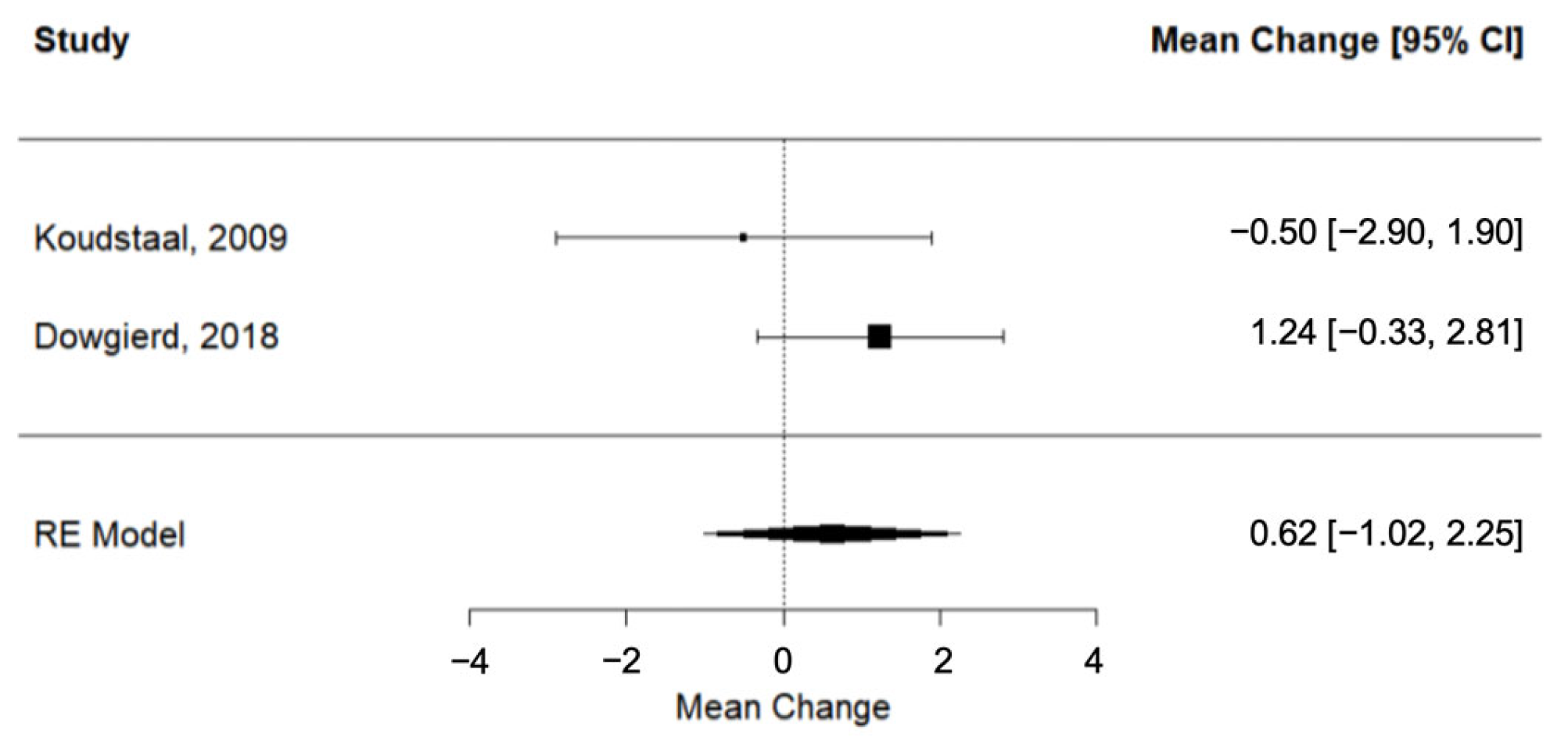
Two trials [25,28] provided evidence on the observed differences in radiographic measurements during post-SARME retention. The pooled SNA outcome did not suggest a statistically significant change (SNA: RC = 0.62; 95% CI= lower: −1.02, upper: 2.25; p-value = 0.46; I2 = 29.33%) (Figure 4).
Figure 4.
Forest plot for meta-analysis of SNA values from two trials [25,28].
3.5.4. Publication Bias
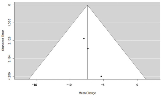
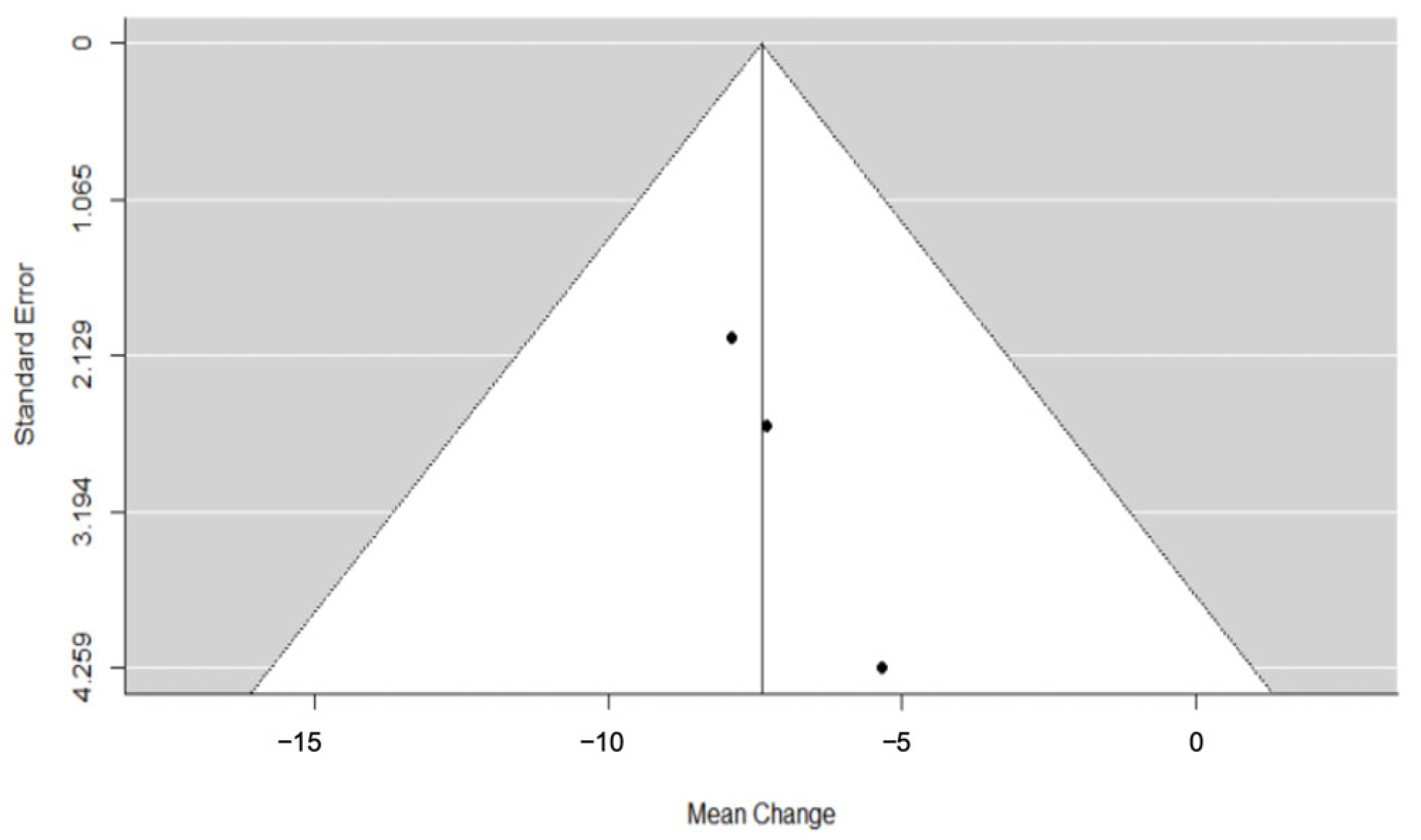
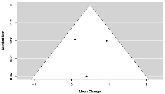
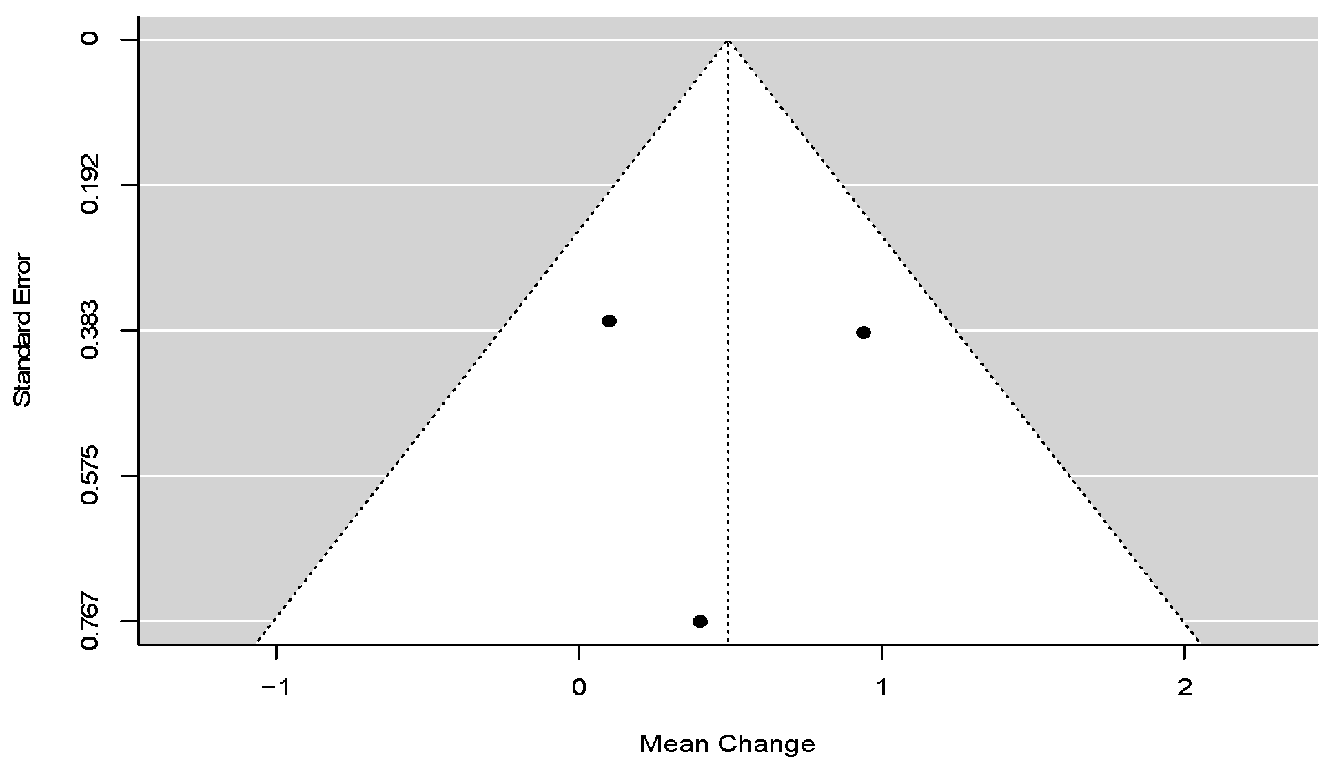
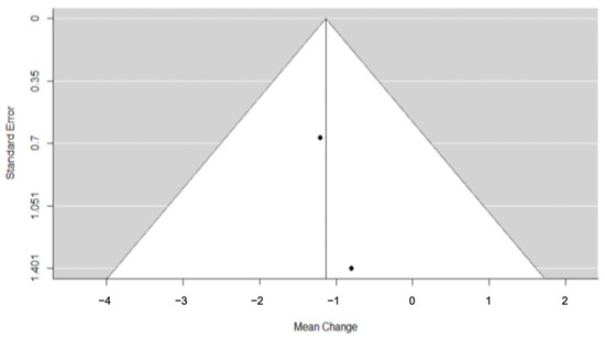
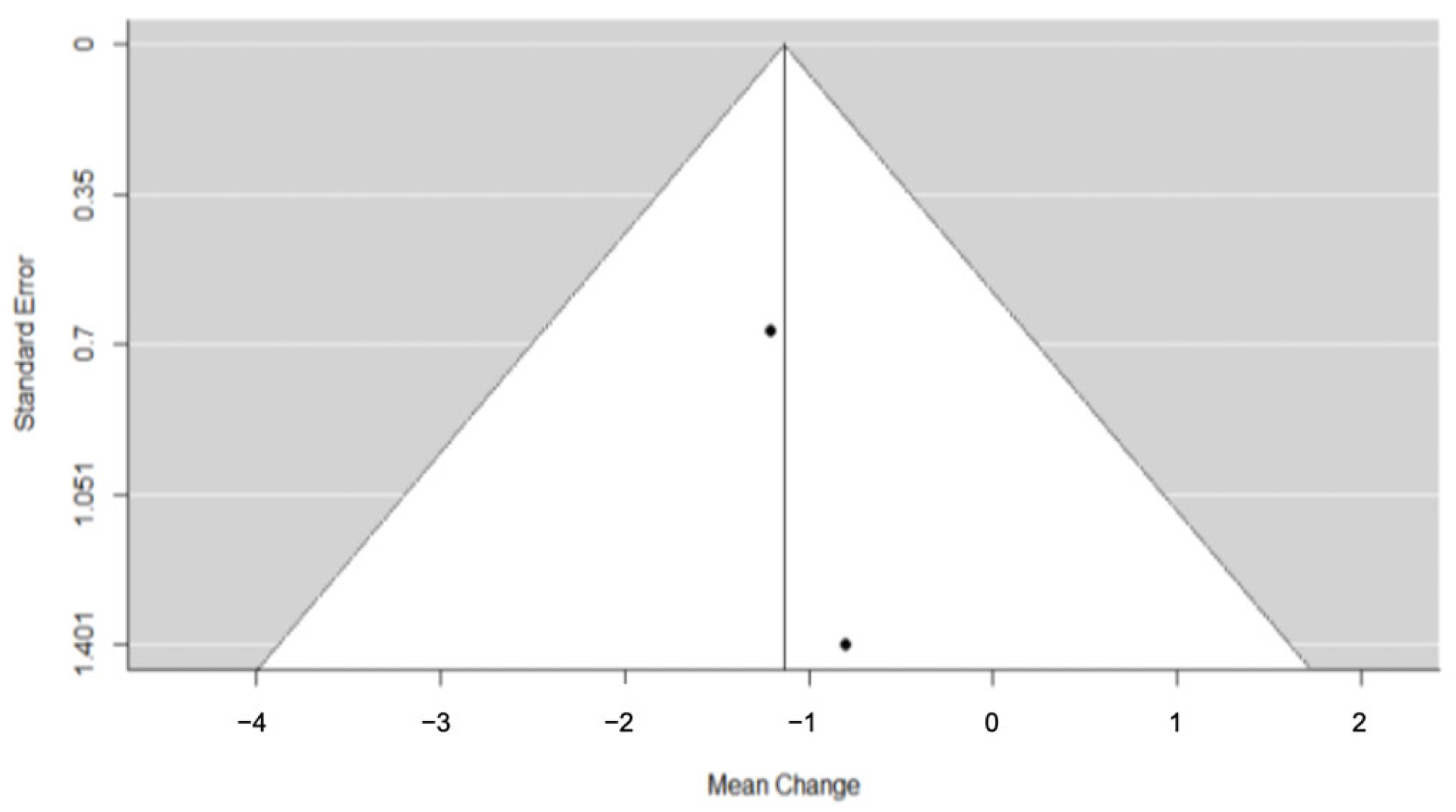
Upon inspection, none of the resulting funnel plots demonstrated obvious signs of asymmetry that would suggest the existence of severe publication bias. Nonetheless, given the small number of the included studies per individual meta-analysis, these conclusions should be regarded with caution (Figure 5, Figure 6 and Figure 7).
Figure 5.
Funnel plot for meta-analysis of arch perimeter from three trials.
Figure 6.
Funnel plot for meta-analysis of palatal depth from three trials.
Figure 7.
Funnel plot for meta-analysis of SNA from two trials.
3.5.5. Sensitivity and Subgroup Analyses
The analyses planned to investigate the robustness of the results and the possible sources of heterogeneity, respectively, were ultimately proven infeasible owing to the small number of relevant trials available.
3.5.6. Quality of Evidence
The quality of evidence for the meta-analyses was evaluated with the GRADE (Grading of Recommendations Assessment, Development, and Evaluation) approach. The domains’ risk of bias, directness of evidence, consistency and precision of results, and risk of publication bias were assessed for each outcome. The judgments for each outcome can be found in Table 5. The overall quality of evidence was deemed to be low.
Table 5.
GRADE summary of findings.
GRADE Working Group grades of evidence
High certainty: We are very confident that the true effect lies close to that of the estimate of the effect.
Moderate certainty: We are moderately confident in the effect estimate: The true effect is likely to be close to the estimate of the effect, but there is a possibility that it is substantially different.
Low certainty: Our confidence in the effect estimate is limited: The true effect may be substantially different from the estimate of the effect.
Very low certainty: We have very little confidence in the effect estimate: The true effect is likely to be substantially different from the estimate of effect.
Explanations
- Downgraded by one point for some concerns of bias (with regards to the randomization process and confounding).
- Downgraded by one point for imprecision (small sample of participants).
- Downgraded by one point for some concerns of bias (with regards to the randomization process, confounding, and selection of participants).
4. Discussion
The aim of this systematic review was to assess the evidence available from clinical studies on the objective dental, skeletal, and soft tissue changes following bone-borne surgically assisted rapid maxillary expansion (SARME) in orthodontic patients. The comparisons were conducted without a control group; instead, individuals were analyzed at different time points to examine intervention outcomes. Overall, 27 studies satisfied the inclusion criteria of human clinical trials reporting outcomes for short-term and/or long-term effects due to SARME procedures and were included in the qualitative review. The risk of bias of the non-randomized trials included in the qualitative synthesis ranged between moderate (20 trials) and serious (4 trials), while for one trial, there was no adequate information for a bias judgment. For the two included RCTs, there were some concerns of bias, mainly due to the lack of blinding and issues with the randomization process. Trials with outcomes measured at the same landmarks within the scope of the prespecified timeframe for short-term and/or long-term effects were deemed eligible for further quantitative synthesis. Out of the eight trials initially selected, five trials [24,25,28,41,45] reported their outcomes using the same variables and were eventually included in the meta-analyses.
From the results of this meta-analysis, it can be inferred that SARME was associated with a significant lengthening of the dental arch perimeter immediately after expansion, as well as with a marginally significant decrease in palatal depth during the post-SARME retention period. The existing data with regards to observed changes in SNA values, which were obtained via radiographic measurements during the post-SARME retention period, did not denote any statistically significant changes. The present evidence indicates that SARME, as a treatment option for transverse deficiencies, may achieve some of the intended effects. However, these results should be viewed with caution owing to the small number of studies included in the analyses and the concerns of bias.
The present study is the first one to meta-analyze the available bibliographic evidence pertaining to changes observed in the measurements of the arch perimeter, the palatal depth, and the SNA angle after bone-borne SARME. Owing to the specificity of the meta-analytic data, no direct comparisons to other published papers regarding this particular method or other alterative interventions (e.g., bone-borne SARME) could be drawn. It could be noted that the review by Vilani et al. (2012) [14], which similarly investigated post-SARME changes in dental and skeletal structures using the metrics of patients at different time points as self-comparators, reported a significant long-term increase with regards to the maxillary alveolar width and to the interdental widths at the canine and molar level. Nevertheless, the results of their study were reported without differentiating between tooth-borne and bone-borne appliances. Bortolotti et al. (2020) [15], in their meta-analysis of RCTs regardless of type of anchorage, also found significant maxillary expansion on a skeletal and inter-molar level, with the latter being the main contributor to the overall effect. Even though the results from the individual studies included in the present systematic review likewise suggest that bone-borne SARME causes a significant increase in dental and skeletal width parameters, those data did not satisfy our criteria for pooling, as the researchers did not use the same landmarks or similar time-points in their measurements. It is worth mentioning that evidence from studies with multiple timepoints, even though they show some relapse of the gained expansion at their longest follow-ups, is not significant, thus suggesting that the overall effect of the modality is net positive and relatively stable [22,24,42].
The limited number and quality of the eligible studies yielded during this systematic review highlights the need for additional original research into the precise effects of SARME. Randomized controlled trials that meet high-quality standards could further establish the treatment scope of the modality and provide strong guidance to clinicians with regards to patient selection and expected results. Possible suggestions for future studies include the investigation of both short- and long-term SARME outcomes, with a specific focus on the effect of differentiated parameters, such as surgical techniques and the type of bone-borne distractor.
Among the strengths attributed to this meta-analysis can be considered the a priori registration protocol in PROSPERO, a scrupulous database and hand search, the strict inclusive and exclusive criteria, and the use of the GRADE approach to assess the quality of the evidence. However, several limitations also exist. First and foremost, there was no exclusion of non-randomized trials, leading to a greater extent of methodological limitations and bias. Several studies deemed eligible for qualitative synthesis reported insufficient control for confounding factors, less than optimal participant selection, and lack of blinding, thus introducing further concerns of possible bias. Furthermore, the varied measurement techniques used to assess skeletal, dental, and soft-tissue changes made it impossible to compare and synthesize all selected studies, which could have strengthened the quality of evidence. The limited number of included studies and heterogeneity in measurement techniques may introduce restrictions to the generalization of the results. Finally, subgroup analyses (including, e.g., different types of distractors, activation protocols, appliances used for retention, time period, and surgical techniques) could not be performed because of the small number of included studies.
5. Conclusions
According to the results of the current meta-analysis, there is low evidence that the dental arch perimeter immediately post-expansion exhibits a significant increase as a result of bone-borne SARME. Generally, the findings indicate that bone-borne SARME constitutes an effective treatment modality for adult patients with maxillary transverse deficiency, hence it may be used to successfully expand a constricted maxilla. Nevertheless, the restricted number of high-quality trials impedes drawing strong conclusions that could offer definite clinical guidance. Future long-term randomized controlled trials with a robust methodology and standardized protocols are needed to minimize bias and further elucidate the precise effects of SARME.
Supplementary Materials
The following supporting information can be downloaded at: https://www.mdpi.com/article/10.3390/dj11060143/s1.
Author Contributions
Conceptualization, O.-E.K. and A.C.; methodology, O.-E.K. and A.C.; investigation, C.S. and M.-M.K.; resources, C.S. and M.-M.K.; data curation, C.S. and M.-M.K.; writing—original draft preparation, C.S. and M.-M.K.; writing—review and editing, C.S., M.-M.K., A.C. and O.-E.K.; visualization, O.-E.K. and A.C.; supervision, O.-E.K. and A.C.; project administration, O.-E.K. and A.C. All authors have read and agreed to the published version of the manuscript.
Funding
This research received no external funding.
Institutional Review Board Statement
Not applicable.
Informed Consent Statement
Not applicable.
Data Availability Statement
The data that support the findings of this study are available from the authors upon request.
Acknowledgments
The authors would like to thank Evaggelos Akrivos for performing the statistical analysis of the collected data.
Conflicts of Interest
The authors declare no conflict of interest.
Appendix A
Table A1.
Search strategy used in each database.
Table A2.
Outcomes of studies included in the qualitative synthesis.
References
- Möhlhenrich, S.; Modabber, A.; Kniha, K.; Peters, F.; Steiner, T.; Hölzle, F.; Fritz, U.; Raith, S. Simulation of three surgical techniques combined with two different bone-borne forces for surgically assisted rapid palatal expansion of the maxillofacial complex: A finite element analysis. Int. J. Oral Maxillofac. Surg. 2017, 46, 1306–1314. [Google Scholar] [CrossRef] [Scilit] [PubMed]
- Suri, L.; Taneja, P. Surgically assisted rapid palatal expansion: A literature review. Am. J. Orthod. Dentofac. Orthop. 2008, 133, 290–302. [Google Scholar] [CrossRef] [Scilit] [PubMed]
- Koudstaal, M.J.; Poort, L.J.; van der Wal, K.G.H.; Wolvius, E.B.; Prahl-Andersen, B.; Schulten, A.J.M. Surgically assisted rapid maxillary expansion (SARME): A review of the literature. Int. J. Oral Maxillofac. Surg. 2005, 34, 709–714. [Google Scholar] [CrossRef] [Scilit] [PubMed]
- Barone, T.R.; Cahali, M.B.; Vasconcelos, C.; Barone, J.R. A comparison of tooth-borne and bone-anchored expansion devices in SARME. Oral Maxillofac. Surg. 2020, 24, 181–187. [Google Scholar] [CrossRef] [Scilit]
- Muñoz-Pereira, M.; Haas-Junior, O.; Meirelles, L.D.S.; Machado-Fernández, A.; Guijarro-Martínez, R.; Hernández-Alfaro, F.; de Oliveira, R.; Pagnoncelli, R. Stability and surgical complications of tooth-borne and bone-borne appliances in surgical assisted rapid maxillary expansion: A systematic review. Br. J. Oral Maxillofac. Surg. 2021, 59, e29–e47. [Google Scholar] [CrossRef] [Scilit]
- Zandi, M.; Miresmaeili, A.; Heidari, A.; Lamei, A. The necessity of pterygomaxillary disjunction in surgically assisted rapid maxillary expansion: A short-term, double-blind, historical controlled clinical trial. J. Craniomaxillofac. Surg. 2016, 44, 1181–1186. [Google Scholar] [CrossRef] [Scilit]
- Gogna, N.; Johal, A.S.; Sharma, P.K. The stability of surgically assisted rapid maxillary expansion (SARME): A systematic review. J. Craniomaxillofac. Surg. 2020, 48, 845–852. [Google Scholar] [CrossRef] [Scilit]
- Jia, H.; Zhuang, L.; Zhang, N.; Bian, Y.; Li, S. Comparison of skeletal maxillary transverse deficiency treated by microimplant-assisted rapid palatal expansion and tooth-borne expansion during the post-pubertal growth spurt stage. Angle Orthod. 2021, 91, 36–45. [Google Scholar] [CrossRef] [Scilit]
- de Oliveira, C.B.; Ayub, P.; Ledra, I.M.; Murata, W.H.; Suzuki, S.S.; Ravelli, D.B.; Santos-Pinto, A. Microimplant assisted rapid palatal expansion vs surgically assisted rapid palatal expansion for maxillary transverse discrepancy treatment. Am. J. Orthod. Dentofac. Orthop. 2021, 159, 733–742. [Google Scholar] [CrossRef] [Scilit]
- Fastuca, R.; Campobasso, A.; Zecca, P.A.; Caprioglio, A. 3D facial soft tissue changes after rapid maxillary expansion on primary teeth: A randomized clinical trial. Orthod. Craniofac. Res. 2018, 21, 140–145. [Google Scholar] [CrossRef] [Scilit]
- Hamedi-Sangsari, A.; Chinipardaz, Z.; Carrasco, L. Following Surgically Assisted Rapid Palatal Expansion, Do Tooth-Borne or Bone-Borne Appliances Provide More Skeletal Expansion and Dental Expansion? J. Oral Maxillofac. Surg. 2017, 75, 2211–2222. [Google Scholar] [CrossRef] [Scilit]
- Krüsi, M.; Eliades, T.; Papageorgiou, S.N. Are there benefits from using bone-borne maxillary expansion instead of tooth-borne maxillary expansion? A systematic review with meta-analysis. Prog. Orthod. 2019, 20, 9. [Google Scholar] [CrossRef] [Scilit]
- Blæhr, T.L.; Mommaerts, M.Y.; Kjellerup, A.D.; Starch-Jensen, T. Surgically assisted rapid maxillary expansion with bone-borne versus tooth-borne distraction appliances—A systematic review. Int. J. Oral Maxillofac. Surg. 2019, 48, 492–501. [Google Scholar] [CrossRef] [Scilit]
- Vilani, G.N.L.; Mattos, C.T.; Ruellas, A.C.O.; Maia, L.C. Long-term dental and skeletal changes in patients submitted to surgically assisted rapid maxillary expansion: A meta-analysis. Oral Surg. Oral Med. Oral Radiol. 2012, 114, 689–697. [Google Scholar] [CrossRef] [Scilit]
- Bortolotti, F.; Solidoro, L.; Bartolucci, M.L.; Parenti, S.I.; Paganelli, C.; Alessandri-Bonetti, G. Skeletal and dental effects of surgically assisted rapid palatal expansion: A systematic review of randomized controlled trials. Eur. J. Orthod. 2020, 42, 434–440. [Google Scholar] [CrossRef] [Scilit]
- Higgins, J.; Thomas, J.; Cumpston, M.; Li, T.; Page, M.; Welch, V. (Eds.) Cochrane Handbook for Systematic Reviews of Interventions, Version 6.2. Available online: https://training.cochrane.org/handbook (accessed on 2 May 2021).
- Page, M.J.; McKenzie, J.E.; Bossuyt, P.M.; Boutron, I.; Hoffmann, T.C.; Mulrow, C.D.; Shamseer, L.; Tetzlaff, J.M.; Akl, E.A.; Brennan, S.E.; et al. The PRISMA 2020 statement: An updated guideline for reporting systematic reviews. BMJ 2021, 372, n71. [Google Scholar] [CrossRef] [Scilit]
- Sterne, J.A.C.; Savović, J.; Page, M.J.; Elbers, R.G.; Blencowe, N.S.; Boutron, I.; Cates, C.J.; Cheng, H.Y.; Corbett, M.S.; Eldridge, S.M.; et al. RoB 2: A revised tool for assessing risk of bias in randomised trials. BMJ 2019, 366, l4898. [Google Scholar] [CrossRef] [Scilit]
- Sterne, J.A.C.; Hernán, M.A.; Reeves, B.C.; Savović, J.; Berkman, N.D.; Viswanathan, M.; Henry, D.; Altman, D.G.; Ansari, M.T.; Boutron, I.; et al. ROBINS-I: A tool for assessing risk of bias in non-randomised studies of interventions. BMJ 2016, 355, i4919. [Google Scholar] [CrossRef] [Scilit]
- Koletsi, D.; Fleming, P.S.; Michelaki, I.; Pandis, N. Heterogeneity in Cochrane and non-Cochrane meta-analyses in orthodontics. J. Dent. 2018, 74, 90–94. [Google Scholar] [CrossRef] [Scilit]
- Schünemann, H.; Brożek, J.; Guyatt, G.; Oxman, A. Grading quality of evidence and strength of recommendations for diagnostic tests and strategies. BMJ 2008, 336, 1106–1110. [Google Scholar] [CrossRef] [Scilit]
- Aras, A.; Akay, M.C.; Çukurova, I.; Günbay, T.; Işıksal, E.; Aras, I. Dimensional Changes of the Nasal Cavity after Transpalatal Distraction Using Bone-Borne Distractor: An Acoustic Rhinometry and Computed Tomography Evaluation. J. Oral Maxillofac. Surg. 2010, 68, 1487–1497. [Google Scholar] [CrossRef] [Scilit] [PubMed]
- Aras, I.; Ölmez, S.; Akay, M.C.; Günbay, T.; Aras, A. The effects of maxillary expansion on the soft tissue facial profile. J. Istanb. Univ. Fac. Dent. 2017, 51, 1–10. [Google Scholar] [CrossRef] [Scilit] [PubMed]
- Asscherickx, K.; Govaerts, E.; Aerts, J.; Vannet, B.V. Maxillary changes with bone-borne surgically assisted rapid palatal expansion: A prospective study. Am. J. Orthod. Dentofac. Orthop. 2016, 149, 374–383. [Google Scholar] [CrossRef] [Scilit] [PubMed]
- Dowgierd, K.; Borowiec, M.; Kozakiewicz, M. Bone changes on lateral cephalograms and CBCT during treatment of maxillary narrowing using palatal osteodistraction with bone-anchored appliances. J. Craniomaxillofac. Surg. 2018, 46, 2069–2081. [Google Scholar] [CrossRef] [Scilit]
- Hansen, L.; Tausche, E.; Hietschold, V.; Hotan, T.; Lagravère, M.; Harzer, W. Skeletally-anchored Rapid Maxillary Expansion using the Dresden Distractor. J. Orofac. Orthop. 2007, 68, 148–158. [Google Scholar] [CrossRef] [Scilit]
- Huizinga, M.P.; Meulstee, J.W.; Dijkstra, P.U.; Schepers, R.H.; Jansma, J. Bone-borne surgically assisted rapid maxillary expansion: A retrospective three-dimensional evaluation of the asymmetry in expansion. J. Craniomaxillofac. Surg. 2018, 46, 1329–1335. [Google Scholar] [CrossRef] [Scilit]
- Koudstaal, M.J.; Wolvius, E.B.; Schulten, A.J.M.; Hop, W.C.J.; van der Wal, K.G.H. Stability, tipping and relapse of bone-borne versus tooth-borne surgically assisted rapid maxillary expansion; a prospective randomized patient trial. Int. J. Oral Maxillofac. Surg. 2009, 38, 308–315. [Google Scholar] [CrossRef] [Scilit]
- Kunz, F.; Linz, C.; Baunach, G.; Böhm, H.; Meyer-Marcotty, P. Expansion patterns in surgically assisted rapid maxillary expansion. J. Orofac. Orthop. 2016, 77, 357–365. [Google Scholar] [CrossRef] [Scilit]
- Landes, C.A.; Laudemann, K.; Petruchin, O.; Mack, M.G.; Kopp, S.; Ludwig, B.; Sader, R.A.; Seitz, O. Comparison of Bipartite Versus Tripartite Osteotomy for Maxillary Transversal Expansion Using 3-Dimensional Preoperative and Postexpansion Computed Tomography Data. J. Oral Maxillofac. Surg. 2009, 67, 2287–2301. [Google Scholar] [CrossRef] [Scilit]
- Landes, C.A.; Laudemann, K.; Schübel, F.; Petruchin, O.; Mack, M.; Kopp, S.; Sader, R.A. Comparison of Tooth- and Bone-Borne Devices in Surgically Assisted Rapid Maxillary Expansion by Three-Dimensional Computed Tomography Monitoring. J. Craniofac. Surg. 2009, 20, 1132–1141. [Google Scholar] [CrossRef] [Scilit]
- Laudemann, K.; Petruchin, O.; Mack, M.G.; Kopp, S.; Sader, R.; Landes, C.A. Evaluation of surgically assisted rapid maxillary expansion with or without pterygomaxillary disjunction based upon preoperative and post-expansion 3D computed tomography data. Oral Maxillofac. Surg. 2009, 13, 159–169. [Google Scholar] [CrossRef] [Scilit]
- Laudemann, K.; Petruchin, O.; Nafzger, M.; Ballon, A.; Kopp, S.; Sader, R.A.; Landes, C.A. Long-term 3D cast model study: Bone-borne vs. tooth-borne surgically assisted rapid maxillary expansion due to secondary variables. Oral Maxillofac. Surg. 2010, 14, 105–114. [Google Scholar] [CrossRef] [Scilit]
- Laudemann, K.; Santo, G.; Revilla, C.; Harth, M.; Kopp, S.; Sader, R.A.; Landes, C.A. Assessment of Surgically Assisted Rapid Maxillary Expansion Regarding Pterygomaxillary Disjunction Using Thin Volume-Rendering Technique: In Variance Analysis and in Reliability, Accuracy, and Validity. J. Oral Maxillofac. Surg. 2011, 69, 2631–2643. [Google Scholar] [CrossRef] [Scilit]
- Matteini, C.; Mommaerts, M.Y. Posterior transpalatal distraction with pterygoid disjunction: A short-term model study. Am. J. Orthod. Dentofac. Orthop. 2001, 120, 498–502. [Google Scholar] [CrossRef] [Scilit]
- Nada, R.M.; Fudalej, P.S.; Maal, T.J.J.; Bergé, S.J.; Mostafa, Y.A.; Kuijpers-Jagtman, A.M. Three-dimensional prospective evaluation of tooth-borne and bone-borne surgically assisted rapid maxillary expansion. J. Craniomaxillofac. Surg. 2012, 40, 757–762. [Google Scholar] [CrossRef] [Scilit]
- Nada, R.M.; van Loon, B.; Maal, T.J.; Bergé, S.J.; Mostafa, Y.A.; Kuijpers-Jagtman, A.M.; Schols, J.G. Three-dimensional evaluation of soft tissue changes in the orofacial region after tooth-borne and bone-borne surgically assisted rapid maxillary expansion. Clin. Oral Investig. 2013, 17, 2017–2024. [Google Scholar] [CrossRef] [Scilit]
- Nikolaev, A.V.; Andreishchev, A.R.; Kutukova, S.I. Comparative biomechanical study of surgically assisted rapid palatal expansion with tooth-borne and bone-borne expanders. Stomatologiia 2017, 96, 48. [Google Scholar] [CrossRef] [Scilit]
- Parhiz, A.; Schepers, S.; Lambrichts, I.; Vrielinck, L.; Sun, Y.; Politis, C. Lateral cephalometry changes after SARPE. Int. J. Oral Maxillofac. Surg. 2011, 40, 662–671. [Google Scholar] [CrossRef] [Scilit]
- Petrick, S.; Hothan, T.; Hietschold, V.; Schneider, M.; Harzer, W.; Tausche, E. Bone density of the midpalatal suture 7 months after surgically assisted rapid palatal expansion in adults. Am. J. Orthod. Dentofac. Orthop. 2011, 139, S109–S116. [Google Scholar] [CrossRef] [Scilit]
- Pinto, P.X.; Mommaerts, M.Y.; Wreakes, G.; Jacobs, W.V.G.J.A. Immediate postexpansion changes following the use of the transpalatal distractor. J. Oral Maxillofac. Surg. 2001, 59, 994–1000. [Google Scholar] [CrossRef] [Scilit]
- Ploder, O.; Winsauer, H.; Juengling, K.; Grill, F.; Bissinger, O.; Wolff, K.-D.; Kolk, A. Is There a Significant Difference in Relapse and Complication Rate of Surgically Assisted Rapid Palatal Expansion Using Tooth-Borne, Bone-Borne, and Orthodontic Mini-Implant–Borne Appliances? J. Oral Maxillofac. Surg. 2021, 79, 213–224. [Google Scholar] [CrossRef] [Scilit] [PubMed]
- Seeberger, R.; Abe-Nickler, D.; Hoffmann, J.; Kunzmann, K.; Zingler, S. One-stage tooth-borne distraction versus two stage bone-borne distraction in surgically assisted maxillary expansion (SARME). Oral Surg. Oral Med. Oral Pathol. Oral Radiol. 2015, 120, 693–698. [Google Scholar] [CrossRef] [Scilit] [PubMed]
- Tausche, E.; Deeb, W.; Hansen, L.; Hietschold, V.; Harzer, W.; Schneider, M. CT Analysis of Nasal Volume Changes after Surgically-assisted Rapid Maxillary Expansion. J. Orofac. Orthop. 2009, 70, 306–317. [Google Scholar] [CrossRef] [Scilit] [PubMed]
- Wallner, J.; Denoiseux, B.; Van de Casteele, E.; Bila, M.; Van Hemelen, G.; Nadjmi, N.; Vercruysse, H., Jr. Do Gender and Age Influence Nasal Soft Tissue Widening after Bone-Borne Transpalatal Distraction? J. Oral Maxillofac. Surg. 2022, 80, 1613–1627. [Google Scholar] [CrossRef] [Scilit]
- Xi, T.; Laskowska, M.; van de Voort, N.; Ghaeminia, H.; Pawlak, W.; Bergé, S.; Maal, T. The effects of surgically assisted rapid maxillary expansion (SARME) on the dental show and chin projection. J. Craniomaxillofac. Surg. 2017, 45, 1835–1841. [Google Scholar] [CrossRef] [Scilit]
- Zandi, M.; Miresmaeili, A.; Heidari, A. Short-term skeletal and dental changes following bone-borne versus tooth-borne surgically assisted rapid maxillary expansion: A randomized clinical trial study. J. Craniomaxillofac. Surg. 2014, 42, 1190–1195. [Google Scholar] [CrossRef] [Scilit]

Disclaimer/Publisher’s Note: The statements, opinions and data contained in all publications are solely those of the individual author(s) and contributor(s) and not of MDPI and/or the editor(s). MDPI and/or the editor(s) disclaim responsibility for any injury to people or property resulting from any ideas, methods, instructions or products referred to in the content. |
© 2023 by the authors. Licensee MDPI, Basel, Switzerland. This article is an open access article distributed under the terms and conditions of the Creative Commons Attribution (CC BY) license (https://creativecommons.org/licenses/by/4.0/).