Article

Knowledge, Attitude, and Practice towards Chelating Agents in Endodontic Treatment among Dental Practitioners

1
Department of Therapeutic Dentistry, I.M. Sechenov First Moscow State Medical University (Sechenov University), 119991 Moscow, Russia
2
Institute of Linguistics and Intercultural Communication, I.M. Sechenov First Moscow State Medical University (Sechenov University), 119991 Moscow, Russia
*
Author to whom correspondence should be addressed.
These authors contributed equally to this work.
Dent. J. 2023, 11(7), 156; https://doi.org/10.3390/dj11070156
Submission received: 4 May 2023 / Revised: 16 June 2023 / Accepted: 20 June 2023 / Published: 21 June 2023
(This article belongs to the Special Issue Modern Endodontics)

Abstract

:
The use of chelating agents (CAs) in the endodontic irrigation protocol is required to dissolve the inorganic components of the smear layer. We aimed to assess the knowledge, attitude, and practice of dental professionals regarding the use of CAs during root canal treatment. A cross-sectional anonymous online survey was conducted among specialized endodontists and general dentists who routinely perform endodontic treatment and work in government-funded or private clinics in Moscow. The 8 min survey consisted of four parts: basic demographic data, knowledge (five items), attitude (four items), and practice (five items). We collected 376 completed questionnaires; a majority of the respondents were general dentists (87.5%) and worked in private clinics (77.4%). Most respondents (83.5%) showed a fair knowledge of the CAs used in endodontics, while 16.5% showed a poor knowledge of the topic. Small yet significant differences were found between endodontists and general practitioners and between dentists employed by private and government-funded clinics. A majority of dental practitioners (83%) demonstrated a positive attitude towards the use of CAs in endodontic treatment, and there were no differences among the study subgroups. Almost a third of the respondents always used chelating solutions during endodontic treatment, while 17% of the respondents did not use them at all. There were significant differences in this parameter between dentists working in private and government-funded clinics. Practice significantly correlated with attitude towards chelating agents and with knowledge of the topic. In conclusion, dental practitioners demonstrated a fair knowledge of CAs. Despite a positive attitude, 71% of the respondents did not use CAs for all endodontic patients.

Graphical Abstract

1. Introduction

The primary goal of root canal treatment (RCT) is the prevention and/or elimination of periapical infection [1]. This can be achieved by mechanical debridement, shaping, irrigation, and filling of the entire root canal system.
Proper irrigation of the root canals during shaping and cleansing is one of the key factors in endodontic success. It is aimed at removing debris and microorganisms as well as the smear layer. The smear layer is formed on the root canal walls during endodontic instrumentation [2]. It is composed of dentin debris and pulpal remnants and microorganisms, i.e., organic and inorganic components. In the literature, there are two positions regarding the need to remove the smear layer during root canal treatment. On the one hand, the smear layer may contribute to bacterial adhesion and colonization and result in root canal obturation failure [3,4,5]. Therefore, smear layer removal may increase the penetration of irrigants and medicaments into the complex anatomy of the root canal system and promote good adaptation of the obturating material to the root canal wall [6,7]. On the other hand, its removal increases dentinal permeability, allowing microorganisms to penetrate into the dentinal tubules, compromising treatment outcome [8,9]. Despite this controversy, the literature in general sides toward smear layer removal before obturation [10,11]. Therefore, an ideal endodontic irrigant solution should not only disinfect dentin and dentinal tubules, but also dissolve pulp tissue and dentin debris in order to remove the smear layer [12]. However, none of the existing irrigating solutions can be regarded as optimal.
Surveys conducted in different countries have shown that sodium hypochlorite is the most widely used root canal irrigant due to its activity against endodontic pathogens and its pulp-dissolving properties [13,14,15,16,17,18]. However, sodium hypochlorite dissolves only organic tissue, and thus the supplementary use of demineralizing or chelating agents (CAs) is required to dissolve the inorganic components of the smear layer [19,20]. The mechanism of action of chelating agents is based on their reaction with calcium ions and the formation of soluble calcium chelates, thus resulting in the decalcification of dentin [21]. Five to ten milliliters [22,23,24] of 10–17% solution of ethylenediaminetetraacetic acid (EDTA) disodium salt applied for at least 1 min [20,25,26] is most commonly recommended for this purpose [13,27,28,29,30,31,32,33,34,35]. Different techniques may be used to enhance the action of EDTA, e.g., ultrasonic [7,36] or sonic activation [37], negative pressure irrigation [38], laser activation [39], and manual dynamic agitation [40]. At the same time, EDTA activation may also cause the surface disintegration of root canal dentin [41].
Although EDTA does not exhibit a significant antimicrobial effect [42,43,44], it disrupts the biofilm polysaccharide matrix [45,46], thus enhancing the anti-biofilm effect of sodium hypochlorite. At the same time, combined irrigation with these solutions results in a chemical reaction, leading to the loss of the antimicrobial and tissue-dissolving properties of sodium hypochlorite [27,47,48]. Moreover, the use of EDTA can cause dentin erosion and the softening and denaturation of collagen fibers, thus decreasing the adhesion of the obturating material to the root canal walls [49,50,51].
Apart from EDTA, other chelating agents may be used in endodontics, e.g., citric acid [52,53,54] and maleic acid [55,56]. Both acids have been reported to effectively remove the smear layer; however, maleic acid provided better removal of the smear layer from the apical third [57] and demonstrated antibiofilm potential [43,58,59]. Like EDTA, both citric and maleic acids react with sodium hypochlorite and decrease its effect [47,60]. Recently, weak chelators such as etidronic acid and tetrasodium EDTA have been proposed for root canal irrigation to overcome this limitation [19,47,61]. Weak chelators can be mixed with sodium hypochlorite without loss of its antimicrobial and tissue-dissolving properties [47,61,62,63,64]. A mixture of sodium hypochlorite with one of these CAs can be potentially used as the only irrigant throughout root canal chemomechanical instrumentation [47,61,62,65,66]. However, further studies are needed to establish its clinical effect.
To sum up, although extensive research has been conducted on the use of CAs in root canal treatment, there is no single accepted evidence-based protocol of root canal irrigation describing the choice of irrigants and their sequence. Moreover, the surveys on endodontic irrigation among dental professionals have shown that some clinicians are still not aware of the importance of smear layer removal [13,67,68]. To the best of our knowledge, there have been no earlier knowledge, attitude, and practice (KAP) surveys among dental practitioners directly focusing on CAs.
The aim of our study was to assess the knowledge, attitude, and practice of dental professionals regarding the use of chelating agents during root canal treatment.

2. Materials and Methods

A cross-sectional online survey was conducted between 20 October 2022 and 10 January 2023 after ethical approval (2202) was obtained from the Ethics Committee of Sechenov University, Moscow, Russia. A questionnaire was developed in Russian based on the contemporary literature on the topic [19,31,48,69,70]. It was further reviewed for face and content validity by 4 independent reviewers who were members of the dental faculty. A pilot test of the questionnaire was initially performed on a group of 30 dental professionals who were excluded from the main sample. Feedback was collected regarding the clarity and order of questions and the time taken to complete the questionnaire. Modifications were made according to the comments received to enable better understanding.
The 8 min survey consisted of four parts: basic demographic data, knowledge, attitude, and practice regarding the use of CAs in endodontics.
The demographic data included questions on gender, years of clinical experience, specialty (endodontist or general practitioner), and workplace (government-funded or private clinic).
The knowledge section consisted of 5 questions on the types of CAs used in endodontics and EDTA concentrations and properties. Two items were single-choice questions, and three items were multiple-choice questions. To obtain a total knowledge score, each correct answer was given a score of 1 and each incorrect answer was given a score of 0. The scores of all items were summed, with a maximum total score of 24. The total knowledge score was categorized as poor if it was lower than 50% (<12 points), fair if it was between 50% and 75% (≥12 and ≤17 points), and good if it was higher than 75% (>17 points).
The attitude section consisted of 4 statements concerning the use of CAs in endodontics. The study participants could agree or disagree with the statements using a five-point Likert scale. Two items were positive-attitude statements (ranging from 5—“strongly agree”—to 1—“strongly disagree”), and two items were negative-attitude statements (ranging from 1—“strongly agree”—to 5—“strongly disagree”). The overall attitude was regarded as positive if the score was higher than 50% (>10 points) or negative if the score was lower than 50% (≤10 points).
The practice section consisted of 5 questions on the actual use of CAs by the respondents while performing endodontic procedures, with a maximum possible score of 8.
Convenience sampling was used to recruit the participants through personal contacts and social media platforms. Data collection was performed using Google forms. A link was sent to dental professionals using different methods, including emails, text messages, and posts on professional social networks. All data were collected anonymously and treated confidentially. The respondents were informed that completion of the questionnaire indicated consent to participate in the study and to have their responses (used in aggregate form) published in a journal article.
The targeted samples included specialized endodontists and general dentists who routinely performed endodontic treatment and worked in government-funded and private clinics in Moscow, Russia.
The total number of the study population was estimated to be around 5000 basing on the information from sberhealth.ru.
A minimum sample size of 357 respondents was calculated assuming a 95% CI and a margin of error of 5% using the following formula:
Sample   size = z 2 × p 1 p e 2 1 + z 2 × p 1 p e 2 N ,
where
  • z (z-score) = 1.96 (95% CI);
  • p (standard deviation) = 0.5;
  • e (a margin of error) = 0.05;
  • N (a population size) = 5000.
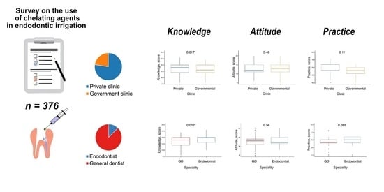
Data manipulation was performed through MS Excel version 16.71 (23031200) and R version 3.6.0 (26 April 2019) with the following packages: “doBy,” “rstatix,” “stats,” and using RStudio version 2023.03.0 + 386 (2023.03.0 + 386). The data were presented as means, medians, standard deviations, 25th and 75th percentiles (quantitative variables), and counts and percentages (qualitative variables). A Shapiro–Wilk test was used to assess the normality of distribution of the quantitative variables (knowledge, attitude, and practice overall scores), and a Mann–Whitney U test was performed to compare these variables in the groups. Fisher’s exact test was used to compare the proportions. Spearman’s rank correlation coefficient was calculated to reveal pair-wise correlation between knowledge, attitude, and practice scores.

3. Results

Of the 378 questionnaires received, 2 were omitted because they were incomplete. A majority of the participants were female, accounting for 69.4% (n  =  261) of the study sample, while men accounted for 30.6% (n  =  115) of the study sample. Almost half of the participants (42.6%) had less than 5 years of clinical experience, 97 (25.8%) had 5–10 years of clinical experience, 75 (19.9%) had 11–20 years, and 44 (11.7%) had more than 20 years of clinical experience. A majority of the respondents were general dentists (n  =  329, 87.5%) and worked in private clinics (n = 291, 77.4%). Table 1 shows a detailed distribution of the participants’ demographic characteristics.
Endodontists demonstrated better knowledge compared with general practitioners, as specified in Table 2. The median number of correct answers was 14 (12; 15) out of the possible 24, with small yet significant differences between endodontists and general practitioners (p = 0.01185) and between dentists employed in private and government-funded clinics (p = 0.01742). A majority of the respondents (83.5%) showed fair knowledge of the use of CAs in endodontics, while 16.5% showed poor knowledge of the topic.
Table 3 shows the distribution of responses to knowledge questions about chelating solutions across the study subgroups. Most respondents in all subgroups correctly identified EDTA properties and concentrations. However, a significantly smaller proportion of general practitioners (76.4%) compared with endodontists (93.6) correctly chose the concentrations of EDTA solutions used for root canal irrigation (p = 0.004399). None of the respondents chose all the chelating solutions used in endodontics correctly, and only 9% of the respondents chose five of the six solutions correctly. A small proportion of the dentists were aware of the effects of the interaction between EDTA and sodium hypochlorite (23.1%) and between EDTA and chlorhexidine (5.3%).
Out of the four correct options, the most recognized CAs used in endodontics were citric acid and EDTA (Figure 1). Almost 20% of the respondents were aware that etidronic acid could be used as an endodontic CA. However, only 14 respondents (3.7%) were aware of the use of maleic acid for that purpose.
A majority of dental practitioners (83%) demonstrated a positive attitude towards the use of CAs in endodontic treatment (Table 4). No differences were found between the mean attitude scores of endodontists and general dentists (p = 0.5555) or between those of clinicians working and private and government-funded clinics (p = 0.4837).
More than half of the respondents agreed or strongly agreed that root canal irrigation with sodium hypochlorite should be alternated with root canal irrigation using CAs (Table 5). At the same time, 27% of the respondents considered CA use appropriate only in sclerotized root canals, approximately 45% of dentists believed that CAs can decrease the antimicrobial action and tissue dissolution capacity of sodium hypochlorite, and 37% of dentists thought that CAs adversely affect the mechanical properties of dentin.
Table 6 shows the use of chelating substances by practitioners during RCT. No differences were found in mean practice scores between the study subgroups.
Almost a third of the respondents always used chelating solutions during endodontic treatment, while 17% of the respondents did not use chelating solutions at all (Figure 2). There were no significant differences in this parameter between the study subgroups. However, a greater proportion of the dentists working in private clinics indicated that they used chelating irrigating solutions “always” (30.6%) or “sometimes” (29.9%), while a greater proportion of the dentists working in government-funded clinics used chelating solutions “rarely” (34.1%) or “never” (24.7%).
EDTA was the most popular chelating solution used for root canal irrigation in RCT and root canal retreatment (reRCT) (Figure 3). A small proportion of dentists reported the use of citric acid in both RCT and reRCT. Interestingly, seven dentists (1.9%) also reported the use of etidronic acid in reRCT.
Regarding the preferred EDTA forms, a majority of the respondents used EDTA solution, 23% used EDTA gel, and 4% did not use this CA (Figure 4). Dental practitioners working in private clinics preferred liquid EDTA forms (81%), while dentists working in government-funded clinics used liquid (47.1%) and gel-type (43.5%) forms in equal proportions (p < 0.001). Endodontists used EDTA solution more frequently (87.2%) than general dentists (71.4), although the differences were not significant (p = 0.06661).
There was a significant weak positive correlation between attitude and practice (r = 0.26, p < 0.001) and between knowledge and practice (r = 0.24, p < 0.001) (Figure 5). However, no correlation was found between knowledge and attitude (r = 0.051, p < 0.32).

4. Discussion

The aim of our study was to assess the knowledge, attitude, and practice of Moscow-based dental professionals towards the use of CAs during root canal treatment. We surveyed endodontists and general dentists employed in private and government-funded clinics. Surprisingly, none of the respondents in our study had a good knowledge of the topic. Dental practitioners had a fair knowledge of chelating endodontic agents, which was significantly better among specialized endodontists than among general dentists. No difference was found between the levels of knowledge of dentists employed in private and government-funded clinics.
To the best of our knowledge, there have not been any cross-sectional KAP surveys on CAs among dental practitioners that are directly comparable to ours. Earlier surveys partially comparable to ours focused mainly on preferred endodontic and irrigation practices. Most of these studies also reported a fair knowledge of the endodontic standards [71,72,73,74]. Bansal et al. reported that 43% of endodontists and 47% of general dentists had a moderate knowledge of evidence-based endodontic practices, while a high level of knowledge was demonstrated by 35% and 13% of dental practitioners, respectively [73]. A study by Al-Omari et al. found that dentists practicing in North Jordan did not comply with international quality standards [74]. Albahiti et al. found moderately satisfying knowledge of decontamination during RCT, with no differences in this parameter between dentists working in government and private sectors [71].
Apart from the general knowledge score, we also analyzed participants’ answers to particular questions. A majority of the respondents correctly identified EDTA properties and concentrations used in RCT. However, less than a third of the respondents were aware of the reaction occurring between EDTA and sodium hypochlorite, leading to the loss of the antibacterial and tissue-dissolving properties of the latter. None of the participants correctly listed all chelating irrigants used in endodontics. The most recognized endodontic chelating solution was citric acid (80.0%), followed by EDTA (59.8%) and etidronic acid (19.9%). Maleic acid was recognized as an endodontic chelating solution by only 3.7% of the respondents. This may be due to the fact that solutions containing this acid are not currently available in the Russian Federation.
As was mentioned above, the use of CAs is required to dissolve the inorganic components of the smear layer. However, the question of whether to retain or to remove the smear layer remains controversial [10,75]. Pashley hypothesized that a smear layer on the root canal walls can prevent bacterial invasion of the dentinal tubules [76]. In contrast, Williams and Goldman found that the smear layer could not prevent bacterial penetration, but only delayed it [77]. Meryon and Brook did not confirm the effect of the smear layer on the ability of oral bacteria to invade dentine tubules in vitro [78]. On the other hand, a systematic review and meta-analysis by Shahravan et al. concluded that smear layer removal had a significant influence on the effective sealing of the root canal system [10]. A systematic review by Violich and Chandler also advocated the removal of the smear layer for better disinfection of the root canal and better adaptation of filling materials to the canal walls [21]. At the same time, these authors admitted the absence of clinical trials to support this recommendation. According to Virdee et al., a majority of the dental schools in the UK and Ireland taught the irrigation protocol, including the use of both sodium hypochlorite and a chelating agent [79].
In our survey, more than 80% of dental practitioners demonstrated a positive attitude towards the use of CAs in endodontic irrigation. No differences were found between the study subgroups in this parameter. Similarly, the majority of the dentists (81%) in the study by Willershausen indicated that removal of the smear layer played an important role in root canal disinfection [13]. According to our results, more than half of dental practitioners agreed or strongly agreed that root canal irrigation with sodium hypochlorite should be alternated with irrigation using CAs. This approach is recommended in several reports [24,80,81,82].
However, this is not in agreement with the studies showing that EDTA should be used at the end of the shaping instead of alternating EDTA and sodium hypochlorite [18,41].
Also, 45% of the respondents believed that CAs could decrease the effect of sodium hypochlorite and 37% of the respondents believed that CAs could weaken dentin structure. Despite their belief that EDTA has deleterious effects, almost a third of the respondents always used chelating solutions in endodontic irrigation, while only 17% of the respondents did not use chelating solutions at all. In a study by Tošić et al., EDTA was the least-applied irrigant in the two surveys conducted in Serbia [83]. In some of the earlier studies, dental practitioners did not report the use of chelating solutions in their irrigation protocols [84,85].
Several surveys have assessed the removal of the smear layer and 20% to 93% of dental practitioners answered that they aimed to remove it [13,15,16,67,68,86,87]. However, some respondents indicated that they removed the smear layer, using paste- or gel-type chelating agents [14,86,87,88]. According to the literature, paste- or gel-based CAs possess only a lubricating effect and do not remove the smear layer effectively when compared to liquid EDTA [21]. Therefore, the use of these CAs does not result in smear layer removal [88]. In our questionnaire, we avoided the question about smear layer removal and asked the participants about preferred forms of CAs (gel or solution) and the type of chelating solutions used. Our survey revealed that dentists working in the private sector used chelating irrigating solutions more frequently and preferred liquid forms of EDTA to gel-type forms. Regarding the specialties, endodontists used EDTA solution more frequently (87.2%) than general dentists (71.4), although the differences were not significant (p = 0.06661). Similarly, in a study by De Grigorio et al., endodontists mainly used EDTA or citric acid solutions (62.2%), while 44.5% of general dentists used EDTA gel and only 24.4% used EDTA solution [87]. Moss et al. reported that EDTA solution was used by 76.1% of endodontists, while gel-type EDTA was used by 15.2% [88]. According to our findings, EDTA was the most commonly used chelating solution, followed by citric acid (irrespective of the diagnosis). This finding is in agreement with those of other surveys, stating that EDTA and citric acid were the most widely used chelating irrigating solutions in endodontics [13,86,87,88].
Most previous studies have reported that the diagnosis could influence the clinician’s choice of the irrigation protocol [13,15,67], but these protocols mainly differed in the use of sodium hypochlorite. In contrast, Vasundhara et al. found that a majority of the respondents did not change their choice of irrigants depending on pulpal and periapical diagnosis [89]. Currently, there is no evidence concerning the benefits of any chelating solution in specific clinical situations. However, we found some differences in the choice of chelating solutions by the survey respondents depending on the diagnosis and clinical case (RCT or reRCT). A slightly greater proportion of dentists preferred using EDTA and citric acid when treating teeth with necrotic pulps (apical periodontitis), but not when treating teeth with vital pulps (pulpitis). This is in agreement with the findings of Moss et al., who reported that some dental schools recommended the combined use of EDTA and sodium hypochlorite specifically in nonvital cases [88]. Interestingly, some dental practitioners (1.9%) reported the use of etidronic acid in reRCT, but not in cases of RCT.
We found a significant correlation between the use of CAs (practice) and the attitude towards CAs and between the use of CAs and the knowledge of CAs. These results are in agreement with Unal et al., who concluded that dentists mainly used techniques, products, and materials currently favored by expert opinion, which influenced their attitudes [90].
We readily acknowledge several limitations to our survey. First, it included dental practitioners from one city, so the results cannot be generalized to the whole country. Second, convenience sampling was used, which is why the survey population was not balanced and included mostly general dentists and dentists working in private clinics. Also, the use of self-reporting data could lead to response bias and affect the accuracy of the findings.

5. Conclusions

Dental practitioners showed fair knowledge of the chelating agents used in root canal treatment. Endodontists and dentists employed in private clinics demonstrated significantly better knowledge compared with general dentists and dentists employed in government-funded clinics. The attitude of dental practitioners towards chelating agents was positive irrespective of the specialty and workplace. No differences were found in practice between endodontists and general practitioners; however, the use of chelating agents differed significantly between dentists working in private and government-funded clinics. Practice significantly correlated with attitude towards chelating agents and with knowledge of the topic.

Author Contributions

Conceptualization, K.B. and N.N.; methodology, I.M. and N.N.; software, I.M.; validation, M.P.; formal analysis, M.P., I.M. and A.Z.; investigation, A.M. (Anna Mikheikina) and M.P.; resources, A.M. (Anna Mikheikina); data curation, A.M. (Anna Mikheikina), I.S., A.M. (Anastasia Mun) and A.Z.; writing—original draft preparation, K.B., A.M. (Anna Mikheikina), N.N., M.P., I.M., I.S., A.M. (Anastasia Mun) and A.Z.; writing—review and editing, K.B., N.N., I.M. and A.Z.; visualization, I.S. and A.M. (Anastasia Mun); supervision, K.B. and N.N.; project administration, K.B. All authors have read and agreed to the published version of the manuscript.

Funding

This research received no external funding.

Institutional Review Board Statement

The study was conducted in accordance with the Declaration of Helsinki and approved by the Institutional Ethics Committee of I.M. Sechenov First Moscow State Medical University (Sechenov University) (Protocol No. 20-22 (20 October 2022)).

Informed Consent Statement

Informed consent was obtained from all subjects involved in the study.

Data Availability Statement

The datasets used and/or analyzed during the current study are available from the corresponding author upon request.

Conflicts of Interest

The authors declare no conflict of interest.

References

  1. Chugal, N.; Mallya, S.M.; Kahler, B.; Lin, L.M. Endodontic Treatment Outcomes. Dent. Clin. N. Am. 2017, 61, 59–80. [Google Scholar] [CrossRef]
  2. McComb, D.; Smith, D.C. A Preliminary Scanning Electron Microscopic Study of Root Canals after Endodontic Procedures. J. Endod. 1975, 1, 238–242. [Google Scholar] [CrossRef] [PubMed]
  3. Martinho, F.C.; Chiesa, W.M.M.; Marinho, A.C.S.; Zaia, A.A.; Ferraz, C.C.R.; Almeida, J.F.A.; Souza-Filho, F.J.; Gomes, B.P.F.A. Clinical Investigation of the Efficacy of Chemomechanical Preparation with Rotary Nickel-Titanium Files for Removal of Endotoxin from Primarily Infected Root Canals. J. Endod. 2010, 36, 1766–1769. [Google Scholar] [CrossRef]
  4. Aksel, H.; Küçükkaya Eren, S.; Puralı, N.; Serper, A.; Azim, A.A. Efficacy of Different Irrigant Protocols and Application Systems on Sealer Penetration Using a Stepwise CLSM Analysis. Microsc. Res. Tech. 2017, 80, 1323–1327. [Google Scholar] [CrossRef] [PubMed]
  5. Lopes, R.M.V.; Marins, F.C.; Belladonna, F.G.; Souza, E.M.; De-Deus, G.; Lopes, R.T.; Silva, E.J.N.L. Untouched Canal Areas and Debris Accumulation after Root Canal Preparation with Rotary and Adaptive Systems. Aust. Endod. J. 2018, 44, 260–266. [Google Scholar] [CrossRef]
  6. Wang, Z.; Shen, Y.; Haapasalo, M. Effect of Smear Layer against Disinfection Protocols on Enterococcus Faecalis-Infected Dentin. J. Endod. 2013, 39, 1395–1400. [Google Scholar] [CrossRef]
  7. Machado, R.; Garcia, L.d.F.R.; da Silva Neto, U.X.; Cruz Filho, A.d.M.d.; Silva, R.G.; Vansan, L.P. Evaluation of 17% EDTA and 10% Citric Acid in Smear Layer Removal and Tubular Dentin Sealer Penetration. Microsc. Res. Tech. 2018, 81, 275–282. [Google Scholar] [CrossRef]
  8. Kumar, P.; Prasad, N.; Darawade, A.; Bhagat, S.K.; Narayana, N.; Darawade, P. The Effect of Four Commonly Used Root Canal Irrigants on the Removal of Smear Layer: An In-Vitro Scanning Electron Microscope Study. J. Int. Oral. Health 2015, 7, 88. [Google Scholar]
  9. Vieira, A.R.; Siqueira, J.F.; Ricucci, D.; Lopes, W.S.P. Dentinal Tubule Infection as the Cause of Recurrent Disease and Late Endodontic Treatment Failure: A Case Report. J. Endod. 2012, 38, 250–254. [Google Scholar] [CrossRef]
  10. Shahravan, A.; Haghdoost, A.-A.; Adl, A.; Rahimi, H.; Shadifar, F. Effect of Smear Layer on Sealing Ability of Canal Obturation: A Systematic Review and Meta-Analysis. J. Endod. 2007, 33, 96–105. [Google Scholar] [CrossRef] [PubMed]
  11. Morago, A.; Ordinola-Zapata, R.; Ferrer-Luque, C.M.; Baca, P.; Ruiz-Linares, M.; Arias-Moliz, M.T. Influence of Smear Layer on the Antimicrobial Activity of a Sodium Hypochlorite/Etidronic Acid Irrigating Solution in Infected Dentin. J. Endod. 2016, 42, 1647–1650. [Google Scholar] [CrossRef]
  12. Grossman, L.I.; Meiman, B.W. Solution of Pulp Tissue by Chemical Agents. J. Am. Dent. Assoc. 1941, 28, 223–225. [Google Scholar] [CrossRef]
  13. Willershausen, I.; Wolf, T.G.; Schmidtmann, I.; Berger, C.; Ehlers, V.; Willershausen, B.; Briseño, B. Survey of Root Canal Irrigating Solutions Used in Dental Practices within Germany. Int. Endod. J. 2015, 48, 654–660. [Google Scholar] [CrossRef]
  14. Savani, G.M.; Sabbah, W.; Sedgley, C.M.; Whitten, B. Current Trends in Endodontic Treatment by General Dental Practitioners: Report of a United States National Survey. J. Endod. 2014, 40, 618–624. [Google Scholar] [CrossRef]
  15. Natanasabapathy, V.; Durvasulu, A.; Krithikadatta, J.; Namasivayam, A.; Deivanayagam, K.; Manali, S.; Sureshbabu, N.M. Current Trends in the Use of Irrigant Activation Techniques among Endodontists & Post-Graduate Dental Students in India—A Knowledge, Attitude and Practice Based Survey. Eur. Endod. J. 2020, 5, 73–80. [Google Scholar] [CrossRef] [PubMed]
  16. Zaugg, L.K.; Savic, A.; Amato, M.; Amato, J.; Weiger, R.; Connert, T. Endodontic Treatment in Switzerland. A National Survey. Swiss Dent. J. 2019, 130, 18–29. [Google Scholar]
  17. Gbadebo, S.O.; Ajayi, D.M. Endodontic Practice amongst Nigerian Dentists Undergoing Postgraduate Training. Pan Afr. Med. J. 2021, 39. [Google Scholar] [CrossRef]
  18. Haapasalo, M.; Shen, Y.; Qian, W.; Gao, Y. Irrigation in Endodontics. Dent. Clin. N. Am. 2010, 54, 291–312. [Google Scholar] [CrossRef] [PubMed]
  19. Ali, A.; Bhosale, A.; Pawar, S.; Kakti, A.; Bichpuriya, A.; Agwan, M.A. Current Trends in Root Canal Irrigation. Cureus 2022, 14, e24833. [Google Scholar] [CrossRef]
  20. Zehnder, M. Root Canal Irrigants. J. Endod. 2006, 32, 389–398. [Google Scholar] [CrossRef]
  21. Violich, D.R.; Chandler, N.P. The Smear Layer in Endodontics—A Review. Int. Endod. J. 2010, 43, 2–15. [Google Scholar] [CrossRef] [PubMed]
  22. Mello, I.; Kammerer, B.A.; Yoshimoto, D.; Macedo, M.C.S.; Antoniazzi, J.H. Influence of Final Rinse Technique on Ability of Ethylenediaminetetraacetic Acid of Removing Smear Layer. J. Endod. 2010, 36, 512–514. [Google Scholar] [CrossRef] [PubMed]
  23. Goldman, M.; Goldman, L.B.; Cavaleri, R.; Bogis, J.; Sun Lin, P. The Efficacy of Several Endodontic Irrigating Solutions: A Scanning Electron Microscopic Study: Part 2. J. Endod. 1982, 8, 487–492. [Google Scholar] [CrossRef] [PubMed]
  24. Yamada, R.S.; Armas, A.; Goldman, M.; Lin, P.S. A Scanning Electron Microscopic Comparison of a High Volume Final Flush with Several Irrigating Solutions: Part 3. J. Endod. 1983, 9, 137–142. [Google Scholar] [CrossRef] [PubMed]
  25. Niu, W.; Yoshioka, T.; Kobayashi, C.; Suda, H. A Scanning Electron Microscopic Study of Dentinal Erosion by Final Irrigation with EDTA and NaOCl Solutions. Int. Endod. J. 2002, 35, 934–939. [Google Scholar] [CrossRef]
  26. Kovac, J.; Kovac, D. Effect of Irrigating Solutions in Endodontic Therapy. Bratisl. Lek. Listy 2011, 112, 410–415. [Google Scholar]
  27. Grawehr, M.; Sener, B.; Waltimo, T.; Zehnder, M. Interactions of Ethylenediamine Tetraacetic Acid with Sodium Hypochlorite in Aqueous Solutions. Int. Endod. J. 2003, 36, 411–417. [Google Scholar] [CrossRef]
  28. Dutner, J.; Mines, P.; Anderson, A. Irrigation Trends among American Association of Endodontists Members: A Web-Based Survey. J. Endod. 2012, 38, 37–40. [Google Scholar] [CrossRef]
  29. Calt, S.; Serper, A. Time-Dependent Effects of EDTA on Dentin Structures. J. Endod. 2002, 28, 17–19. [Google Scholar] [CrossRef] [PubMed]
  30. De-Deus, G.; Zehnder, M.; Reis, C.; Fidel, S.; Fidel, R.A.S.; Galan, J.J.; Paciornik, S. Longitudinal Co-Site Optical Microscopy Study on the Chelating Ability of Etidronate and EDTA Using a Comparative Single-Tooth Model. J. Endod. 2008, 34, 71–75. [Google Scholar] [CrossRef] [Green Version]
  31. Hülsmann, M.; Heckendorff, M.; Lennon, Á. Chelating Agents in Root Canal Treatment: Mode of Action and Indications for Their Use. Int. Endod. J. 2003, 36, 810–830. [Google Scholar] [CrossRef]
  32. Tiwari, S.; Nikhade, P.; Chandak, M.; Sudarshan, C.; Shetty, P.; Gupta, N.K. Impact of Various Irrigating Agents on Root Fracture: An In Vitro Study. J. Contemp. Dent. Pract. 2016, 17, 659–662. [Google Scholar] [CrossRef]
  33. Generali, L.; Prati, C.; Pirani, C.; Cavani, F.; Gatto, M.R.; Gandolfi, M.G. Double Dye Technique and Fluid Filtration Test to Evaluate Early Sealing Ability of an Endodontic Sealer. Clin. Oral. Investig. 2017, 21, 1267–1276. [Google Scholar] [CrossRef]
  34. Mandras, N.; Pasqualini, D.; Roana, J.; Tullio, V.; Banche, G.; Gianello, E.; Bonino, F.; Cuffini, A.M.; Berutti, E.; Alovisi, M. Influence of Photon-Induced Photoacoustic Streaming (PIPS) on Root Canal Disinfection and Post-Operative Pain: A Randomized Clinical Trial. J. Clin. Med. 2020, 9, 3915. [Google Scholar] [CrossRef]
  35. Gandolfi, M.G.; Taddei, P.; Pondrelli, A.; Zamparini, F.; Prati, C.; Spagnuolo, G. Demineralization, Collagen Modification and Remineralization Degree of Human Dentin after EDTA and Citric Acid Treatments. Materials 2019, 12, 25. [Google Scholar] [CrossRef] [Green Version]
  36. Matos, F.d.S.; da Silva, F.R.; Paranhos, L.R.; Moura, C.C.G.; Bresciani, E.; Valera, M.C. The Effect of 17% EDTA and QMiX Ultrasonic Activation on Smear Layer Removal and Sealer Penetration: Ex Vivo Study. Sci. Rep. 2020, 10, 10311. [Google Scholar] [CrossRef] [PubMed]
  37. Alakshar, A.; Saleh, A.R.M.; Gorduysus, M.O. Debris and Smear Layer Removal from Oval Root Canals Comparing XP-Endo Finisher, EndoActivator, and Manual Irrigation: A SEM Evaluation. Eur. J. Dent. 2020, 14, 626–633. [Google Scholar] [CrossRef] [PubMed]
  38. Konstantinidi, E.; Psimma, Z.; Chávez de Paz, L.E.; Boutsioukis, C. Apical Negative Pressure Irrigation versus Syringe Irrigation: A Systematic Review of Cleaning and Disinfection of the Root Canal System. Int. Endod. J. 2017, 50, 1034–1054. [Google Scholar] [CrossRef] [Green Version]
  39. Badami, V.; Akarapu, S.; Kethineni, H.; Mittapalli, S.P.; Bala, K.R.; Fatima, S.F. Efficacy of Laser-Activated Irrigation Versus Ultrasonic-Activated Irrigation: A Systematic Review. Cureus 2023, 15, e36352. [Google Scholar] [CrossRef]
  40. Solakoğlu, E.; Topçuoğlu, H.S.; Düzgün, S. Effect of Different Final Irrigation Agitation Techniques on Root Canal Dentin Tubule Penetration of Nanoparticle Calcium Hydroxide Dressing. Aust. Endod. J. 2023. [Google Scholar] [CrossRef] [PubMed]
  41. Widbiller, M.; Rosendahl, A.; Schlichting, R.; Schuller, C.; Lingl, B.; Hiller, K.A.; Buchalla, W.; Galler, K.M. Impact of Endodontic Irrigant Activation on Smear Layer Removal and Surface Disintegration of Root Canal Dentine In Vitro. Healthcare 2023, 11, 376. [Google Scholar] [CrossRef]
  42. Arias-Moliz, M.T.; Ferrer-Luque, C.M.; Espigares-Rodríguez, E.; Liébana-Ureña, J.; Espigares-García, M. Bactericidal Activity of Phosphoric Acid, Citric Acid, and EDTA Solutions against Enterococcus Faecalis. Oral Surg. Oral Med. Oral Pathol. Oral Radiol. Endod. 2008, 106, e84–e89. [Google Scholar] [CrossRef]
  43. Arias-Moliz, M.T.; Ferrer-Luque, C.M.; Espigares-García, M.; Baca, P. Enterococcus Faecalis Biofilms Eradication by Root Canal Irrigants. J. Endod. 2009, 35, 711–714. [Google Scholar] [CrossRef]
  44. Ordinola-Zapata, R.; Bramante, C.M.; Cavenago, B.; Graeff, M.S.Z.; Gomes de Moraes, I.; Marciano, M.; Duarte, M.A.H. Antimicrobial Effect of Endodontic Solutions Used as Final Irrigants on a Dentine Biofilm Model. Int. Endod. J. 2012, 45, 162–168. [Google Scholar] [CrossRef]
  45. Bryce, G.; O’Donnell, D.; Ready, D.; Ng, Y.-L.; Pratten, J.; Gulabivala, K. Contemporary Root Canal Irrigants Are Able to Disrupt and Eradicate Single- and Dual-Species Biofilms. J. Endod. 2009, 35, 1243–1248. [Google Scholar] [CrossRef] [PubMed]
  46. Busanello, F.H.; Petridis, X.; So, M.V.R.; Dijkstra, R.J.B.; Sharma, P.K.; van der Sluis, L.W.M. Chemical Biofilm Removal Capacity of Endodontic Irrigants as a Function of Biofilm Structure: Optical Coherence Tomography, Confocal Microscopy and Viscoelasticity Determination as Integrated Assessment Tools. Int. Endod. J. 2019, 52, 461–474. [Google Scholar] [CrossRef] [PubMed] [Green Version]
  47. Zehnder, M.; Schmidlin, P.; Sener, B.; Waltimo, T. Chelation in Root Canal Therapy Reconsidered. J. Endod. 2005, 31, 817–820. [Google Scholar] [CrossRef]
  48. Boutsioukis, C.; Arias-Moliz, M.T. Present Status and Future Directions—Irrigants and Irrigation Methods. Int. Endod. J. 2022, 55, 588–612. [Google Scholar] [CrossRef] [PubMed]
  49. Viola, K.S.; Coaguila-Llerena, H.; Rodrigues, E.M.; Santos, C.S.; Chávez-Andrade, G.M.; Magro, M.G.; Tanomaru-Filho, M.; Guerreiro-Tanomaru, J.M.; Faria, G. Different Formulations of Peracetic Acid: Effects on Smear Layer Removal, Dentine Erosion, Cytotoxicity and Antibiofilm Activity. J. Appl. Oral. Sci. 2022, 30, e20210575. [Google Scholar] [CrossRef]
  50. Gawdat, S.I.; Bedier, M.M. Influence of Dual Rinse Irrigation on Dentinal Penetration of a Bioceramic Root Canal Sealer: A Conofocal Microscopic Analysis. Aust. Endod. J. 2022, 48, 481–486. [Google Scholar] [CrossRef]
  51. Barcellos, D.P.D.C.; Farina, A.P.; Barcellos, R.; Souza, M.A.; Borba, M.; Bedran-Russo, A.K.; Bello, Y.D.; Pimenta Vidal, C.D.M.; Cecchin, D. Effect of a New Irrigant Solution Containing Glycolic Acid on Smear Layer Removal and Chemical/Mechanical Properties of Dentin. Sci. Rep. 2020, 10, 7313. [Google Scholar] [CrossRef]
  52. Wayman, B.E.; Kopp, W.M.; Pinero, G.J.; Lazzari, E.P. Citric and Lactic Acids as Root Canal Irrigants in vitro. J. Endod. 1979, 5, 258–265. [Google Scholar] [CrossRef] [PubMed]
  53. Pérez-Heredia, M.; Ferrer-Luque, C.M.; González-Rodríguez, M.P. The Effectiveness of Different Acid Irrigating Solutions in Root Canal Cleaning after Hand and Rotary Instrumentation. J. Endod. 2006, 32, 993–997. [Google Scholar] [CrossRef]
  54. Prado, M.; Gusman, H.; Gomes, B.P.F.A.; Simão, R.A. Scanning Electron Microscopic Investigation of the Effectiveness of Phosphoric Acid in Smear Layer Removal When Compared with EDTA and Citric Acid. J. Endod. 2011, 37, 255–258. [Google Scholar] [CrossRef] [Green Version]
  55. Ballal, N.V.; Kandian, S.; Mala, K.; Bhat, K.S.; Acharya, S. Comparison of the Efficacy of Maleic Acid and Ethylenediaminetetraacetic Acid in Smear Layer Removal from Instrumented Human Root Canal: A Scanning Electron Microscopic Study. J. Endod. 2009, 35, 1573–1576. [Google Scholar] [CrossRef]
  56. Ballal, N.V.; Jain, I.; Tay, F.R. Evaluation of the Smear Layer Removal and Decalcification Effect of QMix, Maleic Acid and EDTA on Root Canal Dentine. J. Dent. 2016, 51, 62–68. [Google Scholar] [CrossRef] [PubMed]
  57. Kaushal, R.; Bansal, R.; Malhan, S. A Comparative Evaluation of Smear Layer Removal by Using Ethylenediamine Tetraacetic Acid, Citric Acid, and Maleic Acid as Root Canal Irrigants: An in vitro Scanning Electron Microscopic Study. J. Conserv. Dent. 2020, 23, 71–78. [Google Scholar] [CrossRef]
  58. Ferrer-Luque, C.M.; Arias-Moliz, M.T.; González-Rodríguez, M.P.; Baca, P. Antimicrobial Activity of Maleic Acid and Combinations of Cetrimide with Chelating Agents against Enterococcus Faecalis Biofilm. J. Endod. 2010, 36, 1673–1675. [Google Scholar] [CrossRef] [PubMed]
  59. Sceiza, M.F.; Daniel, R.L.; Santos, E.M.; Jaeger, M.M. Cytotoxic Effects of 10% Citric Acid and EDTA-T Used as Root Canal Irrigants: An In Vitro Analysis. J. Endod. 2001, 27, 741–743. [Google Scholar] [CrossRef] [PubMed]
  60. Ballal, N.V.; Moorkoth, S.; Mala, K.; Bhat, K.S.; Hussen, S.S.; Pathak, S. Evaluation of Chemical Interactions of Maleic Acid with Sodium Hypochlorite and Chlorhexidine Gluconate. J. Endod. 2011, 37, 1402–1405. [Google Scholar] [CrossRef]
  61. Wright, P.P.; Cooper, C.; Kahler, B.; Walsh, L.J. From an Assessment of Multiple Chelators, Clodronate Has Potential for Use in Continuous Chelation. Int. Endod. J. 2020, 53, 122–134. [Google Scholar] [CrossRef]
  62. Biel, P.; Mohn, D.; Attin, T.; Zehnder, M. Interactions between the Tetrasodium Salts of EDTA and 1-Hydroxyethane 1,1-Diphosphonic Acid with Sodium Hypochlorite Irrigants. J. Endod. 2017, 43, 657–661. [Google Scholar] [CrossRef] [Green Version]
  63. Solana, C.; Ruiz-Linares, M.; Baca, P.; Valderrama, M.J.; Arias-Moliz, M.T.; Ferrer-Luque, C.M. Antibiofilm Activity of Sodium Hypochlorite and Alkaline Tetrasodium EDTA Solutions. J. Endod. 2017, 43, 2093–2096. [Google Scholar] [CrossRef]
  64. Tartari, T.; Oda, D.F.; Zancan, R.F.; da Silva, T.L.; de Moraes, I.G.; Duarte, M.A.H.; Bramante, C.M. Mixture of Alkaline Tetrasodium EDTA with Sodium Hypochlorite Promotes In Vitro Smear Layer Removal and Organic Matter Dissolution during Biomechanical Preparation. Int. Endod. J. 2017, 50, 106–114. [Google Scholar] [CrossRef]
  65. Arias-Moliz, M.T.; Ordinola-Zapata, R.; Baca, P.; Ruiz-Linares, M.; Ferrer-Luque, C.M. Antimicrobial Activity of a Sodium Hypochlorite/Etidronic Acid Irrigant Solution. J. Endod. 2014, 40, 1999–2002. [Google Scholar] [CrossRef]
  66. Tartari, T.; Guimarães, B.M.; Amoras, L.S.; Duarte, M.A.H.; Silva e Souza, P.A.R.; Bramante, C.M. Etidronate Causes Minimal Changes in the Ability of Sodium Hypochlorite to Dissolve Organic Matter. Int. Endod. J. 2015, 48, 399–404. [Google Scholar] [CrossRef]
  67. Alzamzami, Z.T.; Alqurashi, A.A.; Almansour, L.A.; Ashi, H.M.; Abulhamael, A.M.; Alghamdi, F.T.; Albahiti, M.T. Current Trends in Irrigation Solution and Adjunct Use During Endodontic Therapy Among Dental Professionals in Jeddah, Saudi Arabia: A Cross-Sectional Study. Cureus 2022, 14, e32168. [Google Scholar] [CrossRef]
  68. Gopikrishna, V.; Pare, S.; Pradeep Kumar, A.; Lakshmi Narayanan, L. Irrigation Protocol among Endodontic Faculty and Post-Graduate Students in Dental Colleges of India: A Survey. J. Conserv. Dent. 2013, 16, 394–398. [Google Scholar] [CrossRef] [Green Version]
  69. Mohammadi, Z.; Shalavi, S.; Yaripour, S.; Kinoshita, J.-I.; Manabe, A.; Kobayashi, M.; Giardino, L.; Palazzi, F.; Sharifi, F.; Jafarzadeh, H. Smear Layer Removing Ability of Root Canal Irrigation Solutions: A Review. J. Contemp. Dent. Pract. 2019, 20, 395–402. [Google Scholar] [CrossRef]
  70. Basrani, B.; Haapasalo, M. Update on Endodontic Irrigating Solutions. Endod. Topics 2012, 27, 74–102. [Google Scholar] [CrossRef]
  71. Albahiti, M.H. Knowledge and Practices of Decontamination during Root Canal Treatment by Dentists in Jeddah. Saudi Dent. J. 2020, 32, 213–218. [Google Scholar] [CrossRef]
  72. Dogra, M.; Sawai, D.; Ganapathy, S.; Sharma, U.; Singh, I.; Gupta, P. Knowledge, Attitude, and Practice of General Dental Practitioners toward Following Proper Standards of Endodontic Practice and Use of Latest Technology in Dehradun: A Cross-Sectional Study. J. Fam. Med. Prim. Care 2020, 9, 282. [Google Scholar] [CrossRef]
  73. Bansal, N.; Dogra, M.; Mehta, P.; Khan, S.; Jhunjhunwala, N.; Gupta, P. Perception, Knowledge, and Practice of Endodontists and General Dental Practitioners toward Evidence-Based Practice and Factors Associated with It—A Cross-Sectional Study. J. Fam. Med. Prim. Care 2020, 9, 2958. [Google Scholar] [CrossRef]
  74. Al-Omari, W.M. Survey of Attitudes, Materials and Methods Employed in Endodontic Treatment by General Dental Practitioners in North Jordan. BMC Oral Health 2004, 4, 1. [Google Scholar] [CrossRef] [Green Version]
  75. Drake, D.R.; Wiemann, A.H.; Rivera, E.M.; Walton, R.E. Bacterial Retention in Canal Walls In Vitro: Effect of Smear Layer. J. Endod. 1994, 20, 78–82. [Google Scholar] [CrossRef]
  76. Pashley, D.H. Dentin-Predentin Complex and Its Permeability: Physiologic Overview. J. Dent. Res. 1985, 64, 613–620. [Google Scholar] [CrossRef]
  77. Williams, S.; Goldman, M. Penetrability of the Smeared Layer by a Strain of Proteus Vulgaris. J. Endod. 1985, 11, 385–388. [Google Scholar] [CrossRef]
  78. Meryon, S.D.; Brook, A.M. Penetration of Dentine by Three Oral Bacteria In Vitro and Their Associated Cytotoxicity. Int. Endod. J. 1990, 23, 196–202. [Google Scholar] [CrossRef]
  79. Virdee, S.S.; Ravaghi, V.; Camilleri, J.; Cooper, P.; Tomson, P. Current Trends in Endodontic Irrigation amongst General Dental Practitioners and Dental Schools within the United Kingdom and Ireland: A Cross-Sectional Survey. Br. Dent. J. 2020, 1–7. [Google Scholar] [CrossRef]
  80. White, R.R.; Goldman, M.; Lin, P.S. The Influence of the Smeared Layer upon Dentinal Tubule Penetration by Plastic Filling Materials. J. Endod. 1984, 10, 558–562. [Google Scholar] [CrossRef]
  81. Baumgartner, J.C.; Mader, C.L. A Scanning Electron Microscopic Evaluation of Four Root Canal Irrigation Regimens. J. Endod. 1987, 13, 147–157. [Google Scholar] [CrossRef] [PubMed]
  82. Cengiz, T.; Aktener, B.O.; Piskin, B. Effect of Dentinal Tubule Orientation on the Removal of Smear Layer by Root Canal Irrigants. A Scanning Electron Microscopic Study. Int. Endod. J. 1990, 23, 163–171. [Google Scholar] [CrossRef] [PubMed]
  83. Tošić, G.; Miladinović, M.; Kovačević, M.; Stojanović, M. Choice of Root Canal Irrigants by Serbian Dental Practitioners. Vojnosanit. Pregl. 2016, 73, 47–52. [Google Scholar] [CrossRef] [PubMed]
  84. Al-Fouzan, K.S. A Survey of Root Canal Treatment of Molar Teeth by General Dental Practitioners in Private Practice in Saudi Arabia. Saudi Dent. J. 2010, 22, 113–117. [Google Scholar] [CrossRef] [Green Version]
  85. Ravanshad, S.; Sahraei, S.; Khayat, A. Survey of Endodontic Practice amongst Iranian Dentists Participating Restorative Dentistry Congress in Shiraz, November 2007. Iran. Endod. J. 2008, 2, 135–142. [Google Scholar] [PubMed]
  86. Tsotsis, P.; Dunlap, C.; Scott, R.; Arias, A.; Peters, O.A. A Survey of Current Trends in Root Canal Treatment: Access Cavity Design and Cleaning and Shaping Practices. Aust. Endod. J. 2021, 47, 27–33. [Google Scholar] [CrossRef]
  87. De Gregorio, C.; Arias, A.; Navarrete, N.; Cisneros, R.; Cohenca, N. Differences in Disinfection Protocols for Root Canal Treatments between General Dentists and Endodontists: A Web-Based Survey. J. Am. Dent. Assoc. 2015, 146, 536–543. [Google Scholar] [CrossRef]
  88. Moss, H.D.; Allemang, J.D.; Johnson, J.D. Philosophies and Practices Regarding the Management of the Endodontic Smear Layer: Results from Two Surveys. J. Endod. 2001, 27, 537–539. [Google Scholar] [CrossRef]
  89. Vasundhara, V.; Kumar, A.; Kamal Badiyani, B. Assessment of Irrigation Practice among Dentists in South India. Int. J. Med. Oral Res. 2020, 5, 1–3. [Google Scholar]
  90. Unal, G.C.; Kaya, B.U.; Tac, A.G.; Kececi, A.D. Survey of Attitudes, Materials and Methods Preferred in Root Canal Therapy by General Dental Practice in Turkey: Part 1. Eur. J. Dent. 2012, 6, 376–384. [Google Scholar] [CrossRef] [Green Version]
Figure 1. Respondents’ knowledge of acids used as chelating agents in endodontics (EDTA—ethylenediaminetetraacetic acid; GDs—general dentists).
Dentistry 11 00156 g001
Figure 2. The frequency of chelating solutions used by the survey participants.
Dentistry 11 00156 g002
Figure 3. The use of chelating solutions in different clinical cases (EDTA—ethylenediaminetetraacetic acid; reRCT—root canal retreatment).
Dentistry 11 00156 g003
Figure 4. The forms of ethylenediaminetetraacetic acid preferred by the respondents.
Dentistry 11 00156 g004
Figure 5. The correlation between knowledge, attitude, and practice of dental practitioners towards the use of chelating agents in endodontics: (a) the correlation between attitude and knowledge; (b) the correlation between practice and knowledge; (c) the correlation between practice and attitude.
Dentistry 11 00156 g005
Table 1. Participants’ demographic characteristics.
Sociodemographic DataCount, nPercentage, %
Gender
Male11530.6
Female26169.4
Specialty
Endodontist4712.5
General practitioner32987.5
Years of clinical experience
<516042.6
5–109725.8
11–207519.9
>204414.9
Clinic
Private8522.6
Government-funded29177.4
Table 2. Respondents’ knowledge of chelating agents in endodontic treatment among dental practitioners.
KnowledgeTotalSpecialtyClinic
EndodontistsGeneral PractitionersPrivateGovernment-Funded
Score, points
Mean (sd)13.5 (2.4)14.3 (1.7)13.4 (2.1)13.7 (2.0)13.0 (2.2)
Median (Q1; Q3)14 (12; 15)15 (13; 15)14 (12; 15)13 (12; 15)14 (13; 15)
Min, Max8, 1810, 288, 178, 188, 17
p-value 1 0.011850.01742
Level, n (%)
Poor62 (16.5)2 (4.3)60 (18.2)42 (14.4)20 (23.5)
Fair314 (83.5)45 (95.7)269 (81.8)249 (85.6)65 (76.5)
Good-----
p-value 2 0.011570.06625
1 according to Mann–Whitney U test; 2 according to Fisher’s exact test.
Table 3. Distribution of responses to knowledge questions about chelating agents in endodontic treatment among dental practitioners, n (%).
QuestionTotalSpecialtyClinic
EndodontistsGeneral PractitionersPrivateGovernment-Funded
EDTA properties
Correct313 (83.2)44 (93.6)269 (81.8)248 (85.2)65 (76.5)
Incorrect63 (16.8)3 (6.4)60 (18.2)43 (14.8)20 (23.5)
p-value 1 0.057440.06907
EDTA concentration
Correct295 (78.5)44 (93.6)251 (76.4)235 (80.8)60 (70.6)
Incorrect81 (21.5)3 (6.4)78 (23.6)56 (19.2)25 (29.4)
p-value 1 0.0043990.05161
Chelating solutions used for root canal irrigation
Correct 234 (9.0)7 (14.9)27 (8.3)30 (10.3)4 (4.7)
Incorrect342 (81.0)40 (85.1)302 (91.8)261 (89.7)81 (95.3)
p-value 1 0.16830.1346
Interaction between EDTA and NaOCl
Correct87 (23.1)14 (29.8)73 (22.2)71 (24.4)16 (18.8)
Incorrect289 (76.9)33 (70.2)256 (77.8)220 (75.6)69 (81.2)
p-value 1 0.26820.3096
Interaction between EDTA and CHX
Correct20 (5.3)4 (8.5)16 (4.9)14 (4.8)6 (7.1)
Incorrect309 (94.7)43 (91.5)313 (95.1)277 (95.2)79 (92.9)
p-value 1 0.2950.4148
1 according to Fisher’s exact test; 2 respondents chose at least 3 chelating agents out of 4 correctly; EDTA—ethylenediaminetetraacetic acid; NaOCl—sodium hypochlorite; CHX—chlorhexidine.
Table 4. Respondents’ attitude toward chelating agents in endodontic treatment.
AttitudeTotalSpecialtyClinic
EndodontistsGeneral PractitionersPrivateGovernment-Funded
Score
Mean (sd)12.8 (2.6)13.1 (2.8)12.8 (2.5)12.8 (2.5)13.0 (2.8)
Median (Q1; Q3)13 (11; 15)12 (11.5; 15)13 (11; 14)12 (11; 15)13 (11; 15)
Min, Max5, 208, 205, 206, 205, 20
p-value 1 W = 8139, p-value = 0.5555W = 12980, p-value = 0.4837
Level, n (%)
Negative64 (17.0)10 (21.3)54 (16.4)50 (17.2)14 (16.5)
Positive312 (83.0)37 (78.7)275 (83.6)241 (82.8)71 (83.5)
p-value 2 0.40891.0
1 according to Mann–Whitney U test; 2 according to Fisher’s exact test.
Table 5. Distribution of responses to attitude questions about chelating agents in endodontic treatment among dental practitioners, n (%).
ItemRespondents’ Answers
n (%)
SAANDSD
NaOCl should be combined with CAs125 (33.2)86 (22.9)84 (22.3)49 (13.1)32 (8.5)
CAs weaken dentin structure56 (14.9)83 (22.1)98 (26.1)72 (19.1)67 (17.8)
CAs should be used only in sclerotized root canals40 (10.6)60 (16.0)66 (17.6)82 (21.8)128 (34.0)
CAs decrease the effect of NaOCl94 (25.0)76 (20.2)104 (27.6)42 (11.2)60 (16.0)
CAs—chelating agents; NaOCl—sodium hypochlorite; SA—strongly agree; A—agree; N—neutral; D—disagree; SD—strongly disagree.
Table 6. Dental practitioners’ practice scores.
PracticeTotalSpecialtyClinic
EndodontistsGeneral PractitionersPrivateGovernment-Funded
Mean (sd)5.1 (1.7)4.8 (1.2)4.3 (1.5)4.5 (1.4)4.0 (1.7)
Median (Q1; Q3)5 (5; 6)5 (4; 6)4 (4; 5)6 (4; 8)4 (3; 5)
Min, Max0, 80, 60, 80, 80, 6
p-value 1 0.065360.1104
1 according to Mann–Whitney U test.
Disclaimer/Publisher’s Note: The statements, opinions and data contained in all publications are solely those of the individual author(s) and contributor(s) and not of MDPI and/or the editor(s). MDPI and/or the editor(s) disclaim responsibility for any injury to people or property resulting from any ideas, methods, instructions or products referred to in the content.

