Quantitative Analysis of Velopharyngeal Movement by Applying Principal Component Analysis to Range Images Produced by a Three-Dimensional Endoscope
Abstract
1. Introduction
2. Methods
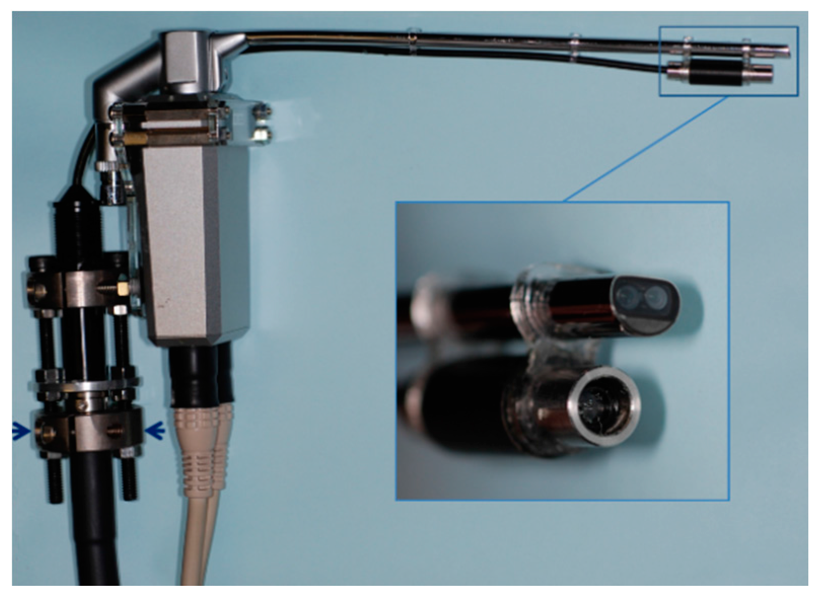
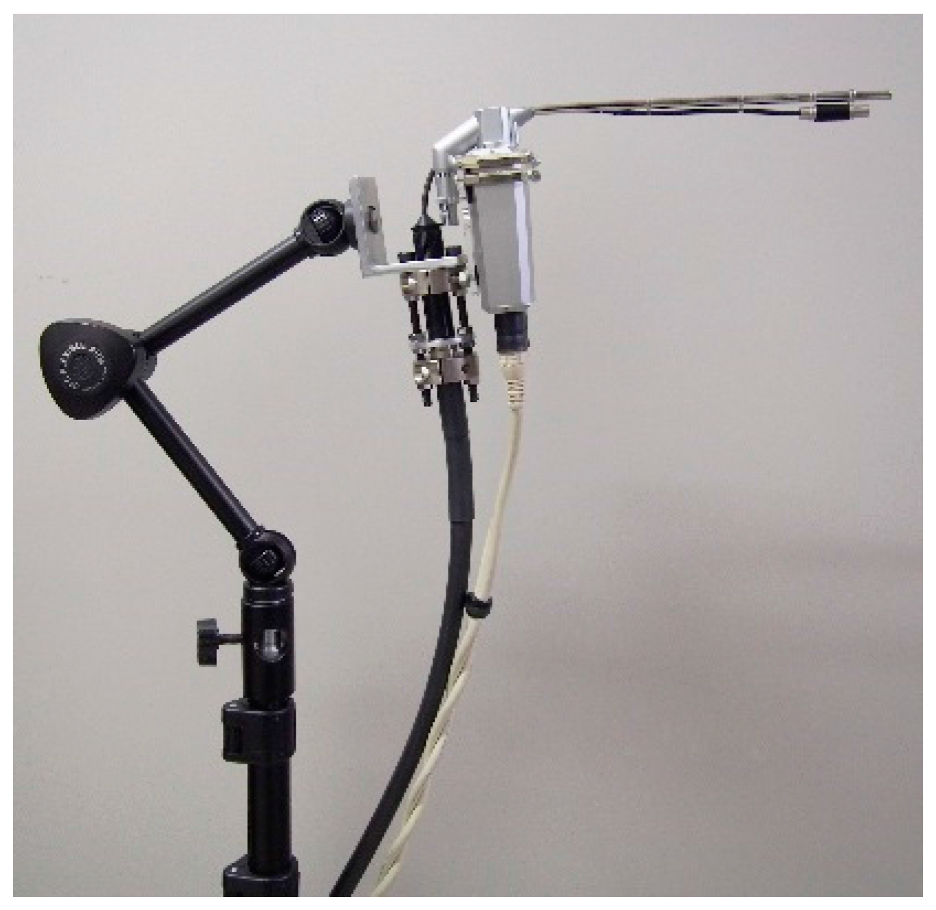
2.1. Production of Range Images
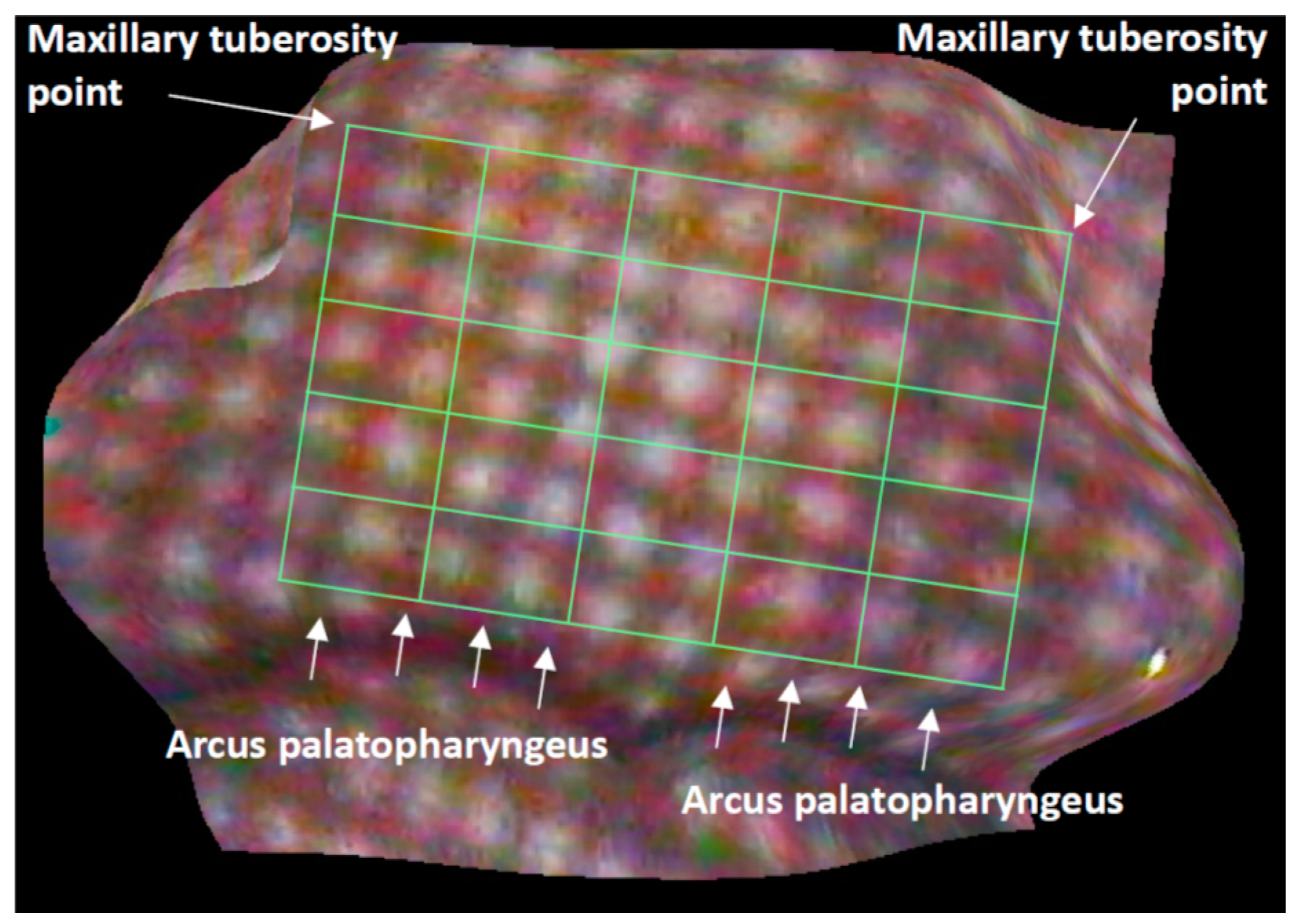
2.2. Analyses of Velopharyngeal Motion
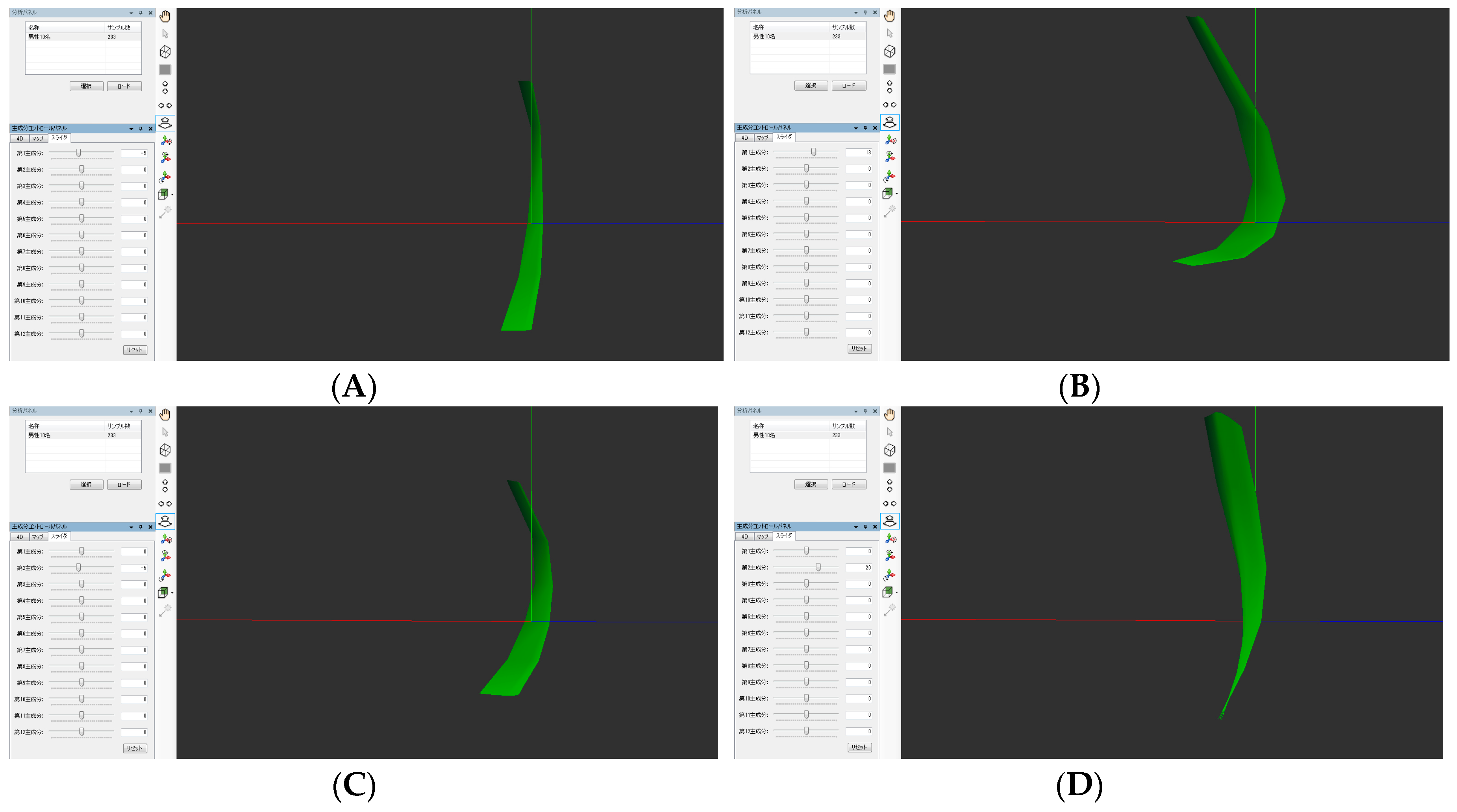
2.3. Principal Component Analyses
3. Results
4. Discussion
5. Conclusions
Acknowledgments
Author Contributions
Conflicts of Interest
References
- Phua, Y.S.; de Chalain, T. Incidence of oronasal fistulae and velopharyngeal insufficiency after cleft palate repair: An audit of 211 children born between 1990 and 2004. Cleft Palate Craniofac. J. 2008, 45, 172–178. [Google Scholar] [CrossRef] [Scilit] [PubMed]
- Inman, D.S.; Thomas, P.; Hodgkinson, P.D.; Reid, C.A. Oro-nasal fistula development and velopharyngeal insufficiency following primary cleft palate surgery—An audit of 148 children born between 1985 and 1997. Br. J. Plast. Surg. 2005, 58, 1051–1054. [Google Scholar] [CrossRef] [Scilit] [PubMed]
- Hoopes, J.E.; Dellon, A.L.; Fabrikant, J.L.; Soliman, A.H. Cineradiographic assessment of combined island flap pushback and pharyngeal flap in the surgical management of submucous cleft palate. Br. J. Plast. Surg. 1970, 23, 39–44. [Google Scholar] [CrossRef] [Scilit]
- Pegoraro-Krook, M.I.; Dutka-Souz, J.C.; Marino, V.C. Nasoendoscopy of velopharynx before and during diagnostic therapy. J. Appl. Oral Sci. 2008, 16, 181–188. [Google Scholar] [CrossRef] [Scilit] [PubMed]
- Pannbacker, M.D.; Lass, N.J.; Hansen, G.G.; Mussa, A.M.; Robison, K.L. Survey of speech-language pathologists’ training, experience, and opinions on nasopharyngoscopy. Cleft Palate Craniofac. J. 1993, 30, 40–45. [Google Scholar] [CrossRef] [Scilit]
- Mishima, K.; Nakano, A.; Shiraishi, R.; Ueyama, Y. Range image of the velopharynx produced using a 3D endoscope with pattern projection. Laryngoscope 2013, 123, 122–126. [Google Scholar] [CrossRef] [Scilit] [PubMed]
- Nakano, A.; Mishima, K.; Shiraishi, R.; Ueyama, Y. Quantitative analysis of velopharyngeal movement using a stereoendoscope: Accuracy and reliability of range images. Comput. Aided Surg. 2015, 20, 29–33. [Google Scholar] [CrossRef] [Scilit] [PubMed]
- Trotman, C.-A.; Faraway, J.J. Modeling facial movement. I. A dynamic analysis of differences based on skeletal characteristics. J. Oral Maxillofac. Surg. 2004, 62, 1372–1379. [Google Scholar] [CrossRef] [Scilit] [PubMed]
- Popat, H.; Henley, E.; Richmond, S.; Benedikt, L.; Marshall, D.; Rosim, P.L. A comparison of the reproducibility of verbal and nonverbal facial gestures using three-dimensional motion analysis. Otolaryngol. Head Neck Surg. 2010, 142, 867–872. [Google Scholar] [CrossRef] [Scilit] [PubMed]
- Mishima, K.; Yamada, T.; Matsumura, T.; Moritani, N. Analysis of lip motion using principal component analyses. J. Craniomaxillofac. Surg. 2011, 39, 232–236. [Google Scholar] [CrossRef] [Scilit] [PubMed]
- Tsai, R.Y. A versatile camera calibration technique for high-accuracy 3D machine vision metrology using off-the-shelf TV camera and lenses. IEEE J. Robot. Autom. 1987, 3, 323–344. [Google Scholar] [CrossRef] [Scilit]
- Birchfield, S.; Tomasi, C. Depth discontinuities by pixel-to-pixel stereo. Int. J. Comput. Vis. 1999, 35, 269–293. [Google Scholar] [CrossRef] [Scilit]
- Mishima, K.; Yamada, T.; Ohura, A.; Sugahara, T. Production of a range image for facial motion analysis: A method for analyzing lip motion. Comput. Med. Imag. Graph. 2006, 30, 53–59. [Google Scholar] [CrossRef] [Scilit] [PubMed]
- Bettens, K.; Wuyts, F.L.; Van Lierde, K.M. Instrumental assessment of velopharyngeal function and resonance: A review. J. Commun. Disord. 2014, 52, 170–183. [Google Scholar] [CrossRef] [Scilit] [PubMed]
- Kao, D.S.; Soltysik, D.A.; Hyde, J.S.; Gosain, A.K. Magnetic resonance imaging as an aid in the dynamic assessment of the velopharyngeal mechanism in children. Plast. Reconstr. Surg. 2008, 122, 572–577. [Google Scholar] [CrossRef] [Scilit] [PubMed]
- Perry, J.L.; Kuehn, D.P. Magnetic resonance imaging and computer reconstruction of the velopharyngeal mechanism. J. Craniofac. Surg. 2009, 20, 1739–1746. [Google Scholar] [CrossRef] [Scilit] [PubMed]
- Rahimian, P.; Perry, J.L.; Kollara, L.; Tabrizi, N.M. Using Synchronized Audio Mapping to Track and Predict Velar and Pharyngeal Wall Locations during Dynamic MRI Sequences. Int. J. Speech Lang. Pathol. Audiol. 2016, 4, 1–10. [Google Scholar]
- Jolliffe, I.T. Principal Component Analysis; Springer: New York, NY, USA, 2002. [Google Scholar]

© 2017 by the authors. Licensee MDPI, Basel, Switzerland. This article is an open access article distributed under the terms and conditions of the Creative Commons Attribution (CC BY) license (http://creativecommons.org/licenses/by/4.0/).
Share and Cite
Nakano, A.; Mishima, K.; Shiraishi, M.; Umeda, H.; Nakano, H.; Ueyama, Y. Quantitative Analysis of Velopharyngeal Movement by Applying Principal Component Analysis to Range Images Produced by a Three-Dimensional Endoscope. Dent. J. 2017, 5, 14. https://doi.org/10.3390/dj5020014
Nakano A, Mishima K, Shiraishi M, Umeda H, Nakano H, Ueyama Y. Quantitative Analysis of Velopharyngeal Movement by Applying Principal Component Analysis to Range Images Produced by a Three-Dimensional Endoscope. Dentistry Journal. 2017; 5(2):14. https://doi.org/10.3390/dj5020014
Chicago/Turabian StyleNakano, Asuka, Katsuaki Mishima, Mami Shiraishi, Hirotsugu Umeda, Hiroyuki Nakano, and Yoshiya Ueyama. 2017. "Quantitative Analysis of Velopharyngeal Movement by Applying Principal Component Analysis to Range Images Produced by a Three-Dimensional Endoscope" Dentistry Journal 5, no. 2: 14. https://doi.org/10.3390/dj5020014
APA StyleNakano, A., Mishima, K., Shiraishi, M., Umeda, H., Nakano, H., & Ueyama, Y. (2017). Quantitative Analysis of Velopharyngeal Movement by Applying Principal Component Analysis to Range Images Produced by a Three-Dimensional Endoscope. Dentistry Journal, 5(2), 14. https://doi.org/10.3390/dj5020014