Communication

Fluorescent and Colorimetric Dual-Mode Strategy Based on Rhodamine 6G Hydrazide for Qualitative and Quantitative Detection of Hg2+ in Seafoods

1
Key Laboratory of Food Quality and Health of Tianjin, Tianjin University of Science and Technology, Tianjin 300457, China
2
State Key Laboratory of Food Nutrition and Safety, Tianjin University of Science & Technology, Tianjin 300457, China
*
Author to whom correspondence should be addressed.
Foods 2023, 12(5), 1085; https://doi.org/10.3390/foods12051085
Submission received: 17 January 2023 / Revised: 26 February 2023 / Accepted: 1 March 2023 / Published: 3 March 2023

Abstract

:
In this study, a rapid fluorescent and colorimetric dual-mode detection strategy for Hg2+ in seafoods was developed based on the cyclic binding of the organic fluorescent dye rhodamine 6G hydrazide (R6GH) to Hg2+. The luminescence properties of the fluorescent R6GH probe in different systems were investigated in detail. Based on the UV and fluorescence spectra, it was determined that the R6GH has good fluorescence intensity in acetonitrile and good selective recognition of Hg2+. Under optimal conditions, the R6GH fluorescent probe showed a good linear response to Hg2+ (R2 = 0.9888) in the range of 0–5 μM with a low detection limit of 2.5 × 10⁻² μM (S/N = 3). A paper-based sensing strategy based on fluorescence and colorimetric analysis was developed for the visualization and semiquantitative analysis of Hg2+ in seafoods. The LAB values of the paper-based sensor impregnated with the R6GH probe solution showed good linearity (R2 = 0.9875) with Hg2+ concentration in the range of 0–50 μM, which means that the sensing paper can be combined with smart devices to provide reliable and efficient Hg2+ detection.

1. Introduction

In recent years, the rapid development of industry has brought serious pollution to the natural and food production environment. Unlike other types of pollution, heavy metal pollution can circulate within the environment and have the characteristic of being nondegradable, thus causing more serious harm and impact on human beings [1]. Heavy metal ions are capable of denaturing proteins in the living organisms. When these harmful heavy metals accumulate and concentrate in the human body, they can produce an accumulation of toxicity and cause very serious diseases [2,3]. Heavy metal contamination in food has been an important factor affecting food safety. Mercury (Hg) is one of the common heavy metals that can easily cause environmental pollution [4]. Usually, microorganisms in the soil can methylate the Hg element, making it easy to easily be absorbed by microorganisms and thus enter the food chain. By accumulating in the human body for a long time, high concentrations of Hg can produce very serious hazards [5]. Therefore, it is necessary and significant to monitor and detect Hg levels in the environment and food samples.
Currently, analytical strategies such as mass spectrometry and spectroscopy based on large-scale instruments, such as inductively coupled plasma mass spectrometry (ICP-MS) [6,7,8], atomic absorption spectrometry (AAS) [9,10], and atomic fluorescence spectrometry (AFS) [11,12], are still the main means for accurate detection of heavy metals (including Hg) in various samples, and such methods have unparalleled advantages in terms of detection accuracy and sensitivity. However, such advanced instruments are usually expensive and large, and require relatively complex sample pretreatment processes, which are inadequate for low-cost screening, in situ detection, and large-scale penetration. To avoid these problems, there is an urgent need to establish a fast, sensitive, and portable detection method for heavy metal targets [13,14]. The paper-based sensing strategy based on fluorescent and colorimetric dual mode can achieve naked-eye visualization of the target and portable, low-cost semiquantitative detection [15,16]; on the other hand, this dual-mode spectral signal output can largely guarantee the reliability of the detection results.
Due to the long emission wavelength, low biotoxicity, and pronounced color change, rhodamine-based compounds are commonly used as fluorescent and colorimetric labeling reagents in visualization assays [17,18,19]. These compounds can specifically complex or bind metal ions or organic small molecules while processing certain recognition abilities. In particular, rhodamine derivatives with spiro ring structure have different optical properties in open and closed loops, making them ideal materials for the construction of optical sensors. It is worth noting that rhodamine 6G hydrazide (R6GH) is one of the most important intermediates of rhodamine compounds, which attracted extensive attention in heavy metal detection studies [20,21]. The amide spiral ring structure with rhodamine as the parent nucleus has an “On–Off” feature. When specific metal ions are added, the amide ring will be opened, resulting in the rupture of the organic dye and enhanced fluorescence [22]. In our previous work [23], the R6GH dye with a spiral ring structure was found to be opened by Pb2+, causing a significant fluorescence signal (Ex: 552 nm). Based on this, a dual-mode fluorescence and colorimetric detection strategy was further designed and constructed for the rapid and efficient detection of Pb2+ in water and food samples.
Since heavy metal Hg2+ can also trigger the fluorescence switch of the R6GH probe, this study continued to explore the fluorescence response performance of the R6GH probe to the heavy metal Hg2+ under different solution systems and thus developed an effective fluorescence analysis strategy for Hg2+ (Figure 1). The study further developed portable detection test strips that not only allowed for the naked-eye colorimetric semiquantitative analysis of the Hg2+ content but also can be combined with a portable color reader for the rapid screening of target Hg2+ in a large number of samples. This study is of great interest for the development of effective strategies for the on-site detection and large-scale screening of trace hazardous substances in food.

2. Materials and Methods

2.1. Reagents and Materials

The reagents and solvent nitrates of target Hg2+ and other ions Cu2+, V2+, Cd2+, Mn2+, Zn2+, Cr3+, Co2+, Ag+, and K+; hydrazine hydrate (85%); rhodamine 6G (R6G, 99.5%); and ethylenediaminetetraacetic acid (EDTA) were purchased from Shanghai Aladdin Biochemical Technology Co., Ltd. (Shanghai, China). Organic reagents such as tetrahydrofuran (THF), methanol (MeOH), ethanol (EtOH), and acetonitrile (ACN) were purchased from Sinopharm Chemical Reagent Company. All the reagents used for R6GH probe synthesis and analysis were of analytical grade or higher and were not further purified.

2.2. Instruments

A Shimadzu UV-vis spectrophotometer (Tokyo, Japan, UV-2600) was used to record UV absorbance data, and a Thermo fluorescence spectrometer (Boston, MA, USA, Lumina) was applied for fluorescence analysis. An electric blast dryer (WG II-45BE, Tianjin, China) and a Teflon digestion tank were used for sample pretreatment. An Agilent inductively coupled plasma mass spectrometer (ICP-MS) (Santa Clara, CA, USA, 7700x) was used to compare and validate the data from the Hg2+ analysis strategy established in this study. An ordinary quantitative filter paper was used in the visual analysis, and a portable colorimeter from FRU Weifu Optoelectronics (WR-10, Wuxi, China) was used for LAB analysis in the paper-based assays.

2.3. Preparation of R6GH Probe

The R6GH fluorescent probe was prepared according to the method reported [23] and is briefly described as follows: accurately weighed R6G (0.5 g) was fully dissolved in EtOH (30.0 mL) in a 100 mL round-bottom glass flask, and a solution of hydrazine hydrate (85%, 2.0 mL) was added for thorough mixing. The mixture was cooled to room temperature and magnetically stirred for 10 h until the color of the mixed solution disappeared. After filtering under reduced pressure and washing with EtOH three times, the white solid product was collected as the R6GH fluorescent probe.

2.4. Optimization of Dual-Mode Detection System

The prepared R6GH fluorescent probes were dispersed in THF/H2O (v/v, 1:1), ACN, and MeOH/H2O (v/v, 3:1), respectively. The concentration of R6GH was controlled at 1.0 mM as a stock solution and diluted to the desired concentration for testing. Subsequently, equal volumes of R6GH solutions at 20 μM concentration in the three systems were mixed with different concentrations (0–60 μM) of Hg2+ to obtain the resulting mixtures with Hg2+ concentrations ranging from 0 to 30 μM. The mixture was reacted at room temperature for 10 min and then analyzed by UV and fluorescence spectroscopy. By comparing the results, the optimal system for the detection of Hg2+ was finally determined and used in the subsequent experiments.

2.5. Detection Procedure for Hg2+

Different concentration gradients (0–10 μM) of Hg2+ were added to the ACN system of the R6GH probe (20 μM), thoroughly mixed, and left at room temperature for fluorescence intensity detection. The common metal ions Cu2+, V2+, Cd2+, Mn2+, Zn2+, Cr3+, Co2+, Ag+, and K+ (10 μM) in food were added to the R6GH probe solution in the ACN system in the same way, and after sufficient reaction, the fluorescence intensity was compared to assess the interference of different metal ions with Hg2+.
The reproducibility and reversible mechanism of the R6GH probe for the detection of Hg2+ were also investigated using ethylenediaminetetraacetic acid (EDTA) to assess the reversibility of R6GH for the detection of Hg2+.

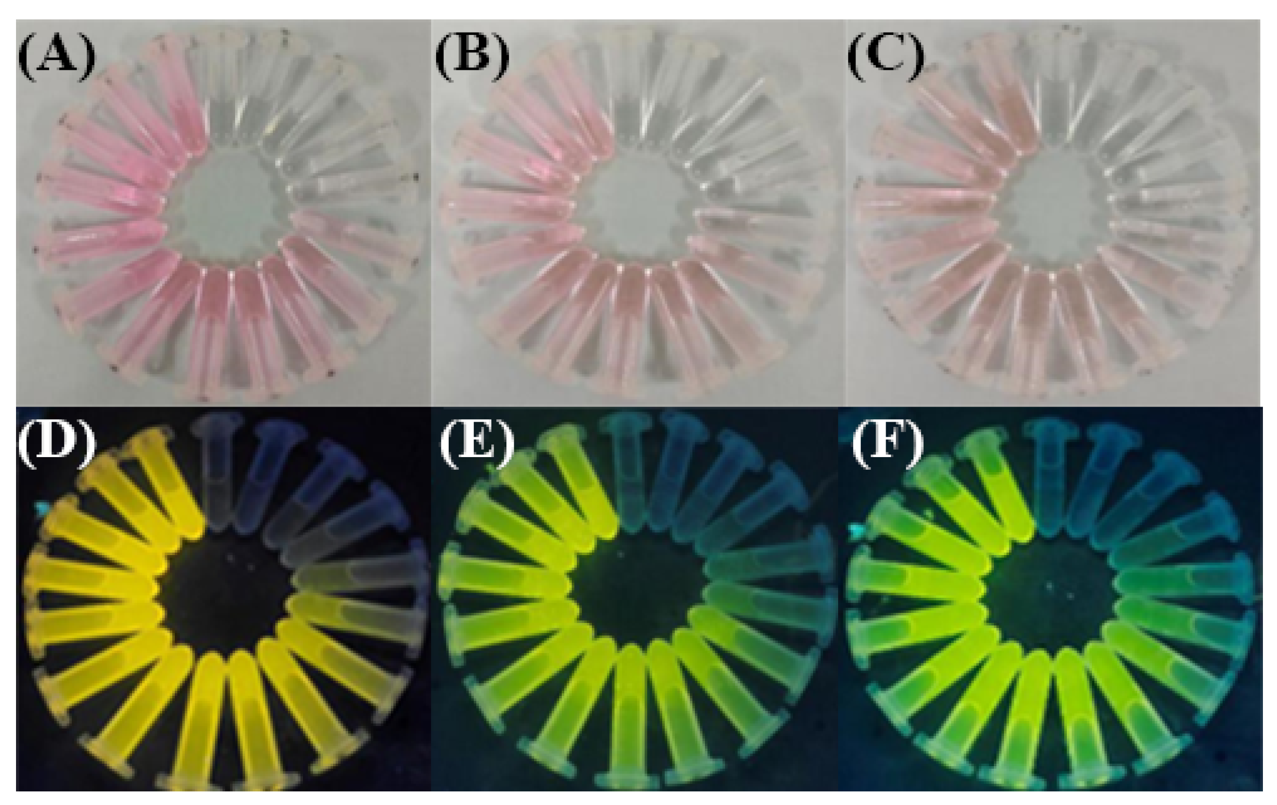
2.6. Preparation of Test Strips for Visual Detection of Heavy Metal Ions

Test strips (1 cm × 1 cm) were infiltrated in the solution of the R6GH probe (20 µM) in the THF/H2O (v/v, 1:1) solution and left for 10 min at room temperature. The solvent was removed from the test strips and allowed to dry. The paper with the R6GH probe immobilized was cut into strips and fixed, infiltrated in Hg2+ standard solutions with different concentration gradients (0–100 µM), and naturally dried to obtain color-developed immobilized paper-based sensors that can be used for naked-eye or fluorescence (365 nm UV) analysis and comparison of Hg2+.

2.7. Actual Sample Pretreatment

Seafood samples of oysters, yellow croaker, and prawn were purchased from a local market in Tianjin and stored in a refrigerator at 4 °C. The samples were first nitrated by adding 0.5 g of the actual sample and 10.0 mL of HNO3 together at room temperature into a Teflon beaker for an overnight treatment, then boiled until all dissolved, and the cooled solution was centrifuged at 8000 r/min to obtain the supernatant, which was adjusted to pH 6.0 using the NaOH solution (1 mol/L) and diluted to 50 mL with ultrapure water to prepare different concentration gradients of Hg2+.

3. Results and Discussion

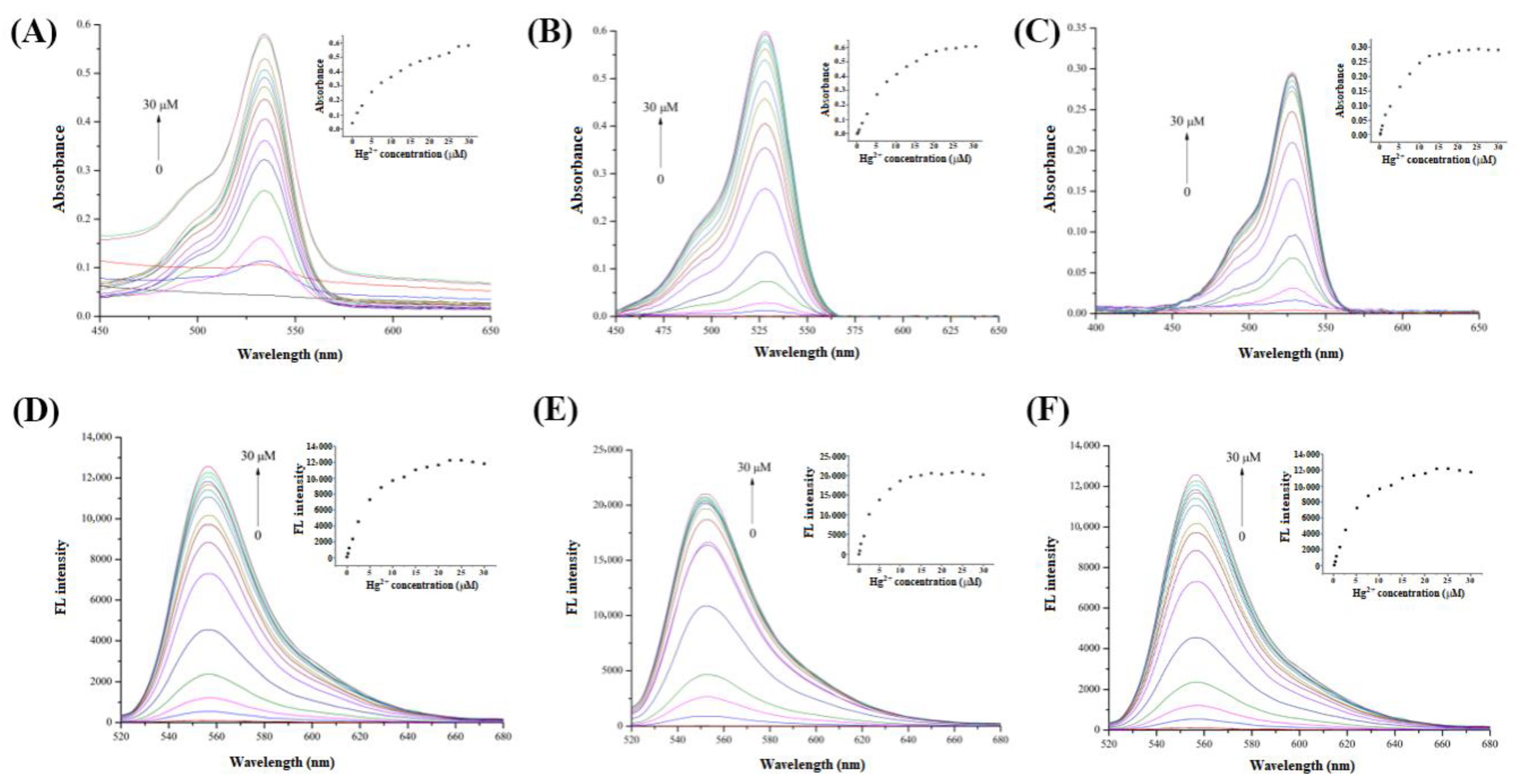
3.1. Optimization of Hg2+ Assay System

The prepared R6GH probe can react with the target Hg2+, which can open the ring structure it possesses, leading to the enhanced fluorescence. The fluorescence response of the R6GH probe to the target Hg2+ in three solutions of THF/H2O (v/v, 1:1), ACN, and MeOH/H2O (v/v, 3:1) was investigated, and the sensitivity and accuracy for the dual-mode detection of Hg2+ in different systems were compared based on the Hg2+-induced changes in color and fluorescence intensity of the R6GH probe observed under natural and UV light.
Figure 2 has shown the color change of the R6GH probe in the three detection systems under natural light and UV after Hg2+ induced the colorimetric and fluorescence switching of the R6GH probe in the “On” state. When Hg2+ was added into the solution of the R6GH probe, the solution color changed from colorless to pink under natural light, and the fluorescence in the solution changed from no fluorescence to bright yellow fluorescence under the excitation of 365 nm UV light. Meanwhile, with the increase in Hg2+ concentration (0–30 µM), the solution color of the R6GH probe gradually deepened and stabilized. From these results, the color change of R6GH in the THF/H2O (v/v, 1:1) system was more obvious, with higher chromogen and brighter fluorescence produced, which was easier to observe. Therefore, the THF/H2O (v/v, 1:1) solution was considered more suitable for the visualization and fluorescent colorimetric analysis of Hg2+ by the R6GH probe and used for the detection process of Hg2+ colorimetric test strips.
To further explore the Hg2+ recognition performance of the R6GH probe in fluorescence and colorimetric detection, the UV absorbance and fluorescence intensity of the R6GH probe were investigated in three systems under the colorimetric and fluorescence “On” states induced by Hg2+, and it was found that the UV absorption and fluorescence intensity significantly increased with the addition of Hg2+. As illustrated in Figure 3, the R6GH probe has no UV absorption and fluorescence emission ability but appeared a UV absorption band near 530 nm and a clear fluorescence emission peak near 556 nm. By comparing the UV absorption and fluorescence spectra in the three detection systems, the ACN system obtained the highest absorbance and fluorescence intensity. This was because the ACN was one nonprotonic solvent that did not provide nor spontaneously transfer protons in the reaction, thus having good solubility for metal cations. Meanwhile, considering the good solubility of ACN for the R6GH probe, it allowed Hg2+ to form ion-dipole bonds with the solvent system, thus increasing the contact area of the complexation reaction between the R6GH fluorescent probe and Hg2+, which made the reaction faster and more adequate.

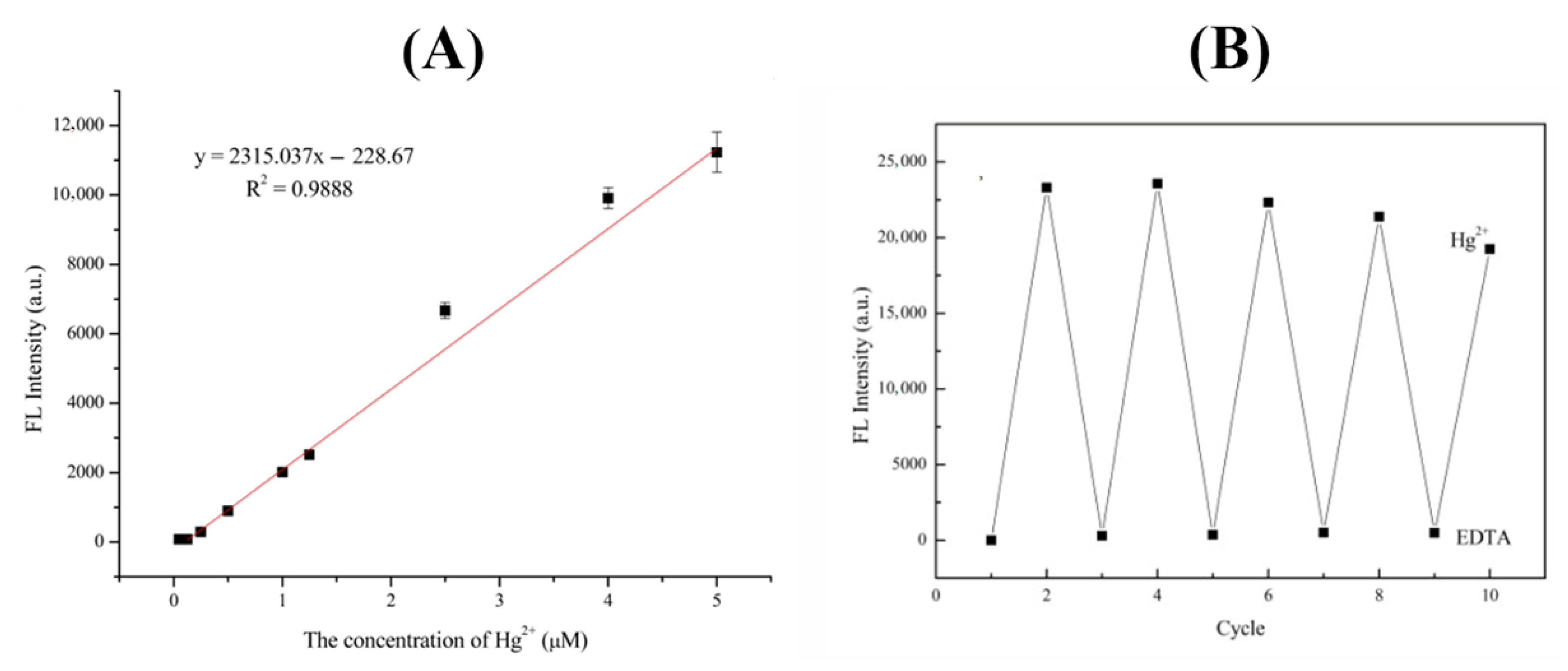
3.2. Establishment of R6GH-Based Fluorescence Strategy for Hg2+ Detection

Hg2+ can ligand-complex with the O atom of -COOH and the N atom of -NH2 in the R6GH probe [24], which induces the conversion of the rhodamine-based amide spiro ring structure from “Off” to “On” state, resulting in the color change or fluorescence enhancement of the originally colorless and nonfluorescent R6GH probe. The fluorescence intensity of the R6GH probe in ACN gradually increased with the increase in Hg2+ concentration, having a good linear relationship with Hg2+ concentration in the range of 0–5 μM with R2 of 0.9888 (Figure 4A). The limit of detection (LOD, S/N = 3) of Hg2+ reached 2.5 × 10⁻² μM, indicating that the prepared R6GH fluorescent probe can sensitively respond to Hg2+.
The selectivity of the R6GH fluorescent probe for Hg2+ in the ACN system was further evaluated for common metal ions (Cd2+, Mn2+, V2+, Cu2+, Zn2+, Cr3+, Co2+, Ag+, and K+) in the study. It was found that these metal ions did not significantly enhance the fluorescence intensity of R6GH. When Hg2+ (10 μM) induced the fluorescence conversion of the R6GH probe to “open loop”, its fluorescence intensity was as high as 9435.1, which indicated that Hg2+ had a relatively obvious fluorescence enhancement effect on the R6GH probe. Except for the selected Cd2+, Mn2+, V2+, and Cu2+, which had weak enhancement on the fluorescence of R6GH [23] (approximately 20–50% of the fluorescence intensity of the same concentration of Hg2+), the other tested ions could not produce fluorescence enhancement on the R6GH probe. Furthermore, the interference factor K (Fothers/FHg2+) was used to compare and assess the interference of other metal ions on the fluorescence response of target Hg2+. Based on the fluorescence intensity of tested metal ions at 5.0 μM, the calculated K values 0.47 (Cd2+), 0.29 (Mn2+), 0.27 (V2+), 0.24 (Cu2+), 0.002 (Ag+), 0.0014 (Zn2+), 0.0012 (K+), 0.00094 (Co2+), and 0.0008 (Cr3+) were all less than 1.0, further demonstrating that the prepared R6GH probe has good selectivity for Hg2+, which provides a great feasibility and theoretical basis for its future practical application.
EDTA can coordinate with a variety of metal ions to form complexes and was used to examine the fluorescence reversibility of the R6GH probe [25]. The results showed that the fluorescence intensity of the R6GH–Hg2+ system to ACN sharply decreased after the addition of EDTA and even almost restored to the original state of the R6GH probe (Figure 4B). This indicated that EDTA could release Hg2+ from the R6GH–Hg2+ complex and turn off the fluorescence switch of the R6GH probe. When Hg2+ was added again, the fluorescence intensity of the solution recovered to be close to that of the R6GH probe solution when only Hg2+ was present, indicating that EDTA can cause the demetallization of the R6GH probe and regeneration of the spirolactam ring. After five cycles, the fluorescence of the R6GH probe solution did not significantly increase or decrease, indicating that the R6GH probe can release Hg2+ through competition with EDTA for reversible cycling of Hg2+ detection.

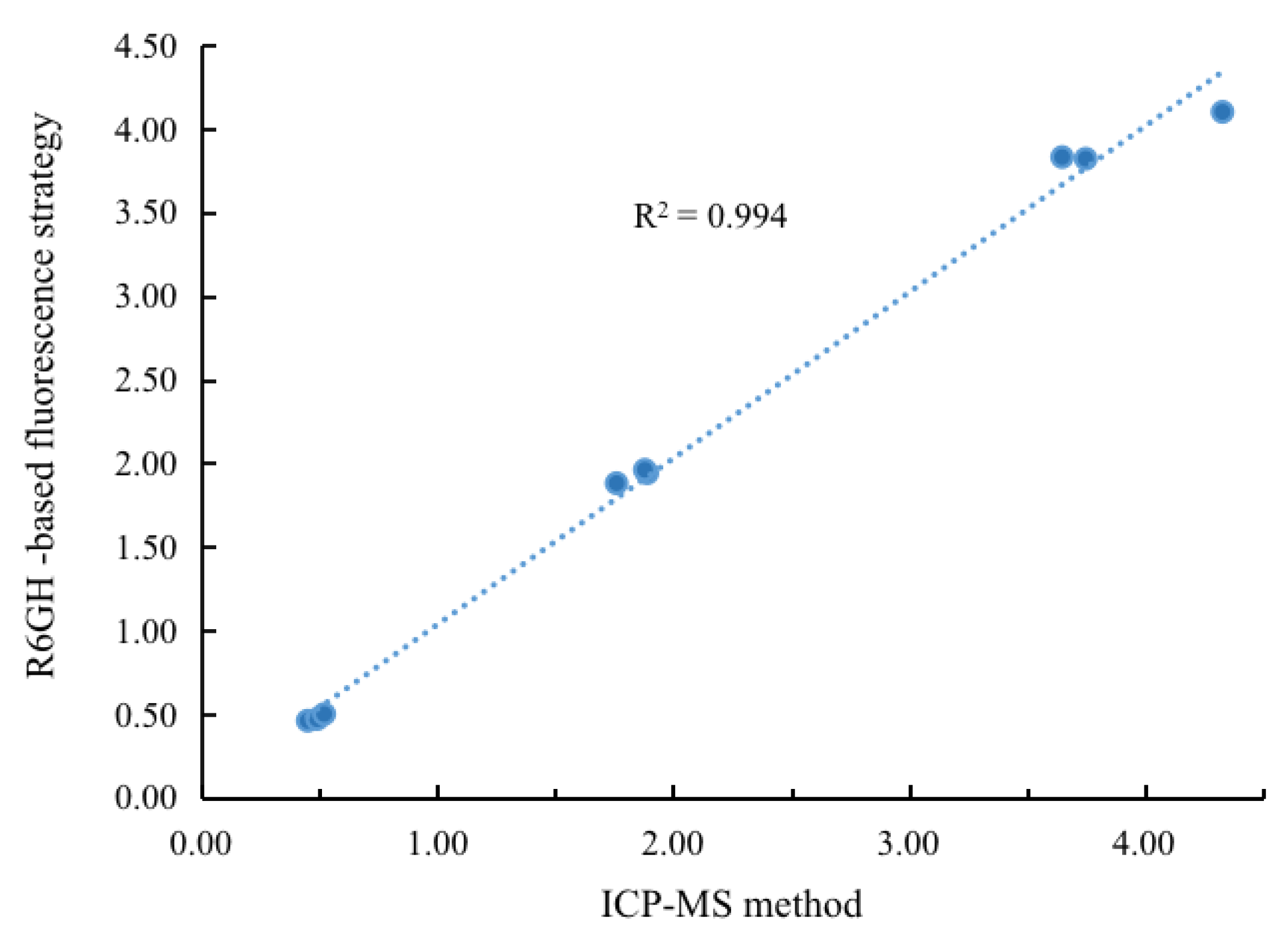
3.3. Detection of Hg2+ in Seafoods Using R6GH-Based Fluorescent Probes

To verify the application capability of the prepared R6GH-based fluorescent probe, seafoods including oysters, yellow croaker, and prawn were selected and spiked Hg2+ with different concentrations (0.5, 2.0, and 4.0 μM) to perform the recovery experiments. The spiked samples were simply pretreated and used for the constructed R6GH-based fluorescent probes and widely accepted ICP-MS methods for detection. As shown in Table 1, the proposed R6GH-based fluorescent probe obtained acceptable recoveries (88.0–108.3%) of Hg2+ in each selected seafood with RSDs all below 5% (n = 3). Compared with the results obtained from the conventional ICP-MS method, good correlation was achieved with r2 > 0.99 (Figure 5). These results indicated that the proposed R6GH probe-based fluorescence strategy was an ideal tool for providing accurate and reliable detection of Hg2+ in food matrices. Table 2 has compared the merits of different strategies for Hg2+ detection, signifying that the proposed R6GH-based probe can offer a rapid, sensitive, and effective strategy for Hg2+.

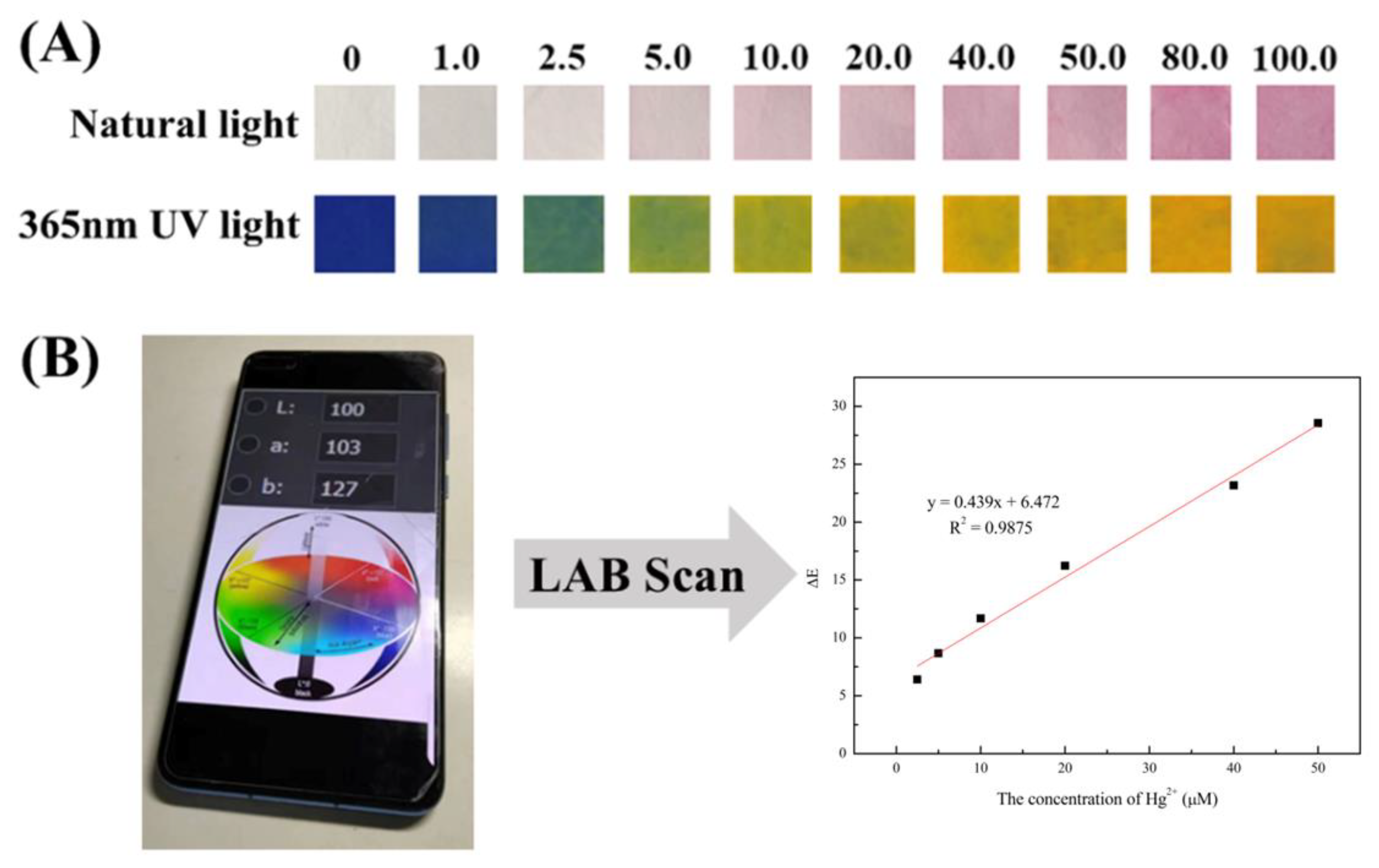
3.4. Development of Visualization Paper-Based Sensor for Hg2+ Detection

Fluorescence sensing analysis suffers from the shortcoming that single signal readings are susceptible to environmental and human factors; thus, there is an urgent need to develop new sensing platforms capable of rapid, on-site, and reliable heavy metal ion detection and analysis. Based on the constructed fluorescence sensing system of R6GH–Hg2+, an intelligent, low-cost, and portable paper-based sensing platform was developed to realize fluorescent and colorimetric dual-mode signal output for more accurate, reliable, and convenient detection of heavy metal Hg2+. As shown in Figure 6A, the paper-based sensor constructed with a filter paper infiltrated with the R6GH–Hg2+ solution as a substrate (the paper used in the experiment has blue background fluorescence) showed a colorless to light pink change (natural light) and a blue to yellow-green change (UV light, 365 nm) with increasing Hg2+ concentration from 0 to 100 μM.
For visualization of the results, colorimetric signals can be recorded and analyzed using a colorimeter to provide the LAB values of the test strips. The LAB color model consists of three elements, luminance L and the associated colors A and B [30,31]. The LAB color space defining the color change can be further linked to digital cameras and smartphones, thus facilitating remote monitoring and online analysis. Thus, the chromatic aberration parameter ∆E can be used as a reference for the visual detection of Hg2+ by paper-based sensors. The chromaticity difference at different concentrations of Hg2+ can be calculated according to the equation ( Δ E = Δ L 2 + Δ A 2 + Δ B 2 ) [32]. The results showed a good linear relationship (y = 0.439x + 6.472, R2 = 0.9875) between the chromatic aberration parameter ∆E and Hg2+ concentration for the paper-based sensor infiltrated using the R6GH–acetonitrile probe solution in the Hg2+ concentration range of 2.5–50 μM (Figure 6B). Therefore, the paper-based array sensor constructed based on fluorescence and colorimetric detection strategies combined with a smart device can achieve visualized semiquantitative detection of target Hg2+. This dual-mode sensing method based on R6GH enabled more convenient, reliable, and accurate analysis of target Hg2+.

4. Conclusions

In summary, this study successfully synthesized the R6GH probe that generates fluorescence and color signals with Hg2+ and developed a fluorescence-colorimetric dual-mode sensing platform that can be used for rapid, accurate, and sensitive detection of Hg2+ in seafood. This R6GH probe was used not only to construct a fluorescence analysis platform with good linearity, accuracy, and sensitivity for Hg2+ but also to develop a paper-based visual semiquantitative analysis strategy that can work in conjunction with a small and portable color reader, providing an ideal tool for rapid, low-cost, and convenient analysis of Hg2+. The R6GH fluorescent probe-based strategy and research method proposed in this study can be extended to the detection of other targets in other fields, providing a new direction for the research of high-performance and intelligent analytical strategies and detection devices.

Author Contributions

Conceptualization, Z.Z. and M.P.; methodology, Z.Z.; validation, Z.Z. and R.H.; formal analysis, S.C.; data curation, Z.Z. and F.Z.; writing—original draft preparation, Z.Z. and X.M.; writing—review and editing, M.P. and S.W.; supervision, S.W.; project administration, M.P.; funding acquisition, M.P. All authors have read and agreed to the published version of the manuscript.

Funding

This work was supported by the National Natural Science Foundation of China (No. 31972147 and 32272416), Project of Tianjin Science and Technology Plan (No. 22ZYJDSS00030), and Project Program of Key Laboratory of Tianjin Key Laboratory of Food Quality and Health, China (No. TJS202205).

Institutional Review Board Statement

Not applicable.

Informed Consent Statement

Not applicable.

Data Availability Statement

Not applicable.

Conflicts of Interest

The authors declare no conflict of interest.

References

  1. Ng’ang’a, J.; Fombong, F.; Kiiru, S.; Kipkoech, C.; Kinyuru, J. Food safety concerns in edible grasshoppers: A review of microbiological and heavy metal hazards. Int. J. Trop. Insect Sci. 2021, 41, 2103–2111. [Google Scholar] [CrossRef]
  2. Nduka, J.K.; Orisakwe, O.E. Heavy metal hazards of pediatric syrup administration in Nigeria: A look at chromium, nickel and manganese. Int. J. Environ. Res. Public Health 2009, 6, 1972–1979. [Google Scholar] [CrossRef] [PubMed] [Green Version]
  3. Awad, M.; El-Sayed, M.M.; Li, X.; Liu, Z.; Mustafa, S.K.; Ditta, A.; Hessini, K. Diminishing heavy metal hazards of contaminated soil via biochar supplementation. Sustainability 2021, 13, 12742. [Google Scholar] [CrossRef]
  4. Suren, S.; Punyain, W.; Maneeintr, K.; Nootong, K.; Pancharoen, U. The simultaneous elimination of arsenic and mercury ions via hollow fiber supported liquid membrane and their reaction mechanisms: Experimental and modeling based on DFT and generating function. Arabian J. Chem. 2022, 16, 104501. [Google Scholar] [CrossRef]
  5. Liu, G.; Cai, Y.; O’Driscoll, N.J. Environmental chemistry and toxicology of mercury. Ecotoxicol. Environ. Saf. 2020, 202, 110926. [Google Scholar]
  6. Pereiro, I.R. Optimization of the coupling of multicapillary GC with ICP-MS for mercury speciation analysis in biological materials. J. Anal. At. Spectrom. 1999, 14, 851–857. [Google Scholar]
  7. Fu, L.; Shi, S.Y.; Chen, X.Q. Accurate quantification of toxic elements in medicine food homologous plants using ICP-MS/MS. Food Chem. 2018, 245, 692–697. [Google Scholar] [CrossRef]
  8. Pérez-Álvarez, E.P.; García, R.; Barrulas, P.; Dias, C.; Cabrita, M.J.; Garde-Cerdán, T. Classification of wines according to several factors by ICP-MS multi-element analysis. Food Chem. 2019, 270, 273–280. [Google Scholar] [CrossRef]
  9. Domanico, F.; Forte, G.; Majorani, C.; Senofonte, O.; Petrucci, F.; Pezzi, V.; Alimonti, A. Determination of mercury in hair: Comparison between gold amalgamation-atomic absorption spectrometry and mass spectrometry. J. Trace Elem. Med. Biol. 2017, 43, 3–8. [Google Scholar] [CrossRef]
  10. Frois, C.F.; Boschetti, W.; dos Passos, A.S.; Potes, M.L.; Vale, M.G.R.; Silva, M.M. A comparison between chemical and photochemical vapor generation techniques for mercury determination using univariate and multivariate optimization. Microchem. J. 2020, 157, 105029. [Google Scholar] [CrossRef]
  11. Grijalba, A.C.; Quintas, P.Y.; Fiorentini, E.F.; Wuilloud, R.G. Usefulness of ionic liquids as mobile phase modifiers in HPLC-CV-AFS for mercury speciation analysis in food. J. Anal. At. Spectrom. 2018, 33, 822–834. [Google Scholar] [CrossRef]
  12. da Silva, D.L.F.; da Costa, M.A.P.; Silva, L.O.B.; Dos Santos, W.N.L. Simultaneous determination of mercury and selenium in fish by CVG AFS. Food Chem. 2019, 273, 24–30. [Google Scholar] [CrossRef] [PubMed]
  13. Pak, K.R.; Bartha, R. Mercury methylation and demethylation in anoxic lake sediments and by strictly anaerobic bacteria. Appl. Environ. Microbiol. 1998, 64, 1013–1017. [Google Scholar] [CrossRef] [Green Version]
  14. Pundi, A.; Chang, C.J. Recent advances in synthesis, modification, characterization, and applications of carbon dots. Polymers 2022, 14, 2153. [Google Scholar] [CrossRef] [PubMed]
  15. Zhang, X.; Deng, J.; Xue, Y.; Shi, G.; Zhou, T. Stimulus response of Au-NPs@ GMP-Tb core–shell nanoparticles: Toward colorimetric and fluorescent dual-mode sensing of alkaline phosphatase activity in algal blooms of a freshwater lake. Environ. Sci. Technol. 2016, 50, 847–855. [Google Scholar] [CrossRef]
  16. Azmi, A.A.; Daud, A.I.; Khairul, W.M.; Hamzah, S.; WMA, W.M.K.; Hairom, N.H.H. Silica–silver core–shell nanoparticles incorporated with cellulose filter paper as an effective colorimetric probe for mercury ion detection in aqueous media: Experimental and computational evaluations. Environ. Nanotechnol. Monit. Manag. 2022, 19, 100762. [Google Scholar] [CrossRef]
  17. Li, H.; Chen, Q.; Hassan, M.M.; Ouyang, Q.; Jiao, T.; Xu, Y.; Chen, M. AuNS@ Ag core-shell nanocubes grafted with rhodamine for concurrent metal-enhanced fluorescence and surfaced enhanced Raman determination of mercury ions. Anal. Chim. Acta 2018, 1018, 94–103. [Google Scholar] [CrossRef]
  18. Zhang, Q.; Liu, X.J.; He, R.C.; Guo, C.B.; Zhao, W.Z.; Zeng, C.C.; Yin, L.P. Development of a fluorescent-type sensor based on rhodamine B for Fe (III) determination. Chem. Lett. 2018, 47, 122–125. [Google Scholar] [CrossRef]
  19. Zhao, M.; Guo, Y.S.; Fu, G.D.; Wang, Q.; Sheng, W.L.; Guo, D.S. Development of a Si-rhodamine-based NIR fluorescence probe for highly specific and quick response of Hg2+ and its applications to biological imaging. Microchem. J. 2021, 171, 106855. [Google Scholar] [CrossRef]
  20. Wang, S.; Li, C.; Xu, L.; Xu, T.; Lv, Y.; Son, Y.A.; Cao, D. A photochromic fluorescent probe for Hg2+ based on dithienylethene-rhodamine b dyad and its application in live cells imaging. Sci. Adv. Mater. 2017, 9, 533–540. [Google Scholar] [CrossRef]
  21. Meng, X.; Li, Z.; Ma, W. A highly sensitivity fluorescent probe based on rhodamine for naked-eye detection of Hg2+ in aqueous solution. Int. J. Environ. Anal. Chem. 2021, 1–11. [Google Scholar] [CrossRef]
  22. Rajasekar, M. Recent trends in Rhodamine derivatives as fluorescent probes for biomaterial applications. J. Mol. Struct. 2021, 1235, 130232. [Google Scholar] [CrossRef]
  23. Xie, X.; Pan, M.; Hong, L.; Liu, K.; Yang, J.; Wang, S.; Wang, S. An “off–on” rhodamine 6G hydrazide-based output platform for fluorescence and visual dual-mode detection of lead (II). J. Agric. Food Chem. 2021, 69, 7209–7217. [Google Scholar] [CrossRef] [PubMed]
  24. Xu, Y.; Niu, X.; Zhang, H.; Xu, L.; Zhao, S.; Chen, H.; Chen, X. Switch-on fluorescence sensing of glutathione in food samples based on a graphitic carbon nitride quantum dot (g-CNQD)–Hg2+ chemosensor. J. Agric. Food Chem. 2015, 63, 1747–1755. [Google Scholar] [CrossRef] [PubMed]
  25. Lu, B.; Xia, D.; Zhao, S.; Yang, Z.; Chen, Z.; Huang, S.; Zhang, Z. The influence mechanism of ethylenediaminetetraacetic acid (EDTA) and ferrous iron on the bioavailability of Fe in the process of low rank coal fermentation. Biochem. Eng. J. 2022, 185, 108520. [Google Scholar] [CrossRef]
  26. Zhou, L.; Lin, Y.; Huang, Z.; Ren, J.; Qu, X. Carbon nanodots as fluorescence probes for rapid, sensitive, and label-free detection of Hg2+ and biothiols in complex matrices. Chem. Commun. 2012, 48, 1147–1149. [Google Scholar] [CrossRef]
  27. Duan, Q.Q.; Zhou, J.L.; Li, P.W.; Sun, L.; Zhuo, K.; Zhang, Y.X.; Sang, S.B. High-sensitivity mercury ion detection system using unmodified gold nanorods. Chinese. J. Anal. Chem. 2018, 46, e1874–e1879. [Google Scholar] [CrossRef]
  28. Wang, Y.; Yang, L.; Liu, B.; Yu, S.; Jiang, C. A colorimetric paper sensor for visual detection of mercury ions constructed with dual-emission carbon dots. New J. Chem. 2018, 42, 15671–15677. [Google Scholar] [CrossRef]
  29. Cao, R.G.; Zhu, B.; Li, J.; Xu, D. Oligonucleotides-based biosensors with high sensitivity and selectivity for mercury using electrochemical impedance spectroscopy. Electrochem. Commun. 2009, 11, 1815–1818. [Google Scholar] [CrossRef]
  30. Kim, H.; Kim, H.; Choi, J.; Inn, K.S.; Seong, J. Visualization of autophagy progression by a red–green–blue autophagy sensor. ACS Sens. 2020, 5, 3850–3861. [Google Scholar] [CrossRef]
  31. Ulrici, A.; Foca, G.; Ielo, M.C.; Volpelli, L.A.; Fiego, D.P.L. Automated identification and visualization of food defects using RGB imaging: Application to the detection of red skin defect of raw hams. Innov. Food Sci. Emerg. Technol. 2012, 16, 417–426. [Google Scholar] [CrossRef]
  32. Wee, A.G.; Lindsey, D.T.; Kuo, S.; Johnston, W.M. Color accuracy of commercial digital cameras for use in dentistry. Dent. Mater. 2006, 22, 553–559. [Google Scholar] [CrossRef] [PubMed]
Figure 1. Schematic of R6GH synthesis and fluorescent and colorimetric dual-mode detection of Hg2+ for smartphone-integrated sensing system.
Foods 12 01085 g001
Figure 2. Color changes of Hg2+ induced R6GH probes in the three different solutions under natural light ((A): THF/H2O (v/v, 1:1); (B): ACN; (C): MeOH/H2O (v/v, 3:1)) and under 365 nm UV light ((D): THF/H2O (v/v, 1:1); (E): ACN; (F): MeOH/H2O (v/v, 3:1)).
Foods 12 01085 g002
Figure 3. UV absorption spectra ((A) THF/H2O (v/v, 1:1); (B) ACN; (C) MeOH/H2O (v/v, 3:1)) and fluorescence spectra ((D) THF/H2O (v/v, 1:1); (E) ACN; (F) MeOH/H2O (v/v, 3:1)) of the R6GH probe (10 μM) in three tested solutions complexing with different concentrations of Hg2+ (0–30 μM). Inset: Relationship between R6GH absorbance or fluorescence intensity (Em: 556 nm) and Hg2+ concentrations.
Foods 12 01085 g003
Figure 4. (A) Correlation between Hg2+ concentrations and R6GH fluorescence intensity. (B) Fluorescence reversibility of R6GH under alternate addition of Hg2+ and EDTA.
Foods 12 01085 g004
Figure 5. Correlation of Hg2+ detection results in real samples by R6GH-based fluorescence strategy and ICP-MS method.
Foods 12 01085 g005
Figure 6. (A) Colors of immobilized paper-based sensors containing R6GH immersed in different concentrations of Hg2+ under natural and UV light. (B) Calibration curve of LAB ∆E value to Hg2+ concentration.
Foods 12 01085 g006
Table 1. Results of Hg2+ detection in seafood samples using R6GH probe.
SeafoodSpiked Level (μM)R6GH-Based Fluorescence StrategyICP-MS Method
Found (μM)Recovery (%)RSD
(%, n = 3)
Found (μM)Recovery (%)RSD
(%, n = 3)
Oysters0.50.4589.24.70.4692.0 3.6
2.01.7688.03.51.8894.0 3.2
4.03.6591.33.23.8395.8 2.3
Yellow croaker 0.50.4997.24.40.4794.0 4.1
2.01.8994.72.81.9497.0 3.1
4.04.33108.33.34.10102.5 2.5
Prawn0.50.52103.64.00.50100.0 3.7
2.01.8894.03.81.9698.0 2.2
4.03.7593.82.43.8295.51.9
Table 2. Comparison of merits of different Hg2+ detection methods.
MethodsMaterialsLinear RangeLODRequired TimeRef.
Multicapillary GC-ICP-MS -0.002–10 pg mL−10.08 pg-[6]
ICP-MS/MS-1.7–325.6 ng g−10.85 ng L−1-[7]
FluorescentCarbon nanodots0–3 μM4.2 nM~10 min[26]
Ultraviolet spectrophotometryGold Nanorods285 nM–8.00 μM112 nM-[27]
Ratiometric fluorescent paperDual-colored carbon dots0–320 nM0.14 nM~3 min[28]
Electrochemical biosensorPoly-T oligonucleotides1 nM–1.0 mM100 pM~30 min[29]
Fluorimetry and visualization assayR6GH0–5 μM0.025 μM<10 minThis work
Disclaimer/Publisher’s Note: The statements, opinions and data contained in all publications are solely those of the individual author(s) and contributor(s) and not of MDPI and/or the editor(s). MDPI and/or the editor(s) disclaim responsibility for any injury to people or property resulting from any ideas, methods, instructions or products referred to in the content.

