Micronization for Enhancement of Curcumin Dissolution via Electrospraying Technique


1
Department of Chemical Engineering, Nagoya University, Furo-cho, Chikusa-ku, Nagoya 464-8603, Japan
2
Department of Materials Process Engineering, Nagoya University, Furo-cho, Chikusa-ku, Nagoya 464-8603, Japan
*
Author to whom correspondence should be addressed.
ChemEngineering 2018, 2(4), 60; https://doi.org/10.3390/chemengineering2040060
Submission received: 19 October 2018
/
Revised: 28 November 2018
/
Accepted: 6 December 2018
/
Published: 9 December 2018
Abstract
:Curcumin is a hydrophobic polyphenol compound exhibiting a wide range of biological activities such as anti-inflammatory, anti-bacterial, anti-fungal, anti-carcinogenic, anti-HIV, and anti-microbial activity. Recently, electrospraying has been successfully used to produce micro-or nano-sized particles for pharmaceutical use. In this work, polyvinylpyrrolidone (PVP) microspheres containing curcumin were prepared via electrospraying in order to improve the bioavailability of poorly-water-soluble curcumin. The influence of five processing parameters namely curcumin/PVP ratio, tip to collector distance, and electric voltage on physic-chemical properties was investigated. The characterization and aqueous solubility of particles were determined by using scanning electron microscopy (SEM), Fourier transform infrared spectroscopy (FTIR), and UV-Vis spectrophotometer. The result indicated that the spherical particles with particle size distribution of 164 to 730 nm obtained at a curcumin/PVP ratio of 1:30, a polymer solution concentration of 0.4%, electric voltage of 10 kV, and a tip-to-collector distance of 15 cm. Moreover, the dissolution of curcumin/PVP particle generated by electrospraying was higher than that of the original curcumin and pure curcumin particles produced by electrospraying.

1. Introduction
Curcumin is known to process anti-inflammatory, anti–bacterial, antifungal, antimicrobial, anti–carcinogenic activities. Several reports have indicated that curcumin has been used to treat various diseases such as multiple myeloma, Alzheimer’s, psoriasis, myelodyplastic syndrome, and anti–human immunodeficiency virus (Anti–HIV) cycle replication [1,2,3]. Despite these benefits, curcumin has a complex structure, high hydrophobicity and low solubility in water, which results in poor absorption, rapid metabolism and elimination. This limits the usage of curcumin in functional food development and pharmaceutical application. Considering these drawbacks, structural modification of curcumin with hydrophilic polymer could be a solution to improve the solubility and to widen its application in food and pharmaceutical application [4,5].
Polyvinylpyrrolidone (PVP) is water-soluble polymer, which has been utilized in many applications such as medicine, cosmetics and pharmaceutical application. It has been widely used as an encapsulating matrix material because of its physical and chemical properties such as high solubility in water, physiologically compatible, non-toxic, chemically inert, temperature-resistant, pH-stable, non-ionic, and colorless [6]. Moreover, the encapsulation of matrix material with PVP can control the release rate and enhance the solubility of encapsulated material [7].
Various techniques have been proposed to prepare the particles including spray drying, oil in water emulsion, solvent–nonsolvent precipitation, sol-gel-based polymerization, coacervation, and supercritical anti-solvent. However, these techniques have some disadvantages such as expensive equipment, relatively big particle size, degradation of the product due to heating, and it is difficult to remove the organic solvent in the product [8].
In this regard, electrospraying was employed to produce inorganic particles, drug particles, polymeric drug-delivery particles, and particles entrapping the active ingredient [9,10,11]. Electrospraying is a one-step preparation method which generates the particles by using electrical force. The particle size and particle size distribution can be controlled by changing the operational parameters such as polymer solution concentration, flow rate, electric voltage, tip-to-collector distance, and environmental humidity [12,13]. Recently, electrospraying was applied in micronization of curcumin with or without polymer in order to improve its bioavaibility. He et al. [14] produced the cucumin nanoparticle by integrating micromixer and electrospraying and indicated that this method could produce small curcumin particles with a diameter of 100 nm at low flow rate. Gomez-Estaca et al. [9] encapsulated the cucumin in zein nanoparticles by electrohydrodynamic atomization and found that the particles’ size range from 175–900 nm. They also encapsulated the curcumin in gelatin microsphere by electrohydrodynamic atomization and found that the microparticle had a diameter up to 1.2 μm. Yuan et al. [15] fabricated the curcumin in poly(lactic-co-glycolic acid) (PLGA) microparticle by coaxial electrospray process. They could produce the small particle size of 1.87 μm. Mai et al. [16] loaded cucumin in poly lactic acid by electrospray and they could fabricate the microparticles with diameters ranging from 3.8 to 4.4 μm. However, there is no report in the literature that considers the micronization of curcumin with PVP by using electrospraying.
Therefore, the aim of this study is to investigate the micronization of curcumin with PVP by using electrospraying. The effect of curcumin/PVP ratio, tip-to-collector distance, and electric voltage on the microparticles formation was also evaluated. The particle size and particle size distribution were determined by SEM and ImageJ 1.42 image analysis software [17]. The Chemical structure of microparticles was examined by FTIR. Finally, the dissolution of microparticles was carried out by using UV-vis specrophotometer.
2. Materials and Methods
2.1. Materials
Crystalline curcumin and PVP ((C6H9NO)n, average molecular weight 10,000) were purchased from Wako Pure Chemical Industries, Ltd. (Osaka, Japan) and Sigma–Aldrich Co. (St. Louis, MO, USA), respectively. Ethanol (C2H6O, >99.5%) was provided by Wako Pure Chemical Industries, Ltd. (Osaka, Japan). All chemicals were used without further purification.
2.2. Experimental Setup and Procedure
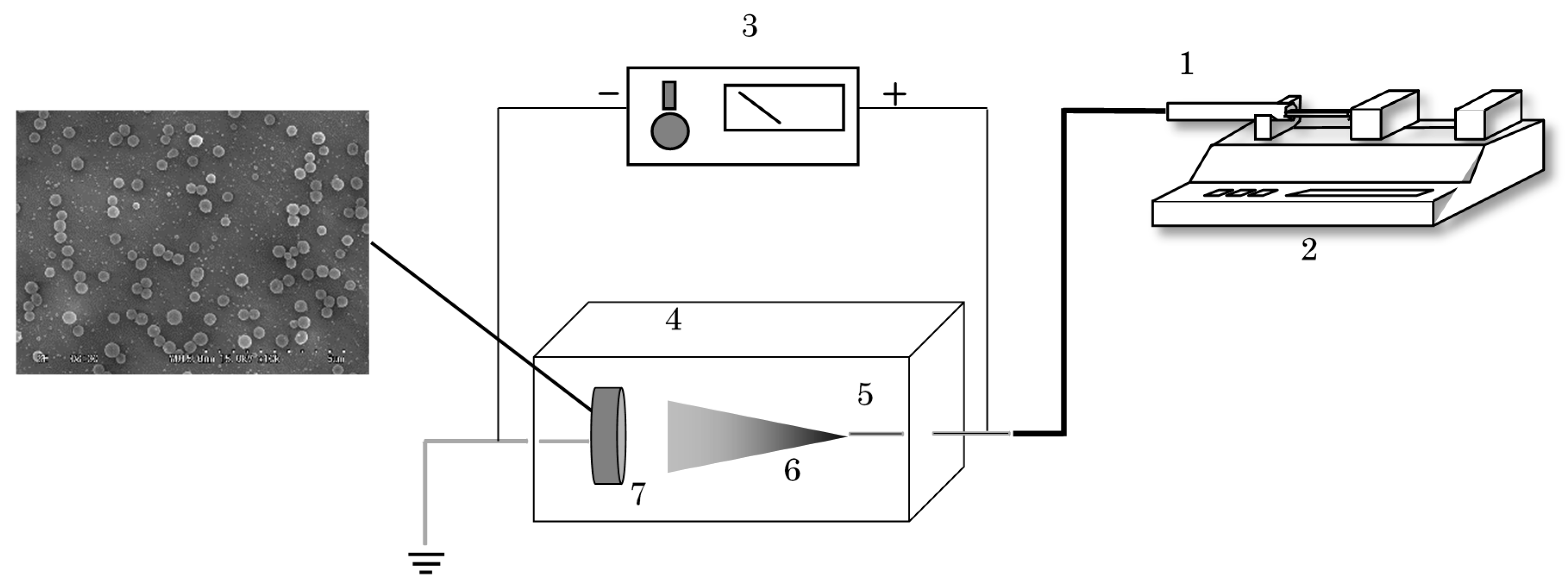
The scheme of the electrospraying apparatus used for the experiments is shown in Figure 1. It consists of a high voltage power supply (Matsusada Precision HARb–30P1, Nagoya, Japan), a high pressure syringe pump (Harvard Apparatus PHD–Ultra 4400, Holliston, MA, USA), a stainless steel syringe, a stainless steel tip, a collector covered with aluminum foil, and an enclosed acrylic chamber. First, mixtures of curcumin and PVP powder at different ratios of 1:10, 1:20, and 1:30 were prepared at concentrations of 0.01 and 0.4% in weight by dissolving them in ethanol to make feed solution. After the feed, solutions were loaded in a 8 mL stainless steel syringe; the syringe outlet was connected to the stainless steel tip (internal diameter, 0.5 mm) by a polyether ether ketone (PEEK) pipe. Before this process, the mixture of curcumin/PVP solution was stirred until completely dissolved. Then, the feed solution was injected to the collector by a high-pressure syringe pump at a constant flow rate of 0.01 mL/min and a distance of 5–15 cm between the stainless steel tip to collector, which was placed in the closed acrylic chamber. At the same time, the electrostatic force of 10–15 kV was supplied to the tip by a high voltage power source. Under this condition, the particles were dried during flying time on the way from the tip to the collector. This process was carried out at the ambient conditions. The dried particles were collected from the collector covered the aluminum foil and stored in the desiccator before further analysis.
2.3. Characterization of Products
Scanning electron microscopy (SEM; JEOL JSM–6390LV, Tokyo, Japan) was used to determine the morphology of particles. Before SEM analysis, the sample was placed in the aluminum holder and coated with gold at the low-pressure evaporator. The particle size and particle size distribution were evaluated using Image J software from SEM photograph for at least 250 particles collected at each experiment. The structure of the original curcumin, PVP, curcumin particles, and curcumin/PVP particles were analyzed using a Spectrum Two FTIR spectrophotometer (PerkinElmer Ltd., Buckinghamshire, England) with the wave number range 4000 to 400 cm−1. This FTIR was equipped by the standard optical system with KBr (potassium bromide) windows and a universal attenuated total reflectance (UATR) sampling accessory for a spectral data collection. Hence, the sample can be characterized and pressed directly between two KBr window plates.
2.4. Dissolution Study
The dissolution of the original curcumin, curcumin particles, and the curcumin/PVP particles were analysed using an UV-vis spectrophotometer (V–550, JASCO, Tokyo, Japan). An accurate amount of sample containing the equivalent of amount of curcumin for drug/polymer ratio was dissolved in 20 mL of distilled water at temperature of 37 °C. After 12 h, the mixer was filtered with 0.2 μm disposal membrane filter. Thereafter, the absorbance of the solution was measured by using UV-vis spectrophotometer at wavelength of 430 nm. The curcumin concentration (expressed as mg curcumin/L) was calculated based on standard calibration curved of curcumin in ethanol.
3. Results and Discussion
3.1. Micronization of Curcumin by Electrospraying
From the preliminary study (data is not shown), the suitable conditions for the production of curcumin particles by the electrospraying process was at contraction of 0.1%, electric voltage of 15 kV, and a tip-to-collector distance of 10 cm. The curcumin particles with irregular shape were formed in the electrospraying process using ethanol as an organic solvent. The SEM image of the original curcumin and the micronized curcumin particles by electrospraying process are shown in Figure 2. The original curcumin were irregular and needle-like with particle size ranging in length from 1.2 to 43 μm (Figure 2a). Figure 2b shows the SEM image of the particle size of curcumin particles obtained after the electrospraying micronization ranged in length from 20 to 499 nm, which was smaller than the original curcumin. In the electrospraying process, the polymer solution flowing out of the tip was subjected to an electric field. During the flight time of the droplet from the tip outlet to the collector, the solution is evaporated; this causes the droplet to shrink and the charge density to increase. The small droplets are produced from parent droplets when the charge density is near to the Rayleigh limited which is the magnitude of charge on drop overcomes the surface tension force. Electrospraying can produce micro or nanoparticles, depending on the liquid flow rate, electric conductivity and the properties of the liquid [14]. These results indicated that the micronization of curcumin particles was successfully prepared by electrospraying, using ethanol as an organic solvent.
3.2. Encapsulation of Curcumin with PVP by Electrospraying
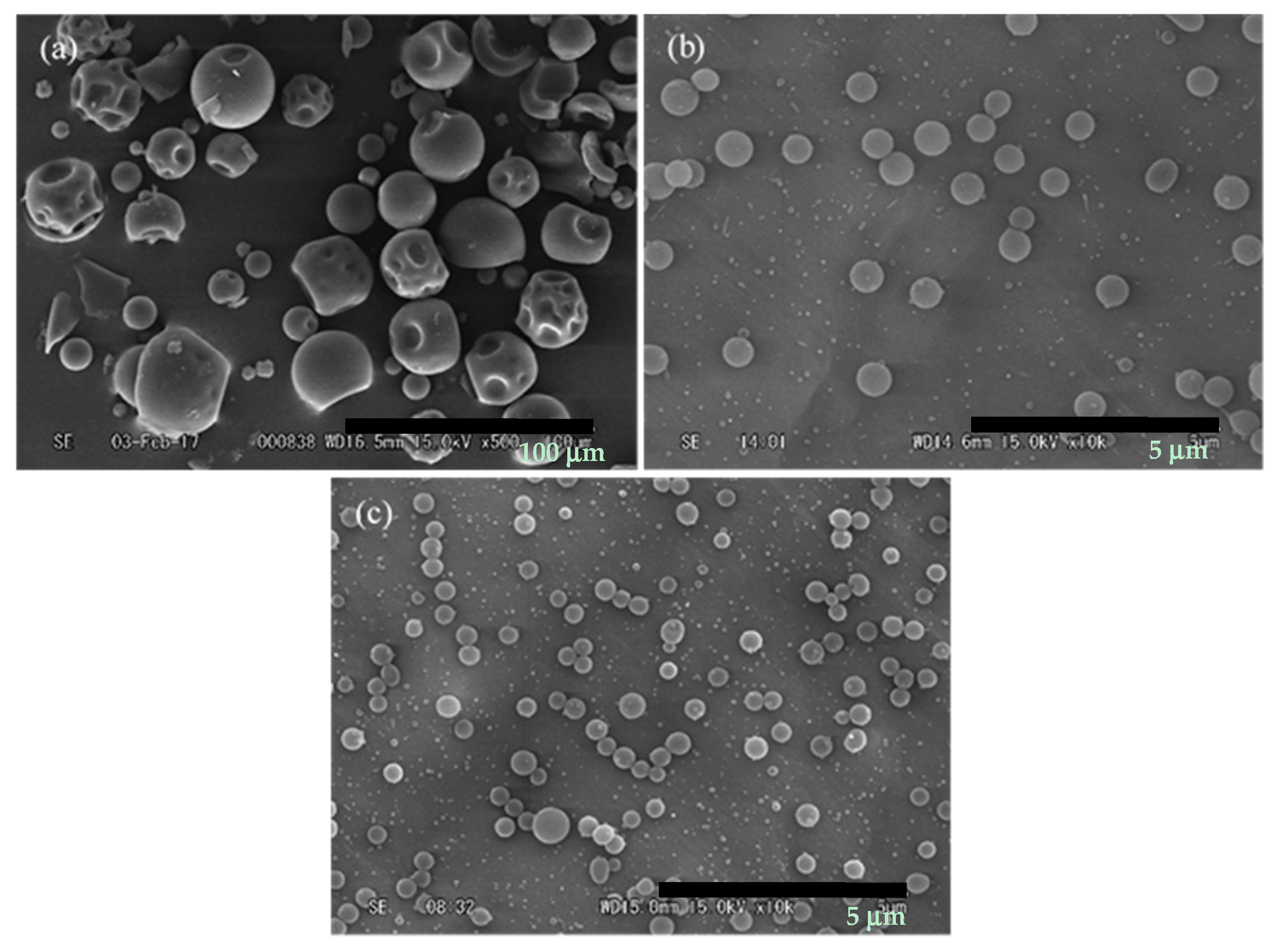
In this section, the curcumin was encapsulated in PVP by using electrospraying in order to compare the physic–chemical properties and solubility with curcumin particles. First, the PVP particles were generated by the electrospraying process using ethanol as an organic solvent at a concentration of 4%, electric voltage of 15 kV, and a tip-to-collector distance of 10 cm (Figure 3b). The result show that spherical PVP particles were obtained by the electrospraying process with particle size ranging from 199–980 nm, which was smaller than the original PVP particles (0.67–42.44 μm) (Figure 3a). When curcumin was added to the PVP solution and micronized by electrospraying under the same conditions, the curcumin/PVP particles with spherical shape were formed and the particle size ranged from 164 to 730 nm (Figure 3c). It is notable that the curcumin/PVP particles were smaller than PVP particles generated by electrospraying. This result could be possible, in that the presence of curcumin in polymer solution could increase the conductivity, resulting in the enhancement of the electrical drawing effect on the jet fluid and a reduction in particle size [18]. The following Section 3.3–Section 3.5 summarize the effect of curcumin/PVP ratio, electric voltage, and tip-to-collector distance on the morphology and particle size distribution of particles in order to find the optimal condition to produce these particles.
3.3. Effect of Curcumin/PVP Ratio
The effect of curcumin/PVP ratio on morphology and particle size distribution was performed at a polymer solution concentration of 0.4%, electric voltage of 10 kV, and a tip-to-collector distance of 15 cm at varying curcumin/PVP ratio of 1:10, 1:20, and 1:30, as shown in Figure 4. When curcumin/PVP ratios of 1:10 and 1:20 were processed, the particles were irregular, spherical and needle-like. However, when the curcumin/PVP ratio increased to 1:30, the spherical particles with particle size ranged from 164 to 730 nm. The irregular shapes of particles were formed in low curcumin/PVP ratio because the proportion of curcumin and PVP was not sufficient in the particle matrix to form spherical particles. Consequently, the irregular structures of particles were attained [18]. This is in agreement with the findings of Huang et al. [19]. They found that the Ketoprofen/Ethyl cellulose particle size was decreasing with increasing the drug/polymer ratio, due to a greater embedding of the drug into the polymer.
3.4. Effect of Tip to Collector Distance
Figure 5 shows the effect of tip-to-collector distance on the morphology and particle distribution at a curcumin/PVP ratio of 1:30, polymer solution concentration of 0.4%, and electric voltage of 10 kV. It is noted that the wet particles with aggregation were found at a low distance of 5 cm. At this working distance, the particle size ranged from 108 to 852 nm. However, when the distance increased to 10 cm, the spherical particles with particle size ranged from 239 to 899 nm. A similar result was obtained when the distance increased to 15 cm. However, the particle size obtained at 15 cm ranged from 164 to 730 nm, which is smaller than that obtained at 10 cm. This phenomenon can be explained by the fact that if the tip-to-collector distance is too short, the solvent might not have enough time to evaporate completely before reaching the collector, resulting in an aggregation of particles. On the other hand, high distance might lead to a reduction of the electric field strength but increase the evaporation time for the solvent. As a result, particles are lost to the surroundings when they travel to the collector. If a sufficient distance is applied in electrospraying, the solvent has adequate time to evaporate entirely; this leads to a decrease in the aggregation of particles and an increase in the collection efficiency [20,21]. In this study, the uniform curcumin/PVP particles with narrow size distribution were achieved at a long tip-to-collector distance of 15 cm. A similar result was obtained by Zhou et al. [21]. They observed that increasing the working distance enhanced the particle size uniformity and decreased the presence of large particles.
3.5. Effect of Electric Voltage
The effect of electric voltage on the morphology and particle size distribution was performed at a curcumin/PVP ratio of 1:30, a polymer solution concentration of 0.4%, and a tip-to-collector distance of 15 cm (Figure 6). The particle size distribution increased from 164–730 nm to 85–902 nm with increasing electric voltage from 10 to 15 kV. In theory, increased electric voltage increased the current through the liquid cone and this effect jet break–up mechanism. The stress ratio at the jet surface, which is given by the ratio of the normal electric stress to the surface tension stress, is the main factor to determine the mode of the electrified jet break up. Varicose instabilities are the jet break up due to azisymmetric instabilities at a low stress ratio value. In this case, a monodisperse droplet was generated with a small number of secondary droplets. However, when the high voltage was applied it increased the current through the jet of liquid and enhanced the surface charge and stress ratio. The jet begins to whip and lateral instabilities contribute to the break-up of the jet above a stress ratio threshold value. As a result, the secondary droplets were increased [9]. From the foregoing discussion of results, it could be concluded that the suitable conditions to produce the curcumin/PVP particles was at a curcumin/PVP ratio of 1:30, polymer solution concentration of 0.4%, electric voltage of 10 kV, and a tip-to-collector distance of 15 cm. The particle size obtained in this condition will be used for analyzing chemical structure and solubility.
3.6. FTIR Analysis
The FTIR analysis of original curcumin, PVP, curcumin particles, curcumin/PVP particles is shown in Figure 7. It has been shown that the FTIR spectra of original curcumin showed the presence of the following peaks: 3529 cm−1 (O–H stretching vibration), 1620 cm−1 (C=C benzene stretching ring), 1511 cm−1 (C=O stretching), 1435 cm–1 (C–H bending), 1292 cm−1 (C–O stretching), 1030 cm−1 (C–O–C stretching vibration), 857 cm−1 (C–H aromatic hydrogen) and the FTIR of original PVP showed characteristic peak at 1667 cm−1 (C=O stretching vibration), 2968 cm−1 (C–H stretching), and 3469 cm−1 (O–H bending). The absorption peak of the original curcumin and curcumin particles prepared by electrospraying had similar wavenumbers, which indicated the structure of curcumin was not changed after the electrospraying process. However, the FTIR spectra of curcumin/PVP particles was completely different from FTIR spectra of the original curcumin, but similar to the FTIR structure of the original PVP. This implied that the curcumin was captured inside the PVP particles. It is also notable that the intensity of FTIR spectra of curcumin/PVP particles is lower than that of the original PVP. This can be probably due to the interaction of O–H absorption of curcumin and the C=O absorption band of PVP occurred when curcumin encapsulated in PVP particles by using electrospraying [22]. Yen et al. [22] developed a novel curcumin nanoparticles system by using a simple precipitation method with PVP as the hydrophilic carrier matrix. They informed that the absorption band related to C=O stretching vibration in the PVP matrix was shifted to a lower wavenumber due to the interaction of the O–H bond of curcumin and the C=O bond of PVP. Next, they concluded that this interaction might enhance the water solubility and the drug release of curcumin and improve its physicochemical properties.
3.7. Dissolution
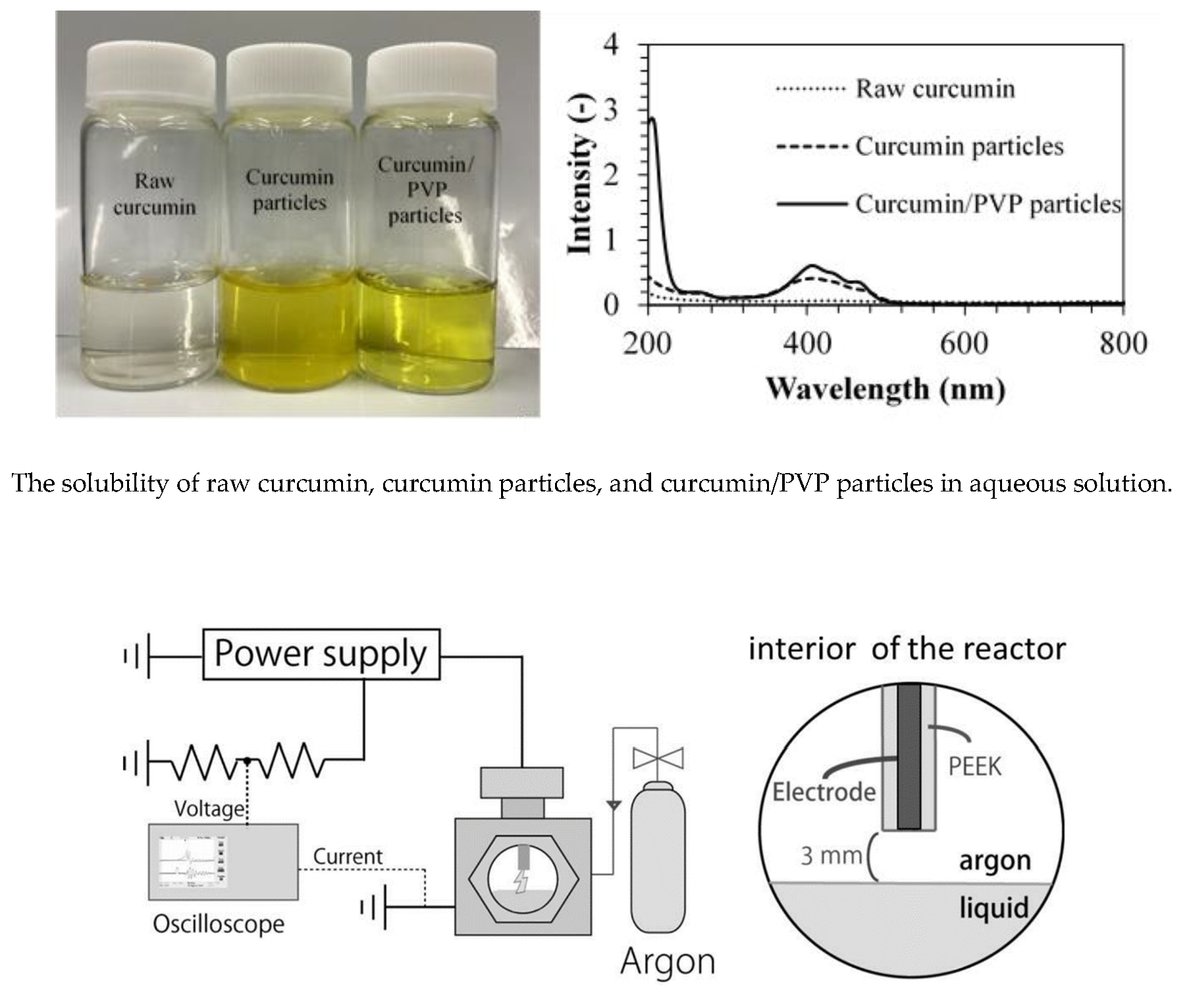
The solubility of the original curcumin, curcumin particles and curcumin/PVP particles obtained by electrospraying was determined and the results are shown in Figure 8. Curcumin dissolved in an aqueous solution after 12 h; the result indicated that the aqueous solubility of curcumin was 0.09 mg/mL. The curcumin processed by electrospraying exhibited a 12.5 times increase (1.13 mg/mL) in aqueous solubility, compared with the original curcumin. Moreover, the curcumin/PVP produced by electrospraying could be enhanced by about 19 times (1.68 mg/mL) compared to raw curcumin. It could be concluded that curcumin/PVP particles had higher aqueous solubility compared to raw curcumin and curcumin particles processed by electrospraying. This result is supported by the fact that curcumin is a hydrophobic phenolic compound; thus it has low solubility in water [3]. Micronization of curcumin by electrospraying could enhance the solubility of curcumin in aqueous solution because electrospraying could reduce the particle size of curcumin by using the electric field. Encapsulation of curcumin in PVP particles had better solubility of curcumin in water than raw curcumin or curcumin particle processed by electrospraying because of a number of factors. One is that PVP was used with curcumin to produce particles, because PVP is a hydrophilic polymer and it has high solubility in aqueous solution. Therefore, when curcumin was combined with PVP, the curcumin structure might have been modified by the presence of PVP, thereby causing an increase in the aqueous solubility of curcumin. Moreover, electrospraying could generate small particles with narrow size distribution, resulting in enhancement of curcumin in aqueous solution [23].
4. Conclusions
The curcumin/PVP particles were successfully produced by the electrospraying process. The morphology and particle size distribution depended on the operational parameters. Increasing the curcumin/PVP ratio to 1:30 may give small particle size with spherical morphology. The higher voltage than 10 kV increased the particle size distribution due to the instability of the current while the long tip-to-collector distance could reduce the particle size. The spherical particles with particle size distribution of 164 to 730 nm obtained from a curcumin/PVP ratio of 1:30, polymer solution concentration of 0.4%, electric voltage of 10 kV, and a tip-to-collector distance of 15 cm was greater than that of curcumin processed by electrospraying. However, the aqueous solubility of curcumin/PVP particles was higher than that of raw curcumin and curcumin particles generated by electrospraying. Thus, it could be concluded that the electrospraying technique enables to encapsulate curcumin in PVP particles with high aqueous solubility of curcumin.
Author Contributions
K.C. and W.D. designed and performed the experiments, analyzed the data and wrote the paper. H.K. and M.G. advised the project.
Funding
This research received no external funding.
Acknowledgments
This research is supported by ASEAN University Network for Southeast Asia Engineering Education Development Network (AUN/SEED–Net) project through the Japan International Cooperation Agency (JICA) and the Precursory Research for Embryonic Science and Technology Program of the Japan Science and Technology Agency (JST).
Conflicts of Interest
The authors declare no conflict of interest.
References
- Moghadamtousi, S.Z.; Kadir, H.A.; Hassandarvish, P.; Tajik, H.; Abubakar, S.; Zandi, K. A review on antibacterial, antiviral, and antifungal activity of curcumin. BioMed Res. Int. 2014. [Google Scholar] [CrossRef]
- Xie, M.; Fan, D.; Zhao, Z.; Li, Z.; Li, G.; Chen, Y.; He, X.; Chen, A.; Li, J.; Lin, X.; et al. Nano–curcumin prepared via supercritical: Improve anti–bacterial, anti–oxidant and anti–cancer efficacy. Int. J. Pharm. 2015, 496, 732–740. [Google Scholar] [CrossRef] [PubMed]
- Kimthet, C.; Diono, W.; Kanda, H.; Goto, M. Extraction of curcumin from Curcuma longa L. using ultrasound assisted supercritical carbon dioxide. AIP Conf. Proc. 2017, 1840, 100001. [Google Scholar] [CrossRef]
- Anand, P.; Kunnumakkara, A.B.; Newman, R.A.; Aggarwal, B.B. Bioavailability of curcumin: Problems and promise. Mol. Pharm. 2007, 4, 807–818. [Google Scholar] [CrossRef]
- Chhouk, K.; Diono, W.; Kanda, H.; Kawasaki, S.; Goto, M. Micronization of curcumin with biodegradable polymer by supercritical anti-solvent using micro swirl mixer. Front. Chem. Sci. Eng. 2018, 12, 184–193. [Google Scholar] [CrossRef]
- Rasekh, M.; Karavasili, C.; Soong, Y.L.; Bouropoulos, N.; Morries, M.; Armitage, D.; Li, X.; Fatourous, D.G.; Ahmad, Z. Electrospun PVP-indomethacin constituents for transdermal dressings and drug delivery devices. Int. J. Pharm. 2014, 473, 95–104. [Google Scholar] [CrossRef]
- Prosapio, V.; Marco, I.D.; Scognamiglio, M.; Reverchon, E. Folic acid-PVP nanostructured composite microparticles by supercritical antisolvent precipitation. Chem. Eng. J. 2015, 277, 286–294. [Google Scholar] [CrossRef]
- Paximada, P.; Echegoyen, Y.; Koutinas, A.A.; Mandala, L.G.; Lagaron, J.M. Encapsulation of hydrophilic and lipophilized catechin into nanoparticles through emulsion electrospraying. Food Hydrocoll. 2017, 64, 123–132. [Google Scholar] [CrossRef]
- Gomez-Estaca, J.; Balaguer, M.P.; Gavara, R.; Hernandez-Muooz, P. Formation of zein nanoparticles by electrohydrodynamic atomization: Effect of the main processing variables and suitability for encapsulating the food coloring and active ingredient curcumin. Food Hydrocoll. 2012, 28, 82–91. [Google Scholar] [CrossRef]
- Gomez-Estaca, J.; Gavara, R.; Hernandez-Muooz, P. Encapsulation of curcumin in electrosprayed gelatin microspheres enhances its bioaccessibility and widens its uses in food applications. IFSET 2015, 29, 302–307. [Google Scholar] [CrossRef]
- Yousaf, A.M.; Mustapha, O.; Kim, D.W.; Kim, D.S.; Kim, K.S.; Jin, S.G.; Yong, C.S.; Youn, Y.S.; Oh, Y.; Kim, J.O.; et al. Novel electrosprayed nanospherule for enhanced aqueous solubility and oral bioavailability of poorly water-soluble fenofibrate. Int. J. Nanomed. 2016, 11, 213–221. [Google Scholar] [CrossRef]
- Gaona-Sanchez, V.A.; Calderon-Dominguez, G.; Morales-Sanchez, E.; Chanona-Perez, J.J.; Arzate-Vazquez, I.; Terres-Rojas, E. Pectin-based films produced by electrospraying. J. Appl. Polym. Sci. 2016, 43779. [Google Scholar] [CrossRef]
- Yao, Z.; Jin, L.; Ahmad, Z.; Huang, J.; Chang, L.J. Ganoderma lucidum polysaccharide loaded sodium alginate micro-particles prepared via electrospraying in controlled deposition environments. Int. J. Pharm. 2017, 524, 148–158. [Google Scholar] [CrossRef] [PubMed]
- He, Y.; Huang, Y.; Wang, W.; Cheng, Y. Integrating micromixer precipitation and electrospray drying toward the continuous production of drug nanoparticles. Chem. Eng. J. 2011, 168, 931–937. [Google Scholar] [CrossRef]
- Yuan, S.; Lei, F.; Liu, Z.; Tong, Q.; Si, T.; Xu, R.X. Coaxial electrospray of curcumin–loaded microparticles for sustained drug release. PLoS ONE 2015, 10, e0132609. [Google Scholar] [CrossRef] [PubMed]
- Mai, Z.; Chen, J.; He, T.; Hu, Y.; Dong, X.; Zhang, H.; Huang, W.; Ko, F.; Zhou, W. Electrospray biodegradable microcapsules loaded with curcumin for drug delivery system with high bioactivity. RSC Adv. 2017, 7, 1724–1734. [Google Scholar] [CrossRef]
- Schneider, C.A.; Rasband, W.S.; Eliceiri, K.W. NIH Image to ImageJ: 25 years of image analysis. Nat. Methods 2012, 9, 671–675. [Google Scholar] [CrossRef] [Green Version]
- Wang, C.; Ma, C.; Wu, Z.; Liang, H.; Yan, P.; Song, J.; Ma, N.; Zhao, Q. Enhanced bioavailability and anticancer effect of curcumin–loaded electrospun nanofiber: In vitro and in vivo study. Nanoscale Res. Lett. 2015, 10, 439. [Google Scholar] [CrossRef]
- Huang, L.Y.; Yu, D.G.; Brandfor-White, C.; Zhu, L.M. Sustained release of ethyl cellulose micro-particulate drug delivery systems prepared using electrospraying. J. Mater. Sci. 2012, 47, 1372–1377. [Google Scholar] [CrossRef]
- Songsurang, K.; Praphairaksit, N.; Siraleartmukul, K.; Muangsin, N. Electrospray fabrication of doxorubicin-Chitosan-Tripolyphosphate nanoparticles for delivery of doxorubicin. Arch. Pharmacal Res. 2011, 34, 583–592. [Google Scholar] [CrossRef]
- Zhou, F.L.; Cristinacce, P.L.H.; Eichhorn, S.J.; Parker, G.J.M. Preparation and characterization of polycaprolactone microspheres by electrospraying. Aerosol Sci. Technol. 2016, l50, 1201–1215. [Google Scholar] [CrossRef] [PubMed]
- Yen, F.L.; Wu, T.; Tzeng, C.W.; Lin, L.; Lin, C. Curcumin nanoparticle improve the physicochemical properties of curcumin and effectively enhance its antioxidant and antithepatoma activities. J. Agric. Food Chem. 2010, 58, 7376–7382. [Google Scholar] [CrossRef] [PubMed]
- Perrut, M.; Jung, J.; Leboeuf, F. Enhancement of dissolution rate of poorly–soluble active ingredients by supercritical fluid processes: Part 1: Micronization of neat particles. Int. J. Pharm. 2005, 288, 3–10. [Google Scholar] [CrossRef] [PubMed]
Figure 1.
Schematic diagram of micronization by electrospraying. (1: Polymer solution; 2: Syringe pump; 3: High voltage power source; 4: Acrylic chamber; 5: Tip; 6: Polymer particles; 7: Collector).
Figure 1.
Schematic diagram of micronization by electrospraying. (1: Polymer solution; 2: Syringe pump; 3: High voltage power source; 4: Acrylic chamber; 5: Tip; 6: Polymer particles; 7: Collector).

Figure 2.
SEM image of (a) original curcumin and (b) curcumin particles generated by electrospraying.
Figure 2.
SEM image of (a) original curcumin and (b) curcumin particles generated by electrospraying.

Figure 3.
SEM image of (a) raw PVP, (b) PVP particles, and (c) curcumin/PVP particles.

Figure 4.
The effect of (a) curcumin/PVP ratio; (b) tip-to-collector distance; and (c) electric voltage on the particle size distribution.
Figure 4.
The effect of (a) curcumin/PVP ratio; (b) tip-to-collector distance; and (c) electric voltage on the particle size distribution.

Figure 5.
SEM image of curcumin/PVP particles at (a) 5 cm; (b) 10 cm, and (c) 15 cm.

Figure 6.
SEM image of curcumin/PVP particles at (a) 10 kV; (b) 12 kV, and (c) 15 kV.

Figure 7.
FTIR spectra of raw curcumin, PVP, curcumin particles, and curcumin/PVP particles.

Figure 8.
The solubility of raw curcumin, curcumin particles, and curcumin/PVP particles.

© 2018 by the authors. Licensee MDPI, Basel, Switzerland. This article is an open access article distributed under the terms and conditions of the Creative Commons Attribution (CC BY) license (http://creativecommons.org/licenses/by/4.0/).
Share and Cite
MDPI and ACS Style
Chhouk, K.; Diono, W.; Kanda, H.; Goto, M. Micronization for Enhancement of Curcumin Dissolution via Electrospraying Technique. ChemEngineering 2018, 2, 60. https://doi.org/10.3390/chemengineering2040060
AMA Style
Chhouk K, Diono W, Kanda H, Goto M. Micronization for Enhancement of Curcumin Dissolution via Electrospraying Technique. ChemEngineering. 2018; 2(4):60. https://doi.org/10.3390/chemengineering2040060
Chicago/Turabian StyleChhouk, Kimthet, Wahyu Diono, Hideki Kanda, and Motonobu Goto. 2018. "Micronization for Enhancement of Curcumin Dissolution via Electrospraying Technique" ChemEngineering 2, no. 4: 60. https://doi.org/10.3390/chemengineering2040060