Article

Characterization and Application of a Disposable Rotating Bed Bioreactor for Mesenchymal Stem Cell Expansion

1
Department for Biotechnology, University of Natural Resources and Life Sciences, Muthgasse 18, 1190 Vienna, Austria
2
Institute for Technical Chemistry, Gottfried Wilhelm Leibniz University Hanover, Callinstrasse 5, 30167 Hanover, Germany
*
Author to whom correspondence should be addressed.
These authors contributed equally to this work.
Bioengineering 2014, 1(4), 231-245; https://doi.org/10.3390/bioengineering1040231
Submission received: 11 September 2014 / Revised: 18 November 2014 / Accepted: 25 November 2014 / Published: 27 November 2014
(This article belongs to the Special Issue Biopharmaceutical Process Development)

Abstract

:
Recruitment of mesenchymal stromal cells (MSC) into the field of tissue engineering is a promising development since these cells can be expanded ex vivo to clinically relevant numbers and, after expansion, retain their ability to differentiate into various cell lineages. Safety requirements and the necessity to obtain high cell numbers without frequent subcultivation of cells raised the question of the possibility of expanding MSC in one-way (single-use) disposable bioreactors. In this study, umbilical cord-derived MSC (UC-MSC) were expanded in a disposable Z®RP 2000 H bioreactor under dynamic conditions. Z®RP was characterized regarding residence time and mixing in order to evaluate the optimal bioreactor settings, enabling optimal mass transfer in the absence of shear stress, allowing an reproducible expansion of MSC, while maintaining their stemness properties. Culture of the UC-MSC in disposable Z®RP 2000 H bioreactor resulted in a reproducible 8-fold increase of cell numbers after 5 days. Cells were shown to maintain specific MSC surface marker expression as well as trilineage differentiation potential and lack stress-induced premature senescence.

Graphical Abstract

1. Introduction

Almost four hundred clinical trials [1] recruiting mesenchymal stem cells (MSC) for the treatment of several diseases such as spinal cord injuries, burns, liver cirrhosis or failure, graft versus host disease, diabetes mellitus, progressive multiple sclerosis and cardiac ischemia are being carried out at the moment. For each treatment, millions of cells are required and optimal expansion methods can help in obtaining MSC of good quality within a short period of time, enhancing chances for treatment optimization and patient survival. The expansion of MSC in bioreactors is a promising development for the future application of MSC in cell-based therapies and tissue engineering. There are numerous types of bioreactors used to expand adherent anchorage-dependent cells including MSC (e.g., WAVE-bioreactor, GE Healthcare, PluriXTM 3D bioreactor, Pluristem and Quantum bioreactor, TerumoBCT) [2,3,4,5]. Dynamic cultivation conditions have several advantages in comparison to static cell expansion. First of all, cultivation in a closed system without the need to manually change the culture medium in a clean bench significantly reduces the risk of possible contamination; Second, the control of all parameters of culture together with continuous documentation using IT systems makes it possible to assure the quality of cells and to avoid human errors; Third, dynamic cultivation systems like bioreactors provide active mass transfer, supplying MSC with gases and nutrients while removing toxic metabolites; Last but not least, there is the argument of production costs - which can be a major obstacle to the spread of MSC clinical applications [6,7,8]. MSC are anchorage-dependant cells, which cannot be expanded in suspension culture. Several techniques including cultivation on microcarriers, three-dimensional matrix and conventional cell culture plastic surfaces (cell factories) were developed over the past few years. These techniques may allow obtaining the necessary cell numbers without manual cultivation of MSC, reducing the risk of bacterial and fungal contamination as well as cross-contamination. Additionally, continuous on-line monitoring and control of important cultivation parameters like pH, temperature, oxygen, CO 2 and metabolite concentrations help to provide the optimal growth conditions for the cells and assure reproducible cell expansion [9,10,11]. Many publications have been issued on that subject reporting a large number of reactor modifications, a wide variety of tissue function as well as biomaterial properties. Bioreactor systems target a reliable and efficient procedure, enabling highly reproducible, automated and controlled process compatible with good manufacturing practice (GMP) requirements [7,12,13]. A new approach in the expansion of adherent MSC is provided by Zellwerk GmbH (Oberkraemer, Germany), where cells are cultivated on rotating stalked polycarbonate cell carrier slides (Figure 1). The Z®RP 2000 H bioreactor is connected to the pH and pO 2 sensor electrodes, which are integrated into the tubing system. Besides cell culture medium mixing via rotation, medium is also constantly circulating via a tubing system, where feed- and waste-flasks are connected, making it possible to culture cells in fed-batch or perfusion modus. Disposable Z®RP 2000 H bioreactor provides a surface of 2000 cm 2 for cell growth, while Z®RP 8000 H offers 8000 cm 2 . Cultivation of cells is performed in a GMP-breeder under fully automated control with documentation. According to the manufacturers data, MSC in a Z®RP 8000 H system can be expanded in 9 days to 400 million cells [14].
Figure 1. Z®RP 2000 H (Zellwerk GmbH, Oberkraemer) bioreactor system with breeder and control tower (a) and disposable bioreactor (b) set up and (c) rotating bed consisted of polycarbonate cell carrier slides (total surface area 2000 cm 2 , reactor size 8.8 × 4.6 cm) (drawings adopted and modified from manufacturers manual and [14]).
Bioengineering 01 00231 g001
Overcoming nutrient limitation is an important factor in all existing bioreactor systems. In vivo cells benefit from blood vessels being located in most tissues within a distance of 100–200 µm from capillaries. Furthermore, MSC are suggested to be associated with blood vessels in vivo [15,16], and consequently are not subjected to major nutrient limitation in their niche. Under conventional, static culture conditions mass transport is controlled by diffusion, being insufficient for high cell densities and high medium volumes [13]. On the other hand, fluid flow induces shear stress to which MSC are very sensitive, responding with biochemical cascades resulting in phenotype chances [17]. Thus, mechanical stimuli play a crucial role in differentiation of MSC, often targeted when bone or cartilage tissue constructs for implantation need to be obtained. On the contrary, these are undesired effects in MSC expansion when undifferentiated cells are requested. It is therefore important to estimate mixing profiles within the bioreactor and possible resulting shear stress prior to bioreactor approval. This work was aimed to characterize the ZRP 2000 H bioreactor as a first step towards comparability and a future scale-up system for MSC expansion in vitro.
We analyzed residence time distribution under different operation conditions, obtaining the Bodenstein number as a dimensional parameter, describing mixing properties within the system. Umbilical cord derived MSC (UC-MSC) were expanded over 5 days without subcultivation in the Z®RP 2000 H bioreactor, whereafter the yield and quality of the expanded cells were studied.

2. Experimental Section
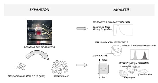
2.1. Z®RP Platform

The Z®RP platform consists of the bioreactor, a GMP-conform breeder and an external control unit. The Z®RP 2000 H bioreactor (Figure 1c) is a disposable rotating bed bioreactor that is working in perfusion mode. The core of the bioreactor is the rotating bed, which consists of polycarbonate cell carrier slides with total surface area 2000 cm 2 . The polycarbonate slides are fixed to a vertical shaft, which is rotated by a non-contact magnetic drive. During culture, bioreactor was placed in a GMP conform breeder. The breeder was thermostated and working as a laminar flow hood allowing manipulations of the bioreactor in a sterile working environment. All process parameters including feeding rate, gassing and rotational speed were monitored and regulated by an external control unit. The temperature within the bioreactor and the breeder as well as the pH-value and oxygen content were controlled by sensor electrodes. pH-value was regulated by overlay gassing with carbon dioxide or air.

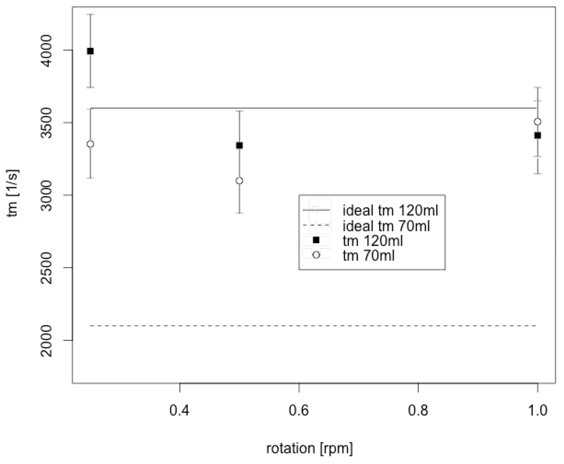
2.2. Residence Time Distribution

Residence time distribution was determined using an approximation of Dirac pulse. The Dirac pulse was approximated by adding the tracer substance methylene blue into the bioreactor in a time frame of 5 s, which results in a slope of about 2 × E(θ). The tracer substance was injected into the bioreactor through an upper injection port. At the beginning of the Dirac pulse the reactor bed disks were oriented horizontally to simulate conditions for residence time distribution like in cell expansion experiments. When starting reactor bed rotation during cell expansion, reactor bed disks were also oriented horizontally, due to cell seeding. Dirac pulse was performed with 200 µL methylene blue (15 mg/mL) for a filled and with 120 µL methylene blue for a half-filled bioreactor, respectively. During the measurement H 2 O was pumped through the bioreactor with a pump rate of 2 mL/min utilizing a peristaltic pump. The concentration gradient of the tracer was measured photometrically at 664 nm. Residence time distribution was determined for different reactor volumes (120 mL, 70 mL) and for different rotation speeds of the bioreactor bed (0.25 rotations per minute (rpm), 0.5 rpm, 1 rpm).

2.3. Cumulative Residence Time Function

Cumulative residence time function was determined performing a displacement measurement. The bioreactor was filled with H 2 O and the tracer substance methylene blue (25 µg/L) was pumped continuously through the bioreactor with a pump rate of 2 mL/min. The concentration gradient of the tracer was measured photometrically at 664 nm. Residence time distribution was determined for different reactor volumes (120 mL, 70 mL) and for different rotation speeds of the bioreactor bed (0.25 rpm, 0.5 rpm, 1 rpm).

2.4. Cell Culture

Human MSC were isolated from the umbilical cords of three different term-deliveries (38–40 weeks) by Cesarean section. All patients delivered their informed consent, as approved by the Institutional Review Board, project #3037 on 17 June 2006 and in an extended permission #443 on 26 February 2009. The isolated populations were extensively characterized as mesenchymal stem cells by surface marker analysis and functional properties [18]. MSC were expanded and cryopreserved until the start of the experiment as described. After thawing, the cells were expanded over two passages. Experiments were performed with cells of passages 3 to 7. Cells were cultivated in αMEM containing 1 g/L glucose (Biochrom, Berlin, Germany), 10% human serum (provided by the Division of Transfusion Medicine, Medical University Hanover, Hanover, Germany) and 50 µg/ml gentamicin (PAA Laboratories GmbH, Pasching, Germany) in a humidified atmosphere containing 5% CO 2 and 21% O 2 at 37 ° C (Incubators: Thermo scientific, Hanau, Germany).

2.5. Expansion of UC-MSC in the Z®RP 2000 H Bioreactor

For the expansion in the Z®RP 2000 H bioreactor, frozen UC-MSC were first revitalized and subsequently cultured over two passages in αMEM (Biochrom, Germany) supplemented with 10% human serum (Division of Transfusion Medicine, Medical University Hanover, Germany) and 0.5% gentamycin (PAA Laboratories GmbH, Pasching, Germany) in T-flasks and then seeded at cell density 1500 cells/cm 2 (total cell number 3 × 10 6 cells; 1.2 × 10 4 cells/mL). Cells were seeded on both sides of polycarbonate cell carrier slides and incubated for 24 h for cell attachment on each side, respectively. For each seeding and attachment period, the rotation bed was stopped resulting in a horizontal position of the carrier slides. Afterwards, the bioreactor was filled with cell suspension (125 mL αMEM supplemented with 10% human serum and 0.5% gentamicin, 37 ° C ). After attachment of the cells on both sides of the slides for 24 h, respectively, bed rotation and medium circulation were started. Medium circulation was set at 0.1 mL/min, bed rotation at 0.1 rpm. Feeding rate with fresh cell culture medium was set to the intermitted mode (starting with 2%) and increased each day depending on glucose consumption and lactate production. Breeder temperature was set at 37 ° C . The overlay gas mixture consisted of 95% air and 5% CO 2 . In total, three cultivations under the same conditions were performed. Cell culture media from the bioreactor was sampled once a day with the help of a syringe via a septum installed on the bioreactor.

2.6. Cell Number, Population Doublings and Doubling Time

After 5 days of expansion, cells were harvested by treatment with accutase (PAA Laboratories GmbH). First of all, the cell culture medium was removed from the bioreactor with the help of the pump, then the bioreactor was filled with warm (37 ° C ) PBS, bed rotation was switched on and cells were washed for 5 min. After washing, PBS was removed, the bioreactor was filled with 80 mL accutase (37 ° C ), and bed rotation was switched on for the next 20 min. After incubation with accutase, the harvested cells were collected in falcon-tubes and centrifuged for 15 min at 300× g. Cell pellets were resuspended in fresh αMEM and cell numbers were estimated by counting in a haemocytometer. Population doublings were calculated according to:
N d = l n ( x x 0 ) l n 2
where N d is the number of population doublings during a Δ t period of time, x 0 is the number of living cells at time t = 0 , and x is the number of living cells at time t (end of cultivation). Population doubling time T d was calculated according to:
T d = Δ t N d

2.7. Cell Differentiation

After expansion in the Z®RP 2000 H bioreactor, UC-MSC were seeded on two-dimensional fibronectin surfaces (Corning, Berlin, Germany) with cell densities of about 3000 cell/cm 2 . Subsequently, UC-MSC were cultivated in DMEM containing 1 g/L glucose (Sigma-Aldrich, Munich, Germany), 10% human serum (provided by the Division of Transfusion Medicine, Medical University Hannover, Germany) and 50 µg/mL gentamicin (PAA Laboratories GmbH, Pasching, Germany) for 4 days. Afterwards UC-MSC were cultivated for three weeks in NH AdipoDiff Medium, NH ChondroDiff Medium and NH OsteoDiff Medium (Miltenyi Biotech, Bergisch Gladbach, Germany), supplemented with 50 µg/mL gentamicin. Medium was changed every second day.

2.8. Histological Stainings

Cell fixation: Cells were fixed with 600 µL paraformaldehyde (4% in PBS) for 30 min at 4 ° C . BODIPY staining: The fixed cell layer was covered with 300 µL of BODIPY solution (5 µM in PBS) (Invitrogen, Darmstadt, Germany). Samples were incubated for 5 min in the dark and thereafter staining solution removed. The fluorescence was detected at an excitation wavelength of 485 nm and an emission wavelength of 535 nm. Alcian Blue staining: The fixed cell layer was incubated for 3 min in 700 µL acidic acid (3%) and subsequently covered with 700 µL Alcianblue 8G solution (Sigma Aldrich, Munich, Germany). Samples were incubated for 30 min at room temperature and thereafter washed with acidic acid (3%). Pictures were taken at a light microscope (Olympus, Tokyo, Japan). Calcein staining: The fixed cell layer was covered with 500 µL of calcein solution (5 µg/mL in ddH 2 O) (Sigma Aldrich, Munich, Germany). Samples were incubated overnight at 4 ° C and thereafter washed extensively with distilled water. The fluorescence of the bounded calcein was detected at an excitation wavelength of 485 nm and an emission wavelength of 535 nm.

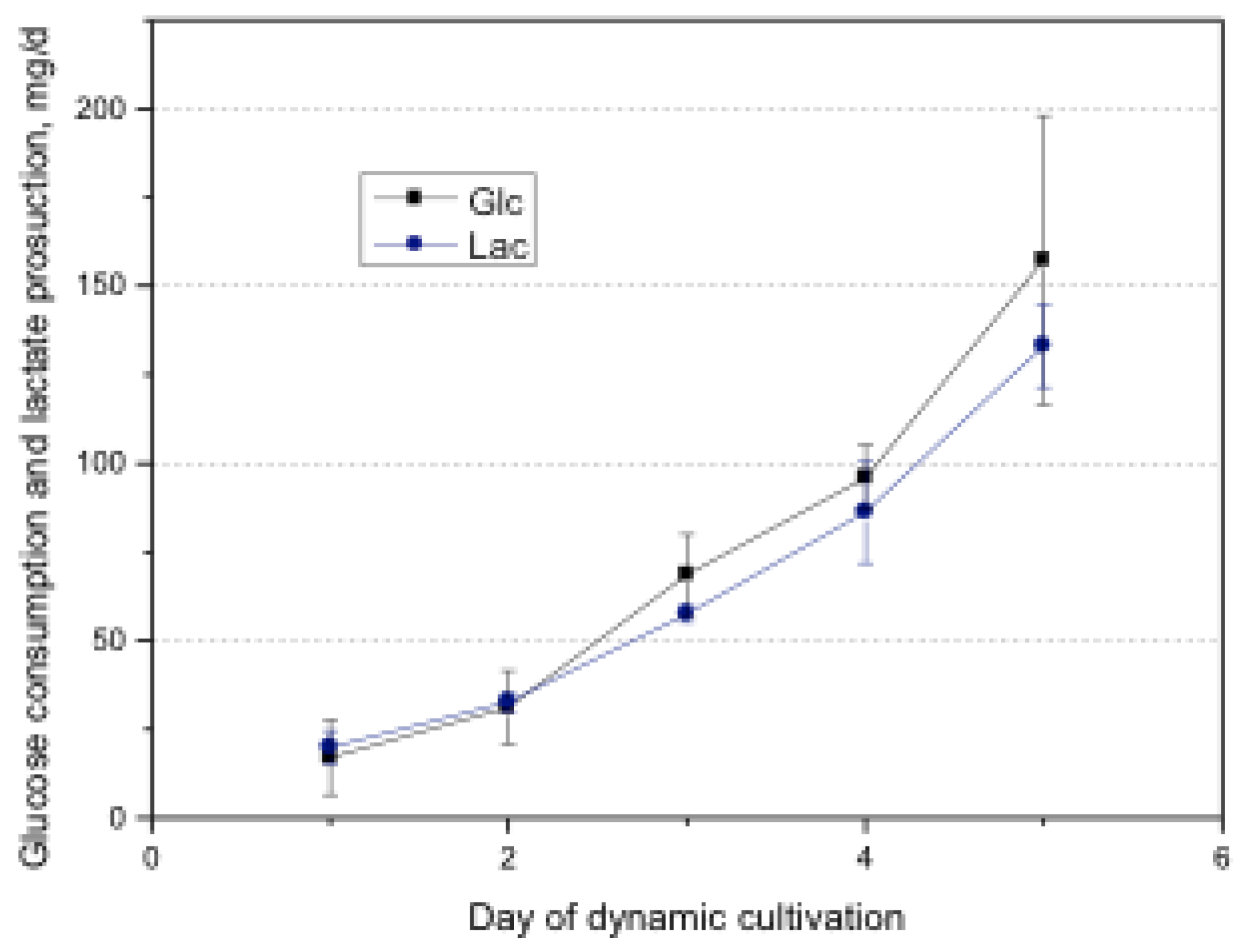
2.9. Glucose and Consumption and Lactate Production

The concentration of glucose and lactate in the medium was measured using an YSI 2700 SELECT analyzer (YSI Inc., Yellow Springs, OH, USA).

2.10. Senescence-Associated β-Galactosidase Staining

Cell senescence was estimated with the use of the Senescence β-Galactosidase Staining Kit (Cell Signaling Technology, Danvers, MA, USA) in accordance to the manufacturer’s instructions. For the staining, harvested cells were seeded at a density of 6000 cells/cm 2 for 48 h, then washed with PBS, fixed with the fixation solution from the kit. Senescence-associated β-galactosidase (SA-β-gal) staining was performed overnight at 37 ° C . Counterstaining was performed with DAPI (Roth GmbH & Co. KG, Karlsruhe, Germany).

2.11. Flow Cytometric Analysis of Surface Antigen Expression

UC-MSC were harvested by accutase treatment, washed twice in a cold blocking buffer and resuspended to a concentration of 10 6 cells per mL. Specific antibody-staining was performed by adding 20 µL of prediluted staining solution (BD Biosciences, Heidelberg, Germany) to 100 µL of cell suspension as described earlier [19]. Cells stained with matched isotype control antibodies served as a negative control. After 20 min incubation at room temperature in the dark, 400 µL of blocking buffer were added and cells were analyzed in an EPICS XL/MCL flow cytometer (Beckman Coulter, Krefeld, Germany). At least, 10,000 gated events were acquired on a LOG fluorescence scale. Generated data were analyzed using the program WinMDI 2.8.

3. Results and Discussion

3.1. Residence Time

Residence time is an important parameter, providing information about homogeneity of the liquid phase within the bioreactor. Residence time distribution (RTD) as well as average residence time were therefore considered. We characterized the RTD in the reactor by conducting a series of washout experiments (Dirac pulse experiments). We considered grade of filling of the bioreactor with liquid as well as rotation speed of the reactor bed to have an influence on the residence time and backmixing within the bioreactor. We therefore achieved characterization of a half-filled reactor (70 mL) and filled bioreactor (120 mL, which is nearly the maximum volume for Z®RP 2000H) (see Figure 2). Rotation was set at 0.25, 0.5 and 1 rpm since these rotation speeds were considered for the later cell culture. Data obtained were compared to the hydrodynamic residence time (τ) (indicating perfectly mixed phase, the ideal condition in case of continuous stirred tank reactor (CSTR)).
τ = V R Q
where V R is the reactor volume (medium volume) and Q the volume flow.
The RTD (E(t)) is obtained from these experimental data by normalization.
E ( t ) = C t - C 0 0 [ C t - C 0 ] d t
where C t represents the concentration of the tracer, C 0 the initial concentration of the tracer and t the time.
Figure 2. Residence time distribution (RTD) in Z®RP 2000H bioreactor depending on rotation speed of the reactor bed (0.25, 0.5, 1 rpm) and medium volume (a) 70 mL and (b) 120 mL; (n = 1). RTD for ideal CSTR is plotted for comparison reasons.
Bioengineering 01 00231 g002
To obtain dimensionless functions the dimensionless time θ is used according to:
θ = t τ
The mean residence time ( t m ) was calculated by integrating the RTD as follows:
t m = 0 t E ( t ) d t
The variance σ 2 of the standard deviation of the RTD is calculated using:
σ 2 = 0 ( t - t m ) 2 E ( t ) d t
The magnitude of this 2nd moment is an indication of the spread of the RTD.
RTD is integrated, resulting in the non-dimensional function F(t) from which mean residence time and the variance can also be deduced (see Figure 3). Additionally, step experiments (Heaviside) for the operation modes were achieved, showing the same results as obtained from integrated pulse response (data not shown).
Mean residence time t m obtained from the experimental data is plotted as a function of reactor bed rotation speed (rpm) for both reactor filling (volumes) in Figure 4. Hydrodynamic residence time τ was calculated according to Equation (3) and additionally plotted for comparison purposes. Hydrodynamic residence time represents the ideal back mixing in the CSTR, which is a desirable condition for cell culture. Regarding high filling volumes (120 mL) and low rotation speed, it is noticeable that the t m is higher than for the ideal reactor. Here we see that t m decreases with increasing rpm until a constant value is reached. t m obtained at higher rotation speeds more match the hydrodynamic values. This indicates a less complete mixing for low rotation speeds. There are no such effects visible for the half filled reactor (70 mL), indicating a more complete mixing at low rotation speeds. However, these data indicate a very uniform concentration distribution throughout the reactor and data match the ideal CSTR. Results from the washout experiments suggest that the liquid phase in the reactor could be considered homogeneous under normal operation conditions. For further analysis of the reactor the dissolved substrate/product concentrations in the liquid phase can hence be assumed to be uniform throughout the reactor.
Figure 3. Accumulated residence time of Z®RP 2000 H bioreactor for operation conditions of (a) 70 mL and (b) 120 mL volume depending on rotation speed of reactor bed (0.25, 0.5, 1 rpm), (n = 1). Accumulated residence time for ideal continuous stirred tank reactor (CSTR) is plotted for comparison reasons.
Bioengineering 01 00231 g003
Figure 4. Mean residence time vs. rotation speed of bioreactor bed for different filling volumes (70 mL, 120 mL). Hydrostatic residence time plotted for comparision. Errorbars represent standard deviation.
Bioengineering 01 00231 g004

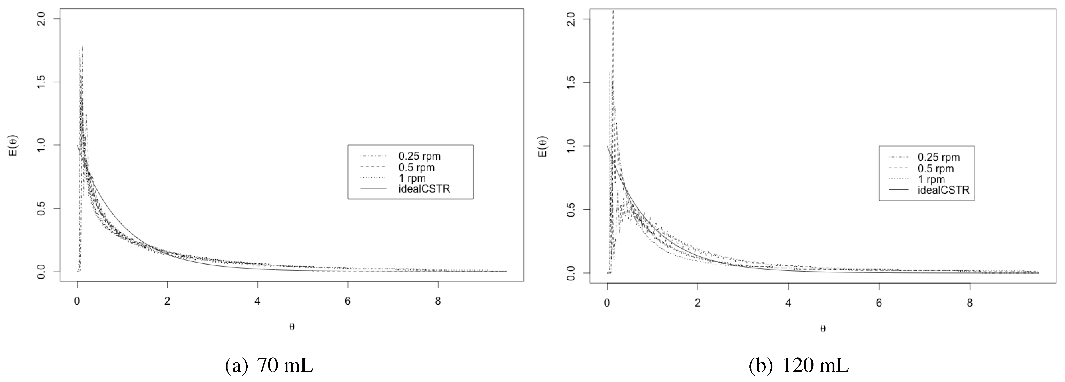
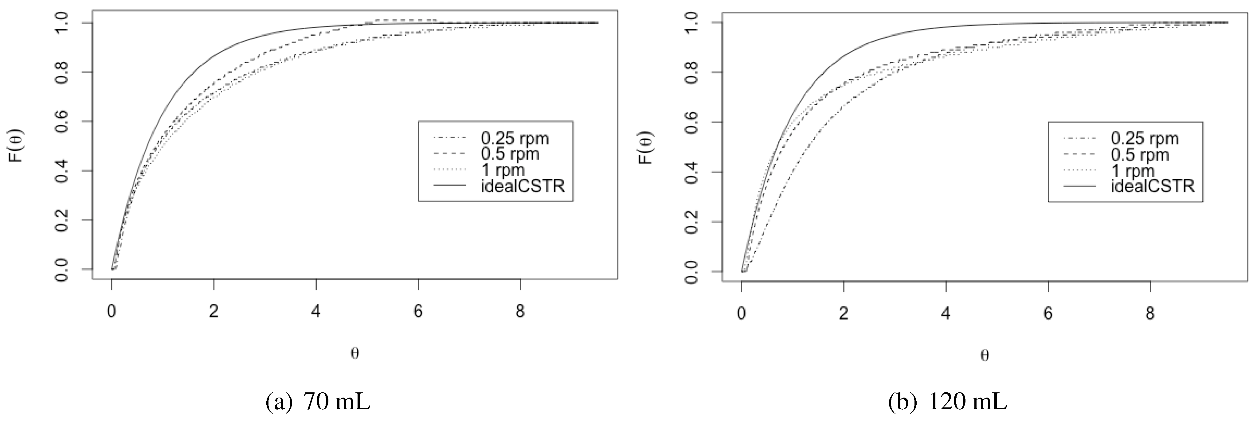
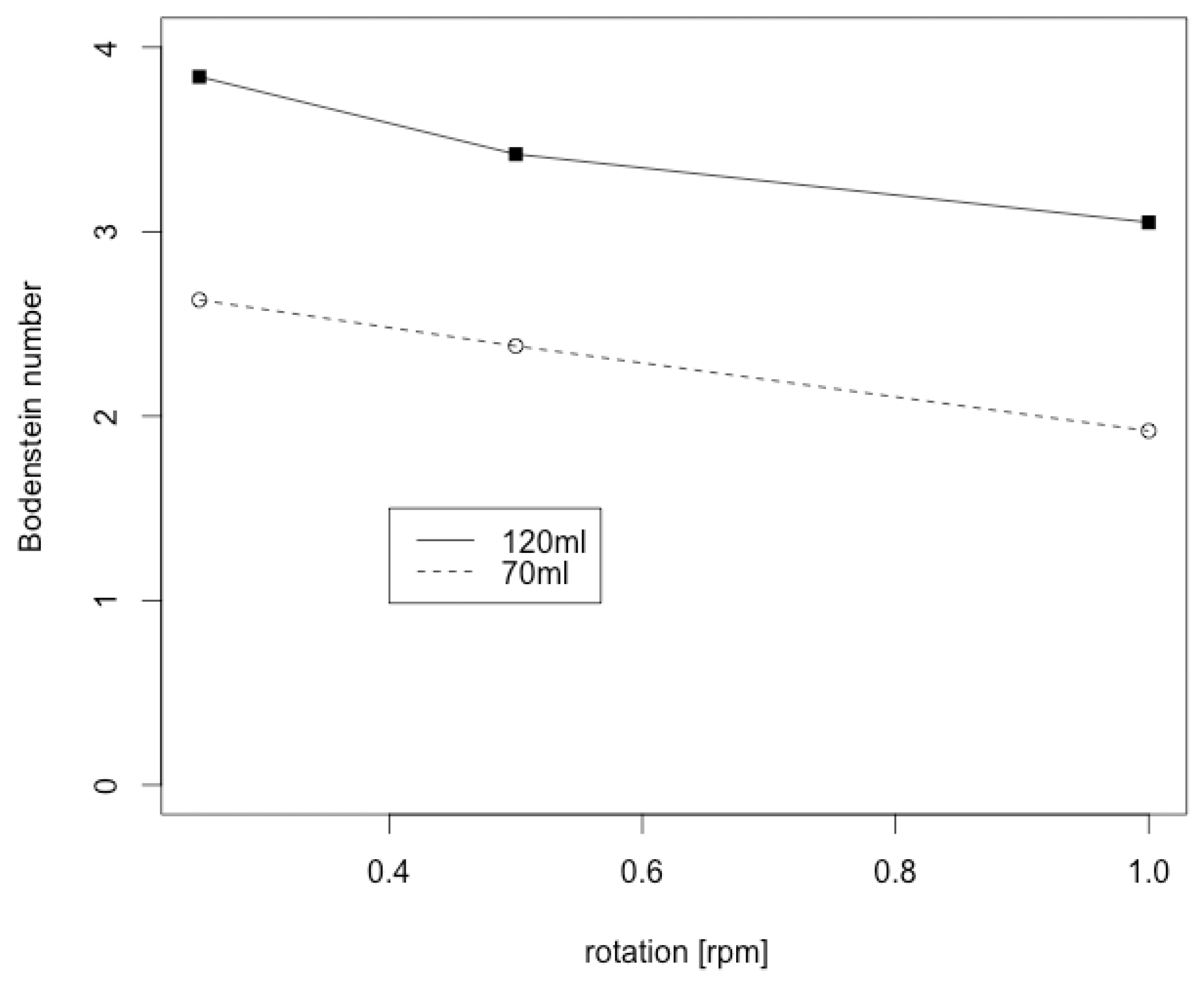
3.2. Bodenstein Number

To further confirm the results obtained from the residence time analysis and examine back mixing properties within the bioreactor, Bodenstein number (Bo) was determined for the operation conditions. Bo is a parameter that measures the extent of axial dispersion. It is inversely proportional to the axial dispersion coefficient. When Bo approaches zero, there is an intense axial dispersion, then the mixed flow prevails. When Bo reaches infinity the flow has negligible axial dispersion, hence becomes plug flow. For MSC culture a low value for Bodenstein number is desirable since an appropriate back mixing is required for nutrient supply of the cells.
Bo was determined according to;
B o = ( 1 + ( 8 · σ Θ 2 ) ) σ Θ 2
σ Θ 2 = ( t - t m ) 2 · E ( t ) d t τ 2
with σ non-dimensional variance of residence time distribution. Results are shown in Figure 5. For all examined operation conditions Bo is below 4, indicating a high back mixing within the bioreactor. The lower the filling volume and the higher the rotation speed, the higher the back mixing. A boundary value of 7 is used in practice to distinguish between CSTR and PFR type [20]. With Bo < 4 the ZRP 2000 H reactor can be classified as CSTR for all operation conditions. The CSTR type favorable over the PFR type for MSC culture, since it allows mass transfer and thus homogenous nutrient supply to the cells. The results obtained by Bodenstein number calculation revealed that even operating with a fully filled (120 mL) bioreactor at the lowest rotation speed (0.1 rpm) provides sufficient back mixing. Since MSC are sensitive to shear stress and can react to the mechanical stimulation with unwanted spontaneous differentiation, 120 mL filling volume and 0.1 rpm rotation speed were chosen for expansion experiments.
Figure 5. Bodenstein numbers of Z®RP 2000 H rotating bed bioreactor. Bodenstein number indicate a high backmixing within the bioreactor.
Bioengineering 01 00231 g005

3.3. Cell Expansion in the Z®RP 2000 H Bioreactor

The total cell number at the end of the dynamic cultivation was 24.6 ± 2.4 × 10 6 cells and 8.2 ± 0.8-fold expansion. Population doubling time was 39.6 ± 1.8 h (in static cultivation 31 h) and the total population doublings were 3.03 ± 0.14. Differentiation potential of UC-MSC was analyzed, after expansion in the Z®RP 2000 H bioreactor. Therefore, cells were cultured after expansion on a two-dimensional fibronectin surface. Collected UC-MSC retained their differentiation capacity towards chondrogenic, adipogenic and osteogenic lineages (see Figure 6). Total glucose consumption and lactate production was increased during cultivation, reaching 157.3 ± 40.7 mg per day for glucose and 133.0 ± 11.6 mg per day for lactate at day 5 (Figure 7).
Figure 6. Differentiation of umbilical cord-derived mesenchymal stromal cells (UC-MSC) cultured in the Z®RP 2000 H bioreactor. (a) Adipogenic differentiation (BODYPI staining) (cultured in NH AdipoDiff medium (Miltenyi Biotech) and 50 µg/mL gentamicin) (b) control (cultured in DMEM, 10% human serum and 50 µg/mL gentamicin); (c) chondrogenic differentiation (Alcian Blue staining) (cultured in NH ChondroDiff medium (Miltenyi Biotech) and 50 µg/mL gentamicin) (d) control (cultured in DMEM, 10% human serum and 50 µg/mL gentamicin); (e) osteogenic differentiation (Calcein staining) (cultured in NH OsteoDiff medium (Miltenyi Biotech) and 50 µg/mL gentamicin) (f) control (phase contrast) (cultured in DMEM, 10% human serum and 50 µg/mL gentamicin) after three weeks of cultivation. MSC retained their differentiation capacity towards chondrogenic, adipogenic and osteogenic lineages after cultivation in the Z®RP 2000 H bioreactor.
Bioengineering 01 00231 g006
Figure 7. Total glucose consumption and lactate production of UC-MSC cultured in the Z®RP 2000 H bioreactor. Glucose consumption indicates cell proliferation throughout the cultivation process.
Bioengineering 01 00231 g007

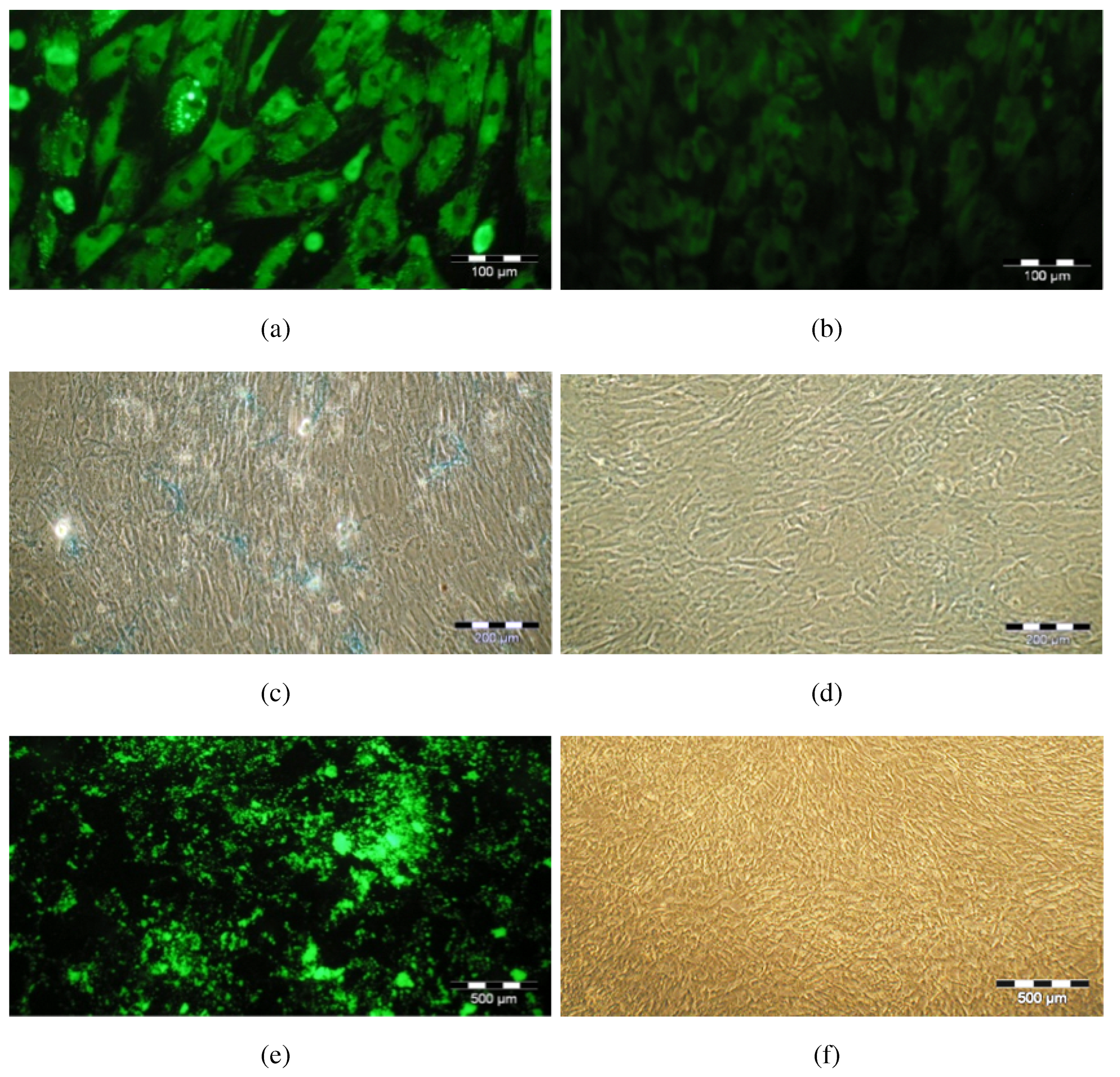
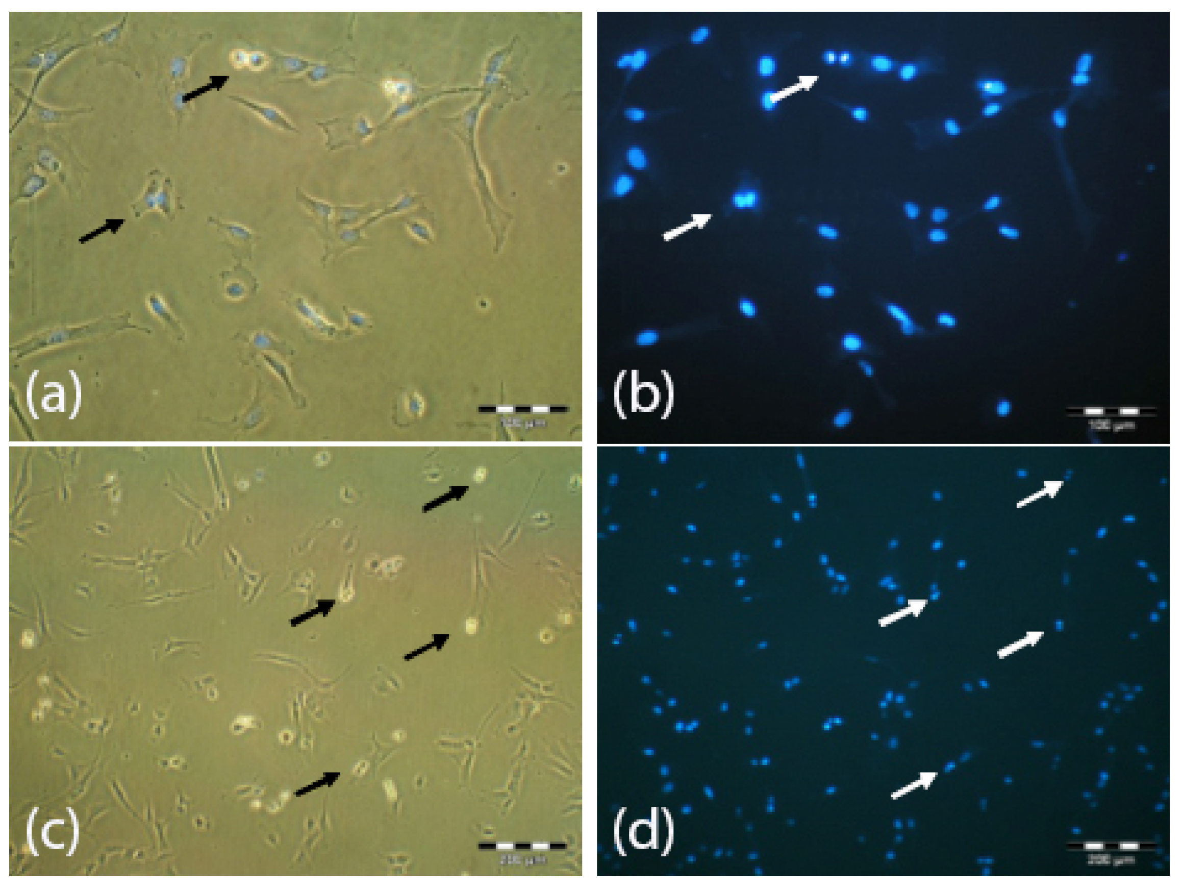
3.4. Cellular Senescence after Expansion in Z®RP 2000 H Bioreactor

The appearance of senescence during short-term expansion is a sign of non-optimal culture conditions, since senescence is one of the responses of the cells to stress (stress-induced premature senescence) [21]. No expression of β-galactosidase and, consequently, no cellular senescence was detected. Moreover, high proportions of cells undergoing cell division (arrows) could be observed, indicating that UC-MSC expanded in the bioreactor did not lose their mitotic activity (Figure 8).
Figure 8. Senescence-associated β-galactosidase staining of UC-MSC cultured in the Z®RP 2000 H bioreactor: (a) β-galactosidase and (b) DAPI-staining, magnification ×50; (c) β-galactosidase and (d) DAPI-staining, magnification ×20. No β-galactosidase and consequently no stress-induced premature senescence could be detected. Cells demonstrate mitotic activity by cell division (arrows).
Bioengineering 01 00231 g008

3.5. Surface Immunophenotype Characterization of UC-MSC after Expansion in the Z®RP 2000 H Bioreactor

After expansion in the Z®RP 2000 H bioreactor, UC-MSC were harvested and analyzed by flow cytometry after staining with FITC- or PE-conjugated antibodies against CD31, CD34, CD44, CD45, CD73, CD90, CD105 and isotype control immunoglobulins. Flow cytometric analysis of UC-MSC showed that they were negative for hematopoietic (CD34, CD45) and endothelial (CD31) markers and were strongly positive for specific immunophenotypic MSC markers (CD44, CD73, CD90 and CD105) (Table 1).
Table 1. Summarized surface antigen expression after dynamic cultivation in Z®RP 2000 H bioreactor. Cells lack hematopoetic and endothelial surface marker expression while expressing specific MSC immunophenotypic markers.
MarkerExpression
CD310.0%
CD340.3%
CD4498.3%
CD450.3%
CD7399.9%
CD9098.1%
CD10599.8%

4. Conclusions

Characterization of the Z®RP bioreactor 2000 H showed that for the purpose of shear stress reduced expansion of MSC it is optimal to cultivate with a completely filled bioreactor chamber and a slow rotation speed. The bioreactor provides properties, comparable to an ideal continuous stirred tank reactor offering beneficial mixing properties and therewith sufficient nutrient supply. The results demonstrate the dynamic cultivation of UC-MSC under these optimized conditions, showing that this system is suitable for UC-MSC expansion, providing high cell yields. Cells obtained from dynamic cultivation demonstrate high quality, maintaining MSC morphology, differentiation potential, mitotic activity as well as MSC surface marker expression. This reproducible cultivation process, offering a controllable environment, has high potential for MSC expansion for future clinical applications.

Author Contributions

A.N. performed data analysis of the bioreactor characterization, contributed in writing and finalized the manuscript. A.L. achieved MSC expansion in the ZRP bioreactor and performed the flow cytometric analysis, proliferation studies as well as histological stainings and contributed in writing and finalized the manuscript. A.H. contributed to the bioreactor characterization and differentiation and histological stainings. M.L. was involved in MSC isolation and cultivation. C.K. designed the study and drafted the manuscript. All authors have read and approved the final manuscript.

Conflicts of Interest

The authors declare no conflict of interest.

References

  1. ClinicalTrials.gov. Avaliable online: http://www.clinicaltrials.gov (accessed on 11 September 2014).
  2. Raviv, L.; Karnieli, O. Cell therapies—The challenges and possible solutions for transferring cell therapy from the bench to the industry. Drug Dev. Deliv. 2014, 3. [Google Scholar]
  3. Ramot, Y.; Meiron, M.; Toren, A.; Steiner, M.; Nyska, A. Safety and biodistribution profile of placental-derived mesenchymal stromal cells (PLX-PAD) following intramuscular delivery. Toxicol. Pathol. 2009, 37, 606–616. [Google Scholar] [CrossRef] [PubMed]
  4. Nold, P.; Brendel, C.; Neubauer, A.; Bein, G.; Hackstein, H. Good manufacturing practice-compliant animal-free expansion of human bone marrow derived mesenchymal stroma cells in a closed hollow-fiber-based bioreactor. Biochem. Biophys. Res. Commun. 2013, 430, 325–330. [Google Scholar] [CrossRef] [PubMed]
  5. Asnaghi, M.A.; Jungebluth, P.; Raimondi, M.T.; Dickinson, S.C.; Rees, L.E.; Go, T.; Cogan, T.A.; Dodson, A.; Parnigotto, P.P.; Hollander, A.P.; et al. A double-chamber rotating bioreactor for the development of tissue-engineered hollow organs: From concept to clinical trial. Biomaterials 2009, 30, 5260–5269. [Google Scholar] [CrossRef] [PubMed]
  6. Martin, I.; Wendt, D.; Heberer, M. The role of bioreactors in tissue engineering. Trends Biotechnol. 2004, 22, 89–86. [Google Scholar] [CrossRef] [PubMed]
  7. Ratcliffe, A.; Niklason, L.E. Bioreactors and bioprocessing for tissue engineering. Ann. N. Y. Acad. Sci. 2002, 961, 210–215. [Google Scholar] [CrossRef] [PubMed]
  8. Hansmann, J.; Groeber, F.; Kahlig, A.; Kleinhans, C.; Walles, H. Bioreactors in tissue engineering—Principles, applications and commercial constraints. Biotechnol. J. 2013, 8, 298–307. [Google Scholar] [CrossRef] [PubMed]
  9. Glindkamp, A.; Riechers, D.; Rehbock, C.; Hitzmann, B.; Scheper, T.; Reardon, K.F. Sensors in disposable bioreactors status and trends. Adv. Biochem. Eng. Biotechnol. 2009, 115, 145–169. [Google Scholar] [PubMed]
  10. Behr, L.; Joeris, K.; Burnett, M.; Scheper, T. A novel in situ probe for oxygen uptake rate measurement in mammalian cell cultures. Biotechnol. Prog. 2012, 28, 581–586. [Google Scholar] [CrossRef] [PubMed]
  11. Babitzky, A.; Lindner, P.; Scheper, T. Cell assessment by at-line microscopy. Methods Mol. Biol. 2014, 1104, 343–353. [Google Scholar] [PubMed]
  12. Committee for Advanced Therapies. Reflection paper on stem cell-based medicinal products. EMA/CAT/571134/2009 2011, 1, 1–14. [Google Scholar]
  13. Naughton, G.K. From lab bench to market: Critical issues in tissue engineering. Ann. N. Y. Acad. Sci. 2002, 961, 372–385. [Google Scholar] [CrossRef] [PubMed]
  14. Reichardt, A.; Polchow, P.; Shakibaei, M.; Henrich, W.; Hetzer, R.; Lueders, C. Large scale expansion of human umbilical cord cells in a rotating bed system bioreactor for cardiovascular tissue engineering applications. Open Biomed. Eng. J. 2013, 7, 50–61. [Google Scholar] [PubMed]
  15. Bianco, P.; Riminucci, M.; Gronthos, S.; Robey, P.G. Bone marrow stromal stem cells: Nature, biology, and potential applications. Stem Cells 2001, 19, 180–192. [Google Scholar] [CrossRef] [PubMed]
  16. Crisan, M.; Yap, S.; Casteilla, L.; Chen, C.-W.; Corselli, M.; Park, T.-S.; Andriolo, G.; Sun, B.; Zheng, B.; Zhang, L.; et al. A perivascular origin for mesenchymal stem cells in multiple human organs. Cell Stem Cell 2008, 3, 301–313. [Google Scholar] [CrossRef] [PubMed]
  17. Yeatts, A.B.; Fisher, J.B. Bone tissue engineering bioreactors: Dynamic culture and the influence of shear stress. Bone 2011, 48, 171–181. [Google Scholar] [CrossRef] [PubMed]
  18. Majore, I.; Moretti, P.; Hass, R.; Kasper, C. Identification of subpopulations in mesenchymal stem cell-like cultures from human umbilical cord. Cell Commun. Signal. 2009, 7, 1–6. [Google Scholar] [CrossRef] [PubMed] [Green Version]
  19. Moretti, P. Establishment of Recombinant Cell Lines and Characterization of Primary Cells for Stem Cell Technology Applications. Ph.D. Thesis, Institute for Technical Chemistry, Leibniz University of Hanover, Hanover, Germany, 2010. [Google Scholar]
  20. Hagen, J. Chemische Reaktionstechnik; VCH: Weinheim, Germany, 1992. [Google Scholar]
  21. Toussaint, O.; Dumont, P.; Remacle, J.; Dierick, J.-F.; Pascal, T.; Frippiat, C.; Magalhaes, J.P.; Zdanov, S.; Chainiaux, F. Stress-induced premature senescence or stress-induced senescence-like phenotype: One in vivo reality, two possible definitions? Sci. World J. 2002, 2, 230–247. [Google Scholar] [CrossRef] [PubMed]

