Radio-Fluorogenic Gel Dosimetry with Coumarin

Abstract
1. Introduction
2. Materials and Methods
2.1. Chemical Preparation
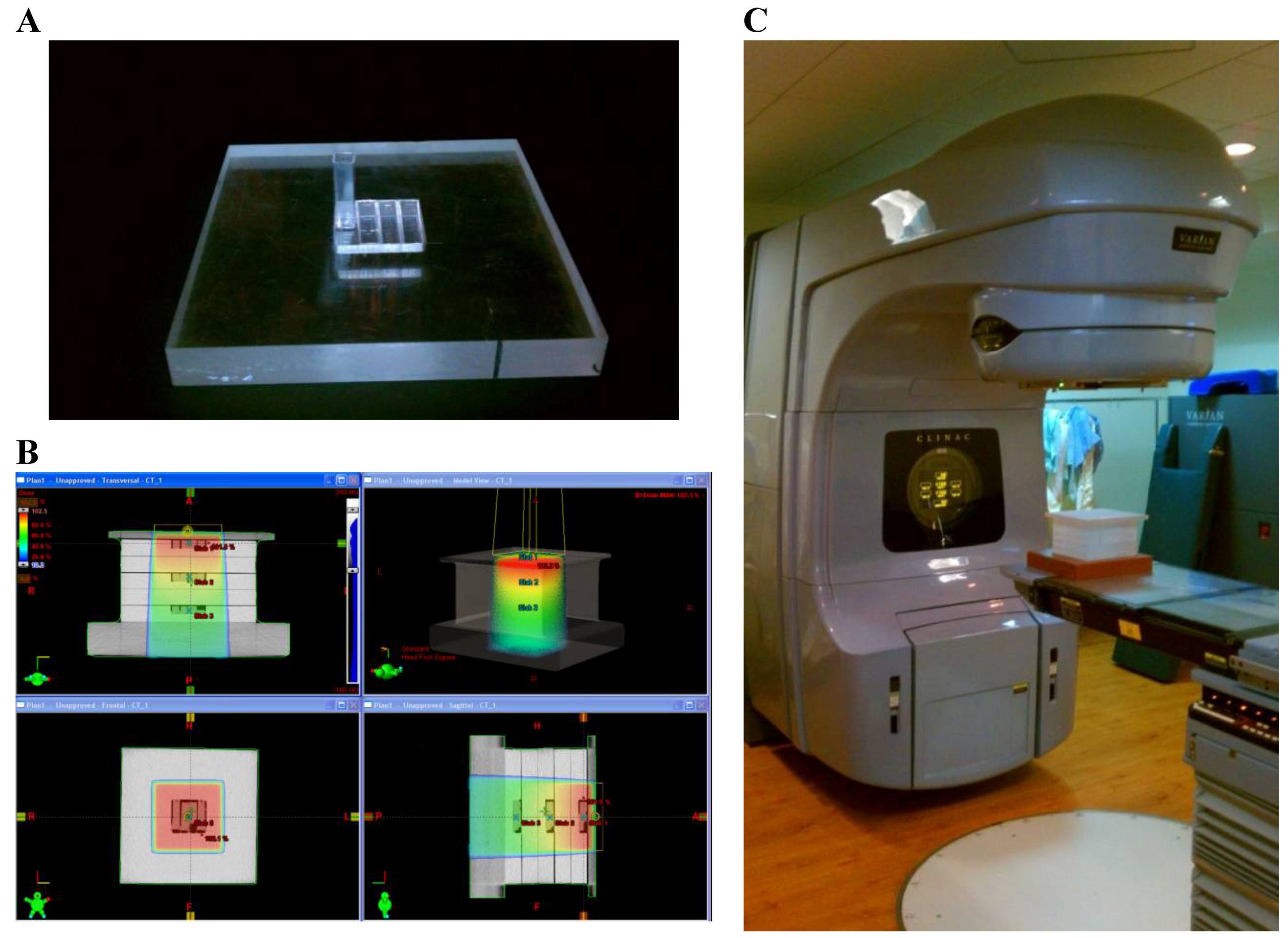
2.2. Irradiation and Analysis
3. Results
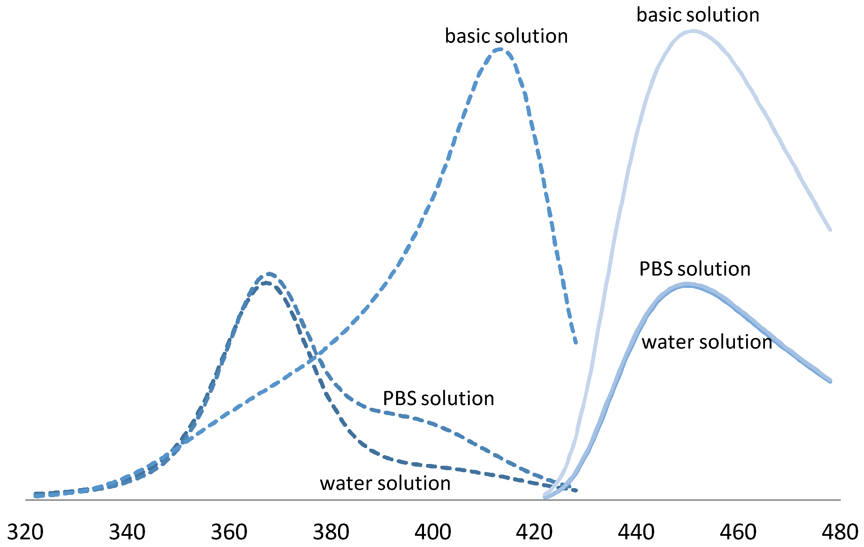
3.1. pH Response
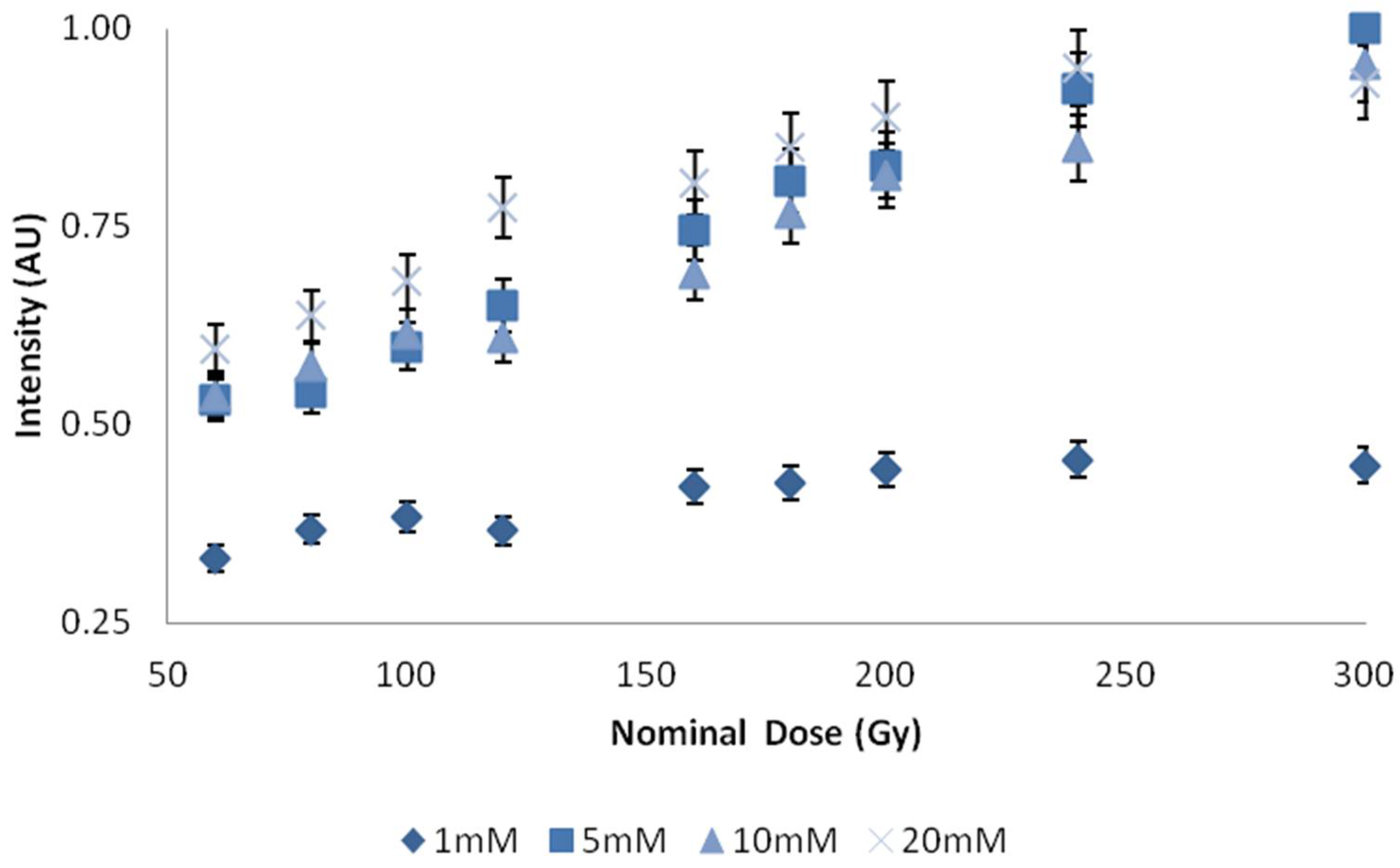
3.2. C3CA Concentration
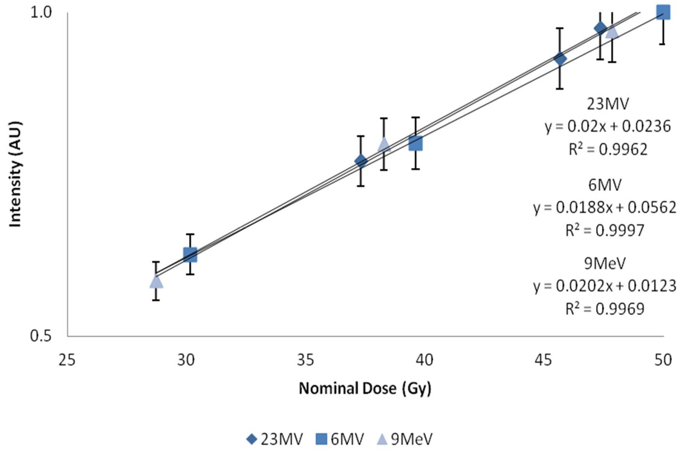
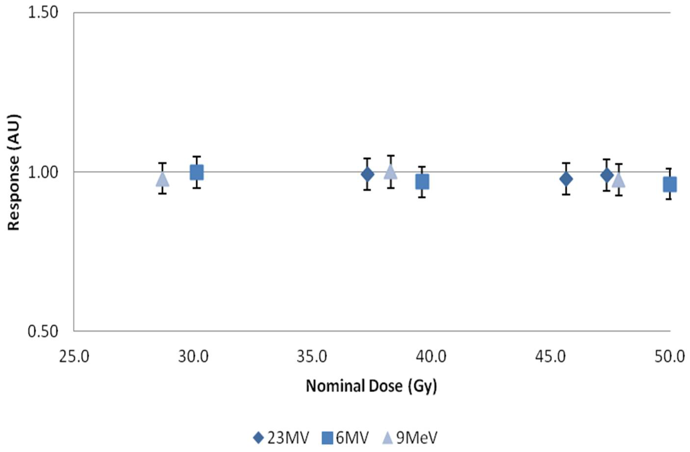
3.3. Dose Response
4. Discussion
5. Conclusions
Author Contributions
Funding
Acknowledgments
Conflicts of Interest
References
- Day, M.J.; Stein, G. Chemical effects of ionizing radiation in some gels. Nature 1950, 166, 146–147. [Google Scholar] [CrossRef] [PubMed]
- Andrews, H.L.; Murphy, R.E.; LeBrun, E.J. Gel dosimeter for depth-dose measurements. Rev. Sci. Instrum. 1957, 28, 329–332. [Google Scholar] [CrossRef]
- Stein, G.; Tomkiewicz, M. Radiation chemistry of gelatin gels containing ferricyanide, radiation research. Radiat. Res. 1970, 43, 25–33. [Google Scholar] [CrossRef] [PubMed]
- Fricke, H.; Hart, E.J. The oxidation of the ferrocyanide, arsenite and selenite ions by the irradiation of their aqueous solutions with X-rays. J. Chem. Phys. 1935, 3, 596. [Google Scholar] [CrossRef]
- Gore, J.C.; Kang, Y.S. Measurement of radiation dose distributions by nuclear magnetic resonance (NMR) imaging. Phys. Med. Biol. 1984, 29, 1189–1197. [Google Scholar] [CrossRef] [PubMed]
- Maryanski, M.J.; Ibbott, G.S.; Eastman, P.; Schulz, R.J.; Gore, J.C. Radiation therapy dosimetry using magnetic resonance imaging of polymer gels. Med. Phys. 1996, 23, 699–705. [Google Scholar] [CrossRef] [PubMed]
- Adamovics, J.; Maryanski, M.J. Characterisation of PRESAGE™: A new 3-D radiochromic solid polymer dosemeter for ionising radiation. Radiat. Prot. Dosim. 2006, 120, 107–112. [Google Scholar] [CrossRef] [PubMed]
- Warman, J.M.; De Haas, M.P.; Luthjens, L.H. High-energy radiation monitoring based on radio-fluorogenic co-polymerization. I: Small volume in situ probe. Phys. Med. Biol. 2009, 54, 3185–3200. [Google Scholar] [CrossRef] [PubMed]
- Doran, S.J. The history and principles of chemical dosimetry for 3-D radiation fields: Gels, polymers and plastics. Appl. Radiat. Isotopes 2009, 67, 393–398. [Google Scholar] [CrossRef] [PubMed]
- Senden, R.J.; De Jean, P.; McAuley, K.B.; Schreiner, L.J. Polymer gel dosimeters with reduced toxicity: A preliminary investigation of the NMR and optical dose–response using different monomers. Phys. Med. Biol. 2006, 51, 3301–3314. [Google Scholar] [CrossRef] [PubMed]
- Veis, A. The Macromolecular Chemistry of Gelatin; Academic Press: New York, NY, USA, 1964. [Google Scholar]
- Djagny, K.B.; Wang, Z.; Xu, S. Gelatin: A valuable protein for food and pharmaceutical industries: Review. Crit. Rev. Food Sci. Nutr. 2001, 41, 481–492. [Google Scholar] [CrossRef] [PubMed]
- Roussenova, M.; Enrione, J.; Diaz-Calderon, P.; Taylor, A.J.; Ubbink, J.; Alam, M.A. A nanostructural investigation of glassy gelatin oligomers: Molecular organization and interactions with low molecular weight diluents. New J. Phys. 2012, 14, 035016. [Google Scholar] [CrossRef]
- Day, M.J.; Stein, G. Chemical Measurement of Ionizing Radiations. Nature 1949, 164, 671–672. [Google Scholar] [CrossRef] [PubMed]
- Stein, G.; Weiss, J. Detection of Free Hydroxyl Radicals by Hydroxylation of Aromatic Compounds. Nature 1950, 166, 1104–1105. [Google Scholar] [CrossRef] [PubMed]
- Armstrong, W.A.; Grant, D.W. A highly sensitive chemical dosimeter for ionizing radiation. Nature 1958, 182, 747. [Google Scholar] [CrossRef] [PubMed]
- Barreto, J.C.; Smith, G.S.; Strobel, N.H.; McQuillin, P.A.; Miller, T.A. Terephthalic acid: A dosimeter for the detection of hydroxyl radicals in vitro. Life Sci. 1994, 56, PL89–PL96. [Google Scholar] [CrossRef]
- Matthews, R.W. Aqueous chemical dosimetry. Int. J. Appl. Radiat. Isotopes 1982, 33, 1159–1170. [Google Scholar] [CrossRef]
- Nadrowitz, R.; Coray, A.; Boehringer, T.; Dunst, J.; Rades, D. A liquid fluorescence dosimeter for proton dosimetry. Phys. Med. Biol. 2012, 57, 1325–1333. [Google Scholar] [CrossRef] [PubMed]
- Lakowicz, J.R. Principles of Fluorescence Spectroscopy, 3rd ed.; Springer: New York, NY, USA, 2006. [Google Scholar]
- Collins, A.K.; Makrigiorgos, G.M.; Svensson, G.K. Coumarin chemical dosimeter for radiation therapy. Med. Phys. 1994, 21, 1741–1747. [Google Scholar] [CrossRef] [PubMed]
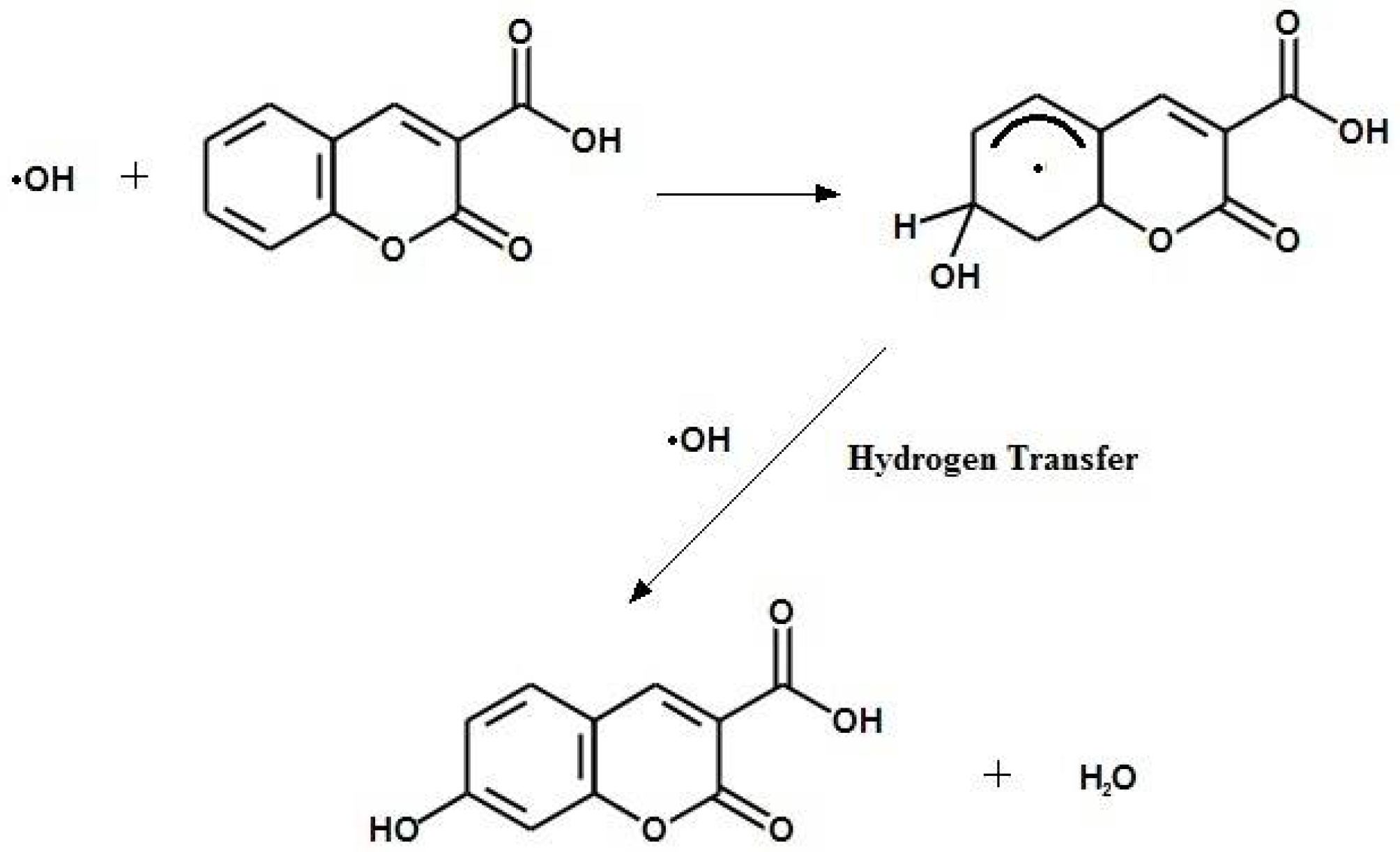
- Yamashita, S.; Baldacchino, G.; Maeyama, T.; Taguchi, M.; Muroya, Y.; Lin, M.; Kimura, A.; Murakami, T.; Katsumura, Y. Mechanism of radiation-induced reactions in aqueous solution of coumarin-3-carboxylic acid: Effects of concentration, gas and additive on fluorescent product yield. Free Radic. Res. 2012, 46, 861–871. [Google Scholar] [CrossRef] [PubMed]
- Perry, C.C.; Tang, V.J.; Konigsfeld, K.M.; Aguilera, J.A.; Milligan, J.R. Use of a coumarin-labeled hexa-arginine peptide as a fluorescent hydroxyl radical probe in a nanoparticulate plasmid DNA condensate. J. Phys. Chem. B 2011, 115, 9889–9897. [Google Scholar] [CrossRef] [PubMed]
- Gallina, M.E.; Kim, T.J.; Vasquez, J.; Tuerkcan, S.; Abbyad, P.; Pratx, G. Single-cell analysis of radiotracers’ uptake by fluorescence microscopy: Direct and droplet approach. In Proceedings of the Imaging, Manipulation, and Analysis of Biomolecules, Cells, and Tissues XV, San Francisco, CA, USA, 16 February 2017; International Society for Optics and Photonics: Bellingham, WA, USA, 2017; Volume 10068, p. 100680Y. [Google Scholar]
- Gallina, M.E.; Kim, T.J.; Shelor, M.; Vasquez, J.; Mongersun, A.; Kim, M.; Tang, S.K.; Abbyad, P.; Pratx, G. Toward a Droplet-Based Single-Cell Radiometric Assay. Anal. Chem. 2017, 89, 6472–6481. [Google Scholar] [CrossRef] [PubMed]
- Dai, X.; Rollin, E.; Bellerive, A.; Hargrove, C.; Sinclair, D.; Mifflin, C.; Zhang, F. Wavelength shifters for water Cherenkov detectors. Nucl. Instrum. Methods Phys. Res. Sect. A Accel. Spectrom. Detect. Assoc. Equip. 2008, 589, 290–295. [Google Scholar] [CrossRef]
- Warman, J.M.; de Haas, M.; Luthjens, L.; Murrer, L. A Radio-Fluorogenic Organic Gel for Real-Time, 3D Radiation Dosimetry. Adv. Mater. 2011, 23, 4953–4955. [Google Scholar] [CrossRef] [PubMed]
- Sandwall, P.A.; Spitz, H.B.; Elson, H.R.; Lamba, M.A.; Connick, W.B.; Fenichel, H. Measuring the photon depth dose distribution produced by a medical linear accelerator in a water-equivalent radio-fluorogenic gel. J. Radioanal. Nucl. Chem. 2016, 307, 2505–2508. [Google Scholar] [CrossRef]
- Sandwall, P.A. Spatial Dosimetry with Violet Diode Laser-Induced Fluorescence of Water-Equivalent Radio-Fluorogenic Gels. Ph.D. Thesis, University of Cincinnati, Cincinnati, OH, USA, 2014. [Google Scholar]
- Sandwall, P.; Spitz, H.; Elson, H.; Lamba, M.; Connick, W.; Fenichel, H. Radio-fluorogenic dosimetry with violet diode laser-induced fluorescence. In Proceedings of the Medical Imaging: Physics of Medical Imaging, San Diego, CA, USA, 19 March 2014; International Society for Optics and Photonics: Bellingham, WA, USA, 2014; Volume 9033, p. 90333Y. [Google Scholar]
- Yao, T.; Gasparini, A.; De Haas, M.P.; Luthjens, L.H.; Denkova, A.G.; Warman, J.M. A tomographic UV-sheet scanning technique for producing 3D fluorescence images of X-ray beams in a radio-fluorogenic gel. Biomed. Phys. Eng. Express 2017, 3. [Google Scholar] [CrossRef]
- Maeyama, T.; Shinnosuke, H. Nanoclay gel-based radio-fluorogenic gel dosimeters using various fluorescence probes. Radiat. Phys. Chem. 2018, 151, 42–46. [Google Scholar] [CrossRef]
- Dahal, E.; Badal, A.; Zidan, A.; Alayoubi, A.; Hagio, T.; Glick, S.J.; Badano, A.; Ghammraoui, B. Stable gelatin-based phantom materials with tunable X-ray attenuation properties and 3D printability for X-ray imaging. Phys. Med. Biol. 2018, 63. [Google Scholar] [CrossRef] [PubMed]


© 2018 by the authors. Licensee MDPI, Basel, Switzerland. This article is an open access article distributed under the terms and conditions of the Creative Commons Attribution (CC BY) license (http://creativecommons.org/licenses/by/4.0/).
Share and Cite
Sandwall, P.A.; Bastow, B.P.; Spitz, H.B.; Elson, H.R.; Lamba, M.; Connick, W.B.; Fenichel, H. Radio-Fluorogenic Gel Dosimetry with Coumarin. Bioengineering 2018, 5, 53. https://doi.org/10.3390/bioengineering5030053
Sandwall PA, Bastow BP, Spitz HB, Elson HR, Lamba M, Connick WB, Fenichel H. Radio-Fluorogenic Gel Dosimetry with Coumarin. Bioengineering. 2018; 5(3):53. https://doi.org/10.3390/bioengineering5030053
Chicago/Turabian StyleSandwall, Peter A., Brandt P. Bastow, Henry B. Spitz, Howard R. Elson, Michael Lamba, William B. Connick, and Henry Fenichel. 2018. "Radio-Fluorogenic Gel Dosimetry with Coumarin" Bioengineering 5, no. 3: 53. https://doi.org/10.3390/bioengineering5030053
APA StyleSandwall, P. A., Bastow, B. P., Spitz, H. B., Elson, H. R., Lamba, M., Connick, W. B., & Fenichel, H. (2018). Radio-Fluorogenic Gel Dosimetry with Coumarin. Bioengineering, 5(3), 53. https://doi.org/10.3390/bioengineering5030053