Retinal Fundus Multi-Disease Image Dataset (RFMiD): A Dataset for Multi-Disease Detection Research

 , , , , , and
Abstract
:1. Summary
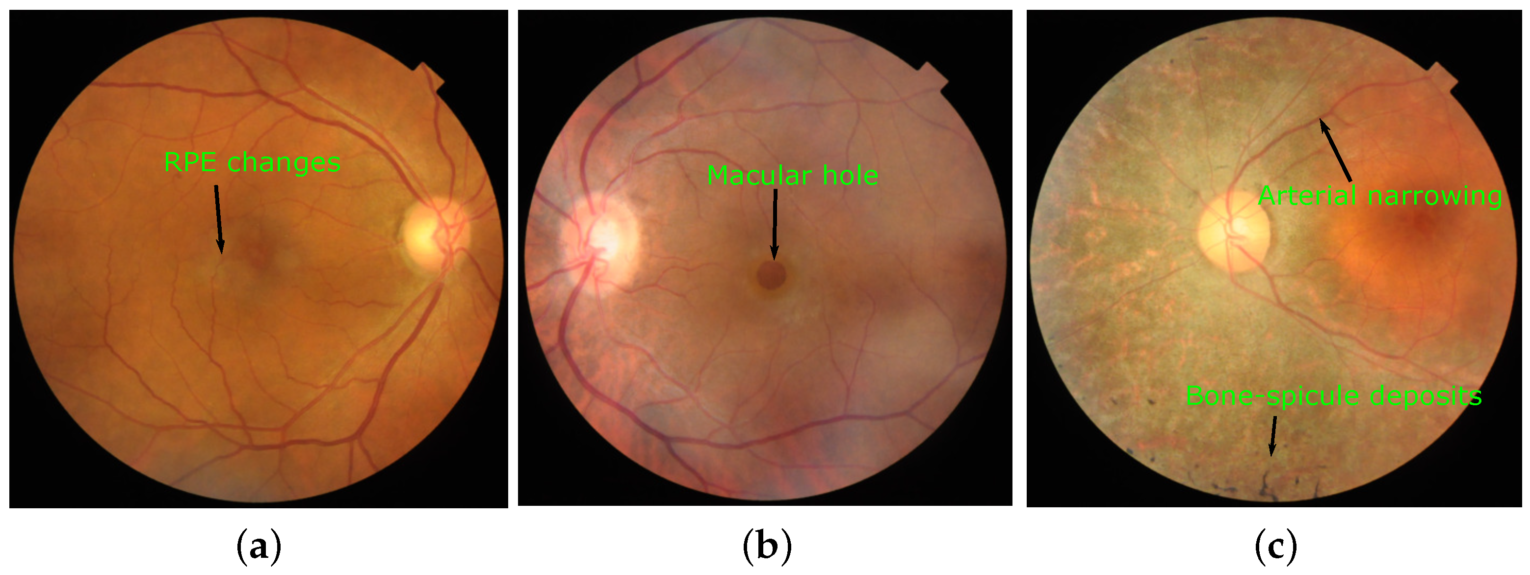
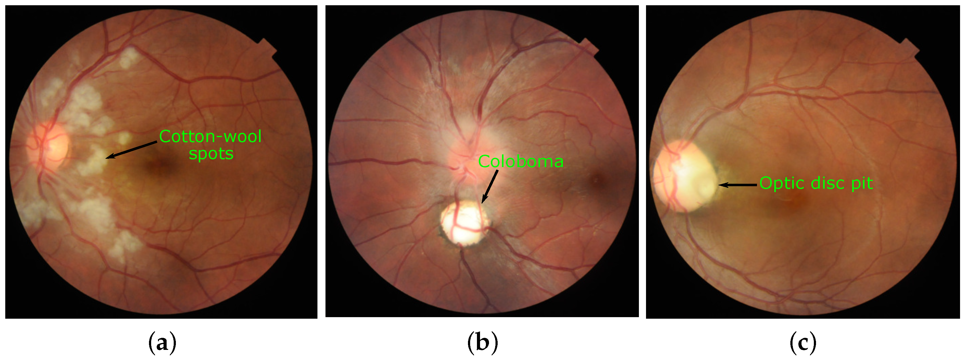
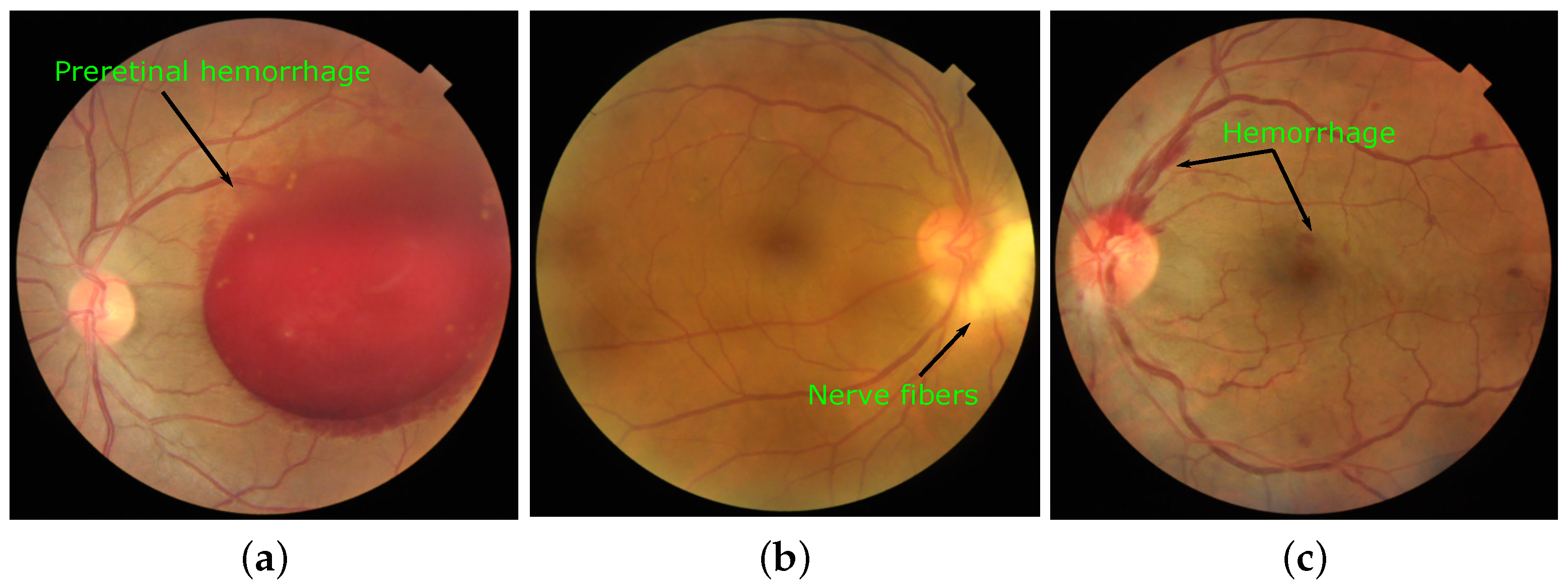
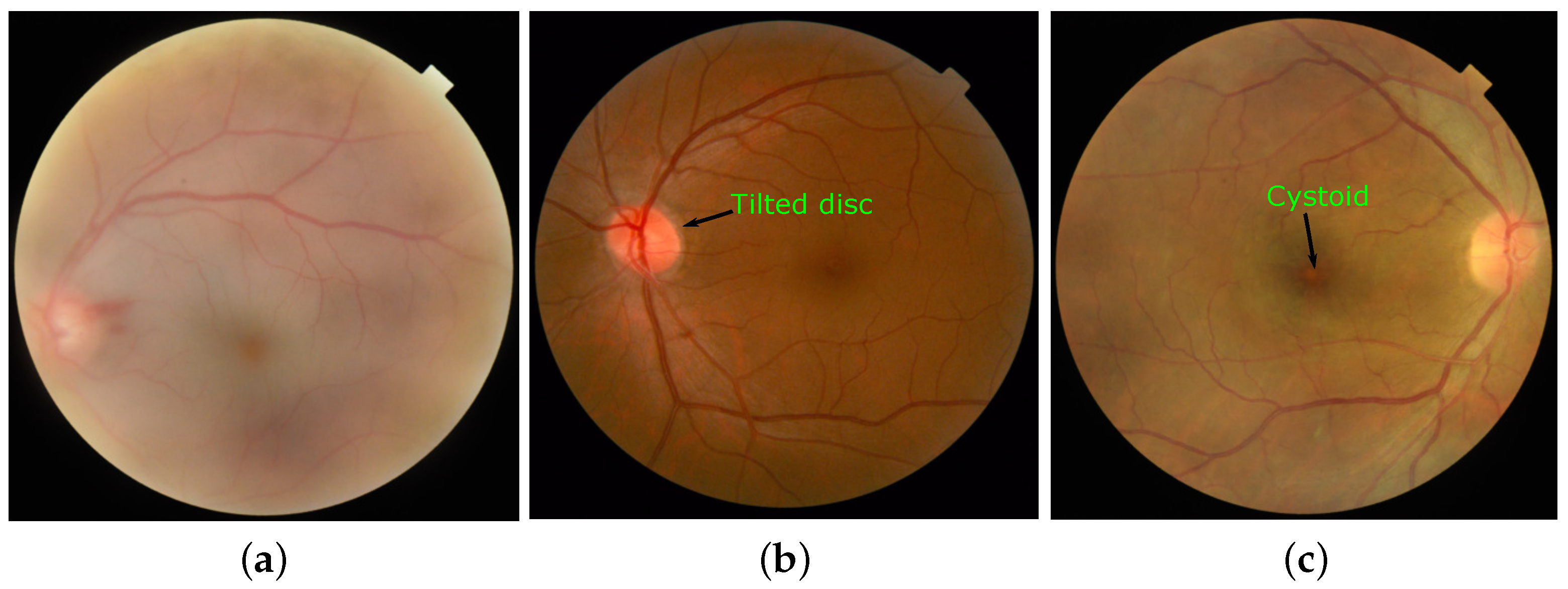
2. Data Description
- Screening of retinal images into normal and abnormal (comprising of 45 different types of diseases/pathologies) categories.
- Classification of retinal images into 45 different categories.
- A.
- ID: Image identity number.
- B.
- Disease_Risk: Presence of disease/abnormality.
- C.
- DR: Presence of diabetic retinopathy.
- D.
- ARMD: Presence of age-related macular degeneration.
- E.
- MH: Presence of media haze.
- F.
- DN: Presence of drusen.
- G.
- MYA: Presence of myopia.
- H.
- BRVO: Presence of branch retinal vein occlusion.
- I.
- TSLN: Presence of tessellation.
- .
- .
- AU.
- CL: Presence of collateral.
3. Experimental Design, Materials, and Methods
3.1. Data Acquisition
- Pretreatment of Samples: Before image acquisition, pupils of most of the subjects were dilated with one drop of tropicamide at 0.5% concentration. The fundus images were captured with position and orientation of the patient sitting upright with 39 mm (Kowa VX–10) and 40.7 mm (TOPCON 3D OCT-2000 and TOPCON TRC-NW300) distance between lenses and examined eye using non-invasive fundus camera.
- Fundus Camera Specifications: Regular retinal fundus images were acquired using three different digital fundus cameras. Details of camera model, hardware used, field of view (FOV), resolution, and number of images included in the dataset are given in Table 2.
- Data Quality: The dataset is formed by extracting 3200 images from the thousands of examinations done during the period 2009–2020. Both high-quality and low-quality images are selected to make the dataset challenging.
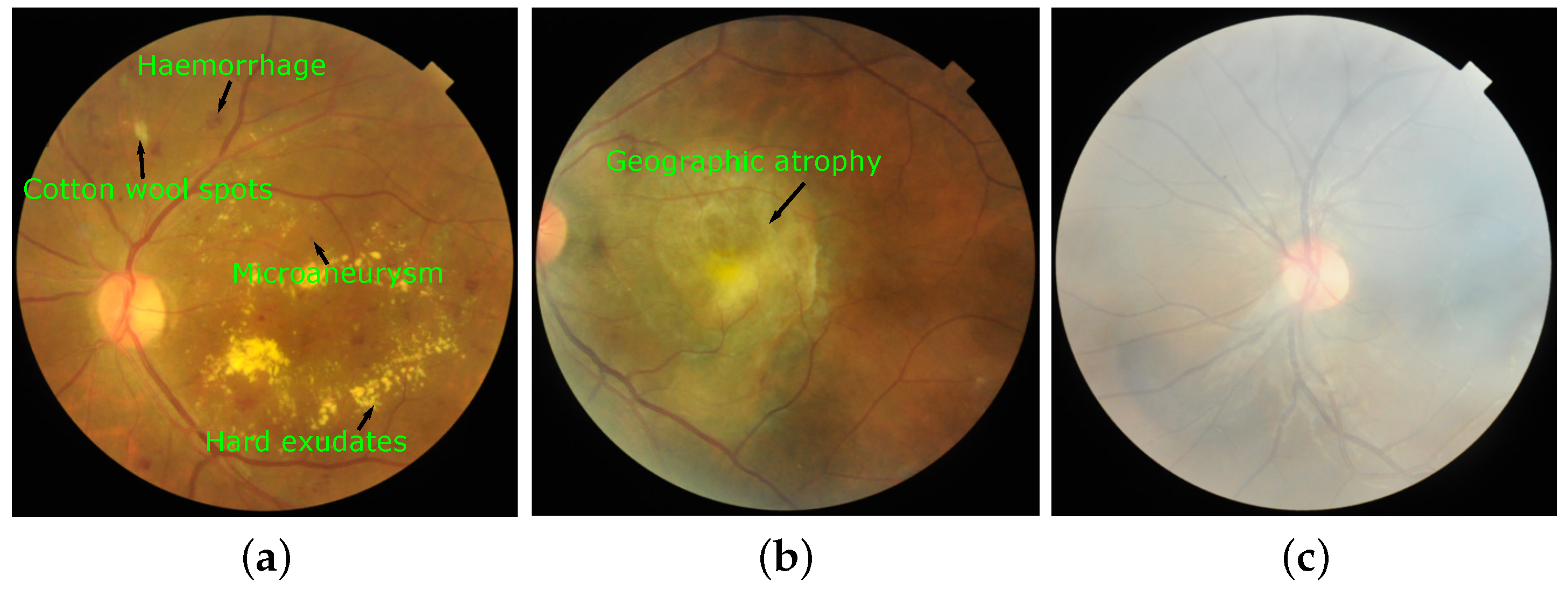
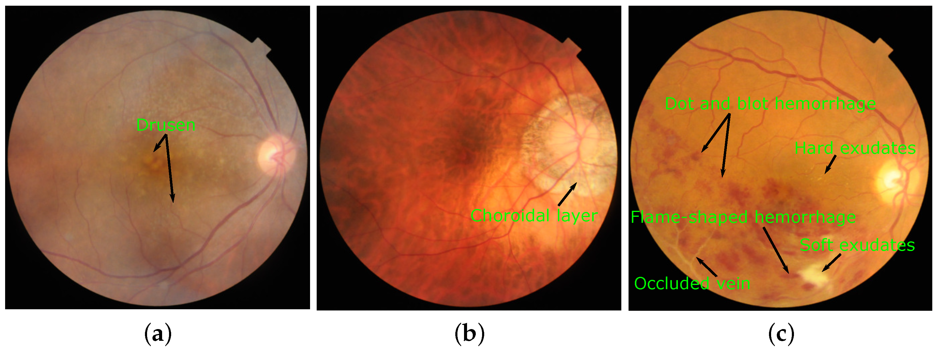
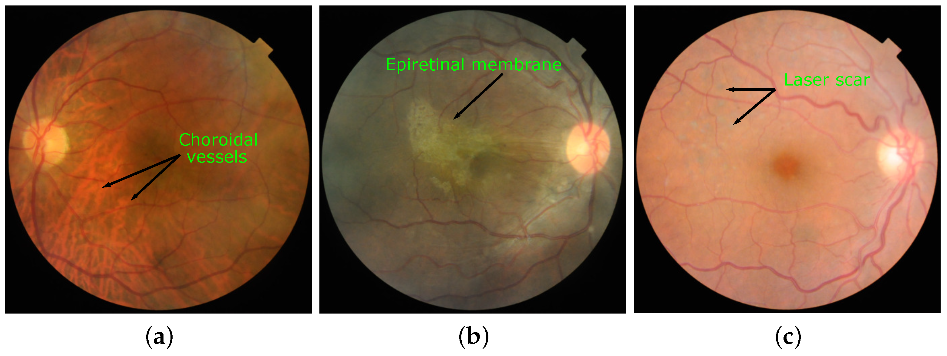
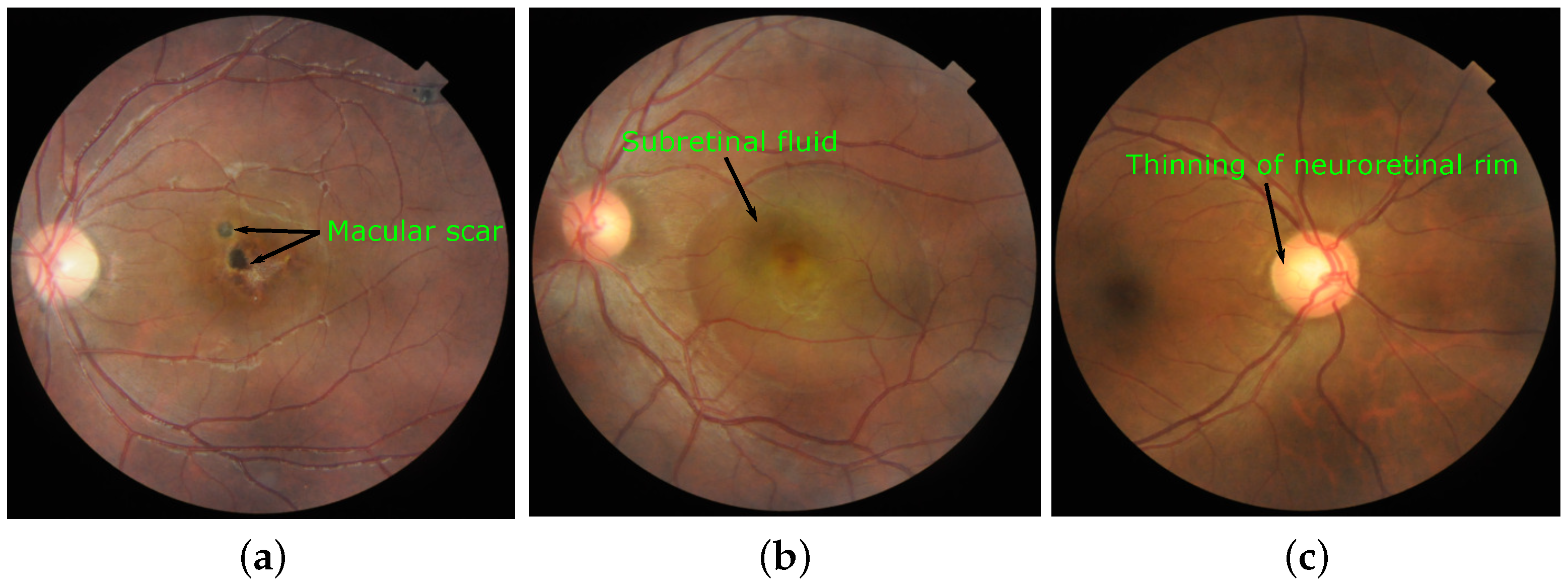
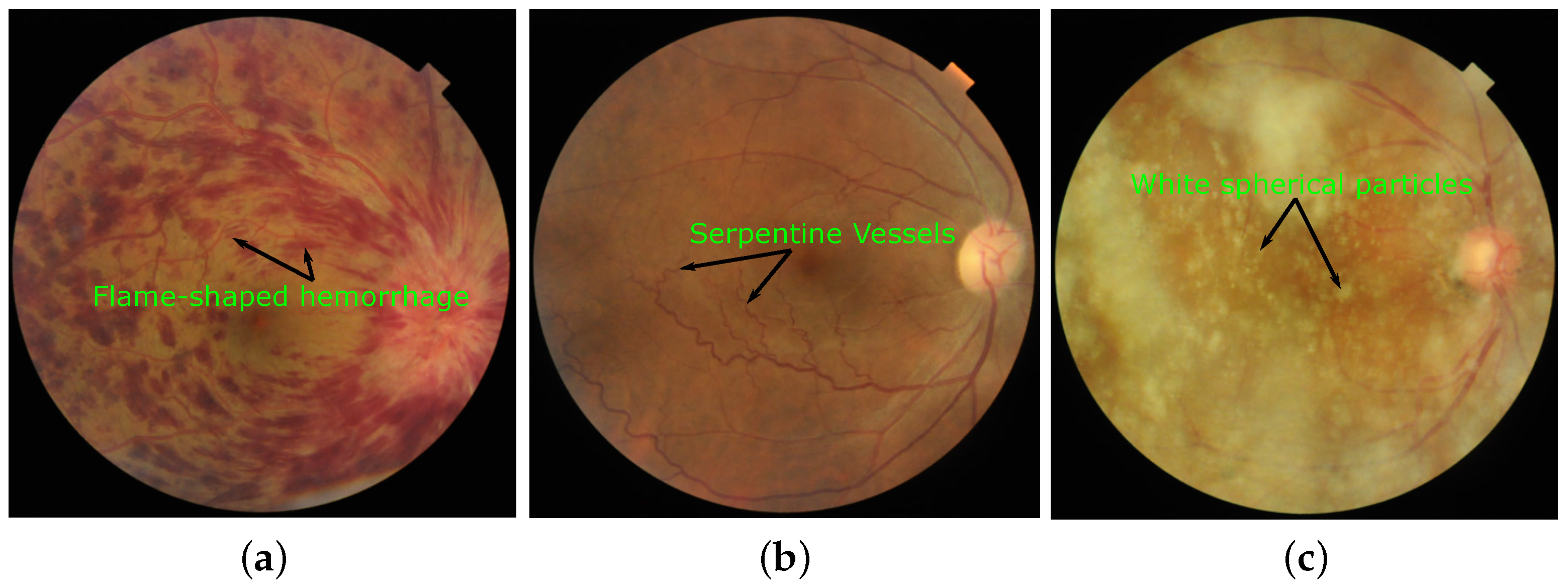
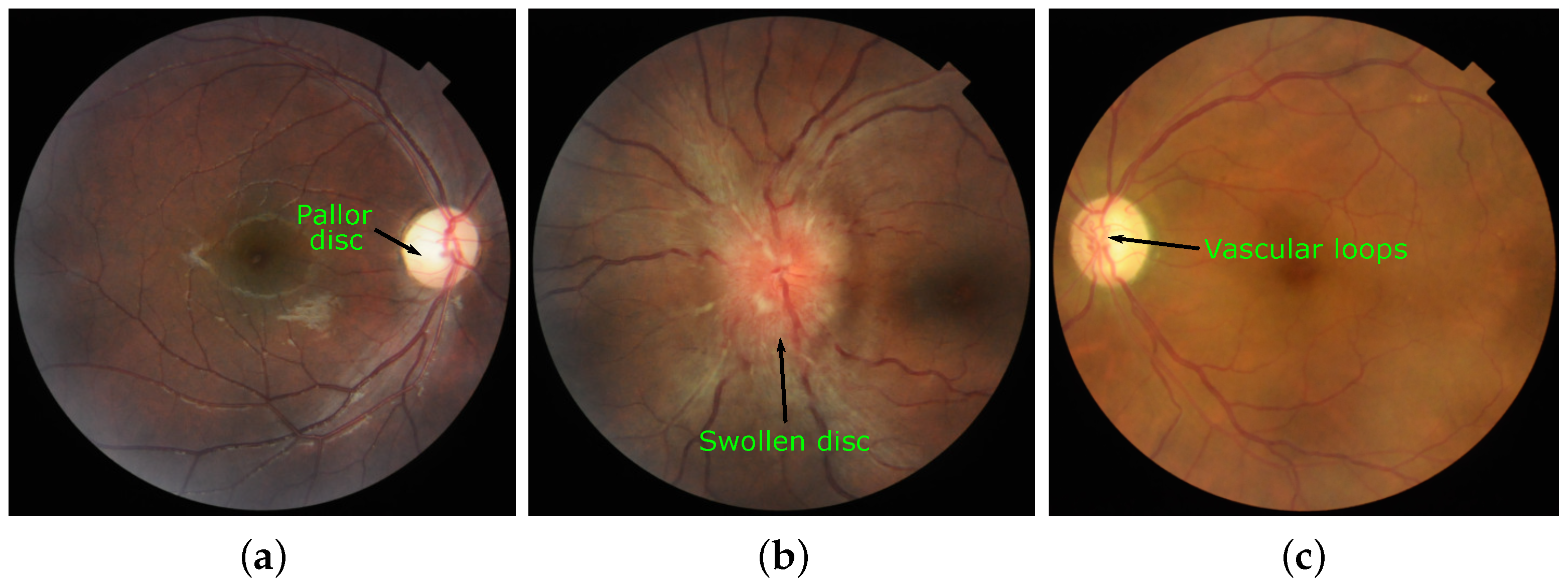
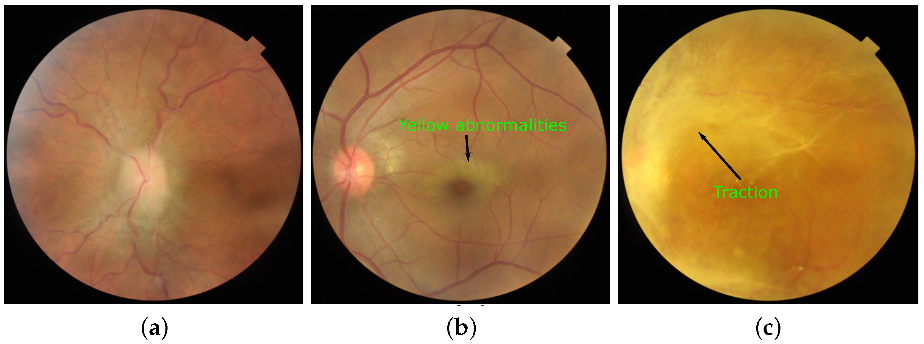
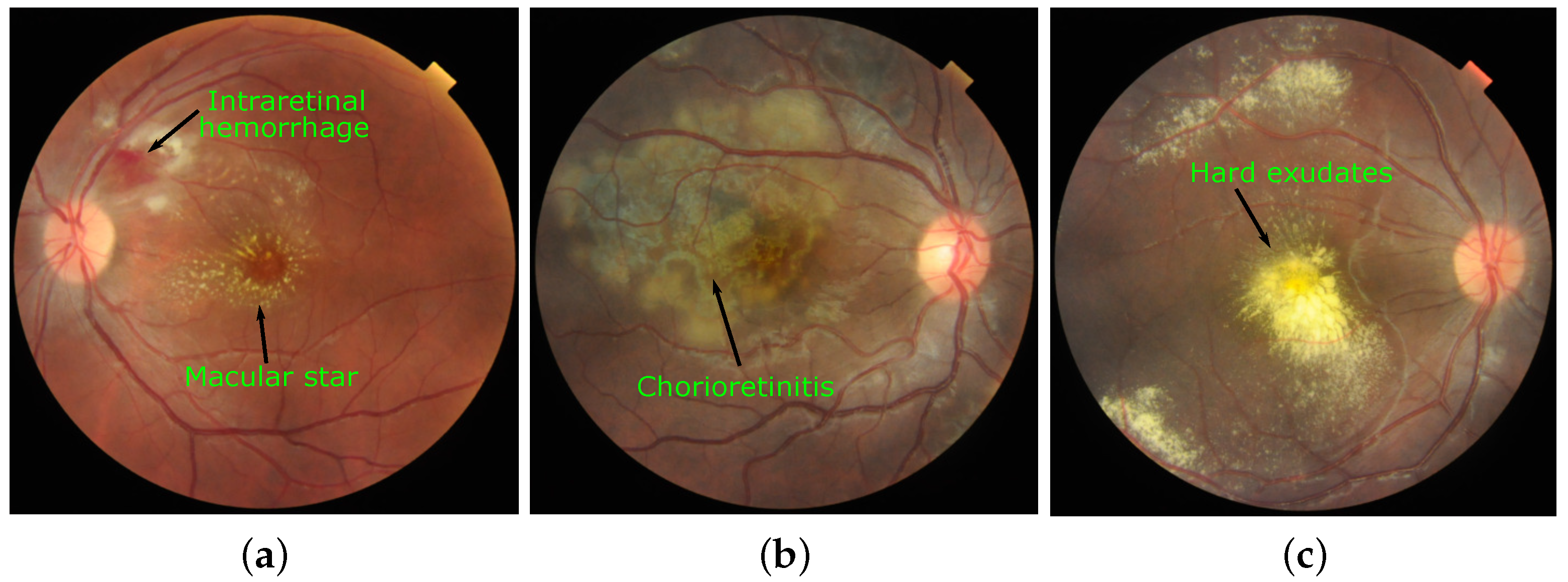
3.2. Annotation of Images
Author Contributions
Funding
Institutional Review Board Statement
Informed Consent Statement
Data Availability Statement
Acknowledgments
Conflicts of Interest
References
- Who Launches First World Report on Vision. Available online: https://www.who.int/blindness/Vision2020_report.pdf (accessed on 29 January 2021).
- MacGillivray, T.; Trucco, E.; Cameron, J.; Dhillon, B.; Houston, J.; Van Beek, E. Retinal imaging as a source of biomarkers for diagnosis, characterization and prognosis of chronic illness or long-term conditions. Br. J. Radiol. 2014, 87, 20130832. [Google Scholar] [CrossRef] [PubMed] [Green Version]
- Yang, W.; Xu, H.; Yu, X.; Wang, Y. Association between retinal artery lesions and nonalcoholic fatty liver disease. Hepatol. Int. 2015, 9, 278–282. [Google Scholar] [CrossRef] [PubMed] [Green Version]
- Chang, Y.S.; Weng, S.F.; Chang, C.; Wang, J.J.; Tseng, S.H.; Wang, J.Y.; Jan, R.L. Risk of retinal vein occlusion following end-stage renal disease. Medicine 2016, 95, e3474. [Google Scholar] [CrossRef] [PubMed]
- Porwal, P.; Pachade, S.; Kamble, R.; Kokare, M.; Deshmukh, G.; Sahasrabuddhe, V.; Meriaudeau, F. Indian diabetic retinopathy image dataset (IDRiD): A database for diabetic retinopathy screening research. Data 2018, 3, 25. [Google Scholar] [CrossRef] [Green Version]
- Decencière, E.; Zhang, X.; Cazuguel, G.; Lay, B.; Cochener, B.; Trone, C.; Gain, P.; Ordonez, R.; Massin, P.; Erginay, A.; et al. Feedback on a publicly distributed image database: The Messidor database. Image Anal. Stereol. 2014, 33, 231–234. [Google Scholar] [CrossRef] [Green Version]
- Cuadros, J.; Bresnick, G. EyePACS: An adaptable telemedicine system for diabetic retinopathy screening. J. Diabetes Sci. Technol. 2009, 3, 509–516. [Google Scholar] [CrossRef] [Green Version]
- Sivaswamy, J.; Krishnadas, S.; Chakravarty, A.; Joshi, G.; Tabish, A.S. A comprehensive retinal image dataset for the assessment of glaucoma from the optic nerve head analysis. JSM Biomed. Imaging Data Pap. 2015, 2, 1004. [Google Scholar]
- Fumero, F.; Alayón, S.; Sanchez, J.L.; Sigut, J.; Gonzalez-Hernandez, M. RIM-ONE: An open retinal image database for optic nerve evaluation. In Proceedings of the 2011 24th International Symposium on Computer-Based Medical Systems (CBMS), Bristol, UK, 27–30 June 2011; pp. 1–6. [Google Scholar]
- Orlando, J.I.; Fu, H.; Breda, J.B.; van Keer, K.; Bathula, D.R.; Diaz-Pinto, A.; Fang, R.; Heng, P.A.; Kim, J.; Lee, J.; et al. Refuge challenge: A unified framework for evaluating automated methods for glaucoma assessment from fundus photographs. Med. Image Anal. 2020, 59, 101570. [Google Scholar] [CrossRef]
- Farnell, D.J.; Hatfield, F.; Knox, P.; Reakes, M.; Spencer, S.; Parry, D.; Harding, S.P. Enhancement of blood vessels in digital fundus photographs via the application of multiscale line operators. J. Frankl. Inst. 2008, 345, 748–765. [Google Scholar] [CrossRef]
- Hoover, A. STARE Database. 1975. Available online: https://cecas.clemson.edu/~ahoover/stare/ (accessed on 29 January 2021).
- Cohen, S. Retina Gallery Full Sized Retina Images. 2017. Available online: https://www.retinagallery.com/ (accessed on 29 January 2021).
- Age-Related Eye Disease Study Research Group. A randomized, placebo-controlled, clinical trial of high-dose supplementation with vitamins C and E, beta carotene, and zinc for age-related macular degeneration and vision loss: AREDS report no. 8. Arch. Ophthalmol. 2001, 119, 1417. [Google Scholar] [CrossRef] [Green Version]
- Choi, J.Y.; Yoo, T.K.; Seo, J.G.; Kwak, J.; Um, T.T.; Rim, T.H. Multi-categorical deep learning neural network to classify retinal images: A pilot study employing small database. PLoS ONE 2017, 12, e0187336. [Google Scholar] [CrossRef] [PubMed] [Green Version]
- Quellec, G.; Lamard, M.; Conze, P.H.; Massin, P.; Cochener, B. Automatic detection of rare pathologies in fundus photographs using few-shot learning. Med. Image Anal. 2020, 61, 101660. [Google Scholar] [CrossRef] [PubMed] [Green Version]
- Wong, T.Y.; Cheung, C.M.G.; Larsen, M.; Sharma, S.; Simó, R. Diabetic retinopathy. Nat. Rev. Dis. Prim. 2016, 2, 16012. [Google Scholar] [CrossRef] [PubMed]
- Lim, L.S.; Mitchell, P.; Seddon, J.M.; Holz, F.G.; Wong, T.Y. Age-related macular degeneration. Lancet 2012, 379, 1728–1738. [Google Scholar] [CrossRef]
- Chen, W.S.; Friberg, T.R.; Eller, A.W.; Medina, C. Advances in retinal imaging of eyes with hazy media: Further Studies. Investig. Ophthalmol. Vis. Sci. 2011, 52, 4036. [Google Scholar]
- Johnson, P.T.; Lewis, G.P.; Talaga, K.C.; Brown, M.N.; Kappel, P.J.; Fisher, S.K.; Anderson, D.H.; Johnson, L.V. Drusen-associated degeneration in the retina. Investig. Ophthalmol. Vis. Sci. 2003, 44, 4481–4488. [Google Scholar] [CrossRef] [PubMed]
- Morgan, I.G.; Ohno-Matsui, K.; Saw, S.M. Myopia. Lancet 2012, 379, 1739–1748. [Google Scholar] [CrossRef]
- Rehak, J.; Rehak, M. Branch retinal vein occlusion: Pathogenesis, visual prognosis, and treatment modalities. Curr. Eye Res. 2008, 33, 111–131. [Google Scholar] [CrossRef]
- Ohno-Matsui, K.; Kawasaki, R.; Jonas, J.B.; Cheung, C.M.G.; Saw, S.M.; Verhoeven, V.J.; Klaver, C.C.; Moriyama, M.; Shinohara, K.; Kawasaki, Y.; et al. International photographic classification and grading system for myopic maculopathy. Am. J. Ophthalmol. 2015, 159, 877–883. [Google Scholar] [CrossRef]
- Foos, R.Y. Vitreoretinal juncture—Simple epiretinal membranes. Albrecht Von Graefes Arch. Für Klin. Und Exp. Ophthalmol. 1974, 189, 231–250. [Google Scholar] [CrossRef]
- Zhang, W.; Liu, H.; Rojas, M.; Caldwell, R.W.; Caldwell, R.B. Anti-inflammatory therapy for diabetic retinopathy. Immunotherapy 2011, 3, 609–628. [Google Scholar] [CrossRef] [PubMed] [Green Version]
- Bahia-Oliveira, L.M.; Rangel, A.L.; Boechat, M.S.; Mangiavacchi, B.M.; Martins, L.M.; Ferraz, F.B.; Almeida, M.B.; Peixoto, E.M.W.; Vieira, F.P.; Peixe, R.G. Immunological and immunogenetic parameters on the diversity of ocular toxoplasmosis: Evidence to support morphological criteria to classify retinal/retinochoroidal scar lesions in epidemiologic surveys. In Toxoplasmosis-Recent Advances; IntechOpen: London, UK, 2012. [Google Scholar]
- Chuang, E.; Sharp, D.; Fitzke, F.; Kemp, C.; Holden, A.; Bird, A. Retinal dysfunction in central serous retinopathy. Eye 1987, 1, 120–125. [Google Scholar] [CrossRef] [PubMed]
- Zhang, Y.X.; Huang, H.B.; Wei, S.H. Clinical characteristics of nonglaucomatous optic disc cupping. Exp. Ther. Med. 2014, 7, 995–999. [Google Scholar] [CrossRef] [PubMed]
- Quinlan, P.M.; Elman, M.J.; Bhatt, A.K.; Mardesich, P.; Enger, C. The natural course of central retinal vein occlusion. Am. J. Ophthalmol. 1990, 110, 118–123. [Google Scholar] [CrossRef]
- Sutter, F.K.; Helbig, H. Familial retinal arteriolar tortuosity: A review. Surv. Ophthalmol. 2003, 48, 245–255. [Google Scholar] [CrossRef]
- Wang, M.; Kador, P.F.; Wyman, M. Structure of asteroid bodies in the vitreous of galactose-fed dogs. Mol. Vis. 2006, 12, 283–289. [Google Scholar]
- Schwartz, B. Cupping and pallor of the optic disc. Arch. Ophthalmol. 1973, 89, 272–277. [Google Scholar] [CrossRef]
- Van Stavern, G.P. Optic disc edema. In Seminars in Neurology; Thieme Medical Publishers, Inc.: New York, NY, USA; Seventh Avenue: New York, NY, USA, 2007; Volume 27, pp. 233–243. [Google Scholar]
- Haskes, C.; Haskes, L.P. Acquired optociliary shunt vessels and their clinical occurrences. Clin. Eye Vis. Care 1995, 7, 69–77. [Google Scholar] [CrossRef]
- Hayreh, S.S. Management of ischemic optic neuropathies. Indian J. Ophthalmol. 2011, 59, 123. [Google Scholar] [CrossRef]
- Nowilaty, S.R.; Al-Shamsi, H.N.; Al-Khars, W. Idiopathic juxtafoveolar retinal telangiectasis: A current review. Middle East Afr. J. Ophthalmol. 2010, 17, 224. [Google Scholar] [CrossRef] [Green Version]
- Pathengay, A.; Jindal, A.; Choudhury, H. A New Clinical Sign in Parafoveal Telangiectasia. Investig. Ophthalmol. Vis. Sci. 2014, 55, 5949. [Google Scholar]
- Mishra, C.; Tripathy, K. Retinal Traction Detachment. Statpearls [Internet]. 2020. Available online: https://www.ncbi.nlm.nih.gov/books/NBK558952/ (accessed on 29 January 2021).
- Sivakumar, R.R.; Prajna, L.; Arya, L.K.; Muraly, P.; Shukla, J.; Saxena, D.; Parida, M. Molecular diagnosis and ocular imaging of West Nile virus retinitis and neuroretinitis. Ophthalmology 2013, 120, 1820–1826. [Google Scholar] [CrossRef] [PubMed]
- Geetha, R.; Tripathy, K. Chorioretinitis. StatPearls [Internet]. 2019. Available online: https://www.ncbi.nlm.nih.gov/books/NBK430685/ (accessed on 29 January 2021).
- Spalter, H.F. Photocoagulation of circinate maculopathy in diabetic retinopathy. Am. J. Ophthalmol. 1971, 71, 242–250. [Google Scholar] [CrossRef]
- Chowdhury, T.; Hopkins, D.; Dodson, P.; Vafidis, G. The role of serum lipids in exudative diabetic maculopathy: Is there a place for lipid lowering therapy? Eye 2002, 16, 689–693. [Google Scholar] [CrossRef] [PubMed] [Green Version]
- Brazis, P.W.; Lee, A.G. Optic disk edema with a macular star. In Mayo Clinic Proceedings; Elsevier: Amsterdam, The Netherlands, 1996; Volume 71, pp. 1162–1166. [Google Scholar]
- Bonilha, V.L. Age and disease-related structural changes in the retinal pigment epithelium. Clin. Ophthalmol. 2008, 2, 413. [Google Scholar] [CrossRef] [PubMed] [Green Version]
- Bikbova, G.; Oshitari, T.; Baba, T.; Yamamoto, S.; Mori, K. Pathogenesis and management of macular hole: Review of current advances. J. Ophthalmol. 2019, 2019, 3467381. [Google Scholar] [CrossRef]
- Ferrari, S.; Di Iorio, E.; Barbaro, V.; Ponzin, D.; Sorrentino, S.F.; Parmeggiani, F. Retinitis pigmentosa: Genes and disease mechanisms. Curr. Genom. 2011, 12, 238–249. [Google Scholar]
- Ioannides, A.; Georgakarakos, N.D.; Elaroud, I.; Andreou, P. Isolated cotton-wool spots of unknown etiology: Management and sequential spectral domain optical coherence tomography documentation. Clin. Ophthalmol. 2011, 5, 1431. [Google Scholar] [CrossRef] [Green Version]
- Wang, K.; Hilton, G. Retinal detachment associated with coloboma of the choroid. Trans. Am. Ophthalmol. Soc. 1985, 83, 49. [Google Scholar]
- Georgalas, I.; Ladas, I.; Georgopoulos, G.; Petrou, P. Optic disc pit: A review. Graefe’s Arch. Clin. Exp. Ophthalmol. 2011, 249, 1113–1122. [Google Scholar] [CrossRef]
- Kuruvilla, O.; Munie, M.; Shah, M.; Desai, U.; Miller, J.A.; Ober, M.D. Nd: YAG membranotomy for preretinal hemorrhage secondary to valsalva retinopathy. Saudi J. Ophthalmol. 2014, 28, 145–151. [Google Scholar] [CrossRef] [PubMed] [Green Version]
- Straatsma, B.R.; Foos, R.Y.; Heckenlively, J.R.; Taylor, G.N. Myelinated retinal nerve fibers. Am. J. Ophthalmol. 1981, 91, 25–38. [Google Scholar] [CrossRef]
- Moss, H.B.; Jara, V.A.K.; Slakter, J.S.; Brucker, A.J.; Rabb, M.F.; Landers, M.B. Clinical Features Of Unilateral Hemorrhagic Retinopathy: A New Retinal Entity? Investig. Ophthalmol. Vis. Sci. 2012, 53, 5199. [Google Scholar]
- Hayreh, S.S.; Zimmerman, M.B. Central retinal artery occlusion: Visual outcome. Am. J. Ophthalmol. 2005, 140, 376. [Google Scholar] [CrossRef] [PubMed]
- Dorrell, D. The tilted disc. Br. J. Ophthalmol. 1978, 62, 16–20. [Google Scholar] [CrossRef] [PubMed] [Green Version]
- Irvine, A.R. Cystoid maculopathy. Surv. Ophthalmol. 1976, 21, 1–17. [Google Scholar] [CrossRef]
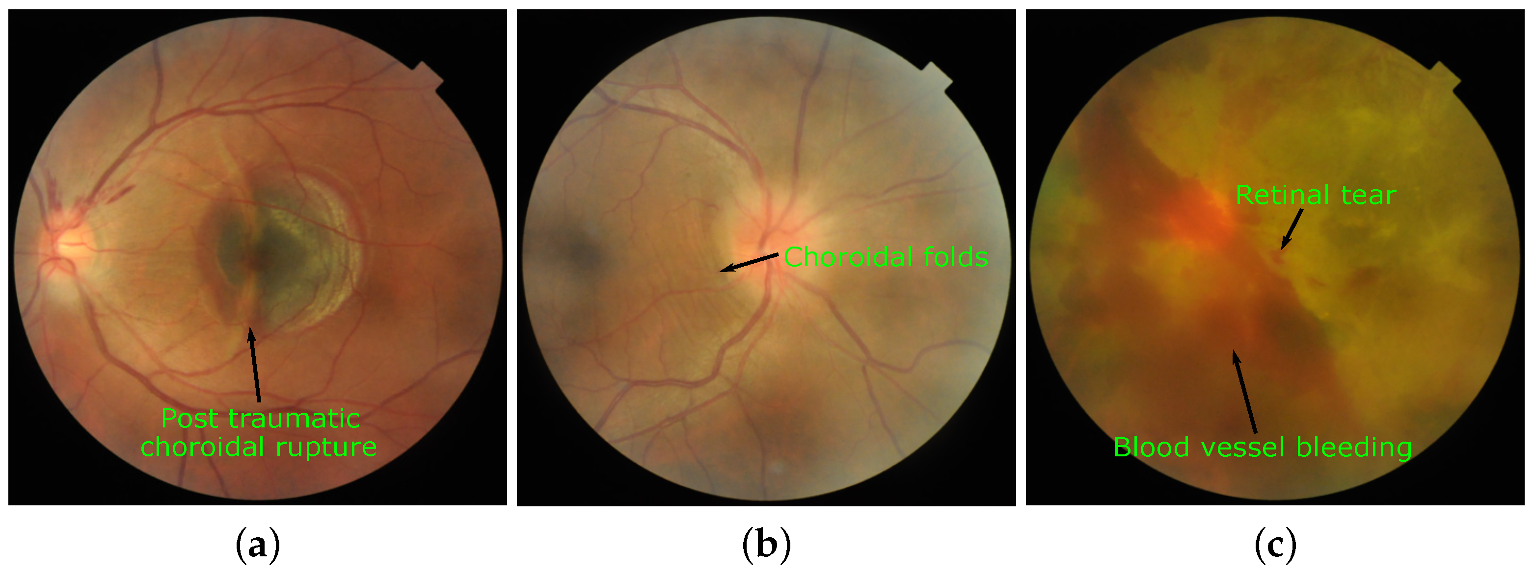
- Chanana, B.; Azad, R.; Kumar, N. Intravitreal bevacizumab for subfoveal choroidal neovascularization secondary to traumatic choroidal rupture. Eye 2009, 23, 2125–2126. [Google Scholar] [CrossRef] [PubMed] [Green Version]
- Cangemi, F.E.; Trempe, C.L.; Walsh, J.B. Choroidal folds. Am. J. Ophthalmol. 1978, 86, 380–387. [Google Scholar] [CrossRef]
- Lascu, R. Vitreous Hemorrhage. Acta Med. Transilv. 2014, 19, 3–39. [Google Scholar]
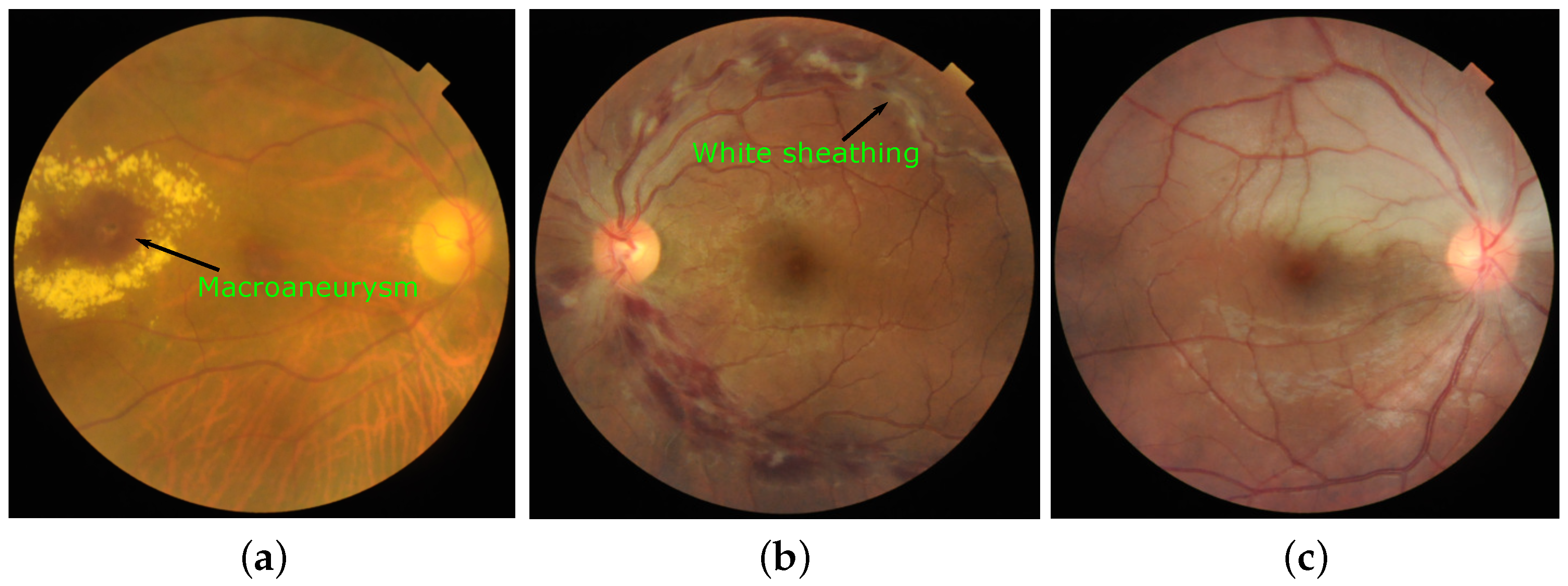
- Yamanaka, E.; Ohguro, N.; Kubota, A.; Yamamoto, S.; Nakagawa, Y.; Tano, Y. Features of retinal arterial macroaneurysms in patients with uveitis. Br. J. Ophthalmol. 2004, 88, 884–886. [Google Scholar] [CrossRef]
- El-Asrar, A.M.A.; Herbort, C.P.; Tabbara, K.F. Differential diagnosis of retinal vasculitis. Middle East Afr. J. Ophthalmol. 2009, 16, 202. [Google Scholar] [PubMed]
- Mason, J.O., III; Shah, A.A.; Vail, R.S.; Nixon, P.A.; Ready, E.L.; Kimble, J.A. Branch retinal artery occlusion: Visual prognosis. Am. J. Ophthalmol. 2008, 146, 455–457. [Google Scholar] [CrossRef] [PubMed]
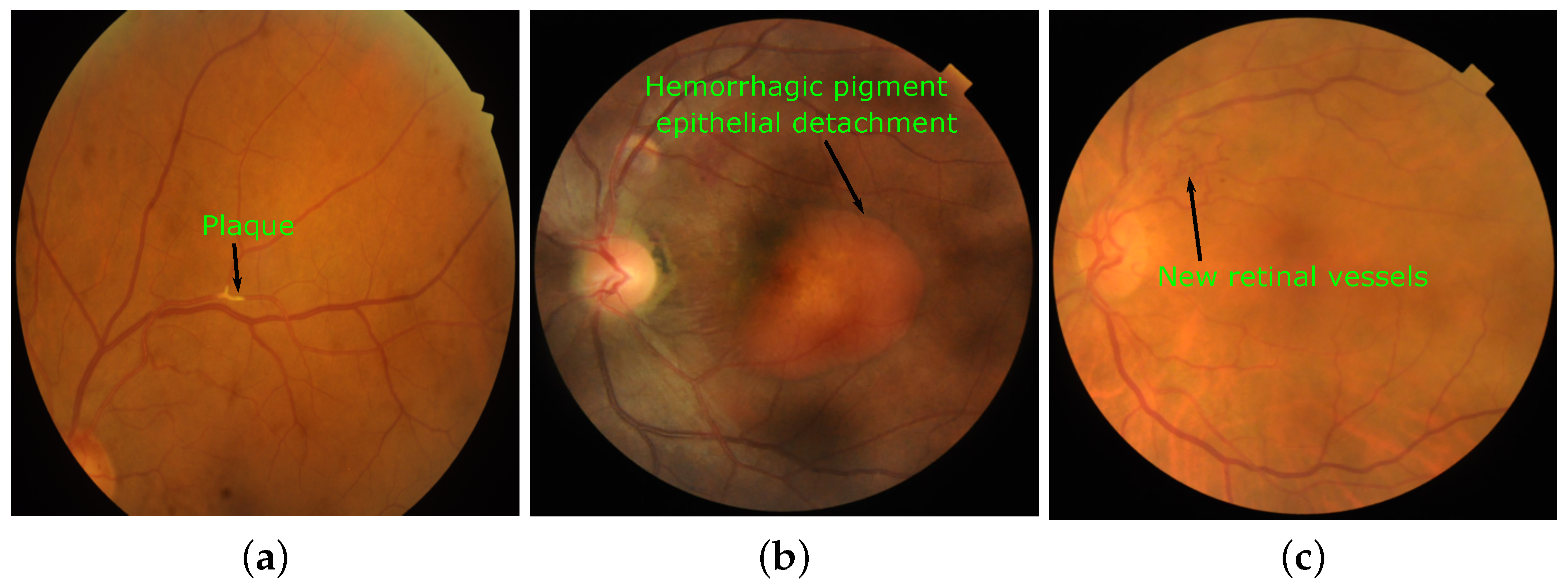
- Hollenhorst, R.W. Significance of bright plaques in the retinal arterioles. JAMA 1961, 178, 23–29. [Google Scholar] [CrossRef] [PubMed] [Green Version]
- Cackett, P.; Htoon, H.; Wong, D.; Yeo, I. Haemorrhagic pigment epithelial detachment as a predictive feature of polypoidal choroidal vasculopathy in a Chinese population. Eye 2010, 24, 789–792. [Google Scholar] [CrossRef] [PubMed]
- Sowka, J.W.; Kabat, A.G. Collateral damage: In addition to vein occlusion, collateral vascularization may be linked to optic disc drusen, diabetes or even tumor formation. By Joseph W. Sowka, OD, and Alan G. Kabat, OD. Rev. Optom. 2014, 151, 85–88. [Google Scholar]


| Subject area | Biomedical Imaging, Ophthalmology |
| More specific subject area | Retinal image analysis for multi-disease detection |
| Type of data | Image, CSV |
| How data was acquired | Three different retinal fundus cameras. Model names: TOPCON 3D OCT-2000, Kowa VX-10 and TOPCON TRC-NW300 |
| Data format | Raw and Manual Annotations |
| Experimental factors | Most of the patients were subjected to mydriasis with one drop of tropicamide at 0.5% concentration |
| Experimental features | The fundus images were captured with position and orientation of the patient sitting upright with 39 mm (Kowa VX-10) and 40.7 mm (TOPCON 3D OCT-2000 and TOPCON TRC-NW300) distance between lenses and examined eye using non-invasive fundus camera |
| Data source location | Eye Clinic, Sushrusha Hospital, and Center of Excellence in Signal and Image Processing, SGGS Institute of Engineering and Technology both located in Nanded, (M.S.), India |
| Data accessibility | https://riadd.grand-challenge.org/download-all-classes/ |
| Model | Hardware | FOV | Resolution (in Pixels) | Number of Images in Dataset |
|---|---|---|---|---|
| TOPCON 3D OCT-2000 | Nikon D7000 digital camera | 45° | 2427 | |
| Kowa VX-10 | Nikon D70s digital camera | 50° | 467 | |
| TOPCON TRC-NW300 | Integrated digital CCD camera | 45° | 306 |
| Sr. No. | Normal/Disease /Marker | # Fundus Images | C1 | C2 | C3 | Sr. No. | Normal/Disease /Marker | # Fundus Images | C1 | C2 | C3 |
|---|---|---|---|---|---|---|---|---|---|---|---|
| 1 | NL | 669 | 333 | 153 | 183 | 24 | CRS | 54 | 46 | 08 | 00 |
| 2 | DR | 632 | 519 | 112 | 01 | 25 | EDN | 24 | 16 | 08 | 00 |
| 3 | ARMD | 169 | 126 | 43 | 00 | 26 | RPEC | 32 | 29 | 03 | 00 |
| 4 | MH | 523 | 425 | 69 | 29 | 27 | MHL | 17 | 15 | 02 | 00 |
| 5 | DN | 230 | 198 | 26 | 06 | 28 | RP | 10 | 08 | 02 | 00 |
| 6 | MYA | 167 | 137 | 26 | 04 | 29 | CWS | 08 | 07 | 01 | 00 |
| 7 | BRVO | 119 | 106 | 13 | 00 | 30 | CB | 02 | 01 | 01 | 00 |
| 8 | TSLN | 304 | 247 | 52 | 05 | 31 | ODPM | 02 | 02 | 00 | 00 |
| 9 | ERM | 26 | 20 | 06 | 00 | 32 | PRH | 05 | 04 | 01 | 00 |
| 10 | LS | 79 | 74 | 05 | 00 | 33 | MNF | 03 | 03 | 00 | 00 |
| 11 | MS | 27 | 25 | 02 | 00 | 34 | HR | 01 | 01 | 00 | 00 |
| 12 | CSR | 61 | 58 | 03 | 00 | 35 | CRAO | 04 | 04 | 00 | 00 |
| 13 | ODC | 445 | 357 | 52 | 36 | 36 | TD | 09 | 07 | 00 | 02 |
| 14 | CRVO | 45 | 43 | 02 | 00 | 37 | CME | 07 | 07 | 00 | 00 |
| 15 | TV | 10 | 09 | 01 | 00 | 38 | PTCR | 06 | 06 | 00 | 00 |
| 16 | AH | 25 | 19 | 04 | 02 | 39 | CF | 06 | 04 | 02 | 02 |
| 17 | ODP | 115 | 94 | 20 | 01 | 40 | VH | 04 | 04 | 00 | 00 |
| 18 | ODE | 96 | 91 | 05 | 00 | 41 | MCA | 01 | 01 | 00 | 00 |
| 19 | ST | 11 | 07 | 03 | 01 | 42 | VS | 04 | 04 | 00 | 00 |
| 20 | AION | 26 | 25 | 01 | 00 | 43 | BRAO | 04 | 03 | 01 | 00 |
| 21 | PT | 17 | 15 | 02 | 00 | 44 | PLQ | 02 | 01 | 01 | 00 |
| 22 | RT | 25 | 23 | 02 | 00 | 45 | HPED | 01 | 01 | 00 | 00 |
| 23 | RS | 71 | 71 | 00 | 00 | 46 | CL | 02 | 01 | 01 | 00 |
Publisher’s Note: MDPI stays neutral with regard to jurisdictional claims in published maps and institutional affiliations. |
© 2021 by the authors. Licensee MDPI, Basel, Switzerland. This article is an open access article distributed under the terms and conditions of the Creative Commons Attribution (CC BY) license (http://creativecommons.org/licenses/by/4.0/).
Share and Cite
Pachade, S.; Porwal, P.; Thulkar, D.; Kokare, M.; Deshmukh, G.; Sahasrabuddhe, V.; Giancardo, L.; Quellec, G.; Mériaudeau, F. Retinal Fundus Multi-Disease Image Dataset (RFMiD): A Dataset for Multi-Disease Detection Research. Data 2021, 6, 14. https://doi.org/10.3390/data6020014
Pachade S, Porwal P, Thulkar D, Kokare M, Deshmukh G, Sahasrabuddhe V, Giancardo L, Quellec G, Mériaudeau F. Retinal Fundus Multi-Disease Image Dataset (RFMiD): A Dataset for Multi-Disease Detection Research. Data. 2021; 6(2):14. https://doi.org/10.3390/data6020014
Chicago/Turabian StylePachade, Samiksha, Prasanna Porwal, Dhanshree Thulkar, Manesh Kokare, Girish Deshmukh, Vivek Sahasrabuddhe, Luca Giancardo, Gwenolé Quellec, and Fabrice Mériaudeau. 2021. "Retinal Fundus Multi-Disease Image Dataset (RFMiD): A Dataset for Multi-Disease Detection Research" Data 6, no. 2: 14. https://doi.org/10.3390/data6020014