Article

The Anticoccidial Effect of Alcoholic Vitis vinifera Leaf Extracts on Eimeria papillate Oocysts Isolated in Mice In Vitro and In Vivo

Department of Zoology, College of Science, King Saud University, P.O. Box 2455, Riyadh 11451, Saudi Arabia
*
Author to whom correspondence should be addressed.
Vet. Sci. 2023, 10(2), 97; https://doi.org/10.3390/vetsci10020097
Submission received: 14 December 2022 / Revised: 21 January 2023 / Accepted: 26 January 2023 / Published: 29 January 2023
(This article belongs to the Special Issue The Effects of Microbiota on Animal Health)

Abstract

:

Simple Summary

Coccidiosis is a disease affecting an enormous group of animals such as birds and mammals. It is caused by an Eimeria spp. infection, which induces gastrointestinal issues that chiefly affect the intestines such as diarrhea, reduced growth performance, weight loss, and reduced feed conversion efficiency; in extreme cases, it can lead to death. In addition, this disease causes substantial economic losses globally. The extensive use of the currently obtainable anticoccidial treatments has led to expansive medication resistance, prompting the investigation of new treatments. Vitis vinifera leaf extract is an interesting potential candidate for an alternative treatment approach. Our study examined the pharmacological potential of VVLE and its effects on E. papillata oocyst sporulation by investigating its ability to inhibit the sporulation of coccidian oocysts in vitro and in mice.

Abstract

Eimeria spp. causes eimeriosis in the guts of numerous domestic mammals and poultry, and the employment of medication and the effects of certain aspects of synthetic anticoccidials in the treatment of eimeriosis have given rise to the appearance of resistant parasites that require the search for alternate remedies. Natural products, which are safe and have no negative impact on the environment, may be utilized in the therapy of an enormous range of parasitic infections. This research aimed to assess the effectiveness of VVLE on the oocyst sporulation of an E. papillate infection in the mouse jejunum. In addition, obtaining the ideal concentration will interrupt the parasite’s life cycle and limit infection. In vitro: Collected unsporulated oocysts (1 × 10³) of E. papillata were given six different concentrations (w/v) of Vitis vinifera leaf extract (10, 25, 50, 100, 150, and 200 mg/mL) and toltrazuril (25 mg/mL), three replicates per group, whereas the control group received 2.5% potassium dichromate solution. In vivo: The mice were separated into six groups; the first and second groups did not receive infection, whilst the third, fourth, fifth, and sixth groups were each given 1 × 10³ sporulated oocysts of E. papillate in the experiment. In addition, an oral dosage of 100 and 200 mg/kg VVLE were given to the fourth and fifth groups, while the sixth group was given toltrazuril at 25 mg/kg. On the fifth day, unpopulated oocysts were collected from each mouse separately. The incubation period and treatments had considerable impacts on the rate of sporulation. The infrared spectroscopy of V. vinifera extract revealed many expected active classes of chemical compounds. Further, the infection of mice with E. papillata caused an oocyst output of nearly 2 × 10⁴ oocysts/g of faeces. VVLE significantly decreased the oocyst output to nearly 88%. In addition, we detected an inhibitory effect on the sporulation (%) and harm (%) of E. papillata oocysts in a dosage-dependent modality compared with the control group. Furthermore, they destroyed the oocyst morphology in terms of the shape, size, and quantity of sporocysts. The results indicate that grape vines have powerful activity as anticoccidials.

1. Introduction

Coccidiosis is a dangerous disease that affects a wide range of animals [1]. It is caused by Eimeria spp. infection, which invades host cells in the infectious stages and causes gastrointestinal issues such as diarrhea, leads to decreased growth performance, and, in extreme cases, leads to death [2,3]. Moreover, Eimeria spp. infections can lead to opportunistic infections of other pathogens such as bacteria [4]. In addition, this disease causes substantial economic losses globally [5]. It has a life cycle that consists of asexual and sexual stages of reproduction, and it produces resistant parasite stages known as oocysts that are released into the environment, thus facilitating the spread of infection. Controlling the Eimeria oocysts is difficult due to their relative resistance to environmental factors [6]. As an outcome, deactivating the sporulation operation is an essential step by which these parasites can be controlled [7]. Furthermore, the extensive prophylactic usage of anticoccidial-added feed has led to extensive medication resistance [5], which has presently been reported against attainable drugs [4,8], e.g., amprolium, sulfonamides, diclazuril, halofuginone, decoquinate, clopidol, and toltrazuril; along with the difficulty of finding an accessible and scalable anticoccidial vaccine, it may be costly and inaccessible to rural farmers and others in the poultry industry [8,9]. An increased interest in safe and effective alternatives aimed at controlling coccidiosis has led to the use of plant extracts, essential oils, and traditional medicinal products [10]. Medicinal plants are the primary source of all alternative or natural medical systems, and they are regarded as therapeutic approaches for obtaining effective chemical ingredients [11]. These products exhibit organ-protective characteristics in Eimeria-infected hosts and target parasites [12].
Lately, some emphasis has been placed on herbal therapy. Plants such as Pinus radiata and Aloe vera [13], Saccharum officinarum [14], Rumex nervosus [15], Cinnamomum Verum [16], and Calotropis Procera [17] were reported as having good antiparasitic and immunomodulatory effects against coccidiosis [18]. Several reviews have described the phytochemical and therapeutic effects of grape (Vitis vinifera) and the active constituents in different parts of the fruit, including the skin, seeds, pomace, and stems [19]. Its primary ingredients have a variety of bioactivities, such as cardioprotection, anticancer, anti-inflammation, antiaging, and antimicrobial activities, which are closely related to disease prevention [20]. In addition to these possible efficacies, it has as an antiparasitic agent [21,22].
Grape leaves contain many compounds with antioxidant activity, such as polyphenols, which include monomeric flavonoids (catechin and epicatechin), tannic acid, ellagitannin, dimeric, trimeric, and polymeric procyanidins, and phenolic acids such as ellagic and Gallic acid [23]. Phenolic compounds and the useful influences from V. vinifera leaves contribute directly to the protective effect observed in the brains of mice, in addition to lowering lipid and protein damage and modifying the status of the antioxidant enzymes investigated [24].
The V. vinifera leaves have been utilized in the treatment of a wide variety of diseases such as in the treatment of hypertension, diarrhea, hemorrhage, varicose veins [21,22], and inflammatory turmoil [25], and to lower blood glucose levels [26]. The leaves of V. vinifera have also been shown to be hepatoprotective against acetaminophen-induced hepatic DNA damage [27].
The present investigation was carried out to assess the pharmacological potential of grape leaves and their effects on E. papillata oocyst sporulation and viability in vitro and in infected mice.

2. Materials and Methods

Thirty-five mice were bought from animal markets. The mice were screened for seven days before the start of the experiment to confirm that they were free of Eimeria infection through fecal investigation, and no Eimeria oocysts were obtained.

2.1. Ethical Approval

The research was carried out in accordance with the ethical criteria for the use of animals established by the Kingdom of Saudi Arabia (Ethic Committee King Saud University, ethical approval number: KSU-SE-21-86).

2.2. Preparation of Extract

The preparation of the grape leaf extract followed the procedure that was outlined by [28], using fresh leaves collected from Shaqra, Saudi Arabia, and a classification scientist at King Saud University’s Botany Department validated the plant’s botanical identity. The air-dried leaves (200 g) were ground into a powder and then extracted by percolating with 70% methanol at room temperature and then conserved for 24 h at 4 °C with occasional stirring. The resulting extract was then concentrated and dried using a rotating vacuum evaporator (Yamato RE300, Tokyo, Japan) under reduced pressure at 40 °C. The obtained crude extracts were weighed and stored at (−20 °C) until used for experiment purposes. A solution of potassium dichromate (K2Cr2O7) and distilled water were employed to dissolve the powder for the different trials.

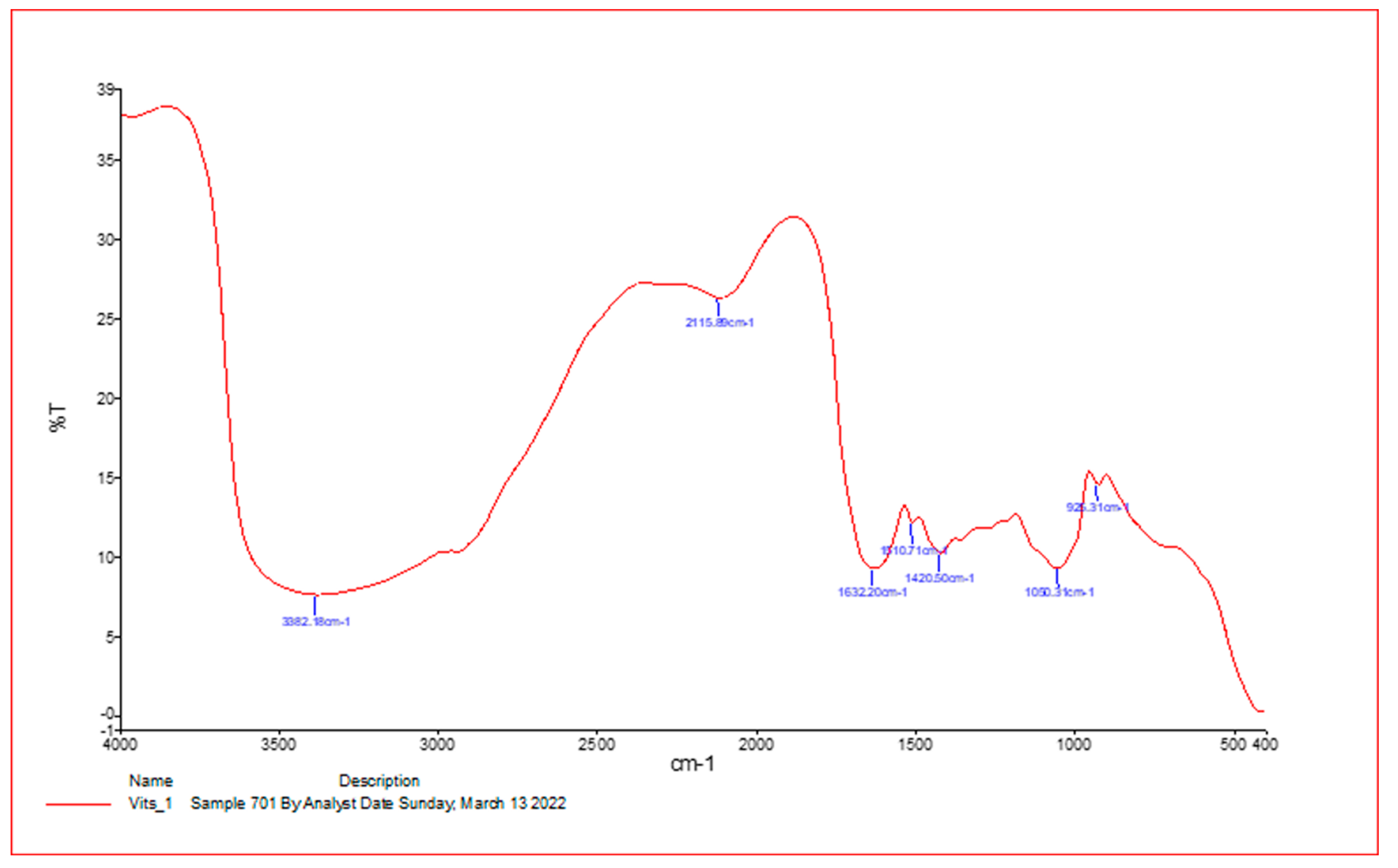
2.3. Infrared Spectroscopy

After processing, a tiny piece of the material was homogenized by combining it with an excessive amount of potassium bromide powder (1:99 wt%). The material was then given a rough mashing before being put in a pellet-forming die. The optical spectrometer NICOLET 6700 Fourier-transform infrared spectroscopy from Thermo Scientific was used to examine infrared (IR) (FT-IR) to predict the likely compound classes. The maximum absorption is expressed as the number of waves (cm−1). At 25 °C, spectra with a resolution of 4 cm were recorded, spanning from 4000 cm−1 to 400.

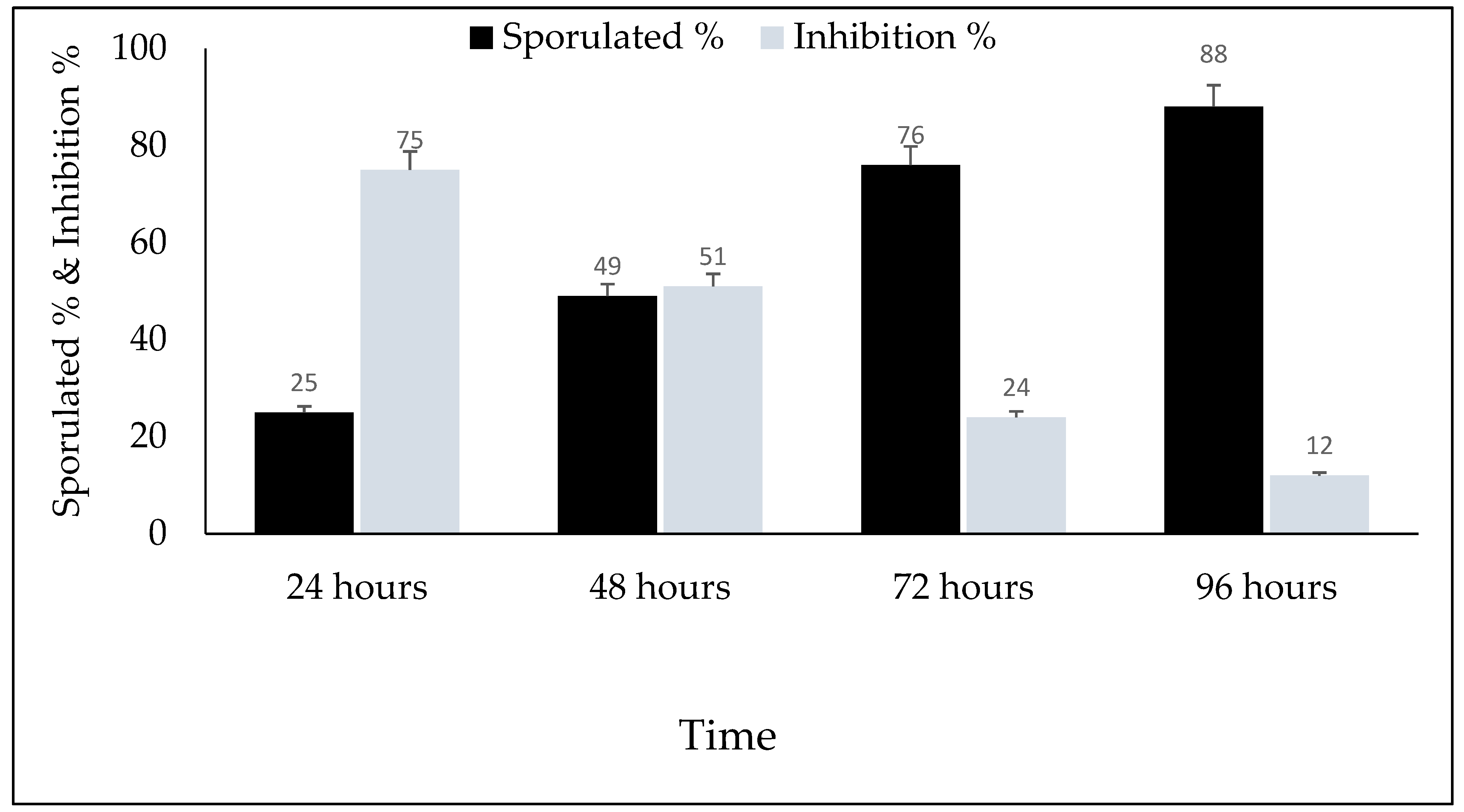
2.4. Oocyst Sporulation

Unpopulated Eimeria papillata oocysts were found by Prof. Mehlhorn, University of Duesseldorf (Duesseldorf, Germany), and the parasite was kept alive by the passage in mice [29]. The treatment was used in vitro through oocysts and was investigated using dichromate. In vivo, sporulation was performed prior to giving it to the mice in order to activate and propagate parasite oocysts.

2.5. In Vitro

A suspension of 654 µL of freshly sporulated oocysts of E. papillata containing 1 × 10³ oocysts was added to each six-well plate and then these dishes were incubated in 3 mL of potassium dichromate containing one of the following concentrations of VVLE: 10%, 25%, 50%, 100%, 150%, or 200%. The potassium dichromate solution (K2Cr2O7) was only used in the negative control group, while toltrazuril (Veterinary Agriculture Products Company (VAPCO)) was used as a positive control group (eight groups). Three replicates were employed for each concentration. All six-well plates for all groups were incubated at 25–29 °C and 65–75% relative humidity and all were semi-covered, with periodic shaking [27]. From each group, a 10 µL suspension was examined microscopically under a light microscope (BX51TF, OLYMPUS, Tokyo, Japan) at 40× magnification, after 24, 48, 72, and 96 h. The viability (oocysts with sporocysts, distorted walls, and inhibitory oocysts) was examined under the light microscope using a McMaster chamber, and the percentages of sporocysts and inhibitory oocysts were calculated using the following equations:
Sporulation (%) = number of sporulated oocysts/total number of oocysts × 100.
The inhibition of sporulation (SI%) = Sporulation of control-sporulation of treated/sporulation of control × 100 as described by [28].

2.6. In Vivo

In this study, adult male C57BL/6 mice aged 10–11 weeks with an average weight of 26–27 g/mice were subjected to the experiment. The mice were divided into five groups with seven mice in each group. The first group was inoculated only with sterile saline (0.9% NaCl) and served as the control group. The second group (noninfected) was treated by oral gavage with 100 μL V. vinifera leaf extract (200 mg/kg) daily for 5 days. The third, fourth, and fifth groups were infected with 1 × 10³ sporulated oocysts of E. papillata and treated either with sterile saline, V. vinifera leaf extract, or 25 mg/kg toltrazuril daily for 5 days, respectively.

2.7. Sample Collection

Fresh fecal pellets were collected every 24 h and weighed for all mice, and the bedding of the mice was changed to prevent reinfection. On day five after infection, each mouse was removed to a small cage, fresh fecal samples were collected from the mice, and the number of oocysts shed per 1 g of feces was calculated according to [30]. The feces pellets from each individual mouse were suspended and diluted in saturated sodium chloride (NaCl) solution for oocyst flotation. The oocysts were counted using a McMaster chamber, and the upshots are expressed as the number of oocysts per gram of feces. To assess the mice’s weight change, each mouse was weighed separately before starting the experiment and again at 5 days post-infection.

2.8. Statistical Analysis
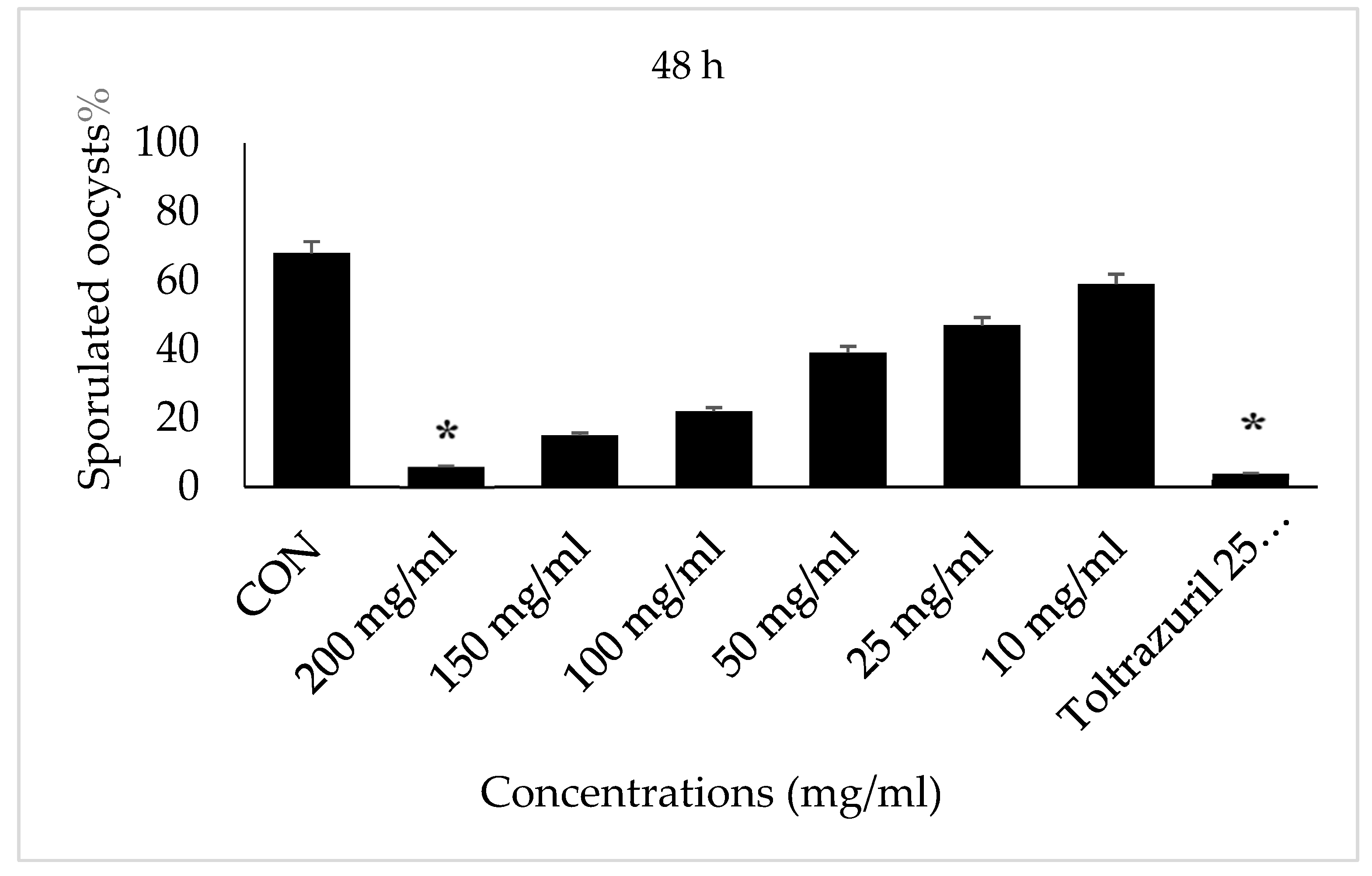
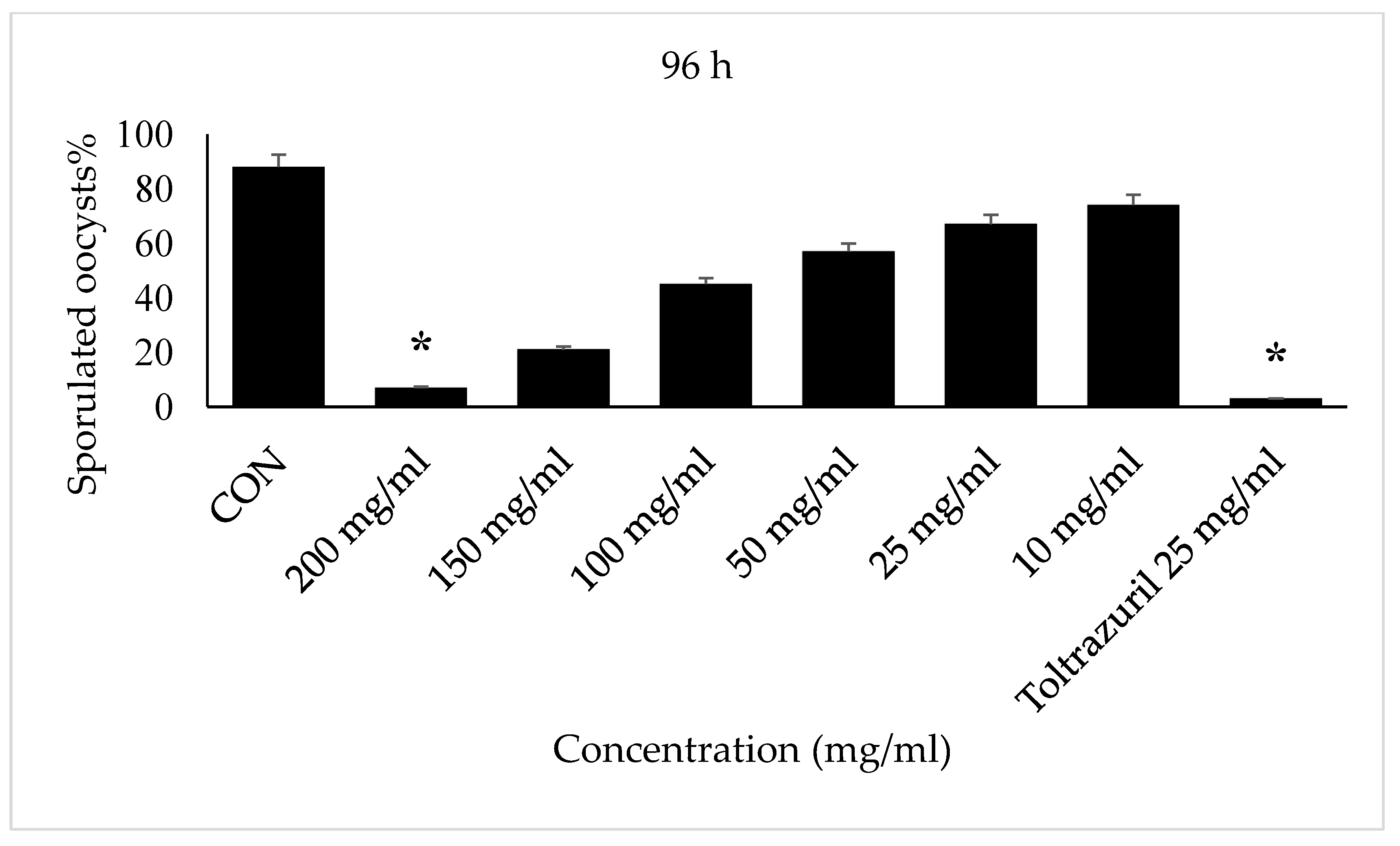
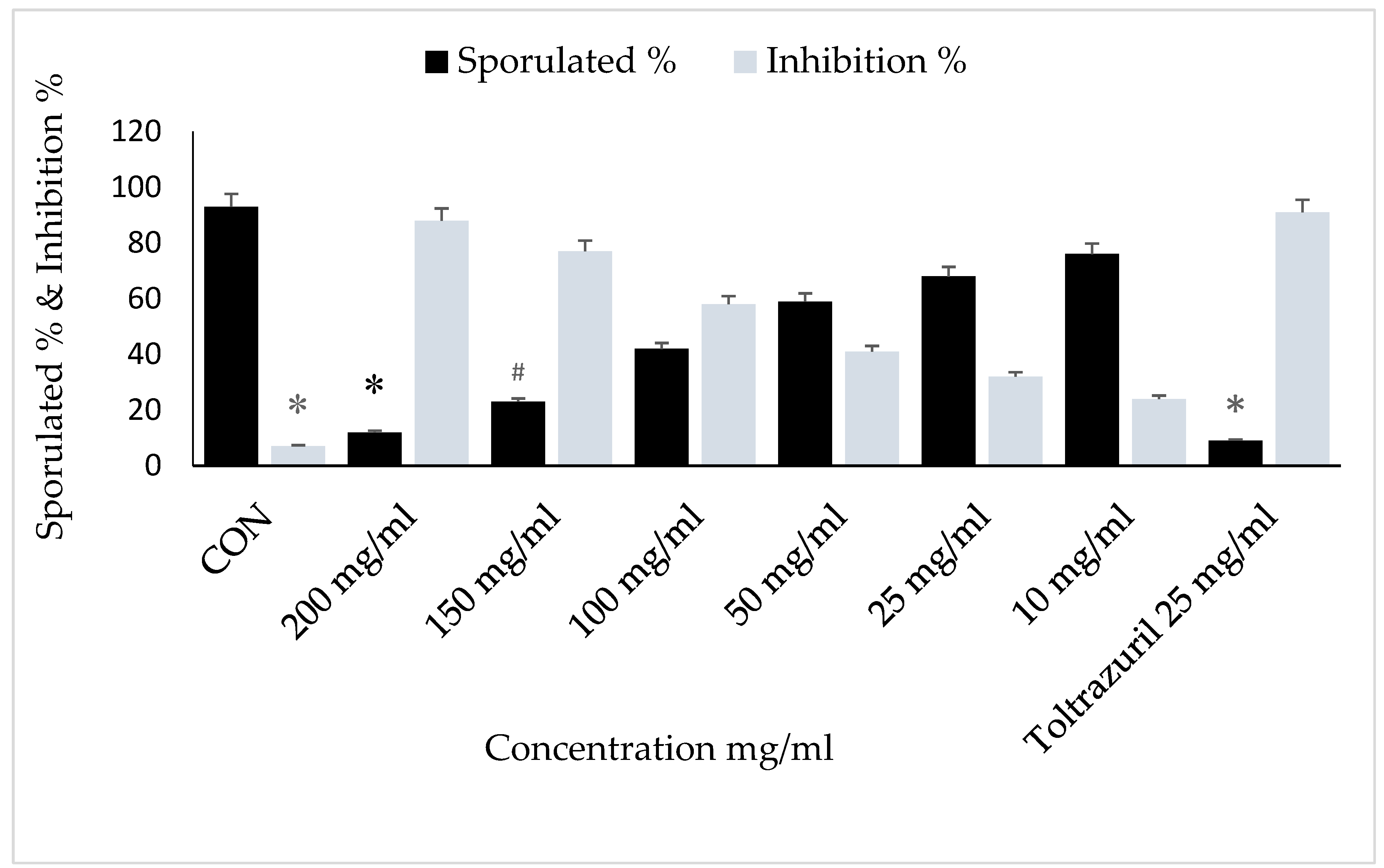
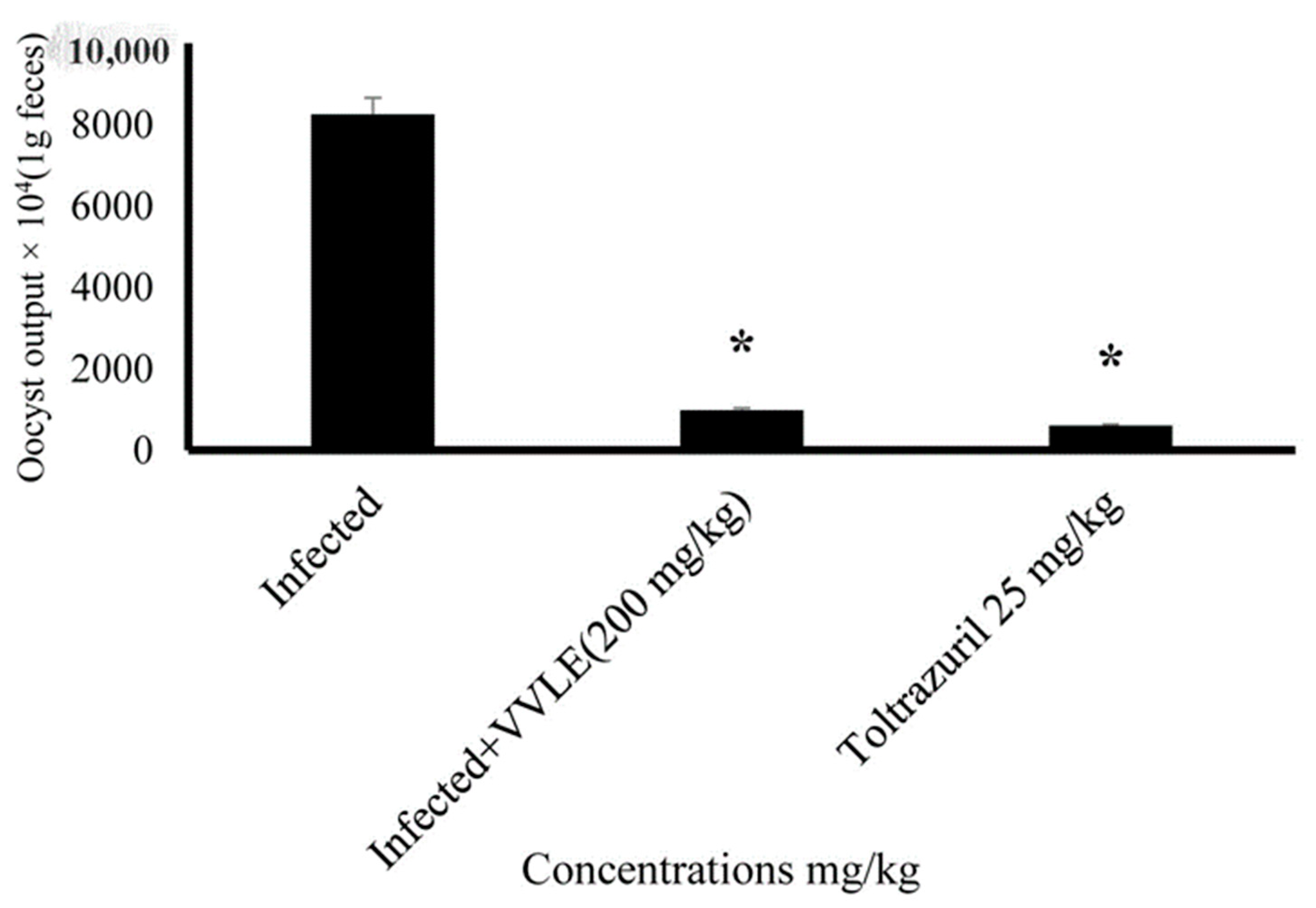
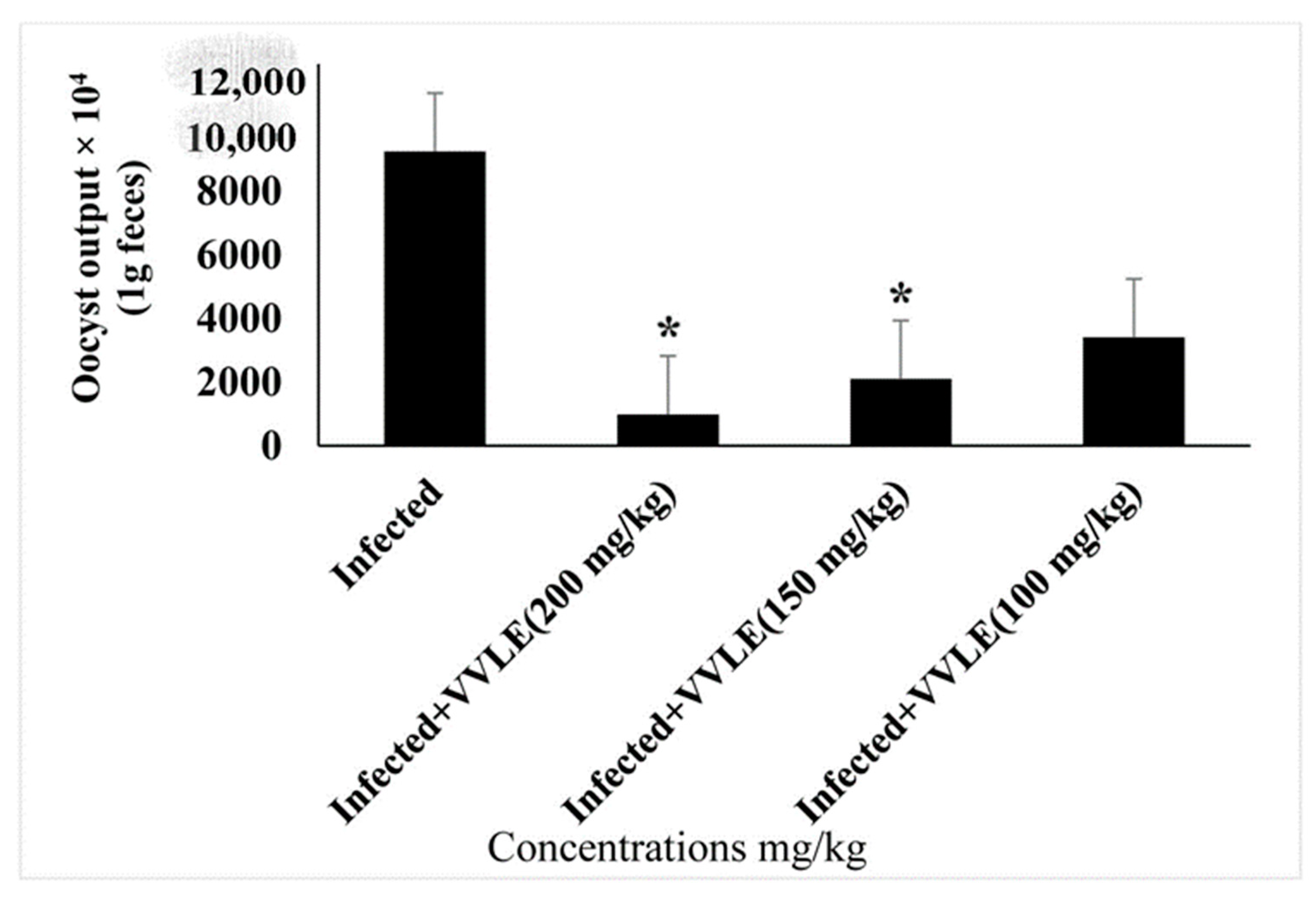
The SPSS software (IBM SPSS, Version 23) was used. Tukey’s method and variables were analyzed using the following experimental design through a two-way ANOVA model for in vitro study:
Yijk = μ + Gi + Tj + GTij + εijk
where Yijk is the value of any observation, μ is the population mean of the measurements, Gi is the effect of the ith treatment groups, Tj is the effect of the jth time of incubation, GTij is the effect of the ijth interaction between treatment and incubation time, and εij is random error.
In vivo study model:
Yij = μ + Ti + εij
where Yij is the value of any observation, μ is the population mean of the measurements, Ti is the effect the ith treatment groups, and εij is random error.
In order to evaluate the overall effects of the treatment in vitro and in vivo, comparisons were made between all groups.
The arithmetic mean (M) and standard deviation (SD) were used to represent the results. Statistically significant differences (p ≤ 0.05) were discovered between treatment groups and control groups.

3. Results

An FT-IR spectrometer was used to perform spectroscopy on the VVLE, analyzed with major bands at 3382.18 cm−1, 2119.89 cm−1, 1632.20 cm−1, 1510.71 cm−1, 1420.50 cm−1, and 1030.31 cm−1, respectively, as shown in Figure 1. The active phytochemical components were obtained as follows: N-H stretching, N=C=S stretchy, C=C stretching, C-O stretching tertiary, CO-O-CO stretching, and C-H bending, with an absorbance of 400–4000/cm−1 (Table 1).
The proportion of sporulated oocysts was calculated for the control and experimental groups at 24, 48, 72, and 96 h. It is important to note that oocysts started to sporulate after 48 h of incubation. The lowest sporulated oocyst percentages were noted in concentrations of 10, 25, and 50 mg/mL. The concentrations of the grape leaf extracts were inversely proportional to the percentage of sporulated oocysts.
After 24 h of E. papillata oocyst incubation in concentrations of 200 mg/mL with VVLE, and toltrazuril 25 mg/mL, there was no sporulation. However, a concentration of 150 mg/mL showed less than 5% sporulation. Concentrations of 10, 25, 50, 100, and 150 mg/mL, showed different levels of sporulation. While the control group (2.5% potassium dichromate solution) remained unsporulated throughout the incubation time until 96 h (Figure 2), we note no significant differences between treatments with all concentrations of VVLE and drug and potassium dichromate (2.5%).
After 48 h of incubation with VVLE (200 µg/mL and toltrazuril 25 mg/mL), the E. papillate oocysts showed high levels of inhibition in comparison with the 2.5% potassium dichromate (control), which showed high levels of sporulated oocysts, while oocysts incubated with VVLE (150,100, 50, 25, and 10 mg/mL) showed low levels of sporulation.
Incubation for 72 h with VVLE at concentrations of 200 mg/mL and toltrazuril inhibited sporulation by nearly 70–80%, whilst incubation with VVLE at the same concentration and toltrazuril for 96 h inhibited sporulation by nearly 88% compared to the control group in which oocysts were incubated with potassium dichromate (2.5%). A reduction in the population of parasites was also obtained at various concentrations throughout 72–96 h for the extract compared with toltrazuril (Figure 3 and Figure 4).
The main effects of the time of sporulation and experimental groups on sporulation (%) and the sporulation inhibition (%) of E. papillata oocysts are shown in Figure 5, which shows that the sporulation percentage increased with an increasing incubation time; conversely, as shown in Figure 6 and Figure 7, the inhibition percentage increased significantly with an increasing incubation time of up to 72 h (p ≤ 0.05). Therefore, the sporulation inhibition rate did not differ significantly between 72 and 96 h exposures. There were interactions between the timing of sporulation and the experimental groups on the sporulation and inhibition of E. papillata oocysts.
The influence of grape leaf extract on the outcome of E. papillata infections was studied. There was no fecal output of oocysts during the first 4 days post-infection. On day five post-infection, the highest oocyst output was 8.5 × 10⁵ ± 29.7 oocysts/g of feces in infected mice. While the grape treatment significantly reduced oocyst output to about 65% on day five post-infection (Table 2), the treatment VVLE at doses of 100, 150, and 200 mg/kg significantly (p < 0.01) reduced the oocyst output to 78.8%, 86.5%, and 96.4%, respectively (Figure 5).
Generally, the experimental groups significantly affected the rates of sporulation (%) and inhibition (%). Concentrations of 200, 150, and 100 mg/mL of VVLE had the highest inhibition rates and lowest sporulation rates (p ≤ 0.05). Concentrations of 50, 25, and 10 mg/mL of VVLE had higher rates of sporulation and the lowest inhibition of sporulation (p ≤ 0.05) (Figure 8 and Figure 9).

4. Discussion

The in vitro and in vivo anticoccidial effects of Vitis vinifera extracts were measured in terms of the number and percentage of Eimeria oocysts damaged and sporulation inhibition. Eimeriosis has caused great economic losses in many countries as it affects most animals [31]. When sporozoites from swallowed sporulated oocysts are released, they enter the jejunum epithelial cells and rapidly multiply, causing E. papillata infections before oocysts are produced. Progeny occurs within the oocysts outside of the host, and they become contagious [32]. In many studies, Eimeriosis drug resistance has been reported [5,33]. Therefore, attention is being paid to using natural products as antiparasitic agents since they are more effective, less toxic, and have fewer adverse effects than conventional chemical agents [12]. Several plant extracts have been used in recent years for the control and treatment of infectious disorders in poultry animals, such as coccidiosis. Many findings reveal that the methanolic extract from the grape plant has anticoccidial properties [34,35]. Our findings revealed a dose-dependent inhibitory effect on Eimeria oocyst sporulation and damage. V. vinifera extracts were utilized to treat mice with experimentally induced coccidiosis. It possessed many several active chemical classes that could be conceivably expected.
This corroborates the findings of several authors who claimed that some plants, such as Saccharum officinarum, had anticoccidial properties [36,37,38], including Pinus radiata [39] and Aloe vera [40,41]. Additionally, oocysts showed various degrees of damage, including cell wall damage, and sometimes the oocyst wall completely disappeared and only free sporocysts were seen [42]. This harm may be caused by anaerobic conditions induced by the grape crude leaf extract. This could be because of the active components in the leaf extract. Anaerobic digestion, which is made up of a sequence of microbial processes, turns the waste’s organic content into biogas and has a detrimental impact on pathogens. These were compared to controls, which were normal E. papillata oocysts at various stages. A reduction in the parasite population was also observed at various doses of the plant extract over a 96-h period compared to toltrazuril and the control.
We found that VVLE, at a concentration of 200 mg/mL, had an influence on oocyst sporulation. This effect may be attributed to the existence of several bioactive phytochemical ingredients [43]. VVLE showed anti-coccidiosis efficacy following the therapy of mice, as confirmed by a significant decrease in the output of E. papillata oocysts in the faeces and oocysts of infected mice. These results agree with those of other studies that investigated Azadirachta indica [44], Punica granatum plant [45], Ziziphus Spina-Christi extract [46], Mulberry extract [47], and grape [42] extract as possible sources of anticoccidial agents, as well as Calotropis procera leaf extract [17].
Various herbal extracts are commonly employed in chicken diets to increase growth rates and animal health, especially when health difficulties are present. Several studies have found that plant extracts improve poultry performance, diet, and conversion efficiency [48,49]. Since VVLE has an anticoccidial action, it could be possible to employ it as a food additive in chicken feed.
Similar in vitro results were reported by [13], who found the sporulation of Eimeria oocysts was inhibited by aqueous pine bark extract. The in vitro anticoccidial efficacy of the ethnomedicinal plants was found to be concentration-dependent.
This gives rise to the possibility that the impact is pharmacologically mediated, demonstrating that the concentrations of the bioactive components may be higher in the plant extracts that have higher dilutions (i.e., 200 and 150 mg/kg). Similar results were obtained by [50], and it also was found that Aloe vera and Thonningia sanguine extracts showed concentration-dependent effects on the oocysts of E. tenella, and P. biglobosa extracts had lower anticoccidial effectiveness [51,52].
This might be a result of the lack of bioactive substances in them or of low levels of such compounds. In contradiction to what was stated in [53], there was in fact anticoccidial action seen in vitro of K. senegalensis, the current investigation found that the plant extracts have anticoccidial activity against E. papillata oocysts. This may be an outcome of the permeation of the cell walls of the oocysts by the fundamental plant components causing damage to the cytoplasm, which was evidenced by the manifestation of abnormal sporocysts in oocysts exposed to 200 and 150 dilutions in most of the plant extracts [42].
Our findings show that grape leaves have potent antiparasitic and anti-sporulation leverage. More research is required to understand the histological and molecular mechanisms of Vitis vinifera leaf extract sporulation suppression and protective effects against E. papillata-induced small intestine damage.

5. Conclusions

Experiments in vitro and in vivo have shown that the extracts have strong effects on the prevention of oocyst sporulation. More extensive pharmacological research should be conducted to determine the new pharmacodynamic effects of bioactive components. Additionally, the primary mechanisms underlying the bioactivities should be investigated in a comprehensive manner. It is necessary to clarify the histological and molecular mechanisms of sporulation inhibition by Vitis vinifera (grape) and its protective influence against E. papillata-induced intestinal contagion.

Author Contributions

Conceptualization, S.A.-Q. and M.M.; methodology, M.M.; software, M.M.; validation, M.M.M. and M.M.; formal analysis, M.M.; investigation, S.A.-Q.; resources, H.M.A.A.; data curation, J.A.; writing—original draft preparation, M.M.; writing—review and editing, M.M.; visualization, M.M.; supervision, S.A.-Q.; project administration, S.A.-Q.; funding acquisition, S.A.-Q. All authors have read and agreed to the published version of the manuscript.

Funding

The funding for this project came from King Saud University in Riyadh, Saudi Arabia, and it was given the Researchers Supporting Project number (RSP2023R3).

Institutional Review Board Statement

The animal study protocol was approved by the Ethics Committee of King Saud University (protocol code KSU-SE-21-86, 9/12/2021–9/12/2022).

Informed Consent Statement

Not applicable.

Data Availability Statement

Not applicable.

Conflicts of Interest

The authors declare no conflict of interest.

References

  1. Mehlhorn, H. Encyclopedic Reference of Parasitology, 6th ed.; Springer: Berlin, Germany, 2014. [Google Scholar]
  2. Allen, P.C.; Fetterer, R. Recent advances in biology and immunobiology of Eimeria species and in diagnosis and control of infection with these coccidian parasites of poultry. Clin. Microbiol. Rev. 2002, 15, 58–65. [Google Scholar] [CrossRef] [PubMed] [Green Version]
  3. Kulkarni, R.R.; Taha-Abdelaziz, K.; Shojadoost, B.; Astill, J.; Sharif, S. Gastrointestinal diseases of poultry: Causes and nutritional strategies for prevention and control. In Improving Gut Health in Poultry; Burleigh Dodds Science Publishing: Cambridge, UK, 2019; pp. 205–236. [Google Scholar]
  4. Collier, C.; Hofacre, C.; Payne, A.; Anderson, D.; Kaiser, P.; Mackie, R.; Gaskins, H. Coccidia-induced mucogenesis promotes the onset of necrotic enteritis by supporting Clostridium perfringens growth. Vet. Immunol. Immunopathol. 2008, 122, 104–115. [Google Scholar] [CrossRef] [PubMed]
  5. Chapman, H. Milestones in avian coccidiosis research: A review. Poult. Sci. 2014, 93, 501–511. [Google Scholar] [CrossRef] [PubMed]
  6. Graat, E.; Henken, A.; Ploeger, H.; Noordhuizen, J.; Vertommen, M. Rate and course of sporulation of oocysts of Eimeria acervulina under different environmental conditions. Parasitology 1994, 108, 497–502. [Google Scholar] [CrossRef] [PubMed]
  7. Mai, K.; Sharman, P.A.; Walker, R.A.; Katrib, M.; Souza, D.D.; McConville, M.J.; Wallach, M.G.; Belli, S.I.; Ferguson, D.J.; Smith, N.C. Oocyst wall formation and composition in coccidian parasites. Mem. Do Inst. Oswaldo Cruz 2009, 104, 281–289. [Google Scholar] [CrossRef] [PubMed] [Green Version]
  8. McDonald, V.; Shirley, M. Past and future: Vaccination against Eimeria. Parasitology 2009, 136, 1477–1489. [Google Scholar] [CrossRef]
  9. Kostadinović, L.; Lević, J. Effects of phytoadditives in poultry and pigs diseases. J. Agron. 2018, 5, 1–7. [Google Scholar]
  10. Sidiropoulou, E.; Skoufos, I.; Marugan-Hernandez, V.; Giannenas, I.; Bonos, E.; Aguiar-Martins, K.; Lazari, D.; Blake, D.P.; Tzora, A. In vitro anticoccidial study of oregano and garlic essential oils and effects on growth performance, fecal oocyst output, and intestinal microbiota in vivo. Front. Vet. Sci. 2020, 7, 420. [Google Scholar] [CrossRef]
  11. Mosaddeghi, P.; Eslami, M.; Farahmandnejad, M.; Akhavein, M.; Ranjbarfarrokhi, R.; Khorraminejad-Shirazi, M.; Shahabinezhad, F.; Taghipour, M.; Dorvash, M.; Sakhteman, A. A systems pharmacology approach to identify the autophagy-inducing effects of Traditional Persian medicinal plants. Sci. Rep. 2021, 11, 336. [Google Scholar] [CrossRef]
  12. Wunderlich, F.; Al-Quraishy, S.; Steinbrenner, H.; Sies, H.; Dkhil, M.A. Towards identifying novel anti-Eimeria agents: Trace elements, vitamins, and plant-based natural products. Parasitol. Res. 2014, 113, 3547–3556. [Google Scholar] [CrossRef] [PubMed]
  13. Molan, A.L.; Liu, Z.; De, S. Effect of pine bark (Pious radiata) extracts on sporulation of coccidian oocysts. Folia Parasitol. 2009, 56, 1. [Google Scholar] [CrossRef] [PubMed]
  14. Zhao, Y.; Chen, M.; Zhao, Z.; Yu, S. The antibiotic activity and mechanisms of sugarcane (Saccharum officinarum L.) bagasse extract against food-borne pathogens. Food Chem. 2015, 185, 112–118. [Google Scholar] [CrossRef] [PubMed]
  15. Qasem, M.A.; Dkhil, M.A.; Al-Shaebi, E.M.; Murshed, M.; Mares, M.; Al-Quraishy, S. Rumex nervosus leaf extracts enhance the regulation of goblet cells and the inflammatory response during infection of chickens with Eimeria tenella. J. King Saud Univ.-Sci. 2020, 32, 1818–1823. [Google Scholar] [CrossRef]
  16. Qaid, M.M.; Al-Mufarrej, S.I.; Azzam, M.M.; Al-Garadi, M.A. Anticoccidial effectivity of a traditional medicinal plant, Cinnamomum verum, in broiler chickens infected with Eimeria tenella. Poult. Sci. 2021, 100, 100902. [Google Scholar] [CrossRef] [PubMed]
  17. Murshed, M.; Al-Quraishy, S.; Qasem, M.A. In vitro: Anti-coccidia activity of Calotropis procera leaf extract on Eimeria papillata oocysts sporulation and sporozoite. Open Chem. 2022, 20, 1057–1064. [Google Scholar] [CrossRef]
  18. Abbas, R.Z.; Abbas, A.; Raza, M.A.; Khan, M.K.; Saleemi, M.K.; Saeed, Z. In vitro anticoccidial activity of Trachyspermum ammi (Ajwain) extract on oocysts of Eimeria species of Chicken. Adv. Life Sci. 2019, 7, 44–47. [Google Scholar]
  19. Xia, E.-Q.; Deng, G.-F.; Guo, Y.-J.; Li, H.-B. Biological activities of polyphenols from grapes. Int. J. Mol. Sci. 2010, 11, 622–646. [Google Scholar] [CrossRef]
  20. Nassiri-Asl, M.; Hosseinzadeh, H. Review of the pharmacological effects of Vitis vinifera (Grape) and its bioactive constituents: An update. Phytother. Res. 2016, 30, 1392–1403. [Google Scholar] [CrossRef]
  21. Başoğlu, F.; Şahin, İ.; Korukluoğlu, M.; Uylaser, V.; Akpinar, A. A research on the effects of fermentation type and additives on quality and preservation and development of adequate technique in brined vine-leaves production. Turk. J. Agric. For. 1996, 20, 535–545. [Google Scholar]
  22. Koşar, M.; Küpeli, E.; Malyer, H.; Uylaşer, V.; Türkben, C.; Başer, K.H.C. Effect of brining on biological activity of leaves of Vitis vinifera L. (Cv. Sultani Cekirdeksiz) from Turkey. J. Agric. Food Chem. 2007, 55, 4596–4603. [Google Scholar] [CrossRef]
  23. Crozier, A.; Jaganath, I.B.; Clifford, M.N. Dietary phenolics: Chemistry, bioavailability and effects on health. Nat. Prod. Rep. 2009, 26, 1001–1043. [Google Scholar] [CrossRef] [PubMed]
  24. Dani, C.; Oliboni, L.; Agostini, F.; Funchal, C.; Serafini, L.; Henriques, J.; Salvador, M. Phenolic content of grapevine leaves (Vitis labrusca var. Bordo) and its neuroprotective effect against peroxide damage. Toxicol. Vitr. 2010, 24, 148–153. [Google Scholar] [CrossRef] [PubMed]
  25. Bombardelli, E.; Morazzoni, P.; Carini, M.; Aldini, G.; Maffei Facino, R. Biological activity of procyanidins from Vitis vinifera L. BioFactors 1997, 6, 429–431. [Google Scholar] [CrossRef]
  26. Walker, J.B. Evaluation of the ability of seven herbal resources to answer questions about herbal products asked in drug information centers. Pharmacother. J. Hum. Pharmacol. Drug Ther. 2002, 22, 1611–1615. [Google Scholar] [CrossRef] [PubMed]
  27. Ercisli, S.; Orhan, E. Chemical composition of white (Morus alba), red (Morus rubra) and black (Morus nigra) mulberry fruits. Food Chem. 2007, 103, 1380–1384. [Google Scholar] [CrossRef]
  28. Manikandan, P.; Letchoumy, P.V.; Gopalakrishnan, M.; Nagini, S. Evaluation of Azadirachta indica leaf fractions for in vitro antioxidant potential and in vivo modulation of biomarkers of chemoprevention in the hamster buccal pouch carcinogenesis model. Food Chem. Toxicol. 2008, 46, 2332–2343. [Google Scholar] [CrossRef] [PubMed]
  29. Dkhil, M.; Abdel-Baki, A.; Wunderlich, F.; Sies, H.; Al-Quraishy, S. Anticoccidial and antiinflammatory activity of garlic in murine Eimeria papillata infections. Vet. Parasitol. 2011, 175, 66–72. [Google Scholar] [CrossRef]
  30. Schito, M.L.; Barta, J.R.; Chobotar, B. Comparison of four murine Eimeria species in immunocompetent and immunodeficient mice. J. Parasitol. 1996, 82, 255–262. [Google Scholar] [CrossRef]
  31. Andrews, A. Some aspects of coccidiosis in sheep and goats. Small Rumin. Res. 2013, 110, 93–95. [Google Scholar] [CrossRef]
  32. Pakandl, M. Selection of a precocious line of the rabbit coccidium Eimeria flavescens Marotel and Guilhon (1941) and characterisation of its endogenous cycle. Parasitol. Res. 2005, 97, 150–155. [Google Scholar] [CrossRef] [PubMed]
  33. Williams, R. A compartmentalised model for the estimation of the cost of coccidiosis to the world's chicken production industry. Int. J. Parasitol. 1999, 29, 1209–1229. [Google Scholar] [CrossRef] [PubMed]
  34. Pieri, F.; Silva, V.; Vargas, F.; Veiga Junior, V.; Moreira, M. Antimicrobial activity of Copaifera langsdorffii oil and evaluation of its most bioactive fraction against bacteria of dog’s dental plaque. Pak. Vet. J. 2014, 34. [Google Scholar]
  35. Xiao, C.; Ji, Q.; Rajput, Z.; Wei, Q.; Liu, Y.; Bao, G. Antifungal Efficacy of Phellodendron amurense Ethanol Extract against Trichophyton mentagrophytes in Rabbits. Pak. Vet. J. 2014, 34, 165–169. [Google Scholar]
  36. Fornazier, R.F.; Ferreira, R.R.; Vitoria, A.; Molina, S.; Lea, P.; Azevedo, R. Effects of cadmium on antioxidant enzyme activities in sugar cane. Biol. Plant. 2002, 45, 91–97. [Google Scholar] [CrossRef]
  37. El-Abasy, M.; Motobu, M.; Sameshima, T.; Koge, K.; Onodera, T.; Hirota, Y. Adjuvant effects of sugar cane extracts (SCE) in chickens. J. Vet. Med. Sci. 2003, 65, 117–119. [Google Scholar] [CrossRef]
  38. Hikosaka, K.; El-Abasy, M.; Koyama, Y.; Motobu, M.; Koge, K.; Isobe, T.; Kang, C.B.; Hayashidani, H.; Onodera, T.; Wang, P.C. Immunostimulating effects of the polyphenol-rich fraction of sugar cane (Saccharum officinarum L.) extract in chickens. Phytother. Res. Int. J. Devoted Pharmacol. Toxicol. Eval. Nat. Prod. Deriv. 2007, 21, 120–125. [Google Scholar] [CrossRef] [PubMed]
  39. Wang, L.; Gong, T.; Chen, R.Y. Two new prenylflavonoids from Morus nigra L. Chin. Chem. Lett. 2009, 20, 1469. [Google Scholar] [CrossRef]
  40. Molan, A.; Sivakumaran, S.; Spencer, P.; Meagher, L. Green tea flavan-3-ols and oligomeric proanthocyanidins inhibit the motility of infective larvae of Teladorsagia circumcincta and Trichostrongylus colubriformis in vitro. Res. Vet. Sci. 2004, 77, 239–243. [Google Scholar] [CrossRef]
  41. Narsih, K.S.; Wignyanto, W.S. Identification of aloin and saponin and chemical composition of volatile constituents from Aloe vera (L.) Peel. J. Agric. Food Tech. 2012, 2, 79–84. [Google Scholar]
  42. Abbas, R.; Abbas, A.; Iqbal, Z.; Raza, M.; Hussain, K.; Ahmed, T.; Shafi, M. In vitro anticoccidial activity of Vitis vinifera extract on oocysts of different Eimeria species of broiler chicken. J. Hell. Vet. Med. Soc. 2020, 71, 2267–2272. [Google Scholar] [CrossRef]
  43. Olaku, O.O.; Ojukwu, M.O.; Zia, F.Z.; White, J.D. The role of grape seed extract in the treatment of chemo/radiotherapy induced toxicity: A systematic review of preclinical studies. Nutr. Cancer 2015, 67, 730–740. [Google Scholar] [CrossRef] [PubMed]
  44. Dkhil, M.A.; Al-Quraishy, S.; Abdel Moneim, A.E.; Delic, D. Protective effect of Azadirachta indica extract against Eimeria papillata-induced coccidiosis. Parasitol. Res. 2013, 112, 101–106. [Google Scholar] [CrossRef] [PubMed]
  45. Amer, O.S.; Dkhil, M.A.; Hikal, W.M.; Al-Quraishy, S. Antioxidant and anti-inflammatory activities of pomegranate (Punica granatum) on Eimeria papillata-induced infection in mice. BioMed Res. Int. 2015, 2015, 219670. [Google Scholar] [CrossRef] [PubMed] [Green Version]
  46. Alzahrani, F.; Al-Shaebi, E.M.; Dkhil, M.A.; Al-Quraishy, S. In vivo anti-eimeria and in vitro anthelmintic activity of Ziziphus spina-christi leaf extracts. Pak. J. Zool. 2016, 48. [Google Scholar]
  47. Thagfan, F.A.; Al-Megrin, W.A.; Al-Quraishy, S.; Dkhil, M.A.M. Mulberry extract as an ecofriendly anticoccidial agent: In vitro and in vivo application. Rev. Bras. De Parasitol. Veterinária 2020, 29, e009820. [Google Scholar] [CrossRef] [PubMed]
  48. Alcicek, A.; Bozkurt, M.; Çabuk, M. The effect of a mixture of herbal essential oils, an organic acid or a probiotic on broiler performance. S. Afr. J. Anim. Sci. 2004, 34, 217–222. [Google Scholar]
  49. Gracia, M.; Millán, C.; Sánchez, J.; Guyard-Nicodeme, M.; Mayot, J.; Carre, Y.; Csorbai, A.; Chemaly, M.; Medel, P. Efficacy of feed additives against Campylobacter in live broilers during the entire rearing period: Part B. Poult. Sci. 2016, 95, 886–892. [Google Scholar] [CrossRef]
  50. Abdullateef, S.; Taib Tahir, M.; A Hasso, R. The effect of nandrolone decanoate on liver of rabbitsusing histological and ultrasound methods. Ann. Coll. Med. Mosul 2009, 35, 160–166. [Google Scholar] [CrossRef]
  51. Marizvikuru, M.; Patrick, J.M. Point prevalence study of gastro-intestinal parasites in village chickens of Centane district, South Africa. Afr. J. Agric. Res. 2011, 6, 2033–2038. [Google Scholar]
  52. Onan, K.S.; Toure, A.; Ouattara, K.; Djaman, A.J.; Nrsquo; Guessan, J.D. In vitro anticoccidial activity of Thonningia sanguinea extract on Eimeria tenella and Eimeria necatrix sporozoites cell. Afr. J. Microbiol. Res. 2012, 6, 6247–6251. [Google Scholar]
  53. Otu, M.O.; Lawal, I.A.; George, B.D.; Abubakar, M.S.; Adeyinka, I.A.; Abeke, F.O.; Sekoni, A.A.; Nwagu, B.I.; Adejoh-Ubani, E.O.; Oluntunmogun, A.K.; et al. Effects of N-Butanol and Aqueous Fractions of Khaya senegalensis, Guiera senegalensis and Tamarindus Indica Leaves Extracts on Eimeria tenella Oocyst Sporulation in Vitro. Niger. Vet. J. 2020, 41, 175–191. [Google Scholar] [CrossRef]
Figure 1. The results of undertaking infrared spectroscopy on VVLE samples. An FT-IR spectrometer was used to collect the data, and the findings were collected in the range of 400–4000/cm−1.
Vetsci 10 00097 g001
Figure 2. Effect of VVLE on the sporulation of E. papillata oocysts at 48 h in vitro. The significance was determined by comparing it to a negative control consisting of potassium dichromate 2.5% and a positive control consisting of toltrazuril 25 mg/mL. Significance is in relation to the group that served as a control (p < 0.05). Significance (*): p-value ≤ 0.05. (VVLE): Vitis vinifera leaf extract. (h): hours.
Vetsci 10 00097 g002
Figure 3. Effect of VVLE on sporulation of E. papillata oocysts at 48 h in vitro. The significance was compared to potassium dichromate 2.5% as a negative control and toltrazuril 25 mg/mL as a positive control. Significance is in comparison to the control group (p < 0.05). Significance (*): p-value ≤ 0.05. (VVLE): Vitis vinifera leaf extract. (h): hours.
Vetsci 10 00097 g003
Figure 4. Effect of VVLE on sporulation of E. papillata oocysts at 72 h in vitro. The significance was determined by comparing it to a negative control consisting of potassium dichromate 2.5% and a positive control consisting of toltrazuril 25 mg/mL. Significance is in relation to the group that served as a control (p < 0.05). Significance (*): p-value ≤ 0.05. (VVLE): Vitis vinifera leaf extract. (h): hours.
Vetsci 10 00097 g004
Figure 5. Effect of VVLE on sporulation of E. papillata oocysts at 96 h in vitro. The significance was determined by comparing it to a negative control consisting of potassium dichromate 2.5% and a positive control consisting of toltrazuril 25 mg/mL. When compared to the control group, significance was found (p < 0.05). Significance (*): p-value ≤ 0.05. (VVLE): Vitis vinifera leaf extract. (h): hours.
Vetsci 10 00097 g005
Figure 6. In vitro main contact time effects of VVLE on sporulation %, inhibition %, of E. papillata oocysts at different times for all concentrations. The significance was determined by comparing it to a negative control consisting of potassium dichromate 2.5% and a positive control consisting of toltrazuril 25 mg/mL. Significance is in comparison to the control group. Significance (*): p-value ≤ 0.01. (#) p-value ≤ 0.05. (VVLE): Vitis vinifera leaf extract. (h): hours.
Vetsci 10 00097 g006
Figure 7. In vitro main effects of VVLE on sporulation %, inhibition%, of E. papillata oocysts at different concentrations during 24, 48, 72, and 96 h. The significance was compared to potassium dichromate 2.5% as a negative control and toltrazuril 25 mg/mL as a positive control. (VVLE): Vitis vinifera leaf extract. (h): hours.
Vetsci 10 00097 g007
Figure 8. The effect of different VVLE doses on oocyst output on day five after E. papillata oocyst infection. * Significance is in comparison to the infected group (p ≤ 0.05).
Vetsci 10 00097 g008
Figure 9. Sporulation oocysts count in feces of infected, VVLE, and toltrazuril-treated mice. *, the difference between the infected treated groups is significant at p ≤ 0.05.
Vetsci 10 00097 g009
Table 1. IR spectrum of Vitis vinifera leaf extracts by frequency range.
Absorption (cm−1)AppearanceTransmittance (%)GroupCompound Class
3425.53medium12N-H stretchingAliphatic primary amine
2093.10strong47N=C=S stretchyIsothiocyanate
1641.41strong25C=C stretchingAlkene
1209.12strong37C-O stretching tertiaryalcohol
1045.64Strong broad35CO-O-CO stretchingAnhydride
410.42strong3C-H bending1,2-disubstituted
Table 2. Effect of VVLE on oocyst output on day five post-infection with E. papillata oocysts.
Weight of Mice (g)Oocyst Output/g Feces
Day 0Day 5
Noninfected (−vitis)28.19 ± 1.428.70 ± 1.40
Noninfected (+vitis)26.9 ± 1.1627.06 ± 0.980
Infected (−vitis)26.5 ± 1.426.3 ± 1.1828.75 ± 54.668
Infected (+vitis)26.51 ± 0.7426.83 ± 0.74628.808 ± 6.785 *
Toltrazuril 25 mg/mL26.17 ± 1.1526.98 ± 1.526.743 ± 6.321 *
* Significance against the infected group (p < 0.01).
Disclaimer/Publisher’s Note: The statements, opinions and data contained in all publications are solely those of the individual author(s) and contributor(s) and not of MDPI and/or the editor(s). MDPI and/or the editor(s) disclaim responsibility for any injury to people or property resulting from any ideas, methods, instructions or products referred to in the content.

