
Prevalence of Presenting Conditions in Grey Seal Pups (Halichoerus grypus) Admitted for Rehabilitation

Abstract
:1. Introduction
2. Methods
2.1. Seals
2.2. Data
3. Results

| Neonate | White-Coat | Mid-Moult | Moulted | Total | |
|---|---|---|---|---|---|
| Female | 7 | 17 | 11 | 52 | 87 |
| Male | 4 | 11 | 19 | 84 | 118 |
| Total | 11 | 28 | 30 | 136 | 205 |
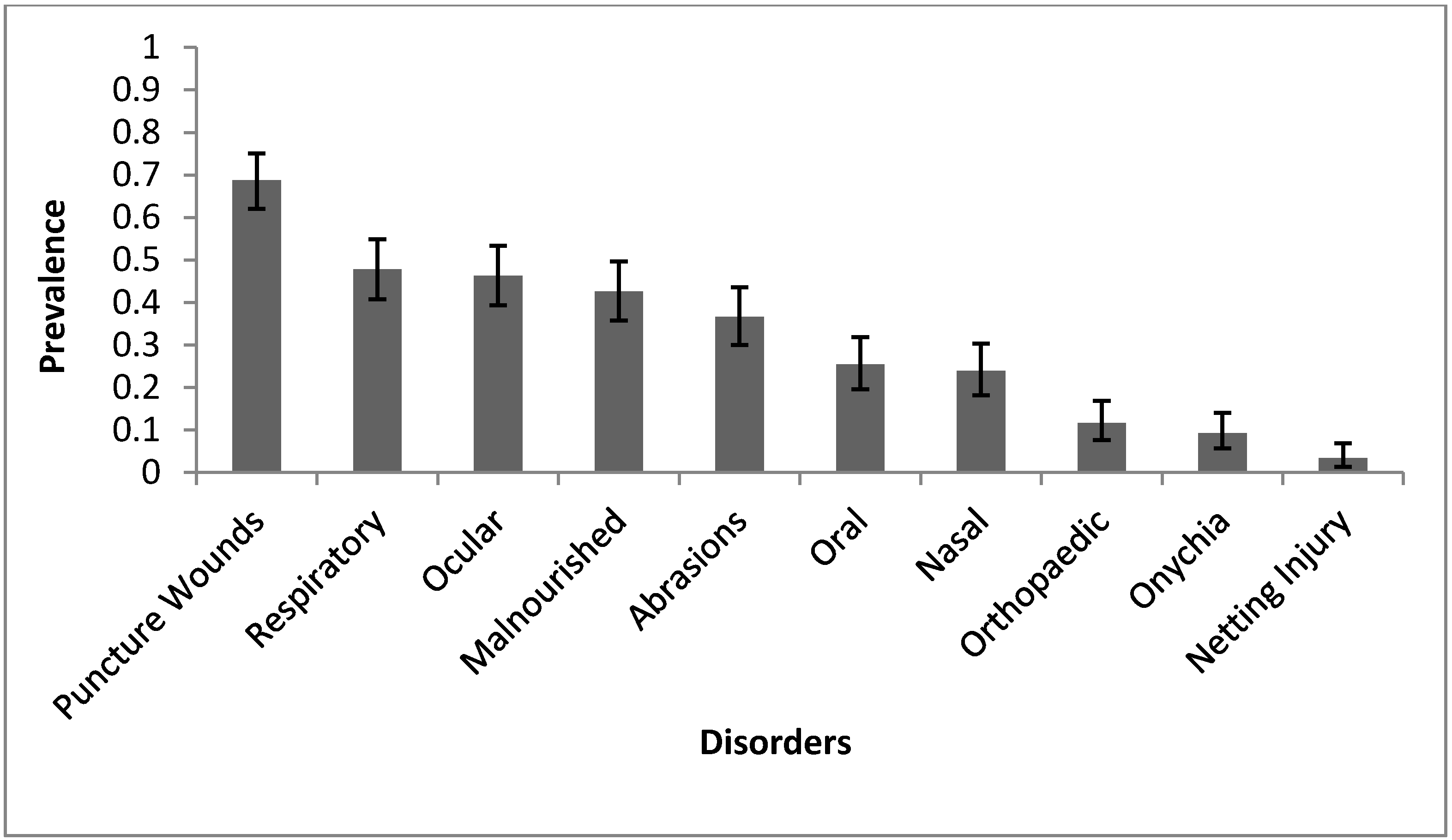
3.1. Prevalence

3.2. Gender
3.3. Age
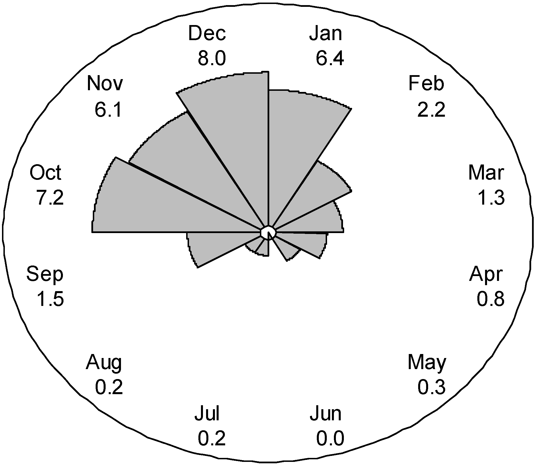
3.4. Season
| Disorder | Factor | Variable | n | OR (95% CI) | p | |
|---|---|---|---|---|---|---|
| Nasal | Age | White-Coat and Neonate | 4 | 0.28 (0.09–0.85) | 0.025 | ** |
| Mid-Moult | 6 | 0.62 (0.24–1.64) | 0.336 | |||
| Moulted | 39 | Ref | ||||
| Oral | Gender | F | 15 | 0.46 (0.23–0.90) | 0.023 | ** |
| M | 37 | Ref | ||||
| Age | White-Coat and Neonate | 4 | 0.25 (0.08–0.74) | 0.012 | ** | |
| Mid-Moult | 5 | 0.43 (0.16–1.21) | 0.109 | |||
| Moulted | 43 | Ref | ||||
| Respiratory | Gender | F | 35 | 0.59 (0.34–1.03) | 0.063 | * |
| M | 63 | Ref | ||||
| Age | White-Coat and Neonate | 8 | 0.19 (0.08–0.44) | <0.001 | ** | |
| Mid-Moult | 11 | 0.42 (0.18–0.95) | 0.036 | ** | ||
| Moulted | 79 | Ref | ||||
| Puncture Wounds | Age | White-Coat and Neonate | 13 | 0.15 (0.07–0.33) | <0.001 | ** |
| Mid-Moult | 24 | 1.23 (0.46–3.27) | 0.677 | |||
| Moulted | 104 | Ref | ||||
| Abrasions | Gender | F | 22 | 0.42 (0.23–0.76) | 0.004 | ** |
| M | 53 | Ref |

3.5. Temperature
| Temperature Classification | Temperature Range (°C) | n |
|---|---|---|
| Marked Hypothermic | <35 | 5 |
| Mildly Hypothermic | 35–35.9 | 7 |
| Normal | 36–37.9 | 105 |
| Mildly Hyperthermic | 38–38.9 | 71 |
| Marked Hyperthermic | >39 | 14 |
| Not Recorded | 3 |
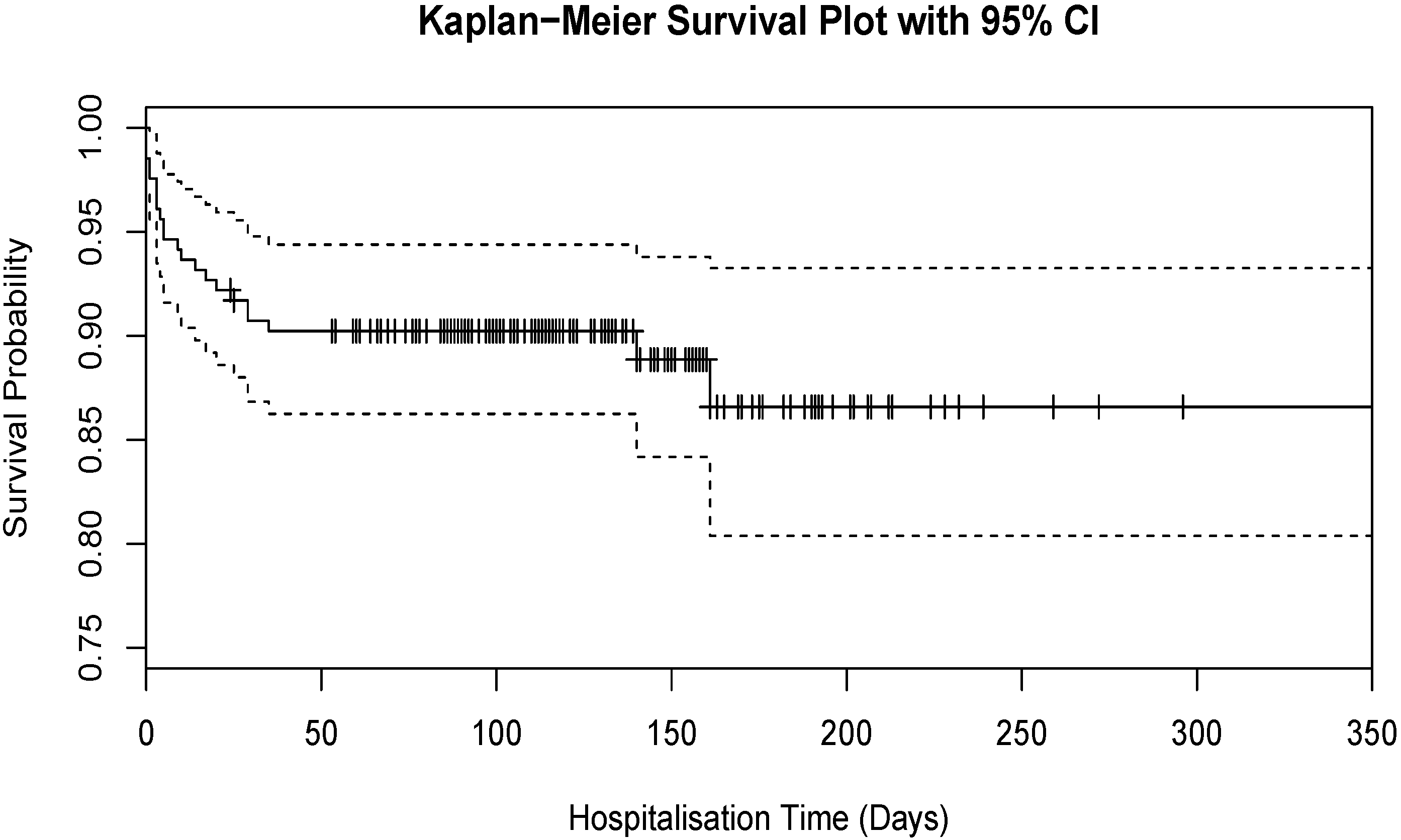
3.6. Outcome

3.7. Multivariable Binary Logistic Regression
4. Discussion
5. Conclusions
Acknowledgments
Author Contributions
Conflicts of Interest
References
- Sea Mammal Research Unit. Scientific Advice on Matters Related to the Management of Seal Populations; Natural Environment Research Council: Swindon, UK, 2013. [Google Scholar]
- Barnett, J.E.F.; Woodley, A.J.; Hill, T.J.; Turner, L. Conditions in grey seal pups (Halichoerus grypus) presented for rehabilitation. Vet. Rec. 2000, 147, 98–104. [Google Scholar] [CrossRef] [PubMed]
- Hammond, J.A.; Pomeroy, P.; Hall, A.; Smith, V.J. Identification and real-time PCR quantification of Phocine distemper virus from two colonies of Scottish grey seals in 2002. J. Gen. Virol. 2005, 86, 2563–2567. [Google Scholar] [CrossRef] [PubMed]
- Lonergan, M.; Hall, A.; Thompson, H.; Thompson, P.; Pomeroy, P.; Harwood, J. Comparison of the 1988 and 2002 phocine distemper epizootics in British harbour seal Phoca vitulina populations. Dis. Aquat. Org. 2010, 88, 183–188. [Google Scholar] [CrossRef] [PubMed]
- Harvell, C.D.; Kim, K.; Burkholder, J.M.; Colwell, R.R.; Epstein, P.R.; Grimes, D.J.; Hofmann, E.E.; Lipp, E.K.; Osterhaus, A.D.; Overstreet, R.M.; et al. Emerging marine diseases—Climate links and anthropogenic factors. Science 1999, 285, 1505–1510. [Google Scholar] [CrossRef] [PubMed]
- Gulland, F.M.D.; Hall, A. The Role of Infectious Disease in Influencing Status and Trend. In Marine Mammal Research: Conservation beyond Crisis; Reynolds, J.E., III, Perrin, W.F., Reeves, R.R., Montgomery, S., Ragen, T.J., Eds.; Johns Hopkins University Press: Baltimore, MD, USA, 2005; pp. 47–62. [Google Scholar]
- Harvell, D.; Altizer, S.; Cattadori, I.M.; Harrington, L.; Weil, E. Climate change and wildlife diseases: When does the host matter the most? Ecology 2009, 90, 912–920. [Google Scholar] [CrossRef] [PubMed]
- Microsfot Excel 2010; Microsoft Corp: Redmond, MA, USA, 2010.
- King, J.E. Seals of the World, 2nd ed.; University of Queensland Press: St. Lucia, Australia, 1983. [Google Scholar]
- Westcott, S. The Grey Seals of the West Country: And Their Neighbours; Cornwall Wildlife Trust: Truro, England, UK, 1997. [Google Scholar]
- Barnett, J. Treatment of sick and injured marine mammals. Practice 1998, 20, 200–211. [Google Scholar] [CrossRef]
- R Development Core Team. R: A Language and Environment for Statistical Computing; R Foundation for Statistical Computing: Vienna, Austria, 2008. [Google Scholar]
- Barnett, A.G.; Dobson, A.J. Analysing Seasonal Health Data; Statistics for Biology and Health; Springer: Berlin, Heidelberg, Germany, 2010. [Google Scholar]
- Minitab 16 Statistical Software; Minitab, Inc.: State College, PA, USA, 2009.
- Barnett, J.E.F.; Booth, P.; Brewer, J.I.; Chanter, J.; Cooper, T.; Crawshaw, T.; Davison, N.J.; Greenwood, A.; Riley, P.; Smith, N.H.; et al. Mycobacterium bovis infection in a grey seal pup (Halichoerus grypus). Vet. Rec. 2013, 173, 168–168. [Google Scholar] [CrossRef] [PubMed]
- Sayer, S.; Hockley, C.; Witt, M.J. Monitoring Grey Seals (Halichoerus grypus) in the Isles of Scilly during the 2010 Pupping Season; Natural England Comissioned Reports NECR103; Natural England: Sheffield, UK, 2012. [Google Scholar]
- Westcott, S. Prodcedural Guidelines for Studying Grey Seals in Southwest England, 2006; Natural England: Sheffield, UK, 2008. [Google Scholar]
- Barnett, J.; Knight, A.; Stevens, M. British Drivers Marine Life Rescue: Marine Mammal Medic Handbook, 7th ed.; British Divers Marine Life Rescue: Uckfield, UK, 2014. [Google Scholar]
- Baker, J.R.; Jepson, P.D.; Simpson, V.R.; Kuiken, T. Causes of mortality and non-fatal conditions among grey seals (Halichoerus grypus) found dead on the coasts of England, Wales and the Isle of Man. Vet. Rec. 1998, 142, 595–601. [Google Scholar] [CrossRef] [PubMed]
- Alonso-Farré, J.M.; D’Silva, J.I.D.; Gestal, C. Naso-pharyngeal mites Halarachne halichoeri (Allman, 1847) in Grey seals stranded on the NW Spanish Atlantic Coast. Vet. Parasitol. 2012, 183, 317–322. [Google Scholar] [CrossRef] [PubMed] [Green Version]
- Baily, J. The Pathology and Occurrence of Pathogens in Scottish Grey Seals (Halichoerus grypus); University of St. Andrews: St. Andrews, UK, 2014. [Google Scholar]
- Bowen, W.D.; Iverson, S.J.; Mcmillan, J.I.; Boness, D.J. Reproductive performance in grey seals: Age-related improvement and senescence in a capital breeder. J. Anim. Ecol. 2006, 75, 1340–1351. [Google Scholar] [CrossRef] [PubMed]
- Rothman, K. No adjustments are needed for multiple comparisons. Epidemiology 1990, 1, 43–46. [Google Scholar] [CrossRef]
- Bender, R.; Lange, S. Adjusting for multiple testing—When and how? J. Clin. Epidemiol. 2001, 54, 343–349. [Google Scholar] [CrossRef] [PubMed]
© 2015 by the authors; licensee MDPI, Basel, Switzerland. This article is an open access article distributed under the terms and conditions of the Creative Commons Attribution license (http://creativecommons.org/licenses/by/4.0/).
Share and Cite
Silpa, M.A.C.; Thornton, S.M.; Cooper, T.; Hedley, J. Prevalence of Presenting Conditions in Grey Seal Pups (Halichoerus grypus) Admitted for Rehabilitation. Vet. Sci. 2015, 2, 1-11. https://doi.org/10.3390/vetsci2010001
Silpa MAC, Thornton SM, Cooper T, Hedley J. Prevalence of Presenting Conditions in Grey Seal Pups (Halichoerus grypus) Admitted for Rehabilitation. Veterinary Sciences. 2015; 2(1):1-11. https://doi.org/10.3390/vetsci2010001
Chicago/Turabian StyleSilpa, Marc A. C., Susan M. Thornton, Tamara Cooper, and Joanna Hedley. 2015. "Prevalence of Presenting Conditions in Grey Seal Pups (Halichoerus grypus) Admitted for Rehabilitation" Veterinary Sciences 2, no. 1: 1-11. https://doi.org/10.3390/vetsci2010001