Article

Effect of Vaccination against Glässer’s Disease in a Farm Suffering from Polyserositis in Weaned Pigs

by
Jasmine Hattab
1,
Giuseppe Marruchella
1,*,
Abigail Rose Trachtman
1,
Luigino Gabrielli
2,
Nicola Bernabò
3,
Francesco Mosca
1 and
Pietro Giorgio Tiscar
1
1
Department of Veterinary Medicine, University of Teramo, Loc. Piano d’Accio, 64100 Teramo, Italy
2
Veterinary Practitioner, 63073 Offida, Italy
3
Department of Bioscience and Agro-Food and Environmental Technology, University of Teramo, via Renato Balzarini 1, 64100 Teramo, Italy
*
Author to whom correspondence should be addressed.
Vet. Sci. 2022, 9(12), 691; https://doi.org/10.3390/vetsci9120691
Submission received: 17 November 2022 / Revised: 9 December 2022 / Accepted: 10 December 2022 / Published: 12 December 2022
(This article belongs to the Section Veterinary Microbiology, Parasitology and Immunology)

Abstract

:

Simple Summary

This study mainly aims to investigate the impact and the etiology of swine polyserositis, focusing on three causative agents: Glaesserella parasuis, Streptococcus suis and Mycoplasma hyorhinis. Results indicate that the etiology of polyserositis is an intricate puzzle of pathogens, and that each pig herd likely represents a unique scenario. Therefore, the correct diagnosis of polyserositis can be really challenging and often makes the implementation of therapeutic and control strategies frustrating.

Abstract

Polyserositis mostly affects 4–8 weeks old piglets and is usually caused by Glaesserella parasuis, and/or Streptococcus suis, and/or Mycoplasma hyorhinis. The present study aimed to investigate the prevalence and etiology of polyserositis in a tricky pig herd. The concurrent effect of vaccination for Glässer’s disease was also assessed. A total of 46 sows and 387 piglets were herein investigated, subdivided into three groups based on their immune status (i.e., vaccination of sows and piglets). All the piglets found spontaneously dead between the 2nd and 16th week of age were recorded and necropsied. Whenever polyserositis was diagnosed, biomolecular investigations were carried out to detect the above-mentioned pathogens. Mycoplasma hyorhinis was detected most frequently (n = 23), often as the only causative agent (n = 15), whereas S. suis was observed in 8 cases (6 as the only pathogen). Moreover, Glaesserella parasuis was demonstrated in 6 piglets, always in combination with Mycoplasma hyorhinis and/or Streptococcus suis. Vaccination did not significantly affect mortality rates. Overall, our data indicate that polyserositis is likely caused by an intricate puzzle of pathogens, even when dealing with a small herd and during a short time span. That makes it challenging to achieve the correct diagnosis and to properly manage this health issue.

1. Introduction

Serous membranes (i.e., the pericardium, pleura, and peritoneum) are prone to inflammatory injuries, which are characterized by the effusion of fibrinous and/or suppurative exudates. Serositis is usually due to the hematogenous spreading of bacterial pathogens. Less commonly, it can result from penetrating wounds or direct extension of adjacent inflammatory foci [1].
The concurrent inflammation of two or more serous membranes is called polyserositis and represents a widespread and economically relevant disease condition in modern pig farming. Swine polyserositis mostly affects 4–8 weeks old piglets and has long been identified with Glässer’s disease by Glaesserella (previously known as “Haemophilus”) parasuis (G. parasuis) [2]. However, Streptococcus suis (S. suis) [3] and Mycoplasma hyorhinis (M. hyorhinis) [4] can often induce similar disease conditions. To a lesser extent, polyserositis can be caused by other pathogens, such as Escherichia coli [5], Pasteurella multocida [6], and Actinobacillus pleuropneumoniae [7]. The clinical onset of polyserositis is usually sudden and often includes neurological signs and swollen leg joints, due to the concurrent localization of the pathogen in the central nervous system and synovial membranes [8].
Recently, Salogni et al. [9] carried out a large-scale study (154 pigs from 80 herds) about the etiology of swine polyserositis in Northern Italy, showing that G. parasuis and M. hyorhinis were most frequently detected at the same prevalence rate. In our opinion, the paper by Salogni et al. provides a useful and reliable picture of a high-density breeding area, although unlikely representative of each pig herd. Considering that, the present study aimed to investigate the prevalence and etiology of polyserositis in a tricky pig herd under strict field conditions. The concurrent effect of vaccination for Glässer’s disease was also assessed.

2. Materials and Methods
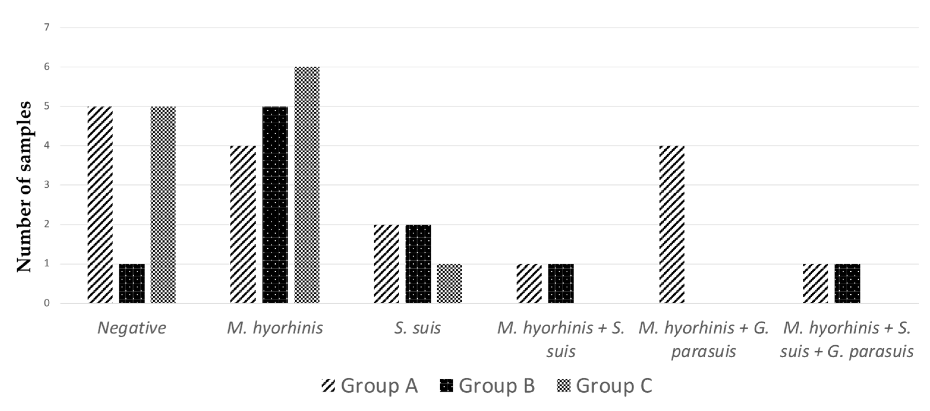
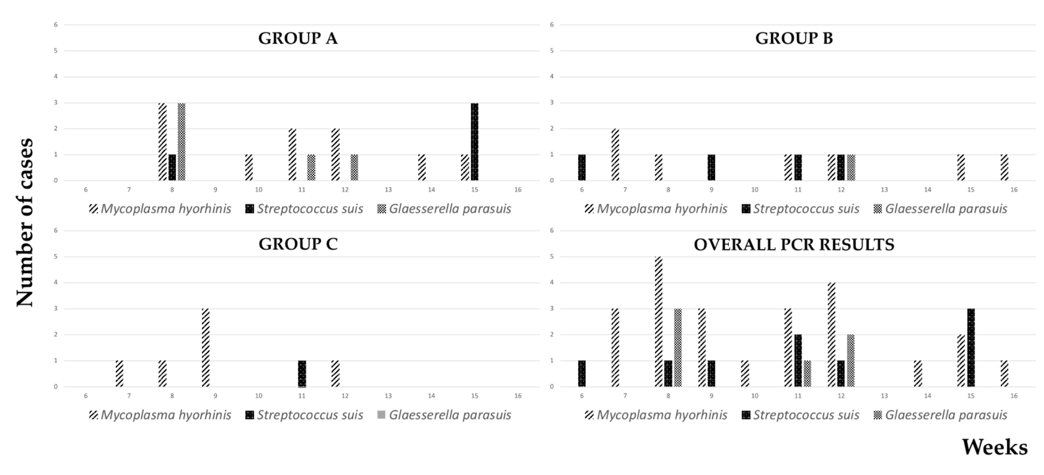
The study was carried out in a farrow-to-finish pig herd located in Central Italy. The breeding stock consisted of 70 sows (Landrace x Large White x Duroc) and 3 boars (Mora Romagnola breed). Piglets were weaned at 4 weeks and reared indoors until 16 weeks of age. Thereafter, pigs were moved outdoors until they reached the market weight of 160 kgs.
Sows were vaccinated against colibacillosis and clostridiosis (Suiseng, Hipra, Girona, Spain), whereas three weeks old piglets were vaccinated against porcine circovirus type 2 (PCV-2), Mycoplasma hyopneumoniae, and porcine reproductive and respiratory syndrome virus (PRRSv; 3FLEX®®, Boehringer Ingelheim, Ingelheim am Rhein, Germany) according to the manufacturer’s recommendations. Compulsory vaccination against Aujeszky’s disease (Aujeszky A-Suivax GI, Fatro, Bologna, Italy) was implemented as per Italian law.
Newly weaned piglets were fed a diet containing amoxicillin for one week (50 g/100 kgs of food), whereas sick piglets were usually treated with amoxicillin by intramuscular injection (Longocillina L.A. 150, 1 mL/10 kgs on alternate days; Ceva Salute Animale S.p.A., Agrate Brianza, Italy).
During the few months prior to the study, postweaning mortality rates were very high and several cases of Glässer’s disease and streptococcosis had been diagnosed, as confirmed by culture of G. parasuis and S. suis, respectively (data provided to the farmer by the “Istituto Zooprofilattico Sperimentale dell’Umbria e delle Marche” in the framework of its routine diagnostic activities).
Between September 2020 and March 2021, a total of 46 sows and 387 piglets were included in the present investigation. During this period, the Veterinary Practitioner decided to vaccinate sows and weaned piglets for Glässer’s disease (Porsilis Glasser, MSD Animal Health, Rahway, NJ, USA; see Table 1 for details). Sows were vaccinated twice, 6–8 and 4 weeks before farrowing, whereas piglets were vaccinated only once at 4–5 weeks of age.
As a routine, piglets from the same litter were identified by means of ear tags. All piglets spontaneously dead between the 2nd and 16th week of age were recorded and As a routine, piglets from the same litter were identified by means of ear tags. All piglets spontaneously dead between the 2nd and 16th week of age were recorded and necropsied on a weekly basis, upon freezing whenever needed. Samples (i.e., pericardium, heart, and fibrin) were taken from all polyserositis-affected pigs, placed into single tubes, kept on ice, and delivered to the laboratory (Veterinary Teaching Hospital, University of Teramo, Italy).
Total DNA was extracted using ExgeneTM Tissue SV (GeneAll®, Seoul, South Korea) according to the manufacturer’s protocol. Biomolecular investigations (i.e., polymerase chain reactions, PCR) were carried out to detect the most common causative agents of polyserositis—namely, G. parasuis, M. hyorhinis, S. suis—following previously published protocols [10,11,12,13]. PCR reactions were prepared as a total volume of 25 µL, consisting of 4 µL of template DNA, 12.5 µL of master mix (GoTaq® Green Master Mix, Promega Corporation, Madison, WI, USA), and 0.5 µL of forward and 0.5 µL of reverse primers (see Table 2 for details).
Positive and negative controls were included in each PCR run. All G. parasuis-positive samples were further tested for serotyping by means of previously described PCR protocols [14,15].
Finally, data were submitted to statistical analysis. Fisher’s exact test was used to compare the mortality rates, as well as the prevalence of different pathogens among groups. The following cut-off points were established for significance: values differ significantly at p ≤ 0.05 and at the trend level at 0.05 < p < 0.1.

3. Results

The weekly rates of total mortality are shown in Figure 1.
During the study period, the overall mortality rate was 10.6% in suckling piglets (41 out of 387 piglets), most deaths being recorded during the first week of age (35 out of 41 dead piglets). After weaning, the overall mortality rate was 19.9% (69 out of 346 weaned piglets), with a prominent peak at 8 weeks of age (see Table 3 for details).
At necropsy, polyserositis was frequently diagnosed in weaned piglets (39 out of 69 cadavers), whereas it was never observed in suckling piglets (Figure 2).
As shown in Figure 3, most cases of polyserositis affected 6-to-8- and 15-week-old piglets.
No significant difference was detected among the three groups regarding polyserositis-specific (i.e., mortality due to polyserositis) and polyserositis-proportionate (i.e., the proportion of deaths attributable to polyserositis) mortality rates (see Table 3 for further details).
The results of the PCR tests are graphically shown in Figure 4. Overall, the genome of M. hyorhinis, S. suis and G. parasuis was detected in 23, 8, and 6 polyserositis-affected piglets, respectively. G. parasuis was always detected in combination with other pathogens. On the contrary, M. hyorhinis and S. suis were detected as the only causative agent in 15 and 5 piglets, respectively.
As shown in Figure 5, M. hyorhinis and S. suis were observed during almost the entire postweaning period, with peaks of incidence at 8 and 15 weeks of age, respectively. G. parasuis was detected between 8 and 12 weeks of age, the highest incidence being observed at 8 weeks of age.
Overall, the detection of M. hyorhinis was significantly higher when compared with G. parasuis (p < 0.01) and S. suis (p < 0.01). In particular, the detection of M. hyorhinis was significantly more frequent than G. parasuis in group B (p = 0.02) and group C (p = 0.01).
A single piglet from group B proved to be positive for G. parasuis, whereas G. parasuis was never detected in group C. The detection of G. parasuis was lower in group C at trend level (p = 0.05) when compared with group A. Moreover, the occurrence of co-infections was significantly lower in group C when compared with group A (p = 0.02).
Biomolecular tests for serotyping G. parasuis always yielded negative results.

4. Discussion

Although mortality is among the foremost causes of economic loss in livestock production, it is difficult to estimate its physiologic rate in pig farming. Preweaning mortality usually ranges between 10% and 20% and is affected by a vast number of interrelated factors (e.g., birth weight, litter size, heating of farrowing crates, sow health, etc.) [16,17]. In the USA, a mortality rate of 3.6% was reported in nursery pigs after a long large-scale data collection [18]. Considering that, the pig herd under study showed an acceptable rate of preweaning mortality, which was mostly restricted to the first week of age, as commonly observed worldwide [17]. Conversely, postweaning mortality was undoubtedly too high, with most of the deaths falling in the critical period between 7-to-9 weeks of age, when passive immunity decreases, and the development of self-antibodies is not yet effective [19,20].
The present study confirms that polyserositis can frequently occur in weaned piglets, and it is likely caused by an intricate puzzle of pathogens, even when dealing with a small herd and during a brief time span.
Over recent years, rising relevance has been given to M. hyorhinis, at least partially due to the widespread use of sensitive diagnostic tools, which overcome the difficulty of culturing this pathogen in vitro. M. hyorhinis is a commensal bacterium in swine and mainly causes polyserositis and polyarthritis in nursery pigs between 8 and 10 weeks of age [21]. Based on our data, M. hyorhinis could play a major role in the etiology of polyserositis, alone or in combination with other pathogens, and during a longer time span. Reasonably, the administration of beta-lactams held off the infections by S. suis and G. parasuis and further increased the proportionate prevalence of M. hyorhinis-induced polyserositis. Although vaccination against M. hyorhinis can reduce clinical signs and lesions [22], commercial vaccines are currently unavailable in Europe. Therefore, M. hyorhinis-induced diseases are usually treated with antimicrobials [23]. A recent study carried out in European countries (including Italy) indicates that doxycycline, oxytetracycline, and tiamulin are the most effective compounds, at least in vitro [24].
Vaccination can reduce mortality by Glässer’s disease. The efficacy of G. parasuis vaccines is serovar-specific, cross-protection being debated and likely limited [25,26,27]. In Italy, G. parasuis serovar 4 was reported as the most prevalent one, followed by serovar 13 and serovar 5, whereas about 27% of strains were non-typeable through agar gel diffusion test [28]. In the present study, G. parasuis serovars remained unidentified, and vaccination weakly affected mortality rates. Reasonably, the presence of M. hyorhinis as the main causative agent accounted for the low efficacy of vaccination, whereas G. parasuis played a less relevant role. However, we remark that G. parasuis was never detected in vaccinated piglets, and vaccination significantly reduced the occurrence of co-infections.
Herein, a high portion of cases of polyserositis (about 28%) proved to be negative for M. hyorhinis, G. parasuis, and S. suis and were likely caused by other pathogens, which should be further investigated. Unfortunately, additional bacteriological investigations were seldom carried out, and Trueperella pyogenes was cultured in a single piglet (data not shown). Moreover, the causative role of viruses (e.g., PRRSv, PCV-2) should be considered, as they often predispose to bacterial infections. In particular, PRRSv often contributes to the etiology of polyserositis, whereas the impact of PCV-2 is likely mitigated by the widespread use of effective vaccines [9,29].
The multi-faceted etiology of polyserositis greatly complicates its control and therapy. It seems that targeting a single pathogen (by means of vaccine and/or antimicrobials) might change the balance among causative agents without affecting the severity of polyserositis. As an example, herein the administration of amoxicillin and the vaccination for Glässer’s disease likely limited the role of S. suis and G. parasuis while enhancing the etiological relevance of M. hyorhinis. Therefore, the effective control of polyserositis should always include the improvement of herd management, minimizing stressful situations that worsen most diseases in intensive pig farming [8].

5. Conclusions

The present study confirms that the etiology of polyserositis is multi-factorial and suggests that each pig herd represents a unique scenario. A suitable diagnostic approach is crucial to properly manage this health issue, aiming to counteract the most relevant pathogens. However, our data indicate that this could be challenging, as “one-shot” investigation on a single or few animals might be insufficient, if not misleading. In our opinion, M. hyorhinis should be regarded as an “emerging” and relevant pathogen. Vaccination against Glässer’s disease could be effective when G. parasuis plays a key etiological role.

Author Contributions

Conceptualization, G.M. and P.G.T.; methodology, G.M. and P.G.T.; validation, G.M., J.H., A.R.T., F.M., L.G. and P.G.T.; formal analysis, F.M. and N.B.; investigation, G.M., J.H., A.R.T. and F.M.; resources, G.M. and P.G.T.; data curation, F.M. and N.B.; writing—original draft preparation, G.M., J.H. and F.M.; writing—review and editing, G.M., J.H. and F.M.; visualization, G.M., J.H., A.R.T., F.M., L.G. and P.G.T.; supervision, G.M. and P.G.T. All authors have read and agreed to the published version of the manuscript.

Funding

This research received no external funding.

Institutional Review Board Statement

Not applicable since all animals examined in this study were naturally deceased.

Informed Consent Statement

Written informed consent was obtained from the owners of the animals described in this study for the procedures undertaken.

Data Availability Statement

Not applicable.

Conflicts of Interest

The authors declare no conflict of interest.

References

  1. Zachary, J.F.; McGavin, M.D. Pathologic Basis of Veterinary Disease, 5th ed.; Elsevier: Amsterdam, The Netherlands, 2012; pp. 536–537. [Google Scholar]
  2. Oliveira, S.; Pijoan, C. Haemophilus parasuis: New trends on diagnosis, epidemiology and control. Vet. Microbiol. 2004, 99, 1–12. [Google Scholar] [CrossRef] [PubMed]
  3. Reams, R.Y.; Glickman, L.T.; Harrington, D.D.; Thacker, H.L.; Bowersock, T.L. Streptococcus suis infection in swine: A retrospective study of 256 cases. Part II. Clinical signs, gross and microscopic lesions, and coexisting microorganisms. J. Vet. Diagn. Investig. 1994, 6, 326–334. [Google Scholar] [CrossRef] [PubMed] [Green Version]
  4. Kobisch, M.; Friis, N.F. Swine mycoplasmoses. Rev. Sci. Tech. 1996, 15, 1569–1605. [Google Scholar] [CrossRef] [PubMed]
  5. Nielsen, N.C.; Bille, N.; Riising, H.J.; Dam, A. Polyserositis in pigs due to generalized Escherichia coli infection. Can. J. Comp. Med. 1975, 39, 421–426. [Google Scholar] [PubMed]
  6. Oliveira Filho, J.X.; Morés, M.A.Z.; Rebellato, R.; Kich, J.D.; Cantão, M.E.; Klein, C.S.; Carvalho Guedes, R.M.; Coldebella, A.; Santos Neves de Barcellos, D.E.; Morés, N. Pathogenic variability among Pasteurella multocida type A isolates from Brazilian pig farms. BMC Vet. Res. 2018, 14, 244. [Google Scholar] [CrossRef] [PubMed]
  7. Hoeltig, D.; Rohde, J.; Frase, R.; Nietfeld, F.; Waldmann, K.H.; Valentin-Weigand, P.; Means, J. Multi-organ spreading of Actinobacillus pleuropneumoniae serovar 7 in weaned pigs during the first week after experimental infection. Vet. Res. 2018, 49, 97. [Google Scholar] [CrossRef] [Green Version]
  8. Costa-Hurtado, M.; Barba-Vidal, E.; Maldonado, J.; Aragon, V. Update on Glässer’s disease: How to control the disease under restrictive use of antimicrobials. Vet. Microbiol. 2020, 242, 108595. [Google Scholar] [CrossRef]
  9. Salogni, C.; Lazzaro, M.; Giovannini, S.; Vitale, N.; Boniotti, M.B.; Pozzi, P.; Pasquali, P.; Alborali, G.L. Causes of swine polyserositis in a high-density breeding area in Italy. J. Vet. Diagn. Investig. 2020, 32, 594–597. [Google Scholar] [CrossRef]
  10. Oliveira, S.; Galina, L.; Pijoan, C. Development of a PCR test to diagnose Haemophilus parasuis infections. J. Vet. Diagn. Investig. 2001, 13, 495–501. [Google Scholar] [CrossRef] [Green Version]
  11. Olvera, A.; Pina, S.; Macedo, N.; Oliveira, S.; Aragon, V.; Bensaid, A. Identification of potentially virulent strains of Haemophilus parasuis using a multiplex PCR for virulence-associated autotransporters (vtaA). Vet. J. 2012, 191, 213–218. [Google Scholar] [CrossRef]
  12. Fourour, S.; Fablet, C.; Tocqueville, V.; Dorenlor, V.; Eono, F.; Eveno, E.; Kempf, I.; Marois-Créhan, C. A new multiplex real-time TaqMan® PCR for quantification of Mycoplasma hyopneumoniae, M. hyorhinis and M. flocculare: Exploratory epidemiological investigations to research mycoplasmal association in enzootic pneumonia-like lesions in slaughtered pigs. J. Appl. Microbiol. 2018, 125, 345–355. [Google Scholar] [CrossRef]
  13. Okwumabua, O.; O’Connor, M.; Shull, E. A polymerase chain reaction (PCR) assay specific for Streptococcus suis based on the gene encoding the glutamate dehydrogenase. FEMS Microbiol. Lett. 2003, 218, 79–84. [Google Scholar] [CrossRef]
  14. Howell, K.J.; Peters, S.E.; Wang, J.; Hernandez-Garcia, J.; Weinert, L.A.; Luan, S.L.; Chaudhuri, R.R.; Angen, Ø.; Aragon, V.; Williamson, S.M.; et al. Development of a Multiplex PCR Assay for Rapid Molecular Serotyping of Haemophilus parasuis. J. Clin. Microbiol. 2015, 53, 3812–3821. [Google Scholar] [CrossRef] [Green Version]
  15. Lee, C.Y.; Ooi, P.T.; Zunita, Z.; Noordin, M.M. Molecular detection of Haemophilus parasuis serotypes 4, 5 or 12 and 13 in Peninsular Malaysia. Trop Biomed. 2019, 36, 482–487. [Google Scholar]
  16. Johnson, A.K.; Colpoys, J.D.; Edwards-Callaway, L.N.; Calvo-Lorenzo, M.; McGlone, J.J.; Millman, S.T.; Phillips, C.E.; Ritter, M.J.; Sutherland, M.A.; Tucker, A.L.; et al. Behavior and Welfare. In Diseases of Swine, 11th ed.; Zimmerman, J.J., Karriker, L.A., Ramirez., A., Schwartz, K.J., Stevenson, G.W., Zhang, J., Eds.; John Wiley & Sons, Inc.: Hoboken, NJ, USA, 2019; pp. 17–41. [Google Scholar]
  17. Bruns, C.E.; Stalder, K.J. Genetics and health. In Diseases of Swine, 11th ed.; Zimmerman, J.J., Karriker, L.A., Ramirez, A., Schwartz, K.J., Stevenson, G.W., Zhang, J., Eds.; John Wiley & Sons, Inc.: Hoboken, NJ, USA, 2019; pp. 42–49. [Google Scholar]
  18. Knauer, M.T.; Hostetler, C.E. US swine industry productivity analysis, 2005 to 2010. J. Swine Health Prod. 2013, 21, 248–252. [Google Scholar]
  19. Day, M.J. Immune system ontogeny and neonatal immunology. In Veterinary Immunology. Principles and Practice, 2nd ed.; Day, M.J., Ed.; CRC Press: Boca Raton, FL, USA, 2014; pp. 213–220. [Google Scholar] [CrossRef]
  20. Muirhead, M.R.; Alexander, T.J.L. Managing health and disease. In Managing Pig Health, 2nd ed.; Carr, J., Ed.; 5m Publishing: Sheffield, UK, 2013; pp. 73–130. [Google Scholar]
  21. Pieters, M.G.; Maes, D. Mycoplasmosis. In Diseases of Swine Swine, 11th ed.; Zimmerman, J.J., Karriker, L.A., Ramirez, A., Schwartz, K.J., Stevenson, G.W., Zhang, J., Eds.; John Wiley & Sons, Inc.: Hoboken, NJ, USA, 2019; pp. 863–883. [Google Scholar] [CrossRef]
  22. Martinson, B.; Minion, F.C.; Kroll, J.; Hermann, J. Age susceptibility of caesarian derived colostrum deprived pigs to Mycoplasma hyorhinis challenge. Vet. Microbiol. 2017, 210, 147–152. [Google Scholar] [CrossRef]
  23. Martinson, B.; Zoghby, W.; Barrett, K.; Bryson, L.; Christmas, R.; Minion, F.C.; Kroll, J. Efficacy of an inactivated Mycoplasma hyorhinis vaccine in pigs. Vaccine 2018, 36, 408–412. [Google Scholar] [CrossRef]
  24. Klein, U.; Földi, D.; Belecz, N.; Hrivnák, V.; Somogyi, Z.; Gastaldelli, M.; Merenda, M.; Catania, S.; Dors, A.; Siesenop, U.; et al. Antimicrobial susceptibility profiles of Mycoplasma hyorhinis strains isolated from five European countries between 2019 and 2021. PLoS ONE 2022, 17, e0272903. [Google Scholar] [CrossRef]
  25. Liu, H.; Xue, Q.; Zeng, Q.; Zhao, Z. Haemophilus parasuis vaccines. Vet. Immunol. Immunopathol. 2016, 180, 53–58. [Google Scholar] [CrossRef]
  26. Zhao, Z.; Liu, H.; Xue, Y.; Chen, K.; Liu, Z.; Xue, Q.; Wang, C. Analysis of efficacy obtained with a trivalent inactivated Haemophilus parasuis serovars 4, 5, and 12 vaccine and commercial vaccines against Glässer’s disease in piglets. Can. J. Vet. Res. 2017, 81, 22–27. [Google Scholar]
  27. Olvera, A.; Ballester, M.; Nofrarías, M.; Sibila, M.; Aragon, V. Differences in phagocytosis susceptibility in Haemophilus parasuis strains. Vet. Res. 2009, 40, 24. [Google Scholar] [CrossRef] [PubMed]
  28. Luppi, A.; Bonilauri, P.; Dottori, M.; Iodice, G.; Gherpelli, Y.; Merialdi, G.; Maioli, G.; Martelli, P. Haemophilus parasuis serovars isolated from pathological samples in Northern Italy. Transbound. Emerg. Dis. 2013, 60, 140–142. [Google Scholar] [CrossRef] [PubMed]
  29. Yu, J.; Wu, J.; Zhang, Y.; Guo, L.; Cong, X.; Du, Y.; Li, J.; Sun, W.; Shi, J.; Peng, J.; et al. Concurrent highly pathogenic porcine reproductive and respiratory syndrome virus infection accelerates Haemophilus parasuis infection in conventional pigs. Vet. Microbiol. 2012, 158, 316–321. [Google Scholar] [CrossRef] [PubMed]
Figure 1. Weekly-based mortality rates. All groups show a peak of mortality around 8 weeks of age. In group A, an additional peak is observed at 15 weeks of age.
Vetsci 09 00691 g001
Figure 2. Piglets. Necropsy. (A) Polyserositis-affected piglet. Large amounts of fibrin are seen upon both pleural sheets, the epicardium, as well as the peritoneum lining the spleen and the liver. (B) A piglet dead with no evidence of polyserositis is shown for comparison.
Vetsci 09 00691 g002
Figure 3. Postweaning mortality due to polyserositis. The trends of polyserositis-specific mortality largely overlap those shown in Figure 1. In group (A), and to a lesser extent in group (B), mortality shows a swinging pattern, whereas it is mostly restricted to at 8-9 weeks of age in group (C).
Vetsci 09 00691 g003
Figure 4. Results of biomolecular investigations. M. hyorhinis was detected most frequently, alone or in combination with other pathogens. Conversely, G. parasuis was seldom detected. In 11 cases, PCR tests yielded negative results.
Vetsci 09 00691 g004
Figure 5. Weekly-based results of biomolecular investigations. Overall, M. hyorhinis was detected from the 7th to the 16th week of age, with the only exception of the 13th week. S. suis infection was unevenly demonstrated, whereas G. parasuis was mostly observed at 8 weeks of age in group A.
Vetsci 09 00691 g005
Table 1. Animals included in the present study. Based on vaccination for Glasser’s disease, three groups were distinguished: (A) non-vaccinated piglets born to non-vaccinated sows; (B) non-vaccinated piglets born from vaccinated sows; and (C) vaccinated piglets born to vaccinated sows.
GroupSows (Number)Piglets (Number)
VaccinatedNot vaccinatedVaccinatedNot Vaccinated
A//19//172
B12////99
C15//116//
Table 2. Primers used for PCR tests.
Primer NamePrimer Sequence (5′-3′)Amplicon Size (bp)Targeted
Pathogen
Reference
Hps-fGTGATGAGGAAGGGTGRTGT822Glaesserella parasuis[10]
Hps-rGGCTTCGTCRCCCTCTGT
YADAF 1TTTAGGTAAAGATAAGCAAGGAAATCC406Glaesserella parasuis; vtA domain group 1[11]
PADHR 1CCACACAAAACCTACCCCTCCTCC
YADAF 2AGCTTAATATCTCAGCACAAGGTGC294Glaesserella parasuis; vtA domain group 2[11]
PADHR 1CCACACAAAACCTACCCCTCCTCC
YADAF 3AATGGTAGCCAGTTGTATAATGTTGC291Glaesserella parasuis; vtA domain group 3[11]
PADHR 1CCACACAAAACCTACCCCTCCTCC
Mhr_p37 fTTCTATTTTCATCTATATTTTCGC101Mycoplasma hyorhinis[12]
Mhr_p37 rTCATTGACCTTGACTAACTG
JP4GCAGCGTATTCTGTCAAACG688Streptococcus suis[13]
JP5CCATGGACAGATAAAGATGG
Table 3. Postweaning mortality rates. Total mortality rate was higher in group C (*) at a trend level (p = 0.078) when compared with group A.
Group AGroup BGroup C
Total mortality rate15.7222.0924.75 *
Polyserositis-specific mortality rate10.6911.6211.88
Polyserositis-proportionate mortality68.0052.6348.00
Publisher’s Note: MDPI stays neutral with regard to jurisdictional claims in published maps and institutional affiliations.

