Review

Cellular Senescence in Cardiovascular Diseases: From Pathogenesis to Therapeutic Challenges

1
Department of Cardiovascular Medicine, Lanzhou University Second Hospital, Lanzhou 730030, China
2
Department of Cardiac Surgery, Lanzhou University Second Hospital, Lanzhou 730030, China
*
Author to whom correspondence should be addressed.
J. Cardiovasc. Dev. Dis. 2023, 10(10), 439; https://doi.org/10.3390/jcdd10100439
Submission received: 16 September 2023 / Revised: 17 October 2023 / Accepted: 19 October 2023 / Published: 23 October 2023

Abstract

:
Cellular senescence (CS), classically considered a stable cell cycle withdrawal, is hallmarked by a progressive decrease in cell growth, differentiation, and biological activities. Senescent cells (SNCs) display a complicated senescence-associated secretory phenotype (SASP), encompassing a variety of pro-inflammatory factors that exert influence on the biology of both the cell and surrounding tissue. Among global mortality causes, cardiovascular diseases (CVDs) stand out, significantly impacting the living quality and functional abilities of patients. Recent data suggest the accumulation of SNCs in aged or diseased cardiovascular systems, suggesting their potential role in impairing cardiovascular function. CS operates as a double-edged sword: while it can stimulate the restoration of organs under physiological conditions, it can also participate in organ and tissue dysfunction and pave the way for multiple chronic diseases under pathological states. This review explores the mechanisms that underlie CS and delves into the distinctive features that characterize SNCs. Furthermore, we describe the involvement of SNCs in the progression of CVDs. Finally, the study provides a summary of emerging interventions that either promote or suppress senescence and discusses their therapeutic potential in CVDs.

1. Introduction

CS entails cycle arrest and concurrent changes in various metabolic pathways, leading to morphological and functional alteration [1]. Under physiological conditions, CS facilitates cellular turnover and organ repair, forming a closely regulated process influenced by many kinds of cells. Nevertheless, SNCs often evade timely elimination and replacement within aging tissues or pathological conditions, resulting in their accumulation and subsequent organ dysfunction [2].
Among multiple chronic fatal diseases, CVDs are the most prevalent, emerging as a significant factor in elderly mortality rates, especially as global demographics increasingly lean toward older age groups [3]. Across a range of cardiovascular conditions, both ischemic and non-ischemic cardiomyopathies, clinical data have revealed a significant relationship between cellular senescence and unfavorable cardiac outcomes [2]. Nevertheless, the exact function of SNCs in these conditions remains enigmatic, as certain instances exhibit both harmful and advantageous effects [4,5,6,7,8,9]. Therefore, although the multiple causes of CVDs have been elucidated due to advances in genomics, epigenomics, proteomics, and single-cell analysis, in this review we prioritize the role of CS in CVDs (Figure 1). Regulatory signals and possible points of intervention in different types of cardiovascular SNCs are reviewed. Furthermore, it outlines various promising targets for manipulating the process of CS. The ultimate goal is to offer a fresh perspective for the development of compounds that regulate senescence and enhance the clinical transformation of cardioprotective lead compounds.

2. Definition and Mechanisms of CS

Although knowledge about the role of CS in health and disease is controversial, its basic definitions and principles still exhibit a certain degree of consensus. In a historical context, senescence was first defined by Hayflick over 70 years ago. His discovery revolved around the observation that human diploid fibroblasts exhibited a limited ability to divide, primarily attributed to telomere shortening [10]. However, senescence can be triggered even when there is no observable telomere loss or dysfunction, in various circumstances. This specific form of senescence has been categorized as ‘premature’ due to its occurrence prior to the stage when it is initiated by the shortening of telomeres [11]. Unlike the former, this variant is not linked to ongoing telomere shortening, but rather to non-telomeric DNA damage and sustained mitogenic stimulation, a form known as stress-induced premature senescence (SIPS) [12]. Fundamentally, CS embodies a cellular state marked by a durable halt in proliferation as a response to diverse stressors, frequently accompanied by the generation of a related secretome recognized as the SASP [13]. In this section, the discussion will briefly cover the involvement of cellular stressors in triggering CS, with particular emphasis on factors such as telomere shortening, oxidative-related senescence, DNA damage, chemotherapy, oncogene activation, mitochondrial dysfunction, epigenetics, the SASP process, and other factors (Figure 2).

2.1. Replicative CS

In line with Hayflick’s hypothesis, contemporary knowledge suggests that, as human cells are cultured over time, telomeres experience gradual shortening, culminating in cells reaching their “Hayflick limit”. This occurrence is denoted as replicative senescence (RS), given that it emerges as a consequence of replication [10]. Telomeres, situated at the termini of linear chromosomes, serve as protective structures. They are composed of repetitions of the tandem sequence TTAGGG and are linked to the shelterin complex [10]. Such an arrangement acts as a protective barrier against chromosomal damage, as it prevents fusion between chromosomes and degradation, thereby preserving genome integrity [14]. With each round of replication, there is a decrease in the number of telomeres repeating, as DNA polymerase cannot fully replicate the lagging strands. One of the most extensively characterized mechanisms in RS is the notable reduction in telomere length that occurs with each successive cellular proliferation [10].
The gradual shortening of telomeres results in the uncovering of DNA termini, ultimately triggering a DNA damage response (DDR). This reaction results in the occurrence of chromosomal unsteadiness and the formation of atypical connections between chromosomes, along with the triggering of inhibitors that regulate cell cycle progression such as p53, p21CIP1, WAF1, or p16INK4a/retinoblastoma protein (Rb). These inhibitors induce cell cycle halt at the G1 phase [15]. The fact that RS depends on telomere shortening becomes clear when it is overcome by the ectopic expression of the catalytic subunit of the human telomerase reverse transcriptase (hTERT) holoenzyme, which elongates telomeres and lessens the impact of the end replication problem [16]. The constrained longevity of the majority of primary human cells is linked to the absence of telomerase expression in human somatic cells, unlike stem cells. Consequently, human somatic cells cannot sustain telomeres at a length that is adequate to suppress DDR [17]. In conclusion, the key molecular events underlying RS seem to encompass telomere shortening and the repression of telomerase during the process of biological aging [11].

2.2. Premature CS

2.2.1. DNA Damage-Induced Senescence

Among the heart and blood vessels, the genome is uninterruptedly exposed to diverse internal and external factors, encompassing ischemia, lipids, blood flow, and aging. These factors inevitably trigger genome instability and transcription disorders [2]. The senescent phenotype can be induced by any stressor that results in persistent DNA damage [18]. DNA damage refers to modifications made to the DNA structure, which can result in changes to its coding properties or its capability of involvement in transcription or replication [19]. Various forms of DNA damage have been identified, including single-strand breaks (SSBs), double-strand breaks (DSBs), DNA–protein cross-links, and insertion or deletion mismatches [20]. Among these, the DNA damage type most consistently associated with aging are DSBs [21].
The DDR is defined by the activation of sensor kinases (ataxia–telangiectasia mutated [ATM] or ataxia–telangiectasia Rad3-related [ATR]), the accumulation of DNA damage foci that contain phosphorylated histone H2AX (γH2AX), and the engagement of checkpoint proteins like p53 and the CDK inhibitor p21 (CDKN1A). Eventually, this cascade culminates in the halting of the cell cycle, resulting in senescence [22]. As a result, DNA damage foci exhibiting positive γ-H2AX staining have been validated as dependable indicators of sustained DNA damage and senescence [23]. Mechanistically, DNA damage and senescence establish a feedback loop. Senescence can disrupt cytoplasmic DNA clearance, activating the senescence-sensing mechanism through cyclic GMP–AMP synthase (cGAS) and triggering the stimulator of interferon genes (STING). This activation promotes senescence and the secretion of SASP [24].
Furthermore, telomere shortening represents a form of DNA damage. Consequently, the existence of DNA damage foci within the telomere region acts as a distinguishing feature of CS referred to as telomere-associated foci (TAF). Detecting TAF at telomeres offers solid proof for quantifying telomere damage as a response to stress. Co-localization studies, often utilized to confirm the existence of TAF, encompass the examination of DDR factors (such as p53-binding protein 1 (p53BP1) and γ-H2AX) as well as telomere repeats [25]. The targeted induction of TAF using the TRF1–Fok1 fusion protein triggers CS in cardiomyocytes, without regard to the length of telomere. These findings indicate that ongoing DNA damage at telomeres could initiate CS in cardiomyocytes [26].

2.2.2. Oncogene-Induced Senescence

In 1997, it was discovered that, in early passage normal human and murine fibroblasts, oncogenic Ras leads to an initial phase of hyperproliferation, along with subsequent essentially irreversible growth arrest. This arrest closely resembles the phenotypic characteristics of RS. This phenomenon has since been referred to as oncogene-induced senescence (OIS) [27]. The activation of oncogenes initiates a hyper-replicative phase, subsequent to the activation of the DNA damage checkpoint response. This phase leads to an elevation in the quantity of active replicons and alterations in the progression of DNA replication forks [28]. In contrast to RS, OIS is not preventable by the expression of hTERT, underscoring its dissociation from telomere erosion [29]. The involvement of the p53 and p16INK4A-RB pathways is a common feature observed in cells experiencing both RS and OIS, albeit within specific contexts [30]. Multiple studies have suggested that CS can impede tumor formation by inducing a prolonged cell cycle withdrawal and restricting the generation of cancer stem cells [31,32,33,34].

2.2.3. Oxidative Stress-Induced Senescence

The hypothesis of the free radical theory of aging, advanced earliest by Denham Harman, posits that an excess of ROS causes harm to macromolecules. The gradual accumulation of this macromolecular damage subsequently results in cellular and organ dysfunction as time progresses [35]. Senescence can be induced by oxidative stress through direct effects on telomeres, irrespective of their length, or by impacting genomic or mitochondrial DNA (mtDNA) [36]. ROS production can be induced by both exogenous and endogenous stressors, via either mitochondrial or non-mitochondrial sources [37]. ROS can play a signaling role in both the induction and maintenance of senescence by directly causing oxidative modifications in subcellular structures, such as nucleic acids and enzymes. This oxidation, in turn, triggers the activation of oncogenes, DNA damage, and reduced telomerase activity [38]. If the presence of stress is persistent and surpasses the antioxidant capacity to counteract it, the process of senescence persists. Alternatively, when antioxidant defenses are sufficient, the senescence process transitions towards autophagy, restoring a proliferative state while reducing the expression of senescence markers [39]. Consequently, oxidative stress stands as one of the essential and pivotal mechanisms contributing to stressor-induced CS.

2.2.4. Paracrine Senescence

Within the SASP, there are three primary groups of secretory proteins: soluble signaling factors, proteases, and insoluble proteins or extracellular membrane (ECM) constituents. The secreted elements originating from SASP influence the nearby tissue microenvironment surrounding SNCs, thereby causing phenotypic changes in neighboring non-SNCs. These molecules and pathways associated with SASP critically mediate the pathophysiological impact of SNCs in tissue damage, chronic inflammation, and tumorigenesis. This is accomplished through intercellular communication and the creation of distinct microenvironmental conditions [40]. One advantageous role of SASP is its capacity to recruit immune cells, promote tissue homeostasis, initiate tissue repair and remodeling via the elimination of dysfunctional cells, and impede tumor growth by preventing the transmission of genetic alterations to future cell generations [41]. Nonetheless, in addition to the advantages, SASP can also exert detrimental effects. It can stimulate a pro-inflammatory microenvironment, perpetuate chronic inflammation, and facilitate tumor progression. In terms of mechanisms, SASP serves as a two-sided coin, generating both advantageous and disadvantageous effects on human diseases [41].

2.2.5. Mitochondrial Dysfunction-Associated Senescence (MiDAS)

Maintaining normal mitochondrial and cardiac function requires mitochondrial fusion, while fission supports the elimination of dysfunctional, depolarized mitochondria through the mechanism of mitophagy [42,43]. A disruption in the balance between mitochondrial fission and fusion that causes an excess of mitochondrial fusion, similar to what is seen in senescence, brings about the retention of impaired mitochondria and the build-up of oxidized proteins. This aggregation could potentially worsen the senescent phenotype [43]. Mitochondrial dysfunction is strongly correlated with an augmented generation of ROS, resulting in heightened oxidative damage. This damage encompasses sulfhydryl oxidation, lipid peroxidation, and mutations in mtDNA [44,45]. Metabolic dysfunction and irregular mitochondrial dynamics are particularly essential in facilitating the senescence of cardiomyocytes.

2.2.6. Epigenetically Induced Senescence

Epigenetic modifications involve DNA methylation, histone acetylation, chromatin remodeling, and non-coding RNAs [46]. SNCs harbor extensive alterations in DNA methylation compared to proliferating cells. These changes include global hypomethylation and hypermethylation of CpG islands [47]. Histone modification directly modifies chromatin structure and attracts adaptor or effector proteins with binding domains that aid in chromatin remodeling [46]. In addition to their involvement in senescence and the progression of CVDs, non-coding RNAs, including miRNAs and lncRNAs, contribute to regulatory processes [48,49,50].

2.2.7. Other Factors
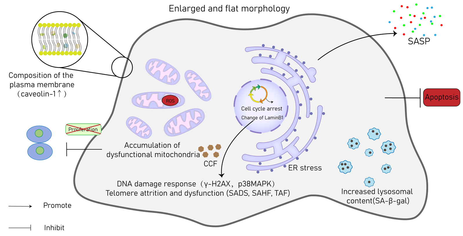
In addition to the mechanisms described above, new studies propose that CS can result from other factors. As an illustration, according to aging theory, excessive activation of nutrient sensing pathways, such as targets of insulin/insulin-like growth factor 1 (IGF-1) signaling and rapamycin, is linked to accelerated aging and a reduced lifespan [51]. Consequently, pursuing moderate dietary restriction, inducing genetic mutations, or employing chemical agents to lower the activity of nutrition-sensitive signaling pathways can serve to extend the lifespan of organisms [52]. Additionally, the dysregulation of systemic humoral pathways has been closely linked to CS, including cases involving a deficiency of Klotho [53]. Finally, protein folding is implicated in CS processes, with ER stress being involved in initiating or sustaining aging phenotypes [54].

3. Hallmarks of CS

Various conditions trigger CS, and SNCs exhibit several distinguishing characteristics that enable their detection in both in vitro and in vivo settings (Figure 3). In the heart and other tissues, specific hallmark characteristics are indicative of SNCs, serving as indirect evidence of the onset of senescence.
A prominent characteristic of CS is the distinct morphological alterations that transpire as it advances. Ball observed that senescent fetal cardiomyocytes consistently display an enlarged and vacuolized cell body [55]. Various senescent cardiovascular cells also show a flattened morphology, an enlarged volume, and the presence of vacuoles [56,57,58]. Consequently, these morphological changes could serve as markers for identifying SNCs in CVDs, even in the absence of cell-type specificity. The most prevalent modification in the plasma membrane composition of SNCs is the heightened expression of caveolin-1, which was consistently observed [59]. An increased number of mitochondria is evident in SNCs, relative to non-SNCs [60]. Even though there is an increased quantity of mitochondria in SNCs, their membrane potential is reduced, leading to the liberation of mitochondrial enzymes such as EndoG and an augmented production of ROS [61]. Mitochondrial dysfunction is considered both a marker and an inducer of stem cell aging [62]. The diminished mitophagy brings about the gathering of aged and impaired mitochondria, contributing primarily to the increased mitochondrial content [63]. SNCs exhibit elevated activity of senescence-associated β-galactosidase (SA-β-gal) due to heightened lysosomal density. Therefore, SA-β-gal is widely used as a prominent hallmark for identifying SNCs [64]. Sudan Black B (SBB) is an alternative marker that can be employed to detect the accumulation of lysosomes. It provides another means of identifying the presence and abundance of lysosomes in cells [65]. Although the elevated activity of lysosomes, indicated by elevated SA-β-gal expression, is commonly linked to SNCs, it is not a definitive hallmark of CS, since the constitutive expression of SA-β-gal has also been observed in non-SNCs [66]. The emergence of cytoplasmic chromatin fragments (CCFs) is a common characteristic observed in SNCs, which is accompanied by the depletion of lamin B1, an essential nuclear structural protein [67]. These CCFs have the ability to be released into the external environment through exosomes and trigger DDR in other cells [68].
As previously described, most stressors that lead to CS activate either the p53/p21 or p16Ink4a/retinoblastoma protein pathways. The buildup of cell cycle-suppressing proteins, including p16 (p16INK4A), p53, or p21, is frequently employed as a hallmark of senescence [69]. Alongside p53, p21, and p16Ink4a, other widely recognized markers of CS encompass elevated levels of the p38 mitogen-activated protein kinase (p38MAPK) or γ-H2AX, which reflect the activation of DDRs. Senescence-associated heterochromatin foci (SAHF) and senescence-associated distention of satellites (SADs) are also recognized as markers of SNCs [70,71].
Telomere-related DNA damage, referred to as telomere dysfunction-induced foci (TAF), is identified through the co-localization of γ-H2AX and p53-binding protein 1 (p53BP1) with telomeres. In fibroblasts, exposure to irradiation or application of hydrogen peroxide (H2O2) results in an elevated formation of TAF. This phenomenon has also been noted to increase in the liver, gut, and heart as individuals age [26,72]. SNCs cease to divide. A senescent phenotype is indicated by the reduction in proliferation markers, like Ki67, along with the lack of incorporation of 5-bromodeoxyuridine (BrdU) or ethynyl deoxyuridine (EdU) [30]. The SASP encompasses a multitude of proteins, which are secreted into the extracellular milieu by SNCs. Although SASP is critically involved in the pathophysiological behavior of SNCs, its nonspecific and heterogeneous character hinders its application as an unequivocal marker for senescence [73]. Nevertheless, evaluating the composition of SASP quantitatively could serve as a means to differentiate between different senescence programs [74].
The characterization of cells as senescent is a multifaceted process. As there is no single specific biomarker capable of definitively identifying SNCs, a combination of markers is typically employed to confirm the presence of a senescent cellular phenotype.

4. Senescence in Specific Cardiac Cell Types

The impact of senescence on cells is highly influenced by their cell type, tissue composition, and organ context, leading to considerable variation in its effects. Different cell types make up the intricate structure of the heart, including cardiomyocytes, endothelial cells, fibroblasts, vascular smooth muscle cells (VSMCs), immune cells, and cardiac progenitor cells (CPCSs) [75]. Extensive in vitro and in vivo studies have demonstrated that all types of cardiovascular cells, both during the natural aging process and in the context of CVDs, can undergo senescence [1]. Hence, the function of CS in age-related CVDs can arise from alterations in multiple cell types (Table 1).

4.1. Cardiomyocytes

CS has traditionally been recognized as an irreversible G1 arrest state in mitotic cells. Nevertheless, in the heart, a significant proportion of cardiomyocytes differentiate into terminally differentiated cells that cease to undergo cell division immediately after birth. Despite being mostly non-dividing, cardiomyocytes from aged rats and mice exhibit telomere shortening, similar to normal human heart tissue [10]. Despite enduring debate that span decades, an increasing body of research indicates that cardiomyocytes possess mechanisms that can trigger senescence [11].
The manifestation of impaired contractility and disrupted conduction patterns in senescent cardiomyocytes can give rise to cardiomyopathies or arrhythmias. As a case in point, in a mouse model of Duchenne muscular dystrophy (DMD), the absence of dystrophin in cardiomyocytes resulted in the emergence of a senescent phenotype. Moreover, mutations in lamin A (LMNA) are linked to dilated cardiomyopathy and this may be linked to genomic instability that occurs as a result of nuclear membrane disruption. This disruption results in increased regions of open chromatin [76]. Compelling evidence indicates that CS is critically involved in mediating cardiomyopathy induced by chemotherapy and radiotherapy, such as with anthracyclines and doxorubicin.

4.2. Endothelial Cells

By releasing vasoactive compounds and growth factors, endothelial cells have a crucial function in regulating vascular tone and vasodilation [77]. The continuous exposure of these cells to various stressors renders them susceptible to damage, which has the potential to induce senescence. Therefore, well-vascularized tissues, especially those with a high density of endothelial cells, tend to bear a greater load of SNCs [78]. The heightened secretion of endothelin-1 (ET-1) and reduced production of nitric oxide by senescent endothelial cells create a substantial interplay between these SNCs and the surrounding cardiac cell populations [78]. Consequently, vascular inflammation and impaired vasodilation ensue because of this phenomenon, establishing a self-amplifying cycle where the amassment of senescent endothelial cells leads to vascular disorder, and subsequently also the other way around [79]. Endothelial cell senescence results in compromised vasodilation and vascular dysfunction, contributing to conditions like atherosclerosis, heart failure (HF) with preserved ejection fraction (HFpEF), or pulmonary hypertension (PH) [80].

4.3. Cardiac Fibroblasts

Cardiac fibroblasts are crucial in regulating ECM remodeling, reorganization, and paracrine communication within the cardiac microenvironment [81]. To aid in adhesion, SNCs express integrins and matrix metalloproteinases (MMPs) that help preserve ECM structure and integrity [82]. Following an acute myocardial infarction (MI), the p53 and p21 pathways are upregulated, culminating in the senescence of cardiac fibroblasts [83]. Inducing senescence in cardiac fibroblasts can have positive and negative impacts—advantageous in the context of chronic wound healing after MI, yet potentially harmful during myocardial fibrosis associated with aging [76]. Cardiac fibroblasts also engage in paracrine signaling to modulate processes like proliferation, hypertrophic growth, and cardiomyocyte senescence [81]. The transition of activated cardiac fibroblasts from a pro-inflammatory condition to an anti-inflammatory condition is crucial in the formation of scar tissue following an acute MI [84]. Navitoclax, a senolytic agent administered systemically, has been found to improve outcomes following ischemia-reperfusion injury by clearing SNCs. Thus, it is essential to strike a balance between the beneficial effects of fibroblast senescence and the potentially detrimental consequences of senescence [85].

4.4. VSMCs

VSMCs collaborate with endothelial cells to regulate critical aspects of vascular function, including arterial pressure regulation, vascular tone maintenance, and blood flow coordination [86]. CS can be triggered in VSMCs through various mechanisms, like telomeric shortening, DNA damage, oxidative stress, and dysfunction in autophagy [87]. Elevated SA-β-gal activity and dysregulated transcriptome involving p16, p21, and Rb proteins are observed in senescent VSMCs. Additionally, they show increased expression of inflammatory cytokines [88]. VSMCs senescence contributes to vascular diseases like atherosclerosis and pulmonary hypertension [89]. The secretion of monocyte chemoattractant protein-1 (MCP-1) and macrophage inflammatory protein-1α/β (MIP1α/β) stimulates the engagement of monocytes, macrophages, and lymphocytes, consequently promoting plaque growth and aggravating the likelihood of rupture [90]. Moreover, the CS of VSMCs may also play a role in the pathophysiology of PH through SASP [91].

4.5. Immune Cells

Immune cells, particularly macrophages and T cells with senescent-like characteristics, have gained significant attention in recent studies for their substantial roles in CVDs [76]. The susceptibility of atherosclerotic plaques to rupture is heightened in cases where there is a higher abundance of macrophages, as elucidated by a recent study [92,93,94]. Notably, in the atrioventricular node, cardiac macrophages have been shown to enhance electrical conduction. In the context of CVD, leukocytes exhibiting short telomeres, which are a characteristic feature of senescence, have been identified within atherosclerotic coronary arteries [95]. A positive feedback loop can be established through the secretion of inflammatory cytokines, chemokines, and metalloproteinases by senescent foamed macrophages exhibiting senescence markers within atherosclerotic plaques [96]. Apart from macrophages, senescent-like T cells can also contribute to the development of chronic inflammatory diseases, encompassing CVDs [97] such as atherosclerotic disease, hypertension, coronary artery disease (CAD), acute coronary syndromes, and HF [98,99,100,101].

4.6. Cardiac Progenitor Cells

CPCs are a unique type of progenitor cell that can differentiate into cardiac myocytes and endothelial cells. The lifespan of these cells is influenced by telomerase activity and telomere length, like other cell types [102]. As humans age, CPCs in the heart tend to become senescent, which can lead to an inability to sustain homeostasis, repair damaged tissue, and regenerate after injury [103]. Various research groups have investigated the use of senolytic medications to enhance CPC function, indicating that CPC senescence could be a viable target for future interventions targeting CVDs [104].
Communication between cells in the myocardial microenvironment is significant in regulating cardiac homeostasis and the aging process. Non-myocyte cells can modulate the physiological functions and senescence of cardiomyocytes. At the same time, cardiomyocytes also exert their influence on non-myocytes, partially through paracrine signaling [105].

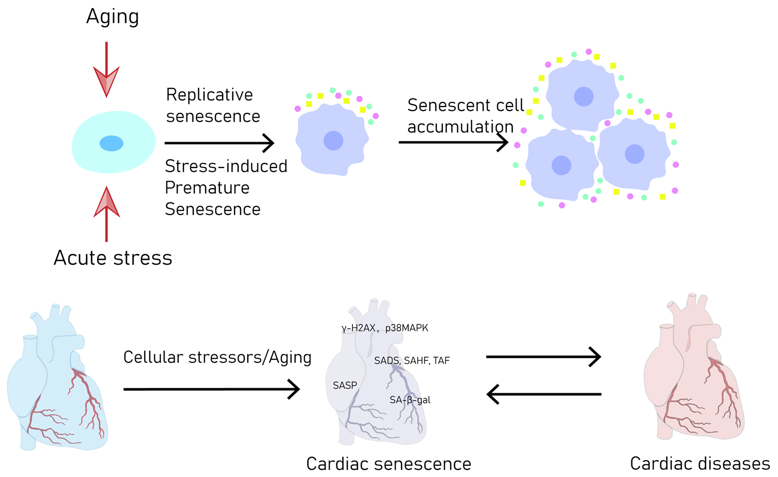
5. Physiology and Pathophysiology of CS in the Cardiovascular System

5.1. Senescence in Heart Development

Numerous investigations have explored the involvement of CS in cardiac embryonic development and the remodeling of cardiac tissues. Senescence assumes a crucial role during embryogenesis, often referred to as developmental senescence, wherein the elimination of SNCs holds significance for morphogenesis [5]. Both SNCs and numerous standard components of the SASP are discernible in the developing embryos of experimental animals [106]. Additional support stems from observations highlighting that abnormalities in senescence-related proteins throughout the developmental process are connected to birth defects of the heart [107,108]. Hence, it becomes evident that senescence actively participates in the process of embryonic development of the four-chambered mammalian heart, whereby aberrant senescence signaling is linked to various congenital heart defects in this context [1].
While CS indeed contributes to organismal remodeling and wound healing, compelling data strongly indicate that persistent senescence in the process of aging is also intertwined with a variety of pathological processes. Considering its unique role in each of these processes, CS is postulated to be a crucial detrimental mechanism that fosters the development of CVDs [109].

5.2. Senescence in Cardiac Pathology

5.2.1. HF

HF emerges from various stressors and underlying factors that lead to the progressive impairment of cardiomyocytes. This impairment leads to the heart’s inability to effectively meet the demands of the body [110]. Multiple research projects have established a correlation between CS and pathways associated with HF pathophysiology [111]. On the one hand, mice with a global deficiency in kinases, which have a protective impact on mitochondria and telomere length, experience advanced HF at the age of six months. These mice exhibit elevated levels of p16, p53, and SA-β-gal, along with a decline in the structure and function of mitochondria within cardiac tissue [112]. In a similar fashion, diabetic cardiomyopathy in mice is characterized by high levels of senescence markers in both cardiomyocytes and CPCs, coupled with impaired mitochondrial function and elevated ROS generation in the heart [113]. On the other hand, the utilization of an angiotensin II receptor blocker and navitoclax has demonstrated the potential to enhance cardiac function and address electrophysiological abnormalities [114]. Furthermore, increased autophagy has been shown to safeguard cardiac function, reducing p16, p21, p53, and several constituents of the SASP in aged mice. Notably, elevated levels of SASP components align with the severity of HF [115].
Collectively, senescence may intersect with several pathophysiological pathways that contribute to HF, such as mitochondrial dysfunction, autophagy, and activation of the neurohumoral system. Nevertheless, whether targeting cell senescence could serve as a viable therapeutic approach for HF remains a subject requiring further investigation.

5.2.2. MI

Acute MI stands as the foremost contributor to global morbidity and mortality. A lot of data strongly imply a connection between CS and the consequences of myocardial ischemia and MI. Generally, MI causes a considerable depletion of cardiomyocytes. Ischemic injury initiates DNA damage, oxidative stress, and disturbances in mitochondrial performance, collectively influencing the predisposition to cardiomyocyte senescence. In a study by Zhu et al., it was suggested that fibroblast senescence, regulated by p53, acts as a barrier against collagen deposition and cardiac fibrosis. This intricate process culminates in cardiac rupture and impaired cardiac dysfunction post-MI [83].
The role of CS in MI pathogenesis remains a topic of debate, as conflicting reports indicate both positive and negative impacts. Notably, mice treated with navitoclax exhibit significantly enhanced survival rates and reduced functional deterioration post-MI. This observation implies that SNCs contribute to unfavorable remodeling. Moreover, standard drug therapies used to mitigate cardiac dysfunction following myocardial infarction (MI) also cause a decline in the presence of markers of CS in cardiac tissue [1]. Conversely, several studies propose a favorable role for cell senescence post-MI. Specifically, the diminished production of SASP components is linked to increased systolic dysfunction and cardiac fibrosis in the post-MI phase. This association suggests that the components of the SASP within the peri-infarct region exert antifibrotic and cardioprotective effects [116]. While a significant amount of data underscores the connection between CS and the repercussions of myocardial ischemia and infarction, much of this evidence is correlational rather than causal.

5.2.3. Hypertrophic Cardiomyopathy

The physiological response of cardiac hypertrophy to biomechanical stress results in an augmentation of the left ventricular mass and a lowering of cardiac compliance [117]. Several pieces of evidence strongly suggest that senescence actively drives the progression of cardiac hypertrophy. To eliminate SNCs in aged mice, Anderson et al. employed a combination of the INK-ATTAC transgenic model and navitoclax treatment. This intervention led to a decrease in cardiomyocyte size and fibrosis, with cardiac function and left ventricular mass remaining relatively stable [26]. From these findings, the researchers reached the conclusion that CS is a factor in age-related cardiac hypertrophy. They also suggested that the removal of SNCs supports cardiomyocyte regeneration, as evidenced by an increase in mononuclear cardiomyocytes positive for proliferation markers Ki67 and EdU [26]. Numerous studies underscore the significance of ROS and mitochondrial dysfunction in instigating age-related senescence in cardiomyocytes [118,119,120,121]. In summary, CS does indeed contribute to age-related cardiac hypertrophy. Nevertheless, the mechanisms governing senescence in cardiomyocytes and their pathological implications warrant further exploration [1,2].

5.2.4. Cancer Therapy-Induced Cardiotoxicity

The intricate molecular mechanisms that underlie cardiotoxicity are constantly under investigation, encompassing facets like mitochondrial dysfunction, oxidative stress, disrupted autophagy, and telomere dysfunction [122]. CS has recently gained recognition as a pivotal contributor to the development of cardiotoxicity resulting from cancer treatments. Cells induced into senescence by anticancer therapies share common attributes with those prompted by other stimuli. Pertinently, eliminating SNCs hold clinical significance as it can ameliorate cardiac systolic dysfunction [26]. In addition, the molecular signatures and patterns exhibited by SNCs, in conjunction with the recognition of specific molecules within blood samples and other biological fluids, hold promise as emerging novel biological markers in the realm of cardio-oncology [74]. Compared to the relatively abundant research examining the role of senescence in doxorubicin-induced cardiotoxicity, there has been less attention given to the function of senescence in cardiac disease induced by radiotherapy. Nevertheless, whether stemming from radiation or chemotherapy, the comprehensive removal of CS can potentially mitigate cardiotoxicity [26].

5.2.5. Cardiac Fibrosis

Cardiac fibrosis is a multifaceted phenomenon distinguished by the excessive generation of extracellular matrix proteins in the myocardial tissue. The proliferation and activation of cardiac fibroblasts mediate this phenomenon [123]. Several investigations have provided evidence linking the presence of senescent fibroblasts to the occurrence of fibrosis [124,125]. However, the nature of the involvement of senescence in fibrogenesis remains uncertain in these investigations. The use of transgenesis or treatment involving navitoclax has demonstrated a reduction in fibrosis, providing evidence for the possible involvement of SNCs in the advancement of cardiac fibrosis. Furthermore, cell cycle arrest brought on by cellular senescence may have the ability to lessen fibroblast proliferation and, in turn, suppress the progression of fibrosis [1].

5.2.6. Diabetic Cardiomyopathy

Cardiac myopathy can arise as a consequence of diabetes mellitus, a specific clinical condition marked by diminished muscle mass, expanded chambers, and compromised ventricular function, all occurring in the absence of CAD [2]. The various elements that play a part in the onset and advancement of diabetic cardiomyopathy encompass a spectrum of elements. These include compromised cardiac insulin metabolic signaling, endoplasmic reticulum stress, elevated oxidative stress, mitochondrial dysfunction, inflammation, and microvascular dysfunction [125]. The diabetic heart exhibits premature telomeric shortening in its CPCs, evident through telomeric shortening and the presence of senescence-associated proteins, namely p53 and p16INK4A. This phenomenon, in turn, escalates the count of senescent myocytes, thus promoting premature myocardial aging and contributing to HF. The suppression of p53 in a mouse model of streptozotocin (STZ)-induced type 1 diabetes (T1DM) prevented cardiac apoptosis during the early phases of diabetes. Moreover, it attenuated cellular senescence induced by diabetes and averted glycolytic and angiogenetic dysfunction. These effects were achieved by enhancing the stability of the hypoxia-inducible factor-1α (HIF-1α) protein and facilitating HIF-1α-mediated genomic transcription [126]. This insight potentially lends theoretical support to diagnosing and treating diabetic cardiomyopathy.

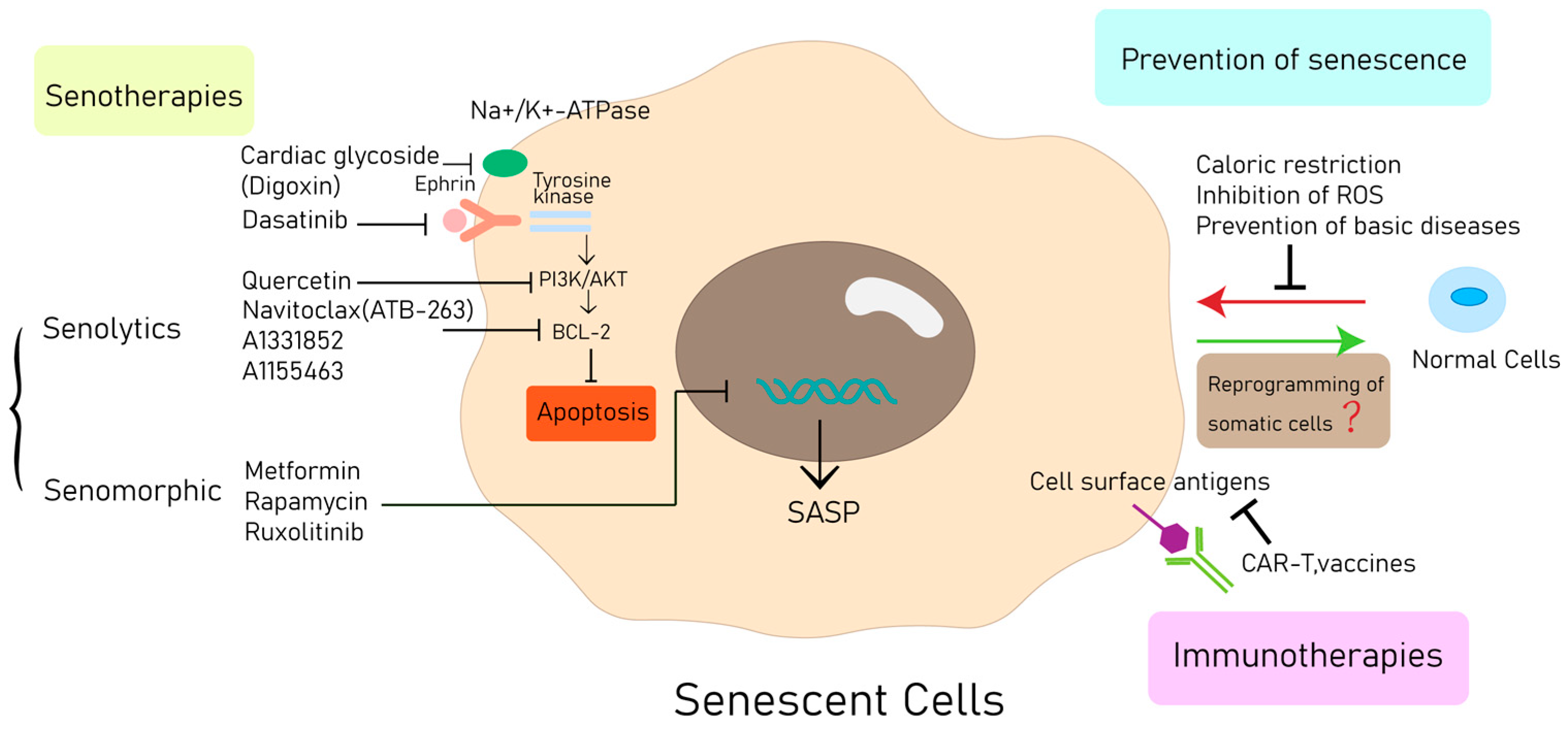
6. Therapeutic Approaches

SNCs have been recognized as impactful contributors to aging and age-related CVDs in the last two decades. The supporting evidence for this assertion stems from the identification of SNCs biomarkers in affected tissues, the genetic interventions that disrupt senescence and thereby alter disease pathophysiology, the modification of disease progression by means of transgene-mediated SNC elimination, and the modification of pathological processes through the administration of senolytics. Collaboratively, these advancements strongly indicate that SNCs present a possible target for senotherapy, holding the potential to treat and even prevent CVDs [127].
The therapeutic intervention aimed at SNCs is gaining recognition as a promising and pioneering approach to hindering cardiovascular aging and the advancement of associated diseases. Presently, a range of methods is being utilized to eradicate cardiovascular SNCs in in vitro and in vivo models [128] (Figure 4).

6.1. Prevention of Senescence

One potential therapeutic approach involves the prevention of senescence by mitigating pro-senescent stressors. For instance, caloric restriction has demonstrated the ability to effectively inhibit CS by diminishing the activation of the mammalian target of rapamycin (mTOR) signaling pathway via the AMP-activated protein kinase (AMPK) [102,129]. In vitro investigations have indicated that inhibiting the accumulation of ROS, using the ROS scavenger N-acetyl-l-cysteine, can prevent CS in human tissues [130]. In mice with diabetes mellitus or heart failure, the advancement of CS can be decelerated by preventing hypertension and inhibiting the sympathetic nervous system [131]. Furthermore, various methods to inhibit the anthracycline-induced CS of VSMCs and cardiomyocytes have been identified, such as prednisolone and the amplification of LncRNA-MALAT1 [132,133].

6.2. Senotherapies

In SNCs, glycolysis is elevated, DNA damage response pathways are heightened, and SASP is present, offering potential avenues for therapeutic intervention [134]. Senotherapeutics constitute a class of medications aimed at eliminating SNCs (senolytics) and mitigating the external effects caused by SNCs (senomorphics or senotatics) [135,136]. Ongoing efforts encompass the screening of repurposed medications and natural products, as well as the development of novel compounds targeting SNCs [11].

6.2.1. Senolytics

SNCs’ anti-apoptotic pathways (SCAPs) have led to the elimination of a significant portion of SNCs that possess pro-apoptotic qualities and are detrimental to tissues, which is achieved through apoptosis. Distinct categories of pro-apoptotic SNCs in humans rely on specific SCAPs for their survival [137]. A variety of techniques have been designed for senolysis, focusing on anti-apoptotic signaling molecules as targets to effectively eliminate SNCs in the context of aging [11]. Dasatinib and quercetin are two examples of agents that promote apoptosis in SNCs by suppressing ephrin receptor-dependent tyrosine kinases and PI3K/mTOR pathways, respectively [138]. Considering their recognized targets, the combination of quercetin and dasatinib has exhibited greater efficacy in eliminating senescence across diverse pathological models. In terms of administration, single or intermittent doses demonstrate considerable advantages compared to continuous senolytic treatment, as they entail fewer side effects [134,139,140,141]. A recent study demonstrated the testing of dasatinib in conjunction with quercetin in clinical scenarios involving dysfunction related to idiopathic pulmonary fibrosis, implying that anti-senescent agents might soon become accessible for human use. Consequently, the optimization of anti-senescent protocols for impending clinical translation holds significant promise.
By targeting the B-cell lymphoma 2 (BCL-2) family of proteins, navitoclax or ABT263 is able to effectively induce apoptosis and eliminate SNCs [142]. This approach has been shown to delay the onset and progression of CVDs such as atherosclerosis, myocardial infarction (MI), ischemia-reperfusion injury, and cardiac aging [26,96,142,143].
A new noteworthy senolytic candidate is digoxin, a cardiac glycoside with a history of application in cardiac disease treatment. The repurposing of established drugs like digoxin holds the potential to expedite clinical translation. In mouse models, the administration of digoxin has demonstrated the capacity to eradicate transplanted SNCs [144,145]. Similar to many other senolytic regimens, intermittent administration of senolytic therapy with digoxin, using a ‘hit-and-run’ approach, has the potential to yield improved clinical outcomes [11].
This avenue of treatment holds substantial promise. After the original hypothesis-driven drug discovery method, many more senolytic compounds have been identified through various methods, including high-throughput screening. Other small molecules that have been found for the targeted elimination of SNCs include specific BCL-xL inhibitors such as A1331852 and A1155463, the flavonol fisetin, piperlongumine, procyanidin C1, a FOXO4-related peptide, and several other compounds [127,146,147,148].

6.2.2. Manipulation of the SASP

SASP inhibitors, often called “senomorphic” drugs, do not have a direct effect on the removal of SNCs. Instead, they function by altering elements of the SNCs secretome that contribute to chronic inflammation and tissue deterioration. Specific drugs among these target the transcription factor nuclear factor kappa B (NF-κB) [149,150], Janus kinases, or STAT pathways. Additional targets encompass the mTOR pathway, p38MAPK and its associated kinases, molecules associated with mitochondrial complexes 1 or 4, heat shock protein 90 (HSP90), and NAD+/NADH metabolism [38]. Several medications that are approved by the Food and Drug Administration (FDA) have been found to possess senomorphic effects. These include metformin, rapamycin, and ruxolitinib [151,152,153,154]. Building on these encouraging findings, initiatives such as the Targeting Aging with Metformin (TAME) trial, supported by the American Federation for Aging Research (AFAR) and others, are in the planning stages. However, the utilization of certain senomorphic drugs poses challenges related to managing potential off-target effects. This includes scenarios where suppressed inflammation might not be beneficial for certain diseases or tissue repair. In contrast to senolytics, which directly eliminate the SNCs responsible for releasing tissue-damaging SASP factors, the need for continuous treatment could be more pronounced with senomorphic drugs [11].

6.3. Immune Activation of Target SNCs

Immunotherapies initially designed for cancer treatments, including therapies like chimeric antigen receptor T cell (CAR-T), cytotoxic T lymphocyte (CTL), natural killer (NK) cell, and dendritic cell (DC) therapies, which are currently undergoing clinical applications, have the potential to be repurposed as senolytics. One potential strategy for selectively eliminating SNCs is to target SNC-specific antigens, which are referred to as “seno-antigens”. This approach offers the benefit that tailored treatments can be devised upon identification of a target molecule, even if the complete physiological function of the molecule remains uncertain or effective inhibitors are yet to be found. This approach holds the potential to mitigate off-target effects [11]. For example, a senolytic vaccine aimed at the glycoprotein non-metastatic melanoma protein B (GPNMB) exhibited enhanced physical function in aged mice and extended the lifespan of progeroid mice [155]. Similarly, another senolytic vaccine focused on CD153, identified as a hallmark of obesity-related T cells within adipose tissue, successfully decreased the buildup of senescent T cells [156]. Both vaccine treatments demonstrated effectiveness for several months. Moreover, various antibody–drug conjugates (ADCs) have been identified as having the ability to effectively removing SNCs [157,158].

6.4. Bypassing and Reverting Senescence

While CS was initially characterized as an essentially irreversible cell cycle withdrawal, inducing senescence can actually be reversed in vitro, leading to the revival of cell proliferation [159]. Furthermore, a groundbreaking study demonstrated that in vivo reprogramming of somatic cells could reinstate their ability to proliferate [160]. These investigations introduce the intriguing prospect of bypassing senescence therapeutically. Nevertheless, it is essential to exercise prudence with such approaches, as recent studies have indicated that this strategy might heighten the tumor-initiating capacity of cells beyond that of nonsenescent cells [161].
Despite major advances, the translation of senescence-targeting strategies to clinic applications must be undertaken cautiously. This caution arises from the fact that senescence can exert both advantageous and detrimental effects, depending on the specific pathological context [162].

7. Conclusions and Outlook

As life expectancy rises, CVDs have become increasingly prevalent. Many prominent pharmacological treatments, designed to address age-related CVDs, have yielded less than satisfactory outcomes, underscoring the necessity for innovative treatment approaches. As a novel research focus in CVDs, emerging evidence suggests the involvement of CS in the pathological progression of CVDs that were triggered by severe acute respiratory syndrome coronavirus 2 (SARS-CoV-2) infection. CS has even been investigated as the key pathogenic mechanism and a promising therapeutic target for SARS-CoV-2 infection. Therefore, in the present post-epidemic era, there is an increasing urgency to elucidate the mechanisms underlying various pathological processes mediated by CS in the human body [163,164].
CS represents a complex cellular state, serving both physiological and pathophysiological functions in processes such as cellular development, the pathogenesis of diseases, and organ dysfunction [165]. Cardiovascular cell senescence is vital in maintaining homeostasis within cardiovascular tissue during tissue regeneration, embryonic development, and wound healing [166]. However, the persistent buildup of SNCs within cardiovascular tissues can impede their function and has been linked to the development of age-related CVDs [167], such as HF, MI, hypertrophic cardiomyopathy, cardiotoxicity, cardiac fibrosis, arrhythmogenic cardiomyopathy, and diabetic cardiomyopathy. Complex molecular pathways intricately govern cardiovascular cell senescence both in vitro and in vivo. Nevertheless, there remains an inadequate grasp of the precise underlying mechanisms that contribute to the dysregulation of cardiovascular cell senescence during the onset of CVDs. At present, there is a deficiency of dependable selective markers to detect senescent cardiovascular cells in vivo [13]. The spatiotemporal identification and noninvasive quantification of individual SNCs in vivo remain challenging [168]. These challenges collectively impede the development of efficacious treatments for CVDs. The successful development of new treatment strategies for cardiovascular aging cells, to alleviate the substantial clinical significance, depends on a thorough understanding of aging biology and the biological specifics and primary cell types that participate in the pathogenesis of CVDs [128]. The selective elimination of SNCs has emerged as a safer approach to senescence during aging [166].
A comprehensive research approach is imperative to target senescent cardiovascular cells accurately, effectively, and safely. This approach should encompass the following aspects: (1) accounting for discrepancies between animal models and human conditions; (2) investigating cellular and model systems representative of natural aging; (3) investigating the mechanism underlying the senescence of cardiovascular cells and its contribution to the initiation and progression of CVDs; (4) identifying distinct spatiotemporal biomarkers and targets specific to senescence in various cardiovascular cell types in vivo; (5) validating the efficacy of established senolytic agents while considering potential side effects; and (6) assessing the potential risk of cancer escalation following the removal of SNCs [128]. With the possibility of therapeutic interventions via targeting, a comprehensive grasp of the manifold roles and mechanisms associated with CS in CVDs not only aids in the development of novel agents, but also facilitates the formulation of appropriate clinical strategies. Nonetheless, it remains imperative to conduct extensive studies for the formulation of precise and accurate treatment strategies for CVDs.

Author Contributions

D.L. and Y.L. conceived and designed the study. An extensive search of relevant topics was conducted by H.D. and X.Z.; Y.W. and Y.X. carried out the review, wrote the manuscript and undertook extensive editing. All the authors have made important contributions to the writing of the manuscript. All authors have approved the final draft. All authors have read and agreed to the published version of the manuscript.

Funding

National Natural Science Foundation of China (NSFC 82060080), Science and Technology Department of Gansu Province (21JR11RA116,23YFFA0038), Lanzhou City Technology Bureau project (2019-RC36) and the Second Hospital of Lanzhou University’s Cuiying Science and Technology Innovation Plan (CY2022-MS-A06 and CY2022-QN-A17) funding. This study also utilized the cardiac rehabilitation in Gansu Province’s Engineering Research Center (CRQI-C00535), Lanzhou University’s Star of “Innovation” plan (2023CXZX-164), and graduate education support in Lanzhou University’s Medical Innovation and Development Project (lzuyxcx-2022-128).

Institutional Review Board Statement

Not applicable.

Informed Consent Statement

Not applicable.

Data Availability Statement

Not applicable.

Conflicts of Interest

The authors declare no conflict of interest.

Abbreviations

The following abbreviations are used in this manuscript:
AbbreviationMeaning
CScellular senescence
SASPsenescence-associated secretory phenotype
CVDscardiovascular diseases
SIPSstress-induced premature senescence
RSreplicative senescence
DDRDNA damage response
hTERThuman telomerase reverse transcriptase
SSBssingle-strand breaks
DSBsdouble-strand breaks
ATMataxia–telangiectasia mutated
ATRataxia–telangiectasia Rad3-related
γH2AXphosphorylated histone H2AX
CDKN1ACDK inhibitor p21
cGAScyclic GMP–AMP synthase
STINGstimulator of interferon genes
TAFstelomere-associated foci
mTORmammalian target of rapamycin
ROSreactive oxygen species
OISoncogene-induced senescence
mtDNAmitochondrial DNA
ECMextracellular membrane
mi-RNAmicroRNAs
lncRNAlong non-coding RNAs
EndoGendonuclease G
SA-β-galsenescence-associated β-galactosidase
SBBSudan Black B
CCFscytoplasmic chromatin fragments
p38MAPKp38 mitogen-activated protein kinase
SAHFsenescence-associated heterochromatin foci
SADssenescence-associated distention of satellites
H2O2hydrogen peroxide
BrdUbromodeoxyuridine
EdUethynyl deoxyuridine
VSMCsvascular smooth muscle cells
DMDDuchenne muscular dystrophy
LMNAlamin A
ET-1endothelin-1
HFheart failure
HFpEFheart failure with preserved ejection fraction
MMPsmatrix metalloproteinases
MImyocardial infarction
CADcoronary artery disease
T1DMtype 1 diabetes
STZstreptozotocin
HIF-1αhypoxia-inducible factor-1α
SNCssenescent cells
AMPKAMP-activated protein kinase
SCAPssenescent cell anti-apoptotic pathways
BCL-2B-cell lymphoma 2
NF-κBnuclear factor kappa B
HSP90heat shock protein 90
FDAFood and Drug Administration
TAMETargeting Aging with Metformin
AFARAmerican Federation for Aging Research
CAR-Tchimeric antigen receptor T cell
CTLcytotoxic T lymphocyte
NKnatural killer
DCdendritic cell
GPNMBglycoprotein non-metastatic melanoma protein B
ADCsantibody–drug conjugates
TGF-βtransforming growth factor-β
SMADsmall mother against decapentaplegic
AMPKadenosine 5‘-monophosphate (AMP)-activated protein kinase
Rbretinoblastoma protein
MiDASmitochondrial dysfunction-associated senescence
IGF-1insulin-like growth factor 1
CPCScardiac progenitor cells

References

  1. Mehdizadeh, M.; Aguilar, M.; Thorin, E.; Ferbeyre, G.; Nattel, S. The role of cellular senescence in cardiac disease: Basic biology and clinical relevance. Nat. Rev. Cardiol. 2022, 19, 250–264. [Google Scholar] [CrossRef] [PubMed]
  2. Evangelou, K.; Vasileiou, P.V.S.; Papaspyropoulos, A.; Hazapis, O.; Petty, R.; Demaria, M.; Gorgoulis, V.G. Cellular senescence and cardiovascular diseases: Moving to the “heart” of the problem. Physiol. Rev. 2023, 103, 609–647. [Google Scholar] [CrossRef] [PubMed]
  3. Niccoli, T.; Partridge, L. Ageing as a risk factor for disease. Curr. Biol. 2012, 22, R741–R752. [Google Scholar] [CrossRef] [PubMed]
  4. Storer, M.; Mas, A.; Robert-Moreno, A.; Pecoraro, M.; Ortells, M.C.; Di Giacomo, V.; Yosef, R.; Pilpel, N.; Krizhanovsky, V.; Sharpe, J.; et al. Senescence is a developmental mechanism that contributes to embryonic growth and patterning. Cell 2013, 155, 1119–1130. [Google Scholar] [CrossRef]
  5. Van Deursen, J.M. The role of senescent cells in ageing. Nature 2014, 509, 439–446. [Google Scholar] [CrossRef]
  6. Laredo, M.; Waldmann, V.; Khairy, P.; Nattel, S. Age as a Critical Determinant of Atrial Fibrillation: A Two-sided Relationship. Can. J. Cardiol. 2018, 34, 1396–1406. [Google Scholar] [CrossRef]
  7. Kuo, C.L.; Pilling, L.C.; Kuchel, G.A.; Ferrucci, L.; Melzer, D. Telomere length and aging-related outcomes in humans: A Mendelian randomization study in 261,000 older participants. Aging Cell 2019, 18, e13017. [Google Scholar] [CrossRef]
  8. Noly, P.E.; Labbé, P.; Thorin-Trescases, N.; Fortier, A.; Nguyen, A.; Thorin, E.; Carrier, M. Reduction of plasma angiopoietin-like 2 after cardiac surgery is related to tissue inflammation and senescence status of patients. J. Thorac. Cardiovasc. Surg. 2019, 158, 792–802.e5. [Google Scholar] [CrossRef]
  9. Gude, N.A.; Broughton, K.M.; Firouzi, F.; Sussman, M.A. Cardiac ageing: Extrinsic and intrinsic factors in cellular renewal and senescence. Nat. Rev. Cardiol. 2018, 15, 523–542. [Google Scholar] [CrossRef]
  10. Hayflick, L.; Moorhead, P.S. The serial cultivation of human diploid cell strains. Exp. Cell Res. 1961, 25, 585–621. [Google Scholar] [CrossRef]
  11. Hu, C.; Zhang, X.; Teng, T.; Ma, Z.G.; Tang, Q.Z. Cellular Senescence in Cardiovascular Diseases: A Systematic Review. Aging Dis. 2022, 13, 103–128. [Google Scholar] [CrossRef]
  12. Serrano, M.; Blasco, M.A. Putting the stress on senescence. Curr. Opin. Cell Biol. 2001, 13, 748–753. [Google Scholar] [CrossRef] [PubMed]
  13. Gorgoulis, V.; Adams, P.D.; Alimonti, A.; Bennett, D.C.; Bischof, O.; Bishop, C.; Campisi, J.; Collado, M.; Evangelou, K.; Ferbeyre, G.; et al. Cellular Senescence: Defining a Path Forward. Cell 2019, 179, 813–827. [Google Scholar] [CrossRef]
  14. Blackburn, E.H. Structure and function of telomeres. Nature 1991, 350, 569–573. [Google Scholar] [CrossRef] [PubMed]
  15. Rossi, M.; Gorospe, M. Noncoding RNAs Controlling Telomere Homeostasis in Senescence and Aging. Trends Mol. Med. 2020, 26, 422–433. [Google Scholar] [CrossRef]
  16. Bodnar, A.G.; Ouellette, M.; Frolkis, M.; Holt, S.E.; Chiu, C.P.; Morin, G.B.; Harley, C.B.; Shay, J.W.; Lichtsteiner, S.; Wright, W.E. Extension of life-span by introduction of telomerase into normal human cells. Science 1998, 279, 349–352. [Google Scholar] [CrossRef]
  17. Masutomi, K.; Yu, E.Y.; Khurts, S.; Ben-Porath, I.; Currier, J.L.; Metz, G.B.; Brooks, M.W.; Kaneko, S.; Murakami, S.; DeCaprio, J.A.; et al. Telomerase maintains telomere structure in normal human cells. Cell 2003, 114, 241–253. [Google Scholar] [CrossRef]
  18. Harper, J.W.; Elledge, S.J. The DNA damage response: Ten years after. Mol. Cell 2007, 28, 739–745. [Google Scholar] [CrossRef] [PubMed]
  19. Rao, K.S. Genomic damage and its repair in young and aging brain. Mol. Neurobiol. 1993, 7, 23–48. [Google Scholar] [CrossRef]
  20. Soto-Palma, C.; Niedernhofer, L.J.; Faulk, C.D.; Dong, X. Epigenetics, DNA damage, and aging. J. Clin. Investig. 2022, 132, e158446. [Google Scholar] [CrossRef] [PubMed]
  21. Tian, X.; Firsanov, D.; Zhang, Z.; Cheng, Y.; Luo, L.; Tombline, G.; Tan, R.; Simon, M.; Henderson, S.; Steffan, J.; et al. SIRT6 Is Responsible for More Efficient DNA Double-Strand Break Repair in Long-Lived Species. Cell 2019, 177, 622–638.e22. [Google Scholar] [CrossRef] [PubMed]
  22. Shmulevich, R.; Krizhanovsky, V. Cell Senescence, DNA Damage, and Metabolism. Antioxid. Redox Signal. 2021, 34, 324–334. [Google Scholar] [CrossRef] [PubMed]
  23. Celeste, A.; Petersen, S.; Romanienko, P.J.; Fernandez-Capetillo, O.; Chen, H.T.; Sedelnikova, O.A.; Reina-San-Martin, B.; Coppola, V.; Meffre, E.; Difilippantonio, M.J.; et al. Genomic instability in mice lacking histone H2AX. Science 2002, 296, 922–927. [Google Scholar] [CrossRef] [PubMed]
  24. Yang, H.; Wang, H.; Ren, J.; Chen, Q.; Chen, Z.J. cGAS is essential for cellular senescence. Proc. Natl. Acad. Sci. USA 2017, 114, E4612–E4620. [Google Scholar] [CrossRef] [PubMed]
  25. Fumagalli, M.; Rossiello, F.; Clerici, M.; Barozzi, S.; Cittaro, D.; Kaplunov, J.M.; Bucci, G.; Dobreva, M.; Matti, V.; Beausejour, C.M.; et al. Telomeric DNA damage is irreparable and causes persistent DNA-damage-response activation. Nat. Cell Biol. 2012, 14, 355–365. [Google Scholar] [CrossRef]
  26. Anderson, R.; Lagnado, A.; Maggiorani, D.; Walaszczyk, A.; Dookun, E.; Chapman, J.; Birch, J.; Salmonowicz, H.; Ogrodnik, M.; Jurk, D.; et al. Length-independent telomere damage drives post-mitotic cardiomyocyte senescence. EMBO J. 2019, 38, e100492. [Google Scholar] [CrossRef]
  27. Serrano, M.; Lin, A.W.; McCurrach, M.E.; Beach, D.; Lowe, S.W. Oncogenic ras provokes premature cell senescence associated with accumulation of p53 and p16INK4a. Cell 1997, 88, 593–602. [Google Scholar] [CrossRef] [PubMed]
  28. Liu, X.L.; Ding, J.; Meng, L.H. Oncogene-induced senescence: A double edged sword in cancer. Acta Pharmacol. Sin. 2018, 39, 1553–1558. [Google Scholar] [CrossRef]
  29. Wei, S.; Wei, S.; Sedivy, J.M. Expression of catalytically active telomerase does not prevent premature senescence caused by overexpression of oncogenic Ha-Ras in normal human fibroblasts. Cancer Res. 1999, 59, 1539–1543. [Google Scholar]
  30. Kuilman, T.; Michaloglou, C.; Mooi, W.J.; Peeper, D.S. The essence of senescence. Genes Dev. 2010, 24, 2463–2479. [Google Scholar] [CrossRef]
  31. Banito, A.; Rashid, S.T.; Acosta, J.C.; Li, S.; Pereira, C.F.; Geti, I.; Pinho, S.; Silva, J.C.; Azuara, V.; Walsh, M.; et al. Senescence impairs successful reprogramming to pluripotent stem cells. Genes. Dev. 2009, 23, 2134–2139. [Google Scholar] [CrossRef] [PubMed]
  32. Kawamura, T.; Suzuki, J.; Wang, Y.V.; Menendez, S.; Morera, L.B.; Raya, A.; Wahl, G.M.; Izpisúa Belmonte, J.C. Linking the p53 tumour suppressor pathway to somatic cell reprogramming. Nature 2009, 460, 1140–1144. [Google Scholar] [CrossRef]
  33. Marión, R.M.; Strati, K.; Li, H.; Murga, M.; Blanco, R.; Ortega, S.; Fernandez-Capetillo, O.; Serrano, M.; Blasco, M.A. A p53-mediated DNA damage response limits reprogramming to ensure iPS cell genomic integrity. Nature 2009, 460, 1149–1153. [Google Scholar] [CrossRef] [PubMed]
  34. Krizhanovsky, V.; Lowe, S.W. Stem cells: The promises and perils of p53. Nature 2009, 460, 1085–1086. [Google Scholar] [CrossRef]
  35. Harman, D. Aging: A theory based on free radical and radiation chemistry. J. Gerontol. 1956, 11, 298–300. [Google Scholar] [CrossRef]
  36. Erusalimsky, J.D. Vascular endothelial senescence: From mechanisms to pathophysiology. J. Appl. Physiol. 2009, 106, 326–332. [Google Scholar] [CrossRef]
  37. Forman, H.J.; Zhang, H. Targeting oxidative stress in disease: Promise and limitations of antioxidant therapy. Nat. Rev. Drug Discov. 2021, 20, 689–709. [Google Scholar] [CrossRef] [PubMed]
  38. Di Micco, R.; Krizhanovsky, V.; Baker, D.; d’Adda di Fagagna, F. Cellular senescence in ageing: From mechanisms to therapeutic opportunities. Nat. Rev. Mol. Cell Biol. 2021, 22, 75–95. [Google Scholar] [CrossRef]
  39. Ma, D.; Stokes, K.; Mahngar, K.; Domazet-Damjanov, D.; Sikorska, M.; Pandey, S. Inhibition of stress induced premature senescence in presenilin-1 mutated cells with water soluble Coenzyme Q10. Mitochondrion 2014, 17, 106–115. [Google Scholar] [CrossRef]
  40. Zhao, H.; Wu, L.; Yan, G.; Chen, Y.; Zhou, M.; Wu, Y.; Li, Y. Inflammation and tumor progression: Signaling pathways and targeted intervention. Signal Transduct. Target. Ther. 2021, 6, 263. [Google Scholar] [CrossRef]
  41. Kumari, R.; Jat, P. Mechanisms of Cellular Senescence: Cell Cycle Arrest and Senescence Associated Secretory Phenotype. Front. Cell Dev. Biol. 2021, 9, 645593. [Google Scholar] [CrossRef]
  42. Chen, Y.; Liu, Y.; Dorn, G.W., 2nd. Mitochondrial fusion is essential for organelle function and cardiac homeostasis. Circ. Res. 2011, 109, 1327–1331. [Google Scholar] [CrossRef]
  43. Twig, G.; Elorza, A.; Molina, A.J.; Mohamed, H.; Wikstrom, J.D.; Walzer, G.; Stiles, L.; Haigh, S.E.; Katz, S.; Las, G.; et al. Fission and selective fusion govern mitochondrial segregation and elimination by autophagy. EMBO J. 2008, 27, 433–446. [Google Scholar] [CrossRef]
  44. Lesnefsky, E.J.; Chen, Q.; Hoppel, C.L. Mitochondrial Metabolism in Aging Heart. Circ. Res. 2016, 118, 1593–1611. [Google Scholar] [CrossRef]
  45. Floyd, R.A.; West, M.; Hensley, K. Oxidative biochemical markers; clues to understanding aging in long-lived species. Exp. Gerontol. 2001, 36, 619–640. [Google Scholar] [CrossRef]
  46. Zhang, W.; Song, M.; Qu, J.; Liu, G.H. Epigenetic Modifications in Cardiovascular Aging and Diseases. Circ. Res. 2018, 123, 773–786. [Google Scholar] [CrossRef]
  47. Cruickshanks, H.A.; McBryan, T.; Nelson, D.M.; Vanderkraats, N.D.; Shah, P.P.; van Tuyn, J.; Singh Rai, T.; Brock, C.; Donahue, G.; Dunican, D.S.; et al. Senescent cells harbour features of the cancer epigenome. Nat. Cell Biol. 2013, 15, 1495–1506. [Google Scholar] [CrossRef]
  48. Lyu, G.; Guan, Y.; Zhang, C.; Zong, L.; Sun, L.; Huang, X.; Huang, L.; Zhang, L.; Tian, X.L.; Zhou, Z.; et al. TGF-β signaling alters H4K20me3 status via miR-29 and contributes to cellular senescence and cardiac aging. Nat. Commun. 2018, 9, 2560. [Google Scholar] [CrossRef]
  49. Florio, M.C.; Magenta, A.; Beji, S.; Lakatta, E.G.; Capogrossi, M.C. Aging, MicroRNAs, and Heart Failure. Curr. Probl. Cardiol. 2020, 45, 100406. [Google Scholar] [CrossRef]
  50. Lozano-Vidal, N.; Bink, D.I.; Boon, R.A. Long noncoding RNA in cardiac aging and disease. J. Mol. Cell Biol. 2019, 11, 860–867. [Google Scholar] [CrossRef]
  51. Rota, M.; Goichberg, P.; Anversa, P.; Leri, A. Aging Effects on Cardiac Progenitor Cell Physiology. Compr. Physiol. 2015, 5, 1775–1814. [Google Scholar] [CrossRef] [PubMed]
  52. López-Otín, C.; Blasco, M.A.; Partridge, L.; Serrano, M.; Kroemer, G. Hallmarks of aging: An expanding universe. Cell 2023, 186, 243–278. [Google Scholar] [CrossRef] [PubMed]
  53. Saito, Y.; Yamagishi, T.; Nakamura, T.; Ohyama, Y.; Aizawa, H.; Suga, T.; Matsumura, Y.; Masuda, H.; Kurabayashi, M.; Kuro-o, M.; et al. Klotho protein protects against endothelial dysfunction. Biochem. Biophys. Res. Commun. 1998, 248, 324–329. [Google Scholar] [CrossRef] [PubMed]
  54. Pluquet, O.; Pourtier, A.; Abbadie, C. The unfolded protein response and cellular senescence. A review in the theme: Cellular mechanisms of endoplasmic reticulum stress signaling in health and disease. Am. J. Physiol. Cell Physiol. 2015, 308, C415–C425. [Google Scholar] [CrossRef] [PubMed]
  55. Ball, A.J.; Levine, F. Telomere-independent cellular senescence in human fetal cardiomyocytes. Aging Cell 2005, 4, 21–30. [Google Scholar] [CrossRef] [PubMed]
  56. Huang, P.; Bai, L.; Liu, L.; Fu, J.; Wu, K.; Liu, H.; Liu, Y.; Qi, B.; Qi, B. Redd1 knockdown prevents doxorubicin-induced cardiac senescence. Aging 2021, 13, 13788–13806. [Google Scholar] [CrossRef]
  57. Ota, H.; Akishita, M.; Eto, M.; Iijima, K.; Kaneki, M.; Ouchi, Y. Sirt1 modulates premature senescence-like phenotype in human endothelial cells. J. Mol. Cell Cardiol. 2007, 43, 571–579. [Google Scholar] [CrossRef]
  58. Minamino, T.; Yoshida, T.; Tateno, K.; Miyauchi, H.; Zou, Y.; Toko, H.; Komuro, I. Ras induces vascular smooth muscle cell senescence and inflammation in human atherosclerosis. Circulation 2003, 108, 2264–2269. [Google Scholar] [CrossRef]
  59. Ohno-Iwashita, Y.; Shimada, Y.; Hayashi, M.; Inomata, M. Plasma membrane microdomains in aging and disease. Geriatr. Gerontol. Int. 2010, 10 (Suppl. S1), S41–S52. [Google Scholar] [CrossRef]
  60. Tai, H.; Wang, Z.; Gong, H.; Han, X.; Zhou, J.; Wang, X.; Wei, X.; Ding, Y.; Huang, N.; Qin, J.; et al. Autophagy impairment with lysosomal and mitochondrial dysfunction is an important characteristic of oxidative stress-induced senescence. Autophagy 2017, 13, 99–113. [Google Scholar] [CrossRef]
  61. Passos, J.F.; Saretzki, G.; Ahmed, S.; Nelson, G.; Richter, T.; Peters, H.; Wappler, I.; Birket, M.J.; Harold, G.; Schaeuble, K.; et al. Mitochondrial dysfunction accounts for the stochastic heterogeneity in telomere-dependent senescence. PLoS Biol. 2007, 5, e110. [Google Scholar] [CrossRef] [PubMed]
  62. Payne, B.A.; Chinnery, P.F. Mitochondrial dysfunction in aging: Much progress but many unresolved questions. Biochim. Biophys. Acta 2015, 1847, 1347–1353. [Google Scholar] [CrossRef] [PubMed]
  63. Korolchuk, V.I.; Miwa, S.; Carroll, B.; von Zglinicki, T. Mitochondria in Cell Senescence: Is Mitophagy the Weakest Link? eBioMedicine 2017, 21, 7–13. [Google Scholar] [CrossRef] [PubMed]
  64. Lee, B.Y.; Han, J.A.; Im, J.S.; Morrone, A.; Johung, K.; Goodwin, E.C.; Kleijer, W.J.; DiMaio, D.; Hwang, E.S. Senescence-associated beta-galactosidase is lysosomal beta-galactosidase. Aging Cell 2006, 5, 187–195. [Google Scholar] [CrossRef] [PubMed]
  65. Georgakopoulou, E.A.; Tsimaratou, K.; Evangelou, K.; Fernandez Marcos, P.J.; Zoumpourlis, V.; Trougakos, I.P.; Kletsas, D.; Bartek, J.; Serrano, M.; Gorgoulis, V.G. Specific lipofuscin staining as a novel biomarker to detect replicative and stress-induced senescence. A method applicable in cryo-preserved and archival tissues. Aging 2013, 5, 37–50. [Google Scholar] [CrossRef]
  66. Kopp, H.G.; Hooper, A.T.; Shmelkov, S.V.; Rafii, S. Beta-galactosidase staining on bone marrow. The osteoclast pitfall. Histol. Histopathol. 2007, 22, 971–976. [Google Scholar] [CrossRef]
  67. Ivanov, A.; Pawlikowski, J.; Manoharan, I.; van Tuyn, J.; Nelson, D.M.; Rai, T.S.; Shah, P.P.; Hewitt, G.; Korolchuk, V.I.; Passos, J.F.; et al. Lysosome-mediated processing of chromatin in senescence. J. Cell Biol. 2013, 202, 129–143. [Google Scholar] [CrossRef]
  68. Takahashi, A.; Imai, Y.; Yamakoshi, K.; Kuninaka, S.; Ohtani, N.; Yoshimoto, S.; Hori, S.; Tachibana, M.; Anderton, E.; Takeuchi, T.; et al. DNA damage signaling triggers degradation of histone methyltransferases through APC/C(Cdh1) in senescent cells. Mol. Cell 2012, 45, 123–131. [Google Scholar] [CrossRef]
  69. Anderson, R.; Richardson, G.D.; Passos, J.F. Mechanisms driving the ageing heart. Exp. Gerontol. 2018, 109, 5–15. [Google Scholar] [CrossRef]
  70. Swanson, E.C.; Manning, B.; Zhang, H.; Lawrence, J.B. Higher-order unfolding of satellite heterochromatin is a consistent and early event in cell senescence. J. Cell Biol. 2013, 203, 929–942. [Google Scholar] [CrossRef]
  71. Narita, M.; Nũnez, S.; Heard, E.; Narita, M.; Lin, A.W.; Hearn, S.A.; Spector, D.L.; Hannon, G.J.; Lowe, S.W. Rb-mediated heterochromatin formation and silencing of E2F target genes during cellular senescence. Cell 2003, 113, 703–716. [Google Scholar] [CrossRef]
  72. Hewitt, G.; Jurk, D.; Marques, F.D.; Correia-Melo, C.; Hardy, T.; Gackowska, A.; Anderson, R.; Taschuk, M.; Mann, J.; Passos, J.F. Telomeres are favoured targets of a persistent DNA damage response in ageing and stress-induced senescence. Nat. Commun. 2012, 3, 708. [Google Scholar] [CrossRef] [PubMed]
  73. Hernandez-Segura, A.; de Jong, T.V.; Melov, S.; Guryev, V.; Campisi, J.; Demaria, M. Unmasking Transcriptional Heterogeneity in Senescent Cells. Curr. Biol. 2017, 27, 2652–2660.e4. [Google Scholar] [CrossRef] [PubMed]
  74. Hernandez-Segura, A.; Nehme, J.; Demaria, M. Hallmarks of Cellular Senescence. Trends Cell Biol. 2018, 28, 436–453. [Google Scholar] [CrossRef]
  75. Baker, D.J.; Childs, B.G.; Durik, M.; Wijers, M.E.; Sieben, C.J.; Zhong, J.; Saltness, R.A.; Jeganathan, K.B.; Verzosa, G.C.; Pezeshki, A.; et al. Naturally occurring p16(Ink4a)-positive cells shorten healthy lifespan. Nature 2016, 530, 184–189. [Google Scholar] [CrossRef] [PubMed]
  76. Chen, M.S.; Lee, R.T.; Garbern, J.C. Senescence mechanisms and targets in the heart. Cardiovasc. Res. 2022, 118, 1173–1187. [Google Scholar] [CrossRef]
  77. Colliva, A.; Braga, L.; Giacca, M.; Zacchigna, S. Endothelial cell-cardiomyocyte crosstalk in heart development and disease. J. Physiol. 2020, 598, 2923–2939. [Google Scholar] [CrossRef]
  78. Yousefzadeh, M.J.; Zhao, J.; Bukata, C.; Wade, E.A.; McGowan, S.J.; Angelini, L.A.; Bank, M.P.; Gurkar, A.U.; McGuckian, C.A.; Calubag, M.F.; et al. Tissue specificity of senescent cell accumulation during physiologic and accelerated aging of mice. Aging Cell 2020, 19, e13094. [Google Scholar] [CrossRef]
  79. Jia, G.; Aroor, A.R.; Jia, C.; Sowers, J.R. Endothelial cell senescence in aging-related vascular dysfunction. Biochim. Biophys. Acta Mol. Basis Dis. 2019, 1865, 1802–1809. [Google Scholar] [CrossRef]
  80. Lee, O.H.; Woo, Y.M.; Moon, S.; Lee, J.; Park, H.; Jang, H.; Park, Y.Y.; Bae, S.K.; Park, K.H.; Heo, J.H.; et al. Sirtuin 6 deficiency induces endothelial cell senescence via downregulation of forkhead box M1 expression. Aging 2020, 12, 20946–20967. [Google Scholar] [CrossRef]
  81. Saucerman, J.J.; Tan, P.M.; Buchholz, K.S.; McCulloch, A.D.; Omens, J.H. Mechanical regulation of gene expression in cardiac myocytes and fibroblasts. Nat. Rev. Cardiol. 2019, 16, 361–378. [Google Scholar] [CrossRef]
  82. Civitarese, R.A.; Kapus, A.; McCulloch, C.A.; Connelly, K.A. Role of integrins in mediating cardiac fibroblast-cardiomyocyte cross talk: A dynamic relationship in cardiac biology and pathophysiology. Basic Res. Cardiol. 2017, 112, 6. [Google Scholar] [CrossRef] [PubMed]
  83. Zhu, F.; Li, Y.; Zhang, J.; Piao, C.; Liu, T.; Li, H.H.; Du, J. Senescent cardiac fibroblast is critical for cardiac fibrosis after myocardial infarction. PLoS ONE 2013, 8, e74535. [Google Scholar] [CrossRef]
  84. Daseke, M.J., 2nd; Tenkorang, M.A.A.; Chalise, U.; Konfrst, S.R.; Lindsey, M.L. Cardiac fibroblast activation during myocardial infarction wound healing: Fibroblast polarization after MI. Matrix Biol. 2020, 91–92, 109–116. [Google Scholar] [CrossRef] [PubMed]
  85. Dookun, E.; Walaszczyk, A.; Redgrave, R.; Palmowski, P.; Tual-Chalot, S.; Suwana, A.; Chapman, J.; Jirkovsky, E.; Donastorg Sosa, L.; Gill, E.; et al. Clearance of senescent cells during cardiac ischemia-reperfusion injury improves recovery. Aging Cell 2020, 19, e13249. [Google Scholar] [CrossRef]
  86. Brozovich, F.V.; Nicholson, C.J.; Degen, C.V.; Gao, Y.Z.; Aggarwal, M.; Morgan, K.G. Mechanisms of Vascular Smooth Muscle Contraction and the Basis for Pharmacologic Treatment of Smooth Muscle Disorders. Pharmacol. Rev. 2016, 68, 476–532. [Google Scholar] [CrossRef] [PubMed]
  87. Grootaert, M.O.; da Costa Martins, P.A.; Bitsch, N.; Pintelon, I.; De Meyer, G.R.; Martinet, W.; Schrijvers, D.M. Defective autophagy in vascular smooth muscle cells accelerates senescence and promotes neointima formation and atherogenesis. Autophagy 2015, 11, 2014–2032. [Google Scholar] [CrossRef]
  88. Matthews, C.; Gorenne, I.; Scott, S.; Figg, N.; Kirkpatrick, P.; Ritchie, A.; Goddard, M.; Bennett, M. Vascular smooth muscle cells undergo telomere-based senescence in human atherosclerosis: Effects of telomerase and oxidative stress. Circ. Res. 2006, 99, 156–164. [Google Scholar] [CrossRef]
  89. Wang, J.C.; Bennett, M. Aging and atherosclerosis: Mechanisms, functional consequences, and potential therapeutics for cellular senescence. Circ. Res. 2012, 111, 245–259. [Google Scholar] [CrossRef]
  90. Gardner, S.E.; Humphry, M.; Bennett, M.R.; Clarke, M.C. Senescent Vascular Smooth Muscle Cells Drive Inflammation Through an Interleukin-1α-Dependent Senescence-Associated Secretory Phenotype. Arterioscler. Thromb. Vasc. Biol. 2015, 35, 1963–1974. [Google Scholar] [CrossRef]
  91. Saker, M.; Lipskaia, L.; Marcos, E.; Abid, S.; Parpaleix, A.; Houssaini, A.; Validire, P.; Girard, P.; Noureddine, H.; Boyer, L.; et al. Osteopontin, a Key Mediator Expressed by Senescent Pulmonary Vascular Cells in Pulmonary Hypertension. Arterioscler. Thromb. Vasc. Biol. 2016, 36, 1879–1890. [Google Scholar] [CrossRef] [PubMed]
  92. Burke, A.P.; Farb, A.; Malcom, G.T.; Liang, Y.H.; Smialek, J.; Virmani, R. Coronary risk factors and plaque morphology in men with coronary disease who died suddenly. N. Engl. J. Med. 1997, 336, 1276–1282. [Google Scholar] [CrossRef]
  93. Libby, P.; Nahrendorf, M.; Pittet, M.J.; Swirski, F.K. Diversity of denizens of the atherosclerotic plaque: Not all monocytes are created equal. Circulation 2008, 117, 3168–3170. [Google Scholar] [CrossRef] [PubMed]
  94. Newby, A.C.; George, S.J.; Ismail, Y.; Johnson, J.L.; Sala-Newby, G.B.; Thomas, A.C. Vulnerable atherosclerotic plaque metalloproteinases and foam cell phenotypes. Thromb. Haemost. 2009, 101, 1006–1011. [Google Scholar]
  95. Allahverdian, S.; Chaabane, C.; Boukais, K.; Francis, G.A.; Bochaton-Piallat, M.L. Smooth muscle cell fate and plasticity in atherosclerosis. Cardiovasc. Res. 2018, 114, 540–550. [Google Scholar] [CrossRef] [PubMed]
  96. Childs, B.G.; Baker, D.J.; Wijshake, T.; Conover, C.A.; Campisi, J.; van Deursen, J.M. Senescent intimal foam cells are deleterious at all stages of atherosclerosis. Science 2016, 354, 472–477. [Google Scholar] [CrossRef] [PubMed]
  97. Sato, Y.; Oguchi, A.; Fukushima, Y.; Masuda, K.; Toriu, N.; Taniguchi, K.; Yoshikawa, T.; Cui, X.; Kondo, M.; Hosoi, T.; et al. CD153/CD30 signaling promotes age-dependent tertiary lymphoid tissue expansion and kidney injury. J. Clin. Investig. 2022, 132, e146071. [Google Scholar] [CrossRef]
  98. Franceschi, C.; Bonafè, M.; Valensin, S.; Olivieri, F.; De Luca, M.; Ottaviani, E.; De Benedictis, G. Inflamm-aging: An evolutionary perspective on immunosenescence. Ann. N. Y. Acad. Sci. 2000, 908, 244–254. [Google Scholar] [CrossRef]
  99. Brouilette, S.W.; Whittaker, A.; Stevens, S.E.; van der Harst, P.; Goodall, A.H.; Samani, N.J. Telomere length is shorter in healthy offspring of subjects with coronary artery disease: Support for the telomere hypothesis. Heart 2008, 94, 422–425. [Google Scholar] [CrossRef] [PubMed]
  100. Youn, J.C.; Yu, H.T.; Lim, B.J.; Koh, M.J.; Lee, J.; Chang, D.Y.; Choi, Y.S.; Lee, S.H.; Kang, S.M.; Jang, Y.; et al. Immunosenescent CD8+ T cells and C-X-C chemokine receptor type 3 chemokines are increased in human hypertension. Hypertension 2013, 62, 126–133. [Google Scholar] [CrossRef]
  101. Delgobo, M.; Heinrichs, M.; Hapke, N.; Ashour, D.; Appel, M.; Srivastava, M.; Heckel, T.; Spyridopoulos, I.; Hofmann, U.; Frantz, S.; et al. Terminally Differentiated CD4(+) T Cells Promote Myocardial Inflammaging. Front. Immunol. 2021, 12, 584538. [Google Scholar] [CrossRef] [PubMed]
  102. Park, J.H.; Lee, N.K.; Lim, H.J.; Ji, S.T.; Kim, Y.J.; Jang, W.B.; Kim, D.Y.; Kang, S.; Yun, J.; Ha, J.S.; et al. Pharmacological inhibition of mTOR attenuates replicative cell senescence and improves cellular function via regulating the STAT3-PIM1 axis in human cardiac progenitor cells. Exp. Mol. Med. 2020, 52, 615–628. [Google Scholar] [CrossRef] [PubMed]
  103. Cesselli, D.; Beltrami, A.P.; D’Aurizio, F.; Marcon, P.; Bergamin, N.; Toffoletto, B.; Pandolfi, M.; Puppato, E.; Marino, L.; Signore, S.; et al. Effects of age and heart failure on human cardiac stem cell function. Am. J. Pathol. 2011, 179, 349–366. [Google Scholar] [CrossRef]
  104. Suda, M.; Paul, K.H.; Minamino, T.; Miller, J.D.; Lerman, A.; Ellison-Hughes, G.M.; Tchkonia, T.; Kirkland, J.L. Senescent Cells: A Therapeutic Target in Cardiovascular Diseases. Cells 2023, 12, 1296. [Google Scholar] [CrossRef]
  105. Tang, X.; Li, P.H.; Chen, H.Z. Cardiomyocyte Senescence and Cellular Communications Within Myocardial Microenvironments. Front. Endocrinol. 2020, 11, 280. [Google Scholar] [CrossRef] [PubMed]
  106. Lorda-Diez, C.I.; Solis-Mancilla, M.E.; Sanchez-Fernandez, C.; Garcia-Porrero, J.A.; Hurle, J.M.; Montero, J.A. Cell senescence, apoptosis and DNA damage cooperate in the remodeling processes accounting for heart morphogenesis. J. Anat. 2019, 234, 815–829. [Google Scholar] [CrossRef]
  107. Cheng, H.L.; Mostoslavsky, R.; Saito, S.; Manis, J.P.; Gu, Y.; Patel, P.; Bronson, R.; Appella, E.; Alt, F.W.; Chua, K.F. Developmental defects and p53 hyperacetylation in Sir2 homolog (SIRT1)-deficient mice. Proc. Natl. Acad. Sci. USA 2003, 100, 10794–10799. [Google Scholar] [CrossRef]
  108. Wu, Y.; Zhou, X.; Huang, X.; Xia, Q.; Chen, Z.; Zhang, X.; Yang, D.; Geng, Y.J. Pax8 plays a pivotal role in regulation of cardiomyocyte growth and senescence. J. Cell. Mol. Med. 2016, 20, 644–654. [Google Scholar] [CrossRef]
  109. Roy, A.L.; Sierra, F.; Howcroft, K.; Singer, D.S.; Sharpless, N.; Hodes, R.J.; Wilder, E.L.; Anderson, J.M. A Blueprint for Characterizing Senescence. Cell 2020, 183, 1143–1146. [Google Scholar] [CrossRef]
  110. Jalloh, M.B.; Averbuch, T.; Kulkarni, P.; Granger, C.B.; Januzzi, J.L.; Zannad, F.; Yeh, R.W.; Yancy, C.W.; Fonarow, G.C.; Breathett, K.; et al. Bridging Treatment Implementation Gaps in Patients with Heart Failure: JACC Focus Seminar 2/3. J. Am. Coll. Cardiol. 2023, 82, 544–558. [Google Scholar] [CrossRef]
  111. Kosugi, R.; Shioi, T.; Watanabe-Maeda, K.; Yoshida, Y.; Takahashi, K.; Machida, Y.; Izumi, T. Angiotensin II receptor antagonist attenuates expression of aging markers in diabetic mouse heart. Circ. J. 2006, 70, 482–488. [Google Scholar] [CrossRef]
  112. Din, S.; Konstandin, M.H.; Johnson, B.; Emathinger, J.; Völkers, M.; Toko, H.; Collins, B.; Ormachea, L.; Samse, K.; Kubli, D.A.; et al. Metabolic dysfunction consistent with premature aging results from deletion of Pim kinases. Circ. Res. 2014, 115, 376–387. [Google Scholar] [CrossRef] [PubMed]
  113. Rota, M.; LeCapitaine, N.; Hosoda, T.; Boni, A.; De Angelis, A.; Padin-Iruegas, M.E.; Esposito, G.; Vitale, S.; Urbanek, K.; Casarsa, C.; et al. Diabetes promotes cardiac stem cell aging and heart failure, which are prevented by deletion of the p66shc gene. Circ. Res. 2006, 99, 42–52. [Google Scholar] [CrossRef] [PubMed]
  114. Braunwald, E. Biomarkers in heart failure. N. Engl. J. Med. 2008, 358, 2148–2159. [Google Scholar] [CrossRef]
  115. Inuzuka, Y.; Okuda, J.; Kawashima, T.; Kato, T.; Niizuma, S.; Tamaki, Y.; Iwanaga, Y.; Yoshida, Y.; Kosugi, R.; Watanabe-Maeda, K.; et al. Suppression of phosphoinositide 3-kinase prevents cardiac aging in mice. Circulation 2009, 120, 1695–1703. [Google Scholar] [CrossRef]
  116. Cui, S.; Xue, L.; Yang, F.; Dai, S.; Han, Z.; Liu, K.; Liu, B.; Yuan, Q.; Cui, Z.; Zhang, Y.; et al. Postinfarction Hearts Are Protected by Premature Senescent Cardiomyocytes Via GATA 4-Dependent CCN 1 Secretion. J. Am. Heart Assoc. 2018, 7, e009111. [Google Scholar] [CrossRef] [PubMed]
  117. Nakamura, M.; Sadoshima, J. Mechanisms of physiological and pathological cardiac hypertrophy. Nat. Rev. Cardiol. 2018, 15, 387–407. [Google Scholar] [CrossRef] [PubMed]
  118. Dai, D.F.; Chen, T.; Wanagat, J.; Laflamme, M.; Marcinek, D.J.; Emond, M.J.; Ngo, C.P.; Prolla, T.A.; Rabinovitch, P.S. Age-dependent cardiomyopathy in mitochondrial mutator mice is attenuated by overexpression of catalase targeted to mitochondria. Aging Cell 2010, 9, 536–544. [Google Scholar] [CrossRef]
  119. Morin, D.; Long, R.; Panel, M.; Laure, L.; Taranu, A.; Gueguen, C.; Pons, S.; Leoni, V.; Caccia, C.; Vatner, S.F.; et al. Hsp22 overexpression induces myocardial hypertrophy, senescence and reduced life span through enhanced oxidative stress. Free Radic. Biol. Med. 2019, 137, 194–200. [Google Scholar] [CrossRef]
  120. Li, Y.; Ma, Y.; Song, L.; Yu, L.; Zhang, L.; Zhang, Y.; Xing, Y.; Yin, Y.; Ma, H. SIRT3 deficiency exacerbates p53/Parkin-mediated mitophagy inhibition and promotes mitochondrial dysfunction: Implication for aged hearts. Int. J. Mol. Med. 2018, 41, 3517–3526. [Google Scholar] [CrossRef]
  121. Zha, Z.; Wang, J.; Wang, X.; Lu, M.; Guo, Y. Involvement of PINK1/Parkin-mediated mitophagy in AGE-induced cardiomyocyte aging. Int. J. Cardiol. 2017, 227, 201–208. [Google Scholar] [CrossRef] [PubMed]
  122. Herrmann, J. Adverse cardiac effects of cancer therapies: Cardiotoxicity and arrhythmia. Nat. Rev. Cardiol. 2020, 17, 474–502. [Google Scholar] [CrossRef]
  123. Nattel, S. Molecular and Cellular Mechanisms of Atrial Fibrosis in Atrial Fibrillation. JACC Clin. Electrophysiol. 2017, 3, 425–435. [Google Scholar] [CrossRef] [PubMed]
  124. Sawaki, D.; Czibik, G.; Pini, M.; Ternacle, J.; Suffee, N.; Mercedes, R.; Marcelin, G.; Surenaud, M.; Marcos, E.; Gual, P.; et al. Visceral Adipose Tissue Drives Cardiac Aging Through Modulation of Fibroblast Senescence by Osteopontin Production. Circulation 2018, 138, 809–822. [Google Scholar] [CrossRef] [PubMed]
  125. Tan, Y.; Zhang, Z.; Zheng, C.; Wintergerst, K.A.; Keller, B.B.; Cai, L. Mechanisms of diabetic cardiomyopathy and potential therapeutic strategies: Preclinical and clinical evidence. Nat. Rev. Cardiol. 2020, 17, 585–607. [Google Scholar] [CrossRef]
  126. Gu, J.; Wang, S.; Guo, H.; Tan, Y.; Liang, Y.; Feng, A.; Liu, Q.; Damodaran, C.; Zhang, Z.; Keller, B.B.; et al. Inhibition of p53 prevents diabetic cardiomyopathy by preventing early-stage apoptosis and cell senescence, reduced glycolysis, and impaired angiogenesis. Cell Death Dis. 2018, 9, 82. [Google Scholar] [CrossRef]
  127. Childs, B.G.; Li, H.; van Deursen, J.M. Senescent cells: A therapeutic target for cardiovascular disease. J. Clin. Investig. 2018, 128, 1217–1228. [Google Scholar] [CrossRef]
  128. Song, P.; Zhao, Q.; Zou, M.H. Targeting senescent cells to attenuate cardiovascular disease progression. Ageing Res. Rev. 2020, 60, 101072. [Google Scholar] [CrossRef]
  129. Noren Hooten, N.; Martin-Montalvo, A.; Dluzen, D.F.; Zhang, Y.; Bernier, M.; Zonderman, A.B.; Becker, K.G.; Gorospe, M.; de Cabo, R.; Evans, M.K. Metformin-mediated increase in DICER1 regulates microRNA expression and cellular senescence. Aging Cell 2016, 15, 572–581. [Google Scholar] [CrossRef]
  130. Macip, S.; Igarashi, M.; Fang, L.; Chen, A.; Pan, Z.Q.; Lee, S.W.; Aaronson, S.A. Inhibition of p21-mediated ROS accumulation can rescue p21-induced senescence. EMBO J. 2002, 21, 2180–2188. [Google Scholar] [CrossRef]
  131. Katsuumi, G.; Shimizu, I.; Yoshida, Y.; Hayashi, Y.; Ikegami, R.; Suda, M.; Wakasugi, T.; Nakao, M.; Minamino, T. Catecholamine-Induced Senescence of Endothelial Cells and Bone Marrow Cells Promotes Cardiac Dysfunction in Mice. Int. Heart J. 2018, 59, 837–844. [Google Scholar] [CrossRef]
  132. Sung, J.Y.; Kim, S.G.; Kim, J.R.; Choi, H.C. Prednisolone suppresses adriamycin-induced vascular smooth muscle cell senescence and inflammatory response via the SIRT1-AMPK signaling pathway. PLoS ONE 2020, 15, e0239976. [Google Scholar] [CrossRef] [PubMed]
  133. Xia, W.; Chen, H.; Xie, C.; Hou, M. Long-noncoding RNA MALAT1 sponges microRNA-92a-3p to inhibit doxorubicin-induced cardiac senescence by targeting ATG4a. Aging 2020, 12, 8241–8260. [Google Scholar] [CrossRef] [PubMed]
  134. Zhu, Y.; Tchkonia, T.; Pirtskhalava, T.; Gower, A.C.; Ding, H.; Giorgadze, N.; Palmer, A.K.; Ikeno, Y.; Hubbard, G.B.; Lenburg, M.; et al. The Achilles’ heel of senescent cells: From transcriptome to senolytic drugs. Aging Cell 2015, 14, 644–658. [Google Scholar] [CrossRef]
  135. Acelajado, M.C.; Pisoni, R.; Dudenbostel, T.; Dell’Italia, L.J.; Cartmill, F.; Zhang, B.; Cofield, S.S.; Oparil, S.; Calhoun, D.A. Refractory hypertension: Definition, prevalence, and patient characteristics. J. Clin. Hypertens. 2012, 14, 7–12. [Google Scholar] [CrossRef] [PubMed]
  136. Chimenti, C.; Kajstura, J.; Torella, D.; Urbanek, K.; Heleniak, H.; Colussi, C.; Di Meglio, F.; Nadal-Ginard, B.; Frustaci, A.; Leri, A.; et al. Senescence and death of primitive cells and myocytes lead to premature cardiac aging and heart failure. Circ. Res. 2003, 93, 604–613. [Google Scholar] [CrossRef] [PubMed]
  137. Saul, D.; Kosinsky, R.L.; Atkinson, E.J.; Doolittle, M.L.; Zhang, X.; LeBrasseur, N.K.; Pignolo, R.J.; Robbins, P.D.; Niedernhofer, L.J.; Ikeno, Y.; et al. A new gene set identifies senescent cells and predicts senescence-associated pathways across tissues. Nat. Commun. 2022, 13, 4827. [Google Scholar] [CrossRef]
  138. Xu, M.; Pirtskhalava, T.; Farr, J.N.; Weigand, B.M.; Palmer, A.K.; Weivoda, M.M.; Inman, C.L.; Ogrodnik, M.B.; Hachfeld, C.M.; Fraser, D.G.; et al. Senolytics improve physical function and increase lifespan in old age. Nat. Med. 2018, 24, 1246–1256. [Google Scholar] [CrossRef]
  139. Jiang, Y.H.; Jiang, L.Y.; Wang, Y.C.; Ma, D.F.; Li, X. Quercetin Attenuates Atherosclerosis via Modulating Oxidized LDL-Induced Endothelial Cellular Senescence. Front. Pharmacol. 2020, 11, 512. [Google Scholar] [CrossRef]
  140. Roos, C.M.; Zhang, B.; Palmer, A.K.; Ogrodnik, M.B.; Pirtskhalava, T.; Thalji, N.M.; Hagler, M.; Jurk, D.; Smith, L.A.; Casaclang-Verzosa, G.; et al. Chronic senolytic treatment alleviates established vasomotor dysfunction in aged or atherosclerotic mice. Aging Cell 2016, 15, 973–977. [Google Scholar] [CrossRef]
  141. Lewis-McDougall, F.C.; Ruchaya, P.J.; Domenjo-Vila, E.; Shin Teoh, T.; Prata, L.; Cottle, B.J.; Clark, J.E.; Punjabi, P.P.; Awad, W.; Torella, D.; et al. Aged-senescent cells contribute to impaired heart regeneration. Aging Cell 2019, 18, e12931. [Google Scholar] [CrossRef]
  142. Chang, J.; Wang, Y.; Shao, L.; Laberge, R.M.; Demaria, M.; Campisi, J.; Janakiraman, K.; Sharpless, N.E.; Ding, S.; Feng, W.; et al. Clearance of senescent cells by ABT263 rejuvenates aged hematopoietic stem cells in mice. Nat. Med. 2016, 22, 78–83. [Google Scholar] [CrossRef]
  143. Walaszczyk, A.; Dookun, E.; Redgrave, R.; Tual-Chalot, S.; Victorelli, S.; Spyridopoulos, I.; Owens, A.; Arthur, H.M.; Passos, J.F.; Richardson, G.D. Pharmacological clearance of senescent cells improves survival and recovery in aged mice following acute myocardial infarction. Aging Cell 2019, 18, e12945. [Google Scholar] [CrossRef]
  144. Triana-Martínez, F.; Picallos-Rabina, P.; Da Silva-Álvarez, S.; Pietrocola, F.; Llanos, S.; Rodilla, V.; Soprano, E.; Pedrosa, P.; Ferreirós, A.; Barradas, M.; et al. Identification and characterization of Cardiac Glycosides as senolytic compounds. Nat. Commun. 2019, 10, 4731. [Google Scholar] [CrossRef]
  145. Guerrero, A.; Herranz, N.; Sun, B.; Wagner, V.; Gallage, S.; Guiho, R.; Wolter, K.; Pombo, J.; Irvine, E.E.; Innes, A.J.; et al. Cardiac glycosides are broad-spectrum senolytics. Nat. Metab. 2019, 1, 1074–1088. [Google Scholar] [CrossRef] [PubMed]
  146. Yousefzadeh, M.J.; Zhu, Y.; McGowan, S.J.; Angelini, L.; Fuhrmann-Stroissnigg, H.; Xu, M.; Ling, Y.Y.; Melos, K.I.; Pirtskhalava, T.; Inman, C.L.; et al. Fisetin is a senotherapeutic that extends health and lifespan. eBioMedicine 2018, 36, 18–28. [Google Scholar] [CrossRef]
  147. Xu, Q.; Fu, Q.; Li, Z.; Liu, H.; Wang, Y.; Lin, X.; He, R.; Zhang, X.; Ju, Z.; Campisi, J.; et al. The flavonoid procyanidin C1 has senotherapeutic activity and increases lifespan in mice. Nat. Metab. 2021, 3, 1706–1726. [Google Scholar] [CrossRef] [PubMed]
  148. Chaib, S.; Tchkonia, T.; Kirkland, J.L. Cellular senescence and senolytics: The path to the clinic. Nat. Med. 2022, 28, 1556–1568. [Google Scholar] [CrossRef] [PubMed]
  149. Moiseeva, O.; Deschênes-Simard, X.; St-Germain, E.; Igelmann, S.; Huot, G.; Cadar, A.E.; Bourdeau, V.; Pollak, M.N.; Ferbeyre, G. Metformin inhibits the senescence-associated secretory phenotype by interfering with IKK/NF-κB activation. Aging Cell 2013, 12, 489–498. [Google Scholar] [CrossRef]
  150. Huffman, D.M.; Justice, J.N.; Stout, M.B.; Kirkland, J.L.; Barzilai, N.; Austad, S.N. Evaluating Health Span in Preclinical Models of Aging and Disease: Guidelines, Challenges, and Opportunities for Geroscience. J. Gerontol. Biol. Sci. Med. Sci. 2016, 71, 1395–1406. [Google Scholar] [CrossRef]
  151. UK Prospective Diabetes Study (UKPDS) Group. Effect of intensive blood-glucose control with metformin on complications in overweight patients with type 2 diabetes (UKPDS 34). Lancet 1998, 352, 854–865. [Google Scholar] [CrossRef]
  152. Barzilai, N.; Crandall, J.P.; Kritchevsky, S.B.; Espeland, M.A. Metformin as a Tool to Target Aging. Cell Metab. 2016, 23, 1060–1065. [Google Scholar] [CrossRef]
  153. Wang, G.; Zhu, X.; Xie, W.; Han, P.; Li, K.; Sun, Z.; Wang, Y.; Chen, C.; Song, R.; Cao, C.; et al. Rad as a novel regulator of excitation-contraction coupling and beta-adrenergic signaling in heart. Circ. Res. 2010, 106, 317–327. [Google Scholar] [CrossRef]
  154. Xu, M.; Palmer, A.K.; Ding, H.; Weivoda, M.M.; Pirtskhalava, T.; White, T.A.; Sepe, A.; Johnson, K.O.; Stout, M.B.; Giorgadze, N.; et al. Targeting senescent cells enhances adipogenesis and metabolic function in old age. eLife 2015, 4, e12997. [Google Scholar] [CrossRef] [PubMed]
  155. Suda, M.; Shimizu, I.; Katsuumi, G.; Hsiao, C.L.; Yoshida, Y.; Matsumoto, N.; Yoshida, Y.; Katayama, A.; Wada, J.; Seki, M.; et al. Glycoprotein nonmetastatic melanoma protein B regulates lysosomal integrity and lifespan of senescent cells. Sci. Rep. 2022, 12, 6522. [Google Scholar] [CrossRef] [PubMed]
  156. Yoshida, S.; Nakagami, H.; Hayashi, H.; Ikeda, Y.; Sun, J.; Tenma, A.; Tomioka, H.; Kawano, T.; Shimamura, M.; Morishita, R.; et al. The CD153 vaccine is a senotherapeutic option for preventing the accumulation of senescent T cells in mice. Nat. Commun. 2020, 11, 2482. [Google Scholar] [CrossRef]
  157. Poblocka, M.; Bassey, A.L.; Smith, V.M.; Falcicchio, M.; Manso, A.S.; Althubiti, M.; Sheng, X.; Kyle, A.; Barber, R.; Frigerio, M.; et al. Targeted clearance of senescent cells using an antibody-drug conjugate against a specific membrane marker. Sci. Rep. 2021, 11, 20358. [Google Scholar] [CrossRef]
  158. Amor, C.; Feucht, J.; Leibold, J.; Ho, Y.J.; Zhu, C.; Alonso-Curbelo, D.; Mansilla-Soto, J.; Boyer, J.A.; Li, X.; Giavridis, T.; et al. Senolytic CAR T cells reverse senescence-associated pathologies. Nature 2020, 583, 127–132. [Google Scholar] [CrossRef]
  159. Paez-Ribes, M.; González-Gualda, E.; Doherty, G.J.; Muñoz-Espín, D. Targeting senescent cells in translational medicine. EMBO Mol. Med. 2019, 11, e10234. [Google Scholar] [CrossRef] [PubMed]
  160. Lapasset, L.; Milhavet, O.; Prieur, A.; Besnard, E.; Babled, A.; Aït-Hamou, N.; Leschik, J.; Pellestor, F.; Ramirez, J.M.; De Vos, J.; et al. Rejuvenating senescent and centenarian human cells by reprogramming through the pluripotent state. Genes. Dev. 2011, 25, 2248–2253. [Google Scholar] [CrossRef]
  161. Milanovic, M.; Fan, D.N.Y.; Belenki, D.; Däbritz, J.H.M.; Zhao, Z.; Yu, Y.; Dörr, J.R.; Dimitrova, L.; Lenze, D.; Monteiro Barbosa, I.A.; et al. Senescence-associated reprogramming promotes cancer stemness. Nature 2018, 553, 96–100. [Google Scholar] [CrossRef]
  162. Muñoz-Espín, D.; Serrano, M. Cellular senescence: From physiology to pathology. Nat. Rev. Mol. Cell Biol. 2014, 15, 482–496. [Google Scholar] [CrossRef] [PubMed]
  163. Rochette, L.; Ghibu, S. Mechanics Insights of Alpha-Lipoic Acid against Cardiovascular Diseases during COVID-19 Infection. Int. J. Mol. Sci. 2021, 22, 7979. [Google Scholar] [CrossRef] [PubMed]
  164. Lee, S.; Yu, Y.; Trimpert, J.; Benthani, F.; Mairhofer, M.; Richter-Pechanska, P.; Wyler, E.; Belenki, D.; Kaltenbrunner, S.; Pammer, M.; et al. Virus-induced senescence is a driver and therapeutic target in COVID-19. Nature 2021, 599, 283–289. [Google Scholar] [CrossRef] [PubMed]
  165. Wan, R.; Srikaram, P.; Guntupalli, V.; Hu, C.; Chen, Q.; Gao, P. Cellular senescence in asthma: From pathogenesis to therapeutic challenges. eBioMedicine 2023, 94, 104717. [Google Scholar] [CrossRef]
  166. Demaria, M.; Ohtani, N.; Youssef, S.A.; Rodier, F.; Toussaint, W.; Mitchell, J.R.; Laberge, R.M.; Vijg, J.; Van Steeg, H.; Dollé, M.E.; et al. An essential role for senescent cells in optimal wound healing through secretion of PDGF-AA. Dev. Cell 2014, 31, 722–733. [Google Scholar] [CrossRef]
  167. Blagosklonny, M.V. Cell cycle arrest is not senescence. Aging 2011, 3, 94–101. [Google Scholar] [CrossRef]
  168. Biran, A.; Zada, L.; Abou Karam, P.; Vadai, E.; Roitman, L.; Ovadya, Y.; Porat, Z.; Krizhanovsky, V. Quantitative identification of senescent cells in aging and disease. Aging Cell 2017, 16, 661–671. [Google Scholar] [CrossRef]
Figure 1. Accumulation of SNCs leads to CVDs. γH2AX, phosphorylated histone H2AX; p38MAPK, p38 mitogen-activated protein kinase; SADS, senescence-associated distention of satellites; SAHF, senescence-associated heterochromatin foci; TAF, telomere-associated foci; SASP, senescence-associated secretory phenotype; SA-β-gal, senescence-associated β-galactosidase.
Jcdd 10 00439 g001
Figure 2. Overview of molecular mechanisms leading to CS. ATM/ATR, ataxia–telangiectasia mutated or ataxia–telangiectasia Rad3-related; DDR, DNA damage response; OIS, oncogene-induced senescence; ROS, reactive oxygen species; SASP, senescence-associated secretory phenotype; TGF-β, transforming growth factor-β; SMAD, small mother against decapentaplegic; AMPK, adenosine 5′-monophosphate (AMP)-activated protein kinase; Rb, retinoblastoma protein.
Jcdd 10 00439 g002
Figure 3. Hallmarks of SNCs. γH2AX, phosphorylated histone H2AX; p38MAPK, p38 mitogen-activated protein kinase; SADS, senescence-associated distention of satellites; SAHF, senescence-associated heterochromatin foci; TAF, telomere-associated foci; ER, endoplasmic reticulum; CCF, cytoplasmic chromatin fragment; SA-β-gal, senescence-associated β-galactosidase; SASP, senescence-associated secretory phenotype.
Jcdd 10 00439 g003
Figure 4. Novel therapeutic approaches for targeting SNCs. PI3K, phosphoinositide 3-kinase; AKT, protein kinase B; BCL-2, B-cell lymphoma 2; SASP, senescence-associated secretory phenotype; ROS, reactive oxygen species.
Jcdd 10 00439 g004
Table 1. Senescence initiators, pathology, associated CVDs, and hallmarks of specific cardiac cell types. SASP, senescence-associated secretory phenotype; ROS, reactive oxygen species; HFpEF, heart failure with preserved ejection fraction; SA-β-gal, senescence-associated β-galactosidase; TRF1/2, telomeric repeat binding factor 1/2; γH2AX, phosphorylated histone H2AX; SASP, senescence-associated secretory phenotype; TAF, telomere-associated foci; ATM, ataxia–telangiectasia mutated; Chk2, checkpoint kinase 2; TRF2, TTAGGG repeat binding factor-2.
Cell TypeInitiatorsPathologyDiseasesHallmarks
CardiomyocytesMetabolic dysfunction
Telomeric shortening
Epigenetic factors
SASP
Impaired contractility
Abnormal conduction patterns
Fibrosis
Heart failure
Cardiomyopathy
Ischemic heart disease
Arrhythmias
SA-β-gal, p16, Telomerase, TRF1/2, p53, p27, γ-H2AX, Telomere length, SASP, TAF
Endothelial cellsOxidative stress
Vascular inflammation
Metabolic factors
Epigenetic regulation
Vascular dysfunction
Fibronectin accumulation
Vascular inflammation
Impaired vasodilation
Atherosclerosis
HFpEF
Pulmonary hypertension
Peripheral artery disease
Ischemic heart disease
SA-β-gal activity, p53, p21, Cell morphology, p16, SASP, γ-H2AX, ATM, Chk2, Telomerase activity
VSMCsTelomeric shortening
DNA damage
Oxidative stress
Autophagic dysfunction
Inducing local inflammation
Impaired smooth muscle
Contraction
Atherosclerosis
Pulmonary hypertension
Hypertension
Aortic aneurysm
Aortic dissection
Myocardial infarction
Cell morphology, SA-β-gal activity, p53, p21, p16, SASP, p27, γ-H2AX, ATM, TRF2, Telomere length,
FibroblastsOxidative stress
Hypoxia
Limits collagen expression
Prevent excessive fibrosis
Heart failure
Cardiomyopathy
Arrhythmias
SA-β-gal activity, p53, SASP, p21, p16, p19, γ-H2AXCell morphology
Immune cellsTelomeric shortening
Cell debris
ROS
Inflammation
Impaired cardiac electrical
Conduction
Atherosclerosis
Heart failure
Arrhythmias
Flow cytometry, CD8+CD57+ T cells, CD28-T cells, D4+CD57+ T cells, CD8+CD28- T cells
CPCsAgingFibrotic remodeling
Proinflammatory
Ischemic heart diseasesSASP, SA-β-gal, p16, γ-H2AX, Telomere length
Disclaimer/Publisher’s Note: The statements, opinions and data contained in all publications are solely those of the individual author(s) and contributor(s) and not of MDPI and/or the editor(s). MDPI and/or the editor(s) disclaim responsibility for any injury to people or property resulting from any ideas, methods, instructions or products referred to in the content.

