Article

The Response of Naturally Based Coatings and Citrus Fungicides to the Development of Four Postharvest Fungi

by
Lizette Serrano-Molina
1,
Mónica Hernández-López
2,
Dolores Azucena Salazar-Piña
1,
Silvia Bautista-Baños
2,* and
Margarita de Lorena Ramos-García
1,*
1
Facultad de Nutrición, Universidad Autónoma del Estado de Morelos, Calle Iztaccihuatl S/N, Col. Los Volcanes, Cuernavaca 62350, Morelos, Mexico
2
Centro de Desarrollo de Productos Bióticos, Instituto Politécnico Nacional, Carretera Yautepec-Jojutla, km. 6.8, Calle CEPROBI 8, Col. San Isidro, Yautepec 62731, Morelos, Mexico
*
Authors to whom correspondence should be addressed.
J. Fungi 2024, 10(5), 309; https://doi.org/10.3390/jof10050309
Submission received: 14 March 2024 / Revised: 17 April 2024 / Accepted: 22 April 2024 / Published: 24 April 2024
(This article belongs to the Special Issue Toxigenic Fungi and Mycotoxins)

Abstract

:
The tomato (Licopersicon esculentum Mill.) is considered to be one of the products with the highest demand due to its nutritional value; however, it is susceptible to infection by fungi during its pre- and postharvest stages. In this research, three commercial products (1% Citrocover, 1% Citro 80, and 0.002% Microdyn) and two coatings based on 1.0% chitosan/0.1% lime or 0.1% orange essential oils were evaluated in vitro and on Saladette tomatoes that were previously inoculated with four postharvest fungi. The application of the commercial citrus-based product Citrocover was highly effective in reducing the in vitro development of Aspergillus flavus, Fusarium oxysporum, and Colletotrichum gloeosporioides, but not Rhizopus stolonifer. The sanitizer Microdyn promoted infections with most fungi. Citrus-based products were effective in reducing infections with A. flavus in the tomatoes during storage. Overall, mycotoxin production was very low for all treatments. The use of commercial citrus-based products and coatings did not alter the weight loss, firmness, or total soluble solid contents of the treated tomatoes. The changes observed were, rather, associated with the normal ripening process of Saladette tomatoes. The commercial citrus-based products satisfactorily controlled the in vitro growth of the fungi Aspergillus flavus, Fusarium oxysporum, and Colletotrichum gloeosporioides.

1. Introduction

The tomato is a perishable and delicate vegetable; therefore, it is susceptible to bruises and perforations in its skin caused by improper handling at the time of harvest, which makes it susceptible to infection by microorganisms in the various stages of the postharvest chain. In 2016, in Mexico, the Food and Agricultural Organization [1] reported estimated rates of food loss and waste for tomatoes of all cultivated varieties of approximately 10.3% during harvest and 7.6% at different postharvest stages prior to the point of sale. During the postharvest stages, among the main microorganisms that cause damage affecting the quality of tomatoes are the fungi Aspergillus flavus, Fusarium oxysporum, Colletotrichum gloeosporioides, and Rhizopus stolonifer. The first two produce secondary metabolites called mycotoxins, which can be very toxic to consumers [2,3].
Controlling fungi in tomatoes has the purpose of extending their shelf life; generally, such strategies are applied to reduce and inhibit the production of spores and their germination, as well as the development of their mycelia and, consequently, the synthesis of mycotoxins [4]. Synthetic fungicides are the most commonly used in the control of postharvest fungi [5,6]. However, in spite of their favorable results in controlling various pathogens, their use is becoming increasingly questionable, which is mainly because their persistent and indiscriminate use has generated toxic waste and health effects; in addition, they lack effectiveness when fungal strains develop resistance, which makes it necessary to research new fungicides [7,8]. For this reason, it is necessary to evaluate products with fungicidal activity that do not cause negative effects on health.
The plant and essential oil extracts from the Citrus genus are generally rich in vitamin C, anthocyanins, and flavonoids, and they have high antimicrobial activity [9,10]. In addition, in combination with chitosan (a product resulting from the deacetylation of chitin obtained from the exoskeletons of insects and crustaceans) [11], they have shown great antifungal activity in phytopathogenic fungi in vitro and in situ. Some examples of this activity are provided in the following. According to Narvaez et al. [12], the best results for the inhibition of radial growth and spore production in R. stolonifer and C. gloeosporioides were obtained with the essential oils of sweet orange (C. sinensis) and tangerine (C. nobilis) obtained from leaves at 4%. In further experiments, among nine spices, including lime (C. limon), the production of aflatoxins was reduced to 60.6% after 1 h of treatment in naturally contaminated maize [13]. In addition, aqueous and ethanolic extracts of lime peels were tested at concentrations ranging from 62.5 to 500 mg/mL and were found to inhibit the radial growth and spore production of F. oxysporum [10].
Liu et al. [14] reported that aqueous and ethanolic extracts of mandarin orange showed the greatest inhibition of mycelial growth in A. flavus at concentrations from 300 to 400 mg mL−1. Similarly, commercial mandarin essential oil has shown reductions in Penicillium digitatum and P. italicum at concentrations of 40 and 50 ppm, reaching 100% inhibition in the case of P. digitatum [15].
On the other hand, coatings based on a combination of chitosan and citrus extracts reinforced their fungicidal activity when tested on tomatoes and mangoes. For example, a coating based on sweet orange essential oil at a concentration of 100 µL mL−1 with chitosan (2%) remarkably controlled the fungal decay caused by A. niger and P. citrinum in tomatoes during eight days of storage at 25 °C [16]. In another experiment carried out by Cheng et al. [17], pomelo extract (C. grandis) at concentrations of 10 and 20 mL−1 with the incorporation of chitosan suppressed anthracnose during the storage of mango cv ‘Keitt’, and the storage temperature was found to be another important factor for the reduction in anthracnose in the fruit.
Colloidal silver is characterized by its antibacterial action, especially in the inhibition of coliform bacteria in leaves and vegetables such as strawberries and cilantro [18,19]. For fungal pathogens, other commercial products such as Medicer colloidal silver and Craft colloidal silver have shown marked control of various fungi according to the concentrations applied in vitro. In this case, the most susceptible fungi to Craft colloidal silver was Sclerotinia sclerotiorum at 7.5 ppm, followed by Alternaria brassica and Botrytris cinerea at 15 ppm [20].
The objectives of this research were to compare the effectiveness of three commercial products and two coatings based on 1.0% chitosan/0.1% lime or 0.1% orange essential oils in relation to the in vitro development of four postharvest fungi, the incidence of disease, and the ripening process in Saladette tomatoes during daytime storage, as well as the production of mycotoxins (aflatoxins and fumosins). In addition, the ripening process of the treated tomatoes was evaluated.

2. Materials and Methods

2.1. Selection of Fungal Strains

The A. flavus fungus was obtained from the fungal collection of the Laboratory of Postharvest Technology of Agricultural Products at CEPROBI-IPN (San Isidro, Mexico). This strain was activated in tomato fruit and incubated in Czapeck-dox agar medium (BD Bioxon, Mexico City, Mexico) at 20 °C. The remaining three fungal strains were isolated from diseased tomatoes showing different symptomatologies. Molecular identification was carried out by the Laboratorio Nacional de Investigación y Servicios Agroalimentarios y Forestales at Chapingo Autonomous University in the state of Mexico, Mexico. The resulting fungi were Colletotrichum gloeosporioides, Fusarium oxysporum, and Rhizopus stolonifer. For these fungi, potato dextrose agar (BD Bioxon, Mexico City, Mexico) was used as a nutrient medium, and they were incubated at 20 °C.

2.2. Commercial Products

Three commercial products were evaluated: 1% Citrocover, 1% Citro-80 (both from MS Agros S.A. de C.V., Yautepec, Mexico), and 0.0021% Microdyn (Mercancías Salubres S.A. de C.V., Mexico City, Mexico). For 1% Citrocover and 1% Citro-80, 5 mL of the product was added to 500 mL of running water and mixed. For Microdyn, 2.5 mL of water was added and mixed.

2.3. Chitosan Essential Oil Coatings

2.3.1. Chitosan Obtention

First, 1% low-molecular-weight chitosan (Q) (Sigma Aldrich CAS 9012-76-4, Mexico City, Mexico) (degree of deacetylation of 75–85%) was prepared by adding an equal amount (w/v 1:1) of acetic acid (Fermont Chemicals Inc., Monterrey, Mexico) to chitosan. This was stirred for 24 h. The solution was adjusted to pH 5.5 with a 1 N NaOH solution. The chitosan–acetic acid mixture was added to 500 mL of distilled water [21,22].

2.3.2. Essential Oil Obtention

Press extraction was used to isolate the lime essential oil (LEO) and orange essential oil (OEO). The citrus peels were punctured to break the rind. Then, they were mechanically pressed to squeeze out the oils and juices. Once the oil was released, it was collected in a container, and the juice and oils were centrifuged to separate the liquid from the solids. For both essential oils, the final concentration used was 0.1% [23].

2.3.3. Coatings

The above-mentioned 1% chitosan solution was stirred for 24 h. Then, 0.3% glycerol (J.T. Baker®, Mexico City, Mexico) and 0.1% essential oils (orange essential oil/lime essential oil) (v/v) were added. A homogenizer (Virtis, New York, NY, USA) was used at 13,500 rpm for 1 min.

2.4. Treatments

The treatments used in this research were the following: (a) 1% Citrocover, (b) 1% Citro 80, (c) chitosan (Q) + 0.1% orange essential oil (OEO) + 3% glycerol, and (d) 1.0% chitosan (Q) + 0.1% lime essential oil (LEO) + 3% glycerol and control (distilled water).

2.5. Tomato Fruit

Saladette tomatoes were obtained from orchards located in the municipality of Taltizapan (18°24′36″ N 99°04′13″ O) in the state of Morelos, Mexico. The fruit maturity index was full red (80 to 90% of the surface of the fruit had a red color). They were free of bruises, rots, and pests.

2.6. Antifungal Analysis

For these experiments, the variables considered were mycelial growth, spore germination, and disease development. For the in vitro variables, the methodology proposed was that of Cortes-Higareda et al. [21]. For this, 25 mL of each treatment was uniformly added to ten Petri plates (90 × 15 mm in diameter) containing Czapek-Dox or PDA medium. Petri dishes without treatments were used as a control. After drying, 10 μL of a spore suspension with a concentration of 105 CFU/mL from each fungus was placed in the center of the Petri plate, allowed to dry, and incubated at 25 ± 2 °C.
The radial mycelial growth of the fungus for each treatment was measured every day with a Truper Vernier caliper for 7 days. The area was calculated using the formula a = r2 and expressed as mycelial growth (cm2).
For the conidial germination variable, 10 mL of sterile water was added to five Petri dishes that belonged to each treatment. After, conidia were scraped off the agar of each treatment with the aid of a glass rod. The number of spores/mL of the filtrate was adjusted to 105 UFC using a Neubauer hemocytometer. From this spore suspension, aliquots of 30 μL were placed onto six PDA disks with a diameter of 5 mm. Germination was terminated by adding lactophenol–safranin. One hundred observations were conducted per treatment using a Nikon ALPHAPHOT-2YS2-H optical microscope with a 40× objective. For this variable, observations were carried out in 0, 2, 4, 6, 8, and 10 h incubation periods. The results are expressed as the percentage of spore germination.
For disease incidence, the tomatoes were prepared as follows: They were immersed for 15 s in a 2% hypochlorite solution, rinsed in running water, and dried at ambient temperature. Then, they were punctured 3 mm deep with a sterile needle and inoculated with 20 μL of a spore suspension at a concentration of 105 UFC/mL. The same fungi and treatments that were evaluated in vitro were evaluated on the tomatoes. Each fungus was tested in separate experiments. The treatments were applied 24 h after inoculation by immersing the tomatoes in each formulation and they were stored at an ambient temperature of 25 °C. Each treatment was applied to 10 fruits with three repetitions.
The disease incidence variable was evaluated daily by measuring the mycelial growth of the fungi on the tomato with a digital Vernier both longitudinally and transversally. Measurements were taken at 4 days for R. stolonifer, 6 days for F. oxysporum and C. gloeosporioides, and 8 days for A. flavus. Data are expressed as cm2.

2.7. Mycotoxin Production In Vitro and on Inoculated Tomatoes

The aflatoxin and fumonisin production of A. flavus and F. oxysporum, respectively, was also measured in the treatments that were evaluated in vitro and on the inoculated tomatoes. In the latter case, tomatoes that were previously inoculated with A. flavus or F. oxysporum were dehydrated with a hot air flow with a food dehydrator (Hamilton Beach Commercial®, Richmond, VA, USA). Then, they were ground in a food processor (Oster®, Saltillo Coah, Mexico) and stored in a dry place for subsequent evaluations.
For the aflatoxin production, the methodology followed was that reported by Neogen Corporation. For this, 10 g of A. flavus incubated on Czapek-Dox or dehydrated samples was mixed with 50 mL of 70% methanol. The extract was filtered (Whatman paper No. 1) and kept in borosilicate tubes. Then, 100 µL of the conjugate (aflatoxin + monoclonal antibody supplied by the kit) was pipetted into a multiwell and mixed. Next, 100 µL of the calibration standard solution and the extract were transferred into antibody-coated wells and incubated for 10 min. They were rinsed five times with deionized water. Then, 100 µL of the substrate solution K-Blue® was added, incubated for 10 min, and agitated; 100 µL of the stop solution Red Stop® was added. Lastly, the mixed solution was placed into the Stat-Fax 4700 reader (Neogen Corporation, Lansing, MI, USA) at 650 nm. The results were expressed in ppb.
For fumonisins, the quantitative lateral-flow immunoassay test was considered. For this, 10 g of the sample was mixed with 50 mL of 65% ethanol and centrifuged at 1000 RPM (Gusto®, Dawson, IL, USA) for 1 min. The extract was reserved in Eppendorf tubes. Then, 200 µL of extract and 400 µL of diluent were added to the mixing tubes and homogenized. The cartridges with the reagent strips were placed in the Raptor® Integrated Analysis Platform (Neogen Corporation, Lansing, MI, USA), and 400 µL of the mixture was added. The reading was performed automatically and is expressed in ppm.

2.8. Ripening of Treated Tomatoes

The weight loss, firmness, and total soluble solids (TSSs) were assessed according to the methodology of Aparicio-García et al. [24]. For weight loss, 5 tomatoes per treatment with three replicates were weighed every day for 7 days with a scale (OHAUS, Tokyo, Japan). Data are expressed as the percentage weight loss. For firmness, 3 fruits per treatment with two replicates were considered. The value was determined using an analogous penetrometer (KANDPI, Tokyo, Japan). Measurements were taken on both sides of the fruit. This variable was assessed at the end of the experiment. The values are reported in Newtons (N). To determine the TSSs, a drop of juice was extracted from 3 tomatoes and placed in a refractometer (Atago, Tokyo, Japan), and the value was taken. The results are expressed in °Brix.

2.9. Statistical Analysis

The experiments were arranged in a completely randomized design. An analysis of variance (ANOVA) and a Tukey means test (p ≤ 0.05) were then performed by using the statistical package in InfoStat student version 2017. Standard deviations were also calculated.

3. Results

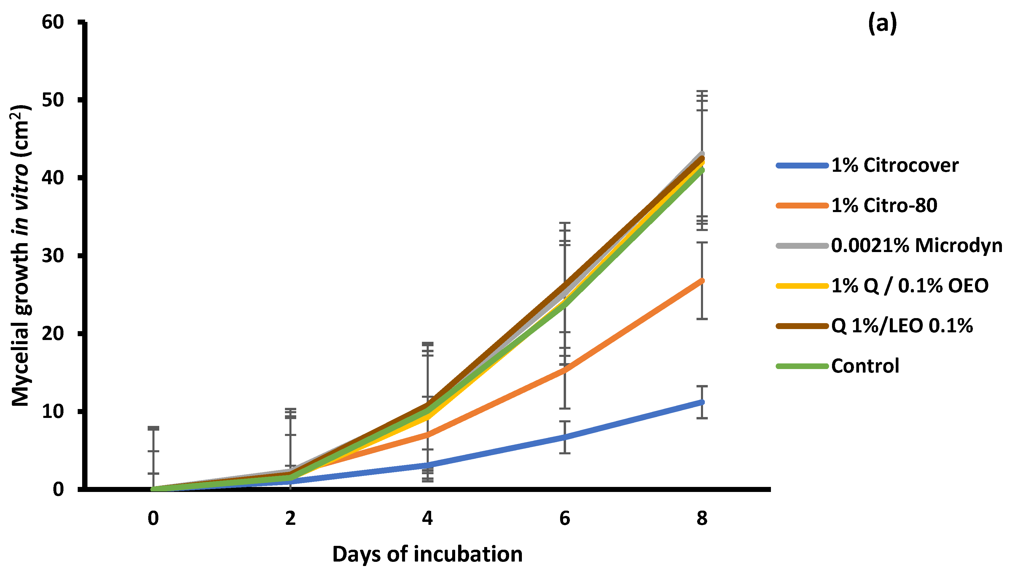
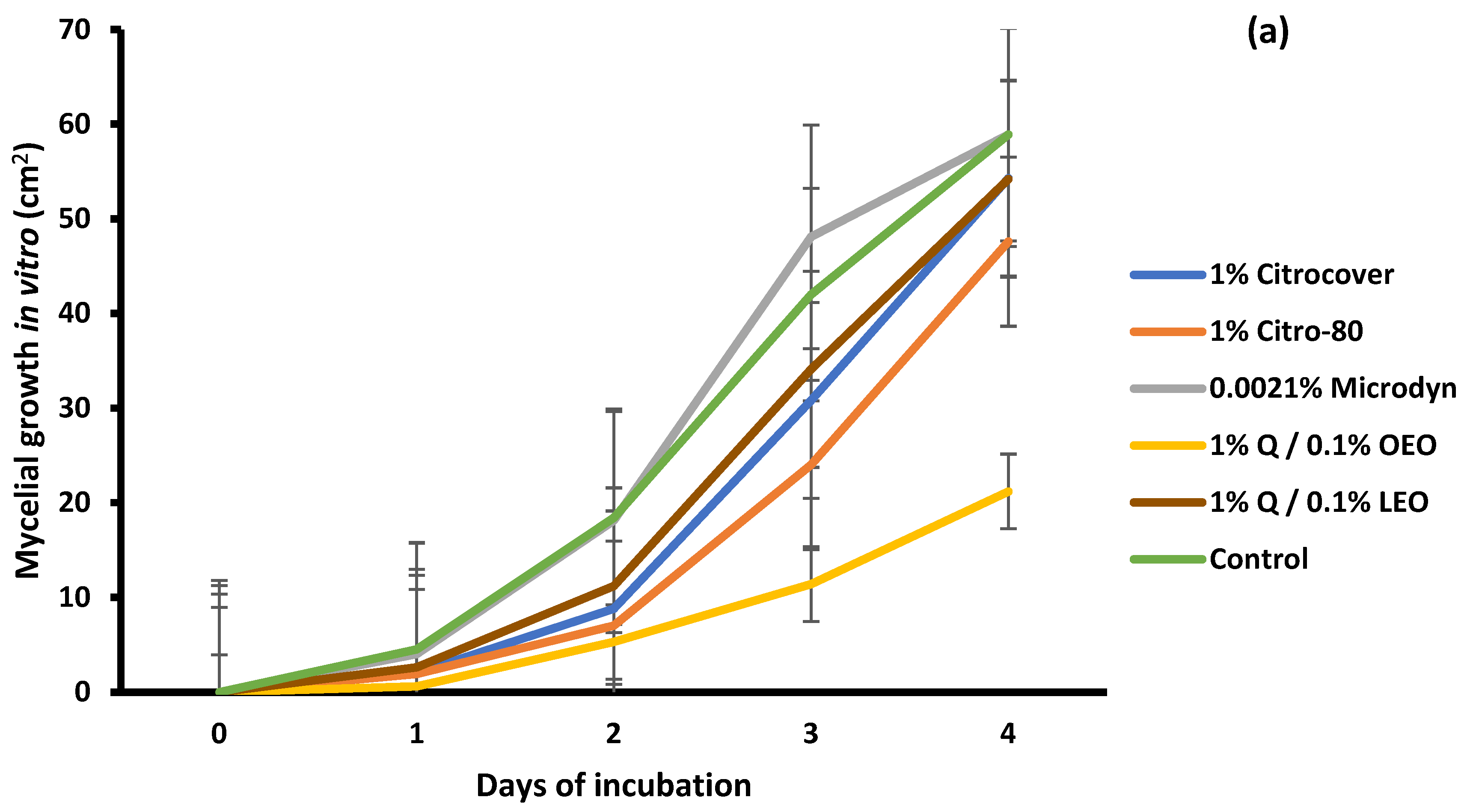
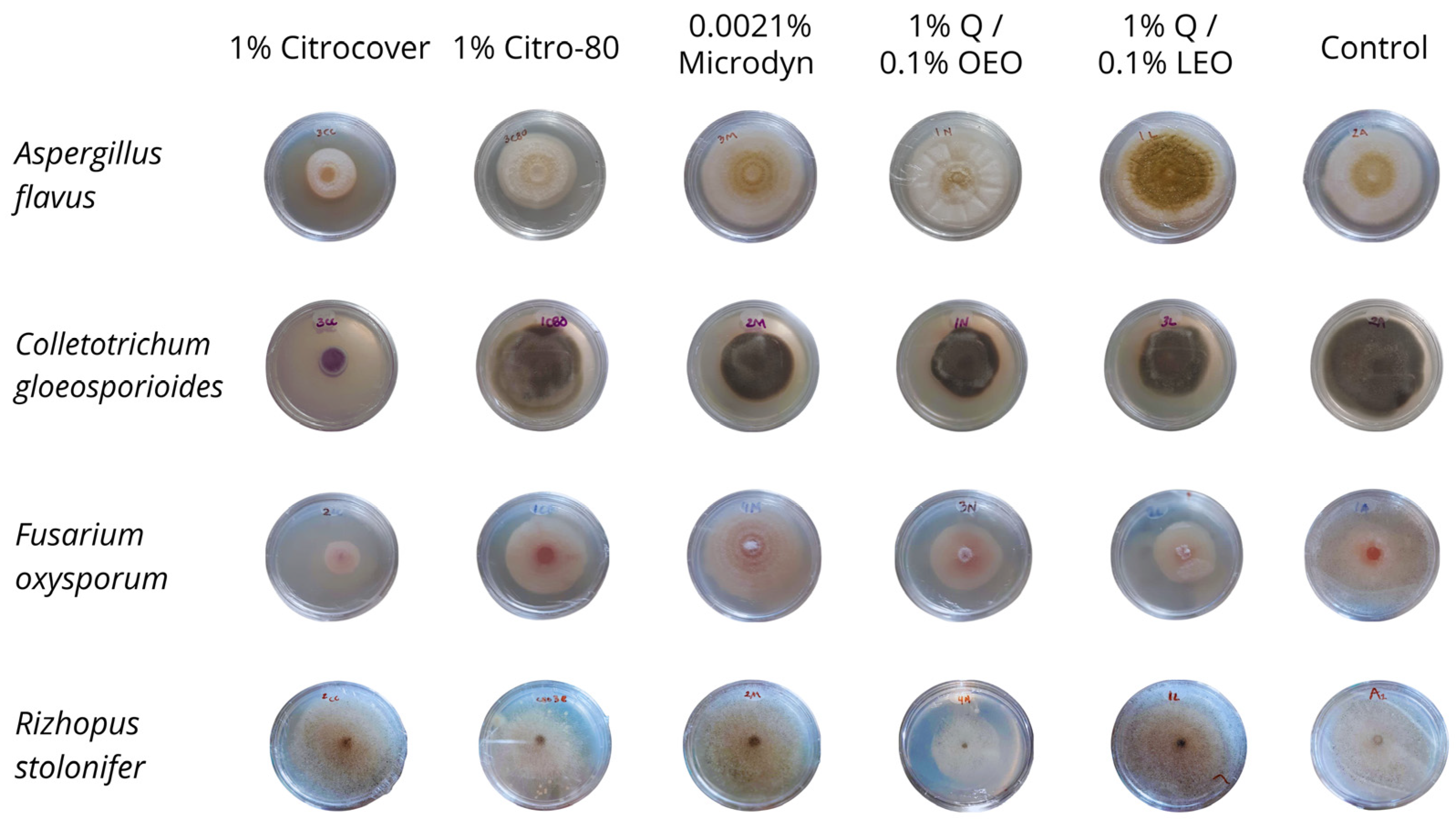
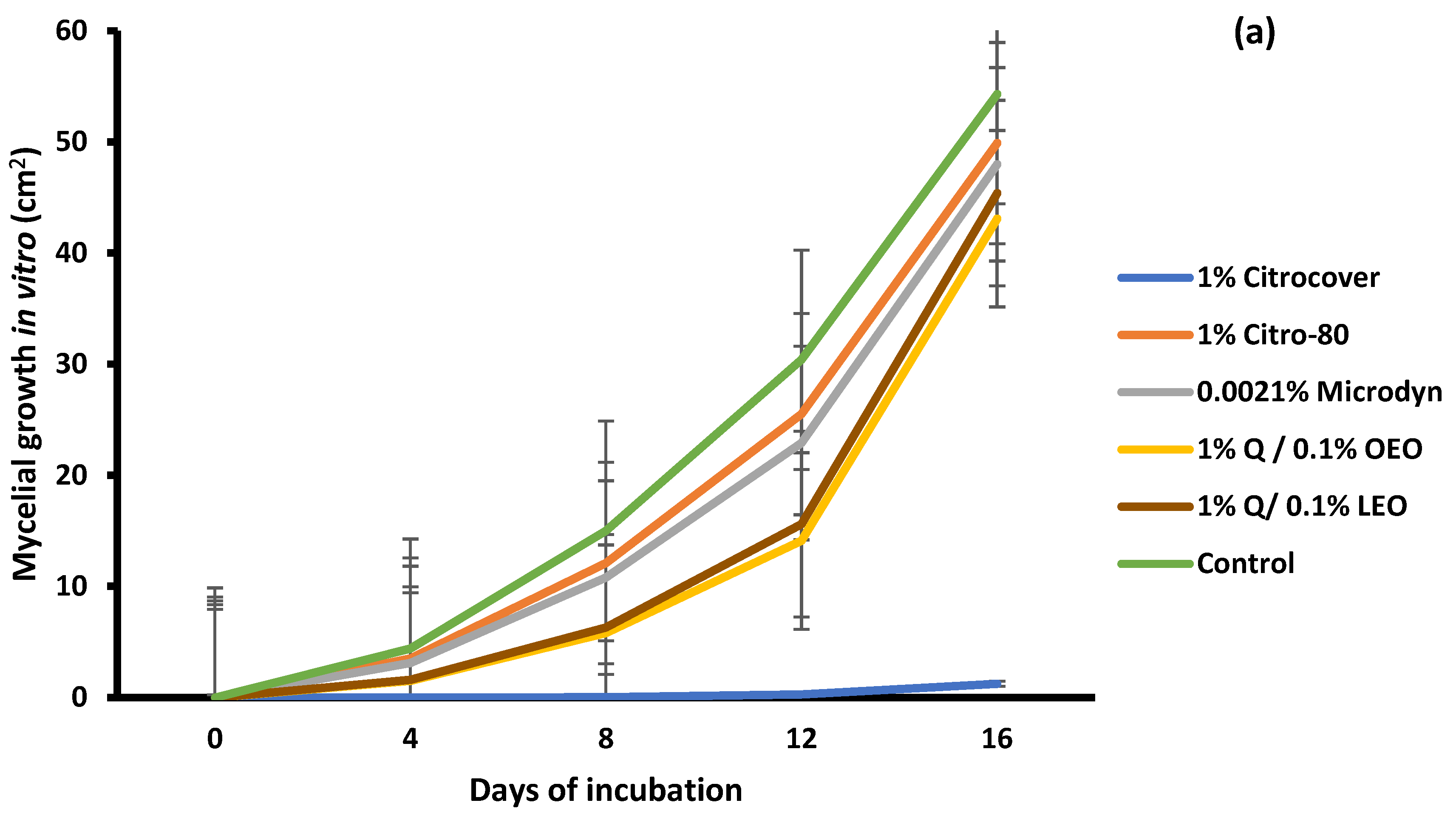
3.1. In Vitro Growth and Disease Development on Inoculated Tomatoes

For A. flavus, the treatment with 1% Citrocover was the one that most inhibited its growth until the end of its 8-day storage, with a final growth of approximately 11.2 cm2, followed by the treatments with 1% Citro-80 with 28 cm2. With the remaining treatments, A. flavus had an average growth of approximately 45.0 cm2 (Figure 1a). When tomatoes were dipped in these same treatments, the infection was noticeably lower in the tomatoes treated with 1% Q/0.1% OEO (Figure 1b). In this case, the final value of the fungal development was approximately 3.5 cm2. In the case of the application of 0.0021% Microdyn, the growth of A. flavus was promoted in comparison with the remaining treatments and the control (8.7–10 cm2), since, at the end of storage, the approximate value was 14.6 cm2.
In relation to C. gloeosporioides, 1% Citrocover significantly (p < 0.05) inhibited the mycelial growth during the whole period, unlike the remaining treatments (Figure 2a). At the end, for the corresponding value, the growth was 1.2 cm2, while in the other treatments, the final average growth was from 43 to 50 cm2. On the other side, except for 0.0021% Microdyne, the growth of C. gloeosporioides on the inoculated tomatoes was minor in all treatments, including the control, during the storage period (Figure 2b). In general, the growth range was from approximately 1.7 to 3.8 cm2.
For F. oxysporum, 1% Citrocover also showed the strongest effect, followed by the 1% Q/0.1% LEO coating, since, at the end of the incubation period of 8 days, the growth achieved was only 3.78 and 15 cm2, respectively, which was statistically different (p < 0.05) among the treatments (Figure 3a). In the treated tomatoes, except for those treated with 0.0021% Microdyn, it was observed that, at the end of the 6-day storage period, the infection was significantly similar (p < 0.05) among the treatments, including the control (Figure 3b). In the case of 0.0021% Microdyne, the infection was significantly greater (16.2 cm2) and was statistically different from the remaining treatments.
Regarding the in vitro growth of R. stolonifer, the treatment in which the fungus showed the least development (20 cm2) at the end of the four-day incubation period was that with 1% Q/0.1% OEO (Figure 4a). In the remaining treatments, including the control, the mycelial growth was 50 and 60 cm2 (Figure 4a and Figure 5). Concerning the development of R. stolonifer in the inoculated tomatoes, regardless of the treatment applied, it advanced rapidly as the days of storage increased, without a marked difference compared to the control, except for 0.0021% Microdine. In this case, the growth of R. stolonifer was the lowest, with a corresponding value of 26 cm2 (Figure 4b and Figure 6). Statistical differences (p < 0.05) were observed among the treatments.

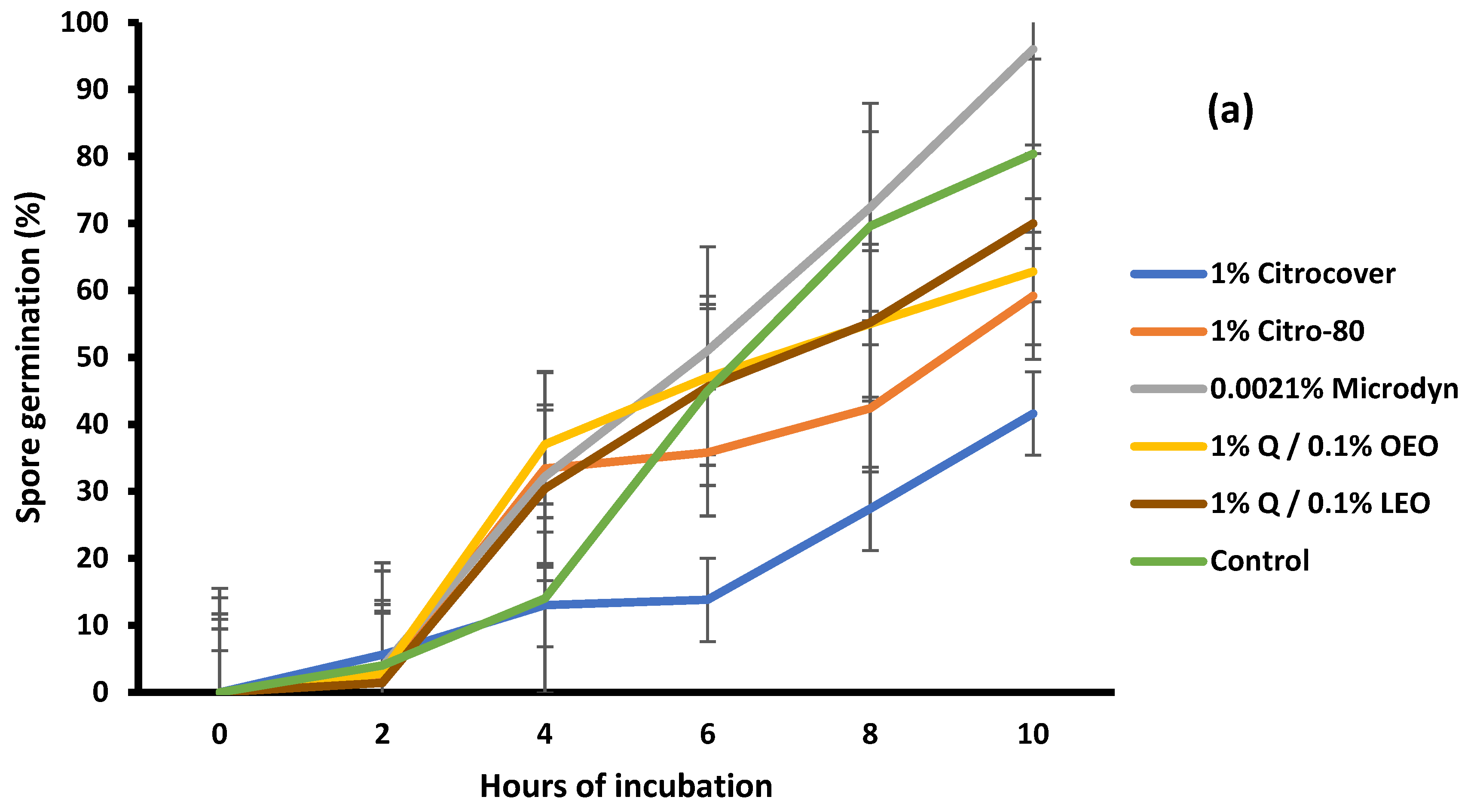
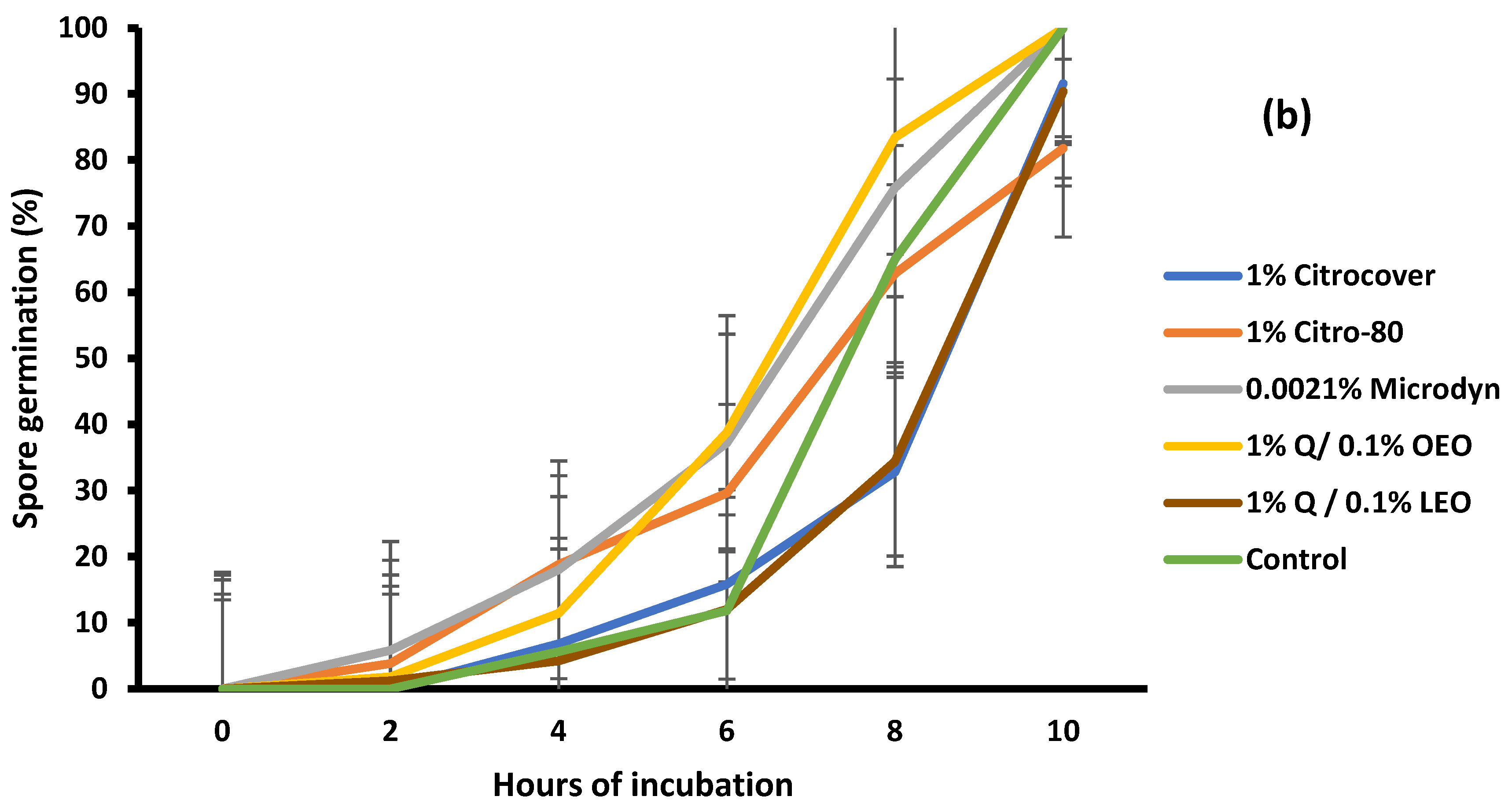
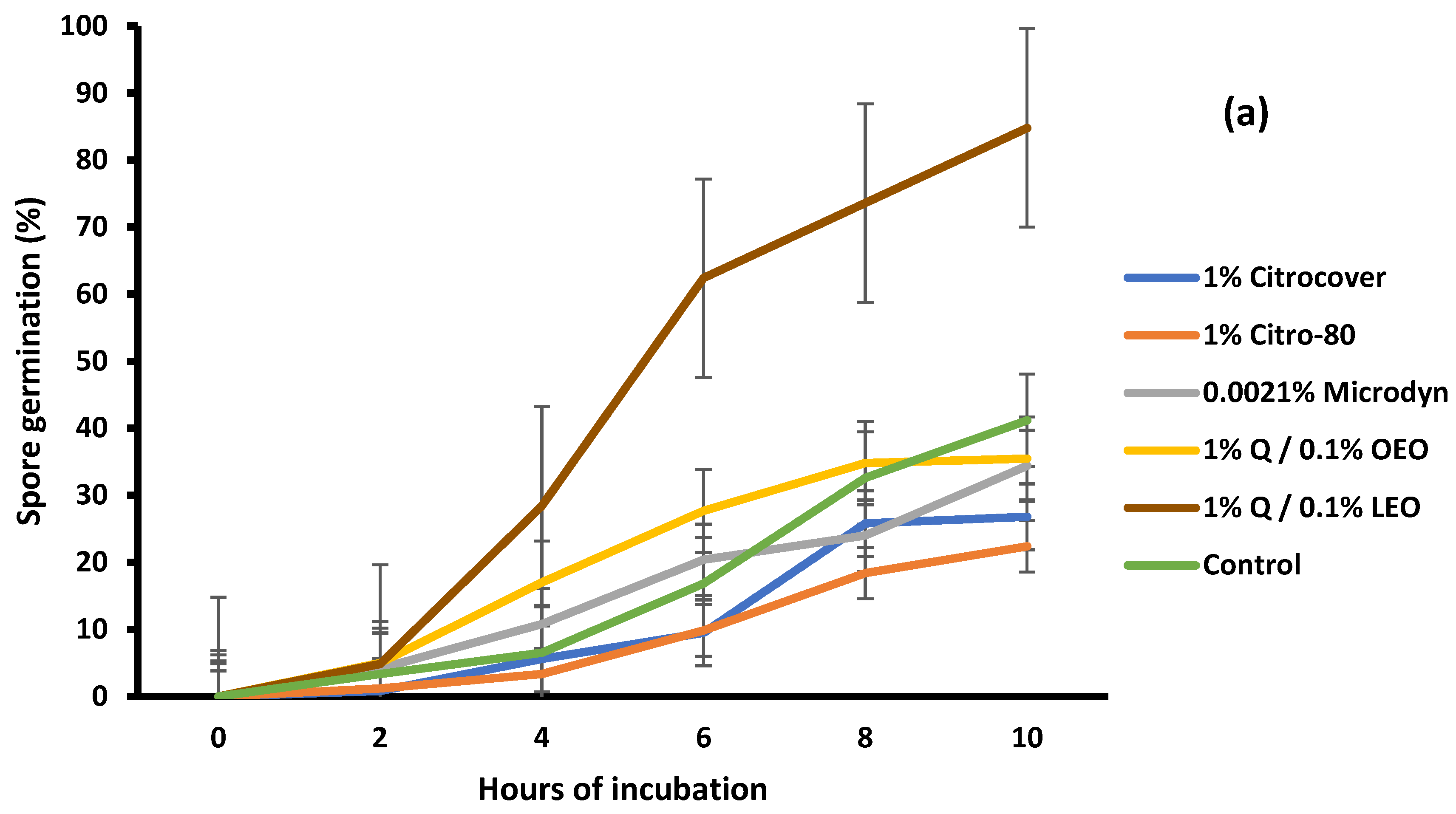
3.2. Spore Germination

The germination of A. flavus spores in Czapek-Dox medium was statistically different (p < 0.05) among the treatments. For the 1% Citrocover treatment, germination was reduced by approximately 60% at the end of the 10 h of incubation (Figure 7a). In relation to the 1% Citro-80 and 1% Q/0.1% OEO treatments, the inhibition was approximately 40%. The spore inhibition for A. flavus under the 0.0021% Microdyne treatment and control was very similar; for the latter, it was promoted. The development of F. oxysporum spores was similar in most treatments, including the untreated samples, at the end of the 10 h of incubation.
For C. gloeosporioides, the spore germination was significantly (p < 0.05) controlled among the treatments. Except for the treatment with 1% Q/0.1% LEO, the germination was inhibited from 60% to 80% with 10 h of incubation (Figure 8a). For this fungus, the 1% Citrocover and 1% Citro-80 products exerted the highest levels of inhibition. The inhibition of the germination of R. stolonifer spores was minimal, since, at the end of the 10 h incubation period, the spores germinated at 100% (Figure 8b). Only the treatment with 1% Citrocover partially inhibited the germination by 30% until the fourth day of incubation.

3.3. Mycotoxin Production In Vitro and on Inoculated and Dehydrated Tomatoes

Regarding the production of aflatoxin and fumonisins by A. flavus and F. oxysporum, statistical differences were observed among the treatments (p < 0.05) in the in vitro and in situ experiments (Table 1).
In the in vitro experiments, the lowest aflatoxin production was obtained with the commercial 1% Citrocover and 1% Citro-80 products, with a corresponding value of 0.1 ppb. In the in situ experiments, the production of aflatoxins in treated and untreated tomatoes was between 1.3 and 1.7 ppb. The lowest aflatoxin production was in tomatoes treated with the 1% Citrcover treatment, and the highest value corresponded to those treated with 0.0021% Microdyn.
With respect to the fumonisin production, the lowest in vitro production corresponded to the 0.0021% Microdyn (0.10 ppb) treatment, while the highest production corresponded to the control (0.21 ppm). The production of fumonisins in tomatoes treated with 1% Citro-80 (0.19 ppm) was the lowest compared to the rest of the treatments. The tomatoes coated with 1% Q/0.1% LEO showed the highest production (0.72 ppm).

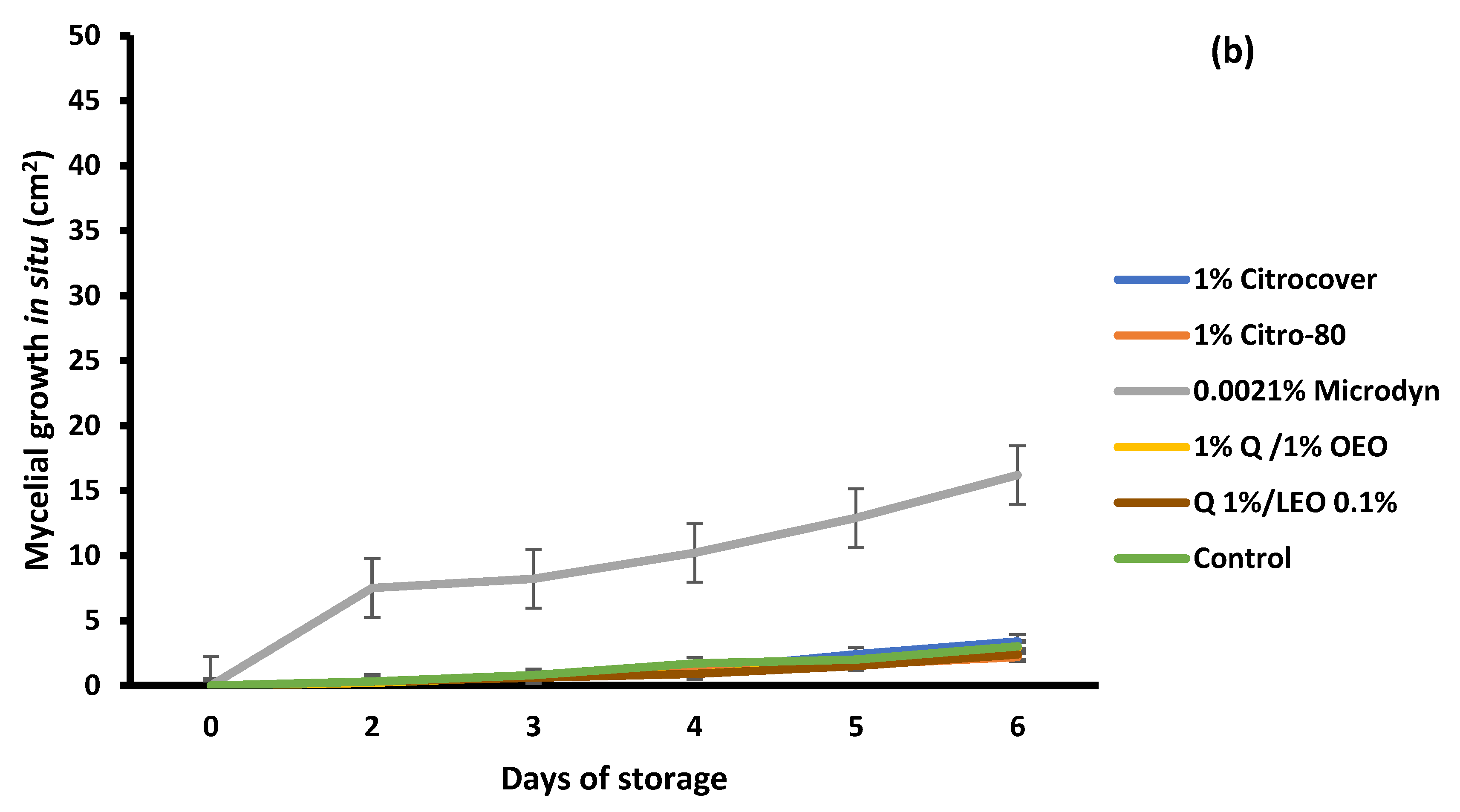
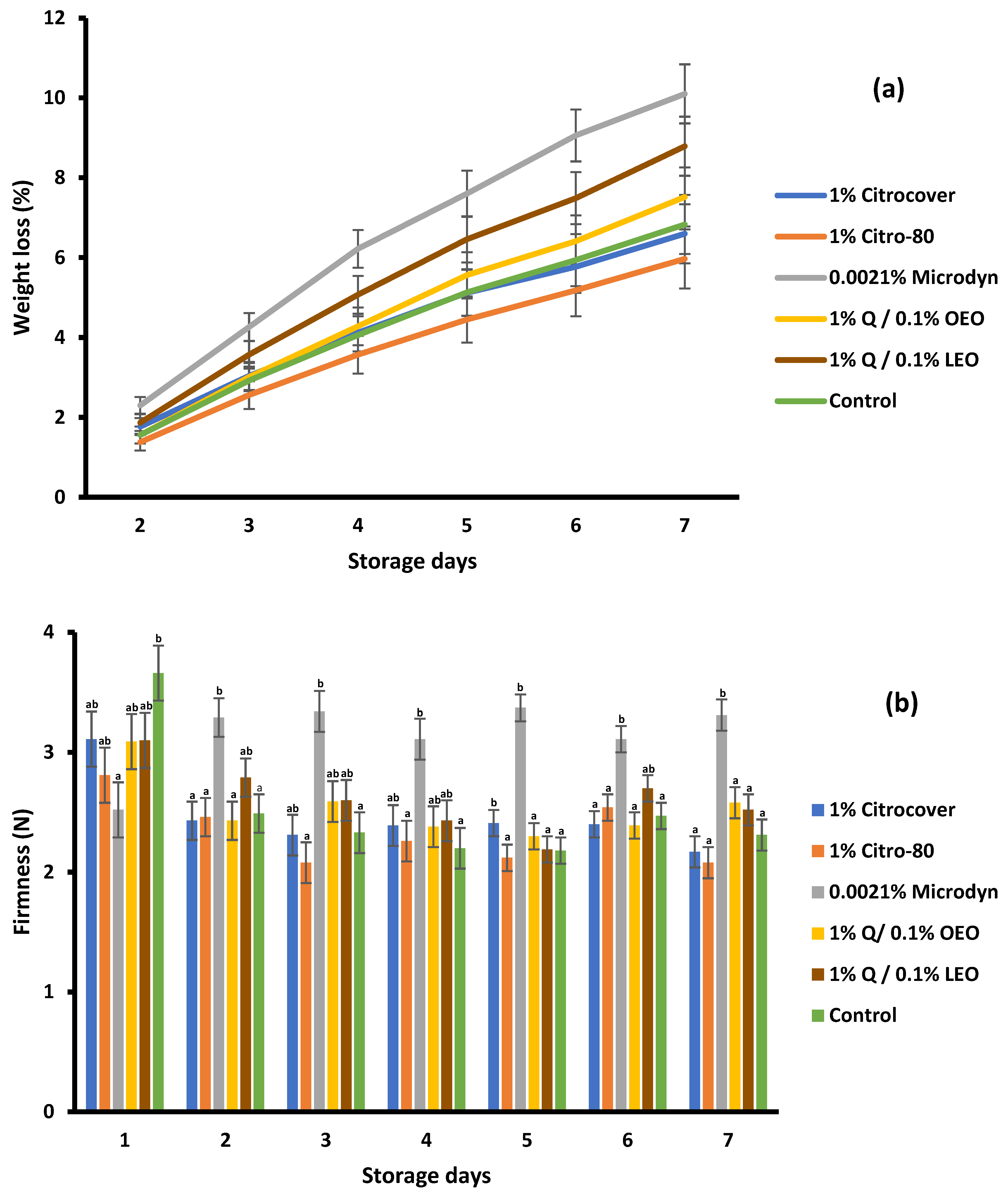
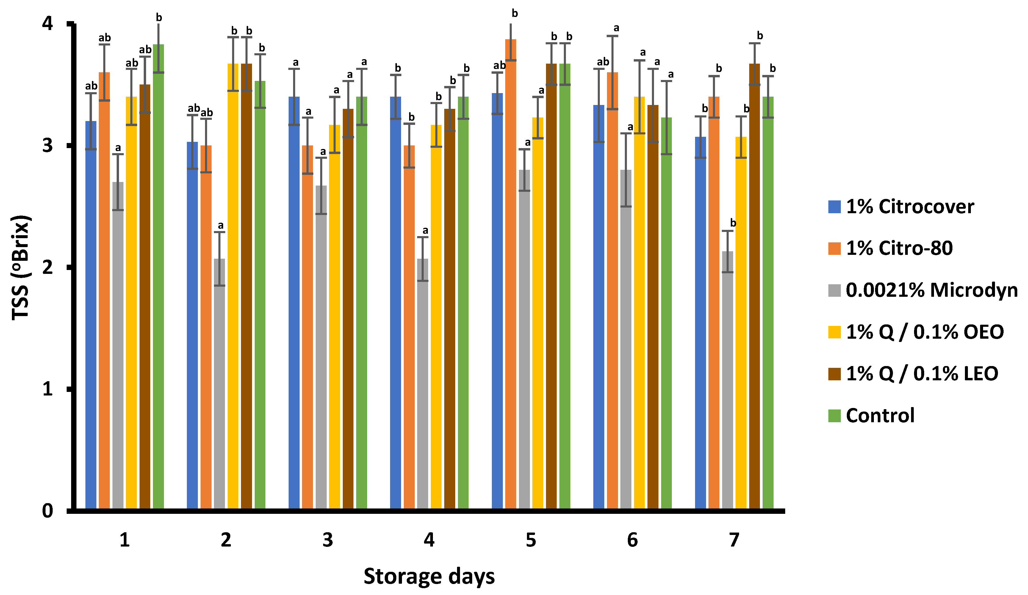
3.4. Storage Behavior of the Treated Tomatoes

In general, the weight loss of tomatoes increased during the 7 days of storage in all treatments (Figure 9a). At the end of this storage period, it was observed that there were no significant differences (p < 0.05) between 1% Citrocover and the control. The greatest weight loss was evidenced in the tomatoes treated with 0.0021% Microdyne (10%), followed by those treated with 1% Q/0.1% LEO and 1% Q/OEO (8.7% and 7.2%, respectively). The lowest weight loss was observed in the tomatoes treated with 1% Citro-80 (5.9%).
There were significant differences (p < 0.05) in the firmness among the treatments (Figure 9b). However, except for that in the 0.00215 Microdyne treatment, the firmness was very similar. In general, the firmness values during the 7 days of storage were in the range from 2.1 N to 3.3 N. Overall, the highest values of firmness in the tomatoes corresponded to the 0.0021% Microdyne treatment.
For the TSSs, the values obtained were statistically different throughout the tomato storage period and across the treatments (Figure 10). There was no pattern in the evolution of the TSSs among the treatments and storage days, but in the case of 0.0021% Microdyne, the TSS content of the treated tomatoes was lower from the first to the last day of storage when compared with the remaining treatments, including the control.

4. Discussion

In this research, differences in fungal growth among treatments were found when measured on incubated Petri plates or in tomatoes. Likewise, the response to the treatments was different according to the genus of the fungus evaluated.
Except for the fungus R. stolonifer, the action of the citrus extracts—especially the commercial product 1% Citrocover—had a notable inhibitory effect in both stages of development in vitro, i.e., the mycelial development and spore viability of the remaining phytopathogenic fungi.
These results agree with those of previous in vitro studies. For example, Rodríguez and Montilla [25] mentioned that the commercial product Citrex, based on seed and pulp extracts of C. paradisi, had a significant effect on the mycelial development of F. oxysporum f. sp. lycopersici. Similarly, Segura-Palacios et al. [26] also evaluated 1% Citrocover in A. flavus isolated from tomatoes, obtaining a notably greater inhibitory effect than that of the control on mycelial growth and sporulation, with corresponding values of 3.0 cm2 and 0.2 × 10⁵ spores mL−1, respectively.
To date, various mechanisms of action against microorganisms (mainly fungi and bacteria) of citrus extracts have been described [27,28]. It has been mentioned that the union of the polyphenols of extracts with ergosterol, glycolipids of the bilayer, and proteins of the membrane of the pathogenic microorganism through covalent or hydrogen bonds resulted in their destabilization and thinning, thus generating a change in the balance of H+ and K+ and causing membrane rupture and a loss of cellular content [29].
In this research, we saw that the production of aflatoxins and fumonisins by A. flavus and F. oxysporum, respectively, in vitro and on dry tomatoes was very low, even in the control. In the case of aflatoxins, the synthesis at the end of storage was in the ranges from 0.1 to 0.5 ppb and from 1.3 to 1.7 ppb (in vitro and in situ, respectively), while the production of fumonisins was from 0.10 to 0.21 ppm and from 0.19 to 0.72 ppm (in vitro and in situ, respectively). Similar results were reported by Villegas-Rascón et al. [30] with the use of chitosan nanoparticles and encapsulated essential oils of cinnamon, thyme, and eucalyptus, which had no effects on the in vitro production of aflatoxins by A. parasiticus. On the other hand, in this research, it was observed that the concentration of mycotoxins in dehydrated tomatoes was slightly higher than that in in vitro studies. This could have been due to the composition of the matrix evaluated, since, in the in vitro studies, the growth of the fungi was based on a culture medium containing approximately 95% water, while in the case of the dehydrated tomatoes, there was a water content of approximately 5%. During the extractions for the immunoassays, a powder with a higher solid content than that of the nutrient medium could have resulted in a higher concentration of mycotoxins.
Nevertheless, the in situ results were not consistent with the in vitro ones or with previous findings that highlighted the effectiveness of citrus-extract-based fungicides in various agricultural products (watermelon, mango, and tomato) [10,16,17,31]. In the present research, the effect of the citrus-based fungicides was not significant in the tomatoes during storage; compared with the control (tomatoes sprayed only with water), only the application of 1% Q/0.1% OEO slightly reduced the development of A. flavus. As for F. oxysporum and C. gloeosporioides, the infection was very similar to that in the control for the citrus-based treatments, but it was stimulated with the application of 0.0021% Mycrodin. This is a commercial disinfectant made from colloidal silver and is indicated to prevent the development of bacteria and fungi. It has been described that the mechanism of action of colloidal silver is related to a cytotoxic function in the cell membrane of microorganisms, and it damages genetic material because free silver ions have an affinity for lipids and proteins. Therefore, they adhere to organic matter very easily [32]. However, scientific studies have reported the low effectiveness of colloidal silver in controlling microorganisms. Rangel-Vargas et al. [18] evaluated the effects of colloidal silver on 13 types of bacteria in pieces of jalapeño pepper; however, colloidal silver did not show significant differences compared with the control (saline solution). Similarly, Venat et al. [20] evaluated the effect of Medicer colloidal silver on pathogenic fungi in plants. The authors reported that colloidal silver resulted in 0% inhibition of mycelial growth in A. flavus, A. niger, and Penicillium digitatum at concentrations of up to 0.015 mL−1. Plata [33] described that microorganisms have a resistance to silver that can be caused by the release of energy-dependent ions from the cell by membrane proteins that function as ATPases.
Overall, the greatest infection at the end of tomato storage was caused by R. stolonifer, since it reached almost 75% of their surface area. The reason for this could be associated with the harvest maturity (full red) of the evaluated tomatoes. In this case, their ripening state could have caused the treated tomatoes to lose their ability to activate their resistance process and, hence, be able to prevent fungal infections by the inoculated fungi [34].
Concerning the three physicochemical variables measured on the tomatoes, except for the treatment with 0.0021% Microdyn, overall, the changes recorded could be associated with normal aspects of the final ripening process. In this regard, López-Vidal et al. [35] mentioned changes during the ripening of Saladette tomatoes through a loss of firmness and an increase in TSSs. In addition, in previous trials to evaluate the shelf life of tomatoes coated with 1% chitosan/0.1% lime essential oil, it was determined that the magnitude of these postharvest changes depends mainly on factors such as the type of coating, the ripening state, and the storage temperature [36].

5. Conclusions

The application of the commercial citrus-based product 1% Citrocover was highly effective in reducing the in vitro development of A. flavus, F. oxysporum, and C. gloeosporioides; this was not the case for R. stolonifer. However, the efficacy of 1% Citrocover and the remaining citrus-based fungicides was lower in all of the tested fungi, since they were evaluated in completely infected fruits.
The 1% Q/0.1% OEO coating was effective in reducing the in vitro development of R. stolonifer.
Except for the 0.0021% Microdyn sanitizer, the use of the commercial citrus-based products did not alter the ripening process of the treated tomatoes. The changes in the losses of weight, firmness, and the TSSs were associated with the normal ripening process of the tomatoes rather than the treatments applied.

Author Contributions

Conceptualization, M.d.L.R.-G., D.A.S.-P. and S.B.-B.; investigation., L.S.-M.; writing—original draft preparation, S.B.-B. and M.H.-L.; writing—review and editing, L.S.-M., M.d.L.R.-G. and S.B.-B.; funding acquisition, D.A.S.-P. and M.d.L.R.-G. All authors have read and agreed to the published version of the manuscript.

Funding

This research was funded by Universidad Autónoma del Estado de Morelos, Facultad de Nutricion and CONAHCYT with project number 316090.

Institutional Review Board Statement

Not applicable.

Informed Consent Statement

Not applicable.

Data Availability Statement

Data are contained within the article.

Acknowledgments

The authors thank CONACYT (Mexico) for the scholarship granted to Lizette Serrano-Molina.

Conflicts of Interest

The authors declare no conflicts of interest.

References

  1. FAO. Organización de las Naciones Unidas para la Alimentación y la Agricultura. Antecedentes|Plataforma Técnica Sobre la Medición y la Reducción de las Pérdidas y el Desperdicio de Alimentos. 2021. Available online: https://www.fao.org/platform-food-loss-waste/background/es (accessed on 13 March 2024).
  2. Barkai-Golan, R. Aspergillus Mycotoxins. In Mycotoxins in Fruits and Vegetables; Barkai-Golan, R., Paster, N., Eds.; Academic Press: Cambridge, MA, USA; Elsevier: Amsterdam, The Netherlands, 2008; pp. 115–151. [Google Scholar]
  3. Ji, F.; He, D.; Olaniran, A.O.; Mokoena, M.O.; Shu, J.; Shi, J. Occurrence, toxicity, production and detection of Fusarium mycotoxin: A review. Food Prod. Process. Nutr. 2019, 1, 6. [Google Scholar] [CrossRef]
  4. Ruiz, M.J.; Vicente, A.V.; Montañéz, S.J.; César Rodríguez, H.R.; Aguilar, G.C.N. Un tesoro perecedero en México: El tomate, tecnologías para prolongar su vida de anaquel. Investig. Cienc. 2012, 20, 57–63. [Google Scholar]
  5. Rodríguez-Guzmán, C.A.; Montaño-Leyva, B.; Velázquez-Estrada, R.M.; Sánchez-Burgos, J.A.; García-Magaña, M.d.L.; González-Estrada, R.R.; Gutiérrez-Martínez, P. Estado actual de métodos alternativos, de control de hongos y su efecto en la calidad postcosecha de frutos de jitomate (Solanum lycopersicum). TIP Rev. Esp. Cienc. Quím. Biol. 2021, 24, 1–15. [Google Scholar] [CrossRef]
  6. Shamurailatpam, D.; Kumar, A. A review on recent methods to control early blight of tomato (Solanum lycopersicum L.). Plant Cell Biotechnol. Mol. Biol. 2020, 21, 136–148. [Google Scholar]
  7. Carvalho, F.P. Agriculture, pesticides, food security and food safety. Environ. Sci. Policy 2006, 9, 685–692. [Google Scholar] [CrossRef]
  8. Shridhar, B.P.; Sharma, M.; Gupta, S.K.; Sharma, S.K. New generation fungicides for the management of buckeye rot of tomato. Indian Phytopathol. 2018, 71, 621–625. [Google Scholar] [CrossRef]
  9. Xu, G.; Liu, D.; Chen, J.; Ye, X.; Shi, J. Composition of major flavanone glycosides and antioxidant capacity of three citrus varieties. J. Food Biochem. 2009, 33, 453–469. [Google Scholar] [CrossRef]
  10. Bashir, L.U.; Abdulkadir, A.; Shah, M.M.; Hamisu, A.; Sharif, U.; Kamalu, A.A. Phytochemical screening and antifungal potentials of Citrus limon peels against Fusarium oxysporum and Rhizopus stolonifer causing rots in water melon (Citrullus lanatus L.). J. Exp. Sci. 2020, 11, 17–21. [Google Scholar] [CrossRef]
  11. Bautista-Baños, S.; Ventura-Aguilar, R.I.; Correa-Pacheco, Z.; Corona-Rangel, M.L. Chitosan: A versatile antimicrobial polysaccharide for fruit and vegetables in postharvest—A review. Rev. Chapingo Ser. Hortic. 2017, 23, 103–121. [Google Scholar] [CrossRef]
  12. Narváez, B.F.J.; Barzola, M.S.E.; Fon-Fay, V.F.M.; Martínez, C.M.J.; Neira, M.J.A.; Sánchez, L.S.N. Potencial antifúngico de Citrus sinensis y Citrus nobilis sobre el crecimiento de Rhizopus stolonifer y Colletotrichum gloeosporioides en papaya. Cienc. Tecnol. 2017, 10, 41–46. [Google Scholar] [CrossRef]
  13. Negera, M.; Washe, A.P. Use of natural dietary spices for reclamation of food quality impairment by aflatoxin. J. Food Qual. 2019, 2019, 4371206. [Google Scholar] [CrossRef]
  14. Liu, Y.; Benohoud, M.; Galani, Y.J.H.; Gong, Y.Y.; Orfila, C. Green extraction of polyphenols from citrus peel by-products and their antifungal activity against Aspergillus flavus. Food Chem. X 2021, 12, 100144. [Google Scholar] [CrossRef] [PubMed]
  15. Velásquez, M.; Álvarez, R.; Tamayo, P.; Carvalho, C. Evaluación in vitro de la actividad fungistática del aceite esencial de mandarina sobre el crecimiento de Penicillium sp. Cienc. Tecnol. Agropecu. 2015, 15, 7–14. [Google Scholar] [CrossRef]
  16. Sheikh, M.; Mehnaz, S.; Sadiq, M.B. Prevalence of fungi in fresh tomatoes and their control by chitosan and sweet orange (Citrus sinensis) peel essential oil coating. J. Sci. Food Agric. 2021, 101, 6248–6257. [Google Scholar] [CrossRef] [PubMed]
  17. Cheng, Y.J.; Wu, Y.J.; Lee, F.W.; Ou, L.Y.; Chen, C.N.; Chu, Y.Y.; Kuan, Y.-C. Impact of storage condition on chemical composition and antifungal activity of pomelo extract against Colletotrichum gloeosporioides and anthracnose in post-harvest mango. Plants 2022, 11, 2064. [Google Scholar] [CrossRef] [PubMed]
  18. Rangel-Vargas, E.; Gutiérrez-Alcántara, E.J.; Gómez-Aldapa, C.A.; Falfán-Cortés, R.N.; Segovia-Cruz, J.A.; Salas-Rangel, L.P.; Castro-Rosas, J. Antibacterial activity of roselle calyx extracts, sodium hypochlorite, colloidal silver and acetic acid against multidrug-resistant salmonella serotypes isolated from coriander. J. Food Saf. 2017, 37, e12320. [Google Scholar] [CrossRef]
  19. Salgado-Escobar, I.; Hernández-Rodríguez, G.; Suárez-López, Y.d.C.; Mancera-Ugarte, M.J.; Guerra-Ramíez, D. Eficacia de métodos de desinfección y los efectos sobre las propiedades nutracéuticas en cilantro y fresa. Rev. Mex. Cienc. Agríc. 2020, 11, 327–337. [Google Scholar] [CrossRef]
  20. Venat, O.; Iacomi, B.; Peticila, A.G. In Vitro studies of antifungal activity of colloidal silver against important plants pathogens. Not. Bot. Horti Agrobot. Cluj-Napoca 2018, 46, 533–537. [Google Scholar] [CrossRef]
  21. Cortes-Higareda, M.; Ramos-García, M.L.; Correa-Pacheco, Z.; Del Rio, J.C.; Bautista-Baños, S. Nanostructured chitosan/propolis formulations: Characterization and effect on the growth of Aspergillus flavus and production of aflatoxins. Heliyon 2019, 5, e01776. [Google Scholar] [CrossRef]
  22. Ventura-Aguilar, R.I.; Gónzalez-Andrade, C.; Hernández-López, M.; Correa-Pacheco, Z.N.; Teksür, P.K.; Ramos-García, M.d.L.; Bautista-Baños, S. Effect of biodegradable coatings on the growth of Aspergillus flavus in vitro, on maize grains, and on the quality of tortillas during storage. Molecules 2022, 27, 4545. [Google Scholar] [CrossRef]
  23. Asfaw, M.D. Basic essential oil extraction techniques and procedures from aromatic plants. J. Chromatogr. Sep. Tech. 2022, 13, 1–5. [Google Scholar]
  24. Aparicio-García, P.F.; Ventura-Aguilar, R.I.; Río-García, J.C.; Hernández-López, M.; Guillén-Sánchez, D.; Salazar-Pina, D.A.; Ramos-García, M.d.L.; Bautista-Banos, S. Edible chitosan/propolis coatings and their effect on ripening, development of Aspergillus flavus, and sensory quality in fig fruit, during controlled storage. Plants 2021, 10, 112. [Google Scholar] [CrossRef] [PubMed]
  25. Rodríguez, D.A.; Montilla, J.O. Disminución de la marchitez causada por Fusarium en tomate con extracto de Citrus paradisi. Integr. Plagas 2002, 46–50. [Google Scholar]
  26. Segura-Palacios, M.A.; Correa-Pacheco, Z.N.; Corona-Rangel, M.L.; Martinez-Ramirez, O.C.; Salazar-Pina, D.A.; Ramos-García, M.d.L.; Bautista-Banos, S. Use of natural products on the control of Aspergillus flavus and production of aflatoxins in vitro and on tomato fruit. Plants 2021, 10, 2553. [Google Scholar] [CrossRef] [PubMed]
  27. Sanver, D.; Murray, B.S.; Sadeghpour, A.; Rappolt, M.; Nelson, A.L. Experimental modeling of flavonoid-biomembrane interactions. Langmuir 2016, 32, 13234–13243. [Google Scholar] [CrossRef] [PubMed]
  28. Makarewicz, M.; Drożdż, I.; Tarko, T.; Duda-Chodak, A. The interactions between polyphenols and microorganisms, especially gut microbiota. Antioxidants 2021, 10, 188. [Google Scholar] [CrossRef] [PubMed]
  29. Tian, F.; Woo, S.Y.; Lee, S.Y.; Park, S.B.; Zheng, Y.; Chun, H.S. Antifungal activity of essential oil and plant-derived natural compounds against Aspergillus flavus. Antibiotics 2022, 11, 1727. [Google Scholar] [CrossRef] [PubMed]
  30. Villegas-Rascón, R.E.; Plascencia-Jatomea, M.; Rosas-Burgos, E.C.; López-Franco, Y.L.; Tánori-Córdova, J.C.; López-Meneses, A.K.; Cortez-Rocha, M.O. Chitosan/essential oils biocomposites for suppressing the growth of Aspergillus parasiticus. Int. Food Res. J. 2020, 27, 316–326. [Google Scholar]
  31. Ruíz-Alvarado, C.; Reyes-Pérez, J.J.; Hernández-Montiel, L.G.; Murillo, B.; Rueda, E. Control of phytopathogenic microorganisms of post-harvest in tomato (Lycopersicon esculentum Mill.) with the use of citrus extract. J. Plant Sci. Phytopathol. 2018, 2, 037–043. [Google Scholar]
  32. Coutiño, R.E.M. Plata Coloidal: Xenobiótico, Antígeno y Disruptor Hormonal. Rev. Educ. Bioquím. 2015, 34, 1–14. [Google Scholar]
  33. Silver, S. Bacterial silver resistance: Molecular biology and uses and misuses of silver compounds. FEMS Microbiol. Rev. 2003, 27, 341–353. [Google Scholar] [CrossRef]
  34. Prusky, D.; Romannazi, G. Induced resistance in fruit and vegetables: A host physiological response limiting postharvest disease development. Annu. Rev. Phytopathol. 2023, 61, 279–300. [Google Scholar] [CrossRef] [PubMed]
  35. López-Vidal, O.; Escalona-Buendía, H.; Pelayo-Zaldívar, C.; Cruz-Salazar, J.; Villa-Hernández, J.M.; Rivera-Cabrera, F.; Villegas-Torres, O.; Alia-Tejacal, I.; Pérez-Flores, L.J.; De León-Sánchez, F.D. Carotenoid content, antioxidant capacity and volatile compounds of the aroma during tomato ripening. Phyton 2014, 83, 185–192. [Google Scholar] [CrossRef]
  36. Bautista-Baños, S.; González-Soto, R.; Ramos-García, M.L. Physical properties of chitosan films with lemon essential oil added and their impact on the shelf life of tomatoes (Lycopersicon esculentum L.). Rev. Mex. Ing. Quim. 2018, 17, 1–11. [Google Scholar] [CrossRef]
Figure 1. Evaluation of the mycelial growth of Aspergillus flavus treated with three commercial fungicides and two naturally based coatings under (a) in vitro conditions and on (b) Saladette tomatoes with an 8 h incubation time and 7 days of storage, respectively, at 25 ± 2 °C. Bars indicate the standard deviation of the mean. Q = chitosan, OEO = orange essential oil, and LEO = lime essential oil.
Jof 10 00309 g001aJof 10 00309 g001b
Figure 2. Evaluation of the mycelial growth of Colletotrichum gloeosporioides treated with three commercial fungicides and two naturally based coatings under (a) in vitro conditions and on (b) Saladette tomatoes with a 16 h incubation time and 6 days of storage, respectively, at 25 ± 2 °C. Bars indicate the standard deviation of the mean. Q = chitosan, OEO = orange essential oil, and LEO = lime essential oil.
Jof 10 00309 g002aJof 10 00309 g002b
Figure 3. Evaluation of the mycelial growth of Fusarium oxysporum treated with three commercial fungicides and two naturally based coatings under (a) in vitro conditions and on (b) Saladette tomatoes with an 8 h incubation time and 6 days of storage, respectively, at 25 ± 2 °C. Bars indicate the standard deviation of the mean. Q = chitosan, OEO = orange essential oil, and LEO = lime essential oil.
Jof 10 00309 g003aJof 10 00309 g003b
Figure 4. Evaluation of the mycelial growth of Rhizopus stolonifer treated with three commercial fungicides and two naturally based coatings under (a) in vitro conditions and on (b) Saladette tomatoes with a 4 h incubation time and 3 days of storage, respectively, at 25 ± 2 °C. Bars indicate the standard deviation of the mean. Q = chitosan, OEO = orange essential oil, and LEO = lime essential oil.
Jof 10 00309 g004aJof 10 00309 g004b
Figure 5. Mycelial growth of four postharvest fungi treated with three commercial fungicides and two naturally based coatings under in vitro conditions. Q = chitosan, OEO = orange essential oil, and LEO = lime essential oil.
Jof 10 00309 g005
Figure 6. Mycelial growth of four postharvest fungi treated with three commercial fungicides and two naturally based coatings on Saladette tomato. Q = chitosan, OEO = orange essential oil, and LEO = lime essential oil.
Jof 10 00309 g006
Figure 7. Spore germination of (a) Aspergillus flavus and (b) Fusarium oxysporum when incubated on PDA amended with three commercial fungicides and two naturally based coatings for 10 h at 25 ± 2 °C. Bars indicate the standard deviation of the mean. Q = chitosan, OEO = orange essential oil, and LEO = lime essential oil.
Jof 10 00309 g007aJof 10 00309 g007b
Figure 8. Spore germination of (a) Colletotrichum gloeosporioides and (b) Rhizopus stolonifer when incubated on PDA amended with three commercial fungicides and two naturally based coatings for 10 h at 25 ± 2 °C. Bars indicate the standard deviation of the mean. Q = chitosan, OEO = orange essential oil, and LEO = lime essential oil.
Jof 10 00309 g008aJof 10 00309 g008b
Figure 9. (a) Weight loss and (b) firmness of Saladette tomatoes treated with three commercial fungicides and two naturally based coatings over 7 days of storage at 25 ± 2 °C. Bars indicate the standard deviation of the mean. Q = chitosan, OEO = orange essential oil, and LEO = lime essential oil.
Jof 10 00309 g009
Figure 10. TSS content of Saladette tomatoes treated with three commercial fungicides and two naturally based coatings over 7 days storage at 25 ± 2 °C. Bars indicate the standard deviation of the mean. Q = chitosan, OEO = orange essential oil, and LEO = lime essential oil.
Jof 10 00309 g010
Table 1. Evaluation of the production of aflatoxin and fumonisins by Aspergillus flavus and Fusarium oxysporum, respectively, in vitro and on tomatoes.
TreatmentsFungi
Aspergillus flavusFusarium oxysporum
In Vitro
(ppb)
Dry Tomatoes
(ppb)
In Vitro
(ppm)
Dry Tomatoes
(ppm)
1% Citrocover 0.1 a,*1.3 a0.16 b0.19 a
1% Citro-800.1 a1.4 a0.15 b0.66 c
0.002% Microdyn 0.3 b1.7 b0.10 a0.56 b
1% Q/0.1% OEO 0.4 c1.4 a0.18 b0.43 c
1% Q/0.1% LEO 0.2 a1.6 b0.18 b0.72 d
Control0.5 d1.6 b0.21 b0.67 c
* Means followed by the same letter are not significantly different (p < 0.05) as determined with Tukey’s multiple-range test.
Disclaimer/Publisher’s Note: The statements, opinions and data contained in all publications are solely those of the individual author(s) and contributor(s) and not of MDPI and/or the editor(s). MDPI and/or the editor(s) disclaim responsibility for any injury to people or property resulting from any ideas, methods, instructions or products referred to in the content.

