Article

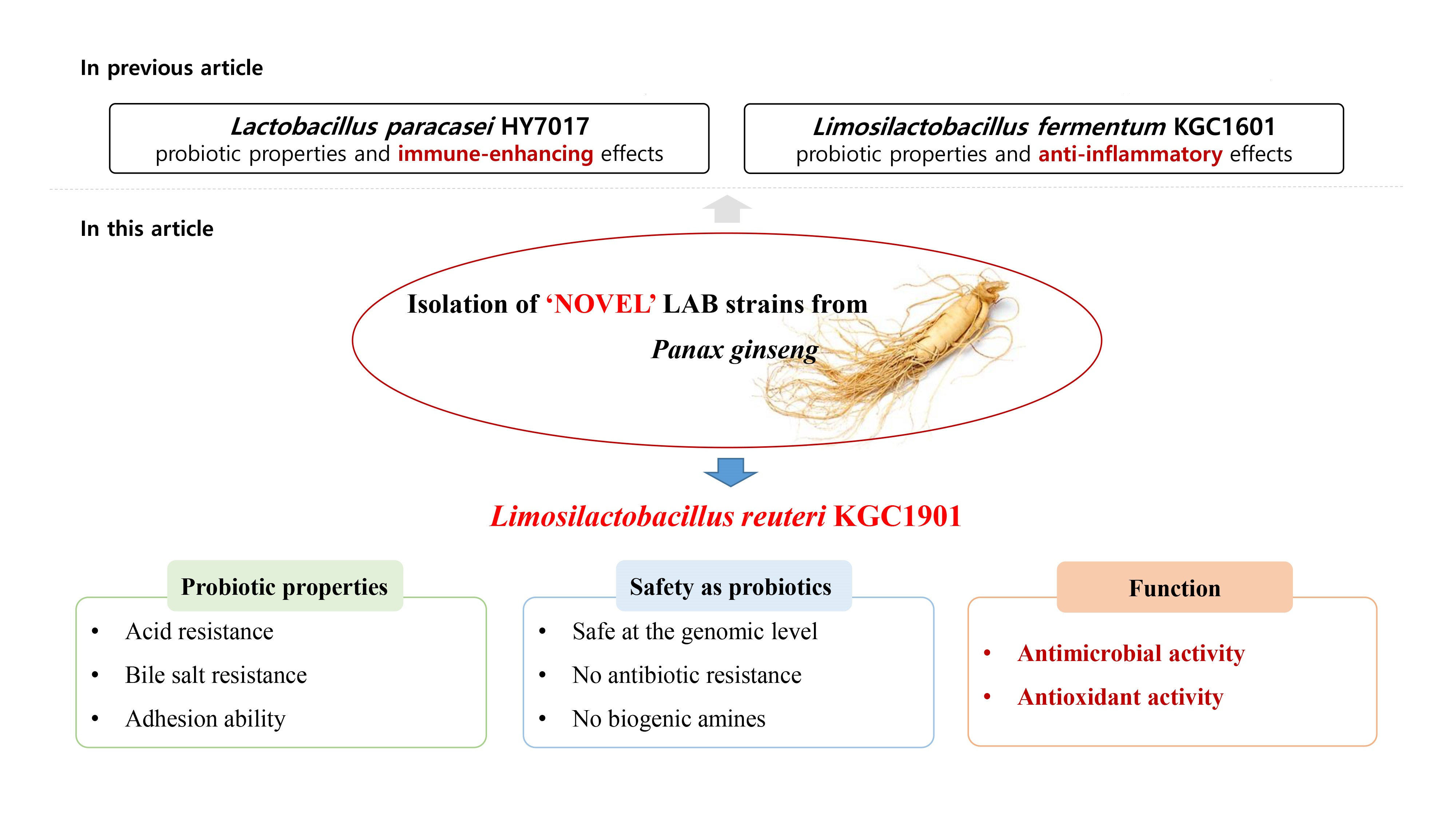
Characterization of Limosilactobacillus reuteri KGC1901 Newly Isolated from Panax ginseng Root as a Probiotic and Its Safety Assessment

1
Laboratory of Efficacy Research, Korea Ginseng Corporation, 30 Gajeong-ro, Yuseong-gu, Daejeon 34128, Republic of Korea
2
Laboratory of Products, Korea Ginseng Corporation, 30 Gajeong-ro, Yuseong-gu, Daejeon 34128, Republic of Korea
*
Author to whom correspondence should be addressed.
Fermentation 2023, 9(3), 228; https://doi.org/10.3390/fermentation9030228
Submission received: 14 February 2023 / Revised: 23 February 2023 / Accepted: 24 February 2023 / Published: 27 February 2023
(This article belongs to the Special Issue Recent Trends in Probiotics and Gut Microbiome for Human Health)

Abstract

:
In this study, we determined the probiotic properties and safety of Limosilactobacillus reuteri KGC1901 isolated from Korean Panax ginseng root. This strain was identified based on its 16s rRNA sequence, and the absence of genes related to antibiotic resistance and virulence was confirmed through whole genome analysis in terms of safety. Moreover, this strain had no antibiotic resistance to eight antibiotics as proposed by the European Food Safety Authority, did not show hemolytic activity on blood agar, and did not produce biogenic amines. L. reuteri KGC1901 also showed the capability to survive at low pH and in presence of bile salts and sufficiently adhered to HT-29 cells and mucin. The adhesion ability to HT-29 was confirmed by immunofluorescence staining. These results indicated the excellent viability of L. reuteri KGC1901 in the human gastrointestinal tract. Additionally, L. reuteri KGC1901 had antimicrobial activity against Clostridium difficile, and the presence of the reuterin (3-HPA) biosynthetic gene cluster in the genome was revealed. Furthermore, the cell-free supernatant of KGC1901 had DPPH scavenging activity and reduced the nitric oxide production in LPS-stimulated RAW 264.7 cells. Based on these results, it was confirmed that L. reuteri KGC1901 derived from ginseng has sufficient potential to be used as a probiotic.

Graphical Abstract

1. Introduction

Microorganisms comprising the human microbiome, including bacteria, archaea, viruses, and eukaryotes, are ubiquitous in the body, including the skin, gastrointestinal tract, respiratory tract, and mammary gland, and interact with their living host [1]. Through this symbiosis, these microbes are closely related to health and disease in humans. The World Health Organization defines probiotics as “live microorganisms which when administered in adequate amounts confer a health benefit on the host” [2]. The most common and traditional genera of probiotic microorganisms are Bifidobacterium and strains belong to lactic acid bacteria (LAB) such as Lactobacillus, Lactococcus, Pediococcus, Enterococcus, and Streptococcus [3,4]. Recent studies also have reported Prevotella, Akkermansia, Feacalibacterium, and Bacteroides as next-generation probiotics [5]. These probiotics play several important roles in the human body such as immune boosting and enhancing metabolic syndromes by regulating the intestinal environment [5,6,7,8]. Previous studies have also shown that probiotics induce the maturation of intestinal epithelium [9]. Nonetheless, as a commercial product, safety remains a major issue. Therefore, probiotic microorganisms should also be evaluated for safety characteristics such as hemolytic activity, antibiotic resistance, and production of toxic metabolites [10,11]. Traditionally, most probiotic strains including Limosilactobacillus reuteri have been isolated from the human gut because these microbes should be colonized and survive in the human gastrointestinal tract [12,13]. However, recently, it has been discovered that autochthonous LAB from raw fruits and vegetables also have several functional features as probiotics: resistance to gastric juice and bile acid, adhesion ability to gut epithelium, and ability to modulate the human immune system. Beatrice et al. reported probiotic potentials of 48 strains of lactic acid bacteria isolated from French beans, carrots, tomatoes, pineapple, cauliflower, and celery [12]. Arshad et al. also reviewed the probiotic properties and human health beneficial effects of Lactobacilli originating from various sources including plant and vegetable matrices [14].
Korean ginseng (Panax ginseng C.A. Meyer) is a well-known traditional herbal medicine that has been used for centuries in East Asia. Ginsenosides, the main active ingredients of ginseng, are known to be effective in oxidative stress and cardiovascular dysfunction [15]. Bacterial endophytes, which have been found in every part of the ginseng plant, including the roots, stem, leaves, and fruits, are known to have the potential to produce ginsenosides and the ability to transform major ginsenosides into a minor form using enzyme activities [16]. The abundant phyla of endophytic bacteria from Korean ginseng are Proteobacteria, Actinobacteria, and Firmicutes, and the dominant endophytic bacterial genera are Bacillus and Pseudomonas [16]. Raw ginseng has a highly acidic matrix and is rich in dietary fiber, so it is suitable for the survival and proliferation of endophytic LAB [17,18]. In recent studies, Lacticaseibacillus paracasei, Limosilactobacillus fermentum, and Lacticaseibacillus casei were isolated from P. ginseng roots in Korea and the health beneficial effects, as well as the probiotic properties and safety of these probiotic bacteria, have been reported [6,19,20].
Clostridium difficile is a Gram-positive, anaerobic, spore-forming bacterium that infects the intestinal tract and causes severe diarrhea and potentially life-threatening complications resulting in death [21]. Antibiotics such as vancomycin or fidaxomicin are administered primarily for the treatment of C. difficile infection, but these broad-spectrum antibiotics eventually destroy the balance of the intestinal microflora and cannot be a fundamental treatment method [22,23]. Nowadays, the application of probiotics has emerged as an alternative to antibiotic therapy [24].
Reactive oxygen species (ROS) are formed naturally as a byproduct of the normal metabolism of oxygen and play important roles in cell signaling [25,26]. However, the overproduction of ROS may cause oxidative stress resulting in cell damage. Eventually, oxidative stress can cause a wide range of diseases such as cancer, aging, and Alzheimer’s disease [27,28]. Supplementation of exogenous antioxidants may sometimes be required to reduce such oxidative stress. In the gut environment, lactic acid bacteria play an important antioxidant role by maintaining the intestinal redox balance [29]. Averina et al. studied the antioxidant potential of lactobacillus and Bifidobacterium and their genetic and biochemical mechanisms [30].
In this study, we isolated Limosilactobacillus reuteri KGC1901 from ginseng root, investigated its probiotic properties, and assessed its safety as a commercial probiotic. We also explored its antimicrobial activity against Clostridium difficile and its antioxidant activity.

2. Materials and Methods

2.1. Isolation of LAB Strains from Raw Ginseng and Identification of L. reuteri KGC1901

We isolated endophytic LAB from 6-year-old ginseng roots (Panax ginseng C.A. Meyer) collected from Anseong, Korea. The collected roots were washed to remove soil debris and then sterilized in 70% ethanol and 3% NaClO. The root samples were sliced and then mixed with a sugar solution containing 1.25% (w/v) sucrose and 2.5% (w/v) glucose. The mixture was fermented at 37 °C for 5 days; then, the fermented solution was serially diluted in 9 mL sterile saline, and the diluted samples were spread on MRS agar medium (BD Difco, Franklin Lakes, NJ, USA) containing 0.5% (w/v) CaCO3. After incubation at 37 °C for 24–48 h, we selected candidate colonies with a halo around the colony. Then, the colonies were identified based on their 16s rRNA sequence (Solgent, Daejeon, Republic of Korea) and deposited in the Korea Collection for Type Culture (KCTC, Jungeup, Republic of Korea). The strain was stored in 25% (v/v) glycerol at −80 °C. The whole genome of L. reuteri KGC1901 was sequenced on the Illumina MiSeq 300 system with 2 × 300 bp paired-end reads using the 600-cycle sequencing kit (MiSeq Reagent Kit v3, CJ Bioscience, Inc., Seoul, Republic of Korea). The genome analysis was performed using the EzBioCloud database (CJ Bioscience, Inc., Seoul, Republic of Korea).

2.2. Carbohydrate Utilization and Enzymatic Activities

The carbohydrate utilization patterns of the strains were determined using the API 50 CHL/CH kit (bioMérieux, Marcy-l’Etoile, France) according to the manufacturer’s instructions. The cell pellets resuspended in 10 mL of API 50 CHL medium were loaded into the API 50CH test strip. The utilization patterns for 49 types of carbohydrates were determined according to the manufacturer’s color reaction chart after 48 h at 37 °C. Enzymatic activities were assessed using the API ZYM kit (bioMérieux, Marcy-l’Etoile, France) according to the manufacturer’s instructions. The media of KGC1901 cultured for 24 h was loaded into the API ZYM strip. After 3 h at 37 °C, a drop of ZYM-A and ZYM-B reagents was added to each well, the mixture was incubated for 5 min at 25 °C, and then the resulting color changes were observed. Enzymatic activities were determined according to the manufacturer’s color reaction chart. In the above two experiments, the type strain, Limosilactobacillus reuteri KCTC3594 purchased from KCTC, was used as a control.

2.3. Acid and Bile Salt Resistance

Acid and bile salt resistance were determined as described by Kim et al. [6], and Lacticaseibacillus rhamnosus GG (LGG) KCTC5033 purchased from KCTC served as a control. Bacterial cell pellets were collected and then resuspended in 10 mM PBS adjusted pH 2.5 and 10 mM PBS with 0.1% (w/v) oxgall, respectively. After incubation at 37 °C for 3 h, the numbers of viable colonies were counted, and the survival rates were determined as the following:
Survival rate (%) = Csurvive/Cinitial
where Csurvive: colony counts after 3 h and Cinitial: colony counts before exposure to acid or oxgall.

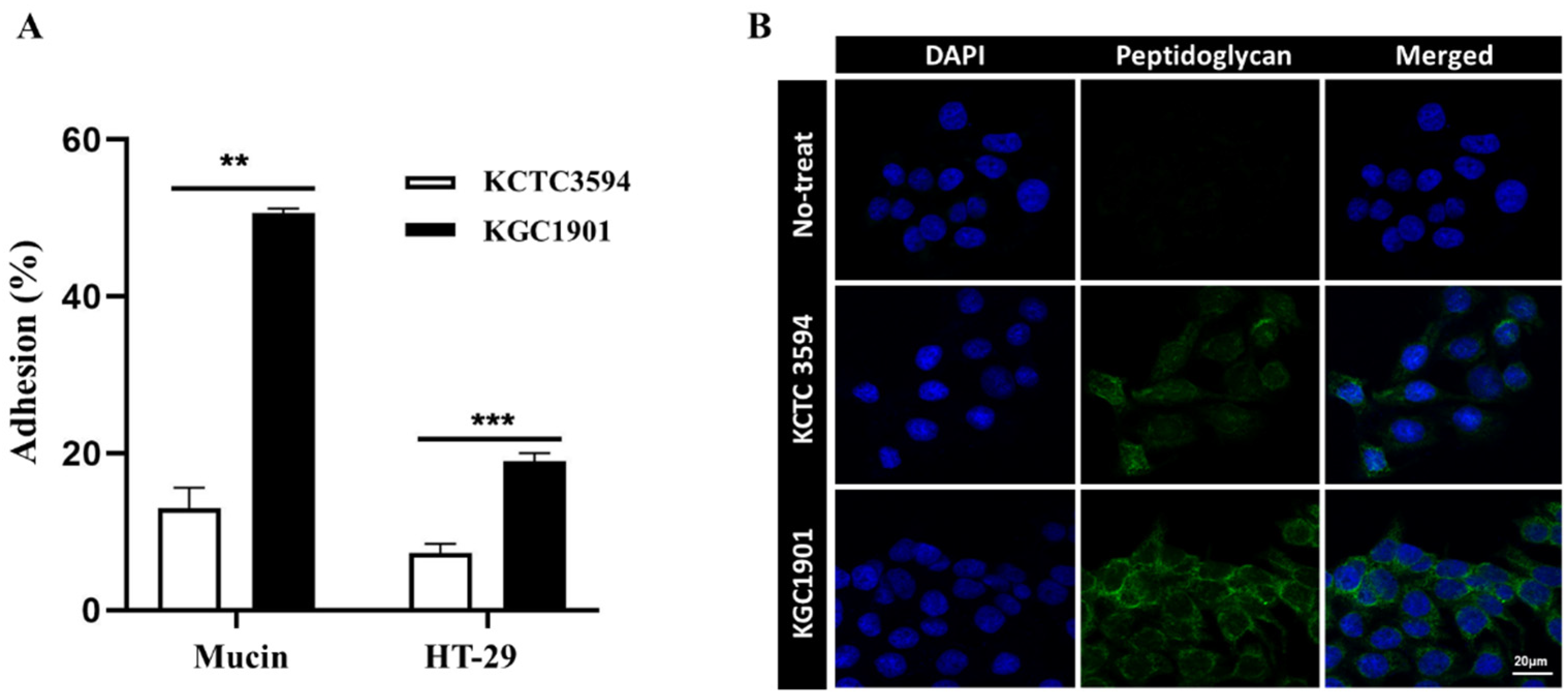
2.4. Intestinal Adhesion and Mucin Binding Ability

The intestinal adhesion property using HT-29 cells (human colonic epithelial cell) was evaluated as described by Kim, et al. [6]. The cultured HT-29 cells (4 × 10⁵ cells/mL) were incubated in RPMI 1640 medium (Hyclone, Logan, UT, USA) without fetal bovine serum and antibiotics at 37 °C in a 5% CO2 air overnight in 12-well tissue culture plates (SPL, Pocheon, Republic of Korea). Each probiotic strain (1 × 10⁷ cells/mL) was added to the HT-29 cell monolayer and incubated at 37 °C in 5% CO2 air. After 2 h of incubation, unattached bacterial cells were removed by PBS, and attached bacterial cells were detached by 0.05% trypsin-EDTA solution (w/v). Mucin binding ability was determined as described by Yasmin et al. with slight modification [31]. In brief, 0.4% mucin solution from porcine stomach type II (Sigma-Aldrich, St Louis, MO, USA) was coated to a 12-well non-coated plate for 24 h at 4 °C. Each probiotic strain (1 × 10⁷ cells/mL) was added to a mucin-coated well and incubated for 2 h at 37 °C in 5% CO2 air. Next, unattached bacterial cells were removed and washed with PBS, and attached bacterial cells were detached by 0.05% trypsin-EDTA solution (w/v). The number of detached viable bacterial cells was measured as follows:
Adhesion (%) = A1A0 × 100
where A1: number of attached bacterial cells and A0: number of initial bacterial cells.
In the above two experiments, Limosilactobacillus reuteri KCTC3594, the type strain purchased from KCTC, was used as a control. All data were obtained from three independent experiments and expressed as mean ± standard error. Significant differences were determined using Student’s t-tests and indicated as * p < 0.05 and ** p < 0.01.

2.5. Immunofluorescence Staining

HT-29 cells were cultured at a density of 2 × 10⁵ cells/well in a 4-well cell culture slide (SPL, Republic of Korea) for 1 day. Lactic acid bacteria were diluted to 1 × 10⁶ CFU/mL with RPMI and inoculated into the HT-29 cells for 2 h. The cells were washed with PBS then fixed by 4% paraformaldehyde and permeabilized with 0.1% Triton X-100 (Sigma-Aldrich, Germany), and they were blocked in 1% Bovine Serum Albumin (Sigma-Aldrich, Germany) for 1 h. Then, they were treated with peptidoglycan antibody (Bio-Rad, Hercules, CA, USA) overnight at 4 °C and goat anti-mouse IgG H&L Alexa Fluor 488 as a secondary antibody for 1 h at room temperature. The slide was mounted using a mounting medium with DAPI (Abcam, Cambridge, UK) and visualized with a confocal laser scanning microscope (LSM 800, Carl Zeiss, Jena, Germany).

2.6. Antibiotic Resistance and Virulence Factor

The European Food Safety Authority (EFSA) has published cut-off MIC values for the following antibiotics: ampicillin, gentamicin, kanamycin, streptomycin, erythromycin, clindamycin, tetracycline, and chloramphenicol. We determined the corresponding MIC values for KGC1901 by performing E-tests [32]. In brief, KGC1901 was spread onto an MRS agar plate, and a test strip (Liofilchem, Roseto degli Abruzzi, Italy) was applied aseptically to the inoculated surface of the agar plate. Following incubation at 37 °C for 24 h, the inhibition zone formed around the strip was observed, and the MIC values were determined on the basis of the concentration of the antibiotic on the strip where the edges of the inhibition zone converged. Acquired antimicrobial resistance genes were detected by comparing the assembled sequences with the reference sequences using ResFinder v.4.1 (https://cge.cbs.dtu.dk/servies/ResFinder (accessed on 1 December 2022)) with a threshold of >90% for %ID and 60% for minimum length [33]. Putative virulence genes were identified using the Virulence Factor Database (version 2020.02.13; http://www.mgc.ac.cn/VFs/ (accessed on 1 December 2022)) by the BLASTn algorithm with conditions of identity > 70%, coverage > 70%, and E-value < 1 × 10⁻⁵ [34].

2.7. Determination of Hemolytic Activity and Biogenic Amine

For determination of hemolytic activity, Escherichia coli KCTC2441 purchased from KCTC and Staphylococcus aureus NCTC10788 from bioMérieux (Marcy-l’Etoile, France) were used as positive controls of α-hemolysis and β-hemolysis, respectively. Each strain was streaked on blood agar plates (KisanBio, Seoul, Republic of Korea), then hemolysis types were determined after incubation. The production of biogenic amines (BAs) was measured as described by Bang et al. [35]. Briefly, BAs from probiotic cell-free supernatant (CFS) were derivatized and analyzed on a high-performance liquid chromatography system (LC-NETI/ADC, Jasco, Macclesfield, UK) equipped with a C18 column (ANPEL Laboratory analysis, Shanghai, China, 4.6 mm × 250 mm C18 column) with a detection wavelength of 254 nm. The mobile phase consisted of an aqueous acetonitrile solution (67:33 of water) flowing at a rate of 0.8 mL/min.

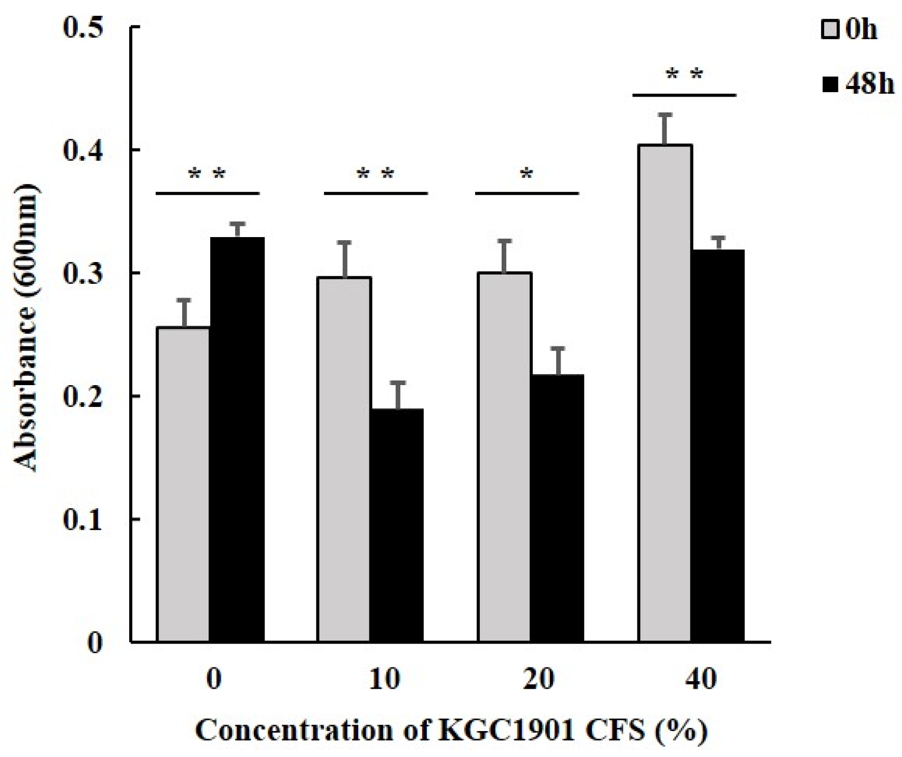
2.8. Antimicrobial Activity of L. reuteri KGC1901 CFS against Clostridium difficile

Clostridium difficile type strain was purchased from the National Culture Collection for Pathogens (Korea) and was cultured in BHI medium (BD Difco, Franklin Lakes, NJ, USA) for 18 h at 37 °C under anaerobic conditions. Then, 1% (v/v) of the cultured broth of C. difficile was transferred to fresh BHI medium with 10%, 20%, and 40% (v/v) of KGC1901 CFS, and the suspensions were incubated for 48 h at 37 °C under anaerobic conditions. Antimicrobial activity was determined by measuring the differences in the absorbance of cultures at 600 nm at the start and end of the incubation period with or without CFS.

2.9. DPPH Radical Scavenging Activity

The antioxidant activity of KGC1901 was determined on the basis of DPPH radical scavenging activity. An overnight culture of KGC1901 was centrifuged at 10,000 rpm for 10 min at 4 °C. The cells were collected, washed twice with PBS, and resuspended in PBS with an absorbance of 1.0 at 600 nm. The cell suspension and DPPH solution (200 μM) were mixed and incubated at 37 °C for 30 min. The mixture was centrifuged at 10,000 rpm for 5 min, and the absorbance of the supernatant was measured at 517 nm. Ascorbic acid was used as the positive control, and scavenging activity was expressed as the percentage of inhibition of the sample relative to the control.

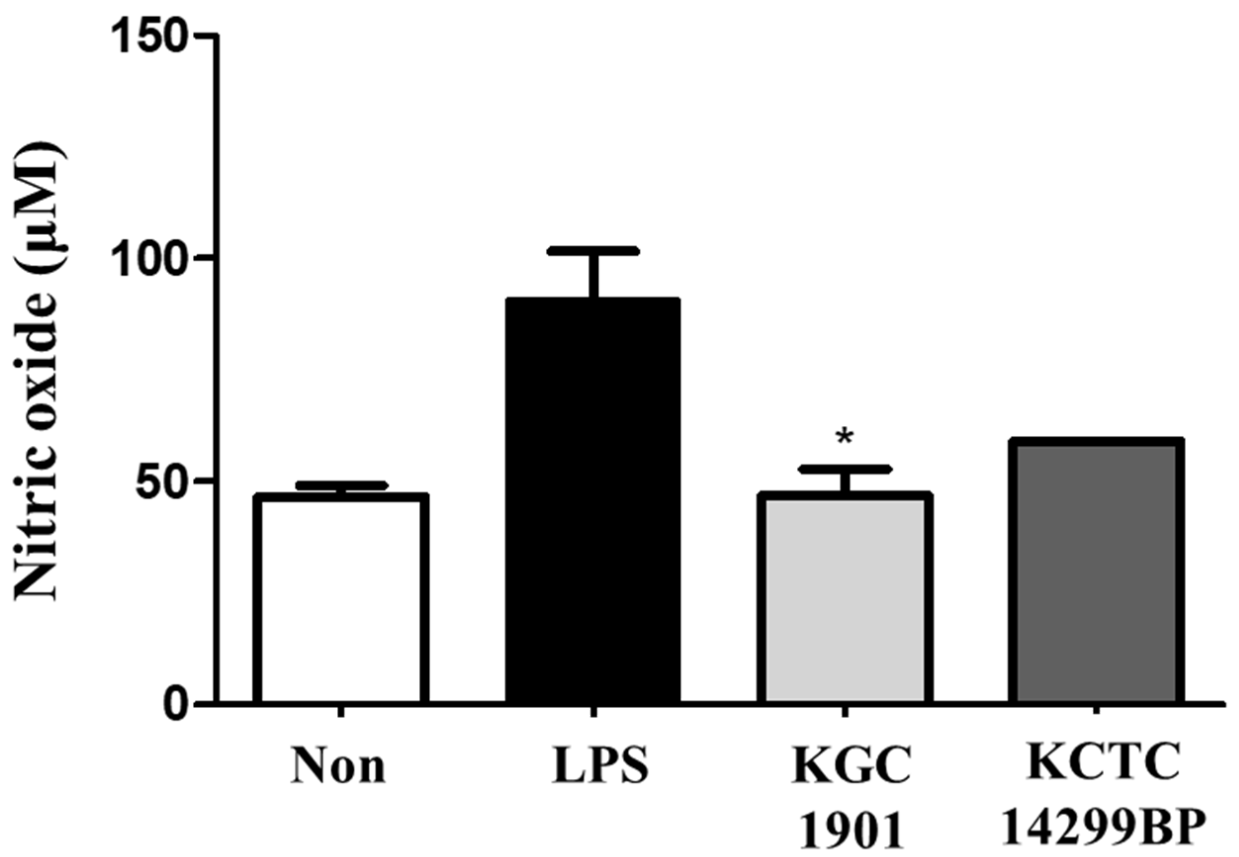
2.10. Cell Culture and Nitric Oxide (NO) Production Assay

The mouse macrophage cell line RAW 264.7 was obtained from the Korean Cell Line Bank (KCLB40071), and the cells were cultured in Dulbecco’s modified Eagle’s medium (Gibco, Waltham, MA, USA) supplemented with 10% (v/v) FBS (Gibco) and 1% (w/v) penicillin/streptomycin solution (Gibco). The cells were incubated at 37 °C under a humidified atmosphere of 5% CO2. RAW 264.7 cells (5.0 × 10⁵ cells/mL) were exposed to 1 µg/mL of lipopolysaccharide (LPS), and then aliquots were treated with 1% of cell-free supernatant obtained from L. reuteri KGC1901 and L. reuteri KCTC14299BP, a positive control purchased from KCTC. NO production was measured using a Griess reagent system as described [36].

3. Results and Discussion

3.1. Isolation and Identification of L. reuteri KGC1901 from Ginseng Root

Based on previous studies that showed endogenous bacteria exist in all parts of ginseng plants [16], we tried to isolate endogenous lactic acid bacteria from ginseng roots. To isolate endophytic LAB from ginseng root, 6-year-old ginseng root samples were fermented and 16s rRNA sequences of candidate colonies forming halos in MRS agar supplemented with CaCO3 were analyzed. Among the candidates, Limosilactobacillus reuteri, which is one of the valuable commercial probiotic strains, was identified, named L. reuteri KGC1901, and the strain was deposited in the Korea Collection for Type Culture (KCTC, Jungeup, Republic of Korea) under the accession number KCTC14652BP (Figure 1).
To determine the genetic characteristics of this strain, whole genome sequencing was performed. The general features and a map of its circular genome are shown in Table 1 and Figure 2, respectively. The length of the genome is approximately 1.96 million base pairs (38.7% G + C content), containing 1944 coding DNA sequences (CDS), 10 rRNA genes, and 61 tRNA genes.

3.2. Carbohydrate Fermentation and Enzymatic Activities

Among 49 carbohydrates, KGC1901 and KCTC3594 differed in their fermentation patterns only on D-mannose; specifically, KGC1901 fermented D-mannose, whereas KCTC3594 did not (Table 2). These results can be used by the industry to optimize culture conditions for enhancing productivity. Microbial enzymes, such as β-glucosidase and β-glucuronidase, can generate carcinogens or increase the possibility of carcinogenesis in the human body [38,39]. Hence, evaluating the activities of extracellular microbial enzymes is critical for product safety. Our results show the absence of β-glucosidase and β-glucuronidase activities in KGC1901 (Table 3). The activities of enzymes involved in carbohydrate metabolism (α-galactosidase, β-galactosidase, and α-glucosidase), lipid metabolism (esterase), and vitamin metabolism (acid phosphatase) were detected in KGC1901. In particular, β-galactosidase and leucine arylamidase activities were stronger than other enzyme activities.

3.3. Probiotic Properties

The gastrointestinal tract of humans is a highly stressful environment for probiotics in terms of oxygen level, nutrient limitations, and pH changes (particularly high acidity) [40,41]. The ability to tolerate acids and bile salts is essential for probiotics to survive in intestinal harsh environments and to ensure their functionalities. In previous studies, only 6 out of 13 probiotic strains isolated from wheat-bran sourdough survived exposure to low pH and bile salts [42]. L. reuteri KGC1901 showed 81.97% and 92.04% survival rates against exposure to pH 2.5 and oxgall, respectively (Table 4). The acid resistance of L. reuteri KGC1901 was similar to that of LGG (84.19%) (p = 0.366), which is one of the most popular probiotic strains in the industrial field and used as a positive control in this study, and its bile salt tolerance is somewhat higher than that of LGG (88.05%) (p = 0.066), under the same conditions.
After passing through the gastrointestinal tract, the probiotics should be attached to the intestinal mucosa to colonize and proliferate [40]. This intestinal adhesion ability can be assessed mainly based on adhesion to HT-29 or Caco-2, human epithelial cell lines, and attachment to the intestinal mucus directly, in vitro [40]. In this study, we used HT-29 cells and 0.4% mucin solution from porcine stomach type II. After 2 h of adhesion, 51% of L. reuteri KGC1901 cells were attached to mucin and 19% of cells were attached to HT-29, whereas 13% and 7% of the cells of L. reuteri KCTC3594 were attached to mucin and HT-29, respectively (Figure 3A). The immunofluorescent image also confirmed that more KGC1901 cells adhered to HT-29 than the type strain (Figure 3B). This indicates that L. reuteri KGC1901 had better adhesion ability to the intestine than the type strain. These results suggest that L. reuteri KGC1901 has the ability to survive in the human gut and has proper properties as a probiotic.

3.4. Safety Assessments

Whole-genome sequencing is as important as biochemical tests in that it can predict the presence of risk factors [43]. In this study, whole-genome sequencing was performed to understand the safety of L. reuteri KGC1901 genetically, and the whole-genome sequences were analyzed using ResFinder and VFDB to identify antibiotic resistance genes and virulence genes, respectively. As a result, the genome of L. reuteri KGC1901 had no putative genes related to antibiotic resistance and virulence. The horizontal transfer of antibiotic resistance genes from commercial probiotics to pathogenic bacteria via plasmid-like mobile elements can be devastating to human health [43,44]. Therefore, the fact that L. reuteri KGC1901 strain does not have an antibiotic resistance gene is very valuable for a safe probiotic material. The antibiotic resistance of L. reuteri KGC1901 was confirmed by measuring the MICs of eight antibiotics together with confirmation at the genome level. As a result, L. reuteri KGC1901 showed MICs below the cut-off values suggested by EFSA for eight antibiotics: ampicillin, gentamicin, kanamycin, streptomycin, erythromycin, clindamycin, tetracycline, and chloramphenicol. Finally, we determined that it is safe against antibiotic resistance (Table 5).
Since hemolytic activity is related to the virulence of bacteria, hemolysis is an important factor that must be considered when selecting a probiotic strain [31]. KGC1901 showed γ-hemolysis (i.e., no hemolytic activity on sheep blood agar), whereas E. coli and S. aureus showed α- and β-hemolysis, respectively (Figure 4).
Biogenic amines (BAs), the biogenic substances containing one or more amine groups, are produced by some lactic acid bacteria during fermentation [45,46,47], and they normally function as neurotransmitters and signal molecules between commensal microbiota in the host gut [48]. However, BAs are also precursors for carcinogenic N-nitroso compounds, and high levels of BAs may cause symptoms such as headache, heart palpitations, vomiting, diarrhea, and hypertensive crises in humans and animals [49]. In this study, BAs such as tyramine, histamine, putrescine, cadaverine, and 2-phenethylamine were not detected in the supernatant of KGC1901. Based on these results, it was confirmed that KGC1901 has sufficient safety as a probiotic strain.

3.5. Antimicrobial Activity against Clostridium difficile

As previously described, the disruption of gut microbiome diversity by broad-spectrum antibiotics is a major risk factor for infection with CD in the gut [50,51]. Reuterin, also known as 3-hydroxypropionaldehyde (3-HPA), a secondary metabolite of L. reuteri, induces ROS production in CD, and this oxidative stress leads to cell membrane disruption, DNA damage, and finally cell death of this pathogen [52,53]. Based on these findings, in this study, the antibacterial activity of L. reuteri KGC1901 isolated from ginseng against CD was tested. As a result, when 10% or more of KGC1901 CFS was added to CD culture media, the absorbance of culture broth was significantly decreased after 48 h compared to the initial time (Figure 5). Reuterin is produced by the bioconversion of glycerol by vitamin B12-dependent glycerol dehydratase [54,55,56]. The biosynthesis of reuterin is controlled by a gene cluster, also known as the pdu-cbi-cob-hem gene cluster, consisting of the glycerol/propanediol utilization (pdu) cluster and the cobalamin biosynthesis cluster. Additionally, glycerol dehydratase is encoded in pduCDE [57]. In this study, the presence of the pdu operon including pduCDE in contig 69 of the KGC1901 genome was confirmed through whole-genome analysis. Moreover, cbi, cob, and hem genes were also detected in contig 68 (Figure 6). Greppi et al. studied the presence and composition of the pdu-cbi-cob-hem gene cluster in the genomes of 65 L. reuteri strains [58]. Most strains possessing complete gene clusters produced 3-HPA, but some strains failed to produce 3-HPA even though they had complete gene clusters. In another study, Yang et al. reported on strain L. reuteri AN417, which does not synthesize reuterin because it lacks a gene cluster encoding reuterin in its genome but has antibacterial activity against oral pathogens. It was confirmed that the antibacterial activity of AN417 was caused by fatty acids or sugars instead of reuterin [59]. Although the mechanism(s) involved in the antimicrobial activity of KGC1901 against C. difficile were not elucidated in this study, the change in absorbance of CD culture media and the presence of reuterin biosynthetic genes in the genome revealed the antimicrobial activity of KGC1901.

3.6. Antioxidant Activity

The DPPH radical scavenging activity of KGC1901 was 13%, higher than that of the reference strain, L. rhamnosus GG (9%) (Table 4). Moreover, in RAW 264.7 cells, LPS treatment stimulated NO production to 79 μM, whereas 1% KGC1901 CFS treatment suppressed NO production to 52.8 μM (Figure 7). This radical scavenging activity and NO production inhibition activity may be useful features to reduce oxidative damage in the human body. Kim et al. studied the free-radical scavenging activity and NO production of 112 probiotic strains and 4 strains were selected [28]. NO production is part of the immune response stimulated by cytokines and microbial substances such as LPS and is regulated by the iNOS gene. NO plays immunologically and physiologically important roles; however, it can also be a reactive radical that causes inflammatory reactions. Therefore, the ability of KGC1901 to suppress NO production in LPS-stimulated RAW 264.7 cells may be useful for suppressing inflammation and oxidative damage. However, additional studies on related mechanisms such as the expression of inflammatory cytokines are needed to demonstrate anti-inflammatory functionality.

4. Conclusions

The aim of this study was to isolate endophytic lactic acid bacteria isolated from the root of ginseng and to investigate the probiotic properties and safety of the isolate based on genomic and phenotypic characteristics as a potential probiotic strain. We also investigated antimicrobial activity against C. difficile and antioxidant activities. On the basis of our findings, it was determined that Limosilactobacillus reuteri KGC1901 isolated from ginseng is a potential probiotic to prevent disease related to C. difficile and oxidative stress.

Author Contributions

Conceptualization, S.-H.L. and S.-K.K.; formal analysis, S.-K.K.; investigation, H.-Y.Y., M.K. and Y.-S.L.; methodology, H.-Y.Y., M.K. and Y.-S.L.; project administration, S.-H.L. and S.-K.K.; supervision, S.-H.L.; writing—original draft preparation, H.-Y.Y.; writing—review and editing, S.-K.K. All authors have read and agreed to the published version of the manuscript.

Funding

This research received no external funding.

Institutional Review Board Statement

Not applicable.

Informed Consent Statement

Not applicable.

Data Availability Statement

The data presented in this study are available in the article.

Conflicts of Interest

The authors declare no conflict of interest.

References

  1. Ogunrinola, G.A.; Oyewale, J.O.; Oshamika, O.O.; Olasehinde, G.I. The human microbiome and its impacts on health. Int. J. Microbiol. 2020, 2020, 8045646. [Google Scholar] [CrossRef] [PubMed]
  2. Hill, C.; Guarner, F.; Reid, G.; Gibson, G.R.; Merenstein, D.J.; Pot, B.; Morelli, L.; Canani, R.B.; Flint, H.J.; Salminen, S. Expert consensus document: The International Scientific Association for Probiotics and Prebiotics consensus statement on the scope and appropriate use of the term probiotic. Nat. Rev. Gastroenterol. Hepatol. 2014, 11, 506–514. [Google Scholar] [CrossRef] [PubMed] [Green Version]
  3. Plaza-Diaz, J.; Ruiz-Ojeda, F.J.; Gil-Campos, M.; Gil, A. Mechanisms of action of probiotics. Adv. Nutr. 2019, 10, S49–S66. [Google Scholar] [CrossRef] [PubMed] [Green Version]
  4. Mathur, H.; Beresford, T.P.; Cotter, P.D. Health benefits of lactic acid bacteria (LAB) fermentates. Nutrients 2020, 12, 1679. [Google Scholar] [CrossRef]
  5. Chang, C.-J.; Lin, T.-L.; Tsai, Y.-L.; Wu, T.-R.; Lai, W.-F.; Lu, C.-C.; Lai, H.-C. Next generation probiotics in disease amelioration. J. Food Drug Anal. 2019, 27, 615–622. [Google Scholar] [CrossRef] [Green Version]
  6. Kim, H.; Lee, Y.-S.; Yu, H.-Y.; Kwon, M.; Kim, K.-K.; In, G.; Hong, S.-K.; Kim, S.-K. Anti-inflammatory effects of Limosilactobacillus fermentum KGC1601 isolated from panax ginseng and its probiotic characteristics. Foods 2022, 11, 1707. [Google Scholar] [CrossRef]
  7. Li, H.-Y.; Zhou, D.-D.; Gan, R.-Y.; Huang, S.-Y.; Zhao, C.-N.; Shang, A.; Xu, X.-Y.; Li, H.-B. Effects and mechanisms of probiotics, prebiotics, synbiotics, and postbiotics on metabolic diseases targeting gut microbiota: A narrative review. Nutrients 2021, 13, 3211. [Google Scholar] [CrossRef]
  8. Wang, X.; Zhang, P.; Zhang, X. Probiotics regulate gut microbiota: An effective method to improve immunity. Molecules 2021, 26, 6076. [Google Scholar] [CrossRef]
  9. Lee, H.; Jung, K.B.; Kwon, O.; Son, Y.S.; Choi, E.; Yu, W.D.; Son, N.; Jeon, J.H.; Jo, H.; Yang, H. Limosilactobacillus reuteri DS0384 promotes intestinal epithelial maturation via the postbiotic effect in human intestinal organoids and infant mice. Gut Microbes 2022, 14, 2121580. [Google Scholar] [CrossRef]
  10. Doron, S.; Snydman, D.R. Risk and safety of probiotics. Clin. Infect. Dis. 2015, 60, S129–S134. [Google Scholar] [CrossRef] [Green Version]
  11. Hotel, A.C.P.; Cordoba, A. Health and nutritional properties of probiotics in food including powder milk with live lactic acid bacteria. Prevention 2001, 5, 1–10. [Google Scholar]
  12. Vitali, B.; Minervini, G.; Rizzello, C.G.; Spisni, E.; Maccaferri, S.; Brigidi, P.; Gobbetti, M.; Di Cagno, R. Novel probiotic candidates for humans isolated from raw fruits and vegetables. Food Microbiol. 2012, 31, 116–125. [Google Scholar] [CrossRef] [PubMed]
  13. Jiang, J.; Li, K.; Xiao, Y.; Zhong, A.; Tang, J.; Duan, Y.; Li, Z. Limosilactobacillus reuteri Regulating Intestinal Function: A Review. Fermentation 2023, 9, 19. [Google Scholar] [CrossRef]
  14. Arshad, F.; Mehmood, R.; Hussain, S.; Khan, M.A.; Khan, M. Lactobacilli as probiotics and their isolation from different sources. Br. J. Res. 2018, 5, 43. [Google Scholar] [CrossRef]
  15. Hyun, S.H.; Bhilare, K.D.; In, G.; Park, C.-K.; Kim, J.-H. Effects of Panax ginseng and ginsenosides on oxidative stress and cardiovascular diseases: Pharmacological and therapeutic roles. J. Ginseng Res. 2022, 46, 33–38. [Google Scholar] [CrossRef] [PubMed]
  16. Chu, L.L.; Bae, H. Bacterial endophytes from ginseng and their biotechnological application. J. Ginseng Res. 2022, 46, 1–10. [Google Scholar] [CrossRef]
  17. Hua, M.; Fan, M.; Li, Z.; Sha, J.; Li, S.; Sun, Y. Ginseng soluble dietary fiber can regulate the intestinal flora structure, promote colon health, affect appetite and glucolipid metabolism in rats. J. Funct. Foods 2021, 83, 104534. [Google Scholar] [CrossRef]
  18. Yu, H.-Y.; Rhim, D.-B.; Kim, S.-K.; Ban, O.-H.; Oh, S.-K.; Seo, J.; Hong, S.-K. Growth promotion effect of red ginseng dietary fiber to probiotics and transcriptome analysis of Lactiplantibacillus plantarum. J. Ginseng Res. 2023, 47, 159–165. [Google Scholar] [CrossRef]
  19. Mo, S.-J.; Nam, B.; Bae, C.-H.; Park, S.-D.; Shim, J.-J.; Lee, J.-L. Characterization of novel Lactobacillus paracasei HY7017 capable of improving physiological properties and immune enhancing effects using red ginseng extract. Fermentation 2021, 7, 238. [Google Scholar] [CrossRef]
  20. Lee, Y.-S.; Yu, H.-Y.; Kwon, M.; Lee, S.-H.; Park, J.-I.; Seo, J.; Kim, S.-K. Probiotic Characteristics and Safety Assessment of Lacticaseibacillus casei KGC1201 Isolated from Panax ginseng. J. Microbiol. Biotechnol. 2023, 33, 1–8. [Google Scholar] [CrossRef]
  21. Czepiel, J.; Dróżdż, M.; Pituch, H.; Kuijper, E.J.; Perucki, W.; Mielimonka, A.; Goldman, S.; Wultańska, D.; Garlicki, A.; Biesiada, G. Clostridium difficile infection. Eur. J. Clin. Microbiol. Infect. Dis. 2019, 38, 1211–1221. [Google Scholar] [CrossRef] [PubMed] [Green Version]
  22. McDonald, L.C.; Gerding, D.N.; Johnson, S.; Bakken, J.S.; Carroll, K.C.; Coffin, S.E.; Dubberke, E.R.; Garey, K.W.; Gould, C.V.; Kelly, C. Clinical practice guidelines for Clostridium difficile infection in adults and children: 2017 update by the Infectious Diseases Society of America (IDSA) and Society for Healthcare Epidemiology of America (SHEA). Clin. Infect. Dis. 2018, 66, e1–e48. [Google Scholar] [CrossRef] [PubMed]
  23. Tartof, S.Y.; Rieg, G.K.; Wei, R.; Tseng, H.F.; Jacobsen, S.J.; Kalvin, C.Y. A comprehensive assessment across the healthcare continuum: Risk of hospital-associated Clostridium difficile infection due to outpatient and inpatient antibiotic exposure. Infect. Control Hosp. Epidemiol. 2015, 36, 1409–1416. [Google Scholar] [CrossRef] [PubMed]
  24. Kalakuntla, A.S.; Nalakonda, G.; Nalakonda, K.; Pidikiti, C.V.; Aasim, S.A. Probiotics and Clostridium difficile: A review of dysbiosis and the rehabilitation of gut microbiota. Cureus 2019, 11, e5063. [Google Scholar] [CrossRef] [Green Version]
  25. Liu, C.F.; Tseng, K.C.; Chiang, S.S.; Lee, B.H.; Hsu, W.H.; Pan, T.M. Immunomodulatory and antioxidant potential of Lactobacillus exopolysaccharides. J. Sci. Food Agric. 2011, 91, 2284–2291. [Google Scholar] [CrossRef]
  26. Tang, W.; Li, C.; He, Z.; Pan, F.; Pan, S.; Wang, Y. Probiotic properties and cellular antioxidant activity of Lactobacillus plantarum MA2 isolated from Tibetan kefir grains. Probiotics Antimicrob. Proteins 2018, 10, 523–533. [Google Scholar] [CrossRef]
  27. Forman, H.J.; Zhang, H. Targeting oxidative stress in disease: Promise and limitations of antioxidant therapy. Nat. Rev. Drug Discov. 2021, 20, 689–709. [Google Scholar] [CrossRef]
  28. Kim, H.; Kim, J.-S.; Kim, Y.; Jeong, Y.; Kim, J.-E.; Paek, N.-S.; Kang, C.-H. Antioxidant and probiotic properties of Lactobacilli and Bifidobacteria of human origins. Biotechnol. Bioprocess Eng. 2020, 25, 421–430. [Google Scholar] [CrossRef]
  29. Fessard, A.; Bourdon, E.; Payet, B.; Remize, F. Identification, stress tolerance, and antioxidant activity of lactic acid bacteria isolated from tropically grown fruits and leaves. Can. J. Microbiol. 2016, 62, 550–561. [Google Scholar] [CrossRef]
  30. Averina, O.V.; Poluektova, E.U.; Marsova, M.V.; Danilenko, V.N. Biomarkers and utility of the antioxidant potential of probiotic Lactobacilli and Bifidobacteria as representatives of the human gut microbiota. Biomedicines 2021, 9, 1340. [Google Scholar] [CrossRef]
  31. Yasmin, I.; Saeed, M.; Khan, W.A.; Khaliq, A.; Chughtai, M.F.J.; Iqbal, R.; Tehseen, S.; Naz, S.; Liaqat, A.; Mehmood, T. In vitro probiotic potential and safety evaluation (hemolytic, cytotoxic activity) of Bifidobacterium strains isolated from raw camel milk. Microorganisms 2020, 8, 354. [Google Scholar] [CrossRef] [PubMed] [Green Version]
  32. Brown, D.F.; Brown, L. Evaluation of the E test, a novel method of quantifying antimicrobial activity. J. Antimicrob. Chemother. 1991, 27, 185–190. [Google Scholar] [CrossRef] [PubMed]
  33. Florensa, A.F.; Kaas, R.S.; Clausen, P.T.L.C.; Aytan-Aktug, D.; Aarestrup, F.M. ResFinder–an open online resource for identification of antimicrobial resistance genes in next-generation sequencing data and prediction of phenotypes from genotypes. Microb. Genom. 2022, 8, 000748. [Google Scholar] [CrossRef] [PubMed]
  34. Liu, B.; Zheng, D.; Zhou, S.; Chen, L.; Yang, J. VFDB 2022: A general classification scheme for bacterial virulence factors. Nucleic Acids Res. 2022, 50, D912–D917. [Google Scholar] [CrossRef]
  35. Bang, W.Y.; Ban, O.-H.; Lee, B.S.; Oh, S.; Park, C.; Park, M.-K.; Jung, S.K.; Yang, J.; Jung, Y.H. Genomic-, phenotypic-, and toxicity-based safety assessment and probiotic potency of Bacillus coagulans IDCC 1201 isolated from green malt. J. Ind. Microbiol. Biotechnol. 2021, 48, kuab026. [Google Scholar] [CrossRef]
  36. Lyons, C.R.; Orloff, G.; Cunningham, J. Molecular cloning and functional expression of an inducible nitric oxide synthase from a murine macrophage cell line. J. Biol. Chem. 1992, 267, 6370–6374. [Google Scholar] [CrossRef]
  37. Tamura, K.; Stecher, G.; Kumar, S. MEGA11: Molecular evolutionary genetics analysis version 11. Mol. Biol. Evol. 2021, 38, 3022–3027. [Google Scholar] [CrossRef]
  38. Cole, C.; Fuller, R. The effect of dietary fat and yoghurt on colonic bacterial enzymes (β-glucosidase and β-glucuronidase) associated with colon cancer. Food Microbiol. 1987, 4, 77–81. [Google Scholar] [CrossRef]
  39. Kim, D.-H.; Jin, Y.-H. Intestinal bacterial β-glucuronidase activity of patients with colon cancer. Arch. Pharm. Res. 2001, 24, 564–567. [Google Scholar] [CrossRef]
  40. Ayyash, M.M.; Abdalla, A.K.; AlKalbani, N.S.; Baig, M.A.; Turner, M.S.; Liu, S.-Q.; Shah, N.P. Invited review: Characterization of new probiotics from dairy and nondairy products—Insights into acid tolerance, bile metabolism and tolerance, and adhesion capability. J. Dairy Sci. 2021, 104, 8363–8379. [Google Scholar] [CrossRef]
  41. Castro-López, C.; Romero-Luna, H.E.; García, H.S.; Vallejo-Cordoba, B.; González-Córdova, A.F.; Hernández-Mendoza, A. Key Stress Response Mechanisms of Probiotics during Their Journey Through the Digestive System: A Review. Probiotics Antimicrob. Proteins 2022, in press. [Google Scholar] [CrossRef] [PubMed]
  42. Manini, F.; Casiraghi, M.; Poutanen, K.; Brasca, M.; Erba, D.; Plumed-Ferrer, C. Characterization of lactic acid bacteria isolated from wheat bran sourdough. LWT-Food Sci. Technol. 2016, 66, 275–283. [Google Scholar] [CrossRef]
  43. Fu, X.; Lyu, L.; Wang, Y.; Zhang, Y.; Guo, X.; Chen, Q.; Liu, C. Safety assessment and probiotic characteristics of Enterococcus lactis JDM1. Microb. Pathog. 2022, 163, 105380. [Google Scholar] [CrossRef] [PubMed]
  44. Jiang, X.; Ellabaan, M.M.H.; Charusanti, P.; Munck, C.; Blin, K.; Tong, Y.; Weber, T.; Sommer, M.O.; Lee, S.Y. Dissemination of antibiotic resistance genes from antibiotic producers to pathogens. Nat. Commun. 2017, 8, 15784. [Google Scholar] [CrossRef] [PubMed] [Green Version]
  45. Spano, G.; Russo, P.; Lonvaud-Funel, A.; Lucas, P.; Alexandre, H.; Grandvalet, C.; Coton, E.; Coton, M.; Barnavon, L.; Bach, B. Biogenic amines in fermented foods. Eur. J. Clin. Nutr. 2010, 64, S95–S100. [Google Scholar] [CrossRef] [Green Version]
  46. Sudo, N. Biogenic amines: Signals between commensal microbiota and gut physiology. Front. Endocrinol. 2019, 10, 504. [Google Scholar] [CrossRef] [Green Version]
  47. Ruiz-Capillas, C.; Herrero, A.M. Impact of biogenic amines on food quality and safety. Foods 2019, 8, 62. [Google Scholar] [CrossRef] [Green Version]
  48. Gardini, F.; Özogul, Y.; Suzzi, G.; Tabanelli, G.; Özogul, F. Technological factors affecting biogenic amine content in foods: A review. Front. Microbiol. 2016, 7, 1218. [Google Scholar] [CrossRef] [Green Version]
  49. Wójcik, W.; Łukasiewicz, M.; Puppel, K. Biogenic amines: Formation, action and toxicity—A review. J. Sci. Food Agric. 2021, 101, 2634–2640. [Google Scholar] [CrossRef]
  50. Pal, R.; Athamneh, A.I.; Deshpande, R.; Ramirez, J.A.; Adu, K.T.; Muthuirulan, P.; Pawar, S.; Biazzo, M.; Apidianakis, Y.; Sundekilde, U.K. Probiotics: Insights and new opportunities for Clostridioides difficile intervention. Crit. Rev. Microbiol. 2022, in press. [Google Scholar] [CrossRef]
  51. Engevik, M.A.; Danhof, H.A.; Shrestha, R.; Chang-Graham, A.L.; Hyser, J.M.; Haag, A.M.; Mohammad, M.A.; Britton, R.A.; Versalovic, J.; Sorg, J.A. Reuterin disrupts Clostridioides difficile metabolism and pathogenicity through reactive oxygen species generation. Gut Microbes 2020, 12, 1795388. [Google Scholar] [CrossRef] [PubMed]
  52. Schaefer, L.; Auchtung, T.A.; Hermans, K.E.; Whitehead, D.; Borhan, B.; Britton, R.A. The antimicrobial compound reuterin (3-hydroxypropionaldehyde) induces oxidative stress via interaction with thiol groups. Microbiology 2010, 156, 1589. [Google Scholar] [CrossRef] [PubMed] [Green Version]
  53. Neumann-Schaal, M.; Metzendorf, N.G.; Troitzsch, D.; Nuss, A.M.; Hofmann, J.D.; Beckstette, M.; Dersch, P.; Otto, A.; Sievers, S. Tracking gene expression and oxidative damage of O2-stressed Clostridioides difficile by a multi-omics approach. Anaerobe 2018, 53, 94–107. [Google Scholar] [CrossRef] [PubMed] [Green Version]
  54. Sun, M.-C.; Hu, Z.-Y.; Li, D.-D.; Chen, Y.-X.; Xi, J.-H.; Zhao, C.-H. Application of the Reuterin System as Food Preservative or Health-Promoting Agent: A Critical Review. Foods 2022, 11, 4000. [Google Scholar] [CrossRef]
  55. Spinler, J.K.; Auchtung, J.; Brown, A.; Boonma, P.; Oezguen, N.; Ross, C.L.; Luna, R.A.; Runge, J.; Versalovic, J.; Peniche, A. Next-generation probiotics targeting Clostridium difficile through precursor-directed antimicrobial biosynthesis. Infect. Immun. 2017, 85, e00303–e00317. [Google Scholar] [CrossRef] [Green Version]
  56. Sun, Y.; Gutierrez-Maddox, N.; Mutukumira, A.N.; Maddox, I.S.; Shu, Q. Influence of Operating Conditions on Reuterin Production Using Resting Cells of Limosilactobacillus reuteri DPC16. Fermentation 2022, 8, 227. [Google Scholar] [CrossRef]
  57. Morita, H.; Toh, H.; Fukuda, S.; Horikawa, H.; Oshima, K.; Suzuki, T.; Murakami, M.; Hisamatsu, S.; Kato, Y.; Takizawa, T. Comparative genome analysis of Lactobacillus reuteri and Lactobacillus fermentum reveal a genomic island for reuterin and cobalamin production. DNA Res. 2008, 15, 151–161. [Google Scholar] [CrossRef]
  58. Greppi, A.; Asare, P.T.; Schwab, C.; Zemp, N.; Stephan, R.; Lacroix, C. Isolation and comparative genomic analysis of reuterin-producing Lactobacillus reuteri from the chicken gastrointestinal tract. Front. Microbiol. 2020, 11, 1166. [Google Scholar] [CrossRef]
  59. Yang, K.M.; Kim, J.-S.; Kim, H.-S.; Kim, Y.-Y.; Oh, J.-K.; Jung, H.-W.; Park, D.-S.; Bae, K.-H. Lactobacillus reuteri AN417 cell-free culture supernatant as a novel antibacterial agent targeting oral pathogenic bacteria. Sci. Rep. 2021, 11, 1631. [Google Scholar] [CrossRef]
Figure 1. The phylogenetic tree is based on the 16s rRNA gene sequence data obtained from the NCBI database, showing the phylogenetic positions of L. reuteri KGC1901. The tree was constructed using the maximum likelihood algorithm and Kimura 2-parameter model and the numbers displayed next to the branch indicate bootstrap values (500 replicates). Bacillus subtilis was used as an outgroup. Phylogenetic analysis was conducted in MEGA11 (Molecular Evolutionary Genetics Analysis) [37].
Fermentation 09 00228 g001
Figure 2. Genome map of L. reuteri KGC1901. From outside to the center: 71 contigs, annotated reference genes (specifically, CDSs) found in the forward strand; in the reverse strand, rRNA and tRNA found in this genome; GC skew metric (higher-than-average values: green, lower-than-average values: red); and GC ratio metric (higher-than-average values: blue, lower-than-average values: yellow).
Fermentation 09 00228 g002
Figure 3. Adhesion ability and immunofluorescent image of L. reuteri KGC1901. (A) Adhesion ability of L. reuteri KGC1901 and L. reuteri KCTC3594 (positive control) to mucin and HT-29 cells. Data are presented as mean ± standard deviation values from triplicate experiments. Significant correlations between the two strains are shown by asterisks (** p < 0.01, and *** p < 0.001). (B) HT-29 cells were stained with DAPI (blue), and the bacterial cells of L. reuteri KGC1901 and L. reuteri KCTC3594 were stained with antibodies against peptidoglycan and goat anti-mouse Alexa fluor 488 (green).
Fermentation 09 00228 g003
Figure 4. Hemolytic activities of L. reuteri KGC1901, Escherichia coli, and Staphylococcus aureus. E. coli shows typical α-hemolysis (A), and S. aureus shows typical β-hemolysis (B). However, L. reuteri KGC1901 (C) does not show any lysis of blood cells.
Fermentation 09 00228 g004
Figure 5. Antimicrobial activity of L. reuteri KGC1901 against Clostridium difficile. C. difficile was cultured in BHI with KGC1901 CFS and the absorbance of the culture soup was measured. As a result, the growth of CD was suppressed on the media added with 10%, 20%, and 40% of KGC1901 CFS. Significant correlations between 0 h and 48 h of each concentration are shown by asterisks (* p < 0.05, and ** p < 0.01).
Fermentation 09 00228 g005
Figure 6. The schematic representation of the pdu-cbi-cbo-hem cluster of L. reuteri KGC1901. The genes encoding cobalamin synthetic genes were located in contig 68 (upper), and the genes encoding propanediol utilization genes were located in contig 69 (lower).
Fermentation 09 00228 g006
Figure 7. Nitric oxide production of L. reuteri KGC1901 in lipopolysaccharide-induced RAW 264.7 cells. Significant correlation between LPS and L. reuteri KGC1901 treatment is shown by asterisk (* p < 0.05).
Fermentation 09 00228 g007
Table 1. Genomic characteristics of Limosilactobacillus reuteri KGC1901.
ItemsResults
Genome size (bp)1,963,544
GC contents (%)38.7
CDS1944
No. of rRNA genes10
No. of tRNA genes61
Table 2. Carbohydrate fermentation of L. reuteri KGC1901.
CarbohydratesStrainsCarbohydratesStrains
KGC
1901
KCTC
3594
KGC
1901
KCTC
3594
Glycerol- *-Salicin--
Erythritol--D-cellobiose--
D-arabinose--D-maltose++
L-arabinose++D-lactose++
D-ribose++D-melibiose++
D-xylose--D-saccharose(sucrose)++
L-xylose--D-trehalose--
D-adonitol--Inulin--
Methyl-βD-Xylopyranoside--D-melezitose--
D-galactose++D-raffinose++
D-glucose++Amidon (starch)--
D-fructose--Glycogen--
D-mannose+-Xylitol--
L-sorbose--Gentiobiose--
L-rhamnose--D-turanose--
Dulcitol--D-lyxose--
Inositol--D-tagatose--
D-mannitol--D-fucose--
D-sorbitol--L-fucose--
MethylαD-mannopyranoside--D-arabitol--
MethylαD-Glucopyranoside--L-arabitol--
N-acetylglucosamine--Potassium gluconatevv
Amygdalin--Potassium 2-ketogluconate--
Arbutin--Potassium 5-ketogluconate--
Esculin ferric citrate++
* -: not utilized; v: weakly utilized; +: strongly utilized.
Table 3. Enzymatic activities of L. reuteri KGC1901.
EnzymesKGC1901EnzymesKGC1901
Alkaline phosphatase- *Naphthol-AS-BI-phosphohydrolase+
Esterase+α-Galactosidase+
Esterase lipase-β-Galactosidase+++
Lipase-β-Glucuronidase-
Leucine arylamidase+++α-Glucosidase+
Valine arylamidase+β-Glucosidase-
Crystinearylamidase-N-acetyl-β-glucosaminidase-
Trypsin-α-Mannosidase-
α-Chymotrypsin-α-Fucosidase-
Acid phosphatase+
* -, no activity; +, poor activity; ++, moderate activity; +++, strong activity.
Table 4. Acid resistance, bile salt tolerance, and antioxidant activity of L. reuteri KGC1901. Data are presented as mean ± standard deviation values from triplicate experiments.
CharacteristicsSurvival Rate (%)
KGC1901LGG
Acid resistance0 h (log CFU/mL)8.26 ± 0.008.03 ± 0.06
3 h (log CFU/mL)6.78 ± 0.116.77 ± 0.13
Survival rate (%)81.97 ± 1.3884.19 ± 2.33
Bile salt tolerance0 h (log CFU/mL)8.26 ± 0.008.03 ± 0.06
3 h (log CFU/mL)7.61 ± 0.107.07 ± 0.02
Survival rate (%)92.04 ± 1.1988.05 ± 0.96
DPPH radical scavenging (%)139
Table 5. Minimal inhibitory concentrations of L. reuteri KGC1901.
Antibiotic 1AMPGENKANSTRERYCLITETCHL
Cut-off value (mg/L) 228646411164
Observed MICs0.01634880.125<0.0161.0–1.50.75
AssessmentS 3SSSSSSS
1 AMP, ampicillin; GEN, gentamycin; KAN, kanamycin; STR, streptomycin; ERY, erythromycin; CLI, clindamycin; TET, tetracycline; CHL, chloramphenicol; 2 European Food Safety Authority Guidelines, 2018; 3 S, Susceptible.
Disclaimer/Publisher’s Note: The statements, opinions and data contained in all publications are solely those of the individual author(s) and contributor(s) and not of MDPI and/or the editor(s). MDPI and/or the editor(s) disclaim responsibility for any injury to people or property resulting from any ideas, methods, instructions or products referred to in the content.

