Article

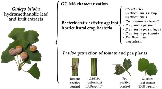
Antibacterial Activity of Ginkgo biloba Extracts against Clavibacter michiganensis subsp. michiganensis, Pseudomonas spp., and Xanthomonas vesicatoria

by
Eva Sánchez-Hernández
1,*,
Vicente González-García
2,
Ana Palacio-Bielsa
2,
Belén Lorenzo-Vidal
3,
Laura Buzón-Durán
1,
Jesús Martín-Gil
1 and
Pablo Martín-Ramos
1
1
Department of Agricultural and Forestry Engineering, ETSIIAA, Universidad de Valladolid, Avda. Madrid 44, 34004 Palencia, Spain
2
Department of Agricultural, Forest and Environmental Systems, Agrifood Research and Technology Centre of Aragón, Instituto Agroalimentario de Aragón—IA2 (CITA-Universidad de Zaragoza), Avda. Montañana 930, 50059 Zaragoza, Spain
3
Servicio de Microbiología, Hospital Universitario Rio Hortega, Calle Dulzaina 2, 47012 Valladolid, Spain
*
Author to whom correspondence should be addressed.
Horticulturae 2023, 9(4), 461; https://doi.org/10.3390/horticulturae9040461
Submission received: 24 February 2023 / Revised: 29 March 2023 / Accepted: 3 April 2023 / Published: 5 April 2023

Abstract

:
Phytopathogenic bacteria represent a risk to global food production by impacting a variety of crops. The aim of this study was to characterize the contents of bioactive constituents in extracts from Ginkgo biloba L. leaves and fruits and test their activity against six phytopathogenic bacteria that affect horticultural crops. Gas chromatography–mass spectrometry (GC−MS) was used for the chemical profiling of the aqueous methanol extracts, and their bacteriostatic activity against Clavibacter michiganensis subsp. michiganensis, Pseudomonas cichorii, Pseudomonas syringae pv. pisi, Pseudomonas syringae pv. syringae, Pseudomonas syringae pv. tomato, and Xanthomonas vesicatoria (formerly Xanthomonas campestris pv. vesicatoria) was determined in vitro through the agar dilution method. The protective effect of the leaf extract was tested in vivo against the most relevant bacterial pathogens based on their economic/scientific importance, i.e., C. michiganensis subsp. michiganensis and P. syringae pv. pisi, in tomato (Solanum lycopersicum L.) and pea (Pisum sativum L.) plants, respectively, under greenhouse conditions. The GC−MS characterization of G. biloba extracts revealed the presence of dihydro-4-hydroxy-2(3H)-furanone, 2,4-dimethyl-3-hexanol, catechol, 3-O-methyl-D-fructose, 4,6-di-O-methyl-α-D-galactose, methyl 2-O-methyl-α-D-xylofuranoside, and 3-methyl mannoside. In vitro growth inhibition tests showed that, while the fruit extract had no activity, the leaf extract exhibited minimum inhibitory concentrations between 500 and 1000 μg mL−1, which may be attributed to the presence of 2,4-dimethyl-3-hexanol and catechol. In vivo tests of the leaf extract demonstrated full protection in tomato and pea plants at 1000 and 1500 μg∙mL−1, respectively. The results indicate that G. biloba leaves may be employed as a biorational source for integrated pest management in horticulture.

Graphical Abstract

1. Introduction

Ginkgo biloba L. is a tree species belonging to the family Ginkgoaceae and the only living species of the Ginkgoales order. Ginkgo biloba trees are disease- and insect-resistant, making them exceptionally long-lived, with some specimens reportedly existing for over 2500 years. They generally reach a height of 20 to 35 m, possess an angular crown, and typically feature deep roots. The leaves are petiolated, with a fan-shaped blade and dichotomous and open venation. It is a dioecious species, where female trees produce fruit each year, while male trees do not (Figure 1). The fruits emit an unpleasant, musty odor due to the presence of butyric acid in the yellowish, fleshy part (sarcotesta), which is not consumed as it can be toxic; rather, the inner, softer portion of the seed (sclerotesta)—which has a greenish color—is eaten.
Ginkgo biloba is a widely used plant remedy throughout the world [1]. Its potential benefits for cardiovascular and neurodegenerative diseases have been substantiated [2,3]. Furthermore, the ginkgo nut has demonstrated potential in the prevention of neurological and cardiovascular diseases, though research in this area is limited [4].
Back in 1992, Gülz et al. initially documented the chemical composition of the waxes of G. biloba leaves [5], but it was not until 2008 that Lin et al. provided a more extensive report on the phytoconstituents of the aqueous methanol extract of the leaves, including forty-five glycosylated flavones and flavonols (predominantly hydroxyflavones), ten biflavones, three flavonol aglycones (myricetin, quercetin, and kaempferol), catechin, dihydroxybenzoic acid, and terpene lactones (ginkgolides and bilobalide, Figure 2) [6].
Regarding standardized G. biloba extract EGb 761 (a leaf extract frequently utilized as a dietary supplement), it is typically composed of flavone glycosides (24%), terpenes (6%), and other substances, such as proanthocyanidins and organic acids. Ginkgolides and bilobalide account for approximately 3.1% and 2.9% of the extract, respectively [7]. It has been suggested that some of the beneficial effects of EGb may be due to antioxidant activities, with an increasing body of evidence pointing to lipid peroxidation reactions and free radicals’ participation in neuronal damage and central and peripheral vascular diseases [8,9].
Ginkgo biloba has been reported to possess antifungal activity by Krauze-Baranowska and Wiwart [10] and Oh et al. [11], and antibacterial activity against Staphylococcus aureus Rosenbach, Bacillus thuringiensis Berliner, Clostridium perfringens (Veillon and Zuber, 1898) Hauduroy et al., 1937, Listeria monocytogenes (Murray et al., 1926) Pirie, 1940, and Haemophilus influenzae (Lehmann and Neumann, 1896) Winslow et al., 1917 [12]. However, to the best of our knowledge, no prior studies have been conducted to assess the antibacterial activity of this plant species on phytopathogenic taxa.
Given the significant global economic losses caused by bacterial diseases of plants—estimated at over one billion dollars worldwide annually [13]—there is an interest in investigating biorationals based on natural products for the integrated pest management of phytopathogenic bacteria that affect horticultural crops. For instance, these products could be used to control Gram-negative bacteria such as Pseudomonas syringae van Hall pathovars (e.g., Pseudomonas syringae pv. pisi (Sackett 1916) Young et al. 1978 Dye & Wilkie; Pseudomonas syringae pv. syringae van Hall 1902; Pseudomonas syringae pv. tomato (Okabe 1933) Young et al. 1978), Pseudomonas cichorii (Swingle 1925) Stapp, 1928; and Xanthomonas vesicatoria (Doidge) Vauterin et al., as well as Gram-positive bacteria such as Clavibacter michiganensis subsp. michiganensis corrig. (Smith 1918) Davis et al. 1984. Among these, P. syringae [14] and P. cichorii [15] can infect a wide range of plant species, while X. vesicatoria (formerly X. campestris pv. vesicatoria) is the causal agent of bacterial leaf spot on tomatoes (Solanum lycopersicum L.) and peppers (Capsicum annuum L.) [16] and C. michiganensis subsp. michiganensis causes bacterial canker of tomatoes, and can also affect other solanaceous plants [17]. It should be noted that P. syringae pathovars and C. michiganensis subsp. michiganensis are considered among the more relevant bacterial pathogens based on their scientific/economic importance [18].
The purpose of this study was twofold: (i) to characterize the phytoconstituents of Ginkgo biloba L. leaf and fruit extracts and (ii) to investigate their activity against the aforementioned six phytopathogenic bacteria that affect horticultural crops.

2. Materials and Methods

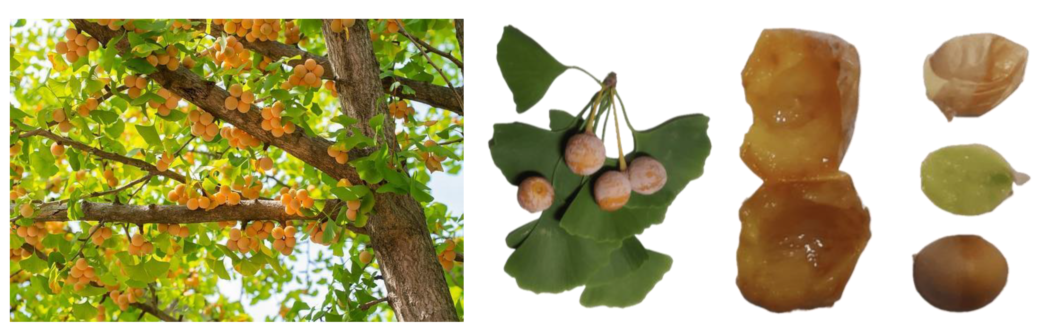
2.1. Plant Materials and Reagents

Ginkgo biloba leaf and fruit samples were collected from 20 female trees—grown under the same environmental conditions, soil type, and agronomic factors—along the Avenida de los Monegros in Huesca City, Aragon, northeastern Spain (42°7′46.9″ N 0°24′6.3″ W) in the late days of September 2021. For drying the samples, the freeze-drying method was chosen, in agreement with Boateng and Yang [19]: samples were initially frozen at −20 °C for 24 h and then freeze-dried at −50 °C.
Eight-week-old tomato (Solanum lycopersicum) cv. “Optima F1” plants and fourteen-week-old pea (Pisum sativum) cv. “Lincoln” plants used in the in vivo experiments were supplied by Agrodepa S.L. (Palencia, Spain).
Catechol (CAS no. 120-80-9) was acquired from Thermo Fisher Scientific (Madrid, Spain). Additionally, 2,4-dimethyl-3-hexanol (CAS no. 13432-25-2), tryptic soy broth (TSB, no. CAS 8013-01-2), and tryptic soy agar (TSA, CAS no. 91079-40-2) were procured from Sigma–Aldrich Química (Madrid, Spain).

2.2. Bacterial Strains

Pseudomonas cichorii (strain CITA Pci-5), P. syringae pv. pisi (strain CITA Pspi-12), P. syringae pv. syringae (strain CITA Psy-9), P. syringae pv. tomato (strain CITA Pst-7), X. vesicatoria (strain CITA Xv-5), and C. michiganensis subsp. michiganensis (strain CITA Cmm-30) were supplied by the Bacteriology Lab at the Agrifood Research and Technology Center of Aragon (CITA, Zaragoza, Spain) as TSA subcultures.

2.3. Extract Preparation

The preparation of the leaf, sarcotesta, and sclerotesta extracts was carried out according to the methodology described in [20]; the powdered sample was digested in a methanol/water (1:1, v/v) solution for 2 h, sonicated for 10 min in pulsed mode using a model UIP1000hdT probe-type ultrasonicator (20 kHz, 1000 W; Hielscher Ultrasonics, Teltow, Germany), and allowed to stand for 24 h. The solution was then centrifuged at 9000 rpm for 15 min, and the supernatant was filtered using Whatman No. 1 paper. Samples of the extracts were freeze-dried to obtain the solid residue. For gas chromatography–mass spectrometry (GC–MS) analyses, 25 mg of the freeze-dried extracts were dissolved in 5 mL of methanol (HPLC grade) to yield a 5 mg·mL−1 solution, which was filtered before use.

2.4. Extract Characterization

The extracts were analyzed using GC–MS at the Research Support Services at the University of Alicante (Alicante, Spain). The equipment utilized was a model 7890A gas chromatograph coupled to a model 5975C quadrupole mass spectrometer (Agilent Technologies; Santa Clara, CA, USA). The conditions for chromatography included 3 injections per vial, 1 µL injection volume, an injector temperature of 280 °C, and an initial oven temperature of 60 °C. The temperature then increased at a rate of 10 °C·min−1 to a final temperature of 300 °C, which was held for 15 min. The chromatographic column employed was an Agilent Technologies HP-5MS UI 30 m in length, 0.250 mm in diameter, and with a 0.25 µm film. The temperatures of the electron impact source of the mass spectrometer and of the quadrupole were set at 230 and 150 °C, respectively; ionization energy was set at 70 eV. For calibration, test mixture 2 for apolar capillary columns according to Grob and PFTBA tuning standards were used. Component identification was based on a comparison of their mass spectra and retention time to those of authentic compounds, and by computer matching using the National Institute of Standards and Technology (NIST11) database.
The infrared spectrum of the dried powdered leaves was acquired using a Nicolet iS50 Fourier-transform infrared spectrometer (Thermo Scientific; Waltham, MA, USA) with a diamond attenuated total reflectance system. The spectra were collected in the 400–4000 cm−1 range, with a 1 cm−1 spectral resolution, by co-adding 64 scans.

2.5. In Vitro Antibacterial Activity Assessment

The bacteriostatic activity was assessed according to the Clinical and Laboratory Standards Institute (CLSI) standard M07–11 [21], using the agar dilution method to determine the minimum inhibitory concentrations (MICs). Colonies of each species were incubated at 28 °C for 24 h in TSB. Serial dilutions of the cell suspension were then performed, beginning from a concentration of 108 CFU·mL−1, to obtain a final inoculum of 104 CFU·mL−1. The bacterial suspensions were then applied to the surface of the TSA plates to which the treatments had previously been added at concentrations between 62.5 and 1500 μg·mL−1. The plates were incubated at 28 °C for 24 h, and readings were taken after 24 h. MICs were determined as the lowest concentrations of the bioactive products at which no bacterial growth was visible. All experiments were conducted in triplicate, with 3 plates per treatment/concentration. Statistical analyses of antibacterial activity data were conducted using IBM (Armonk, NY, USA) SPSS Statistics v.25.
For comparison, the MIC values of five antibiotics commonly used in clinical practice, viz. ampicillin (AM), benzylpenicillin or penicillin G (PG), ciprofloxacin (CI), gentamicin (GM), and tetracycline (TC), were determined using ETEST® strips.

2.6. In Vivo Experiments on Tomato and Pea Plants under Greenhouse Conditions

Tomato plants cv. “Optima F1” were subjected to experiments in accordance with the guidelines in [22], with a few minor modifications. In a greenhouse cabin of biological containment level 2 (NCB2), tomato plants were grown in multi-pots (4 × 4 cm per pot) containing sterilized peat as substrate, at 20 ± 2 °C day/17 ± 2 °C night, with supplementary lighting to give a 12 h day. The last irrigation was carried out 48 h before inoculation, at which time the relative humidity was set at 100%. The G. biloba leaf extract was applied through spraying (3 mL per plant) at two concentrations (MIC and MIC × 2, i.e., 500 and 1000 μg·mL−1, respectively, taking into consideration that some of the treatment may be lost due to evaporation, runoff, degradation, etc., thus necessitating a higher concentration to achieve the same effect as in vitro); control plants were sprayed with water. After two hours, an isolated colony of C. michiganensis subsp. michiganensis was inoculated to the node of the first true leaf with the stem, with the aid of a sterile entomological pin. The plants were then covered with polyethylene bags for five days, and the next irrigation was carried out 24 h after inoculation. Throughout the experiment, the plants were irrigated three times a week. For the pea assay, the experiments were carried out according to the guidelines in [23]. Pea plants were cultivated under the aforementioned greenhouse conditions in 4 × 4 cm pots that contained a mixture of sterilized peat and sand (2:1). The G. biloba leaf extract was sprayed at two concentrations (MIC and MIC × 2, i.e., 750 and 1500 μg·mL−1, respectively) onto treated plants, and control plants were sprayed with water. Two hours after treatment, an isolated colony of P. syringae pv. pisi was inoculated using a sterile entomological pin, by pricking the back of the stem at the two youngest nodes of the plant, at its junction with the stipula. Inoculation of the leaflets or stipules took place at the same time as that of the stems, with 4–8 pricks in the youngest leaflets and stipules. The irrigation regime was similar to that described above for tomato plants.
In both experiments, five plants per treatment (negative control, positive control, MIC, and MIC × 2) were used, with two independent replicates. In regard to the tomato plant trials, the first replicate began on 31 January 2023 and concluded on 10 February 2023; the second replicate began on 3 February 2023, ending on 13 February 2023. Regarding the pea plant trials, the first replicate started on 27 January 2023, concluding on 6 February 2023. The second replicate started on 30 January 2023, ending on 9 February 2023. Concerning the experimental layout, a completely randomized design was used, where each experimental unit (plant) was randomly assigned to one of the treatments.
Disease incidence was evaluated after ten days of inoculation, using the following formula: I (%) = (Number of plants with symptoms/Total number of plants assayed) × 100.

3. Results

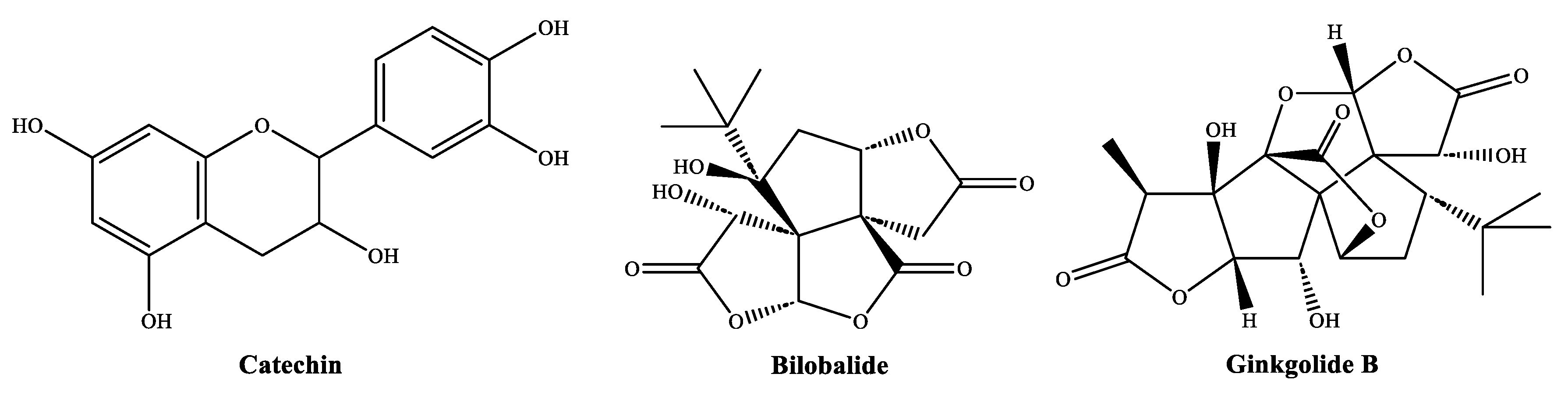
3.1. Extract Phytoconstituents Elucidation by GC−MS

In the GC−MS analysis of the leaf extract (Figure S1, Table S1, Figure 3a), the main identified compounds were: 3-O-methyl-D-fructose (18.1%); 2,4-dimethyl-3-hexanol (18.1%); acetic acid (16.2%); 3-methylmannoside (9.8%); acetic acid, 4-methylphenyl ester (4.4%); catechol (3.3%); 1-hydroxy-2-propanone (2.4%); and dihydro-4-hydroxy-2(3H)-furanone or β-hydroxy-γ-butyrolactone (1.2%).
Regarding the sarcotesta extract chromatogram (Figure S2, Table S2, Figure 3b), the following phytoconstituents were detected: acetic acid (11.9%); 2,3-dihydro-3,5-dihydroxy-6-methyl-4H-pyran-4-one and related compounds (8.7%); formic acid 2-methylpropyl ester and related compounds (5.4%); 1-hepten-4-ol (4.5%); catechol (4.1%); dihydroxyacetone (3.9%); 5-hydroxymethylfurfural (3.6%); 1-hydroxy-2-propanone (3.3%); 2-hydroxy-2-cyclopenten-1-one (3.1%); 4,6-di-O-methyl-α-D-galactose (1.6%); 4,5-dihydro-2-methyl-1H-imidazole (1.3%); 2,4-dihydroxy-2,5-dimethyl-3(2H)-furan-3-one (1.3%); 2,5-dimethyl-4-hydroxy-3(2H)-furanone (1.0%); α-methyl mannofuranoside (1.3%); and azelaic acid (1.1%).
Lastly, GC−MS analysis of the sclerotesta extract (Figure S3, Table S3, Figure 3c) yielded the following main constituents: methyl 2-O-methyl-α-D-xylofuranoside (19.1%); dihydro-4-hydroxy-2(3H)-furanone (11.7%); 4,6-di-O-methyl-α-D-galactose (8.4%); catechol (7.9%); 3-methylmannoside (6.3%); 3-O-methyl-fructose (6.1%); tetrahydro-2-methyl-thiophene (3%); and 2-furanmethanol (2.5%).

3.2. Leaves Vibrational Spectra

Ginkgo biloba leaves showed absorption bands at 3309, 2915, 2849, 1731, 1712, 1630, 1605, 1515, 1465, 1371, 1243, 1166, 1104, 1020, and 832 cm−1. The intense band at 3309 cm−1 was attributed to OH stretching, while the one at 2915 cm−1 was associated with methylene (CH2) stretching, and 2849 cm−1 with symmetric vibration of CH2 groups. The bands at 1731 and 1712 cm−1 were likely due to C = O stretching in unconjugated ketones, esters (cutin), and aldehydes (hemicelluloses); the one at 1605 cm−1, to C = C aromatic skeletal vibrations (syringyl units); and the one at 1515 cm−1, to aromatic skeletal vibrations (guaiacyl rings). The band at 1020 cm−1 can be ascribed to the C–O stretching of secondary alcohols (in cellulose and lignin), despite the absence of the cellulose bands at 1032 cm−1 and 1050 cm−1 (assigned by [24] to the C–O stretching of secondary and primary alcohols, respectively). Other band assignments were: 1630 cm−1 to carbonyl groups of unsaturated acids; 1465 cm−1 to CH2 in-plane bending; 1371 cm−1 to CH2 wagging in lipids and β-1,3 glucans; 1243 cm−1 to cutin and other polysaccharides; 1166 and 1104 cm−1 to asymmetric and symmetric C–O–C in esters (cutin, cellulose, pectin); and 832 cm−1 to C–C or C–N ring stretching or to C–H, O–H, and C = O deformation.

3.3. Antibacterial Activity of the Extracts

The results of the bacterial growth inhibition tests of the G. biloba extracts against C. michiganensis, P. cichorii, the three pathovars of P. syringae, and X. vesicatoria are summarized in Table 1. The hydromethanolic fruit (sarcotesta and sclerotesta) extracts were the least effective, with MIC values above 1500 μg·mL−1. The leaf extract showed higher activity, with MIC values ranging from 500 to 1000 μg·mL−1. Table 2 presents the results of the bacteriostatic activity tests for two of the extract phytoconstituents (2,4-dimethyl-3-hexanol and catechol).
For comparison purposes, the MIC values of the five antibiotics commonly used in clinical practice (namely, benzylpenicillin, ampicillin, gentamicin, ciprofloxacin, and tetracycline) are provided in Table 3.

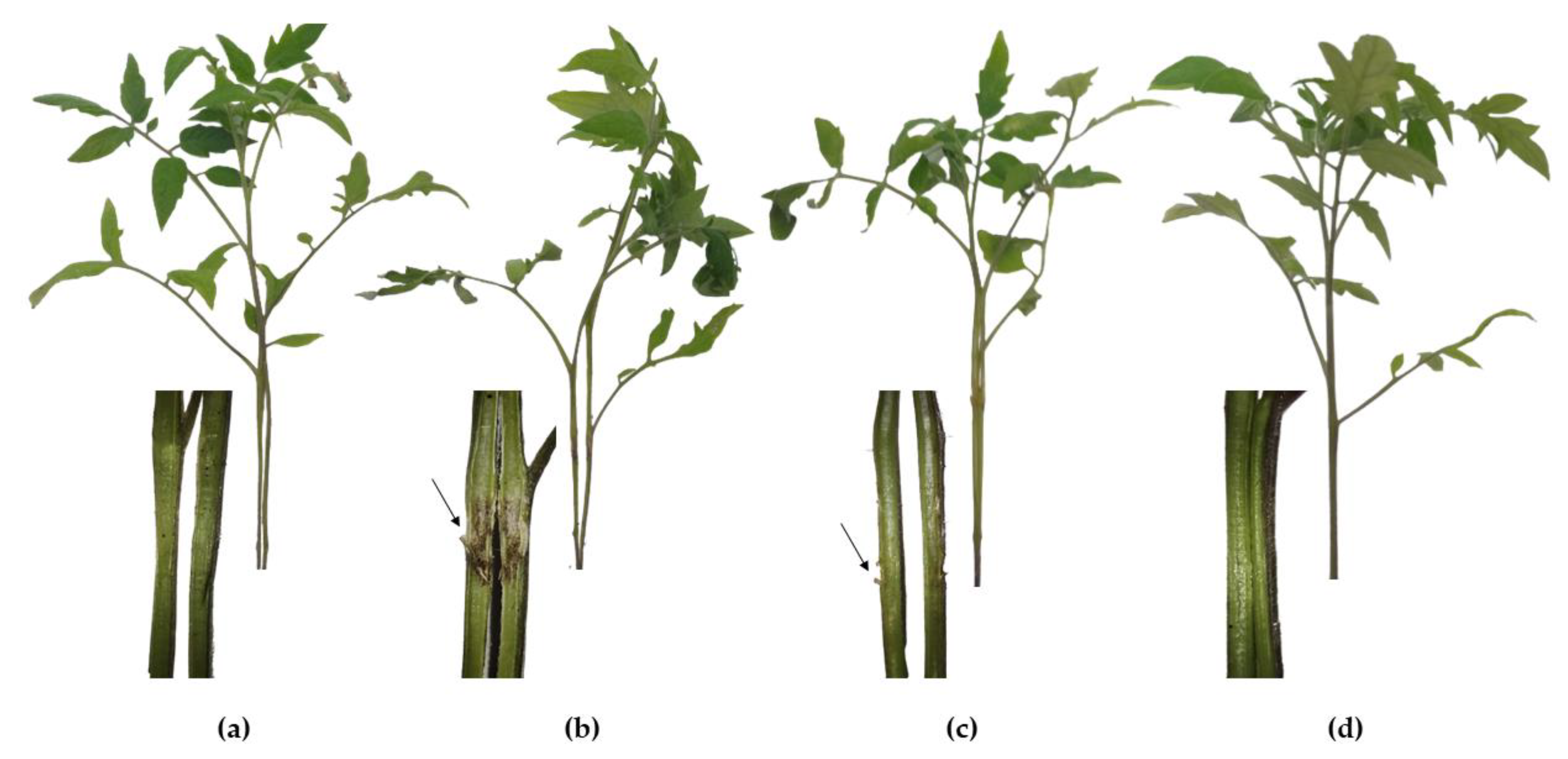
3.4. Tomato Plant Protection against C. michiganensis subsp. michiganensis

Given that G. biloba leaf extract was the most active in the in vitro tests, it was further tested as a protective treatment against bacterial canker in tomato plants cv. “Optima F1” (Figure 4). In the positive control (C. michiganensis subsp. michiganensis artificially inoculated on plants treated with bidistilled water only), all plants exhibited a typical vascular discoloration as brown streaks on the stem (Figure 4b). Plants treated with the extract at a concentration equal to the MIC (500 μg∙mL−1) showed an incidence of 80%, although the stem discoloration was less pronounced compared to the positive control. On the other hand, plants treated with the highest concentration (twice the MIC, i.e., 1000 μg∙mL−1) did not show symptoms of stem decay or phytotoxicity, appearing entirely similar to those of the negative control.

3.5. Pea Plant Protection against P. syringae pv. pisi

Figure 5 shows the results obtained for pea plants cv. “Lincoln” treated with different concentrations of G. biloba leaf extract, particularly MIC and MIC × 2 (i.e., 750 and 1500 μg∙mL−1, respectively). The positive control showed a high susceptibility to bacterial blight, with an incidence of 100% of plants showing the characteristic brown and papery leaflet lesions. The plants treated with the extract at 750 μg∙mL−1 exhibited a relatively lower susceptibility, with an incidence of 60% (i.e., 6 plants out of the 10 tested showed symptoms, albeit with less virulence than that of the positive control). In order to provide full protection, the dose had to be increased up to 1500 μg∙mL−1. As in the case of tomato plants, no phytotoxicity symptoms were observed, with no visual differences between the negative control and the MIC × 2 treated plants.

4. Discussion

4.1. On the Identified Phytochemicals and Their Antimicrobial Activity

The main phytochemicals identified in G. biloba hydromethanolic extracts (Figure 3) were: dihydro-4-hydroxy-2(3H)-furanone (present in both leaf and sclerotesta extracts); 2,4-dimethyl-3-hexanol; catechol (present in the three extracts); 1-hydroxy-2-propanone; 2,3-dihydro-3,5-dihydroxy-6-methyl-4H-pyran-4-one; acetic acid (identified both in leaf and sarcotesta extracts); formic acid 2-methylpropyl ester; 1-hepten-4-ol; 3-O-methyl-D-fructose (present in both leaf and sclerotesta extracts); 4,6-di-O-methyl-α-D-galactose; 2-O-methyl-α-D-xylofuranoside; and 3-methylmannoside (present in both leaf and sclerotesta extracts). Their occurrence in other extracts, together with available information on their antimicrobial activity, is summarized below.
Dihydro-4-hydroxy-2(3H)-furanone was previously identified in Equisetum arvense L. and Urtica dioica L. extracts [25]. The presence of this phytochemical in G. biloba hydromethanolic extracts may be the result of the hydrolysis/breaking of ginkgolide and bilobalide components (Figure 2) [26]. Dihydro-4-hydroxy-2(3H)-furanone is a major constituent in the extract of Crocus sativus L. (22% content), which exhibited antifungal activity against Pyricularia oryzae Cavara, Cryptococcus neoformans (Sanfelice) Vuill., Trichophyton rubrum (Castell.) Sabour., and Aspergillus fumigatus Fresen. [27]. 2,4-dimethyl-3-hexanol, identified in leaf extracts of Silene uniflora Roth and Pentatropis microphylla (Roxb.) Wight & Arn. [28], has been shown to possess considerable biocontrol activity against Aspergillus flavus Link and the generation of aflatoxin in stored rice grains [29,30]. In the activity assays presented here (Table 2), 2,4-dimethyl-3-hexanol yielded MIC values in the 250 to 750 μg·mL−1 range.
The presence of catechol should be attributed to the rupture of catechin (a constituent identified by Lin et al. [6] in the G. biloba leaves aqueous methanol extract) (Figure 2). Catechol had previously been identified in the extracts of Taxus baccata L. [31], Allium sativum L. [32], Sambucus nigra L. [33], and Quercus ilex L. [34], with concentrations ranging from 1.6 to 6.4% (compared to 3.3−7.9% in the extracts reported herein). Catechol oxidizes to melanoid pigments that are derivatives of benzoquinone, which is reported to have antimicrobial properties, slowing the decay of damaged fruits and other parts of plants. Catechol has been found to have antifungal activity against phytopathogenic taxa such as Penicillium italicum Wehmer and Fusarium oxysporum Schlecht. [35]. In our assays, catechol showed MIC values ranging from 187.5 to 500 μg·mL−1 (Table 2).
1-hydroxy-2-propanone is an alpha-hydroxy ketone found, for instance, in T. baccata leaf aqueous ammonia extracts [31]. Hydroxyacetone has also been detected, but not quantified, in several different edible plants, such as black cabbages, pulses, mentha, onion family vegetables, and rape. Additionally, it was identified (alongside acetic acid) as a major constituent of bioactive metabolites from tomato endophytic fungi, which had antibacterial activity against X. vesicatoria [36]. Furthermore, asymmetric reduction of hydroxyacetone leads to (R)-1,2-propanediol, a building block for antibacterial agents [37], and methylglyoxal—produced non-enzymatically from dihydroxyacetone—exhibits high non-peroxide antibacterial activity [38].
2,3-dihydro-3,5-dihydroxy-6-methyl-4H-pyran-4-one was identified in Hibiscus syriacus L. [39], S. nigra [33], Punica granatum L. [40], and T. baccata [31]. In terms of antifungal activity, it is effective against Phytophthora megasperma Drechsler, Verticillium dahliae Kleb., and Diaporthe amygdali (Delacr.) Udayanga, Crous & K.D. Hyde, with MIC values between 375 and 750 μg mL−1 [33].
The presence of acetic acid in contents higher than those detected herein has been reported, for instance, in Artocarpus altilis (Parkinson) Fosberg 1941 methanolic extracts (34.7%) [41]. In the particular case of G. biloba, its presence has been documented in ginkgo vinegar (obtained from the ginkgo seed coat by fermentation) [42]. Research has proven that it has good antibacterial activity against Pseudomonas aeruginosa (J.Schröter) Migula [43] at 1660 μg·mL−1. Concerning formic acid 2-methylpropyl ester (or isobutyl formate), it belongs to a family of esters which provide a sweet, floral, and fruity odor and flavor to foods and beverages, without any known antimicrobial properties.
1-hepten-4-ol was detected in leaf and bark extracts of Moringa concanensis Nimmo [44] and is a volatile constituent of Litchi chinensis Sonn. [45] and Aspalathus linearis (Burm.f.) R.Dahlgren [46]. It was tested against two barley fungal pathogens, Fusarium culmorum (Wm.G.Sm.) Sacc. and Cochliobolus sativus (S.Ito & Kurib.) Drechsler ex Dastur, but demonstrated low activity [47].
3-O-methyl-D-fructose has been found to be present in Ichnocarpus frutescens (L.) W.T.Aiton [48] and in T. baccata [31], and reports have indicated its antibacterial activity [49]; 4,6-di-methyl-α-D-galactose has been found in the Siberian ginseng Eleutherococcus senticosus (Rupr. & Maxim.) Maxim. [50]; and 2-O-methyl-α-D-xylofuranoside has been reported in Alternanthera sessilis (L.) DC. [51] and T. baccata [31]. Additionally, 3-methylmannoside has been found in Psidium guajava L. [52]. None of these polysaccharides have been identified as bactericides. Nonanedioic acid (azelaic acid), however, has been shown to have an inhibitory effect on common dermatophytes, such as Scopulariopsis brevicaulis (Sacc.) Brainier, Candida glabrata (H.W.Anderson) S.A.Mey. & Yarrow, Candida albicans (C.P.Robin) Berkhout, and Pityrosporum ovale (Bizz.) Castell. & Chalm. [53].

4.2. On the Antimicrobial Activity of G. biloba Extracts

In agricultural systems, a study on the antifungal action of G. biloba outer seedcoat extracts against rice sheath blight (which is caused by Rhizoctonia solani J.G. Kuhn) reported significant inhibition at 500 μg·mL−1 [11]. Regarding antibacterial activity, crude G. biloba extracts were tested by Ražná et al. [12] against human and veterinary pathogens, including Gram-positive bacteria such as B. thuringiensis, C. perfringens, H. influenzae, L. monocytogenes, and S. aureus, and Gram-negative bacteria such as Klebsiella pneumoniae (Schroeter, 1886) Trevisan, 1887, Escherichia coli (Migula, 1895) Castellani and Chalmers, 1919, Salmonella enterica (ex Kauffmann and Edwards, 1952) Le Minor and Popoff, 1987, Shigella sonnei (Levine, 1920) Weldin, 1927, and Yersinia enterocolitica (Schleifstein and Coleman, 1939) Frederiksen, 1964. Among these species, S. aureus, E. coli, K. pneumoniae, and Y. enterocolitica were the most susceptible, with the best results being obtained for S. aureus (MIC50 = 64.2 and MIC90 = 72.2 μg·mL−1) and Y. enterocolitica (MIC50 = 85.3 and MIC90 = 99.6 μg·mL−1). These outcomes were consistent with the ones reported herein, in which greater activity was observed against Gram-positive C. michiganensis subsp. michiganensis bacteria (MIC = 500 μg·mL−1) than against Gram-negative P. syringae, P. cichorii, and X. vesicatoria bacteria (whose MIC values ranged from 750 to 1000 μg·mL−1). However, the MIC values for our extracts were higher (i.e., their activity was lower) than those reported by Ražná et al. [12].

4.3. Comparison with Conventional Antibiotics

Upon comparison of the activity of the extract with those of gentamicin, tetracycline, and ciprofloxacin—representative of aminoglycosides, tetracyclines, and quinolones classes, respectively, that are commonly used in plant production in some countries outside of the European Union, as well as to treat human and animal diseases [54]—the efficacy of the conventional antibiotics was orders of magnitude higher (with MIC values in the 0.047 to 24 μg∙mL−1 range, depending on the pathogen). Concerning the other two antibiotics tested, viz. penicillin G and ampicillin, both were very effective against the Gram-positive C. michiganensis subsp. michiganensis; however, resistance was observed for all the Gram-negative bacteria, given that some strains of P. syringae produce β-lactamases [55] (e.g., some reports have indicated that 58% of P. syringae pv. syringae strains are resistant to ampicillin [56]) and X. vesicatoria does too [57]. It is noteworthy that the utilization of conventional antibiotics in agriculture (including tetracyclines, aminoglycosides, β-lactams, lincosamides, macrolides, pleuromutilins, and sulphonamides) may involve risks related to the emergence of resistance. Consequently, while the effectiveness of the natural extracts is not as high, they might be seen as a promising alternative to meet the requirements of the Global Action Plan on Antimicrobial Resistance set out by the World Health Organization (WHO).

5. Conclusions

Bacteriostatic activity was only detected for the G. biloba leaf extract (with MIC values ranging from 500 to 1000 μg mL−1 depending on the bacterial strain), in which 3-O-methyl-D-fructose and 2,4-dimethyl-3-hexanol were the main constituents. In vivo tests on tomato and pea plants artificially inoculated with C. michiganensis subsp. michiganensis and P. syringae pv. pisi, respectively, demonstrated the potential of G. biloba leaf extract as a protective treatment, although higher concentrations than the in vitro-determined MICs were required (1000 and 1500 μg·mL−1, respectively). The reported findings suggest an alternative valorization pathway for G. biloba leaves as biorationals for crop protection in addition to their use as dietary supplements.

Supplementary Materials

The following supporting information can be downloaded at: https://www.mdpi.com/article/10.3390/horticulturae9040461/s1, Figure S1. GC-MS chromatogram of G. biloba leaf hydromethanolic extract; Figure S2. GC-MS chromatogram of G. biloba sarcotesta hydromethanolic extract; Figure S3. GC-MS chromatogram of G. biloba sclerotesta hydromethanolic extract; Table S1. Constituents of G. biloba leaf hydromethanolic extract; Table S2. Constituents of G. biloba sarcotesta hydromethanolic extract; Table S3. Constituents of G. biloba sclerotesta hydromethanolic extract.

Author Contributions

Conceptualization, V.G.-G. and A.P.-B.; methodology, B.L.-V. and A.P.-B.; validation, A.P.-B.; formal analysis, E.S.-H., V.G.-G. and P.M.-R.; investigation, E.S.-H., V.G.-G., A.P.-B., B.L.-V., L.B.-D., J.M.-G. and P.M.-R.; resources, J.M.-G. and A.P.-B.; writing—original draft preparation, E.S.-H., V.G.-G., A.P.-B., B.L.-V., L.B.-D., J.M.-G. and P.M.-R.; writing—review and editing, E.S.-H., V.G.-G. and P.M.-R.; visualization, E.S.-H.; supervision, P.M.-R. All authors have read and agreed to the published version of the manuscript.

Funding

This research was funded by the European Union through the Horizon Program (HORIZON-CL6-2022-FARM2FORK-01) under project ‘Agro-ecological strategies for resilient farming in West Africa (CIRAWA)’, with project ID 101084398.

Data Availability Statement

The data presented in this study are available on request from the corresponding author.

Acknowledgments

The authors thank Pablo Candela and Pilar Blasco from the Technical Research Services of the University of Alicante for conducting the GC–MS analyses.

Conflicts of Interest

The authors declare no conflict of interest. The funders had no role in the design of the study; in the collection, analyses, or interpretation of data; in the writing of the manuscript; or in the decision to publish the results.

References

  1. Singh, B.; Kaur, P.; Gopichand; Singh, R.D.; Ahuja, P.S. Biology and chemistry of Ginkgo biloba. Fitoterapia 2008, 79, 401–418. [Google Scholar] [CrossRef]
  2. Luo, Y.; Smith, J.V. Studies on molecular mechanisms of Ginkgo biloba extract. Appl. Microbiol. Biotechnol. 2004, 64, 465–472. [Google Scholar] [CrossRef] [PubMed]
  3. Chan, P.-C.; Xia, Q.; Fu, P.P. Ginkgo biloba leave extract: Biological, medicinal, and toxicological effects. J. Environ. Sci. Health C 2007, 25, 211–244. [Google Scholar] [CrossRef] [PubMed]
  4. Mahadevan, S.; Park, Y.; Park, Y. Modulation of cholesterol metabolism by Ginkgo biloba L. nuts and their extract. Food Res. Int. 2008, 41, 89–95. [Google Scholar] [CrossRef]
  5. Gülz, P.G.; Müller, E.; Schmitz, K.; Marner, F.J.; Güth, S. Chemical composition and surface structures of epicuticular leaf waxes of Ginkgo biloba, Magnolia grandiflora and Liriodendron tulipifera. Z. Nat. C 1992, 47, 516–526. [Google Scholar] [CrossRef]
  6. Lin, L.-Z.; Chen, P.; Ozcan, M.; Harnly, J.M. Chromatographic profiles and identification of new phenolic components of Ginkgo biloba leaves and selected products. J. Agric. Food. Chem. 2008, 56, 6671–6679. [Google Scholar] [CrossRef] [Green Version]
  7. Drieu, K. Preparation and definition of Ginkgo biloba extract. In Rökan: Ginkgo biloba Recent Results in Pharmacology and Clinic; Fünfgeld, E.W., Ed.; Springer: Berlin/Heidelberg, Germany, 1988; pp. 32–36. [Google Scholar] [CrossRef]
  8. DeFeudis, F.V. Ginkgo biloba Extract (EGb 761): From Chemistry to the Clinic; Ullstein Medical: Wiesbaden, Germany, 1998; Volume 25, p. 401. [Google Scholar]
  9. Maitra, I.; Marcocci, L.; Droy-Lefaix, M.T.; Packer, L. Peroxyl radical scavenging activity of Ginkgo biloba extract EGb 761. Biochem. Pharmacol. 1995, 49, 1649–1655. [Google Scholar] [CrossRef]
  10. Krauze-Baranowska, M.; Wiwart, M. Antifungal activity of biflavones from Taxus baccata and Ginkgo biloba. Z. Nat. C 2003, 58, 65–69. [Google Scholar] [CrossRef]
  11. Oh, T.-S.; Koo, H.-M.; Yoon, H.-R.; Jeong, N.-S.; Kim, Y.-J.; Kim, C.-H. Antifungal action of Ginkgo biloba outer seedcoat on rice sheath blight. Plant Pathol. J. 2015, 31, 61–66. [Google Scholar] [CrossRef] [PubMed] [Green Version]
  12. Ražná, K.; Sawinska, Z.; Ivanišová, E.; Vukovic, N.; Terentjeva, M.; Stričík, M.; Kowalczewski, P.Ł.; Hlavačková, L.; Rovná, K.; Žiarovská, J.; et al. Properties of Ginkgo biloba L.: Antioxidant characterization, antimicrobial activities, and genomic MicroRNA based marker fingerprints. Int. J. Mol. Sci. 2020, 21, 3087. [Google Scholar] [CrossRef]
  13. Martins, P.M.M.; Merfa, M.V.; Takita, M.A.; De Souza, A.A. Persistence in phytopathogenic bacteria: Do we know enough? Front. Microbiol. 2018, 9, 1099. [Google Scholar] [CrossRef] [PubMed]
  14. Xin, X.-F.; Kvitko, B.; He, S.Y. Pseudomonas syringae: What it takes to be a pathogen. Nat. Rev. Microbiol. 2018, 16, 316–328. [Google Scholar] [CrossRef]
  15. Hikichi, Y.; Wali, U.M.; Ohnishi, K.; Kiba, A. Mechanism of disease development caused by a multihost plant bacterium, Pseudomonas cichorii, and its virulence diversity. J. Gen. Plant Pathol. 2013, 79, 379–389. [Google Scholar] [CrossRef]
  16. Nakayinga, R.; Makumi, A.; Tumuhaise, V.; Tinzaara, W. Xanthomonas bacteriophages: A review of their biology and biocontrol applications in agriculture. BMC Microbiol. 2021, 21, 291. [Google Scholar] [CrossRef] [PubMed]
  17. Peritore-Galve, F.C.; Tancos, M.A.; Smart, C.D. Bacterial canker of tomato: Revisiting a global and economically damaging seedborne pathogen. Plant Dis. 2021, 105, 1581–1595. [Google Scholar] [CrossRef]
  18. Mansfield, J.; Genin, S.; Magori, S.; Citovsky, V.; Sriariyanum, M.; Ronald, P.; Dow, M.A.X.; Verdier, V.; Beer, S.V.; Machado, M.A.; et al. Top 10 plant pathogenic bacteria in molecular plant pathology. Mol. Plant Pathol 2012, 13, 614–629. [Google Scholar] [CrossRef] [Green Version]
  19. Boateng, I.D.; Yang, X.M. Effect of different drying methods on product quality, bioactive and toxic components of Ginkgo biloba L. seed. J. Sci. Food Agric. 2021, 101, 3290–3297. [Google Scholar] [CrossRef] [PubMed]
  20. Sánchez-Hernández, E.; Martín-Ramos, P.; Martín-Gil, J.; Santiago-Aliste, A.; Hernández-Navarro, S.; Oliveira, R.; González-García, V. Bark extract of Uncaria tomentosa L. for the control of strawberry phytopathogens. Horticulturae 2022, 8, 672. [Google Scholar] [CrossRef]
  21. CLSI M07; Methods for Dilution Antimicrobial Susceptibility Tests for Bacteria that Grow Aerobically (11th ed.). Clinical and Laboratory Standards Institute: Wayne, PA, USA, 2018.
  22. de León, L.; Siverio, F.; López, M.M.; Rodríguez, A. Comparative efficiency of chemical compounds for in vitro and in vivo activity against Clavibacter michiganensis subsp. michiganensis, the causal agent of tomato bacterial canker. Crop Prot. 2008, 27, 1277–1283. [Google Scholar] [CrossRef]
  23. Martin Sanz, A. Bacteriosis en Guisante (Pisum sativum L.): Situación en Castilla y León, Caracterización de los Patógenos Implicados y Búsqueda de Fuentes de Resistencia. Ph.D. Thesis, Universidad de León, León, Spain, 2008. [Google Scholar]
  24. Maréchal, Y.; Chanzy, H. The hydrogen bond network in I β cellulose as observed by infrared spectrometry. J. Mol. Struct. 2000, 523, 183–196. [Google Scholar] [CrossRef]
  25. Langa-Lomba, N.; Buzón-Durán, L.; Martín-Ramos, P.; Casanova-Gascón, J.; Martín-Gil, J.; Sánchez-Hernández, E.; González-García, V. Assessment of conjugate complexes of chitosan and Urtica dioica or Equisetum arvense extracts for the control of grapevine trunk pathogens. Agronomy 2021, 11, 976. [Google Scholar] [CrossRef]
  26. Lan, K.; Li, X.-J.; Du, G.; Xu, L. Characterizations of the hydrolyzed products of ginkgolide A and ginkgolide B by liquid chromatography coupled with mass spectrometry. J. Pharm. Biomed. Anal. 2016, 118, 113–122. [Google Scholar] [CrossRef] [PubMed]
  27. Zheng, C.-J.; Li, L.; Ma, W.-H.; Han, T.; Qin, L.-P. Chemical constituents and bioactivities of the liposoluble fraction from different medicinal parts of Crocus sativus. Pharm. Biol. 2011, 49, 756–763. [Google Scholar] [CrossRef] [PubMed] [Green Version]
  28. Kalaivanan, C.; Sundhararajan, A.; Kokila, S.; Ahmed, J.S. GC-MS analysis of Pentatropis Microphylla. Int. J. Eng. Res. Technol. 2017, 5, 1–2. [Google Scholar]
  29. Mannaa, M.; Oh, J.Y.; Kim, K.D. Microbe-mediated control of Aspergillus flavusin stored rice grains with a focus on aflatoxin inhibition and biodegradation. Ann. Appl. Biol. 2017, 171, 376–392. [Google Scholar] [CrossRef]
  30. Mannaa, M.; Oh, J.Y.; Kim, K.D. Biocontrol activity of volatile-producing Bacillus megaterium and Pseudomonas protegens against Aspergillus flavus and aflatoxin production on stored rice grains. Mycobiology 2018, 45, 213–219. [Google Scholar] [CrossRef] [PubMed] [Green Version]
  31. Sánchez-Hernández, E.; González-García, V.; Martín-Gil, J.; Lorenzo-Vidal, B.; Palacio-Bielsa, A.; Martín-Ramos, P. Phytochemical screening and antibacterial activity of Taxus baccata L. against Pectobacterium spp. and Dickeya chrysanthemi. Horticulturae 2023, 9, 201. [Google Scholar] [CrossRef]
  32. Teixeira, A.; Sánchez-Hernández, E.; Noversa, J.; Cunha, A.; Cortez, I.; Marques, G.; Martín-Ramos, P.; Oliveira, R. Antifungal activity of plant waste extracts against phytopathogenic fungi: Allium sativum peels extract as a promising product targeting the fungal plasma membrane and cell wall. Horticulturae 2023, 9, 136. [Google Scholar] [CrossRef]
  33. Sánchez-Hernández, E.; Balduque-Gil, J.; González-García, V.; Barriuso-Vargas, J.J.; Casanova-Gascón, J.; Martín-Gil, J.; Martín-Ramos, P. Phytochemical profiling of Sambucus nigra L. flower and leaf extracts and their antimicrobial potential against almond tree pathogens. Int. J. Mol. Sci. 2023, 24, 1154. [Google Scholar] [CrossRef]
  34. Sánchez-Hernández, E.; Balduque-Gil, J.; Barriuso-Vargas, J.J.; Casanova-Gascón, J.; González-García, V.; Cuchí-Oterino, J.A.; Lorenzo-Vidal, B.; Martín-Gil, J.; Martín-Ramos, P. Holm oak (Quercus ilex subsp. ballota (Desf.) Samp.) bark aqueous ammonia extract for the control of invasive forest pathogens. Int. J. Mol. Sci. 2022, 23, 11882. [Google Scholar] [CrossRef]
  35. Kocaçalışkan, I.; Talan, I.; Terzi, I. Antimicrobial activity of catechol and pyrogallol as allelochemicals. Z. Nat. C 2006, 61, 639–642. [Google Scholar] [CrossRef] [PubMed]
  36. Rashid, T.S. Bioactive metabolites from tomato endophytic fungi with antibacterial activity against tomato bacterial spot disease. Rhizosphere 2021, 17, 100292. [Google Scholar] [CrossRef]
  37. Oda, T.; Oda, K.; Yamamoto, H.; Matsuyama, A.; Ishii, M.; Igarashi, Y.; Nishihara, H. Hydrogen-driven asymmetric reduction of hydroxyacetone to (R)-1,2-propanediol by Ralstonia eutropha transformant expressing alcohol dehydrogenase from Kluyveromyces lactis. Microb. Cell Fact. 2013, 12, 2. [Google Scholar] [CrossRef] [PubMed] [Green Version]
  38. Gan, S.H.; Cokcetin, N.N.; Pappalardo, M.; Campbell, L.T.; Brooks, P.; Carter, D.A.; Blair, S.E.; Harry, E.J. The antibacterial activity of Australian Leptospermum honey correlates with methylglyoxal levels. PLoS ONE 2016, 11, e0167780. [Google Scholar] [CrossRef] [Green Version]
  39. Sánchez-Hernández, E.; Buzón-Durán, L.; Lorenzo-Vidal, B.; Martín-Gil, J.; Martín-Ramos, P. Physicochemical characterization and antimicrobial activity against Erwinia amylovora, Erwinia vitivora, and Diplodia seriata of a light purple Hibiscus syriacus L. cultivar. Plants 2021, 10, 1876. [Google Scholar] [CrossRef]
  40. Sánchez-Hernández, E.; Buzón-Durán, L.; Cuchí-Oterino, J.A.; Martín-Gil, J.; Lorenzo-Vidal, B.; Martín-Ramos, P. Dwarf pomegranate (Punica granatum L. var. nana): Source of 5-HMF and bioactive compounds with applications in the protection of woody crops. Plants 2022, 11, 550. [Google Scholar] [CrossRef] [PubMed]
  41. Mainasara, M.M.; Abu Bakar, M.F.; Barau, A.I. GC-MS analysis of phytochemical constituents from ethyl acetate and methanol extract of Artocarpus altilis (Parkinson) Fosberg from Endau Rompin, Johor, Malaysia. Path Sci. 2019, 5, 3001–3010. [Google Scholar] [CrossRef]
  42. Hosoda, S.; Kawazoe, Y.; Shiba, T.; Numazawa, S.; Manabe, A. Anti-obesity effect of ginkgo vinegar, a fermented product of ginkgo seed coat, in mice fed a high-fat diet and 3T3-L1 preadipocyte cells. Nutrients 2020, 12, 230. [Google Scholar] [CrossRef] [Green Version]
  43. Fraise, A.P.; Wilkinson, M.A.C.; Bradley, C.R.; Oppenheim, B.; Moiemen, N. The antibacterial activity and stability of acetic acid. J. Hosp. Infect. 2013, 84, 329–331. [Google Scholar] [CrossRef]
  44. Balamurugan, V.; Balakrishnan, V.; Sundaresan, A. GC-MS analysis of leaf and bark extract of Moringa concanensis Nimmo, a Siddha medicinal plant of South India. Eur. J. Biotechnol. Biosci. 2015, 3, 57–61. [Google Scholar]
  45. Johnston, J.C.; Welch, R.C.; Hunter, G.L.K. Volatile constituents of litchi (Litchi chinesis Sonn.). J. Agric. Food. Chem. 2002, 28, 859–861. [Google Scholar] [CrossRef]
  46. Habu, T.; Flath, R.A.; Mon, T.R.; Morton, J.F. Volatile components of Rooibos tea (Aspalathus linearis). J. Agric. Food. Chem. 1985, 33, 249–254. [Google Scholar] [CrossRef]
  47. Kaddes, A.; Fauconnier, M.-L.; Sassi, K.; Nasraoui, B.; Jijakli, M.H. Antifungal properties of two volatile organic compounds on barley pathogens and introduction to their mechanism of action. Int. J. Environ. Res. Public Health 2019, 16, 2866. [Google Scholar] [CrossRef] [PubMed] [Green Version]
  48. Meher, A.; Behera, B.; Nanda, B.K. GC–MS investigation of phytocomponents present in ethanolic extract of plant Ichnocarpus frutescens (L.) W. T. Aiton aerial part. Int. J. Pharm. Sci. Res. 2020, 11, 104–113. [Google Scholar]
  49. Ghosh, G.; Panda, P.; Rath, M.; Pal, A.; Sharma, T.; Das, D. GC-MS analysis of bioactive compounds in the methanol extract of Clerodendrum viscosum leaves. Pharmacogn. Res. 2015, 7, 110. [Google Scholar] [CrossRef] [Green Version]
  50. Bai, Y.; Tohda, C.; Zhu, S.; Hattori, M.; Komatsu, K. Active components from Siberian ginseng (Eleutherococcus senticosus) for protection of amyloid β(25–35)-induced neuritic atrophy in cultured rat cortical neurons. J. Nat. Med. 2011, 65, 417–423. [Google Scholar] [CrossRef] [PubMed]
  51. Khan, M.; Yusufzai, S.; Kaun, L.; Shah, M.; Idris, R. Chemical composition and antioxidant activity of essential oil of leaves and flowers of Alternanthera sessilis red from Sabah. J. Appl. Pharm. Sci. 2016, 6, 157–161. [Google Scholar] [CrossRef] [Green Version]
  52. Erlina, L.; Paramita, R.I.; Kusuma, W.A.; Fadilah, F.; Tedjo, A.; Pratomo, I.P.; Ramadhanti, N.S.; Nasution, A.K.; Surado, F.K.; Fitriawan, A.; et al. Virtual screening of Indonesian herbal compounds as COVID-19 supportive therapy: Machine learning and pharmacophore modeling approaches. BMC Complement. Med. Ther. 2022, 22, 207. [Google Scholar] [CrossRef]
  53. Brasch, J.; Christophers, E. Azelaic acid has antimycotic properties in vitro. Dermatology 1993, 186, 55–58. [Google Scholar] [CrossRef]
  54. Miller, S.A.; Ferreira, J.P.; LeJeune, J.T. Antimicrobial use and resistance in plant agriculture: A One Health perspective. Agriculture 2022, 12, 289. [Google Scholar] [CrossRef]
  55. Coleman, R.H.; Shaffer, J.; True, H. Properties of β-lactamase from Pseudomonas syringae. Curr. Microbiol. 1996, 32, 147–150. [Google Scholar] [CrossRef] [PubMed]
  56. Hwang, M.S.H.; Morgan, R.L.; Sarkar, S.F.; Wang, P.W.; Guttman, D.S. Phylogenetic characterization of virulence and resistance phenotypes of Pseudomonas syringae. Appl. Environ. Microbiol. 2005, 71, 5182–5191. [Google Scholar] [CrossRef] [PubMed] [Green Version]
  57. Weng, S.-F.; Chen, C.-Y.; Lee, Y.-S.; Lin, J.-W.; Tseng, Y.-H. Identification of a novel β-lactamase produced by Xanthomonas campestris, a phytopathogenic bacterium. Antimicrob. Agents Chemother. 1999, 43, 1792–1797. [Google Scholar] [CrossRef] [PubMed] [Green Version]
Figure 1. (left) Ginkgo biloba tree bearing ripe, fruit-like sarcotestae; (center) close-up of leaves and sarcotestae; (right) fleshy seedcoat (sarcotesta) and seed (sclerotesta).
Horticulturae 09 00461 g001
Figure 2. Specific components of Ginkgo biloba leaf extracts reported by Lin et al. [6].
Horticulturae 09 00461 g002
Figure 3. Chemical structures of several of the main constituents of (a) G. biloba leaf; (b) sarcotesta; and (c) sclerotesta extracts.
Horticulturae 09 00461 g003
Figure 4. Symptoms of bacterial canker produced by C. michiganensis subsp. michiganensis in plants of tomato cv. “Optima F1” after 10 days of inoculation: (a) negative control; (b) positive control; (c) G. biloba leaf extract at 500 μg∙mL−1; and (d) G. biloba leaf extract at 1000 μg∙mL−1.
Horticulturae 09 00461 g004
Figure 5. Symptoms of bacterial blight produced by P. syringae pv. pisi in plants of pea cv. “Lincoln” after 10 days of inoculation: (a) negative control; (b) positive control; (c) G. biloba leaf extract at 750 μg∙mL−1; and (d) G. biloba leaf extract at 1500 μg∙mL−1.
Horticulturae 09 00461 g005
Table 1. Minimum inhibitory concentrations (expressed in μg∙mL−1) of G. biloba extracts against the horticultural pathogens.
BacteriaLeaf
Extract
Sarcotesta
Extract
Sclerotesta
Extract
Clavibacter michiganensis subsp. michiganensis500n.a.n.a.
Pseudomonas cichorii750n.a.n.a.
Pseudomonas syringae pv. pisi750n.a.n.a.
Pseudomonas syringae pv. syringae750n.a.n.a.
Pseudomonas syringae pv. tomato750n.a.n.a.
Xanthomonas vesicatoria1000n.a.n.a.
n.a.: no activity detected at the highest assayed dose.
Table 2. Minimum inhibitory concentrations (expressed in μg∙mL−1) of two G. biloba extract phytoconstituents against the horticultural pathogens.
Bacteria2,4-Dimethyl-3-HexanolCatechol
Clavibacter michiganensis subsp. michiganensis375187.5
Pseudomonas cichorii750500
Pseudomonas syringae pv. pisi750375
Pseudomonas syringae pv. syringae750500
Pseudomonas syringae pv. tomato500500
Xanthomonas vesicatoria250187.5
Table 3. Minimum inhibitory concentrations (expressed in μg∙mL−1) of conventional antibiotics (for clinical use) against the horticultural pathogens.
BacteriaPGAMGMCITC
Clavibacter michiganensis subsp. michiganensis0.1250.0940.380.380.19
Pseudomonas cichorii≥32≥2563624
Pseudomonas syringae pv. pisi≥324810.251
Pseudomonas syringae pv. syringae≥32480.250.0471
Pseudomonas syringae pv. tomato≥32160.1250.0470.75
Xanthomonas vesicatoria≥32≥2560.750.1251
PG: penicillin G; AM: ampicillin; GM: gentamicin; CI: ciprofloxacin; TC: tetracycline.
Disclaimer/Publisher’s Note: The statements, opinions and data contained in all publications are solely those of the individual author(s) and contributor(s) and not of MDPI and/or the editor(s). MDPI and/or the editor(s) disclaim responsibility for any injury to people or property resulting from any ideas, methods, instructions or products referred to in the content.

