
In Vitro Studies Regarding the Effect of Cellulose Acetate-Based Composite Coatings on the Functional Properties of the Biodegradable Mg3Nd Alloys
 , , , , , , ,
  and
and
Abstract
:
1. Introduction
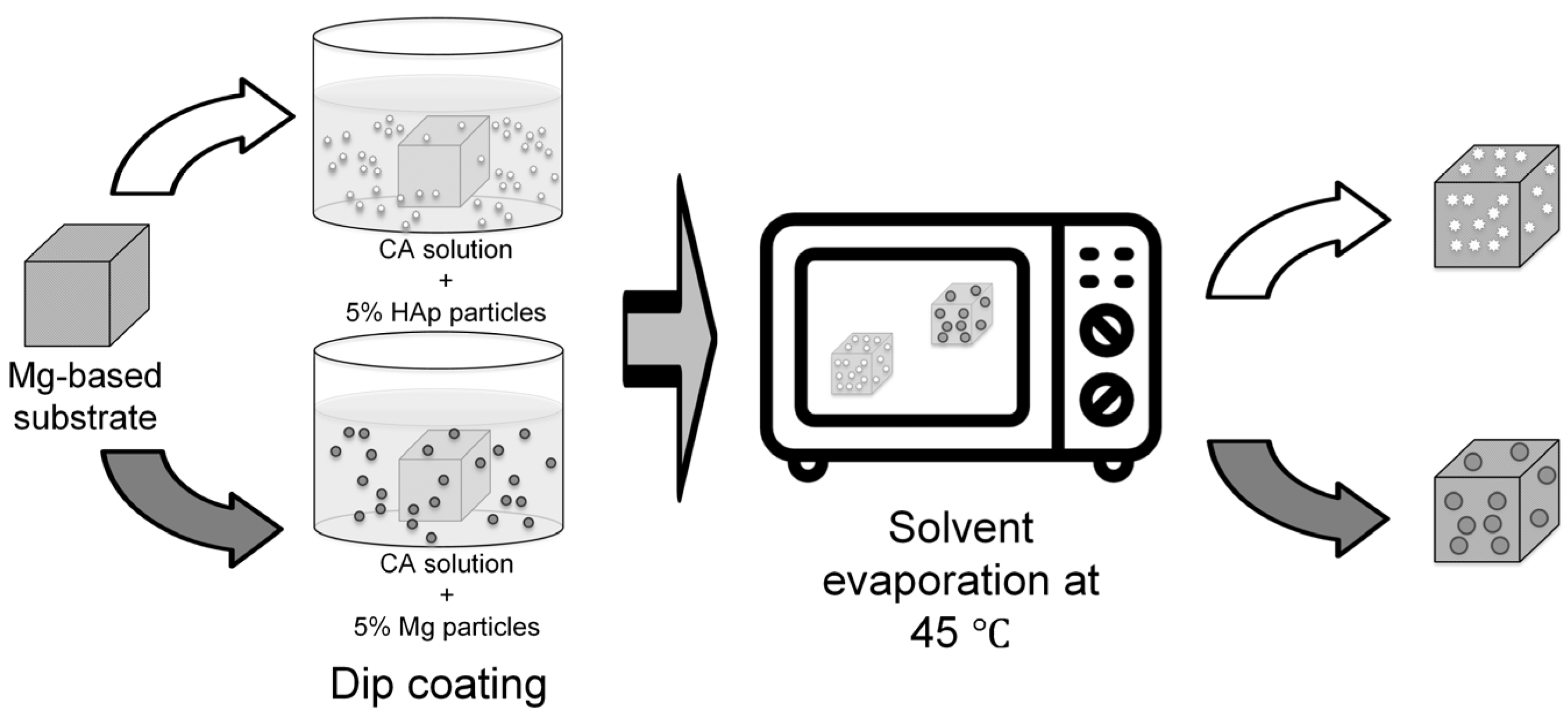
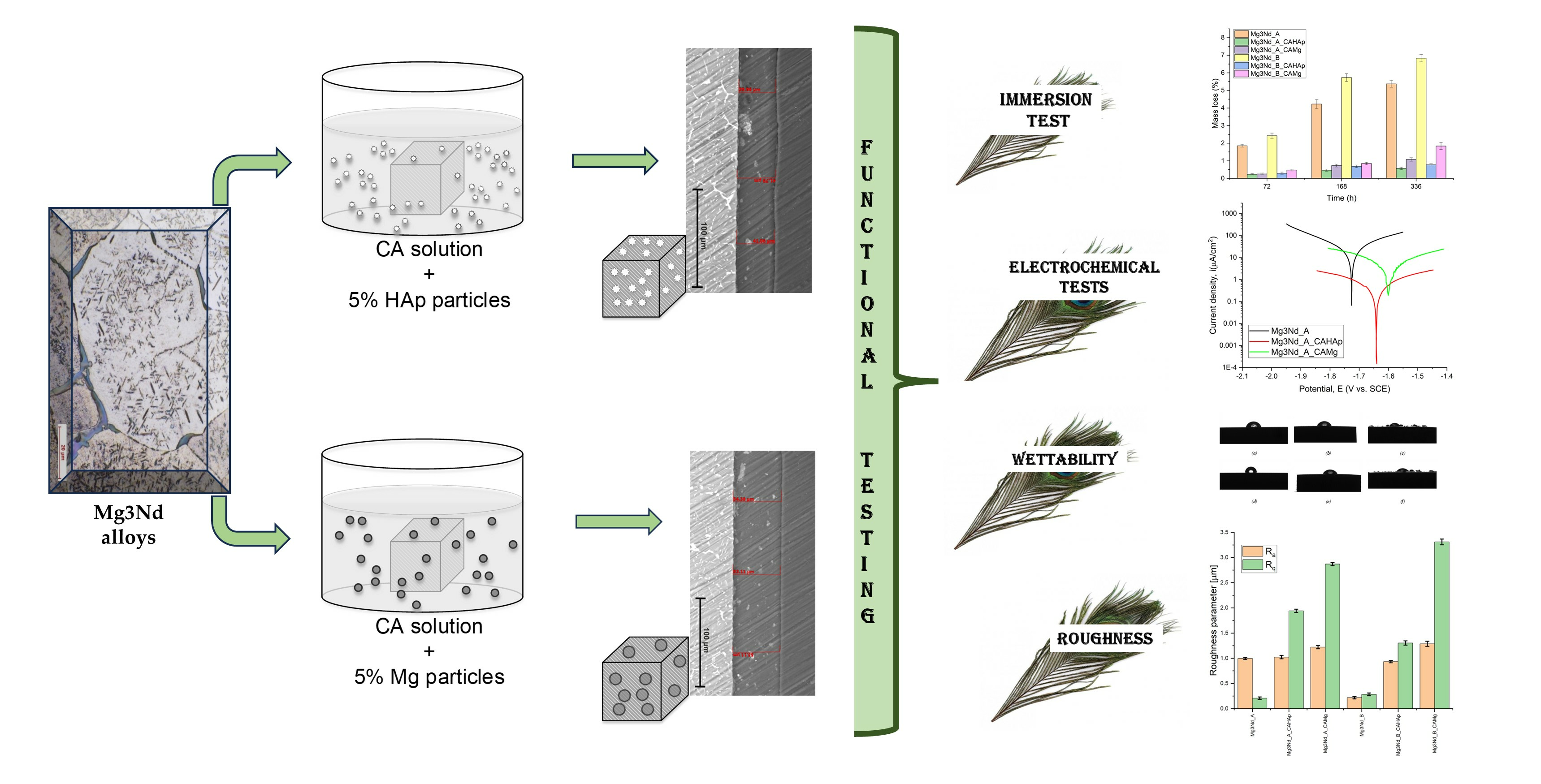
2. Materials and Methods
2.1. Materials Characterization
2.2. Corrosion Behavior of the Experimental Samples
2.2.1. Immersion Test
2.2.2. Electrochemical Tests
2.3. Surface Properties of the Experimental Samples
3. Results and Discussion
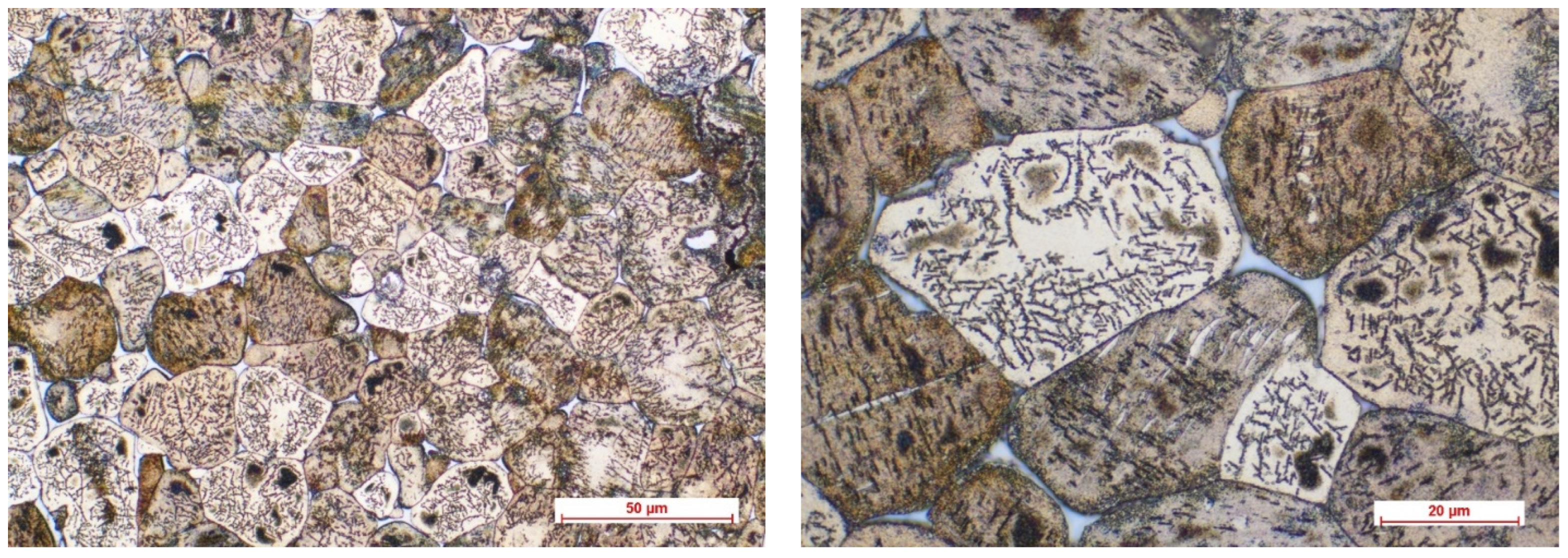
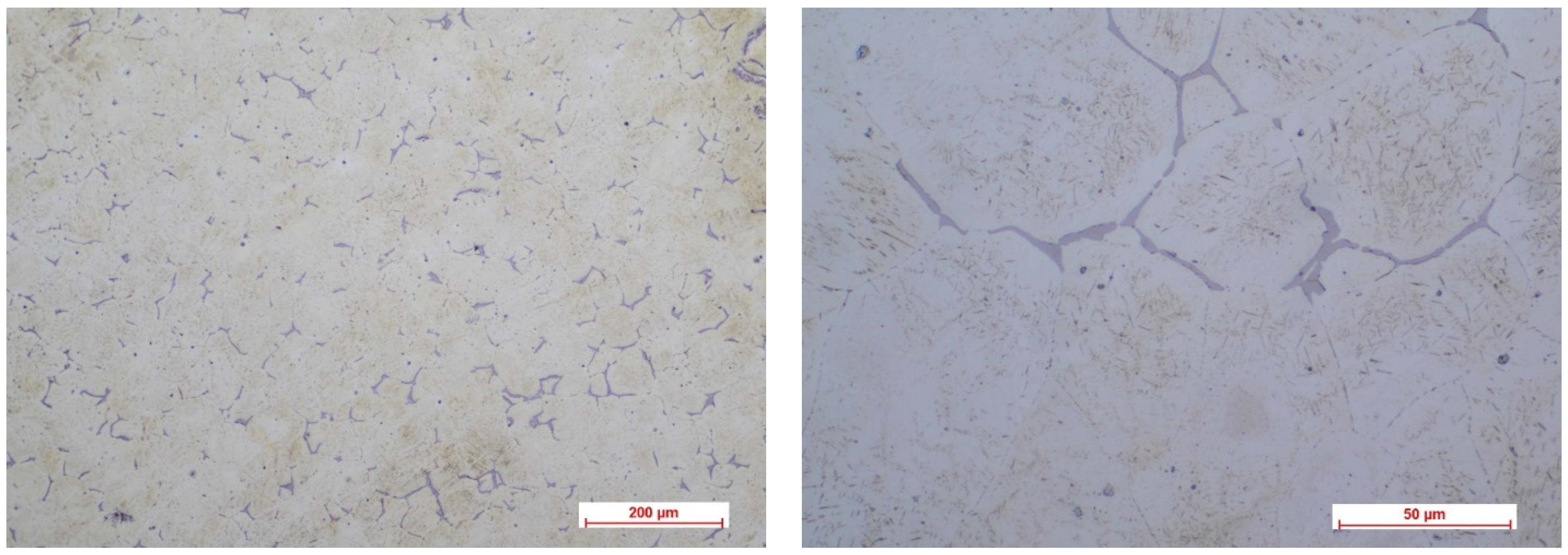
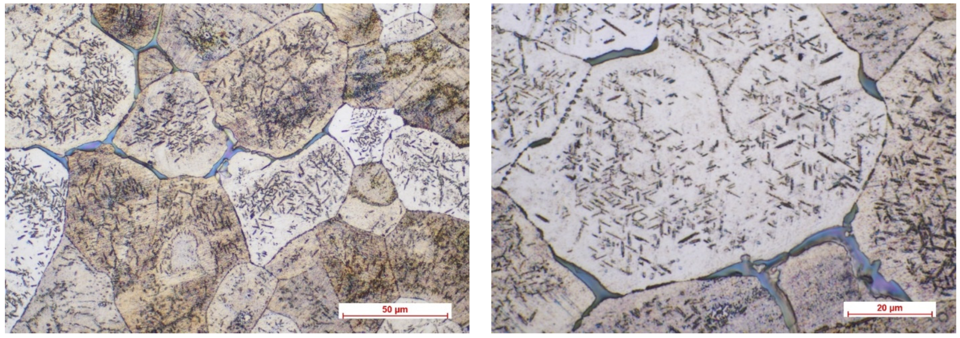
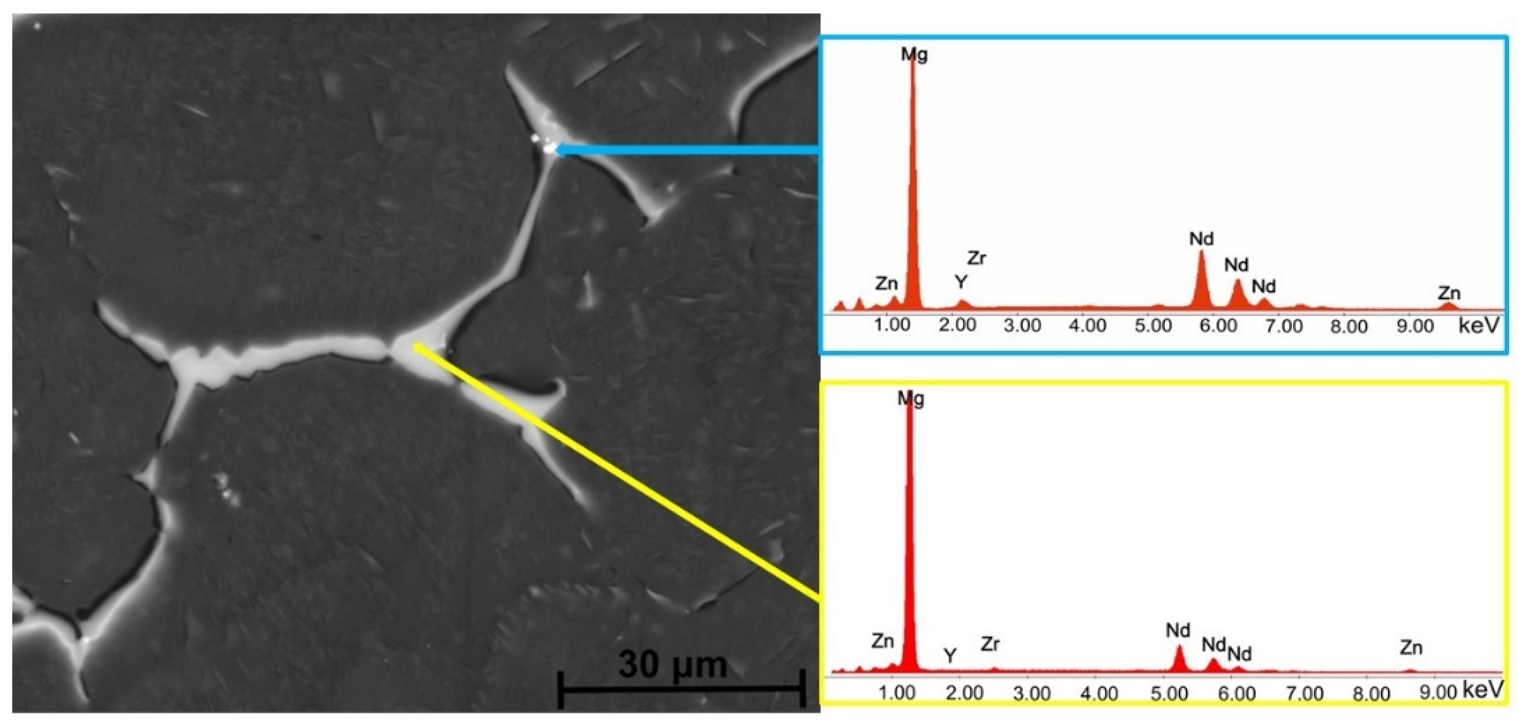
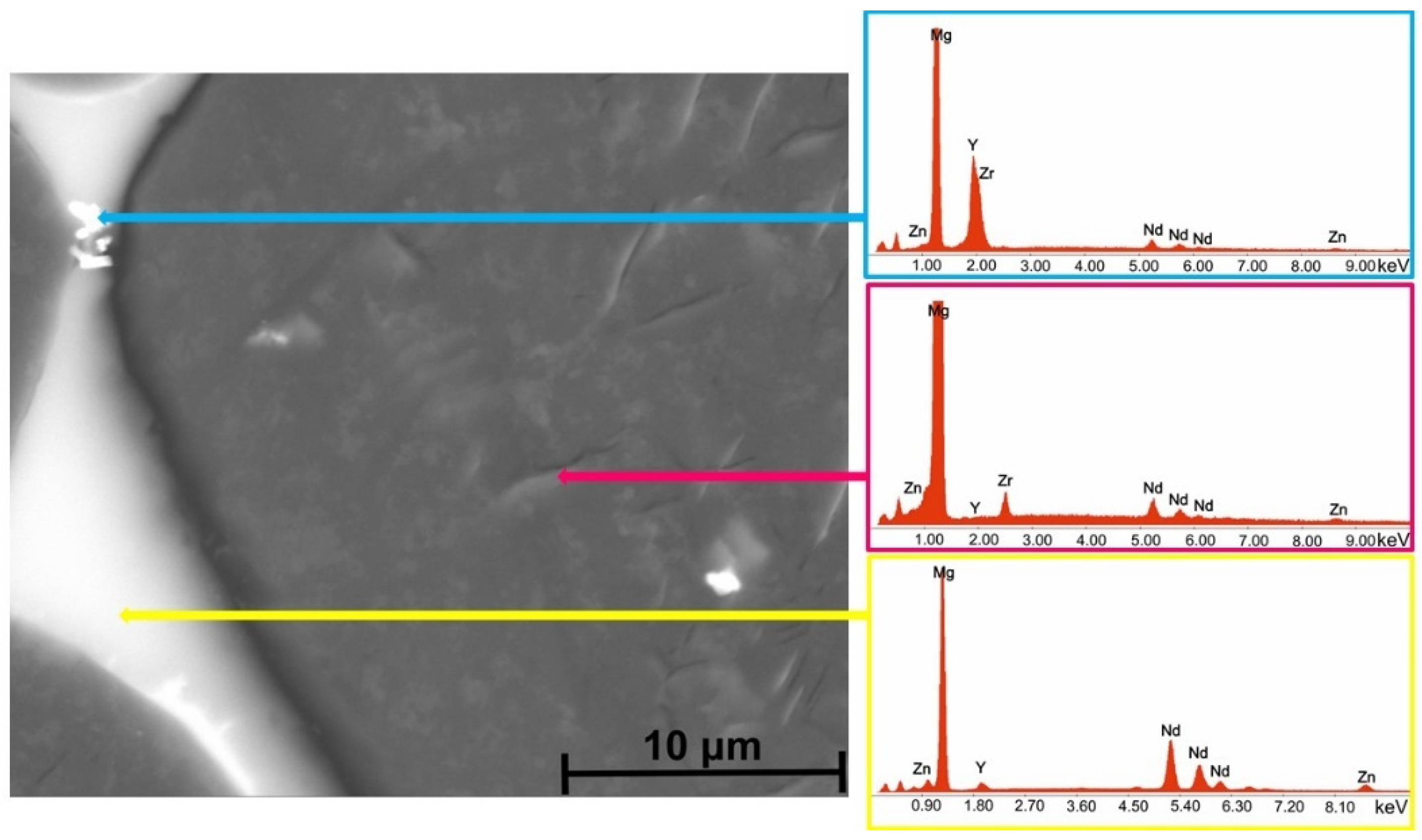
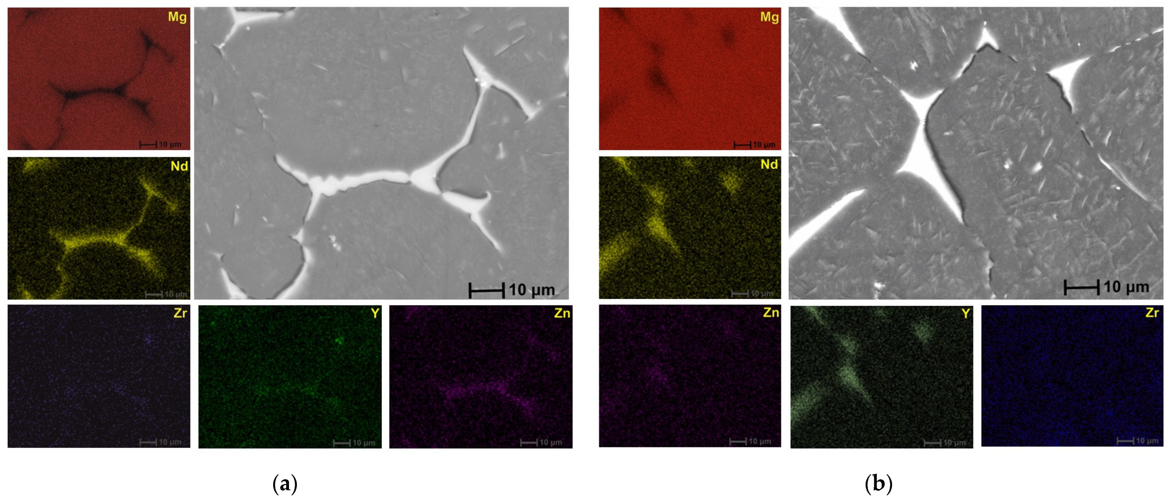
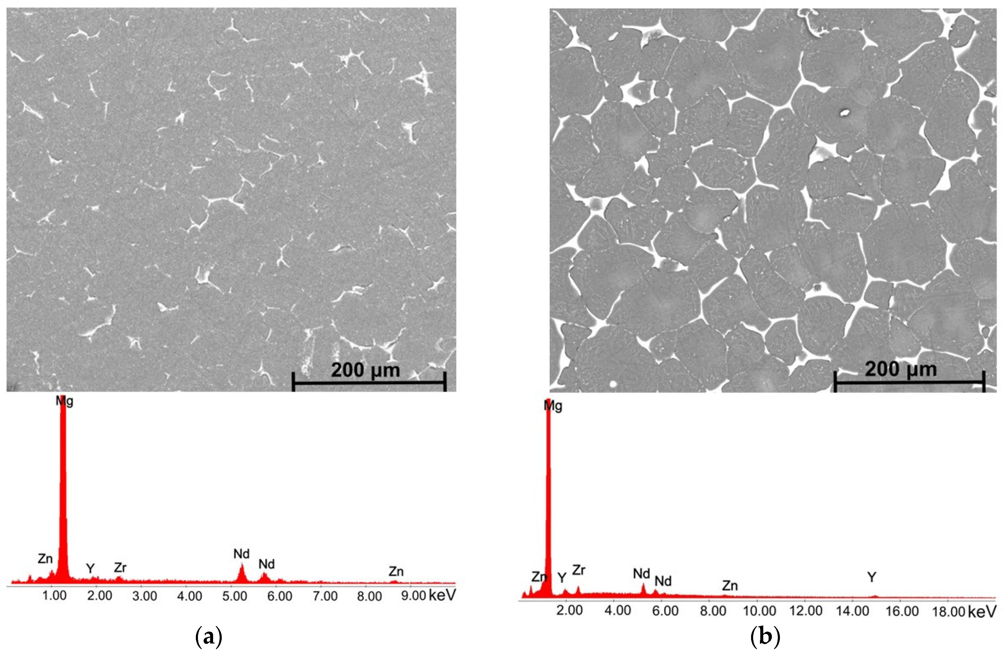
3.1. Microstructural Characterization of the Alloys
3.2. Corrosion Behavior of the Experimental Samples
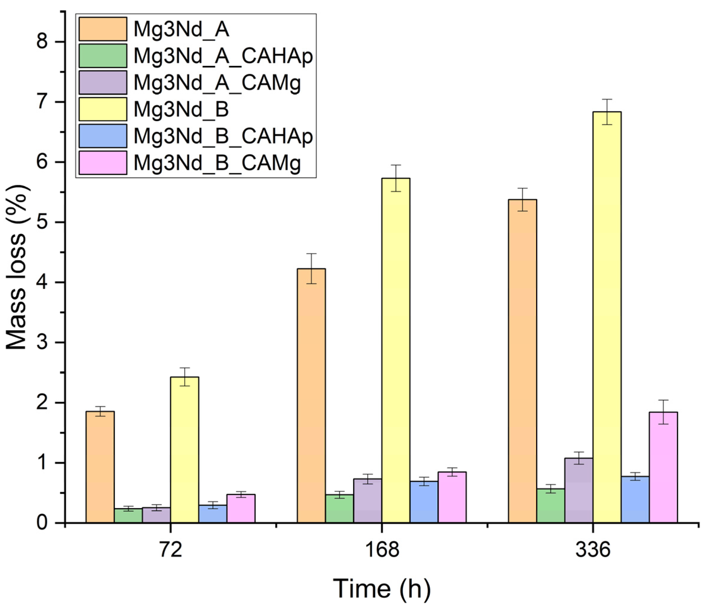
3.2.1. Immersion Test
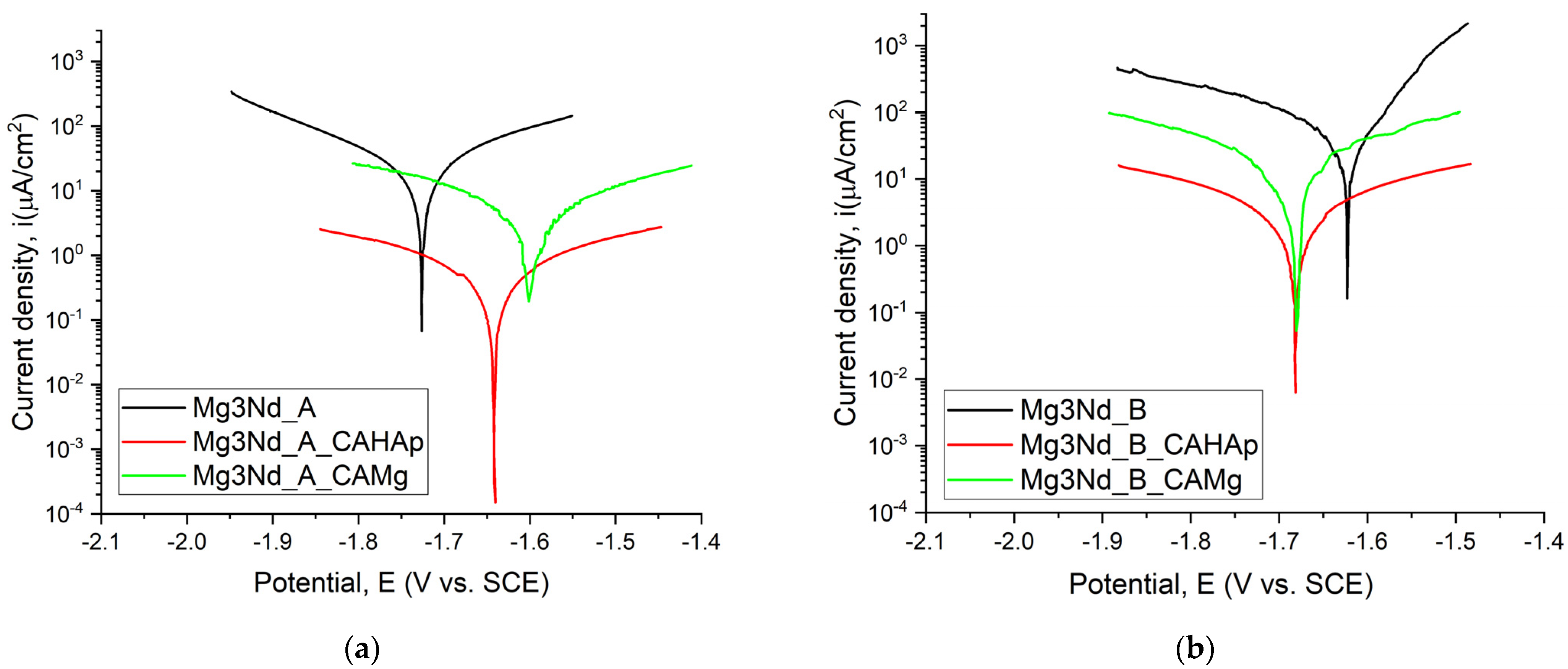
3.2.2. Electrochemical Tests
3.3. Surface Properties of the Experimental Samples
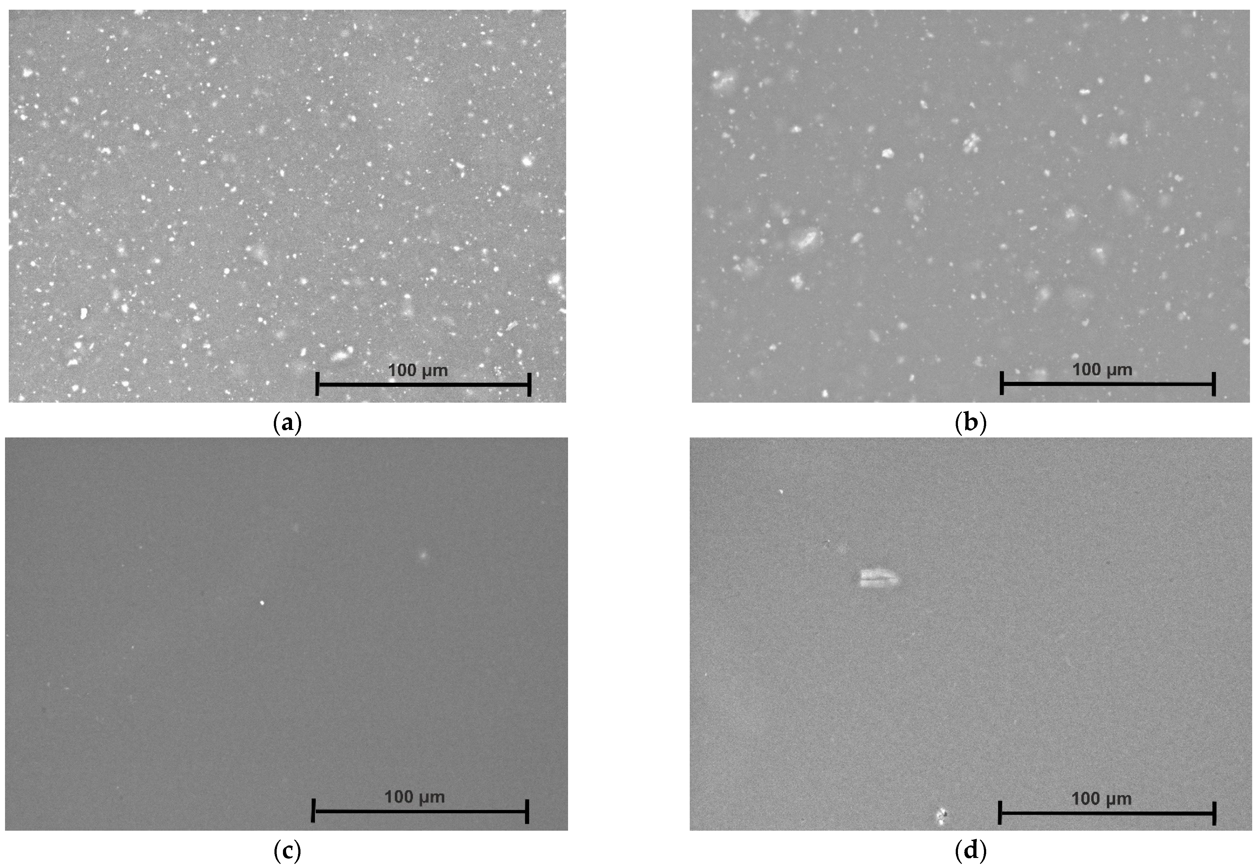
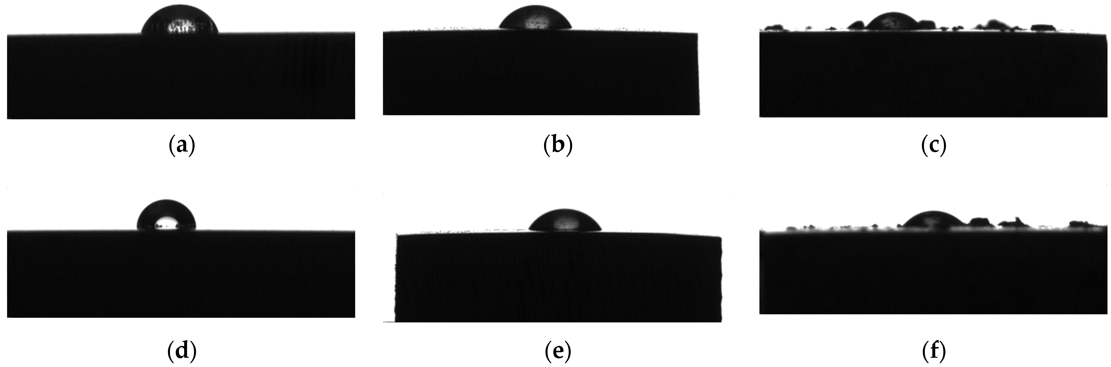
3.3.1. Wettability
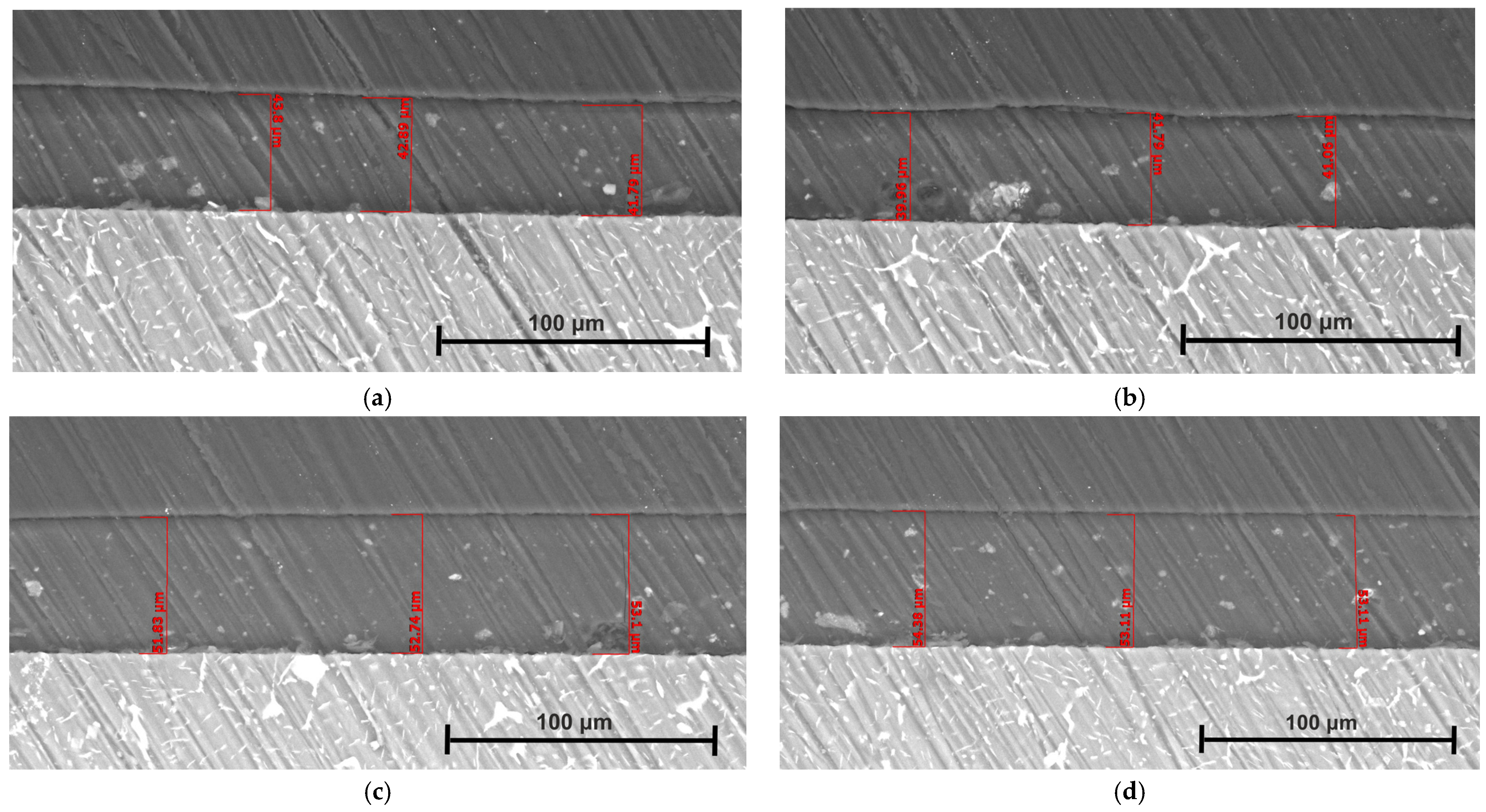
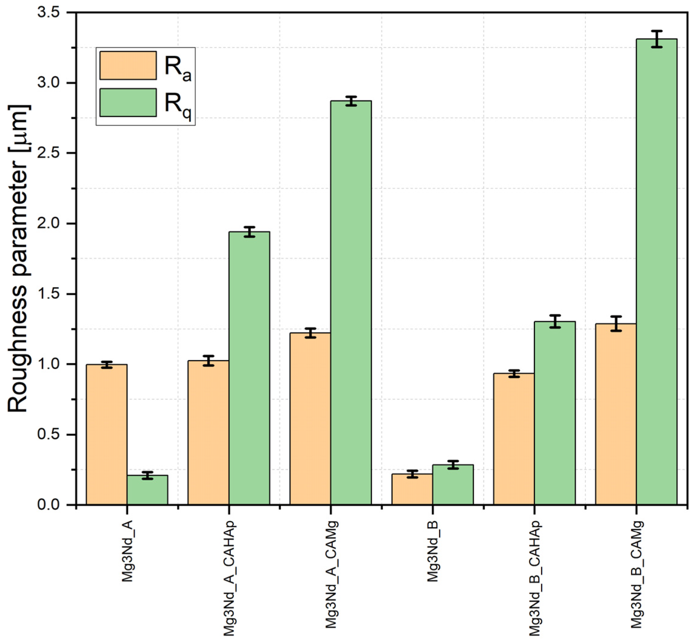
3.3.2. Roughness
4. Conclusions
Author Contributions
Funding
Institutional Review Board Statement
Data Availability Statement
Conflicts of Interest
References
- Prasad, S.V.S.; Prasad, S.B.; Verma, K.; Mishra, R.K.; Kumar, V.; Singh, S. The Role and Significance of Magnesium in Modern Day Research-A Review. J. Magnes. Alloys 2022, 10, 1–61. [Google Scholar] [CrossRef]
- Antoniac, I.; Miculescu, M.; Mănescu, V.; Stere, A.; Quan, P.H.; Păltânea, G.; Robu, A.; Earar, K. Magnesium-Based Alloys Used in Orthopedic Surgery. Materials 2022, 15, 1148. [Google Scholar] [CrossRef] [PubMed]
- Antoniac, I.V.; Filipescu, M.; Barbaro, K.; Bonciu, A.; Birjega, R.; Cotrut, C.M.; Galvano, E.; Fosca, M.; Fadeeva, I.V.; Vadalà, G.; et al. Iron Ion-Doped Tricalcium Phosphate Coatings Improve the Properties of Biodegradable Magnesium Alloys for Biomedical Implant Application. Adv. Mater. Interfaces 2020, 7, 2000531. [Google Scholar] [CrossRef]
- Xing, F.; Li, S.; Yin, D.; Xie, J.; Rommens, P.M.; Xiang, Z.; Liu, M.; Ritz, U. Recent Progress in Mg-Based Alloys as a Novel Bioabsorbable Biomaterials for Orthopedic Applications. J. Magnes. Alloys 2022, 10, 1428–1456. [Google Scholar] [CrossRef]
- Zeng, Z.; Salehi, M.; Kopp, A.; Xu, S.; Esmaily, M.; Birbilis, N. Recent Progress and Perspectives in Additive Manufacturing of Magnesium Alloys. J. Magnes. Alloys 2022, 10, 1511–1541. [Google Scholar] [CrossRef]
- Uppal, G.; Thakur, A.; Chauhan, A.; Bala, S. Magnesium Based Implants for Functional Bone Tissue Regeneration—A Review. J. Magnes. Alloys 2022, 10, 356–386. [Google Scholar] [CrossRef]
- Sekar, P.S.N.; Desai, V. Recent Progress in in Vivo Studies and Clinical Applications of Magnesium Based Biodegradable Implants—A Review. J. Magnes. Alloys 2021, 9, 1147–1163. [Google Scholar] [CrossRef]
- Sahu, M.R.; Kumar, T.S.S.; Chakkingal, U. A Review on Recent Advancements in Biodegradable Mg-Ca Alloys. J. Magnes. Alloys 2022, 10, 2094–2117. [Google Scholar] [CrossRef]
- Atrens, A.; Song, G.-L.; Cao, F.; Shi, Z.; Bowen, P.K. Advances in Mg Corrosion and Research Suggestions. J. Magnes. Alloys 2013, 1, 177–200. [Google Scholar] [CrossRef]
- Bamberger, M.; Dehm, G. Trends in the Development of New Mg Alloys. Annu. Rev. Mater. Res. 2008, 38, 505–533. [Google Scholar] [CrossRef]
- Blawert, C.; Fechner, D.; Höche, D.; Heitmann, V.; Dietzel, W.; Kainer, K.U.; Živanović, P.; Scharf, C.; Ditze, A.; Gröbner, J.; et al. Magnesium Secondary Alloys: Alloy Design for Magnesium Alloys with Improved Tolerance Limits against Impurities. Corros. Sci. 2010, 52, 2452–2468. [Google Scholar] [CrossRef]
- Han, L.; Hu, H.; Northwood, D.O.; Li, N. Microstructure and Nano-Scale Mechanical Behavior of Mg–Al and Mg–Al–Ca Alloys. Mater. Sci. Eng. A 2008, 473, 16–27. [Google Scholar] [CrossRef]
- Kirkland, N.T.; Staiger, M.P.; Nisbet, D.; Davies, C.H.J.; Birbilis, N. Performance-Driven Design of Biocompatible Mg Alloys. JOM 2011, 63, 28–34. [Google Scholar] [CrossRef]
- Radha, R.; Sreekanth, D. Insight of Magnesium Alloys and Composites for Orthopedic Implant Applications—A Review. J. Magnes. Alloys 2017, 5, 286–312. [Google Scholar] [CrossRef]
- Bairagi, D.; Mandal, S. A Comprehensive Review on Biocompatible Mg-Based Alloys as Temporary Orthopaedic Implants: Current Status, Challenges, and Future Prospects. J. Magnes. Alloys 2022, 10, 627–669. [Google Scholar] [CrossRef]
- Sun, J.; Du, W.; Fu, J.; Liu, K.; Li, S.; Wang, Z.; Liang, H. A Review on Magnesium Alloys for Application of Degradable Fracturing Tools. J. Magnes. Alloys 2022, 10, 2649–2672. [Google Scholar] [CrossRef]
- Neil, W.C.; Forsyth, M.; Howlett, P.C.; Hutchinson, C.R.; Hinton, B.R.W. Corrosion of Heat Treated Magnesium Alloy ZE41. Corros. Sci. 2011, 53, 3299–3308. [Google Scholar] [CrossRef]
- Loos, A.; Rohde, R.; Haverich, A.; Barlach, S. In Vitro and In Vivo Biocompatibility Testing of Absorbable Metal Stents. Macromol. Symp. 2007, 253, 103–108. [Google Scholar] [CrossRef]
- Chang, J.; Guo, X.; He, S.; Fu, P.; Peng, L.; Ding, W. Investigation of the Corrosion for Mg–XGd–3Y–0.4Zr (X = 6, 8, 10, 12 wt%) Alloys in a Peak-Aged Condition. Corros. Sci. 2008, 50, 166–177. [Google Scholar] [CrossRef]
- Darrah, T.H.; Prutsman-Pfeiffer, J.J.; Poreda, R.J.; Ellen Campbell, M.; Hauschka, P.V.; Hannigan, R.E. Incorporation of Excess Gadolinium into Human Bone from Medical Contrast Agents. Metallomics 2009, 1, 479. [Google Scholar] [CrossRef]
- Wu, A.; Xia, C. Study of the Microstructure and Mechanical Properties of Mg-Rare Earth Alloys. Mater. Des. 2007, 28, 1963–1967. [Google Scholar] [CrossRef]
- Kawagoe, M.; Hirasawa, F.; Cun Wang, S.; Liu, Y.; Ueno, Y.; Sugiyama, T. Orally Administrated Rare Earth Element Cerium Induces Metallothionein Synthesis and Increases Glutathione in the Mouse Liver. Life Sci. 2005, 77, 922–937. [Google Scholar] [CrossRef]
- Willbold, E.; Gu, X.; Albert, D.; Kalla, K.; Bobe, K.; Brauneis, M.; Janning, C.; Nellesen, J.; Czayka, W.; Tillmann, W.; et al. Effect of the Addition of Low Rare Earth Elements (Lanthanum, Neodymium, Cerium) on the Biodegradation and Biocompatibility of Magnesium. Acta Biomater. 2015, 11, 554–562. [Google Scholar] [CrossRef]
- He, F.; Hu, W.; Liu, L.; Ma, S.; He, W. Effect of Zn Addition on Microstructure and Mechanical Properties of Mg-3Y-2Nd-0.5Zr Alloy. China Foundry 2023, 20, 299–306. [Google Scholar] [CrossRef]
- Chen, Y.; Wu, G.; Liu, W.; Zhang, L.; Zhang, H.; Cui, W. Effects of Minor Y Addition on Microstructure and Mechanical Properties of Mg–Nd–Zn–Zr Alloy. J. Mater. Res. 2017, 32, 3712–3722. [Google Scholar] [CrossRef]
- Tian, K.V.; Passaretti, F.; Nespoli, A.; Placidi, E.; Condò, R.; Andreani, C.; Licoccia, S.; Chass, G.A.; Senesi, R.; Cozza, P. Composition―Nanostructure Steered Performance Predictions in Steel Wires. Nanomaterials 2019, 9, 1119. [Google Scholar] [CrossRef] [PubMed]
- Kirkland, N.T.; Lespagnol, J.; Birbilis, N.; Staiger, M.P. A Survey of Bio-Corrosion Rates of Magnesium Alloys. Corros. Sci. 2010, 52, 287–291. [Google Scholar] [CrossRef]
- Rau, J.V.; Antoniac, I.; Filipescu, M.; Cotrut, C.; Fosca, M.; Nistor, L.C.; Birjega, R.; Dinescu, M. Hydroxyapatite Coatings on Mg-Ca Alloy Prepared by Pulsed Laser Deposition: Properties and Corrosion Resistance in Simulated Body Fluid. Ceram. Int. 2018, 44, 16678–16687. [Google Scholar] [CrossRef]
- Antoniac, I.; Miculescu, F.; Cotrut, C.; Ficai, A.; Rau, J.V.; Grosu, E.; Antoniac, A.; Tecu, C.; Cristescu, I. Controlling the Degradation Rate of Biodegradable Mg–Zn-Mn Alloys for Orthopedic Applications by Electrophoretic Deposition of Hydroxyapatite Coating. Materials 2020, 13, 263. [Google Scholar] [CrossRef]
- Quan, P.H.; Antoniac, I.; Miculescu, F.; Antoniac, A.; Manescu, V.; Robu, A.; Bița, A.I.; Miculescu, M.; Saceleanu, A.; Bodog, A.D.; et al. Fluoride Treatment and In Vitro Corrosion Behavior of Mg-Nd-Y-Zn-Zr Alloys Type. Materials 2022, 15, 566. [Google Scholar] [CrossRef]
- Saberi, A.; Bakhsheshi-Rad, H.R.; Abazari, S.; Ismail, A.F.; Sharif, S.; Ramakrishna, S.; Daroonparvar, M.; Berto, F. A Comprehensive Review on Surface Modifications of Biodegradable Magnesium-Based Implant Alloy: Polymer Coatings Opportunities and Challenges. Coatings 2021, 11, 747. [Google Scholar] [CrossRef]
- Neacsu, P.; Staras, A.; Voicu, S.; Ionascu, I.; Soare, T.; Uzun, S.; Cojocaru, V.; Pandele, A.; Croitoru, S.; Miculescu, F.; et al. Characterization and In Vitro and In Vivo Assessment of a Novel Cellulose Acetate-Coated Mg-Based Alloy for Orthopedic Applications. Materials 2017, 10, 686. [Google Scholar] [CrossRef] [PubMed]
- Alabbasi, A.; Liyanaarachchi, S.; Kannan, M.B. Polylactic Acid Coating on a Biodegradable Magnesium Alloy: An in Vitro Degradation Study by Electrochemical Impedance Spectroscopy. Thin Solid Films 2012, 520, 6841–6844. [Google Scholar] [CrossRef]
- Shi, P.; Niu, B.E.S.; Chen, Y.; Li, Q. Preparation and Characterization of PLA Coating and PLA/MAO Composite Coatings on AZ31 Magnesium Alloy for Improvement of Corrosion Resistance. Surf. Coat. Technol. 2015, 262, 26–32. [Google Scholar] [CrossRef]
- Yazdimamaghani, M.; Razavi, M.; Vashaee, D.; Tayebi, L. Development and Degradation Behavior of Magnesium Scaffolds Coated with Polycaprolactone for Bone Tissue Engineering. Mater. Lett. 2014, 132, 106–110. [Google Scholar] [CrossRef]
- Li, J.N.; Cao, P.; Zhang, X.N.; Zhang, S.X.; He, Y.H. In Vitro Degradation and Cell Attachment of a PLGA Coated Biodegradable Mg–6Zn Based Alloy. J. Mater. Sci. 2010, 45, 6038–6045. [Google Scholar] [CrossRef]
- Chen, L.; Sheng, Y.; Zhou, H.; Li, Z.; Wang, X.; Li, W. Influence of a MAO + PLGA Coating on Biocorrosion and Stress Corrosion Cracking Behavior of a Magnesium Alloy in a Physiological Environment. Corros. Sci. 2019, 148, 134–143. [Google Scholar] [CrossRef]
- Streza, A.; Antoniac, A.; Manescu, V.; Paltanea, G.; Robu, A.; Dura, H.; Verestiuc, L.; Stanica, E.; Voicu, S.I.; Antoniac, I.; et al. Effect of Filler Types on Cellulose-Acetate-Based Composite Used as Coatings for Biodegradable Magnesium Implants for Trauma. Materials 2023, 16, 554. [Google Scholar] [CrossRef] [PubMed]
- Bița, A.-I.; Antoniac, I.; Miculescu, M.; Stan, G.E.; Leonat, L.; Antoniac, A.; Constantin, B.; Forna, N. Electrochemical and In Vitro Biological Evaluation of Bio-Active Coatings Deposited by Magnetron Sputtering onto Biocompatible Mg-0.8Ca Alloy. Materials 2022, 15, 3100. [Google Scholar] [CrossRef]
- Kokubo, T.; Takadama, H. How Useful Is SBF in Predicting in Vivo Bone Bioactivity? Biomaterials 2006, 27, 2907–2915. [Google Scholar] [CrossRef] [PubMed]
- Annamalai, M.; Gopinadhan, K.; Han, S.A.; Saha, S.; Park, H.J.; Cho, E.B.; Kumar, B.; Patra, A.; Kim, S.-W.; Venkatesan, T. Surface Energy and Wettability of van Der Waals Structures. Nanoscale 2016, 8, 5764–5770. [Google Scholar] [CrossRef]
- Xu, D.K.; Liu, L.; Xu, Y.B.; Han, E.H. The Influence of Element Y on the Mechanical Properties of the As-Extruded Mg–Zn–Y–Zr Alloys. J. Alloys Compd. 2006, 426, 155–161. [Google Scholar] [CrossRef]
- Wang, J.; Liu, R.; Dong, X.; Yang, Y. Microstructure and Mechanical Properties of Mg-Zn-Y-Nd-Zr Alloys. J. Rare Earths 2013, 31, 616–621. [Google Scholar] [CrossRef]
- Zhou, H.T.; Zhang, Z.D.; Liu, C.M.; Wang, Q.W. Effect of Nd and Y on the Microstructure and Mechanical Properties of ZK60 Alloy. Mater. Sci. Eng. A 2007, 445–446, 1–6. [Google Scholar] [CrossRef]
- Zengin, H.; Turen, Y. Effect of Y Addition on Microstructure and Corrosion Behavior of Extruded Mg–Zn–Nd–Zr Alloy. J. Magnes. Alloys 2020, 8, 640–653. [Google Scholar] [CrossRef]
- Elkaiam, L.; Hakimi, O.; Goldman, J.; Aghion, E. The Effect of Nd on Mechanical Properties and Corrosion Performance of Biodegradable Mg-5%Zn Alloy. Metals 2018, 8, 438. [Google Scholar] [CrossRef]
- Li, Q.; Wang, Q.; Wang, Y.; Zeng, X.; Ding, W. Effect of Nd and Y Addition on Microstructure and Mechanical Properties of As-Cast Mg–Zn–Zr Alloy. J. Alloys Compd. 2007, 427, 115–123. [Google Scholar] [CrossRef]
- Xu, L.; Liu, X.; Sun, K.; Fu, R.; Wang, G. Corrosion Behavior in Magnesium-Based Alloys for Biomedical Applications. Materials 2022, 15, 2613. [Google Scholar] [CrossRef]
- Gaon, O.; Dror, G.; Davidi, O.; Lugovskoy, A. The Effect of the Local Microstructure of MRI 201S Magnesium Alloy on Its Corrosion Rate. Corros. Sci. 2015, 93, 167–171. [Google Scholar] [CrossRef]
- Zemková, M.; Minárik, P.; Dittrich, J.; Bohlen, J.; Král, R. Individual Effect of Y and Nd on the Microstructure Formation of Mg-Y-Nd Alloys Processed by Severe Plastic Deformation and Their Effect on the Subsequent Mechanical and Corrosion Properties. J. Magnes. Alloys 2023, 11, 509–521. [Google Scholar] [CrossRef]
- Zhang, X.; Li, Y.; Zhang, K.; Wang, C.; Li, H.; Ma, M.; Zang, B. Corrosion and Electrochemical Behavior of Mg–Y Alloys in 3.5% NaCl Solution. Trans. Nonferrous Met. Soc. China 2013, 23, 1226–1236. [Google Scholar] [CrossRef]
- Sudholz, A.D.; Gusieva, K.; Chen, X.B.; Muddle, B.C.; Gibson, M.A.; Birbilis, N. Electrochemical Behaviour and Corrosion of Mg–Y Alloys. Corros. Sci. 2011, 53, 2277–2282. [Google Scholar] [CrossRef]
- Davenport, A.J.; Padovani, C.; Connolly, B.J.; Stevens, N.P.C.; Beale, T.A.W.; Groso, A.; Stampanoni, M. Synchrotron X-ray Microtomography Study of the Role of Y in Corrosion of Magnesium Alloy WE43. Electrochem. Solid-State Lett. 2007, 10, C5. [Google Scholar] [CrossRef]
- Coy, A.E.; Viejo, F.; Skeldon, P.; Thompson, G.E. Susceptibility of Rare-Earth-Magnesium Alloys to Micro-Galvanic Corrosion. Corros. Sci. 2010, 52, 3896–3906. [Google Scholar] [CrossRef]
- Wu, S.; Jang, Y.S.; Lee, M.H. Enhancement of Bone Regeneration on Calcium-Phosphate-Coated Magnesium Mesh: Using the Rat Calvarial Model. Front. Bioeng. Biotechnol. 2021, 9, 652334. [Google Scholar] [CrossRef]
- Kim, S.; Jo, J.; Lee, S.; Kang, M.; Kim, H.; Estrin, Y.; Lee, J.; Lee, J.; Koh, Y. Hydroxyapatite-coated Magnesium Implants with Improved in Vitro and in Vivo Biocorrosion, Biocompatibility, and Bone Response. J. Biomed. Mater. Res. A 2014, 102, 429–441. [Google Scholar] [CrossRef]
- Ishizaki, T.; Saito, N.; Takai, O. Correlation of Cell Adhesive Behaviors on Superhydrophobic, Superhydrophilic, and Micropatterned Superhydrophobic/Superhydrophilic Surfaces to Their Surface Chemistry. Langmuir 2010, 26, 8147–8154. [Google Scholar] [CrossRef] [PubMed]
- Kim, S.-Y.; Kim, Y.-K.; Jang, Y.-S.; Park, I.-S.; Lee, S.-J.; Jeon, J.-G.; Lee, M.-H. Bioactive Effect of Alkali-Heat Treated TiO2 Nanotubes by Water or Acid Treatment. Surf. Coat. Technol. 2016, 303, 256–267. [Google Scholar] [CrossRef]
- Aronov, D.; Rosen, R.; Ron, E.Z.; Rosenman, G. Tunable Hydroxyapatite Wettability: Effect on Adhesion of Biological Molecules. Process Biochem. 2006, 41, 2367–2372. [Google Scholar] [CrossRef]
- Wang, S.; Zhang, X.; Li, J.; Liu, C.; Guan, S. Investigation of Mg–Zn–Y–Nd Alloy for Potential Application of Biodegradable Esophageal Stent Material. Bioact. Mater. 2020, 5, 1–8. [Google Scholar] [CrossRef]
- Indira, K.; Sylvie, C.; Zhongke, W.; Hongyu, Z. Investigation of Wettability Properties of Laser Surface Modified Rare Earth Mg Alloy. Procedia Eng. 2016, 141, 63–69. [Google Scholar] [CrossRef]
- Lorenz, C.; Brunner, J.G.; Kollmannsberger, P.; Jaafar, L.; Fabry, B.; Virtanen, S. Effect of Surface Pre-Treatments on Biocompatibility of Magnesium. Acta Biomater. 2009, 5, 2783–2789. [Google Scholar] [CrossRef] [PubMed]
- Walter, R.; Kannan, M.B.; He, Y.; Sandham, A. Effect of Surface Roughness on the in Vitro Degradation Behaviour of a Biodegradable Magnesium-Based Alloy. Appl. Surf. Sci. 2013, 279, 343–348. [Google Scholar] [CrossRef]
- Laycock, N.J.; Krouse, D.P.; Hendy, S.C.; Williams, D.E. Computer Simulation of Pitting Corrosion of Stainless Steels. Interface Mag. 2014, 23, 65–71. [Google Scholar] [CrossRef]
- Zhao, Y.; Jamesh, M.I.; Li, W.K.; Wu, G.; Wang, C.; Zheng, Y.; Yeung, K.W.K.; Chu, P.K. Enhanced Antimicrobial Properties, Cytocompatibility, and Corrosion Resistance of Plasma-Modified Biodegradable Magnesium Alloys. Acta Biomater. 2014, 10, 544–556. [Google Scholar] [CrossRef]
- Von Der Höh, N.; Bormann, D.; Lucas, A.; Denkena, B.; Hackenbroich, C.; Meyer-Lindenberg, A. Influence of Different Surface Machining Treatments of Magnesium-Based Resorbable Implants on the Degradation Behavior in Rabbits. Adv. Eng. Mater. 2009, 11, B47–B54. [Google Scholar] [CrossRef]
- Shetty, O.; Parekh, R.B.; Tabassum, R. Surface Modifications for Endosseous Dental Implants. Int. J. Oral Implantol. Clin. Res. 2012, 3, 116–121. [Google Scholar] [CrossRef]
- Lacefield, W.R. Materials Characteristics of Uncoated/Ceramic-Coated Implant Materials. Adv. Dent. Res. 1999, 13, 21–26. [Google Scholar] [CrossRef]
- Salerno, M.; Reverberi, A.; Baino, F. Nanoscale Topographical Characterization of Orbital Implant Materials. Materials 2018, 11, 660. [Google Scholar] [CrossRef]
- Hwang, D.Y.; Kim, Y.M.; Shin, D.H. Corrosion Resistance of Plasma-Anodized AZ91 Mg Alloy in the Electrolyte with/without Potassium Fluoride. Mater. Trans. 2009, 50, 671–678. [Google Scholar] [CrossRef]
- Yoo, B.; Shin, K.R.; Hwang, D.Y.; Lee, D.H.; Shin, D.H. Effect of Surface Roughness on Leakage Current and Corrosion Resistance of Oxide Layer on AZ91 Mg Alloy Prepared by Plasma Electrolytic Oxidation. Appl. Surf. Sci. 2010, 256, 6667–6672. [Google Scholar] [CrossRef]
- Supplit, R.; Koch, T.; Schubert, U. Evaluation of the Anti-Corrosive Effect of Acid Pickling and Sol–Gel Coating on Magnesium AZ31 Alloy. Corros. Sci. 2007, 49, 3015–3023. [Google Scholar] [CrossRef]
- Nguyen, T.L.; Blanquet, A.; Staiger, M.P.; Dias, G.J.; Woodfield, T.B.F. On the Role of Surface Roughness in the Corrosion of Pure Magnesium in Vitro. J. Biomed. Mater. Res. B Appl. Biomater. 2012, 100B, 1310–1318. [Google Scholar] [CrossRef]
- Sigmund, W.M.; Hsu, S.-H. Wenzel Model Encyclopedia of Membranes; Drioli, E., Giorno, L., Eds.; Springer: Berlin/Heidelberg, Germany, 2015; ISBN 9783642408724. [Google Scholar]


| Coating | Mg-Based Substrate | Remarks | Ref |
|---|---|---|---|
| PLA | Mg9Al1Zn | The authors investigated the degradation of samples in simulated body fluid (SBF) based on electrochemical impedance spectroscopy (EIS). It was concluded that a thickness of 5 μm for the polymeric coatings determines a corrosion resistance increase of two times compared to the bare Mg alloy. Thicker PLA layers were linked to poor adhesion. | Alabbasi et al. [33] |
| PLA/micro-arc oxidation (MAO) | Mg3Al1Zn | After the MAO process was applied, a 5 μm porous MgO layer was obtained. Then, the samples were immersed in PLA, and a coating with 9 μm thickness was formed. Following the electrochemical corrosion analysis in the case of coated samples, the corrosion potential was about −1.51 V, and the corrosion current density had a very low value, equal to 1.83 μA/cm2. | Shi et al. [34] |
| PCL (3% (w/v)) and PCL (6% (w/v)) | Mg-based scaffolds | It was noticed that the uncoated scaffolds fully degraded after 72 h, and the coated samples degraded in a proportion of 36% and 23%. | Yazdimamaghani et al. [35] |
| PLGA | Mg6Zn | PLGA was dissolved in chloroform with 2 wt.% and 4 wt.% concentrations. The corrosion analysis revealed a very low value for the corrosion current density in the coated samples compared to the control one. | Li et al. [36] |
| (MAO) + PLGA | Mg4Zn0.6Zr0.4Sr | Compared with the uncoated sample, the (MAO) + PLGA coatings increased the corrosion resistance of the alloy by three orders of magnitude, and the stress corrosion cracking susceptibility measurements were reduced by 75% of the ultimate tensile strength. | Chen et al. [37] |
| Composite coatings based on CA, CA-HAp, CA-Mg, and CA-HAp-Mg | - | They noticed that the thermogravimetric analysis proved the coating stability up to 200 °C, highlighting a mass loss with a maximum value of 9% at a maximum temperature of 250 °C. HAp crystals were put in evidence through scanning electron microscopy investigations. The CA-Mg and CA-HAp composite coatings were characterized by a viability of 80% for the MG-63 cell line. | Streza et al. [38] |
| Alloys | Composition (wt.%) | ||||
|---|---|---|---|---|---|
| Zn | Zr | Y | Nd | Mg | |
| Mg3Nd_A | 0.3 | 0.6 | 2.10 | 3.2 | Bal |
| Mg3Nd_B | 0.3 | 0.4 | 0.21 | 3.1 | Bal |
| Sample ID | Eoc (V) | Ecorr (V) | icorr (µA/cm2) | βc (mV) | βa (mV) | CR (mm/Year) | Pe (%) | Rp (kΩcm2) |
|---|---|---|---|---|---|---|---|---|
| Mg3Nd_A | −1.765 | −1.720 | 38.918 | 266.60 | 280.011 | 0.885 | - | 1.530 |
| Mg3Nd_A_CAHAp | −1.647 | −1.641 | 4.497 | 2637 | 1371 | - | 99.63 | 87.210 |
| Mg3Nd_A_CAMg | −1.610 | −1.605 | 16.684 | 625.154 | 676.912 | - | 98.66 | 8.469 |
| Mg3Nd_B | −1.698 | −1.629 | 55.689 | 268.870 | 87.419 | 1.266 | - | 0.535 |
| Mg3Nd_B_CAHAp | −1.683 | −1.681 | 14.971 | 1008 | 788.346 | - | 98.54 | 12.847 |
| Mg3Nd_B_CAMg | −1.696 | −1.678 | 20.607 | 297.275 | 242.764 | - | 97.99 | 2.819 |
Disclaimer/Publisher’s Note: The statements, opinions and data contained in all publications are solely those of the individual author(s) and contributor(s) and not of MDPI and/or the editor(s). MDPI and/or the editor(s) disclaim responsibility for any injury to people or property resulting from any ideas, methods, instructions or products referred to in the content. |
© 2023 by the authors. Licensee MDPI, Basel, Switzerland. This article is an open access article distributed under the terms and conditions of the Creative Commons Attribution (CC BY) license (https://creativecommons.org/licenses/by/4.0/).
Share and Cite
Streza, A.; Antoniac, A.; Manescu, V.; Ciocoiu, R.; Cotrut, C.-M.; Miculescu, M.; Miculescu, F.; Antoniac, I.; Fosca, M.; Rau, J.V.; et al. In Vitro Studies Regarding the Effect of Cellulose Acetate-Based Composite Coatings on the Functional Properties of the Biodegradable Mg3Nd Alloys. Biomimetics 2023, 8, 526. https://doi.org/10.3390/biomimetics8070526
Streza A, Antoniac A, Manescu V, Ciocoiu R, Cotrut C-M, Miculescu M, Miculescu F, Antoniac I, Fosca M, Rau JV, et al. In Vitro Studies Regarding the Effect of Cellulose Acetate-Based Composite Coatings on the Functional Properties of the Biodegradable Mg3Nd Alloys. Biomimetics. 2023; 8(7):526. https://doi.org/10.3390/biomimetics8070526
Chicago/Turabian StyleStreza, Alexandru, Aurora Antoniac, Veronica Manescu (Paltanea), Robert Ciocoiu, Cosmin-Mihai Cotrut, Marian Miculescu, Florin Miculescu, Iulian Antoniac, Marco Fosca, Julietta V. Rau, and et al. 2023. "In Vitro Studies Regarding the Effect of Cellulose Acetate-Based Composite Coatings on the Functional Properties of the Biodegradable Mg3Nd Alloys" Biomimetics 8, no. 7: 526. https://doi.org/10.3390/biomimetics8070526