
Dedicated Protocol for Ultrastructural Analysis of Farmed Rainbow Trout (Oncorhynchus mykiss) Tissues with Red Mark Syndrome: The Skin—Part One

Abstract
:1. Introduction
2. Experimental Design
2.1. Materials
- Millonig’s phosphate buffer (0.1 M pH 7.4, 4) (Electron Microscopy Sciences, 1560 Industry Road, Hatfield, PA, USA, cat. no. 11582-05)
- Glutaraldehyde EM GRADE 25% (Agar Scientific, Cambridge Road, Stansted Essex, UK, cat. no. 605390)
- Dulbecco’s phosphate buffer (Immunological Sciences, Rome, Italy, cat. no. ISL0615-500)
- Osmium tetroxide (OsO4) 4% (Electron Microscopy Sciences, 1560 Industry Road, Hatfield, PA, USA, cat. no. 19150)
- Ethanol absolute anhydrous (Carlo Erba, Milan, Italy, cat. no. 414608)
- EMbed-812 Embedding Kit W/DMP-30 (Electron Microscopy Sciences, 1560 Industry Road, Hatfield, PA, USA, cat. no. 14120)
- Propylene oxide (Electron Microscopy Sciences, 1560 Industry Road, Hatfield, PA, USA, cat. no. 20401)
- Methylene blue (Agar Scientific, Cambridge Road, Stansted Essex, UK, cat. no. 52015)
- Borax (Merck Millipore, Burlington, MA, USA, cat. no. CA1330-43-4)
- Azure II (Merck, Milan, Italy, cat. no. 37247-10-2)
- Sodium hydroxide anhydrous pellets ACS-ISO for analysis (CARLO ERBA REAGENTS S.r.l., Milan, Italy, cat. no. 480507)
- Neo-Mount® (Merck Millipore, Burlington, MA, USA, cat. no. 200869).
- Lead citrate (Agar Scientific, Cambridge Road, Stansted Essex, UK, cat. no. AGR1210)
2.2. Equipment
- Disposable razor blades (Electron Microscopy Sciences, 1560 Industry Road, Hatfield, PA, USA, cat. no. 71960)
- Tweezers (Electron Microscopy Sciences, 1560 Industry Road, Hatfield, PA, USA, cat. no. 78512—4DX)
- BEEM embedding capsules (Electron Microscopy Sciences, 1560 Industry Road, Hatfield, PA, USA, cat. no. 7000-B)
- Glass beakers (250–400 mL) (Sigma-Aldrich, 3050 Spruce St., Saint Louis, MO, USA, cat. no. Z231843)
- Staccup disposable beakers (120 cc) (Electron Microscopy Sciences, 1560 Industry Road, Hatfield, PA, USA, cat. no. 50-996-320)
- Magnetic stir bars (Dutscher SAS, Bernolsheim, France, cat. no. 785588)
- Magnetic stirrer (IKA-Werke GmbH & Co. KG, Staufen, Germany, cat no. 0005020000)
- Balance (Mettler Toledo, Milan, Italy, cat. no. 05361)
- Laboratory shaker (Biosan, Riga, Latvia, cat. no. BS-010130-AAI)
- Chemical fume hood (Asalair, Zetalab, Padova, Italy, cat. no. A-CARBO900)
- Oven (Memmert, Venice, Italy, cat. no. 12616987)
- Microcentrifuge (Tomos, Singapore, cat. no. 3000002)
- Parafilm (American Can Company, SPI supplies, 206 Garfield Ave West Chester, PA 19380-4512, USA, cat. no. 01851-AB)
- Glass knife maker (Leica Microsystem, Milan, Italy, cat. no. K111)
- Ultramicrotome glass knife strips (Agar Scientific, Unit 7, M11 Business Link, Parsonage Lane, Stansted, Essex CM24 8GF, UK, cat. no. AGG336)
- Microscope slides (Fischer Scientific, Rome, Italy, cat. no. 12342108)
- Coverslips (Bioptica, Milan, Italy, cat. no. 09-2040)
- Hot plate (at least 70° C) (IKA-Werke GmbH & Co. KG, 79219 Staufen, Germany, cat no. 0003582000)
- Ultramicrotome (Leica Microsystem, Milan, Italy, cat. no. Leica UC7)
- Diamon knife (Diatome Diamond Knives, Microcontrol, Milan, Italy, cat. no. TDU4530)
- Copper mesh grids (Electron Microscopy Sciences, 1560 Industry Road, Hatfield, PA, USA, cat. no. G200-Cu)
- Fine-pointed tweezers (Electron Microscopy Sciences, 1560 Industry Road, Hatfield, PA, USA, cat. no. 7801-7)
- Filter paper, hardened (Macherey-Nagel, Germany, cat. no. 431012)
- 0.22 μm filter (Fischer Scientific, Rome, Italy, cat. no. 10002261)
- Aluminum rolls (Ted Pella, Inc., Redding, CA, USA, cat. no. 43-100)
- Falcon tubes (Fischer Scientific, Rome, Italy, cat. no. 10788561)
- Microcentrifuge tubes (Fischer Scientific, Rome, Italy, cat. no. 10154671)
- Laboratory cylinder (Fischer Scientific, Rome, Italy, cat. no. 11537832)
- Glass bottles (Dutscher SAS, (Dutscher SAS, Bernolsheim, France, cat. no. 090394)
- Plastic Petri dishes (35 × 10 mm) (Biosigma, Venice, Italy, cat. no. 353001)
- Glass Petri dishes (60 × 15 mm) (Fischer Scientific, Rome, Italy, cat. no. 11740834)
- Plastic Pasteur pipettes (Merck, Milan, Italy, cat. no. BR747775)
- Glass Pasteur pipettes (Merck, Milan, Italy, cat. no. BR747725)
- Eyelash probe (Electron Microscopy Sciences, 1560 Industry Road, Hatfield, PA, USA, cat. no. 71182)
- Grid storage boxes (Electron Microscopy Sciences, 1560 Industry Road, Hatfield, PA, USA, cat. no. 71150)
3. Procedure
3.1. Preparative for Transmission Electron Microscopy
3.1.1. Preparative for TEM: Sampling
3.1.2. Preparative for TEM: Primary Fixation
![Mps 07 00037 i002]() PAUSE STEP: store the skin samples at 4° for 48 h (Table 1) until the start of the post-fixation.
PAUSE STEP: store the skin samples at 4° for 48 h (Table 1) until the start of the post-fixation.

3.1.3. Preparative for TEM: Post-Fixation
![Mps 07 00037 i003]() CRITICAL STEP: carefully place the samples on the laboratory shaker, completely closed for 2 h, and covered with sheets of aluminum.
CRITICAL STEP: carefully place the samples on the laboratory shaker, completely closed for 2 h, and covered with sheets of aluminum.

![Mps 07 00037 i004]() CRITICAL STEP: at this stage, the skin samples derived from rainbow trout affected by RMS will be completely black; this is suggestive that post-fixation procedures are now completed.
CRITICAL STEP: at this stage, the skin samples derived from rainbow trout affected by RMS will be completely black; this is suggestive that post-fixation procedures are now completed.

3.1.4. Preparative for TEM: Washing and Dehydration
![Mps 07 00037 i005]() PAUSE STEP: once the 70% wash is finished, remove the Petri dishes from the laboratory shaker, cover them with parafilm, and store them at 4 °C overnight.
PAUSE STEP: once the 70% wash is finished, remove the Petri dishes from the laboratory shaker, cover them with parafilm, and store them at 4 °C overnight.

3.1.5. Preparative for TEM: Epoxy Resin
![Mps 07 00037 i006]() CRITICAL STEP: the mix should be resuspended gently to avoid bubble formation (bubbles will impede embedding) and should be located overnight on a magnetic stirrer, in contact with a magnetic stir bar.
CRITICAL STEP: the mix should be resuspended gently to avoid bubble formation (bubbles will impede embedding) and should be located overnight on a magnetic stirrer, in contact with a magnetic stir bar.

3.1.6. Preparative for TEM: The Embedding
![Mps 07 00037 i007]() CRITICAL STEP: keep the glass Petri dishes containing the samples closed for 1 h on the laboratory shaker.
CRITICAL STEP: keep the glass Petri dishes containing the samples closed for 1 h on the laboratory shaker.

![Mps 07 00037 i008]() CRITICAL STEP: keep the embedded samples on the laboratory shaker for 1 h and then put them at 60° in the oven for 48 h.
CRITICAL STEP: keep the embedded samples on the laboratory shaker for 1 h and then put them at 60° in the oven for 48 h.

3.1.7. Preparative for TEM: The Ultramicrotome
3.1.8. Preparative for TEM: Semithin Sections
![Mps 07 00037 i009]() CRITICAL STEP: filter the methylene blue solution, previously prepared, with a syringe and a 0.22 μm filter.
CRITICAL STEP: filter the methylene blue solution, previously prepared, with a syringe and a 0.22 μm filter.

3.1.9. Preparative for TEM: Ultrathin Sections
3.1.10. Preparative for TEM: Uranyless and Lead Citrate Staining
3.1.11. Preparative for TEM: TEM Observations
4. Results and Discussion
5. Conclusions
6. Reagent Setup
6.1. Primary Fixation Reagent: 2.5% Glutaraldehyde in PBS
![Mps 07 00037 i010]() PAUSE STEP: then, carefully store the falcon tube containing 2.5% glutaraldehyde at 4 °C until the sampling and fixation procedures.
PAUSE STEP: then, carefully store the falcon tube containing 2.5% glutaraldehyde at 4 °C until the sampling and fixation procedures.

6.2. Dehydrating Solutions: Ethanol 30–100%
- Prepare ethanol 30% in distilled water: by using a plastic Pasteur pipette and falcon tubes (50 mL), dissolve 15 mL of ethanol absolute anhydrous in 35 mL of distilled water. Carefully store the falcon tubes at 4° C until dehydration.
- Prepare ethanol 50% in distilled water: by using a plastic Pasteur pipette and falcon tubes (50 mL), dissolve 25 mL of ethanol absolute anhydrous in 25 mL of distilled water. Carefully store the falcon tubes at 4° C until dehydration.
- Prepare ethanol 70% in distilled water: by using a plastic Pasteur pipette and falcon tubes (50 mL), dissolve 35 mL of ethanol absolute anhydrous in 15 mL of distilled water. Carefully store the falcon tubes at 4° C until dehydration.
- Prepare ethanol 95% in distilled water: by using a plastic Pasteur pipette and falcon tube (50 mL), dissolve 47.5 mL of ethanol absolute anhydrous in 2.5 mL of distilled water. Carefully store the falcon tubes at 4° C until dehydration.
- Prepare ethanol 100%: by using a plastic Pasteur pipette, place in a falcon tube (50 mL) a total volume of 50 mL of ethanol absolute anhydrous. Then, carefully store the falcon tubes at 4° C until dehydration.
6.3. Post-Fixation Reagents: OsO4 1% in Distilled Water
![Mps 07 00037 i011]() PAUSE STEP: then, carefully close and cover the glass bottle containing OsO4 1% in distilled water with aluminum sheets, and store it at 4° C until the post-fixation.
PAUSE STEP: then, carefully close and cover the glass bottle containing OsO4 1% in distilled water with aluminum sheets, and store it at 4° C until the post-fixation.

6.4. LM Staining: Methylene Blue
6.5. TEM Staining: Lead Citrate
- Firstly, prepare sodium hydroxide Na(OH) 1N: dissolve in a glass bottle 4 gr of Na(OH) in 10 mL of cold distilled water. Mix the solution until it is completely dissolved, and cover it with parafilm.
![Mps 07 00037 i012]() CRITICAL STEP: at this stage, the mixture will be hot: mix it until it is completely clear and ready for use.
CRITICAL STEP: at this stage, the mixture will be hot: mix it until it is completely clear and ready for use.

- Prepare lead citrate 0.3% in distilled water: place 30 mL of distilled water, collected in a becker, on a magnetic stirrer and wait until it boils. After this step, transfer all 30 mL of distilled water to a laboratory cylinder and store it at 4° C. Once the solution is completely cold, dissolve in it 1.33 gr of lead nitrate PB(NO3)2 and 1.76 gr of trisodium citrate dihydrate Na3(C6H5O7) 2H2O, with 8 mL of sodium hydroxide Na(OH) 1N, previously prepared, also adding distilled water to reach a total volume of 50 mL.
![Mps 07 00037 i013]() PAUSE STEP: then, store the prepared solution at 4°C until the start of the lead citrate staining.
PAUSE STEP: then, store the prepared solution at 4°C until the start of the lead citrate staining.

Author Contributions
Funding
Institutional Review Board Statement
Informed Consent Statement
Data Availability Statement
Acknowledgments
Conflicts of Interest
Abbreviations
| COVID-19 | coronavirus disease 2019 |
| ddPCR | digital droplet PCR |
| DDSA | dodecenylsuccinic anhydride, specially distilled |
| DMP-30 | 2,4,6-tri(dimethylaminomethyl) phenol |
| LM | light microscopy |
| MLO | Midichloria-like organism |
| Na3(C6H5O7)2 H2O | trisodium citrate dihydrate |
| NMA | methyl-5-norborene-2,3-dicarboxylic anhydride |
| Na(OH) | sodium hydroxide |
| OsO4 | osmium tetroxide |
| PB(NO3)2 | lead nitrate |
| PO | propylene oxide |
| qPCR | quantitative PCR |
| RLO | Rickettsia-like organism |
| TEM | transmission electron microscopy |
References
- Giusti, I.; Bianchi, S.; Nottola, S.A.; Macchiarelli, G.; Dolo, V. Clinical Electron Microscopy in the Study of Human Ovarian Tissues. Euro Mediterr. Biomed. J. 2019, 14, 145–151. [Google Scholar] [CrossRef]
- Franken, L.E.; Grünewald, K.; Boekema, E.J.; Stuart, M.C.A. A Technical Introduction to Transmission Electron Microscopy for Soft-Matter: Imaging, Possibilities, Choices, and Technical Developments. Small 2020, 16, 1906198. [Google Scholar] [CrossRef]
- Mielańczyk, Ł.; Matysiak, N.; Klymenko, O.; Wojnicz, R. Transmission Electron Microscopy of Biological Samples. In The Transmission Electron Microscope—Theory and Applications; InTech: London, UK, 2015; pp. 193–236. [Google Scholar] [CrossRef]
- Graham, L.; Orenstein, J.M. Processing Tissue and Cells for Transmission Electron Microscopy in Diagnostic Pathology and Research. Nat. Protoc. 2007, 2, 2439–2450. [Google Scholar] [CrossRef]
- Winey, M.; Meehl, J.B.; O’Toole, E.T.; Giddings, T.H. Conventional Transmission Electron Microscopy. Mol. Biol. Cell 2014, 25, 319–323. [Google Scholar] [CrossRef] [PubMed]
- Mrazova, K.; Bacovsky, J.; Sedrlova, Z.; Slaninova, E.; Obruca, S.; Fritz, I.; Krzyzanek, V. Urany-Less Low Voltage Transmission Electron Microscopy: A Powerful Tool for Ultrastructural Studying of Cyanobacterial Cells. Microorganisms 2023, 11, 888. [Google Scholar] [CrossRef] [PubMed]
- Torge, D.; Bernardi, S.; Arcangeli, M.; Bianchi, S. Histopathological Features of SARS-CoV-2 in Extrapulmonary Organ Infection: A Systematic Review of Literature. Pathogens 2022, 11, 867. [Google Scholar] [CrossRef] [PubMed]
- Metselaar, M.; Orioles, M.; Galeotti, M.; Adams, A.; Thompson, K.D. Red Mark Syndrome—Current State of Knowledge. Aquaculture 2022, 549, 737748. [Google Scholar] [CrossRef]
- Orioles, M.; Galeotti, M.; Saccà, E.; Bulfoni, M.; Corazzin, M.; Bianchi, S.; Torge, D.; Macchiarelli, G.; Magi, G.E.; Schmidt, J.G. Effect of Temperature on Transfer of Midichloria-like Organism and Development of Red Mark Syndrome in Rainbow Trout (Oncorynchus mykiss). Aquaculture 2022, 560, 738577. [Google Scholar] [CrossRef]
- Metselaar, M.; Thompson, K.D.; Paley, R.; Green, D.M.; Verner-Jeffreys, D.; Feist, S.; Adams, A. Investigating the Involvement of a Midichloria-like Organism (MLO) in Red Mark Syndrome in Rainbow Trout Oncorynchus mykiss. Aquaculture 2020, 528, 735485. [Google Scholar] [CrossRef]
- von Gersdorff Jørgensen, L.; Schmidt, J.G.; Chen, D.; Kania, P.W.; Buchmann, K.; Olesen, N.J. Skin Immune Response of Rainbow Trout (Oncorynchus mykiss) Experimentally Exposed to the Disease Red Mark Syndrome. Vet. Immunol. Immunopathol. 2019, 211, 25–34. [Google Scholar] [CrossRef] [PubMed]
- Galeotti, M.; Manzano, M.; Beraldo, P.; Bulfon, C.; Rossi, G.; Volpatti, D.; Magi, G.E. Ultrastructural and Biomolecular Detection of Rickettsiales-like Organisms in Tissues of Rainbow Trout with Red Mark Syndrome. J. Fish Dis. 2017, 40, 907–917. [Google Scholar] [CrossRef] [PubMed]
- Galeotti, M.; Orioles, M.; Saccà, E.; Byadgi, O.; Pesaro, S.; Di Cerbo, A.; Magi, G.E. Understanding the Pathogenesis of Red Mark Syndrome in Rainbow Trout (Oncorynchus mykiss) through an Integrated Morphological and Molecular Approach. Animals 2023, 13, 1103. [Google Scholar] [CrossRef] [PubMed]
- Galeotti, M.; Sarli, G.; Sirri, R.; Mandrioli, L.; Beraldo, P.; Bronzatti, P.; Giavenni, R.; Orioles, M.; Magi, G.E.; Volpatti, D. Red Mark Syndrome of Trout (Oncorhynchus mykiss; Walbaum, 1792): Histopathological Scoring and Correlation with Gross Lesions. J. Fish Dis. 2021, 44, 1325–1336. [Google Scholar] [CrossRef] [PubMed]
- Oh, T.W.; Giri, S.S.; Yun, S.; Kim, H.J.; Kim, S.G.; Kim, S.W.; Kang, J.W.; Han, S.J.; Kwon, J.; Jun, J.W.; et al. Emergence of Rickettsial Infection in Rainbow Trout (Oncorynchus mykiss) Fry Displaying the Appearance of Red Mark Syndrome in Korea. Microorganisms 2019, 7, 302. [Google Scholar] [CrossRef] [PubMed]
- Cafiso, A.; Sassera, D.; Serra, V.; Bandi, C.; McCarthy, U.; Bazzocchi, C. Molecular Evidence for a Bacterium of the Family Midichloriaceae (Order Rickettsiales) in Skin and Organs of the Rainbow Trout Oncorhynchus mykiss (Walbaum) Affected by Red Mark Syndrome. J. Fish Dis. 2016, 39, 497–501. [Google Scholar] [CrossRef] [PubMed]
- Orioles, M.; Schmidt, J.G.; Tomè, P.; Vendramin, N.; Galeotti, M. A Questionnaire-Based Survey on the Presence of Red Mark Syndrome in Italian Rainbow Trout (Oncorhynchus mykiss) Farms. Bull. Eur. Assoc. Fish Pathol. 2023, 43, 38–50. [Google Scholar] [CrossRef]
- Pardo, A.; Villasante, A.; Romero, J. Skin Microbial Community Associated to Strawberry Disease in Farmed Rainbow Trout (Oncorynchus mykiss Walbaum, 1792). Microorganisms 2024, 12, 217. [Google Scholar] [CrossRef] [PubMed]
- Ortega, Y.; Sandoval, N.; Orioles, M. Red Mark Syndrome (RMS) in Farmed Rainbow Trout (Oncorhynchus mykiss: First Report of Outbreak in Peruvian Salmonid Aquaculture. Bull. Eur. Assoc. Fish Pathol. 2023, 43, 129–136. [Google Scholar] [CrossRef]
- Oidtmann, B.; LaPatra, S.E.; Verner-Jeffreys, D.; Pond, M.; Peeler, E.J.; Noguera, P.A.; Bruno, D.W.; St-Hilaire, S.; Schubiger, C.B.; Snekvik, K.; et al. Differential Characterization of Emerging Skin Diseases of Rainbow Trout—A Standardized Approach to Capturing Disease Characteristics and Development of Case Definitions. J. Fish Dis. 2013, 36, 921–937. [Google Scholar] [CrossRef] [PubMed]
- Verner-Jeffreys, D.; Pond, M.; Peeler, E.; Rimmer, G.; Oidtmann, B.; Way, K.; Mewett, J.; Jeffrey, K.; Bateman, K.; Reese, R.; et al. Emergence of Cold Water Strawberry Disease of Rainbow Trout Oncorynchus mykiss in England and Wales: Outbreak Investigations and Transmission Studies. Dis. Aquat. Organ. 2008, 79, 207–218. [Google Scholar] [CrossRef] [PubMed]
 : critical step of the procedure.
: critical step of the procedure.
: critical step of the procedure.


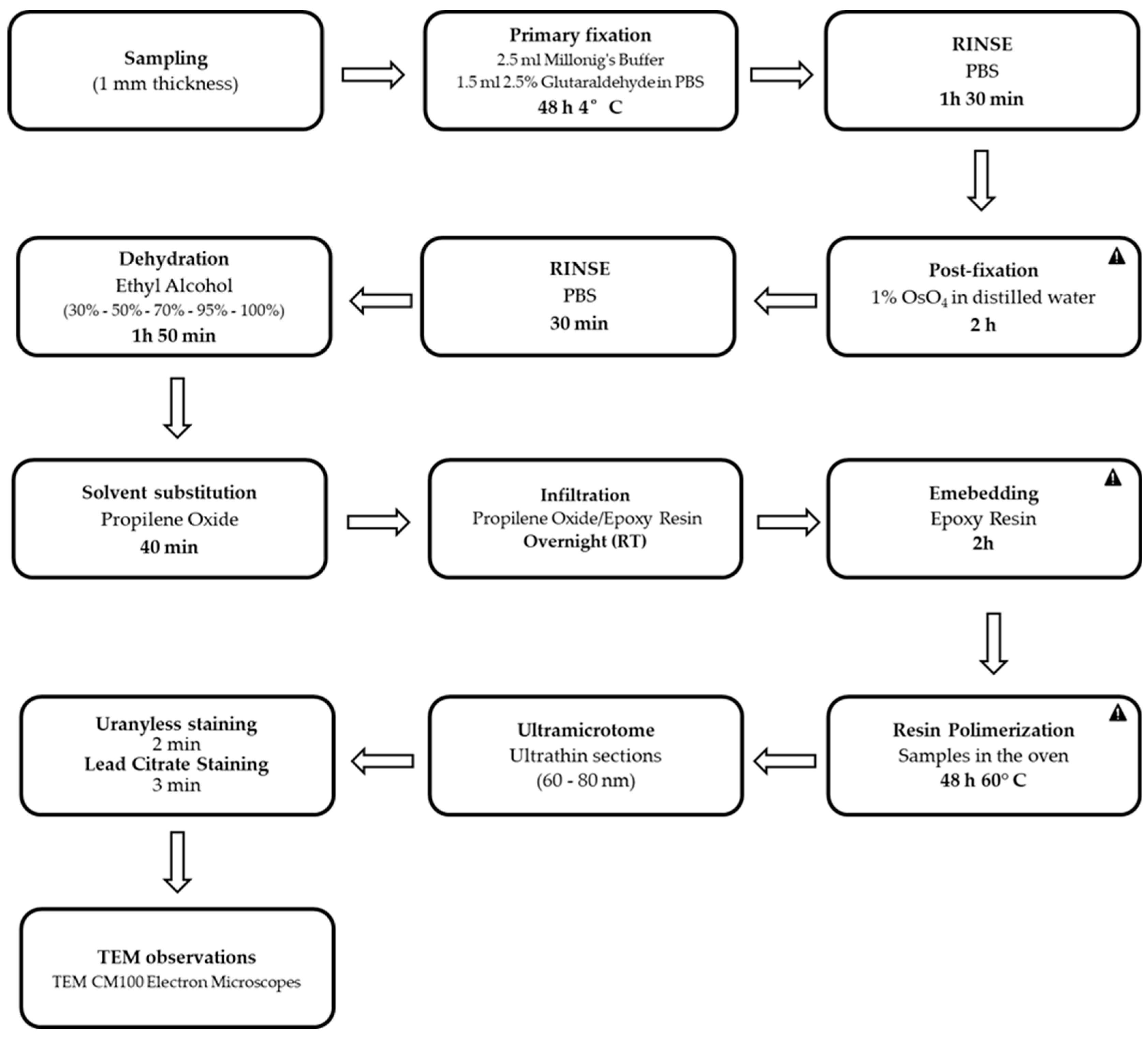
| PROCEDURE | REAGENTS | TIME |
|---|---|---|
| FIXATION | Millonig’s Buffer (0.1 M ph 7.4, 4) and Glutaraldehyde 2.5% in PBS | 48 h at + 4 °C |
| RINSE | PBS | 30 min (3 changes) |
| POST - FIXATION | Osmium Tetroxide 1% in distilled water | 2 h |
| RINSE | PBS | 10 min (3 changes) |
| DEHYDRATION | Ethyl Alcohol 30% Ethyl Alcohol 50% Ethyl Alcohol 70% Ethyl Alcohol 95% Ethyl Alcohol 100% | 10 min 10 min 10 min 10 min (2 changes) 15 min (4 changes) |
| SOLVENT SUBSTITUTION | Propylene Oxide | 20 min (2 changes) |
| INFILTRATION | Propilene Oxide:Epoxy Resin (50/50) | Overnight at RT |
| EMBEDDING | Epoxy Resin | 2 h |
| EMBEDDING AND RESIN POLYMERIZATION | Epoxy Resin | 48 h at 60 °C |
Disclaimer/Publisher’s Note: The statements, opinions and data contained in all publications are solely those of the individual author(s) and contributor(s) and not of MDPI and/or the editor(s). MDPI and/or the editor(s) disclaim responsibility for any injury to people or property resulting from any ideas, methods, instructions or products referred to in the content. |
© 2024 by the authors. Licensee MDPI, Basel, Switzerland. This article is an open access article distributed under the terms and conditions of the Creative Commons Attribution (CC BY) license (https://creativecommons.org/licenses/by/4.0/).
Share and Cite
Torge, D.; Bernardi, S.; Ciciarelli, G.; Macchiarelli, G.; Bianchi, S. Dedicated Protocol for Ultrastructural Analysis of Farmed Rainbow Trout (Oncorhynchus mykiss) Tissues with Red Mark Syndrome: The Skin—Part One. Methods Protoc. 2024, 7, 37. https://doi.org/10.3390/mps7030037
Torge D, Bernardi S, Ciciarelli G, Macchiarelli G, Bianchi S. Dedicated Protocol for Ultrastructural Analysis of Farmed Rainbow Trout (Oncorhynchus mykiss) Tissues with Red Mark Syndrome: The Skin—Part One. Methods and Protocols. 2024; 7(3):37. https://doi.org/10.3390/mps7030037
Chicago/Turabian StyleTorge, Diana, Sara Bernardi, Giulia Ciciarelli, Guido Macchiarelli, and Serena Bianchi. 2024. "Dedicated Protocol for Ultrastructural Analysis of Farmed Rainbow Trout (Oncorhynchus mykiss) Tissues with Red Mark Syndrome: The Skin—Part One" Methods and Protocols 7, no. 3: 37. https://doi.org/10.3390/mps7030037