Article

Exposure to Polymethylmethacrylate Microplastics Induces a Particle Size-Dependent Immune Response in Mediterranean Mussel Mytilus galloprovincialis

1
Center for Marine Research, Ruđer Bošković Institute, 52210 Rovinj, Croatia
2
Faculty of Educational Sciences, Juraj Dobrila University of Pula, 52100 Pula, Croatia
3
Faculty of Mathematics, Natural Sciences and Information Technologies, University of Primorska, 6000 Koper, Slovenia
*
Author to whom correspondence should be addressed.
Fishes 2022, 7(6), 307; https://doi.org/10.3390/fishes7060307
Submission received: 11 September 2022 / Revised: 3 October 2022 / Accepted: 19 October 2022 / Published: 27 October 2022
(This article belongs to the Special Issue Effect of Microplastics and Plastics on Aquatic Animals)

Abstract

:
The widespread occurrence of plastic has become a significant problem in the natural environment and may give rise to a range of deleterious impacts in biota, particularly where plastic particles overlap in size with that of the particles that are naturally ingested by filter-feeders. In this context, the effects of two particle sizes (10 μm and 50 μm) of polymethylmethacrylate (PMMA) on ecologically and commercially significant mussel Mytilus galloprovincialis have been investigated. Mussel health status was evaluated by measuring the condition index and stress-on-stress test during and after 72 h exposure to PMMA microparticles in the 0.1–10 mg L−1 concentration range. The decreased condition and fitness indices in the exposed mussels pointed to significant physiological effects at the entire organism level. The 10 μm and 50 μm PMMA particles were noted to rapidly increase the total haemocytes count in haemolymph, and significantly reduce cell viability at higher concentrations of both particle sizes. The results also indicated a significant increase in levels of vacuolised haemocytes as a result of PMMA exposure. While both microparticles were detected in the haemolymph, only the 10 μm PMMA was observed in the gill tissue and digestive gland by histological cryosections, indicating their rapid uptake, transport, and accumulation in tissue. Lack of accumulation of 50 μm microparticles in tissue may be related to a combination of chemical identity and size considerations, enabling more efficient depuration of microparticles in pseudofaeces. The PMMA particles did not induce significant changes in activity of a range of enzymes involved in neurotransmission and responses to oxidative stress.

Graphical Abstract

1. Introduction

In parallel with the large use of plastics in consumer and industrial products, with an estimated additional 367 million tonnes of world plastic production in 2020 alone [1], the quantity of polymer-based pollution is ever increasing [2]. For example, in the marine environment plastics comprise up to 70% of marine litter [3]. Under the influence of wave action, ocean temperature, and UV radiation, the large plastic debris gradually degrade into smaller fractions, with fragments smaller than 5 mm generally categorised as microplastics [4,5,6,7,8]. In Europe, polyethylene (PE, 30.3%) and polypropylene (PP, 19.7%) together account for about 50% of all plastic consumed, therefore PE and PP are likely to be widespread in the marine environment [1,6]. The abundance of microplastics in the Adriatic Sea has been reported to range from 0.4 ± 0.7 to 1.0 ± 1.8 items m−3 [9] with an average concentration of floating micro litter of 0.127 items m−2 [10]. PE has been reported as the predominant polymer with an overall relative abundance of 52–66.6% of the total amount of detected microplastics, followed by PP (16–19.7%) and synthetic paints (7%) [11]. The presence of paint and paraffin wax suggests high ship-based pollution in the Adriatic Sea, and while fragments of synthetic rubbers, such as ethylene propylenediene monomer (EPDM) and ethylene propylene rubber (EPR), polyacrylates (PMMA), polystyrene (PS), polyvinyl chloride (PVC), and polyethylene terephthalate (PET) were also found; each accounting for less than 1% of the floating polymers [10]. Of these, polymethylmethacrylate (PMMA; widely known under the trade name plexiglass) is a synthetic resin produced from the polymerisation of methyl methacrylate and is widely used as a substitute for glass in products such as lenses, windows, skylights, panels, illuminated signs, acrylic nails, paint, and LCD touch screens, etc., [11].
Currently, it is estimated that about 600 species, including marine mammals, fish, seabirds, turtles, and invertebrates, including plankton and algae, have been affected by plastic material in the marine environment [6,12,13,14,15]. Microplastics have shown the potential to be ingested by different species, to bioaccumulate and to be transferred through the food web to higher trophic levels [15,16,17,18,19]. The consequences of microplastics on marine life have been reported for the past two decades where, due to their small size, they readily enter organisms through the digestive or respiratory systems, subsequently entering tissues and cells leading to a range of toxicological effects [20,21,22,23,24,25,26]. Moreover, consumption of commercial marine organisms such as mussels, oysters, fish, crabs, sea cucumbers, or other products such as sea salt, can have consequences for the health of humans [13,27,28,29]. Thus, not only is the monitoring of microplastics in the marine environment of high importance for human health, but also for assessing their potential to cause a range of deleterious effects in biota and disturb delicate ecological balances [28,29].
Mytilus galloprovincialis is the dominant mussel of the genus Mytilus along the eastern Adriatic coast [30,31] and has been used as a biological indicator in the monitoring of anthropogenic pollution trends in coastal waters [32,33,34,35]. As they are sessile filter feeders they can provide location-specific information, particularly as they have instant exposure to pollutants (e.g., pesticides, toxins, metal(loid)s, and hydrocarbons) that can have several biological impacts on their physiology [33,36,37,38]. Due to their prodigious filtering ability (3–9 L h−1) various environmental pollutants can rapidly accumulate in their tissue [39]. Such accumulation also encompasses microplastics, and may be enhanced in cases where the microparticle size range overlaps with that of particles naturally ingested [40,41].
Investigation of the levels and polymeric composition of microplastics in the native population of mussels, M. galloprovincialis, collected in coastal and in offshore areas in the northern Adriatic Sea has shown that the most recurring polymer type was PE followed by PP and PET, with equal amounts of PS, PVC and other plastics. The highest accumulation of microplastics was observed in organisms collected at coastal sites (1.06–1.33 items g−1 WW) compared to the offshore areas (0.65–0.66 items g−1 WW) [42]. To the best of our knowledge there are no data available on the concentrations of PMMA in intensive mussel mariculture areas such as the Lim Channel in the northern Adriatic Sea.
Field investigation of microplastics in mussels is currently an area of intense research, with recent results indicating that microplastic accumulation in mussels is closely correlated with the degree of pollution in coastal habitats, hence reflecting the real abundance of microplastics in the environment within a certain size range [15,43]. Most of the research literature on the effects of microplastics in mussels has focused on polystyrene [4,12,41,44,45] and polyethylene [46,47] as they are two of the most common consumer plastics, while the effects of PMMA have not been addressed in detail to date. However, there are some reports of the effects of PMMA in other models including mice [48], humans [49], marine diatom [50], polar cod Boreogadus saida [51], and in sea urchins Paracentrotus lividus [52], and Sphaerechinus granularis [53], with results showing mild negative effects over a range of endpoints, such as a slightly elevated level of developmentally delayed embryos or an increased level of developmental (skeletal) defects in the offspring of PMMA microplastic-treated sperm (statistically significant differences to controls in only some cases). Interestingly, despite the low expected concentrations of PMMA microparticles in the marine environment, a study of ingested microplastics in fish in the North Sea found only PMMA microparticles while other types of microplastic (>100 μm) were not evident [54].
As PMMA microplastics have shown the ability to cause a range of negative impacts in a range of models, and PMMA microparticles may in principle be taken up through filtration by bivalves, we hypothesise that such particles will also cause deleterious effects in mussels. The possible pathways of PMMA microparticle transport into cells and the effects at the cellular and tissue level in mussels are scarcely researched; therefore, the aim of this experimental study was to determine, for the first time, if PMMA microparticles can be taken up by the filter feeder M. galloprovincialis, and be transported and accumulate in tissues of the mussel, and importantly, to assess if they provoke an immune response and increase levels of oxidative stress in the organism.

2. Materials and Methods

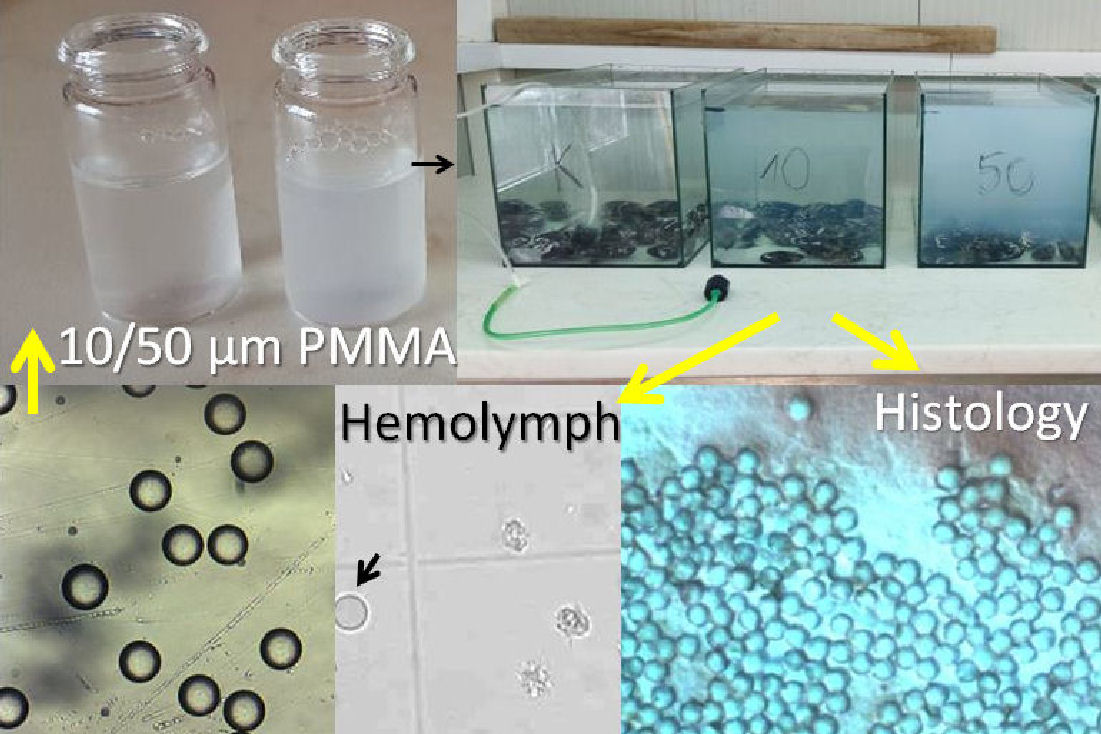
2.1. Microparticles and Chemicals

Virgin polymethylmethacrylate (PMMA) particles with nominal diameters of 10 µm and 50 µm (experimentally determined by light microscopy to be 9.83 ± 0.75 and 51.87 ± 2.69 μm, respectively, (mean ± standard deviation)) were obtained from Microbeads SA, Norway and used without further treatment. Trypan Blue, Neutral Red dye, hematoxylin, eosin, hexane, glycerol gelatine, 5,5′-dithiobis-2-dinitrobenzoic acid, Triton-X-100, acetylthiocholine iodide, hydrogen peroxide, EDTA, 1-Chloro-2,4-dinitrobenzene, β-NADPH, ethanol, and salts for sodium and potassium phosphate buffers were purchased from Sigma-Aldrich, St. Louis, MO, USA.

2.2. The Model Organism and Sampling Site

Mediterranean mussels, Mytilus galloprovincialis, were sampled in May 2019 at the mariculture area of Lim Bay (45°07′50″ N 13°44′10″ E), in the northern Adriatic Sea, Croatia, when the seawater temperature was 15.0 °C and salinity S·36.3. The total number of mussels used for experiments was 374 and all mussels were acclimated for 3 days in aquaria with fresh running seawater (temperature was 14.2 °C and salinity S·36.8 over the course of the experiment) in the Center for Marine Research, Ruđer Bošković Institute, Rovinj, Croatia.

2.3. Experimental Setup and Sampling Procedure

The experiment was set up in seven stand-alone, aerated aquaria comprising: one control aquarium; three aquaria with 10 μm PMMA microplastics (concentrations of 0.1, 1 and 10 mg L−1, respectively); and three aquaria with 50 μm PMMA microplastics (0.1, 1 and 10 mg L−1, respectively). These concentrations, covering several orders of magnitude, were chosen to encompass concentrations which can potentially range from non-toxic to toxic in marine organisms; for example, this range corresponded with similar concentrations used in some urchin embryo development studies [52,53]. Each aquarium contained 50 mussels (6.3 ± 0.5 cm) in a total seawater volume of 37.5 L. The mussels were exposed to the different concentrations and sizes of microplastics for 3 days, with all the seawater changed and the microplastics re-added every 24 h. The mussels were not fed during the experiment. After 24, 48, and 72 h three mussels were taken from each aquarium for haemocyte analysis, and three replicates were made for each mussel. After three days of exposure to PMMA microplastics the remaining 41 mussels in each aquarium were divided as follows: 5 mussels from each aquarium were set aside for determining the condition index (CI), 30 mussels were selected from each aquarium for a stress-on-stress (SOS) test, and 6 mussels from each aquarium were used for both histological and biochemical analyses. The gills and hepatopancreas tissue from these latter mussels were divided and stored at −80 °C until further histological and biochemical analyses.
The lysosomal membrane stability experiments were conducted separately; 3 mussels were exposed in aquaria (2.25 L) to each of the 6 different size and concentration combinations of microplastics along with two control groups (24 mussels in total) for 72 h.

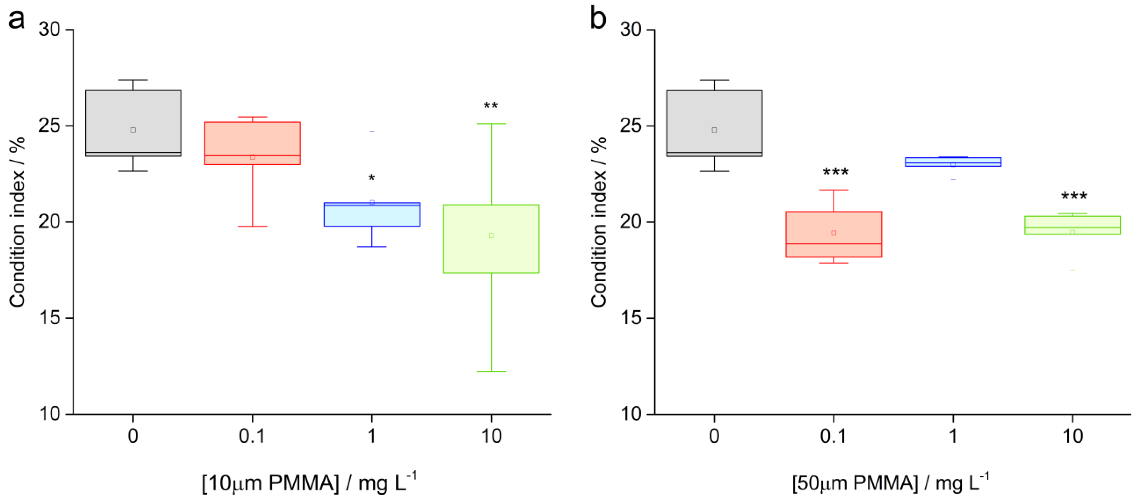
2.4. Condition Index (CI)

For calculating the CI, the ratio between the soft tissue and the whole mass of the five mussels taken from each aquarium was determined. Shell length, width, and height were measured by digital caliper (0.1 mm precision), and total mussel weight (TW) and wet meat weight (WMW) were measured by digital scales (0.01 g precision). The CI was calculated by [35]:
CI (%) = WMW (g) × 100/TW (g)

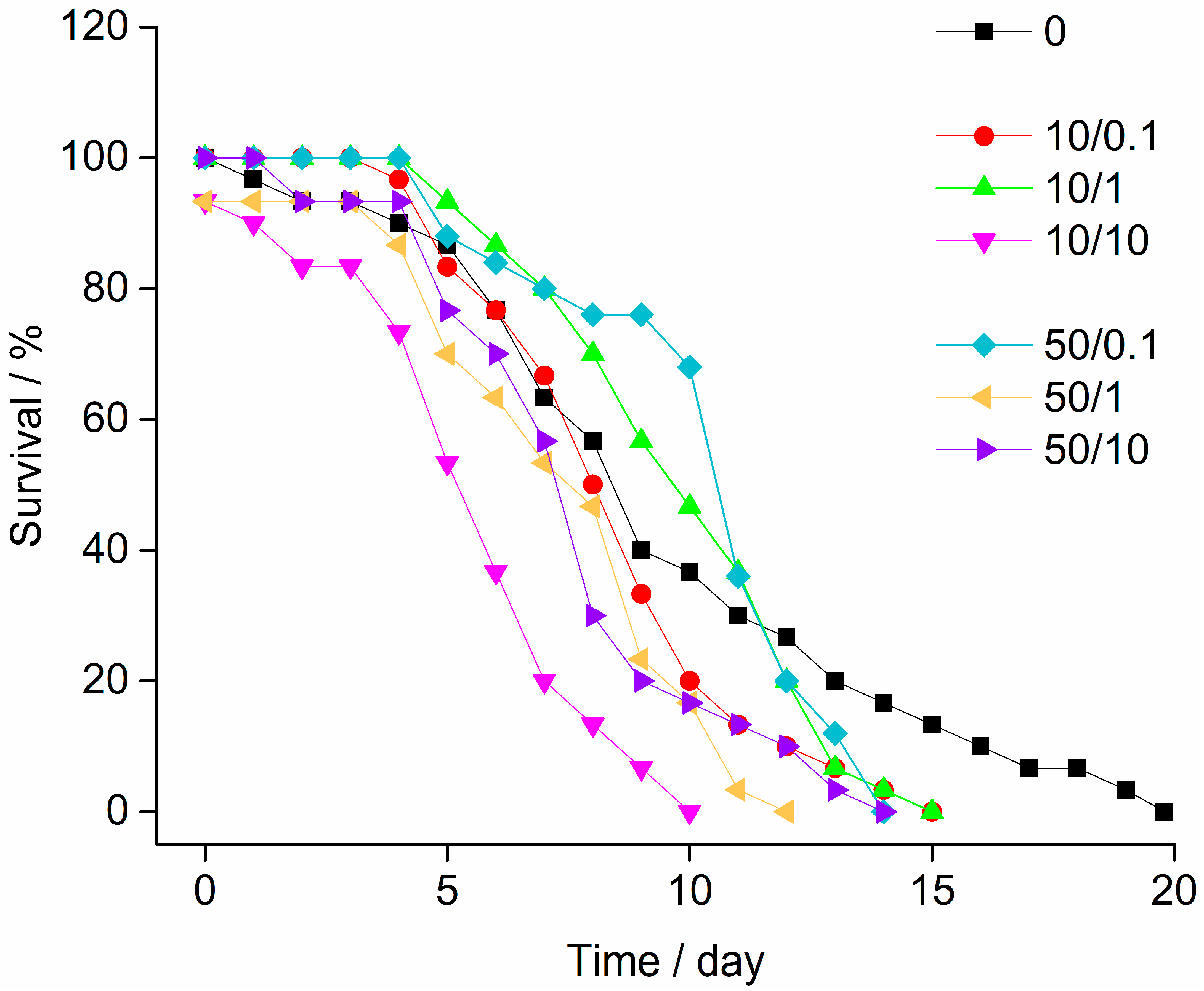
2.5. Stress-on-Stress (SOS) Test

The 30 mussels selected from each aquarium for the SOS test were placed on coarse filter paper in deep polystyrene trays and held at 18 °C in high humidity. Every day at the same time, the number of dead mussels was recorded, and these mussels were discarded. The LT50 values, i.e., the time by which 50% of the mussels died, with the corresponding confidence intervals were calculated [55].

2.6. Haemolymph Analyses

The three mussels taken from each aquarium at the 24, 48 and 72 h time-points were used for haemocyte analysis, where haemolymph was retrieved (0.1–0.5 mL) from the anterior adductor muscle with a 25-gauge needle and a 1 mL plastic syringe. All samples were held on ice to prevent haemocyte clumping. Haemocytes were counted in triplicate samples for each mussel.

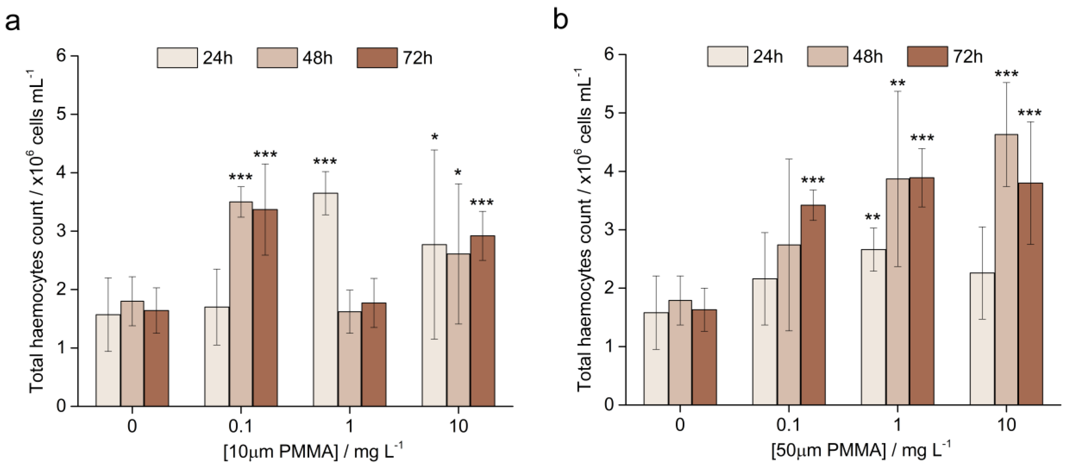
2.6.1. Total Haemocyte Count (THC)

The THC determination was performed on 50 mL aliquots of haemolymph fixed at 1:1 with 4% formaldehyde by counting the cells in triplicate samples from each mussel in an improved Neubauer chamber on a Nikon Diaphot microscope.

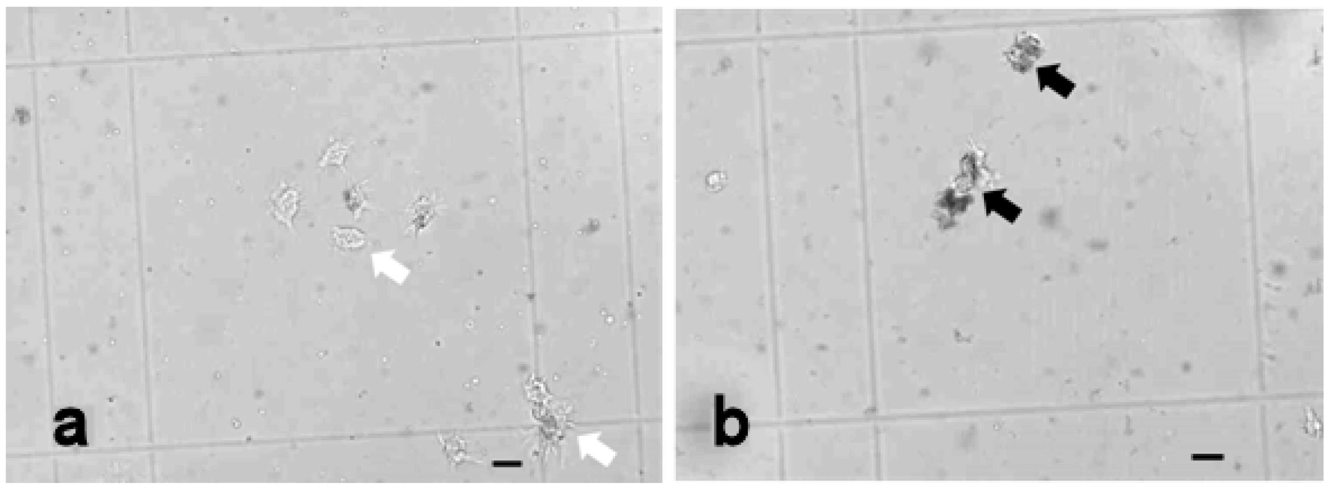
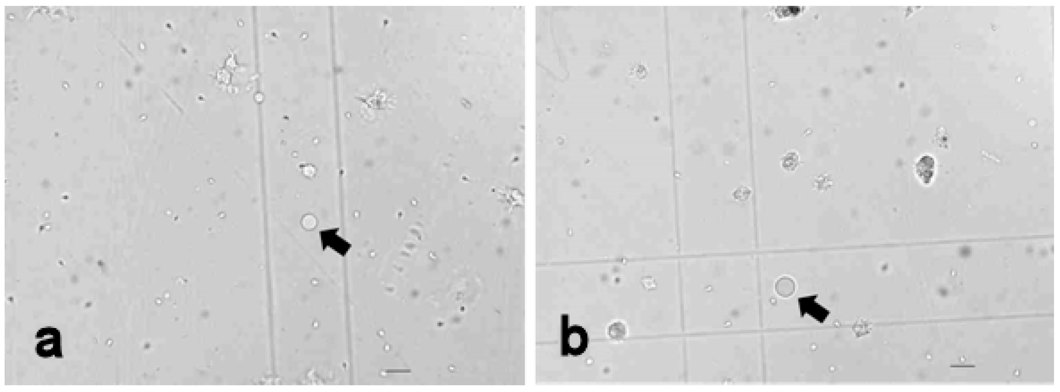
2.6.2. Vacuolised Haemocytes

The occurrence of vacuolation as well as ‘multi nuclei’ (multi-nucleated haemocytes, micronuclei, and bi-nucleated cells), and the deformation of the nucleus such as blebbing, were assessed by a Nikon Diaphot microscope in triplicate samples from each mussel [37,56].

2.6.3. Cell Viability

The viability of the haemocytes, given as a percentage of the total haemocyte number, was assessed by staining triplicate aliquots of unfixed haemolymph (20 µL) from each mussel with trypan blue, where unstained cells represented the viable cells in the suspension [57].

2.6.4. Lysosomal Membrane Stability

The lysosomal membrane stability of haemocytes (in vivo exposure) was assessed by the Neutral Red retention assay. Briefly, microscope slides were treated with poly-L-lysine and incubated in a dark and humid chamber for 30 min. Haemolymph was extracted from the adductor muscle of three mussels from each aquarium (i.e., each experimental exposure), and three 40 μL aliquot replicates from each mussel were transferred onto the microscope slides which were again left in a dark, humid chamber (to attach to the slide surface) for 30 min. The excess haemolymph was removed and 0.0006% w/v Neutral Red solution was added. The slides were incubated in a humidity chamber for 15 min and then a cover slip was added. The slides were scored at 15, 30, 45, 60, 90, 120, 150, and 180 min. for retention or leakage of dye from the cells.

2.7. Histological Analysis

Gill and hepatopancreas tissue for histology analysis was excised from six mussels from each aquarium and flash frozen in N-hexane, pre-chilled in liquid nitrogen, and stored at −80 °C until the preparation of histological sections. Before cryosectioning, the tissue was attached to a microtome carrier and blended into an O.C.T. medium (Microm Inc. GmbH, Germany). The tissue samples were placed on a cryostat (Zeiss Hyrax C50) previously cooled to −30 °C and 10 μm-thick sections were prepared. The frozen cross-sections were placed on slides, heated to room temperature, and washed with hematoxylin and eosin. After staining, the sections were incorporated into glycerol gelatine and the localisation of the microplastics was assessed by polarised light microscopy (Zeiss Axiovert 200) [58].

2.8. Biochemical Analyses—Enzymatic Activities

A portion of the tissue that was excised from the six mussels (from each aquarium) not used for histological analysis was used for conducting enzyme activity assays. The gills and digestive glands were removed, labelled and frozen in liquid nitrogen, and then stored at −80 °C until further analyses. All the tissue analyses were carried out at 0–4 °C. The tissue was homogenised using a Teflon Potter homogeniser in specific buffers depending on the enzyme activity being measured. The samples were assayed for enzyme activities using a Tecan M200 Infinite Pro microplate-reader (Männedorf, Switzerland) (AChE, GST and GR) and a Shimadzu UV-1800 spectrophotometer (Kyoto, Japan) (CAT).

2.8.1. AcetylCholinesterase Activity (AChE)

In determining the AChE, the tissue was weighed and homogenised in NaH2PO4/Na2HPO4 buffer (0.02 M, pH 7) with 0.1% Triton-X-100, with the tissue and buffer volume ratio being 1:3. The homogenate was centrifuged at 10,000× g, for 30 min at 4 °C. The reaction mixture (300 µL) contained 25 µL of sample, 225 µL 5,5′-dithiobis-2-dinitrobenzoic acid (0.665 mM), and 50 µL acetylthiocholine iodide (15.6 mM) as the substrate. The absorbance increase at 415 nm was recorded every 30 s for 4 min and 30 s. AChE activity was expressed as specific activity in nanomoles of hydrolysed acetylthiocholine per minute per mg of protein (nmol−1 min−1 mgprot−1).

2.8.2. The Activity of Catalase (CAT)

CAT was determined using the same sample homogenate as prepared in the AChE assay. The reaction mixture (1.5 mL) contained 20 µL of sample, 700 µL 30 mM hydrogen peroxide, and 780 µL phosphate buffer (50 mM, pH 7). The decrease in absorbance was measured for 30 s at 240 nm. CAT activity was expressed as specific activity in nanomoles per minute per mg of protein (nmol−1 min−1 mgprot−1).

2.8.3. The Activity of Glutathione S–Transferase (GST)

The GST was determined on tissue that was homogenised in 0.05 M KH2PO4/K2HPO4 buffer (pH 7.4) with 2 mM EDTA, in the ratio 1:2 (w/v). The homogenate was centrifuged at 10,000× g, for 30 min at 4 °C. The reaction mixture (300 µL) contained 5 µL sample and 295 µL Master Mix (100 mM 1-Chloro-2,4-dinitrobenzene dissolved in absolute ethanol and 200 mM reduced glutathione dissolved in distilled water). An increase in absorbance was measured every 30 s for 4 min and 30 s at 595 nm. The GST activity was expressed as specific activity in nanomoles per minute per mg of protein (nmol−1 min−1 mgprot−1).

2.8.4. The Activity of Glutathione Reductase (GR)

For determining GR, the same homogenate used for the GST assay was used. The reaction mixture contained 20 µL of sample, 100 µL buffer (sodium phosphate buffer; 0.1 M, pH 7.2), 100 µL GSSG (2 mM), and 10 µL β-NADPH (1 mM) as substrate. The decrease in absorbance was measured every 30 s for 10 min at 340 nm. GR activity was expressed as specific activity in nanomoles per minute per mg of protein (nmol−1 min−1 mgprot−1).

2.9. Statistics

A statistical analysis of parameters including THC, cell viability, vacuolised cells and CI, lysosomal membrane stability, and enzymatic activity was carried out with a ONE WAY ANOVA using the Statistica 9.0 software. The data were checked for normality (Shapiro–Wilk test) and homoscedasticity (Levene’s test), and when these conditions were met, a parametric ANOVA with a post hoc Tukey HSD test was used to determine the significant differences between groups. In the cases where the data did not meet the requirements for parametric ANOVA, a non-parametric Kruskal–Wallis analysis was conducted, followed by a Mann–Whitney U-test when statistical differences were indicated.

3. Results

3.1. Condition Index (CI)

The CI of the mussels showed a trend towards lower values as a function of concentration of microparticles, even after the short experimental exposure reported herein. Compared to the median value of control (CON) mussels (24%), the median CI values of mussels exposed to 1 mg L−1 (MID) and 10 mg L−1 (HIGH) 10 µm PMMA microparticles decreased to 21% and 17%, respectively, while those exposed to the lowest 10 µm microplastics concentration of 0.1 mg L−1 (LOW) microparticles did not show a statistically significant difference to the control (Figure 1a).
Exposure to the larger 50 μm PMMA microparticles resulted in low CI values for all microparticle concentrations except for the 1.0 mg L−1 (MID) exposure (Figure 1b). The low and high microparticle concentrations induced a reduction of CI to 19% and 20% respectively, while the mid-concentrations showed a CI value close to that of the control mussels. Statistically significant differences from the control were found for the 0.1 mg L−1 (LOW) and the 10 mg L−1 (HIGH) treatments at a significance level of p < 0.001.

3.2. Stress-on-Stress (SOS) Test

The times of survival of mussels in the air, which represent an additional stress after 72 h of exposure to 10 µm and 50 µm PMMA microparticles, over three weeks are shown in Figure 2. The mussels treated with lower concentrations of microplastics of both sizes showed LT50 values similar to the control LT50 of 8.32 days (Table 1). The lowest survival over time, and hence the most rapid mortality, was noted for mussels exposed to a concentration of 10 mg L−1 (HIGH) of 10 µm PMMA. After ten days, all the mussels were dead, with a calculated LT50 value of 4.35 days. The greatest negative effect was also noted for the 1 (MID) and 10 mg L−1 (HIGH) concentrations of 50 μm microparticles, where very similar LT50 values of about 6.5 days were determined.

3.3. Haemolymph Analysis

3.3.1. The Total Haemocyte Count (THC)

The THC for the control mussels (CON) and mussels exposed to 0.1 mg L−1 (LOW), 1.0 mg L−1 (MID), and 10 mg L−1 (HIGH) concentrations of 10 µm PMMA particles after 24, 48, and 72 h are given in Figure 3a. The THC in the control group remained relatively constant over 3 days with approximately 1.7 million cells mL−1. By the second and third day, the THC had doubled for the mussels exposed to 0.1 mg L−1 while similar large increases, to approximately 3 million cells mL−1, were also noted for the 10 mg L−1 (HIGH) PMMA microparticle treatment at all time points. In the mussels exposed to the intermediate concentration of 1 mg L−1 (MID), the THC similarly increased to a high value of nearly 4 million cells mL−1, which showed the greatest statistical difference (p < 0.001) from the controls. However, the THC for this microparticle concentration returned to values close to those of the controls after 48 and 72 h. The reason for such a return to normal levels is unclear, and assigning a more rapid removal of microparticles in these mussels as the cause of the reduced impact on the immune system would be speculative, particularly as similar behaviour was not seen after exposure to the 0.1 (LOW) and 10 mg L−1 (HIGH) microparticle concentrations.
For mussels exposed to 50 μm microparticles, similar trends were noted. In particular, the THC increased over time for each tested microparticle concentration, and the THC also increased as a function of concentration (Figure 3b), with the highest values recorded for the 10 mg L−1 (HIGH) after 48 h. For example, the ANOVA indicated that this value was statistically different from the control value at the level p < 0.001.

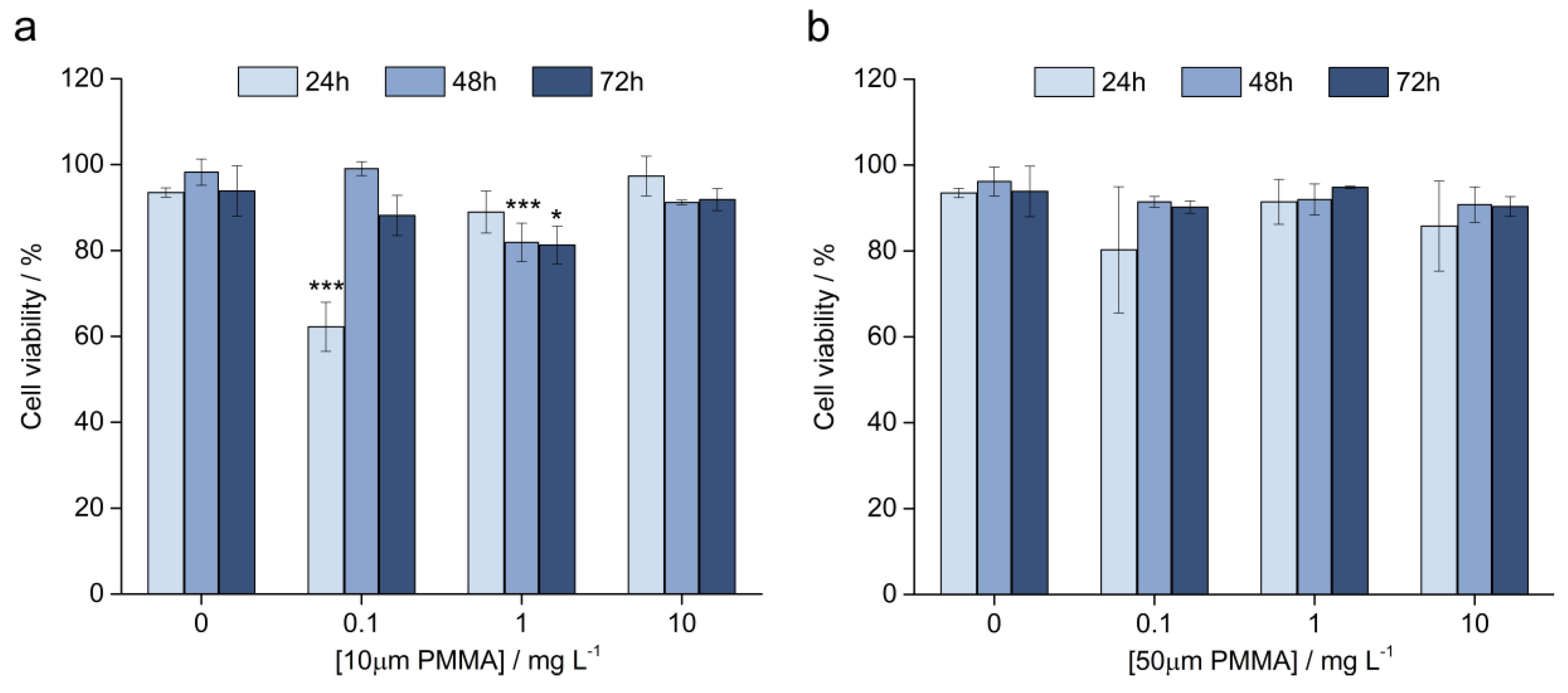
3.3.2. Cell Viability

The cell viability of haemocytes was investigated as a function of exposure time and microparticle diameter. The control group showed cell viability of about 95% over 72 h while in mussels exposed to 0.1 mg L−1 (LOW) microparticles of 10 μm diameter, cell viability decreased to 60% in the first 24 h and then recovered to about 90% when measured after 48 h and 72 h (Figure 4a). For the higher microparticle concentration of 1 mg L−1 (MID) the cell viability gradually decreased over 72 h to about 80%, while the highest microparticle concentration showed a slight decrease to about 90% after 72 h exposure.
For the mussels exposed to 50 μm microparticles, a similar effect for the 0.1 (LOW) and 10 mg L−1 (HIGH) treatments was noted where the number of viable cells decreased (to 80–85%) after 24 h, but then recovered to about 90% after 48 h exposure (Figure 4b). The 1 mg L−1 (MID) treatment showed qualitatively the same behaviour, though with a reduced variability over 72 h, with cells typically showing about 90% viability.

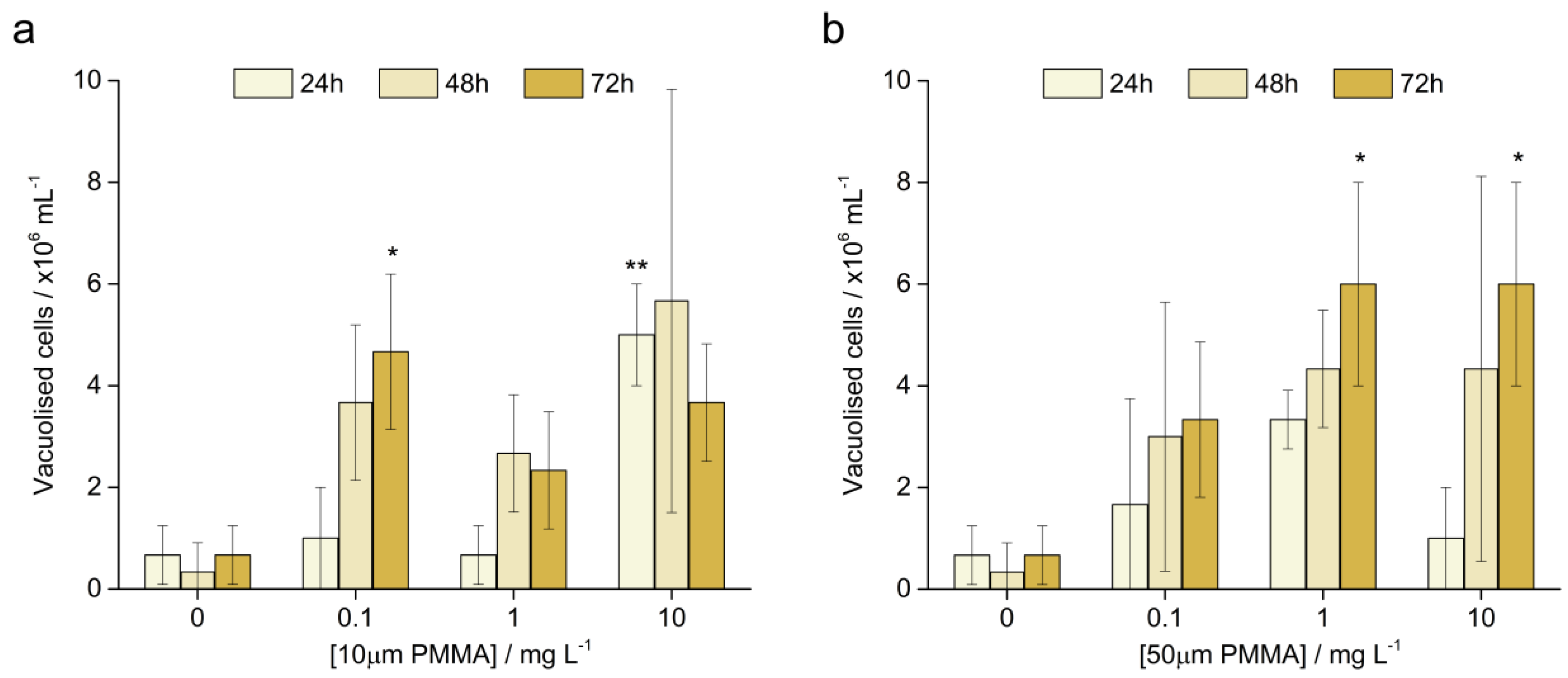
3.3.3. Vacuolised Cells

The occurrence of haemocyte vacuolisation, known as ‘multi nuclei’ (multi-nucleated haemocytes, micronuclei, and bi-nucleated cells), and deformation of the nuclei of haemocytes was observed in exposed mussels (Figure 5).
The number of vacuolised cells in the control mussels (CON) and mussels exposed to 0.1 mg L−1 (LOW), 1.0 mg L−1 (MID), and 10 mg L−1 (HIGH) PMMA microparticles of 10 µm and 50 μm diameters are shown in Figure 6. In the control group, the number of vacuolised cells remained very low to the end of the experiment, while after 48 and 72 h the 0.1 (LOW) and 1 mg L−1 (MID) treatments showed an increase in vacuolisation by a factor of about 4 and 3, respectively. The highest PMMA microparticle concentration of 10 mg L−1 (HIGH) was found to cause the greatest amount of cell vacuolisation, including from the first day of the experimental exposure (p < 0.001; Figure 6a).
Similar values were found for the experimental treatments with 50 μm microparticles. Over the course of 72 h the number of vacuolised cells increased for each individually tested microparticle concentration (Figure 6b). Moreover, the number of vacuolised cells was also noted to be a function of microparticle concentration, with the 0.1, 1, and 10 mg L−1 concentrations increasing the average vacuolisation of the cells to about 3 × 10⁶, 4 × 10⁶, and 4 × 10⁶ cells mL−1, respectively, from the control value of about 1 × 10⁶ cells mL−1.
The stability of the lysosomal membrane of the haemocytes was investigated each day and no statistically significant differences from the controls were found up to the 2 h point (data not shown). While the test is usually run over the course of 2 h, we allowed the test to continue to 3 h, and at this test time point the haemocytes from the mussels exposed to the highest concentration of 10 μm particles indicated that less than 50% of them still maintained membrane stability. After 72 h of mussel exposure, the reduction in membrane stability became more pronounced at the 3 h mark for the 10 mg L−1 (HIGH) 10 μm and 1 mg L−1 (MID) 50 μm treatments. It should be noted that the haemocytes were not exposed to the plastics in vitro, but rather in vivo, hence the milder effects and lack of lysosomal membrane instability.

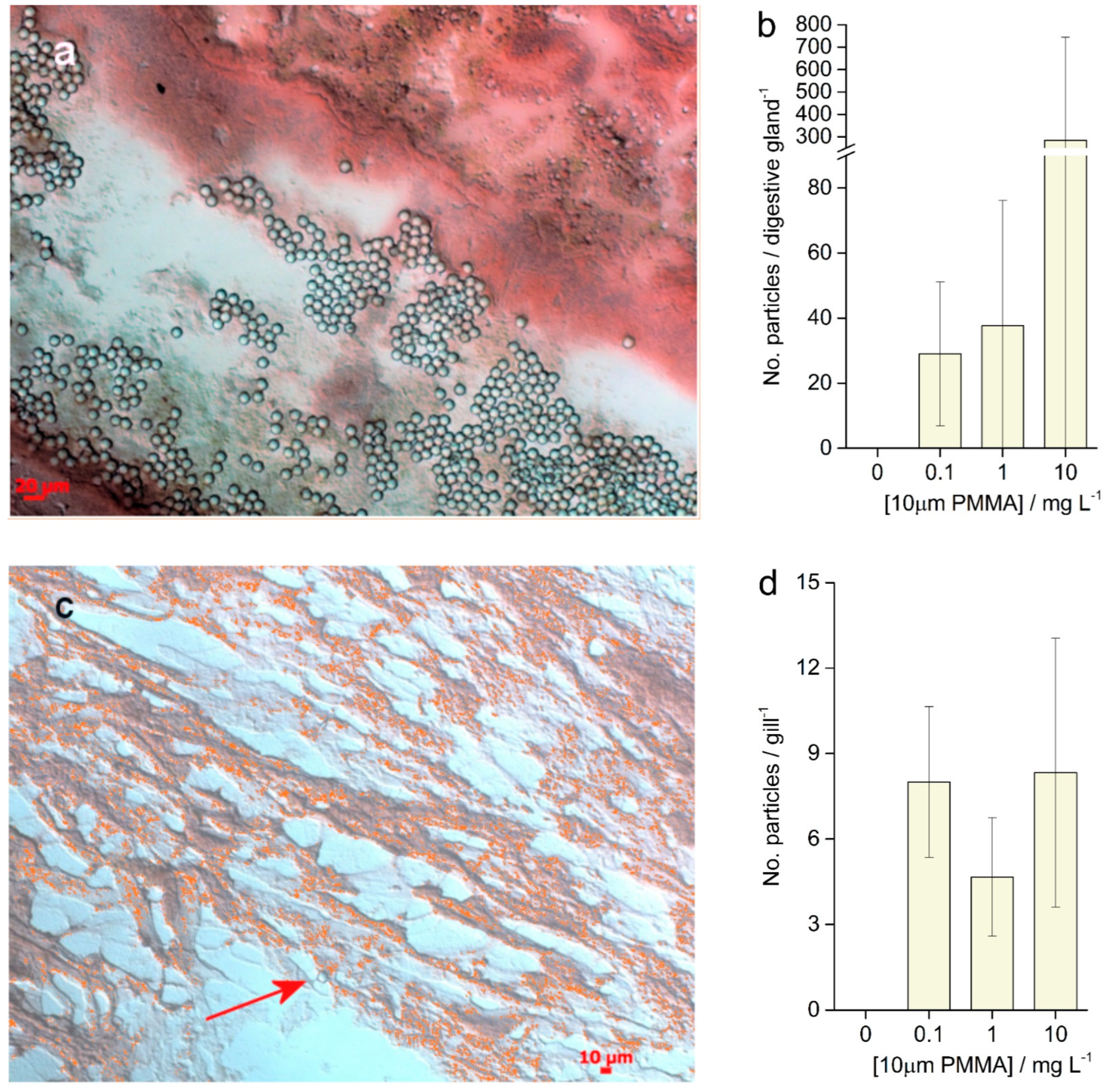
3.4. Histological Analysis

Both particle sizes of PMMA (10 µm and 50 µm) were observed in the mussel haemolymph after the experimental exposures, for example as shown in Figure 7.
Cryosections of the mussel digestive gland (hepatopancreas) and gills after exposure to PMMA microplastics (10 mg L−1 (HIGH) for 24 h) showed the presence of 10 μm particles, while 50 µm PMMA particles were not observed in the mussel tissue (Figure 8a,c). The particle counts in the tubules of the hepatopancreas showed average values of 25 particles hepatopancreas−1 while a large increase in the number of particles was noted for the 10 mg L−1 (HIGH) microparticle treatment (an average of 300 particles hepatopancreas−1; Figure 8b). The gill tissue cryosections showed far fewer microparticles, with averages of 5–8 particles gill−1 observed, and did not show a trend with respect to increasing the exposure concentrations.

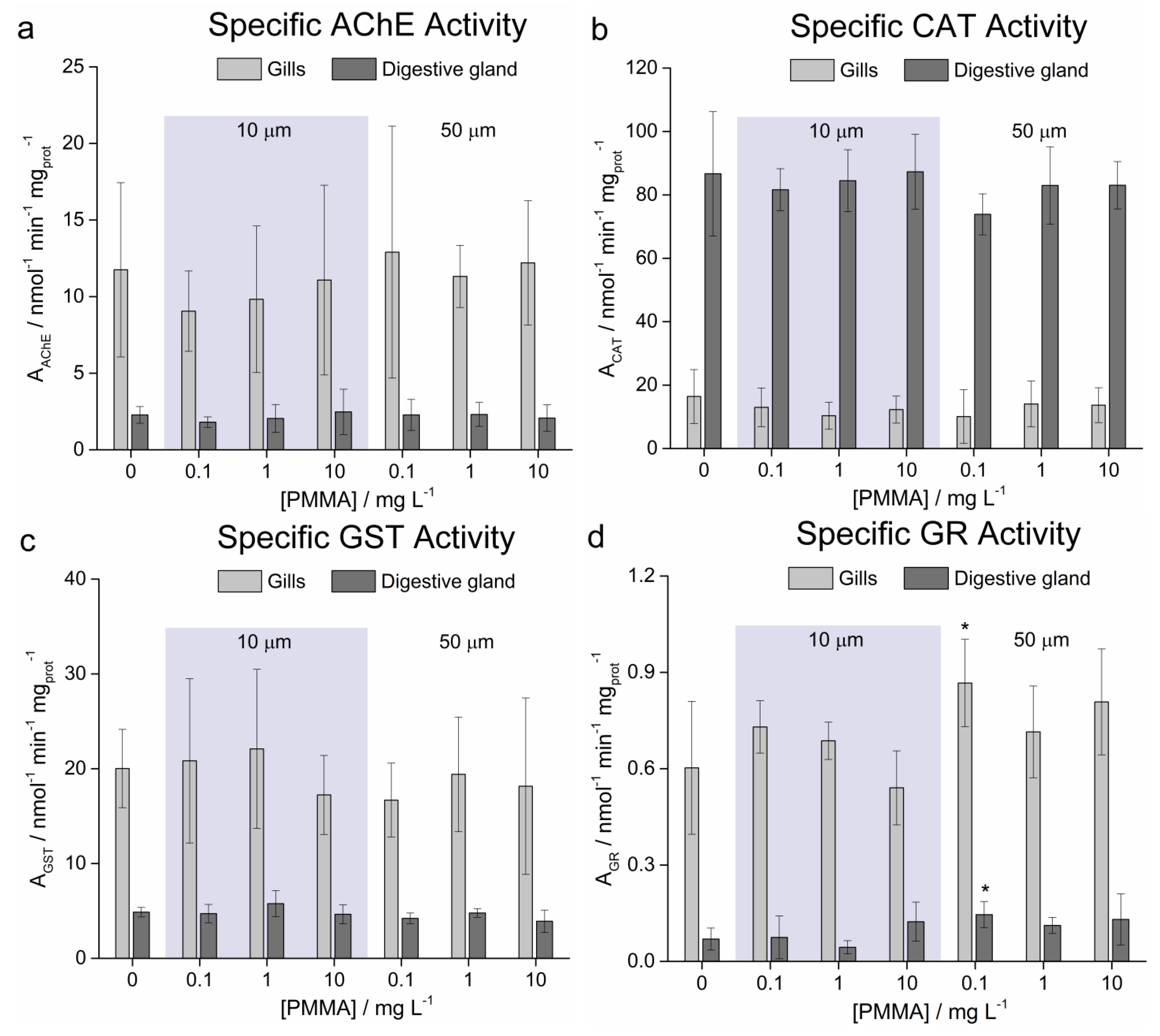
3.5. Enzymatic Activities

The specific activities of a range of enzymes in the mussel gill and hepatopancreas tissue were determined after 72 h exposure of mussels to the 10 μm and 50 μm PMMA microplastics. Acetylcholinesterase activity in the gills was slightly suppressed for the smaller microparticles compared to the larger particles, although it was not statistically different from the control (Figure 9a). Catalase activity also showed values close to the control without any trends apparent (Figure 9b). Glutathione-S-transferase activity in the gill tissue was lower than the control for the highest concentration of 10 μm microparticles and all the 50 μm microparticles (Figure 9c), while glutathione reductase activity in the gills and hepatopancreas was higher than the control in nearly all cases except for the 10 mg L−1 in the gills and the 1 mg L−1 in the hepatopancreas treatments, respectively, with 10 μm particles (Figure 9d). Statistically significant differences from the controls were only found for the 0.1 mg L−1 (LOW) treatment at p < 0.05 to the 50 μm PMMA microplastics.

4. Discussion

The widespread occurrence of microplastics has become a significant problem in the environment, particularly in the case of marine waters where microplastics can have significant ecological impacts [59]. This concern arises not only due to the possibility for larger microplastics to cause obstruction and blockage of the digestive tracts of a wide range of organisms by ingestion, but also at the microscale where very small microplastics may impact directly on the cells, tissues, and organs of marine animals. Furthermore, it is these smaller microplastics, rather than the larger fragments, that have the potential to cause important consequences for marine animals as their size range overlaps with that of the food supply [13].
The Mediterranean mussel, M. galloprovincialis, is a species of high ecological and commercial importance and is utilised worldwide as a sentinel organism of pollution in coastal marine environments [15,55]. As it is widely distributed in urbanised coastal environments it is likely to be exposed to microplastics sources over its entire life cycle. In this context, the effects of the two particle sizes (10 μm and 50 μm) of PMMA in the low (0.1 mg L−1), medium (1 mg L−1), and high (10 mg L−1) concentrations on mussels, M. galloprovincialis, were investigated during and after a 72 h exposure.
The microplastics uptake by the mussel gills and the subsequent transport of these particles to the haemolymph and digestive system is known to cause adverse effects on mussel health. Microplastics can be taken up via the surfaces in the digestive glands of mussels by endocytosis and granulocytomas and then transferred to the lysosomes and the circulatory system or eliminated as pseudofaecal particles [4,43,46,60,61]. The present study found that the smaller particles (10 μm) are easily taken up into the tissues whereby they are captured by the gills, accumulate in the hepatopancreas, and are transported to the haemolymph, leading to negative effects on mussel health. An early study of polystyrene microparticles showed that mussels exposed to microplastic particles had accumulated polystyrene microspheres in their gut cavities and digestive tubules [4]. After three days, the ingested microplastics accumulated in the circulatory fluid of mussels and particles were found in the haemolymph and haemocytes. A later study also observed the presence of high-density polyethylene (HDPE) in the blue mussel, M. edulis, [46]. Particles were found in the intestine, in the lumina of the digestive gland, and in the endocytotic vacuoles of the digestive cells, which suggests that the HDPE microparticles were taken up via the mouth, transported to the gastrointestinal tract, and internalised into cells of the digestive system by endocytosis.
Considering microparticle size as a factor in microplastics uptake, it is likely that the ease of uptake is a function of its decreasing size. For example, only microplastics of the smallest size (10 μm) were detected in mussels exposed to three sizes (10 μm, 30 μm, and 90 μm) used in an exposure experiment [62]. The data reported herein are consistent with that study where the larger particles (50 µm) were not found in the tissues of the gills and the digestive gland, although it should be noted that some of these 50 μm microparticles were observed in the haemolymph.
Mussel health status was evaluated by measuring the condition index (CI) and stress-on-stress test (SOS test) after 72 h exposure to PMMA particles. The CI provides a broad measure of the physiological activity of organisms under given environmental conditions, including pollution, and is an important ecophysiological measure of the health status of mussels. For example, the CI has found use as an important tool for assessing of the quality of meat for the marketplace [63]. Furthermore, it summarises the physiological activity of the organisms under given environmental conditions [64]. Research has shown that abiotic (temperature, salinity, and food availability) and biotic (reproductive cycle) factors may influence mussel CI [30,35,58,65]. It is important to note that the experiment reported herein was performed in May, following a spawning period, after which the mussels had returned to a ‘normal’ physiological state. The CI of mussels exposed to 10 µm and 50 μm PMMA particles showed statistically significant decreased values with increased concentration of microparticles, although it was observed that the effects of the smaller PMMA particle size (10 µm) generally had a lesser influence on mussel CI than the larger particles (50 µm). While the mid- and higher concentrations of 10 µm particles showed differences from the control, more highly significant differences were noted for the low and high concentrations of 50 µm particles. However, other endpoints such as the total haemocyte count, cell viability and vacuolisation indicate significant negative effects at low concentrations of the 10 µm microplastics. Therefore, we cannot make a general statement based on the condition index alone that low concentrations of small PMMA microplastics are of little cause for concern in the environment. The reason for the larger 50 µm particles showing a greater effect on the condition index compared to the smaller 10 µm particles, in broad terms, is likely due to the smaller particles not being retained on the gills, but passing into the digestive gland to eventually be removed, while the larger particles remain attached to the gills, disrupting the organism’s respiration and resulting in a lower mussel fitness. Overall, the range of CI measured in this study is similar to that previously reported for Lim Bay in the spring [66], which was higher than that for a range of other locations, suggesting that this important mariculture area is less polluted (higher CI) than those other locations investigated. In contrast to the present results on PMMA, exposure to HDPE microparticles was not found to negatively affect the CI of mussels after a similar exposure time [46].
As a further means to test the general fitness of the mussels after exposure to microplastics, the mussels were left in air as a means of achieving an elevated general stress condition. This so-called SOS test is a physiological biomarker used for measuring the ability of mussels to survive exposure to air after prior exposure to other stressors such as heavy metals and organic chemicals [55]. The SOS test has found wide acceptance as an indicator of the general physiological state of mussels; for example, any change in ecological factors in the habitat affects the stress levels of the organism, which may be subsequently assessed by the SOS test [67,68,69]. Herein, the SOS test showed that mussels exposed to the highest concentration (10 mg L−1) of 10 μm microparticles lived for a significantly shorter time than mussels in the untreated control group, while the LT50 values for mussels exposed to lower concentrations (0.1 and 1 mg L−1) did not show a statistically significant difference from the controls LT50 values. Similarly, the highest concentration of the larger 50 µm PMMA particles showed a slight reduction in resistance to stress compared to the controls, while the lower concentrations did not result in significantly shorter survival times compared to the control group. Comparing the CI and SOS data, it is clear that these PMMA microparticles can cause elevated levels of overall stress in the organism over short time periods, which clearly affects the general health of the mussel.
Considering the individual systems in the organism, haemocytes are macrophages that play a key role in the immune system, contributing to a defence against parasites, pathogens, and pollutants [57], and provide a convenient way to assess the stressors of the immune system [70]. While cell viability is typically the most used metric [71], other endpoints such as haemocyte vacuolation, expressed in the form of multi-nucleated haemocytes, micronuclei, and binucleated cells, and the deformation of the nucleus such as blebbing, provide a large number of assessment criteria for assessing perturbations in the mussel immune system [37,71]. As haemocytes circulate in the haemolymph throughout the animal and the major organs, any microparticles associated with the haemocytes or in the haemolymph may achieve a widespread distribution in the organism. Studies have shown the uptake of polystyrene particles (3.0 μm and 9.6 µm) in the haemolymph and haemocytes of Mytilus edulis after three days, with the smaller particles observed in significantly higher amounts compared to the larger microspheres [4]. Interestingly, the particles were still detected in the haemolymph after 48 days. A similar study on microplastics which exposed M. galloprovincialis to 3 μm and 45 µm polystyrene microparticles for four days at 1 and 10 particles mL−1 confirmed that even at these low concentrations polystyrene particles could be detected in the haemolymph of all treated mussels [72]. The present study showed that different sizes of PMMA microparticles could also enter the haemolymph, hence indicating that chemical identity might not be a key factor in microplastic uptake. In addition, it was observed that the lowest concentration of PMMA microparticles induced a significant increase in the number of haemocytes relative to the control group after 48 h (10 μm) and 72 h (50 μm), while at higher concentrations the THC had already started to significantly increase within the first 24 h. This immune response against the presence of 10 μm and 50 μm PMMA microparticles not only shows the activation of a highly sensitive defence mechanism, but also indicates that these microplastics are identified as a threat to the organism. Our study confirms that exposure to MPs induced a significant increase in haemocyte formation, thus these increases are correlated with the concentration of PMMA particles over the time of exposure.
Cell viability, as a means to assess the response and health of cells in organisms after treatment with various toxicants, was also monitored, and the smaller PMMA microparticles (10 μm) were found in some cases to significantly reduce the viability of haemocytes. In parallel, there was a general increase in cell vacuolisation with the increasing microparticle concentration and time, for example, for each concentration of 50 μm microparticle. Negative correlations between cell viability and vacuolation of haemocytes have previously been reported for Mytilus trossulus and also assigned to an immune response and the effect of increased pollution levels [37]. For example, treatment of M. galloprovincialis with toxicants such as zinc chloride manifested in the increased mortality of haemocytes such that, after 72 h and 7 days exposure at concentrations of 0.5 and 1 mg mL−1, significant mortality compared to controls was observed [57].
Histological analysis confirmed the presence of 10 µm PMMA microparticles deep in the gill tissue and in the digestive glands of exposed mussels, with the level of abundance in the tissue increasing with the exposure concentration, primarily in digestive tubules in the hepatopancreas. Surprisingly, although 50 μm particles were observed in the haemolymph, they were not found in the tissue samples. However, other studies on polystyrene (3.0 and 9.6 µm) [4] and HDPE (0–80 µm) particles [46] in M. edulis found the presence of these particles in the gut cavity, the intestine, in the lumina of the primary and secondary ducts of the digestive gland, and in the endocytotic vacuoles of digestive epithelial cells. Particles were also found in the blood lacunae of the gills and the areas of lamellar junctions. Not only mussels, but also crabs have shown ingestion of microplastics, with shore crabs, Carcinus maenas, exposed to 8–10 µm polystyrene microspheres having detectable numbers of microspheres on their gills [73]. It is a matter of speculation as to why larger PMMA microparticles were not evident in the tissues in the present study, although it is possible that following uptake the chemical identity of the polymer acted in concert with a size conducive to depuration in pseudofaeces to induce faster expulsion of these particles after uptake. For example, a size effect was recently reported for polystyrene particles where the larvae of M. edulis ingested plastic particles of a nano- and micro-scale, although a higher amount of the 2 µm polystyrene beads was ingested compared to the 100 nm particles [45]. This adds to the evidence that size is an important factor in terms of particle uptake and may point to particles that are commensurate in size with the organisms’ usual prey being more likely to be taken up, particularly during active feeding. Interestingly, a recent study on the impact of 10 μm polystyrene and PMMA particles on embryonic development of sea urchins, Paracentrotus lividus, showed that the PMMA particles were retained in the gut while the uptake or retention of the polystyrene microparticles was not observed, again with hints that the chemical identity of the polymer may in some way play a role in the microparticles’ interaction with the animal [52]. However, in the natural environment, it is likely that polymer microparticles will quickly become encapsulated in an eco-corona of natural organic matter, which will mask the chemical signature of the microplastics surface, thus diminishing the distinguishing features among the chemically different microparticles.
Not only is the size of the microplastic an important factor, but also the size of the organism. For example, the size of mussels also impacts upon the ingestion of microplastics. A significant positive correlation was found between mussel body size and the number of ingested particles, such that larger mussels are prone to accumulating more microparticles than smaller sized mussels [74]. As oftentimes the sizes of the mussels used for experimental exposures are not reported, this represents one aspect of variability among data reported for various studies.
Apart from provoking an immune response in adult organisms such as in this study, other reports have highlighted that the early life phases of biota can also be negatively impacted. Larvae of M. edulis, which was found to ingest polystyrene nano- and micro-particles (100 nm, 2 μm), did not display negative consequences in terms of larval growth, but the number of abnormally developed larvae increased after exposure to the polystyrene beads [45]. Similarly, PMMA microparticles were also recently shown to impact deleteriously on the early development phases in sea urchin, Sphaerechinus granularis, embryos [53]. PMMA microparticle exposure has increased the developmental (skeletal) defects, cytogenetic abnormalities (increased mitotic aberration), and transmissible damage from sperm to (eventual) zygotes.
While the immune system was challenged, the microparticles did not induce significant changes in the activity of a range of enzymes engaged in neurotransmission and the cellular control of reactive oxygen species. Data reported in this study have shown that M. galloprovincialis can effectively ingest and accumulate PMMA particles, suggesting their potential role as microplastic primary consumers/accumulators in marine ecosystems. It has been noted that the proportion of morphotype and polymer types in mussels and seawater are consistent, suggesting that microplastics in mussels can reflect the real pollution status in the environment [43]. For example, the abundance of microbeads in mussels was significantly higher in the high concentration exposure group than in the low concentration group. Furthermore, it has been noted that microplastic abundance in mussels was closely related to human activity, and mussels from areas with intensive human activities contain significantly higher numbers of microplastics than mussels in putatively unpolluted mariculture areas [75]. Thus, a higher number of microplastics in mussels may provide an indication of anthropogenic activity and is closely correlated with the degree of pollution in coastal habitats, reflecting the real abundance of microplastics in the environment within a certain size range [15]. This then represents a more pressing concern as mussels, particularly from coastal mariculture, are a commercially important crop for human consumption and may enable trophic transfer of microplastics (and any adsorbed substances and secondary pollutants) to humans.
Overall, it must be borne in mind that the present study and many other investigations were performed using (virgin) polymer microparticles in controlled laboratory conditions; thus, the observed responses might not necessarily reproduce those occurring in natural systems, where organisms are simultaneously exposed to a broad range of structurally heterogeneous microplastic particles of irregular shape–size and further associated stressors. Nevertheless, the data herein highlight for the first time the vulnerability of bivalves towards the PMMA microplastics, providing baseline information for future investigations addressing their impact on marine ecosystems.

5. Conclusions

While the impact of microplastics such as those of polystyrene and polyethylene on biota have received significant attention to date, the effects of polymethylmethacrylate (PMMA, ‘plexiglass’) microplastics on marine organisms have not yet been sufficiently addressed. As the chemical identity of PMMA microparticles differs from other microplastics, it is important to understand if the surface chemistry or the size are the drivers of toxicity, particularly in filter-feeders such as clams, oysters, scallops, and mussels, which may be especially impacted. The results obtained in this study provide the first evidence that PMMA microparticles are taken up into the gills and digestive gland of the mussel, Mytilus galloprovincialis, and cause significant effects on mussel health. In a broad context, the impacts of the smaller particle sizes (10 μm) on mussel M. galloprovincialis have been observed to induce more significant effects compared to the larger (50 μm) particles at the corresponding concentrations, for example, on THC and cell viability at 0.1 and 1 mg L−1 concentrations, and on SOS LD50 at 10 mg L−1. The general physiological fitness of mussels, as indicated by the CI and SOS data, is rapidly impacted by the PMMA microparticles and decreased with exposure to the higher concentrations and smaller sizes of microparticles. The presence of PMMA microparticles in mussel tissues (only 10 μm) and in the haemolymph correlated with negative physiological effects on mussel health. The uptake of microplastics into the haemolymph induced an immune response as indicated by the reduced cell viability and vacuolisation, with concomitantly increasing THC. While the immune system was challenged, the microparticles did not induce significant changes in activity of a range of enzymes engaged in cellular control of reactive oxygen species. Overall, this study highlights how PMMA microparticle size is important in terms of the different internalisation mechanisms in mussel tissues, which correlates with deleterious effects on animal fitness and health. However, further studies in other animal models would be useful to fully uncover the potential hazards that may arise from PMMA microplastics.

Author Contributions

Conceptualization, D.P.-H.; Methodology, D.P.-H., M.M. and D.M.L.; Formal Analysis, D.P.-H., I.K. and D.M.L.; Investigation, D.P.-H., T.S., I.K. and M.M.; Resources, D.P.-H. and D.M.L.; Writing—Original Draft Preparation, D.P.-H.; Writing—Review & Editing, D.M.L.; Funding Acquisition, D.M.L. All authors have read and agreed to the published version of the manuscript.

Funding

This research has received funding from the Croatian Science Foundation in the framework of project IP-2018-01-5351 “Understanding the physico-chemical behaviour and toxicity of silver, copper and plastic nanoparticles as emerging materials of concern in coastal waters”.

Institutional Review Board Statement

Not applicable.

Data Availability Statement

All data generated and quoted in this study is reported herein.

Conflicts of Interest

The authors declare no conflict of interest.

References

  1. Plastics Europe, Plastics—The Facts 2021. Available online: https://plasticseurope.org/wp-content/uploads/2021/12/Plastics-the-Facts-2021-web-final.pdf (accessed on 30 August 2022).
  2. UNEP Marine Plastic Debris and Microplastics—Global Lessons and Research to Inspire Action and Guide Policy Change; United Nations Environment Programme: Nairobi, Kenya, 2016.
  3. Pham, C.K.; Ramirez-Llodra, E.; Alt, C.H.S.; Amaro, T.; Bergmann, M.; Canals, M.; Company, J.B.; Davies, J.; Duineveld, G.; Galgani, F.; et al. Marine litter distribution and density in European seas, from the shelves to deep basins. PLoS ONE 2014, 9, e95839. [Google Scholar] [CrossRef] [PubMed] [Green Version]
  4. Browne, M.A.; Dissanayake, A.; Galloway, T.S.; Lowe, D.M.; Thompson, R.C. Ingested Microscopic Plastic Translocates to the Circulatory System of the Mussel, Mytilus edulis (L.). Environ. Sci. Technol. 2008, 42, 5026–5031. [Google Scholar] [CrossRef] [PubMed]
  5. Browne, M.A.; Galloway, T.S.; Thompson, R.C. Spatial patterns of plastic debris along estuarine shorelines. Environ. Sci. Technol. 2010, 44, 3404–3409. [Google Scholar] [CrossRef]
  6. Andrady, A.L. Microplastics in the marine environment. Mar. Pollut. Bull. 2011, 60, 1596–1605. [Google Scholar] [CrossRef]
  7. Barnes, D.K.A.; Galgani, F.; Thompson, R.C.; Barlaz, M. Accumulation and fragmentation of plastic debris in global environments. Philos. Trans. R. Soc. B 2009, 364, 1985–1998. [Google Scholar] [CrossRef] [PubMed] [Green Version]
  8. Kershaw, P.J. (Ed.) GESAMP Sources, fate, and effects of microplastics in the marine environment: A global assessment. In IMO/FAO/UNESCO-IOC/UNIDO/WMO/IAEA/UN/UNEP/UNDP Joint Group of Experts on the Scientific Aspects of Marine Environmental Protection; Rep. Stud. GESAMP; FAO: Rome, Italy, 2015; No. 90; p. 96. [Google Scholar]
  9. Suaria, G.; Avio, C.G.; Mineo, A.; Lattin, G.L.; Magaldi, M.G.; Belmonte, G.; Moore, C.J.; Regoli, F.; Aliani, S. The Mediterranean plastic soup: Synthetic polymers in Mediterranean surface waters. Sci. Rep. 2016, 6, 37551. [Google Scholar] [CrossRef] [PubMed] [Green Version]
  10. Palatinus, A.; Viršek, M.K.; Robič, U.; Grego, M.; Bajt, O.; Šiljić, J.; Suaria, G.; Liubartseva, S.; Coppini, G.; Peterlin, M. Marine litter in the Croatian part of the middle Adriatic Sea: Simultaneous assessment of floating and seabed macro and micro litter abundance and composition. Mar. Pollut. Bull. 2019, 139, 427–439. [Google Scholar] [CrossRef]
  11. Vianello, A.; Da Ros, L.; Boldrin, A.; Marceta, T.; Moschino, V. First evaluation of floating microplastics in the northwestern Adriatic Sea. Environ. Sci. Poll. Res. 2018, 25, 28546–28561. [Google Scholar] [CrossRef] [PubMed]
  12. Avio, C.G.; Gorbi, S.; Regoli, F. Plastics and microplastics in the oceans: From emerging pollutants to emerged threat. Mar. Environ. Res. 2016, 128, 2–11. [Google Scholar] [CrossRef]
  13. Sharma, S.; Chatterjee, S. Microplastic pollution, a threat to marine ecosystem and human health; a short review. Environ. Sci. Pollut. Res. 2017, 24, 21530–21547. [Google Scholar] [CrossRef] [PubMed]
  14. Andbumani, S.; Kakkar, P. Ecotoxicological effects of microplastics on biota: A review. Environ. Sci. Pollut. Res. 2018, 25, 14373–14396. [Google Scholar] [CrossRef] [PubMed]
  15. Li, J.; Lusher, A.L.; Rotchell, J.M.; Deudero, S.; Turra, A.; Brate, I.L.N.; Sun, C.; Shahadat Hossain, M.; Li, Q.; Kolandhasamy, P.; et al. Using mussel as a global bioindicator of coastal microplastic pollution. Environ. Pollut. 2019, 244, 522–533. [Google Scholar] [CrossRef] [PubMed]
  16. Li, J.; Yang, D.; Li, L.; Jabeen, K.; Shi, H. Microplastics in commercial bivalves from China. Environ. Pollut. 2015, 207, 190–195. [Google Scholar] [CrossRef] [PubMed]
  17. Luster, A. Marine Anthropogenic Litter, Chapter 10. In Microplastics in the Marine Environment: Distribution, Interactions and Effects; Springer Open: Cham, Switzerland, 2015; pp. 245–307. [Google Scholar] [CrossRef] [Green Version]
  18. Wright, S.L.; Kelly, F.J. Plastic and human health: A micro issue? Environ. Sci. Technol. 2017, 51, 12, 6634–6647. [Google Scholar] [CrossRef] [PubMed]
  19. Tosetto, L.; Williamson, J.E.; Browne, C. Trophic transfer of microplastics does not affect fish personality. Anim. Behav. 2017, 123, 159–167. [Google Scholar] [CrossRef]
  20. Laist, D.W. Overview of the biological effects of lost and discarded plastic debris in the marine environment. Mar. Pollut. Bull. 1987, 18, 319–326. [Google Scholar] [CrossRef]
  21. Derraik, J.G.B. The pollution of the marine environment by plastic debris: A review. Mar. Pollut. Bull. 2002, 44, 842–852. [Google Scholar] [CrossRef]
  22. Thompson, R.C.; Olsen, Y.; Mitchell, R.P.; Davis, A.; Rowland, S.J.; John, A.W.G.; McGonigle, D.; Russell, A.E. Lost at sea: Where is all the plastic? Science 2004, 304, 838–844. [Google Scholar] [CrossRef] [PubMed]
  23. Browne, M.A.; Galloway, T.S.; Thompson, R.C. Microplastic—an emerging contaminant of potential concern? Integr. Environ. Assess. Manag. 2007, 3, 559–561. [Google Scholar] [CrossRef] [PubMed]
  24. Moore, C.J. Synthetic polymers in the marine environment: A rapidly increasing, long-term threat. Environ. Res. 2008, 108, 131–139. [Google Scholar] [CrossRef] [PubMed]
  25. Wright, S.L.; Thompson, R.C.; Galloway, T.S. The physical impacts of microplastics on marine organisms: A review. Environ. Pollut. 2013, 178, 483–492. [Google Scholar] [CrossRef] [PubMed]
  26. Cole, M.; Lindeque, P.; Halsband, C.; Galloway, T.S. Microplastics as contaminants in the marine environment: A review. Mar. Pollut. Bull. 2011, 62, 2588–2597. [Google Scholar] [CrossRef] [PubMed]
  27. Rochman, C.M.; Tahir, A.; Williams, S.L.; Baxa, D.V.; Lam, R.; Miller, J.T.; Teh, F.; Werorilangi, S.; Teh, S.J. Anthropogenic debris in seafood: Plastic debris and fibers from textiles in fish and bivalves sold for human consumption. Sci. Rep. 2015, 5, 14340. [Google Scholar] [CrossRef] [PubMed] [Green Version]
  28. Hodkovicova, N.; Hollerova, A.; Caloudova, H.; Blahova, J.; Franc, A.; Garajova, M.; Lenz, J.; Tichy, F.; Faldyna, M.; Kulich, P.; et al. Do foodborne polyethylene microparticles affect the health of rainbow trout (Oncorhynchus mykiss)? Sci. Total Environ. 2021, 793, 148490. [Google Scholar] [CrossRef] [PubMed]
  29. Seth, K.; Shriwastav, A. Contamination of Indian sea salts with microplastics and a potential prevention strategy. Environ. Sci. Pollut. Res. 2018, 25, 30122–30131. [Google Scholar] [CrossRef]
  30. Pavičić-Hamer, D.; Kovačić, I.; Koščica, L.; Hamer, B. Physiological indices of maricultured mussel Mytilus galloprovincialis Lamarck, 1819 in Istria, Croatia: Seasonal and transplantation effect. J. World Aquac. Soc. 2016, 47, 768–778. [Google Scholar] [CrossRef]
  31. Hamer, B.; Korlević, M.; Durmiši, E.; Nerlović, V.; Bierne, N. Nuclear marker Me 15-16 analyses of Mytilus galloprovincialis populations along the eastern Adriatic coast. Cah. Biol. Mar. 2012, 53, 35–44. [Google Scholar] [CrossRef]
  32. Goldberg, E.D. The mussel wach-a first step in global marine monitoring. Mar. Pollut. Bull. 1975, 6, 111–114. [Google Scholar] [CrossRef]
  33. Hamer, B.; Pavičić-Hamer, D.; Muller, W.E.G.; Batel, R. Stress-70 proteins in marine mussel Mytilus galloprovincialis as biomarkers of environmental pollution: A field study. Environ. Int. 2004, 30, 873–882. [Google Scholar] [CrossRef]
  34. Hamer, B.; Pavičić-Hamer, D.; Muller, W.E.G.; Zahn, R.; Batel, R. Detection of stress-70 proteins of mussel Mytilus galloprovincialis using 2-D gel electrophoresis:a proteomics approach. Fresenius Environ. Bull. 2005, 14, 605–611. [Google Scholar]
  35. Hamer, B.; Jaksic, Z.; Pavicic-Hamer, D.; Peric, L.; Medakovic, D.; Ivankovic, D.; Pavicic, J.; Zilberberg, C.; Schroder, H.C.; Muller, W.E.G.; et al. Effect of hypoosmotic stress by low salinity acclimation of Mediterranean mussels Mytilus galloprovincialison biological parameters used for pollution assessment. Aquat. Toxicol. 2008, 89, 137–151. [Google Scholar] [CrossRef] [PubMed]
  36. Faggio, C.; Tsarpali, V.; Dailianis, S. Mussel digestive gland as a model tissue for assessing xenobiotics: An overview. Sci. Total Environ. 2018, 636, 220–229. [Google Scholar] [CrossRef]
  37. Höher, N.; Turja, R.; Köhler, A.; Lehtonen, K.K.; Broeg, K. Immunological responses in the mussel Mytilus trossulus transplanted at the coastline of the northern Baltic Sea. Mar. Environ. Res. 2015, 112, 113–121. [Google Scholar] [CrossRef] [PubMed]
  38. Livingstone, D.R.; Pipe, R.K. Mussels and environmental contaminants: Molecular and cellular aspects. In Developments in Aquaculture and Fisheries Science; Gosling, E.M., Ed.; Elsevier Science Publishers B.V.: Amsterdam, The Netherlands, 1992; pp. 425–464. [Google Scholar]
  39. Famme, P.; Riisgård, H.U.; Jørgensen, C.B. On direct measurement of pumping rates in the mussel Mytilus edulis. Mar. Biol. 1986, 92, 323–327. [Google Scholar] [CrossRef]
  40. Ward, J.E.; Shumway, S.E. Separating the grain from the chaff: Particle selection in suspension- and deposit-feeding bivalves. J. Exp. Mar. Biol. Ecol. 2004, 300, 83–130. [Google Scholar] [CrossRef]
  41. Gonçalves, C.; Martins, M.; Sobral, P.; Costa, P.M.; Costa, M.H. An assessment of the ability to ingest and excrete microplastics by filter-feeders: A case study with the Mediterranean mussel. Environ. Pollut. 2019, 245, 600–606. [Google Scholar] [CrossRef]
  42. Gomiero, A.; Strafella, P.; Birger Oysaed, K.; Fabi, G. First occurrence and composition assessment of microplastics in native mussels collected from coastal and offshore areas of the northern and central Adriatic Sea. Environ. Sci. Pollut. Res. 2019, 26, 24407–24416. [Google Scholar] [CrossRef]
  43. Qu, X.; Su, L.; Li, H.; Liang, M.; Shi, H. Assessing the relationship between the abundance and properties of microplastics in water and in mussels. Sci. Total. Environ. 2018, 621, 679–686. [Google Scholar] [CrossRef]
  44. Paul-Pont, I.; Lacroix, C.; González Fernández, C.; Hégaret, H.; Lambert, C.; Le Goïc, N.; Frère, L.; Cassone, A.-L.; Sussarellu, R.; Fabioux, C.; et al. Exposure of marine mussels Mytilus spp. to polystyrene microplastics: Toxicity and influence on fluoranthene bioaccumulation. Environ. Pollut. 2016, 216, 724–737. [Google Scholar] [CrossRef] [Green Version]
  45. Rist, S.; Baun, A.; Almeda, R.; Hartmann, N.B. Ingestion and effects of micro- and nanoplastics in blue mussel (Mytilus edulis) larvae. Mar. Pollut. Bull. 2019, 140, 423–430. [Google Scholar] [CrossRef]
  46. von Moos, N.; Burkhardt-Holm, P.; Kohler, A. Uptake and Effects of Microplastics on Cells and Tissue of the Blue Mussel Mytilus edulis L. after an Experimental Exposure. Environ. Sci. Technol. 2012, 46, 11327–11335. [Google Scholar] [CrossRef]
  47. Détrée, C.; Gallardo-Escárate, C. Single and repetitive microplastics exposures induce immune system modulation and homeostasis alteration in the edible mussel Mytilus galloprovincialis. Fish Shellfish Immunol. 2018, 83, 52–60. [Google Scholar] [CrossRef]
  48. Tomazic-Jezic, V.J.; Merritt, K.; Umbreit, T.H. Significance of the type and the size of biomaterial particles on phagocytosis and tissue distribution. J. Biomed. Mater. Res. 2001, 55, 523–529. [Google Scholar] [CrossRef]
  49. FAO. Microplastics in Fisheries and aquaculture. In FAO Fisheries and Aquaculture Technical Paper 615; FAO: Rome, Italy, 2017. [Google Scholar]
  50. O’Neill, P.; Barrett, A.; Sullivan, T.; Regan, F.; Brabazon, D. Rapid Prototyped Biomimetic Antifouling Surfaces for Marine Applications. Mater. Today 2016, 3, 527–532. [Google Scholar] [CrossRef]
  51. Kühn, S.; Schaafsma, F.L.; van Werven, B.; Flores, H.; Bergmann, M.; Egelkraut-Holtus, M.; Tekman, M.B.; van Franeker, J.A. Plastic ingestion by juvenile polar cod (Boreogadus saida) in the Arctic Ocean. Polar Biol. 2018, 41, 1269–1278. [Google Scholar] [CrossRef] [Green Version]
  52. Thomas, P.J.; Oral, R.; Pagano, G.; Tez, S.; Toscanesi, M.; Ranieri, P.; Trifuoggi, M.; Lyons, D.M. Mild toxicity in Paracentrotus lividus early life stages may indicate species-specific sensitivity to polystyrene and polymethylmethacrylate microplastics. Mar. Environ. Res. 2020, 161, 105132. [Google Scholar] [CrossRef]
  53. Trifuoggi, M.; Pagano, G.; Oral, R.; Pavičić-Hamer, D.; Burić, P.; Kovačić, I.; Siciliano, A.; Toscanesi, M.; Thomas, P.J.; Paduano, L.; et al. Microplastic-induced damage in early embryonal development of sea urchin Sphaerechinus granularis. Environ. Res. 2019, 179, 108815. [Google Scholar] [CrossRef] [PubMed]
  54. Hermsen, E.; Pompe, R.; Besseling, E.; Koelmans, A.A. Detection of low numbers of microplastics in North Sea fish using strict quality assurance criteria. Mar. Pollut. Bull. 2017, 122, 253–258. [Google Scholar] [CrossRef] [PubMed]
  55. Viarengo, A.; Canesi, L.; Pertica, M.; Mancinelli, G.; Accomando, R.; Smaal, A.C.; Orunesu, M. Stress on stress response: A simple monitoring tool in the assessment of a general stress syndrome in mussels. Mar. Environ. Res. 1995, 39, 245–248. [Google Scholar] [CrossRef]
  56. Fenech, M.; Chang, W.P.; Kirsch-Volders, M.; Holland, N.; Bonassi, S.; Zeiger, E. HUMN project: Detailed description of the scoring criteria for the cytokinesis block micronucleus assay using isolated human lymphocyte cultures. Mutat. Res. Genet. Toxicol. Environ. Mutagen. 2003, 534, 65–75. [Google Scholar] [CrossRef]
  57. Pagano, M.; Porcino, C.; Briglia, M.; Fiorino, E.; Vazzana, M.; Silvestro, S.; Faggio, C. The influence of exposure of cadmium chloride and zinc chloride on haemolymph and digestive gland cells from Mytilus galloprovincialis. Int. J. Environ. Res. 2017, 11, 207–216. [Google Scholar] [CrossRef]
  58. Kovačić, I.; Pavičić-Hamer, D.; Phannkuchen, A.M.; Usich, M. Mytilus galloprovincialis (Lamarck, 1819) as host of Mytillcola orientalis (Mori, 1935) in the northern Adriatic Sea: Presence and effect. Aquac. Int. 2016, 25, 211–221. [Google Scholar] [CrossRef]
  59. Thomas, P.J.; Perono, G.; Tommasi, F.; Pagano, G.; Oral, R.; Burić, P.; Kovačić, I.; Toscanesi, M.; Trifuoggi, M.; Lyons, D.M. Resolving the effects of environmental micro- and nanoplastics exposure in biota: A knowledge gap analysis. Sci. Total Environ. 2021, 780, 146534. [Google Scholar] [CrossRef] [PubMed]
  60. Avio, C.G.; Gorbi, S.; Milan, M.; Benedetti, M.; Fattorini, D.; d’Errico, G.; Pauletto, M.; Bargelloni, L.; Regoli, F. Pollutants bioavailability and toxicological risk from microplastics to marine mussels. Environ. Pollut. 2015, 198, 211–222. [Google Scholar] [CrossRef] [PubMed]
  61. Beyer, J.; Green, N.W.; Brooks, S.; Allan, I.J.; Ruus, A.; Gomes, T.; Bråte, I.L.N.; Schøyen, M. Blue mussels (Mytilus edulis spp.) as sentinel organisms in coastal pollution monitoring: A review. Mar. Environ. Res. 2017, 130, 338–365. [Google Scholar] [CrossRef] [PubMed]
  62. Van Cauwenberghe, L.; Claessens, M.; Vandegehuchte, M.B.; Janssen, C.J. Microplastics are taken up by mussels (Mytilus edulis) and lugworms (Arenicola marina) living in natural habitats. Environ. Pollut. 2015, 199, 10–17. [Google Scholar] [CrossRef]
  63. Orban, E.; Di Lena, G.; Nevigato, T.; Casini, I.; Marzetti, A.; Caproni, R. Seasonal changes in meat content, condition index and chemical composition of mussels (Mytilus galloprovincialis) cultured in two different Italian sites. Food Chem. 2002, 77, 57–65. [Google Scholar] [CrossRef]
  64. Lucas, A.; Beninger, P.G. The use of physiological condition indices in marine bivalve aquaculture. Aquaculture 1985, 44, 187–200. [Google Scholar] [CrossRef]
  65. Okumuş, İ.; Stirling, H.P. Seasonal variations in the meat weight, condition index and biochemical composition of mussels (Mytilus edulis L.) in suspended culture in two Scottish sea lochs. Aquaculture 1998, 159, 249–261. [Google Scholar] [CrossRef]
  66. Kanduč, T.; Šlejkovec, Z.; Falnoga, I.; Mori, N.; Budič, B.; Kovačić, I.; Pavičić-Hamer, D.; Hamer, B. Ecological status of the Istrian marine environment (NE Adriatic Sea, Croatia): Insights from mussel Mytilus galloprovincialis condition indices, stable isotopes and metal(loid)s. Marine Pollution Bulletin 2017, 126, 525–534. [Google Scholar] [CrossRef] [PubMed]
  67. Thrush, S.F.; Hewitt, J.E.; Hickey, W.C.; Kelly, S. Multiple stressor effects identified from species abundance distributions: Interactions between urban contaminants and species habitat relationships. J. Exp. Mar. Biol. Ecol. 2008, 366, 160–168. [Google Scholar] [CrossRef]
  68. Andrade, M.; Soares, A.; Figueira, E.; Freitas, R. Biochemical changes in mussels submitted to different time periods of air exposure. Environ. Sci. Pollut. Res. 2018, 25, 8903–8913. [Google Scholar] [CrossRef] [PubMed]
  69. Hewitt, J.E.; Thrush, S.F. Do Species’ Abundances become More Spatially Variable with Stress? Open J. Ecol. 2009, 10, 111–118. [Google Scholar] [CrossRef] [Green Version]
  70. Carballal, M.J.; Lopez, M.C.; Azevedo, C.; Villalba, A. Hemolymph cell types of the mussel Mytilus galloprovincialis. Dis. Aquat. Org. 1997, 29, 127–135. [Google Scholar] [CrossRef] [Green Version]
  71. King, M.A. Detection of dead cells and measurement of cell killing by flow cytometry. J. Immunol. Methods 2000, 243, 155–166. [Google Scholar] [CrossRef]
  72. Franzellitti, S.; Capolupo, M.; Wathsala, R.H.G.R.; Valbonesi, P.; Fabbri, E. The multixenobiotic resistance system as a possible protective response triggered by microplastic ingestion in Mediterranean mussels (Mytilus galloprovincialis): Larvae and adult stages. Comp. Biochem. Physiol. C 2019, 219, 50–58. [Google Scholar] [CrossRef] [PubMed]
  73. Watts, A.J.R.; Lewis, C.; Goodhead, R.M.; Beckett, S.J.; Moger, J.; Tyler, C.R.; Galloway, T.S. Uptake and retention of microplastics by the shore crab Carcinus maenas. Environ. Sci. Technol. 2014, 48, 8823–8830. [Google Scholar] [CrossRef] [PubMed]
  74. Brate, L.N.; Hurley, R.; Iversen, K.; Beyer, J.; Thomas, K.V.; Steindal, C.C.; Green, N.W.; Olsen, M.; Lusher, A. Mytilus spp. as sentinels for monitoring microplastic pollution in Norwegian coastal waters: A qualitative and quantitative study. Environ. Pollut. 2018, 243, 383–393. [Google Scholar] [CrossRef] [PubMed]
  75. Li, J.; Qu, X.; Su, L.; Zhang, W.; Yang, D.; Kolandhasamy, P.; Li, D.; Shi, H. Microplastics in mussels along the coastal waters of China. Environ. Pollut. 2016, 214, 177–184. [Google Scholar] [CrossRef]
Figure 1. The CI of the control mussels (CON) and mussels exposed to 0.1 mg L−1 (LOW), 1.0 mg L−1 (MID) and 10 mg L−1 (HIGH) PMMA microparticles of (a) 10 µm diameter and (b) 50 µm diameter. The boxes indicate the 2nd and 3rd quartiles, with the median value shown, and the whiskers denote the non-outlier ranges. The open circles indicate the outliers. Statistical significance is indicated at the levels * p < 0.05, ** p < 0.01, *** p < 0.001.
Fishes 07 00307 g001
Figure 2. Cumulative survival of the control mussels and mussels exposed to different concentrations of 10 µm and 50 µm PMMA particles through time. The legend indicates the microparticle size (10, 50 μm)/concentration (0.1, 1, 10 mg L−1), with the control group denoted as CON.
Fishes 07 00307 g002
Figure 3. The THC (mean ± SD) after 24, 48, and 72 h for control mussels (CON) and mussels exposed to 0.1 mg L−1 (LOW), 1.0 mg L−1 (MID), and 10 mg L−1 (HIGH) PMMA microparticles of (a) 10 µm diameter and (b) 50 µm diameter. Statistical significance is indicated at the levels * p < 0.05, ** p < 0.01, *** p < 0.001.
Fishes 07 00307 g003
Figure 4. Cell viability (mean ± SD) after 24, 48, and 72 h for control mussels (CON) and mussels exposed to 0.1 mg L−1 (LOW), 1.0 mg L−1 (MID), and 10 mg L−1 (HIGH) PMMA microparticles of (a) 10 µm diameter and (b) 50 µm diameter. Statistical significance is indicated at the levels * p < 0.05, *** p < 0.001.
Fishes 07 00307 g004
Figure 5. (a) normal haemocytes (white arrow) from control mussels and (b) malformed haemocytes (black arrow) from mussels exposed to 50 µm PMMA particles with vacuolisation and deformed nuclei. Scale bar 10 µm.
Fishes 07 00307 g005
Figure 6. The number of vacuolised cells mL−1 (mean ± SD) after 24, 48, and 72 h for the control mussels (0 mg L−1) and mussels exposed to 0.1 mg L−1 (LOW), 1.0 mg L−1 (MID), and 10 mg L−1 (HIGH) PMMA microparticles of (a) 10 µm diameter and (b) 50 µm diameter. Statistical significance is indicated at the levels * p < 0.05, ** p < 0.01.
Fishes 07 00307 g006
Figure 7. Micrographs of the haemolymph of mussels exposed for 48 h to 0.1 mg L−1 PMMA microparticles of diameters (a) 10 µm and (b) 50 µm. Microparticles are indicated by the arrows. Scale bar 20 µm.
Fishes 07 00307 g007
Figure 8. Micrographs of 10 µm microparticles in a cryosection of the (a) hepatopancreas (scale bar 20 μm) and (c) gill (arrow indicates microparticle; scale bar 10 μm) for mussels exposed to 10 mg L−1 (HIGH) PMMA, and particle counts (mean ± SD) in (b) hepatopancreas and (d) gill cryosections for mussels exposed to 0.1 mg L−1 (LOW), 1.0 mg L−1 (MID) and 10 mg L−1 (HIGH) microparticles.
Fishes 07 00307 g008
Figure 9. Enzymatic activity in gills and digestive glands after exposure to different sizes (10 and 50 μm)/concentrations (0.1, 1, 10 mg L−1) of PMMA microparticles: (a) acetylcholinesterase (AChE), (b) catalase (CAT), (c) glutathione-S-transferase (GST), and (d) glutathione reductase (GR). Statistical significance is indicated at the level * p < 0.05.
Fishes 07 00307 g009
Table 1. LT50 values of the control mussels and mussels exposed to different concentrations of 10 µm and 50 µm PMMA particles.
LT50 Regression AnalysisControl10/0.110/110/1050/0.150/150/10
Median survival time LT508.327.869.294.359.556.486.76
Lower confidence limit (95%)6.477.388.802.988.37ND5.47
Upper confidence limit (95%)9.988.319.775.5810.88ND7.74
Column headings: particle size (10, 50 μm)/concentration (0.1, 1, 10 mg L−1). ND—not defined.
Publisher’s Note: MDPI stays neutral with regard to jurisdictional claims in published maps and institutional affiliations.

