Article

Astragalus membranaceus Extract (AME) Enhances Growth, Digestive Enzymes, Antioxidant Capacity, and Immunity of Pangasianodon hypophthalmus Juveniles

by
Hany M. R. Abdel-Latif
1,*,
Hamada A. Ahmed
2,
Mustafa Shukry
3,
Md Reaz Chaklader
4,
Rasha M. Saleh
5 and
Mohamed A. Khallaf
6
1
Department of Poultry and Fish Diseases, Faculty of Veterinary Medicine, Alexandria University, Alexandria 22758, Egypt
2
Department of Nutrition and Veterinary Clinical Nutrition, Faculty of Veterinary Medicine, Damanhour University, Damanhour 22511, Egypt
3
Department of Physiology, Faculty of Veterinary Medicine, Kafrelsheikh University, Kafrelsheikh 33516, Egypt
4
Department of Primary Industries and Regional Development, Fleet Street, Fremantle, WA 6160, Australia
5
Department of Physiology, Faculty of Veterinary Medicine, Mansoura University, Mansoura 35516, Egypt
6
Department of Aquatic Animals Medicine and Management, Faculty of Veterinary Medicine, University of Sadat City, Sadat City 32897, Egypt
*
Author to whom correspondence should be addressed.
Fishes 2022, 7(6), 319; https://doi.org/10.3390/fishes7060319
Submission received: 21 September 2022 / Revised: 27 October 2022 / Accepted: 30 October 2022 / Published: 3 November 2022
(This article belongs to the Special Issue Fish Nutrition and Feed Technology)

Abstract

:
The present study evaluated the impacts of powdered Astragalus membranaceus extract (AME) on the growth, physiological responses, and serum immunity of Pangasianodon hypophthalmus juveniles. Four test diets were formulated to include varying AME levels as 0.0 (control), 1.5 (AME1.5), 3.0 (AME3.0), and 4.5 (AME4.5) g/kg. Fish weighing approximately 11.50 g were stocked into four triplicate groups and hand-fed on the test diets three times daily for two months. At 60 days post-feeding, the growth performance, including weight gain and the specific growth rate, was increased quadratically (R2 > 0.90) with increasing AME inclusion levels. An improvement in the feed intake and feed conversion ratio were also noticed in groups fed at different AME levels. The whole-body and amino acid composition were unaffected by the test diets. A significant quadratic trend in the digestive enzymes (lipase, α-amylase, and protease) was found along with increasing AME inclusion levels. Liver enzymes associated with liver functions were improved by AME dietary inclusion levels. Meanwhile, the blood urea nitrogen, uric acid, and creatinine values were unaffected by AME dietary inclusion. On the other hand, serum immunity (lysozyme and total Igs) was elevated with a significant quadratic trend along with increasing AME dietary inclusion levels. Liver MDA levels decreased with increasing AME levels. Liver CAT, GPx, and SOD enzyme activities demonstrated a significant increasing trend along with dietary AME inclusion. The aforementioned effects of dietary AME on P. hypophthalmus health underpinned the potentiality of AME to be used as a phyto-additive to improve the functionality of aquafeed.

1. Introduction

Pangasianodon hypophthalmus is generally known as striped catfish, pangas catfish, or pangasiid catfish. It is a freshwater fish popularly farmed in several Asian countries [1,2]. Its high tolerance to environmental conditions, along with its higher growth rate, lower operational expenditures, and relatively higher economic returns, make it a favorable candidate for finfish aquaculture in several countries [3,4]. This fish is usually cultivated in high stocking density [2], which may increase its susceptibility to various infectious agents, especially bacterial pathogens [5]. However, to achieve the intensification of production, researchers have engaged, in recent years, to find novel immunostimulants to enhance the immunity of cultured species and support antibiotic- and chemotherapeutic-free and disease-resistant sustainable aquaculture. Using plant herbal extracts as immunostimulants has been considered a common practice to improve the aquafeed’s functionality [6,7,8]. Several researchers have assessed a variety of plant- or herbal extract-based diets to promote the growth and immunity of P. hypophthalmus. For example, Zingiber officinale, Euphorbia hirta, Phyllanthus amarus, Azadirachta indica, and Allium sativum were proven to be potential immunostimulants for P. hypophthalmus [9,10]. Moreover, Andrographis paniculata extract also improved the immune responses and disease resistance of juvenile pangas catfish [11]. Our recent study also reported the positive impacts of Silybum marianum extract on the growth, immune performance, antioxidant condition, and intestine morphometry of this valuable fish species [8].
Astragalus membranaceus (AM) is a frequently used medicinal plant in China. It has well-known immunostimulatory, antioxidant, general tonic, and hepato-protective properties due to its phyto-components of Astragalus polysaccharides, flavonoids, phenolics, alkaloids, and saponins [12,13,14]. There is a plethora of studies on the beneficial roles and applications of AM as a functional feed supplement in several finfish and shellfish aquaculture. For example, feeding Oreochromis niloticus on a diet supplemented with 0.1% powdered AME extract increased its resistance to Aeromonas hydrophila [15]. Dietary AM plants also promoted the growth performance, antioxidant responses, immunity, and increased stress tolerance of yellow perch (Perca flavescens) [16]. Furthermore, dietary application of AM (1.5, 3, and 4.5%) promoted growth and antioxidative status, as well as modulated cold water stress tolerance in bluegill sunfish (Lepomis macrochirus) [17]. Dietary supplementation with a dried AM plant (0.25% and 0.5%) also enhanced the growth, immunity, and antioxidant status of Litopenaeus vannamei [18]. However, a recently published study demonstrated that dietary supplementation with 0.1–0.8% AM root extract did not improve the growth, hepatic morphology, or immunity of hybrid groupers (Epinephelus lanceolatus ♂ × E. fuscoguttatus ♀) [19].
Astragalus polysaccharides (ASP) are dietary supplements that are the subject of extensive research and recent publications in finfish and shrimp species. Diet supplementation with 0.05% and 0.10% ASP promoted growth, digestive enzyme activities, and intestinal morphology of larval large yellow croaker (Larimichthys crocea) [20]. Dietary ASP (50 and 150 mg/kg) ameliorated the growth, antioxidant activity, and immunity of turbot (Scophthalmus maximus) [21]. In addition, dietary ASP obtained from dried roots of A. membranaceus (100 mg/kg) enhanced the growth and immunity of Carassius auratus juveniles [22]. Moreover, dietary 0.01% ASP improved the growth, gut health condition, and anti-viral immunity of Danio rerio [23]. Furthermore, dietary ASP (1 g/kg) also enhanced the immunity, intestinal microbiota, and disease resistance of Ctenopharyngodon idella [24]. It was recently reported that dietary ASP (100, 200, and 300 mg/kg) significantly improved the growth, antioxidant responses, and immunity of Catla catla [25]. In addition, dietary supplementation with ASP obtained from dried sliced roots of A. membranaceus (30 g/kg) also promoted the growth and improved disease resistance of L. vannamei [26]. Lately, it has been demonstrated that dietary 0.15% ASP and 0.30% ASP significantly improved the immunity, disease resistance and attenuated toxicity signs in Nile tilapia [27,28].
Various reports regarding emerging issues have jeopardized the aquaculture industry, such as the elaboration of antibiotic- and therapeutics-resistant bacteria and the deposition of antibiotic and chemotherapeutic compounds in the aquaculture products [29]. These concerns have led to efforts to find suitable, effective, economic, and eco-friendly alternatives such as dietary herbal extracts [6,7,8]. Herein, though several researchers have conducted several experimental trials on P. hypophthalmus after its introduction in Egypt [4,8], there are no known reports on the effects of AME as a feed additive for striped catfish juveniles. Consequently, this study was intended to assess the supplementing impacts of powdered AME on the growth performances, serum immunity, hepato-renal functions, and antioxidant responses of P. hypophthalmus juveniles to test its suitability for improving the functionality of striped catfish diets and maintaining sustainable aquaculture.

2. Materials and Methods

2.1. Herbal Extract and Analysis of Its Bioactive Constituents

Powdered Astragalus membranaceus extract (AME) was procured from Free Trade Egypt Co., Alexandria, EGY. The GC-MS method analyzed the bio-active constituents in the ethanolic extract of AME, as illustrated by Gomathi et al. [30]. Bioactive components were characterized and identified by mass spectral and relative retention time (RT) comparisons with the WILEY 09 and NIST14 Mass Spectral databases and libraries. The flavonoid compounds in AME were determined by HPLC analysis following Mattila et al. [31], whilst the phenolic content of AME ethanolic extract was measured in accordance with the methods outlined by Öztürk et al. [32].

2.2. Fish and Adaptation Conditions

At a fish hatchery in Borg El Arab, Alexandria, EGY, three hundred juvenile healthy P. hypophthalmus were raised in spherical black fiberglass tanks with 1000 L of water capacity for fifteen days before starting the experiments (for adaptation). During this period, fish were fed daily on a well-balanced reference diet (30% crude protein (CP), Aller Aqua Co., October City, Egypt). The feed ingredients of this diet were formulated to contain all the nutritional necessities for rearing the fish in accordance with NRC guidelines [33] (Table 1).

2.3. Formulation of AME-Based Diets

The powdered AME was fully blended with the reference diet at varying inclusion levels of 0.0, 1.5, 3.0 and 4.5 g/kg diet to make 4 experimental diets termed AME0.0 (control), AME1.5, AME3.0, and AME4.5, respectively. For 15 min, we thoroughly combined and pulverized all of the feed ingredients. A suitable amount of sunflower oil and warm distilled water (20.0 °C) were added to the feed ingredients to make a dough. The formulated feed pellets of 2.0 mm in diameter and 2.0 mm in length were produced by passing the dough through a meat mincer. The prepared experimental diets were subjected to the sun to dry, then preserved in Ziplock bags, labeled with the group names, and stored in the refrigerator (−18 °C) until used for feeding the fish groups during the feeding trial.

2.4. Fish Rearing and Experimental Design

Juvenile striped catfish with an initial body weight of (11.50 ± 0.5 g) were allocated into four triplicate groups and were cultivated into twelve rectangular glass aquaria. Each glass aquarium was sized 1.0 m × 0.90 m × 0.75 m with a 100 L water capacity. There were 30 individuals per group (10 fish per aquarium). Throughout the duration of the experiment, fish were raised in these aquariums for two months. The lighting schedule was configured to a 12 h cycle of light and dark, whereas fish reared in the day sunlight for 12 h and reared in the night for 12 h using Medium Bi-Pin Fluorescent Lamps with a Power of 36.0 Watt. Two air stones attached to the air pumps supported each aquarium to maintain enough aeration. On the previously formulated test diets, fish were hand-fed three times a day (6:00 a.m., 1:00 p.m., and 8:00 p.m.) with 3% of their wet body weight. Equal meals were given to each group during each feeding time. Every two days, the water column in each replica was exchanged, and well-aerated water was used (for around 33% of the water column). Fecal matter and remaining food particles were siphoned off during water exchange to avoid deterioration of the water quality parameters.

2.5. Water Quality Measurements

Using the HI9829 multiparameter equipment (HANNA instruments, EGY), the daily evaluation of dissolved oxygen (DO) and pH values was performed. A water thermometer measured the water’s temperature (°C). A pH meter was used to check the pH levels (HANNA 8424, Hungary). A DREL portable spectrophotometer 2000 was used to measure the amounts of nitrite (NO2; mg/L) and unionized ammonia (NH3; mg/L) (HACH Co., Loveland, CO, USA). DO, water temperature, pH, NO2, and NH3 values were kept at 6.6 ± 0.4 mg/L, 29.0 ± 1 °C, 8.0 ± 0.05, 0.03 ± 0.01, and 0.02 ± 0.01 mg/L, respectively.

2.6. Determination of Growth, Feed Utilization, and Survival Rates

The final fish weight (FW) was determined by dividing the total fish weight in each aquarium by their number. The following equations were used to estimate the growth, feed utilization and fish survival:
Weight gain (WG; g) = Final weight (FW) − Initial weight (IW);
Weight gain percentage (WG%) = 100 × (FW − IW)/IW;
Specific growth rate (SGR; %/day) = [Ln FW − Ln IW] × 100/60 (experiment period in days);
Feed intake (FI; g feed/fish/day) = The total amount of diets used by fish all over the whole feeding period;
Feed conversion ratio (FCR) = FI (g)/WG (g);
Fish survival rates (SR; %) = [Fish number per group after the feeding trial/their initial number] × 100.

2.7. Proximate Composition of the Whole-Body and Amino Acid Retention

After the end of the feeding trial, three fish per replicate (9 fish per group) were frozen at −20 °C then transferred to the laboratory to analyze the whole-body proximate chemical analysis and amino acid retention. According to AOAC procedures, the proximate chemical composition of the entire fish body, including crude protein (CP), moisture (%), ether extract (EE), and ash (%), was assessed [34]. The composition analysis of the essential amino acids (EAAs) and non-essential amino acids (% of total amino acid) retention in the whole-body of the treated fish was conducted using Amino Acid Analyzer (SupNIR-2700 series) following the guidelines provided by the manufacturer.

2.8. Sampling Procedures

All fish groups fasted for one day after the feeding trial so that blood and liver samples could be taken at a uniform time. Clove oil (50 µL/L; Algomhuria company, Alexandria, Egypt) was used to induce anesthesia. The serum was separated from blood samples. Liver samples and homogenates were taken from fish in a sterile environment.

Serum Collection and Preparation of Tissue Homogenates

Nine fish were sampled for each group. Blood was sampled from their caudal vessels into sterile Eppendorf tubes without using anticoagulant. Blood was centrifuged at 3000 rpm for 10 min at 4 °C, and then the serum samples were kept at −20 °C until used. The liver samples (9 per group) were sampled aseptically and kept on ice. Liver samples were homogenized, centrifuged for 15 min at 5000 rpm at 4 °C, and then supernatants were collected in sterile test tubes and stored at −20 °C until they were used to measure liver antioxidants, while sediments were discarded.

2.9. Serum Biochemical Assays

Specific diagnostic kits (Biodiagnostic Co., Cairo, Egypt) were used to measure enzymatic tests at a wavelength of 540 nm for enzymes such as aspartate transaminase (AST), alanine transaminase (ALT), and alkaline phosphatase (ALP) according to the protocols outlined in [35,36]. The digestive enzymes were assessed in serum samples by diagnostic kits (Cusabio Biotech Co. Ltd., Wuhan, China) following the procedures provided by the supplier. Amylase, lipase, and protease enzyme activities were determined by the methods represented in [37,38,39]. Following the protocols outlined, blood urea nitrogen, creatinine, and uric acid levels were measured using diagnostic kits (Biodiagnostic Co., Cairo, Egypt) [40,41,42].

2.10. Serum Immunity Parameters

Serum lysozyme (LZ) activity was measured using a turbidimetric technique with a Micrococcus lysodeikticus suspension (Sigma-Aldrich, St. Louis, MI, USA) [43]. LZ activities in serum were determined using a standard curve established from LZ extracted from chicken egg white (Sigma-Aldrich, USA). The serum samples’ total immunoglobulin content was measured following the manufacturer’s instructions of the diagnostic kits (Cusabio Biotech Co. Ltd., China) [44,45].

2.11. Hepatic Antioxidant Biomarkers

Diagnostic kits were used to check the levels of the hepatic antioxidant enzymes as catalase (CAT), superoxide dismutase (SOD), and glutathione peroxidase (GPx) (MyBioSource Inc., San Diego, CA, USA) following the manufacturer’s guidelines [46,47,48]. Malondialdehyde (MDA; the lipid peroxidation marker) was measured in liver homogenate using the thiobarbituric acid (TBA) technique at OD 532 nm [49,50].

2.12. Statistical Analysis

Results were analysed as a function of dietary supplementation of AME using complex regression models. Best-fitted models were applied with checking inbuilt options for the normality od residuals using the D’Agostino–Pearson Omnibus Test. Adjusted R squares < 0.2 were rejected in favour of simpler models.

3. Results

3.1. The Phyto-Components, Flavonoids, and Phenolics Present in AME Supplement

GC-MS spectra of AME showed the peaks that exhibited the main constituents of AME as identified by GC-MS analysis (Supplementary Materials). GC-MS chromatogram signifies the separated bioactive constituents of AME. The compounds have been found and cross-referenced with their counterparts in the WILEY 09 and NIST14 mass spectrum databases. The compound names, retention time (RT), Area %, molecular formula, and molecular weight are described in detail (Supplementary Materials). Table 2 shows the HPLC analysis of the concentration (µg/mL), and RT of phenolics and flavonoids detected in the AME feed supplement. An ample number of flavonoids (such as rutin, catechin, quercetin, kaempferol, luteolin, chrysoeriol, naringin, and apigenin) and phenolic compounds (such as syringic acid, caffeic acid, ferulic acid, protocatechuic acid, gallic acid, ellagic acid, p-coumaric acid, resveratrol, vanillic acid, and gentisic acid) have been found in the AME that has been used in the present experiment (Table 2).

3.2. Growth Performance, Feed Utilization, and Survival Rates

The growth performance, including WG (Figure 1A) and SGR (Figure 1B) of juvenile P. hypophthalmus, were increased linearly (R2 > 0.90) with increasing AME inclusion levels, as determined by second-order polynomial regression analysis. A significant linear increase in FI (Figure 1C) and decrease in FCR (Figure 1D) following quadratic regression were found in fish fed with all AME inclusion levels compared to the AME0.0 diet. Interestingly, after the end of the feeding experiment, the fish survival rates (SR%) were not considerably altered between the experimental and the CONT groups. It was found that the SR% was 100% in all groups including the CONT group.

3.3. Whole-Body Proximate Analysis and Amino acid Composition

At the end of the trial, AME dietary supplementation did not influence the body composition, including moisture, CP, EE, and ash of juvenile P. hypophthalmus, manifested by no statistically significant variation and linear trend (Table 3). The effects of AME-based diets on the essential and non-essential amino acid retention in the whole-body of juvenile P. hypophthalmus are exemplified in Table 4. Results showed that none of the tested diets influenced the essential and non-essential amino acid composition, manifested by no statistically significant variations (Table 4).

3.4. Digestive Enzymes

The effect of different supplemental AME levels on the different serum digestive enzymes of juvenile P. hypophthalmus, including lipase E, α-amylase E, and protease E, is presented in Figure 2. Lipase E (Figure 2A), α-amylase E (Figure 2B), and protease E (Figure 2C) activities have been increased with a significant quadratic trend in fish fed diets supplied with different AME inclusion levels when compared with the AME0.0 group.

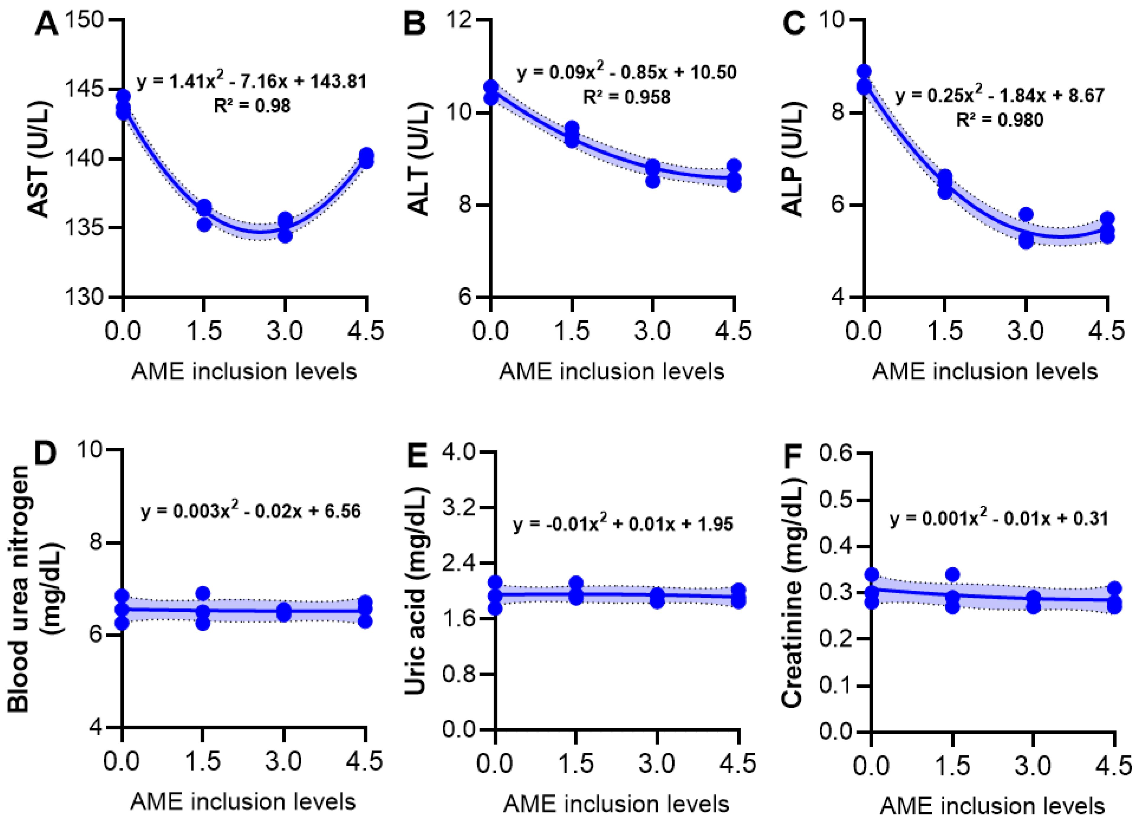
3.5. Serum Biochemical Variables

A panel of serum biochemistry in juvenile P. hypophthalmus fed diets with different AME inclusion levels is presented in Figure 3. Liver enzymes (AST, ALT, and ALP) (Figure 3A–C) were improved by the dietary AME inclusion levels, manifested by a significant quadratic decreasing trend in the levels of those enzymes in fish fed with different AME levels. Meanwhile, there was no relationship between the blood urea nitrogen (Figure 3D), uric acid (Figure 3E), and creatinine (Figure 3F) and the AME inclusion levels.

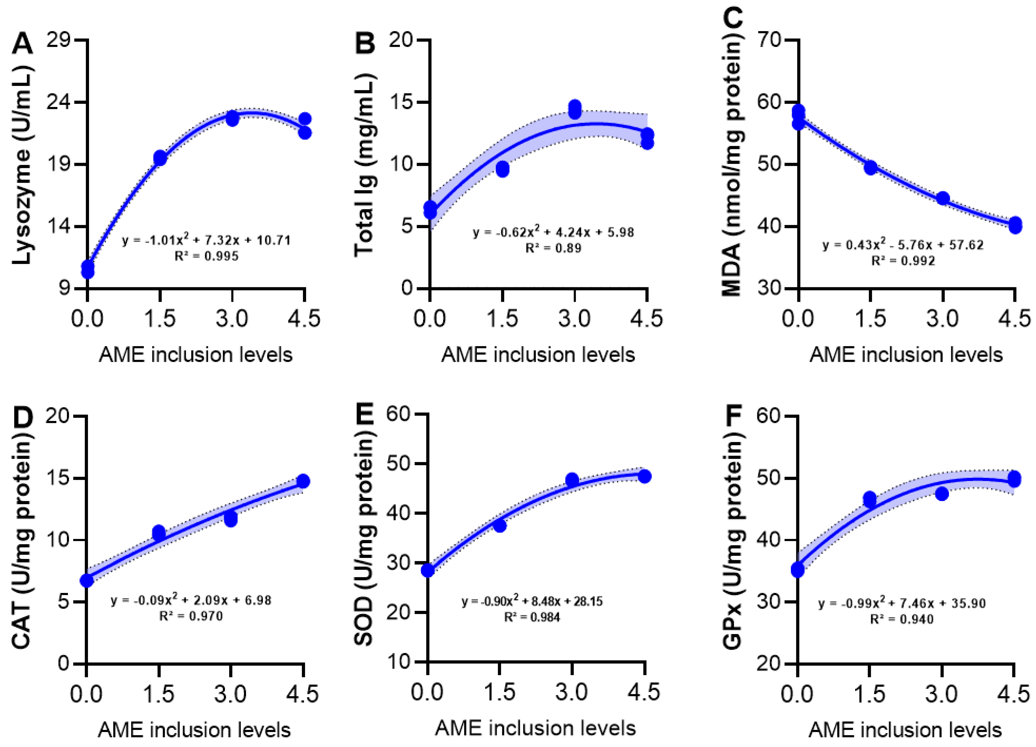
3.6. Serum Immunity and Hepatic Antioxidant Activity

The impacts of dietary AME on serum immune response and antioxidant activity of juvenile P. hypophthalmus is demonstrated in Figure 4. Serum immunity, including lysozyme (Figure 4A) and total Igs (Figure 4B), has been elevated considerably with a strong quadratic trend in fish fed with different AME levels when compared with the control. Hepatic MDA levels (Figure 4C) were decreased with increasing AME levels. Hepatic CAT (Figure 4D) and SOD (Figure 4E) have been increased with a quadratic trend with the inclusion of AME in fish diets. Hepatic GPx (Figure 4F) activity showed a similar result.

4. Discussion

The observed enhancement in the growth performance, in terms of WG and SGR, in juvenile P. hypophthalmus in response to AME-based diets was the result of the improvements recorded in the FI and FCR values. The growth and feed utilization results were concomitant with digestive enzymes observations, suggesting the ability of AME to improve the functionality of diets. The growth-stimulating effects of dietary AM were also confirmed in Nile tilapia [51] and bluegill sunfish [17]. Dietary supplementation with Astragalus polysaccharides (ASP) also supported the growth performance of several finfish species such as Schizothorax prenanti [52], Nile tilapia [28,53], turbot [21], large yellow croaker [20], largemouth bass juveniles [54], crucian carp juveniles [22], and Zebrafish [23]. Differently, in a recently published paper by Sun et al. [19], AME in the diet was shown to have no discernible effect on hybrid grouper’s weight increase or feed efficiency. Variations in fish species, diet, experiment design, time spent feeding, or other factors may all play a role in these disparities. The growth-promoting effects of dietary AME may be attributed to the presence of functional bioactive constituents in the AME, such as phenolic acids and flavonoids, as presented in Table 2. These important phytochemicals have already proven to increase the voluntary feeding intake and feed efficiency and improve protein retention [55]. These functional bioactive compounds could also positively enhance nutrient digestibility, which may, in turn, help improve feed utilization [56]. As reported in our current study, dietary ASP could also improve digestive enzyme activities, which subsequently helped to increase nutrient digestibility [53]. Dietary ASP has been described to be improved the gut health of fish by boosting the intestinal mucosal barrier functions [20] and the abundance of several gut-beneficial microbial communities [23,24], which were not considered in the present study, thus deserving further studies. One hundred percent survival rates across groups at the end of the feeding trial suggested that the treated fish suffered no hazardous effects from receiving AME in their diets. A possible explanation for these results involves the functional bioactive phytochemicals included in the AME, which have potent growth-promoting, hepatoprotective, antioxidant, and immunostimulant properties [55,57,58].
The whole-body composition, including moisture, CP, EE, and ash and amino acids composition, was unchanged by the test diets, suggesting that AME supplementation did not affect the assimilation of whole-body and amino acid composition. Similarly, it was found that diets supplemented with Yucca schidigera or Quillaja saponaria did not significantly affect the whole-body composition of P. hypophthalmus [59,60]. Moreover, dietary AME did not considerably alter CP, moisture, EE, and ash in the hybrid grouper’s whole body and muscles [19]. Our findings were also in harmony with Sun et al. [21], who found that ASP-supplemented diets did not substantially change the whole-body composition of turbot. Farag et al. [28] also found that ASP-based diets did not significantly influence the whole-body proximate composition of Nile tilapia. However, our findings did not correspond to those reported by Liu et al. [20], who found that dietary supplementation with ASP at a dose rate of 0.10 or 0.15% significantly increased the whole-body CP content in large yellow croaker larvae. Several factors may be responsible for the obvious differences in the findings, as mentioned earlier, including different supplementation doses, fish species, experimental setup, feeding duration, and others. Conversely, our results also showed that none of the tested diets had influenced the essential and non-essential amino acid composition. This result is linked to non-significant changes in CP content among experimental groups. The reasons for the insignificant differences in body proximate composition and amino acid composition of groups fed AME-supplemented diets and those fed the control diet are ill-defined and require additional investigation.
It is well-documented that enhancement in the fishes’ digestive enzyme activities is associated with improving the digestibility and availability of nutrients for fish [61]. A significant improvement in lipase, α-amylase, and protease enzyme production in juvenile P. hypophthalmus fed with different AME inclusion levels indicated that AME-based diets might have positively enhanced the nutrient digestibility and promoted nutrient absorption capacity, underpinning the growth-promoting effects of AME. This was proven by a study in which 0.1% of AST supplementation improved intestinal metabolisms by stimulating intestinal mucosal barrier function and beneficial bacteria [62]. Similarly, supplementation with Eleutherine bulbosa, a medicinal herb, improved the intestinal digestive enzymes in P. hypophthalmus [63]. Several previously published studies reported the functional ability of ASP to enhance the digestive enzymes in several other finfish species. For instance, the intestinal protease, amylase, and lipase enzyme activities are elevated in crucian carps fed ASP-based diets [22]. A similar reflection was observed in the intestinal trypsin enzymatic activity in large yellow croaker larvae [20] and intestinal protease and amylase activities in Catla when fed ASP-based diets [25]. Furthermore, dietary ASP (0.10–0.2%) increased the midgut digestive enzymes, for instance, protease and lipase activities in Asian seabass (Lates calcarifer) compared with those fed the reference diet [64]. However, a recent study showed no variations in α-amylase and protease enzyme activities in Nile tilapia-fed ASP-enriched diets associated with the controls [28]. These inconsistencies may be attributable to variables such as dietary supplementation dose and experiment design.
Fish liver enzymes such as AST and ALT are used as indications of liver health [65], and the increases in these enzymes in sera are considered a potential indicator of liver injury, causing leakage of these enzymes into the blood circulation from the hepatocytes [66]. Liver health was improved in P. hypophthalmus juveniles fed AME-based diets, manifested by a considerable decrease in liver enzymes (AST, ALT, and ALP) in fish fed diets with different AME levels compared to those provided with the reference diet. These data may suggest the hepatoprotective impacts of dietary AME, suggesting the potential application of AME as a functional additive to prevent negative impact on the liver caused by intrinsic or extrinsic factors. This could be further strengthened by the study of Jia et al. [67], who observed potential hepatoprotective effects of ASP against CCl4-induced hepatic injury in common carp by inhibiting the elevation of AST, ALT, and lactate dehydrogenase (LDH) enzymes in the hepatocytes. Another study found a similar dietary effect of ASP on the serum ALT and AST enzyme levels in crucian carp juveniles [22]. The functional flavonoids in AME might have potential effects on the improvement in liver functions of P. hypophthalmus juveniles. These flavonoids conferred effective hepatoprotective functions [68]. Moreover, an earlier study also reported that A. membranaceus root had potent hepato-protective effects and protected hepatic cells from pathological injuries [69]. Several other reports have demonstrated the potential hepatoprotective effects of polysaccharides in A. membranaceus [13,67,70]. Another theory showed that AM has potent antioxidant constituents such as astragalosides, flavonoids, and polysaccharides, effectively preventing tissue injury via their antioxidant mechanisms [71]. Jin et al. have reviewed that ASP could increase the enzymatic antioxidant activities, which helps to limit and eliminate oxidative stress caused by free radicals and oxygen radicals [13].
Similar to liver enzymes, blood urea nitrogen, an indicator of the occurrence of renal damage and gill dysfunction [72], was improved by dietary AME, indicating the optimistic impacts of dietary AST supplementation on the kidney functions of the treated fish with no renal injuries. As far as we know, there were no previous reports on the effects of AME on the fish’s kidney functions. A. membranaceus root has been described as used in treating kidney diseases in Chinese medicine [73]. It also has renal protective effects against nephropathy via the modulation of kidney function biomarkers in the blood [74]. In animal models, the positive renal protective effect of A. membranaceus root has been closely associated with the presence of astragalosides (astragalus saponins); they are most well-known for their ability to protect renal tubules from damage caused by free radicals [75]. However, our previously published study showed that kidney function markers such as creatinine, uric acid, and blood urea nitrogen were not significantly altered in common carp fingerlings fed diets supplemented with Origanum vulgare essential oil [65]. Apart from the beneficial effect of AME on enzymes associated with liver and kidney function, further research should consider the histological microstructure of the liver and kidney in relation to stress-relevant gene expression to better understand the actual modes of action of phytochemicals present in AME.
Lysozyme, a mucolytic enzyme excreted by leukocytes, can activate leukocytes and macrophages to lyse the bacterial cell walls [76,77]. Total Igs are a major part of the fish’s humoral immunity playing a significant role in the fish’s immune system defense and are deemed a biomarker of the fish’s adaptive immune responses [44]. Improved serum immunity, including lysozyme activity and total Igs in P. hypophthalmus groups fed diets with different AME levels compared with the CONT, suggested the immunostimulatory roles of dietary AME. It is well documented that plant herbal extracts can stimulate the immune response of P. hypophthalmus. For instance, it was found that dietary supplementation with Euphorbia hirta extract for one month considerably increased serum lysozyme and total Igs content in P. hypophthalmus [9]. Correspondingly, supplementing diets with Psidium guajava and Phyllanthus amarus extracts also improved the serum lysozyme and total Igs in P. hypophthalmus [10]. The immunomodulatory effects of dietary AME or ASP have been described in several studies in other finfish species. Ardó et al. [15] found that dietary A. membranaceus-based diets significantly increased serum lysozyme and total Igs in Nile tilapia. Serum lysozyme pursuits were also increased in Nile tilapia-fed ASP-enriched diets [53]. Furthermore, ASP liposome-based diets significantly increased nitric oxide production and boosted the phagocytic activities of head kidney macrophages as well as serum lysozyme activity in large yellow croaker [78]. It was reported that polysaccharides, saponins, and water decoction of AME significantly increased phagocytic activity and serum lysozyme activities of spotted maigre (Nibea albiflora) [79]. It was recently reported that dietary ASP significantly increased serum lysozyme and total Igs in Nile tilapia [27]. The immunostimulatory functions of AME might be associated with the functional phytochemicals such as polyphenols, flavonoids, and phenolic acids, as mentioned above [55,80]. In addition, astragalosides present in A. membranaceus possess immune-boosting, anti-inflammatory, and immune-regulatory effects [81]. Further challenge trials considering the immune response at a different post-challenge time should be conducted to understand the immunostimulatory effects of AME.
The enzymatic endogenous antioxidant enzymes such as CAT, SOD, and GPx can safeguard the host against oxidative stress [82,83]. MDA is a biomarker of the lipid peroxidation process [84]. An elevation in the hepatic production of CAT, GPx, and SOD enzymes coupled with the lower production of MDA in P. hypophthalmus fed AME-based diets with regard to those reared in the AME0.0 group suggested the antioxidant effects of dietary AME. A study by Wu et al. [51] observed similar responses in SOD, GPx, CAT, and MDA enzyme activities to low-temperature stress in the liver of Nile tilapia when fed AME powder. In a similar pattern, dietary AME increased the enzymatic antioxidant capacity (via increased SOD, GPx, and CAT enzyme activities) of bluegill sunfish exposed to cold-water stress [17]. Astragalus polysaccharides also modulated the antioxidant activity of several finfish species, such as Nile tilapia [53], large yellow croaker [20], and turbot [21]. Furthermore, it was reported that ASP significantly modulated CCl4-induced oxidative stress through increasing SOD enzyme activity and total antioxidant capacity (T-AOC) and decreasing MDA concentrations in the liver of common carp [67]. The antioxidant effects of dietary AME may be associated with ASP, which has potential efficacy in increasing antioxidant enzyme activities and counteracting the negative impacts of free radicals and reactive oxygen species [85]. Notably, the HPLC analysis of AME used in the present study found many flavonoids and phenolics, compounds with strong antioxidant properties [55,86,87,88]. We hypothesize that these phytochemicals can positively enhance the antioxidative capacity of juvenile P. hypophthalmus.

5. Conclusions and Prospects

In summary, feeding AME-supplemented diets for two months improved the growth performance of juvenile P. hypophthalmus. This was supported by improvement in feed utilization with concurrent elevated levels of digestive enzymes. In addition, inclusion of AME in feed improved immunity and antioxidant activity without impacting liver and kidney functions. The overall improvement in fish health might be attributed to the phytochemicals (flavonoids and phenolic acids) found in the AME used in the present study. In addition, dietary supplementation of 1.0–4.5 g AME/kg diet could be regarded as a promising phyto-additive to promote the health condition and welfare of farmed P. hypophthalmus in addition to potentially being used as a functional additive to maintain hepatorenal functions. It may also prevent liver and kidney damage caused by intrinsic and extrinsic factors. Nevertheless, more in-depth experiments and investigations are still necessary to decipher the existent roles of the phytochemicals in AME in influencing the gut health, intestinal histomorphology, and gene expression analysis of the examined fish species.

Supplementary Materials

The following Supplementary Materials are available for download at: https://www.mdpi.com/article/10.3390/fishes7060319/s1.

Author Contributions

Conceptualization, R.M.S., H.M.R.A.-L. and M.S.; methodology, M.S. and H.A.A.; software, M.A.K. and R.M.S.; validation, H.A.A., M.S. and M.R.C.; formal analysis, M.R.C.; investigation, H.M.R.A.-L. and M.S.; resources, M.A.K. and M.S.; data curation, M.R.C.; writing—original draft preparation, H.M.R.A.-L. and M.R.C.; writing—review and editing, H.M.R.A.-L. and M.R.C.; visualization, H.M.R.A.-L.; supervision, R.M.S. and H.A.A.; project administration, H.M.R.A.-L. and R.M.S.; funding acquisition, M.A.K. and R.M.S. Each author has contributed equally to this work. All authors have read and agreed to the published version of the manuscript.

Funding

This research received no external funding.

Institutional Review Board Statement

The experimental methods, procedure, and fish rearing were approved by the Faculty of Veterinary Medicine, Kafrelsheikh University, Egypt. This study was conducted according to the guidelines of the Local Experimental Animal Care Committee, Faculty of Veterinary Medicine, Kafrelsheikh University, Egypt.

Informed Consent Statement

Not applicable.

Data Availability Statement

Data are available upon request.

Acknowledgments

All authors thank Basem Elkhayat, The Fish and Shrimp Consultant at Fish hatchery, Borg El Arab, Alexandria, Egypt, for his aid and support throughout fish rearing and experimental work.

Conflicts of Interest

The authors declare no conflict of interest.

References

  1. Ali, H.; Haque, M.M.; Belton, B. Striped catfish (Pangasianodon hypophthalmus, Sauvage, 1878) aquaculture in Bangladesh: An overview. Aquac. Res. 2013, 44, 950–965. [Google Scholar] [CrossRef]
  2. Phan, L.T.; Bui, T.M.; Nguyen, T.T.T.; Gooley, G.J.; Ingram, B.A.; Nguyen, H.V.; Nguyen, P.T.; De Silva, S.S. Current status of farming practices of striped catfish, Pangasianodon hypophthalmus in the Mekong Delta, Vietnam. Aquaculture 2009, 296, 227–236. [Google Scholar] [CrossRef]
  3. De Silva, S.S.; Phuong, N.T. Striped catfish farming in the Mekong Delta, Vietnam: A tumultuous path to a global success. Rev. Aquac. 2011, 3, 45–73. [Google Scholar] [CrossRef]
  4. Abdel-Latif, H.M.R.; Chaklader, M.R.; Shukry, M.; Ahmed, H.A.; Khallaf, M.A. A multispecies probiotic modulates growth, digestive enzymes, immunity, hepatic antioxidant activity, and disease resistance of Pangasianodon hypophthalmus fingerlings. Aquaculture 2023, 563, 738948. [Google Scholar] [CrossRef]
  5. Dong, H.T.; Nguyen, V.V.; Phiwsaiya, K.; Gangnonngiw, W.; Withyachumnarnkul, B.; Rodkhum, C.; Senapin, S. Concurrent infections of Flavobacterium columnare and Edwardsiella ictaluri in striped catfish, Pangasianodon hypophthalmus in Thailand. Aquaculture 2015, 448, 142–150. [Google Scholar] [CrossRef]
  6. Abdel-Latif, H.M.R.; Abdel-Daim, M.M.; Shukry, M.; Nowosad, J.; Kucharczyk, D. Benefits and applications of Moringa oleifera as a plant protein source in Aquafeed: A review. Aquaculture 2022, 547, 737369. [Google Scholar] [CrossRef]
  7. Abdel-Latif, H.M.R.; Hendam, B.M.; Nofal, M.I.; El-Son, M.A.M. Ginkgo biloba leaf extract improves growth, intestinal histomorphometry, immunity, antioxidant status and modulates transcription of cytokine genes in hapa-reared Oreochromis niloticus. Fish Shellfish Immunol. 2021, 117, 339–349. [Google Scholar] [CrossRef]
  8. Abdel-Latif, H.M.R.; Shukry, M.; Noreldin, A.E.; Ahmed, H.A.; El-Bahrawy, A.; Ghetas, H.A.; Khalifa, E. Milk thistle (Silybum marianum) extract improves growth, immunity, serum biochemical indices, antioxidant state, hepatic histoarchitecture, and intestinal histomorphometry of striped catfish, Pangasianodon hypophthalmus. Aquaculture 2023, 562, 738761. [Google Scholar] [CrossRef]
  9. Nhu, T.Q.; Bich Hang, B.T.; Bach, L.T.; Buu Hue, B.T.; Quetin-Leclercq, J.; Scippo, M.-L.; Phuong, N.T.; Kestemont, P. Plant extract-based diets differently modulate immune responses and resistance to bacterial infection in striped catfish (Pangasianodon hypophthalmus). Fish Shellfish Immunol. 2019, 92, 913–924. [Google Scholar] [CrossRef]
  10. Nhu, T.Q.; Bich Hang, B.T.; Cornet, V.; Oger, M.; Bach, L.T.; Anh Dao, N.L.; Thanh Huong, D.T.; Quetin-Leclercq, J.; Scippo, M.-L.; Phuong, N.T.; et al. Single or Combined Dietary Supply of Psidium guajava and Phyllanthus amarus Extracts Differentially Modulate Immune Responses and Liver Proteome in Striped Catfish (Pangasianodon hyphophthalmus). Front. Immunol. 2020, 11, 797. [Google Scholar] [CrossRef]
  11. Maiti, S.; Saha, S.; Jana, P.; Chowdhury, A.; Khatua, S.; Ghosh, T.K. Effect of dietary Andrographis paniculata leaf extract on growth, immunity, and disease resistance against Aeromonas hydrophila in Pangasianodon hypopthalmus. J. Appl. Aquac. 2021, 1–25. [Google Scholar] [CrossRef]
  12. Fu, J.; Wang, Z.; Huang, L.; Zheng, S.; Wang, D.; Chen, S.; Zhang, H.; Yang, S. Review of the Botanical Characteristics, Phytochemistry, and Pharmacology of Astragalus membranaceus (Huangqi). Phytother. Res. 2014, 28, 1275–1283. [Google Scholar] [CrossRef] [PubMed]
  13. Jin, M.; Zhao, K.; Huang, Q.; Shang, P. Structural features and biological activities of the polysaccharides from Astragalus membranaceus. Int. J. Biol. Macromol. 2014, 64, 257–266. [Google Scholar] [CrossRef] [PubMed]
  14. Zheng, Y.; Ren, W.; Zhang, L.; Zhang, Y.; Liu, D.; Liu, Y. A review of the pharmacological action of Astragalus polysaccharide. Front. Pharmacol. 2020, 11, 349. [Google Scholar] [CrossRef] [Green Version]
  15. Ardó, L.; Yin, G.; Xu, P.; Váradi, L.; Szigeti, G.; Jeney, Z.; Jeney, G. Chinese herbs (Astragalus membranaceus and Lonicera japonica) and boron enhance the non-specific immune response of Nile tilapia (Oreochromis niloticus) and resistance against Aeromonas hydrophila. Aquaculture 2008, 275, 26–33. [Google Scholar] [CrossRef]
  16. Elabd, H.; Wang, H.-P.; Shaheen, A.; Yao, H.; Abbass, A. Feeding Glycyrrhiza glabra (liquorice) and Astragalus membranaceus (AM) alters innate immune and physiological responses in yellow perch (Perca flavescens). Fish Shellfish Immunol. 2016, 54, 374–384. [Google Scholar] [CrossRef] [Green Version]
  17. Elabd, H.; Wang, H.-P.; Shaheen, A.; Yao, H.; Abbass, A. Astragalus membranaceus (AM) enhances growth performance and antioxidant stress profiles in bluegill sunfish (Lepomis macrochirus). Fish Physiol. Biochem. 2016, 42, 955–966. [Google Scholar] [CrossRef]
  18. Angela, C.; Wang, W.; Lyu, H.; Zhou, Y.; Huang, X. The effect of dietary supplementation of Astragalus membranaceus and Bupleurum chinense on the growth performance, immune-related enzyme activities and genes expression in white shrimp, Litopenaeus vannamei. Fish Shellfish Immunol. 2020, 107, 379–384. [Google Scholar] [CrossRef]
  19. Sun, Z.; Wei, Z.; Liu, Q.; Mai, H.; Liu, Y.; Liu, B.; Tan, X.; Ye, C. Effects of dietary Astragalus membranaceus (Fisch.) Bge. root extract on growth performance, plasma biochemical parameters, fish composition, liver and intestinal morphology, and genes expression in head kidney of hybrid grouper (Epinephelus lanceolatus♂ × Epinephelus fuscoguttatus♀). Aquac. Rep. 2022, 22, 100934. [Google Scholar] [CrossRef]
  20. Liu, Y.; Miao, Y.; Xu, N.; Ding, T.; Cui, K.; Chen, Q.; Zhang, J.; Fang, W.; Mai, K.; Ai, Q. Effects of dietary Astragalus polysaccharides (APS) on survival, growth performance, activities of digestive enzyme, antioxidant responses and intestinal development of large yellow croaker (Larimichthys crocea) larvae. Aquaculture 2020, 517, 734752. [Google Scholar] [CrossRef]
  21. Sun, Y.; Wang, X.; Zhou, H.; Mai, K.; He, G. Dietary Astragalus polysaccharides ameliorates the growth performance, antioxidant capacity and immune responses in turbot (Scophthalmus maximus L.). Fish Shellfish Immunol. 2020, 99, 603–608. [Google Scholar] [CrossRef] [PubMed]
  22. Wu, S. Dietary Astragalus membranaceus polysaccharide ameliorates the growth performance and innate immunity of juvenile crucian carp (Carassius auratus). Int. J. Biol. Macromol. 2020, 149, 877–881. [Google Scholar] [CrossRef] [PubMed]
  23. Li, Y.; Ran, C.; Wei, K.; Xie, Y.; Xie, M.; Zhou, W.; Yang, Y.; Zhang, Z.; Lv, H.; Ma, X.; et al. The effect of Astragalus polysaccharide on growth, gut and liver health, and anti-viral immunity of zebrafish. Aquaculture 2021, 540, 736677. [Google Scholar] [CrossRef]
  24. Shi, F.; Lu, Z.; Yang, M.; Li, F.; Zhan, F.; Zhao, L.; Li, Y.; Li, Q.; Li, J.; Li, J.; et al. Astragalus polysaccharides mediate the immune response and intestinal microbiota in grass carp (Ctenopharyngodon idellus). Aquaculture 2021, 534, 736205. [Google Scholar] [CrossRef]
  25. Harikrishnan, R.; Devi, G.; Doan, H.V.; Tapingkae, W.; Balasundaram, C.; Arockiaraj, J.; Ringø, E. Changes in immune genes expression, immune response, digestive enzymes -antioxidant status, and growth of catla (Catla catla) fed with Astragalus polysaccharides against edwardsiellosis disease. Fish Shellfish Immunol. 2022, 121, 418–436. [Google Scholar] [CrossRef]
  26. Pu, Y.; Wu, S. The growth performance, body composition and nonspecific immunity of white shrimps (Litopenaeus vannamei) affected by dietary Astragalus membranaceus polysaccharide. Int. J. Biol. Macromol. 2022, 209, 162–165. [Google Scholar] [CrossRef]
  27. Farag, M.R.; Alagawany, M.; Khalil, S.R.; El-Hady, E.W.; Elhady, W.M.; Ismail, T.A.; Marini, C.; Di Cerbo, A.; Abdel-Latif, H.M.R. Immunosuppressive Effects of Thallium Toxicity in Nile Tilapia Fingerlings: Elucidating the Rescue Role of Astragalus membranaceus Polysaccharides. Front. Vet. Sci. 2022, 9, 843031. [Google Scholar] [CrossRef]
  28. Farag, M.R.; Alagawany, M.; Khalil, S.R.; Moustafa, A.A.; Mahmoud, H.K.; Abdel-Latif, H.M.R. Astragalus membranaceus polysaccharides modulate growth, hemato-biochemical indices, hepatic antioxidants, and expression of HSP70 and apoptosis-related genes in Oreochromis niloticus exposed to sub-lethal thallium toxicity. Fish Shellfish Immunol. 2021, 118, 251–260. [Google Scholar] [CrossRef]
  29. Abdel-Latif, H.M.R.; Dawood, M.A.O.; Menanteau-Ledouble, S.; El-Matbouli, M. The nature and consequences of co-infections in tilapia: A review. J. Fish Dis. 2020, 43, 651–664. [Google Scholar] [CrossRef]
  30. Gomathi, D.; Kalaiselvi, M.; Ravikumar, G.; Devaki, K.; Uma, C. GC-MS analysis of bioactive compounds from the whole plant ethanolic extract of Evolvulus alsinoides (L.) L. J. Food Sci. Technol. 2015, 52, 1212–1217. [Google Scholar] [CrossRef]
  31. Mattila, P.; Astola, J.; Kumpulainen, J. Determination of Flavonoids in Plant Material by HPLC with Diode-Array and Electro-Array Detections. J. Agric. Food Chem. 2000, 48, 5834–5841. [Google Scholar] [CrossRef] [PubMed]
  32. Öztürk, N.; Tunçel, M.; Tunçel, N.B. Determination of Phenolic Acids by a Modified HPLC: Its Application to Various Plant Materials. J. Liq. Chromatogr. Relat. Technol. 2007, 30, 587–596. [Google Scholar] [CrossRef]
  33. NRC. Nutrient Requirements of Fish and Shrimp; National Academy Press: Washington, DC, USA, 2011; 392p. [Google Scholar]
  34. AOAC. Official Methods of Analysis, 13th ed.; Association of Analytical Chemists: Washington, DC, USA, 2012; 1018p. [Google Scholar]
  35. Belfield, A.; Goldberg, D. Colorimetric determination of alkaline phosphatase activity. Enzyme 1971, 12, 561–568. [Google Scholar] [CrossRef]
  36. Reitman, S.; Frankel, S. A colorimetric method for the determination of serum glutamic oxalacetic and glutamic pyruvic transaminases. Am. J. Clin. Pathol. 1957, 28, 56–63. [Google Scholar] [CrossRef]
  37. Bernfeld, P. Amylases, α and β. In Methods in Enzymology; Academic Press: Cambridge, MA, USA, 1955; Volume 1, pp. 149–158. [Google Scholar]
  38. Khantaphant, S.; Benjakul, S. Comparative study on the proteases from fish pyloric caeca and the use for production of gelatin hydrolysate with antioxidative activity. Comp. Biochem. Physiol. Part B Biochem. Mol. Biol. 2008, 151, 410–419. [Google Scholar] [CrossRef]
  39. Shihabi, Z.K.; Bishop, C. Simplified Turbidimetric Assay for Lipase Activity. Clin. Chem. 1971, 17, 1150–1153. [Google Scholar] [CrossRef]
  40. Barham, D.; Trinder, P. Enzymatic determination of uric acid. Analyst 1972, 97, 142–145. [Google Scholar] [CrossRef]
  41. Coulombe, J.J.; Favreau, L. A new simple semimicro method for colorimetric determination of urea. Clin. Chem. 1963, 9, 102–108. [Google Scholar] [CrossRef]
  42. Heinegård, D.; Tiderström, G. Determination of serum creatinine by a direct colorimetric method. Clin. Chim. Acta 1973, 43, 305–310. [Google Scholar] [CrossRef]
  43. Ellis, A. Lysozyme assays. Tech. Fish Immunol. 1990, 1, 101–103. [Google Scholar]
  44. Cuesta, A.; Meseguer, J.; Esteban, M.A. Total serum immunoglobulin M levels are affected by immunomodulators in seabream (Sparus aurata L.). Vet. Immunol. Immunopathol. 2004, 101, 203–210. [Google Scholar] [CrossRef] [PubMed]
  45. Siwicki, A.; Anderson, D. An easy spectrophotometric assay for determining total protein and immunoglobulin levels in fish sera: Correlation to fish health. Tech. Fish Immunol. 1993, 3, 23–30. [Google Scholar]
  46. Aebi, H. Catalase in vitro. In Methods in Enzymology; Academic Press: Cambridge, MA, USA, 1984; Volume 105, pp. 121–126. [Google Scholar]
  47. McCord, J.M.; Fridovich, I. Superoxide Dismutase: An enzymic function for erythrocuprein (hemocuprein). J. Biol. Chem. 1969, 244, 6049–6055. [Google Scholar] [CrossRef]
  48. Paglia, D.E.; Valentine, W.N. Studies on the quantitative and qualitative characterization of erythrocyte glutathione peroxidase. J. Lab. Clin. Med. 1967, 70, 158–169. [Google Scholar] [PubMed]
  49. Ohkawa, H.; Ohishi, N.; Yagi, K. Assay for lipid peroxides in animal tissues by thiobarbituric acid reaction. Anal. Biochem. 1979, 95, 351–358. [Google Scholar] [CrossRef]
  50. Uchiyama, M.; Mihara, M. Determination of malonaldehyde precursor in tissues by thiobarbituric acid test. Anal. Biochem. 1978, 86, 271–278. [Google Scholar] [CrossRef]
  51. Wu, F.; Yang, C.; Wen, H.; Zhang, C.; Jiang, M.; Liu, W.; Tian, J.; Yu, L.; Lu, X. Improving low-temperature stress tolerance of tilapia, Oreochromis niloticus: A functional analysis of Astragalus membranaceus. J. World Aquac. Soc. 2019, 50, 749–762. [Google Scholar] [CrossRef]
  52. Xiang, X.; Chen, J.; Zhou, X.; Wang, W. Effect of astragalus polysaccharides on growth, body composition and immune index in Schizothorax prenanti. Acta Hydrobiol. Sin. 2011, 35, 291–299. [Google Scholar] [CrossRef]
  53. Zahran, E.; Risha, E.; AbdelHamid, F.; Mahgoub, H.A.; Ibrahim, T. Effects of dietary Astragalus polysaccharides (APS) on growth performance, immunological parameters, digestive enzymes, and intestinal morphology of Nile tilapia (Oreochromis niloticus). Fish Shellfish Immunol. 2014, 38, 149–157. [Google Scholar] [CrossRef]
  54. Lin, S.-M.; Jiang, Y.; Chen, Y.-J.; Luo, L.; Doolgindachbaporn, S.; Yuangsoi, B. Effects of Astragalus polysaccharides (APS) and chitooligosaccharides (COS) on growth, immune response and disease resistance of juvenile largemouth bass, Micropterus salmoides. Fish Shellfish Immunol. 2017, 70, 40–47. [Google Scholar] [CrossRef]
  55. Ahmadifar, E.; Yousefi, M.; Karimi, M.; Fadaei Raieni, R.; Dadar, M.; Yilmaz, S.; Dawood, M.A.O.; Abdel-Latif, H.M.R. Benefits of Dietary Polyphenols and Polyphenol-Rich Additives to Aquatic Animal Health: An Overview. Rev. Fish. Sci. Aquac. 2021, 29, 478–511. [Google Scholar] [CrossRef]
  56. Citarasu, T. Herbal biomedicines: A new opportunity for aquaculture industry. Aquac. Int. 2010, 18, 403–414. [Google Scholar] [CrossRef]
  57. Bratkov, V.M.; Shkondrov, A.M.; Zdraveva, P.K.; Krasteva, I.N. Flavonoids from the Genus Astragalus: Phytochemistry and Biological Activity. Pharm. Rev. 2016, 10, 11–32. [Google Scholar] [CrossRef] [Green Version]
  58. Matkowski, A.; Woźniak, D.; Lamer-Zarawska, E.; Oszmiański, J.; Leszczyńska, A. Flavonoids and Phenol Carboxylic Acids in the Oriental Medicinal Plant Astragalus membranaceus Acclimated in Poland. Z. Für Nat. C 2003, 58, 602–604. [Google Scholar] [CrossRef]
  59. Güroy, B.; Mantoğlu, S.; Kayalı, S.; Şahin, İ. Effect of dietary Yucca schidigera extract on growth, total ammonia–nitrogen excretion and haematological parameters of juvenile striped catfish Pangasianodon hypophthalmus. Aquac. Res. 2014, 45, 647–654. [Google Scholar] [CrossRef]
  60. Güroy, B.; Mantoğlu, S.; Merrifield, D.L.; Guroy, D. Effects of dietary Nutrafito Plus on growth, haemotological parameters and total ammonia-nitrogen excretion of juvenile striped catfish Pangasianodon hypophthalmus. Aquac. Res. 2016, 47, 1770–1777. [Google Scholar] [CrossRef]
  61. Hoseinifar, S.H.; Dadar, M.; Ringø, E. Modulation of nutrient digestibility and digestive enzyme activities in aquatic animals: The functional feed additives scenario. Aquac. Res. 2017, 48, 3987–4000. [Google Scholar] [CrossRef]
  62. Shi, H.-T.; Zhao, S.-Z.; Wang, K.-L.; Fan, M.-X.; Han, Y.-Q.; Wang, H.-L. Effects of dietary Astragalus membranaceus supplementation on growth performance, and intestinal morphology, microbiota and metabolism in common carp (Cyprinus carpio). Aquac. Rep. 2022, 22, 100955. [Google Scholar] [CrossRef]
  63. Rudy, A.N.; Meylianawati, O.F.A.; Yanti, P.S.; Esti, H.H. The effects of dietary Eleutherine bulbosa on the growth, leukocyte profile, and digestive enzyme activity of the striped catfish Pangasianodon hypophthalmus. Nusant. Biosci. 2018, 10, 46–51. [Google Scholar]
  64. Yu, W.; Yang, Y.; Zhou, Q.; Huang, X.; Huang, Z.; Li, T.; Wu, Q.; Zhou, C.; Ma, Z.; Lin, H. Effects of dietary Astragalus polysaccharides on growth, health and resistance to Vibrio harveyi of Lates calcarifer. Int. J. Biol. Macromol. 2022, 207, 850–858. [Google Scholar] [CrossRef]
  65. Abdel-Latif, H.M.R.; Abdel-Tawwab, M.; Khafaga, A.F.; Dawood, M.A.O. Dietary oregano essential oil improved the growth performance via enhancing the intestinal morphometry and hepato-renal functions of common carp (Cyprinus carpio L.) fingerlings. Aquaculture 2020, 526, 735432. [Google Scholar] [CrossRef]
  66. Bruslé, J.; Anadon, G.G. The structure and function of fish liver. Fish Morphol. 1996, 76, 545–551. [Google Scholar]
  67. Jia, R.; Cao, L.; Xu, P.; Jeney, G.; Yin, G. In vitro and in vivo hepatoprotective and antioxidant effects of Astragalus polysaccharides against carbon tetrachloride-induced hepatocyte damage in common carp (Cyprinus carpio). Fish Physiol. Biochem. 2012, 38, 871–881. [Google Scholar] [CrossRef] [PubMed]
  68. Davila, J.C.; Lenherr, A.; Acosta, D. Protective effect of flavonoids on drug-induced hepatotoxicity in vitro. Toxicology 1989, 57, 267–286. [Google Scholar] [CrossRef]
  69. Zhen-Lun, Z.; Qi-Zhen, W.; Chang-Xiao, L. Hepatoprotective effects of Astraglus root. J. Ethnopharmacol. 1990, 30, 145–149. [Google Scholar] [CrossRef]
  70. Hamid, M.; Liu, D.; Abdulrahim, Y.; Liu, Y.; Qian, G.; Khan, A.; Gan, F.; Huang, K. Amelioration of CCl4-induced liver injury in rats by selenizing Astragalus polysaccharides: Role of proinflammatory cytokines, oxidative stress and hepatic stellate cells. Res. Vet. Sci. 2017, 114, 202–211. [Google Scholar] [CrossRef]
  71. Shahzad, M.; Shabbir, A.; Wojcikowski, K.; Wohlmuth, H.; C Gobe, G. The antioxidant effects of Radix Astragali (Astragalus membranaceus and related species) in protecting tissues from injury and disease. Curr. Drug Targets 2016, 17, 1331–1340. [Google Scholar] [CrossRef]
  72. Nelson, K.; Jones, J.; Jacobson, S.; Reimschuessel, R. Elevated Blood Urea Nitrogen (BUN) Levels in Goldfish as an Indicator of Gill Dysfunction. J. Aquat. Anim. Health 1999, 11, 52–60. [Google Scholar] [CrossRef]
  73. Zhang, H.W.; Lin, Z.X.; Xu, C.; Leung, C.; Chan, L.S. Astragalus (a traditional Chinese medicine) for treating chronic kidney disease. Cochrane Database Syst. Rev. 2014, CD008369. [Google Scholar] [CrossRef]
  74. Li, M.; Wang, W.; Xue, J.; Gu, Y.; Lin, S. Meta-analysis of the clinical value of Astragalus membranaceus in diabetic nephropathy. J. Ethnopharmacol. 2011, 133, 412–419. [Google Scholar] [CrossRef]
  75. Zhang, J.; Xie, X.; Li, C.; Fu, P. Systematic review of the renal protective effect of Astragalus membranaceus (root) on diabetic nephropathy in animal models. J. Ethnopharmacol. 2009, 126, 189–196. [Google Scholar] [CrossRef] [PubMed]
  76. Alexander, J.B.; Ingram, G.A. Noncellular nonspecific defence mechanisms of fish. Annu. Rev. Fish Dis. 1992, 2, 249–279. [Google Scholar] [CrossRef]
  77. Saurabh, S.; Sahoo, P.K. Lysozyme: An important defence molecule of fish innate immune system. Aquac. Res. 2008, 39, 223–239. [Google Scholar] [CrossRef]
  78. Zhang, W.; Zhang, M.; Cheng, A.; Hao, E.; Huang, X.; Chen, X. Immunomodulatory and antioxidant effects of Astragalus polysaccharide liposome in large yellow croaker (Larimichthys crocea). Fish Shellfish Immunol. 2020, 100, 126–136. [Google Scholar] [CrossRef]
  79. Wang, X.; Ding, L.; Yan, M.; Chai, X.; Lu, R.; Wang, Q.; Li, F. Polysaccharides, saponins, and water decoction of Astragalus membranaceus significantly enhance the non-specific immune response of spotted maigre (Nibea albiflora). Isr. J. Aquac.—Bamidgeh 2012, 64, 1–6. [Google Scholar] [CrossRef]
  80. Chakraborty, S.B.; Hancz, C. Application of phytochemicals as immunostimulant, antipathogenic and antistress agents in finfish culture. Rev. Aquac. 2011, 3, 103–119. [Google Scholar] [CrossRef]
  81. Qi, Y.; Gao, F.; Hou, L.; Wan, C. Anti-inflammatory and immunostimulatory activities of astragalosides. Am. J. Chin. Med. 2017, 45, 1157–1167. [Google Scholar] [CrossRef]
  82. Chowdhury, S.; Saikia, S.K. Oxidative Stress in Fish: A Review. J. Sci. Res. 2020, 12, 145–160. [Google Scholar] [CrossRef] [Green Version]
  83. Hoseinifar, S.H.; Yousefi, S.; Van Doan, H.; Ashouri, G.; Gioacchini, G.; Maradonna, F.; Carnevali, O. Oxidative Stress and Antioxidant Defense in Fish: The Implications of Probiotic, Prebiotic, and Synbiotics. Rev. Fish. Sci. Aquac. 2021, 29, 198–217. [Google Scholar] [CrossRef]
  84. Martínez-Álvarez, R.M.; Morales, A.E.; Sanz, A. Antioxidant Defenses in Fish: Biotic and Abiotic Factors. Rev. Fish Biol. Fish. 2005, 15, 75–88. [Google Scholar] [CrossRef]
  85. Li, R.; Chen, W.-C.; Wang, W.-P.; Tian, W.-Y.; Zhang, X.-G. Antioxidant activity of Astragalus polysaccharides and antitumour activity of the polysaccharides and siRNA. Carbohydr. Polym. 2010, 82, 240–244. [Google Scholar] [CrossRef]
  86. Bors, W.; Michel, C.; Stettmaier, K. Antioxidant effects of flavonoids. BioFactors 1997, 6, 399–402. [Google Scholar] [CrossRef] [PubMed]
  87. Huyut, Z.; Beydemir, Ş.; Gülçin, İ. Antioxidant and Antiradical Properties of Selected Flavonoids and Phenolic Compounds. Biochem. Res. Int. 2017, 2017, 7616791. [Google Scholar] [CrossRef] [Green Version]
  88. Rice-Evans, C.; Miller, N.; Paganga, G. Antioxidant properties of phenolic compounds. Trends Plant Sci. 1997, 2, 152–159. [Google Scholar] [CrossRef]
Figure 1. Growth performance including weight gain (WG) (A) and specific growth rate (SGR) (B) and feed utilization including feed intake (FI) (C) and feed conversion ratio (FCR) (D) of P. hypophthalmus fed diets with different AME inclusion levels. Data lines denote best fit models for the data. Equations and R square in the figures demonstrates the relationship between the measured parameters and AME inclusion levels. Individual marker indicates the number of technical replicates (n = 3).
Fishes 07 00319 g001
Figure 2. Serum lipase E (A), α-amylase E (B), and protease E (C) of juvenile P. hypophthalmus fed diets with different AME inclusion levels over the feeding trial period of 56 days. Data lines denote best fit models for the data. Equations and R square in the figures demonstrate the relationship between the measured parameters and AME inclusion levels. Individual marker indicates the number of technical replicates (n = 3). Individual marker is the mean of two biological replicates.
Fishes 07 00319 g002
Figure 3. Serum biochemistry including AST (A), ALT (B), ALP (C), blood urea nitrogen (D), uric acid (E), and creatinine (F) of juvenile P. hypophthalmus fed diets with different AME inclusion levels over the feeding trial period of 56 days. Data lines denote best fit models for the data. Equations and R square in the figures demonstrate the relationship between the measured parameters and AME inclusion levels. Individual marker indicates the number of technical replicates (n = 3). Individual marker is the mean of two biological replicates.
Fishes 07 00319 g003
Figure 4. Serum immune responses (lysozyme and total Ig) (A,B) and hepatic antioxidant activity (CF) of juvenile P. hypophthalmus fed diets with different AME inclusion levels over the feeding trial period of 56 days. Data lines denote best fit models for the data. Equations and R square in the figures demonstrate the relationship between the measured parameters and AME inclusion levels. Individual marker indicates the number of technical replicates (n = 3). Individual marker is the mean of two biological replicates.
Fishes 07 00319 g004
Table 1. Ration elements, nutrients, and chemical composition (% on dry matter (DM) basis) of the reference diet.
Ration Elements% DM Basis
Fish meal (FM; 72% CP) 110.0
Soybean meal (SBM; 46% CP) 234.0
Corn gluten meal (CGM; 60% CP)3.5
Rice bran14.0
Yellow corn meal15.0
Wheat bran9.0
Wheat flour13.0
Sunflower oil0.70
Vitamin and Mineral premix 30.30
Di-calcium phosphate0.50
Total100
Chemical composition (% on DM basis)
Dry matter (DM)90.19
Crude protein (CP)30.82
Ether extract (EE)6.93
Ash6.59
Crude fiber (CF)8.81
Nitrogen-free extract (NFE) 446.85
Gross energy (GE; KJ/g diet DM) 518.06
Protein to energy ratio (P/E ratio) 6 17.06
Formerly published in our study [8].1 Danish FM (contains 72.0% CP), procured from TripleNine Fish Protein, DK-6700 (Esbjerg, Denmark).2 Egyptian soybean flour (contains 46.0% CP), procured from Cargill Trading Egypt Co. (Katameya, Cairo, Egypt).3 AGRI-VET CO. for manufacturing Vitamins and Feed additives (10th of Ramadan City A2, Egypt). The vitamin premix mixture (contains per 1 kg): Vitamin A (17,000 IU); Vitamin D3 (2400 IU); Vitamin E (240 mg); Vitamin K3 (11 mg); Vitamin B1 (24 mg); Vitamin B2 (52 mg); Vitamin B3 (275 mg); Vitamin B6 (25 mg); Vitamin B12 (0.05 mg); Vitamin C (220 mg); Folic acid (15 mg); Calcium d–pantothenate (55 mg); Biotin (1.5 mg); Inositol (125 mg), and Choline chloride (2500 mg). The mineral mixture (contains per 1 kg) is composed of the following: Iron (74.50 mg/kg); Copper (12.5 mg/kg); Manganese (200 mg/kg); Zinc (80 mg/kg); Iodine (2 mg/kg); Selenium (0.330 mg/kg), and Cobalt (1.5 mg/kg).4 NFE = 100 − (CP + EE + CF + Ash). 5 GE was estimated on the basis of 23.60, 39.40, and 17.20 kJ/g of CP, EE, and NFE, respectively following NRC guidelines [33].6 P/E ratio was estimated as mg crude protein/KJ.
Table 2. HPLC results of the flavonoids and phenolics present in Astragalus membranaceus extract (AME) used in the present study.
PhytochemicalsRT (min)Concentration (µg/mL)
Flavonoids
Rutin4.65.23
Catechin12.014.14
Quercetin6.99.21
Kaempferol8.14.05
Luteolin9.06.09
Chrysoeriol15.019.08
Naringin5.24.12
Apigenin10.012.45
Phenolics
Syringic acid3.08.69
Caffeic acid4.73.22
Ferulic acid6.85.66
Protocatechuic acid7.83.31
Gallic acid9.03.77
Ellagic acid11.06.33
p-Coumaric acid4.06.14
Resveratrol13.87.01
Vanillic acid14.66.79
Gentisic acid2.06.55
Table 3. Whole body composition analysis of striped catfish juveniles fed diets supplemented with AME inclusion of levels after 2 months feeding trial.
ParametersAST0.0AST1.5AST3.0AST4.5
Moisture (%)73.20 ± 0.2073.55 ± 0.7174.47 ± 0.7573.69 ± 0.64
Crude protein (%)14.24 ± 0.2114.68 ± 0.2114.58 ± 0.2914.63 ± 0.52
Ether extract (%)4.60 ± 0.174.62 ± 0.244.66 ± 0.204.54 ± 0.21
Ash (%)3.10 ± 0.033.17 ± 0.033.15 ± 0.023.17 ± 0.03
Table 4. Effects of dietary AME levels on the amino acid composition (% of total amino acids) retention in the whole-body of striped catfish juveniles.
Amino Acid ContentExperimental Groups
AME0.0AME1.5AME3.0AME4.5
Essential amino acids (% of total amino acids)
Threonine2.86 ± 0.822.07 ± 0.012.11 ± 0.062.16 ± 0.03
Valine3.11 ± 0.692.56 ± 0.022.54 ± 0.072.65 ± 0.08
Methionine1.00 ± 0.061.10 ± 0.011.12 ± 0.051.16 ± 0.08
Phenylalanine1.67 ± 0.141.84 ± 0.031.93 ± 0.102.00 ± 0.19
Lysine3.25 ± 0.113.33 ± 0.033.42 ± 0.123.51 ± 0.13
Leucine3.18 ± 0.073.27 ± 0.023.35 ± 0.113.46 ± 0.11
Histidine0.99 ± 0.041.14 ± 0.041.11 ± 0.041.12 ± 0.01
Arginine3.41 ± 0.023.44 ± 0.013.53 ± 0.073.52 ± 0.01
Tryptophan0.31 ± 0.010.34 ± 0.0030.36 ± 0.040.41 ± 0.04
Non-essential amino acids (% of total amino acids)
Aspartic Acid4.55 ± 0.044.68 ± 0.034.70 ± 0.104.77 ± 0.04
Alanine3.63 ± 0.614.25 ± 0.024.23 ± 0.024.22 ± 0.11
Isoleucine1.63 ± 0.091.72 ± 0.021.81 ± 0.081.87 ± 0.15
Serine2.20 ± 0.102.08 ± 0.042.15 ± 0.032.16 ± 0.07
Glutamate6.67 ± 0.106.50 ± 0.106.62 ± 0.106.76 ± 0.17
Glycine5.76 ± 0.155.58 ± 0.025.71 ± 0.045.52 ± 0.14
Tyrosine1.32 ± 0.061.23 ± 0.031.31 ± 0.041.29 ± 0.03
Proline2.81 ± 0.112.70 ± 0.012.79 ± 0.032.75 ± 0.07
Cysteine0.40 ± 0.010.40 ± 0.010.42 ± 0.010.45 ± 0.02
Values are depicted as means ± standard error of means of three technical replicates.
Publisher’s Note: MDPI stays neutral with regard to jurisdictional claims in published maps and institutional affiliations.

