Article

Untargeted Metabolomics Associated with Behavioral and Toxicological Studies Yield Insights into the Impact of 2,6-Dichloro-3-hydroxy-1,4-benzoquinone Disinfection By-Product on Zebrafish Larvae

by
Ieremias Chousidis
1,
Theodoros Chatzimitakos
2,
Constantine Stalikas
2 and
Ioannis Leonardos
1,*
1
Laboratory of Zoology, Biological Applications and Technology Department, University of Ioannina, 45110 Ioannina, Greece
2
Laboratory of Analytical Chemistry, Department of Chemistry, University of Ioannina, 45110 Ioannina, Greece
*
Author to whom correspondence should be addressed.
Fishes 2022, 7(6), 368; https://doi.org/10.3390/fishes7060368
Submission received: 13 October 2022 / Revised: 25 November 2022 / Accepted: 30 November 2022 / Published: 2 December 2022
(This article belongs to the Special Issue Omics in Fish Aquaculture and Fisheries)

Abstract

:
The disinfection by-product 2,6-dichloro-3-hydroxy-1,4-benzoquinone (2,6-DCBQ-OH) is a halobenzoquinone that emerges after chlorination. Therefore, it will inevitably come into contact with aquatic organisms. The aim of this study was to investigate the effect of 2,6-DCBQ-OH on zebrafish embryos. The dose-dependent toxicity was recorded, and the LC50 value was found to be 186 μg/L. Toxicity was accompanied with morphological, developmental, and behavioral abnormalities, and metabolic alterations. The association of phenotypic alterations with metabolic alterations was investigated through metabolomic study. In the control group, 25 metabolic pathways were identified, and 10 of them remained unaffected upon exposure to the halobenzoquinone. The upregulation of the glutathione pathway suggested that 2,6-DCBQ-OH can cause oxidative stress. In addition, the upregulation of the β-alanine metabolism pathway may be associated with the observed reduced swimming activity observed. Likewise, the downregulation of pathways associated with glycerophospholipids and butyrate can result in endocrine disruption, ineffective regulation of weight and body composition, and glucose homeostasis. The fatty acid elongation and synthesis of essential amino acids are downregulated, which can be associated with insufficient organogenesis at early life stages. In conclusion, exposure of zebrafish to 2,6-DCBQ-OH results in dysregulation and metabolic collapse of the organism, which ultimately result in developmental, morphological, behavioral, and other abnormalities.

Graphical Abstract

1. Introduction

Chlorination is a necessary process to remove the microbial load of water, so as to make it potable and safe for public health. However, during the chlorination process, chlorine reacts with the natural organic load that exists in water and induces the synthesis of chlorination by-products. Exposure to such by-products has been associated with reproductive and developmental toxicity and several forms of cancer, such as colon and pancreatic [1,2,3,4]. To date, their toxicity has been tested in mouse cell lines, human cancer cells, and nerve cell lines [5]. Recent studies, performed in integrated in vivo systems of organisms (rodents, zebrafish), also resulted in worrying outcomes concerning toxicity [6].
Halobenzoquinones is one class of chlorination by-products. This class of compounds not only exhibits toxicity towards (micro)organisms but it has also been proved that it can further react with the leftover organic load of water, leading to the production of secondary by-products [7]. The production of secondary metabolites depends on the physicochemical characteristics of the water (temperature, pH, etc.). Moreover, in cases where personal care products, such as deodorants, cosmetics, etc., are added to the water (for instance in swimming pools) the production of secondary by-products is more pronounced [7].
The 2,6-dichloro-1,4-benzoquinone (DCBQ) is one such halobenzoquinone, which can usually be detected in chlorinated water. However, this compound can readily be hydroxylated under UV irradiation, yielding 2,6-dichloro-3-hydroxy-1,4-benzoquinone (2,6-DCBQ-OH). Although hydroxylated benzoquinones are considered to be less toxic compared to halobenzoquinones [4], more data is needed to back up these claims and establish a safer environment for humans exposed to chlorinated water [4,5,8,9].
The model organism, zebrafish (Danio rerio), is a vertebrate organism that is considered a “high-throughput” model system, highly suitable for ecotoxicological, drug, and chemical toxicity studies, since nearly 70% of its genome is similar to that of humans [10,11,12]. Moreover, zebrafish have many more benefits, such as transparency, allowing direct in vivo monitoring, etc., corroborating its enhanced suitability [13]. In addition, zebrafish larvae are used as an experimental model organism in behavioral studies due to the high developmental rate, and the chance to explore their locomotor activity [12,14]. The use of zebrafish larvae also surpasses major ethical issues with animal laboratory use; studies implementing zebrafish as a model organism have greatly increased in the past years [15].
Although conducting toxicity studies would produce valuable results, more data is needed in order to shed light on the mechanism via which toxicity is inducted. Metabolomics is the field of research studying the alterations of low molecular weight endogenous metabolites, able to provide an overview of the metabolic status of a biological system [16]. Data generated from omics can be linked together through bioinformatic analysis to generate an overall approach to events occurring within a given organism [17]. Due to the valuable results that can be obtained, the number of studies revolving around toxicometabolomics is increasing rapidly. There are two main reasons for the increasing interest of toxicologists in metabolomics [18]. The first one is that metabolic pathways can be used to unravel signatures of toxicity that could emerge as useful diagnostic biomarkers and predict toxicity from dangerous and probably unknown chemical compounds. The second is that metabolomics can improve our understanding of the mechanisms of drug toxicity through the identification of the cellular pathways that are affected by the toxicants [19].
In this study, the effect of 2,6-DCBQ-OH on the viability of zebrafish embryo and larvae was examined, in an effort to shed light on its potential toxicity. Moreover, the toxicity study was accompanied by heart rate measurements, behavioral analysis, and a locomotor activity study. Furthermore, an untargeted metabolomic approach was employed in order to gain a better overview of the metabolic alterations that take place, to which the observed phenotypes are attributed.

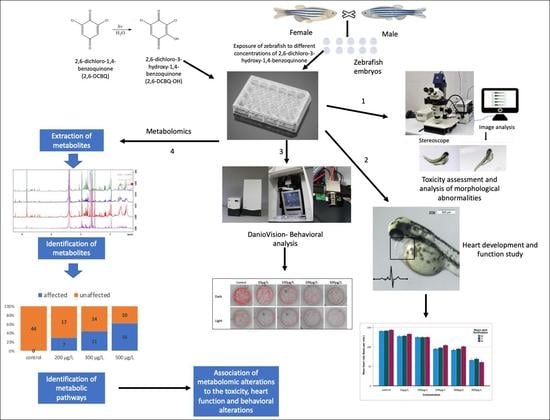
2. Materials and Methods

2.1. Chemicals

For the preparation of 2,6-DCBQ-OH working solutions, at first, a standard solution of 2,6-DCBQ (97%, w/w) (Alfa Aesar, Karlsruhe, Germany) was prepared in ethanol at a concentration of 20.0 mg/mL. After the complete dissolution of 2,6-DCBQ, an appropriate amount of the solution was transferred to a screw-capped conical flask, and water was added, so as to prepare a solution with a concentration of 1.0 mg/mL. The flask was left under daylight for 24 h, so as to enhance the hydroxylation rate. The complete transformation of 2,6-DCBQ to 2,6-DCBQ-OH was validated by recording molecular absorbance of the solution at 530 nm, which corresponds to the wavelength where 2,6-DCBQ-OH absorbs. Diluted solutions were prepared, for zebrafish exposure, in standard zebrafish E3 buffer solution (5 mM NaCl, 0.33 mM CaCl2, 0.33 mM MgSO4·7H2O, 0.17 mM KCl).

2.2. Animal Housing and Husbandry

Adult zebrafish (of the wild-type strain AB) were maintained in a colony room, in a recirculated system, at 28 ± 1 °C, pH 6.5–7.5, conductivity 500 ± 50 μS cm−1, with a 14 h light/10 h dark photoperiod (lights on at 8:00 a.m.). Fish were fed daily twice with zebrafish feed (Sparos Zebrafeed, Olhao, Portugal). Sexually mature zebrafish (at least 3 months old) were used as genitors. After spawning, the genitors were removed and the eggs were placed into the standard zebrafish E3 buffer solution. Treatments were carried out into a water bath with constant temperature set to 28 ± 1 °C.

2.3. Zebrafish Toxicity Testing
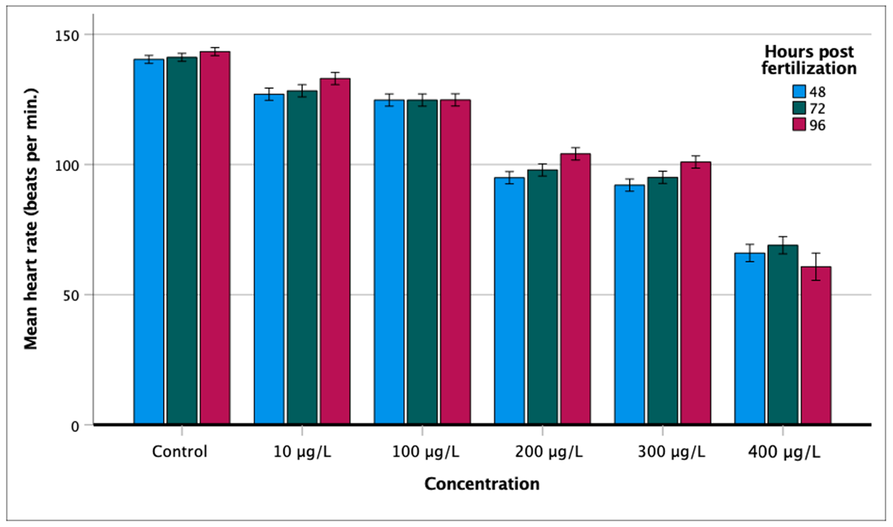
The collected zebrafish eggs were inspected; the unfertilized eggs were removed. At 24 hpf (hours post-fertilization) the dechorionation process of the eggs was carried out. Details about the toxicity testing are provided in the Supplementary Materials.

2.4. Heart Rate

The effect of 2,6-DCBQ-OH on the heart rate (number of beats per minute (bmp)) as a function of its concentration and time hours post fertilization (hpf) was investigated to evaluate the effect on developmental ontogeny. Details about the toxicity testing are provided in the Supplementary Materials.

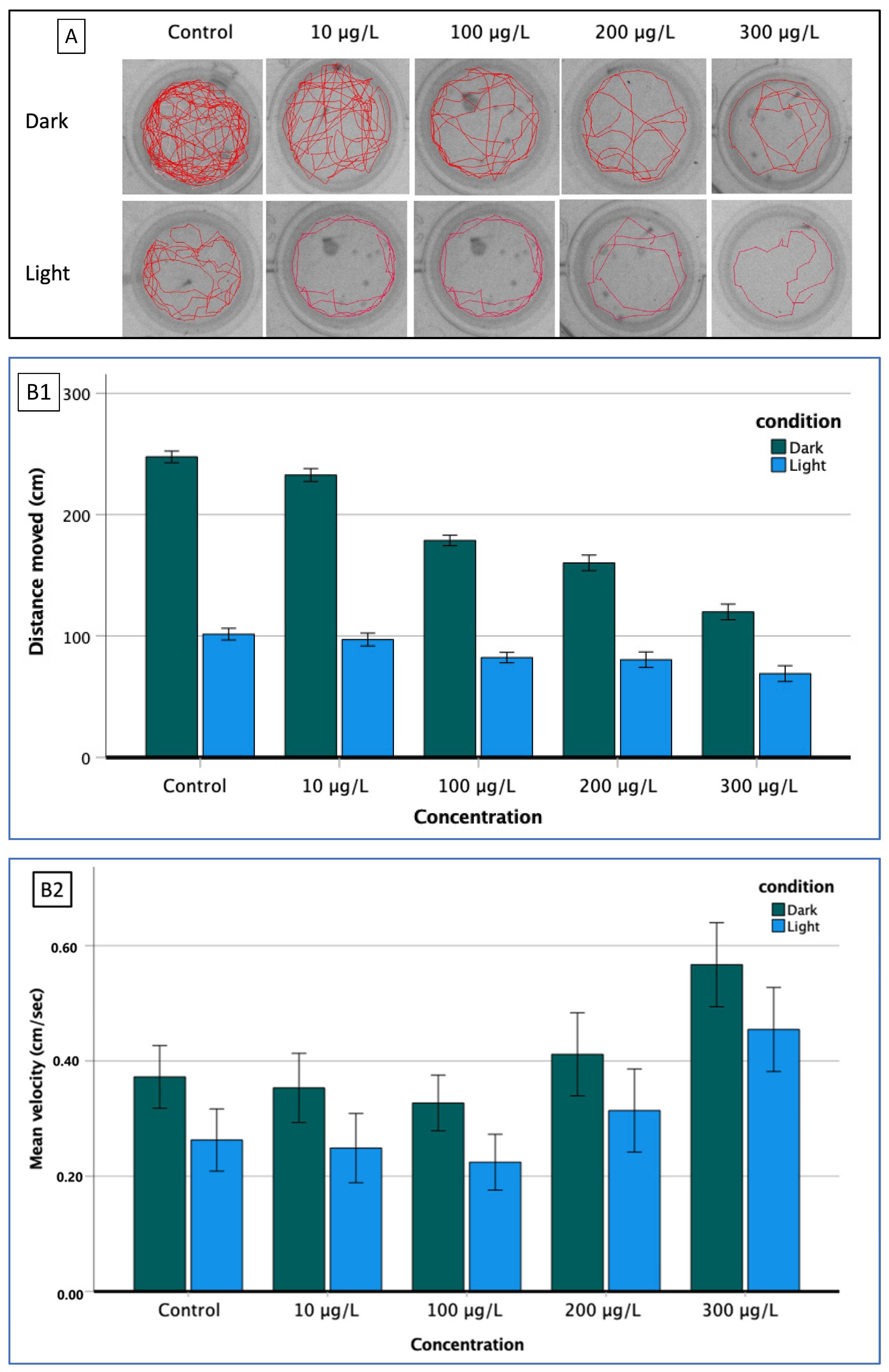
2.5. Behavior Screening

The effect of 2,6-DCBQ-OH was investigated on the motor function and reflexes of the zebrafish larvae through behavioral screening. To this end, motor function was analyzed in relation to 2,6-DCBQ-OH concentration by performing various behavioral trials.

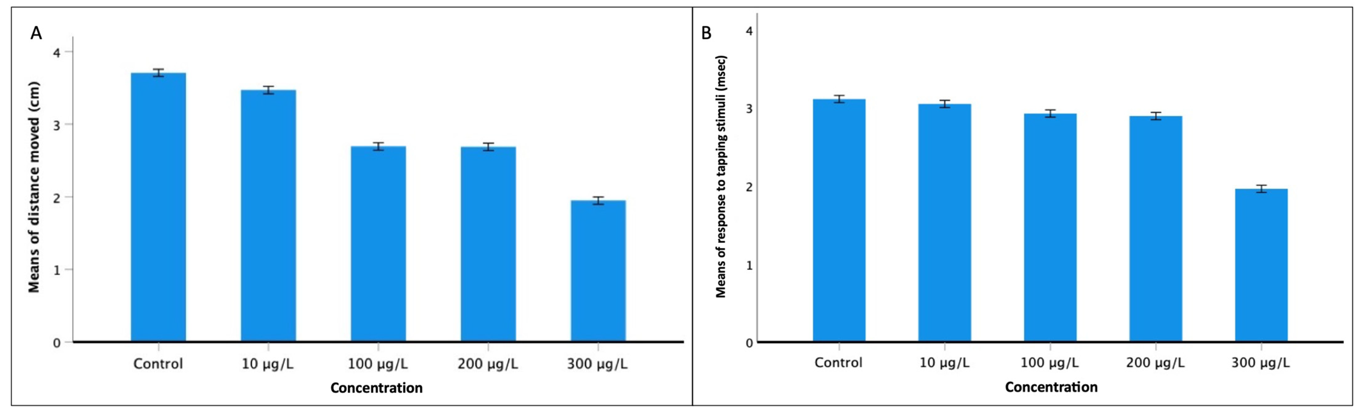
2.5.1. Touching Motor Response (TMR)

The touching motor response (TMR) was evaluated by performing a light touch stimulus test applied to the rostral head area by a glass capillary injection needle to determine the responsivity of larvae to this stimulus. Larvae lacking any response to a touch or those that were phenotypically unable to swim were excluded from further behavioral assessment.

2.5.2. Locomotor Activity

The larval swimming activity (distance moved, velocity) was studied in relation to dark and light conditions to evaluate the basal locomotor activity (BLA). Moreover, a visual motor response (VMR) test was employed to explore their ability to adapt to changing light conditions (dark/light alternation) after exposure to various concentrations of 2,6-DCBQ-OH. The larvae were then subjected to a vibrational startle response (VSR) test. Vibrational stimulus is provoked by a piston installed in the behavior recording system (DanioVision Tapping Device DVOC-004x/T, Noldus, Wageningen, The Netherlands). Details about the BLA, VMR, and VSR tests are presented in the Supplementary Materials.

2.6. Metabolomic Study and Data Processing

2.6.1. Metabolite Extraction

The procedure followed is similar to that of our previous studies [16,20,21]. In brief, larvae (144 hpf) were transferred to falcon tubes and washed with water. After euthanizing the larvae by snap freezing, the samples were lyophilized. Metabolites were extracted following the Bligh and Dyer extraction method [22]. Briefly, chloroform (1 mL) and methanol (2 mL) were added to the dry samples and vortexed. Then, chloroform (1 mL) and water (1 mL) were added. After ultrasonication and vortex mixing, the samples were centrifuged (3000 rpm for 5 min). The upper phase was collected, divided into two equal portions, and evaporated to dryness using a gentle nitrogen stream. In the first sub-sample (intended for NMR spectra), 600 μL of D2O (containing trimethylsilyl propionate (TSP)) was added and transferred in an NMR tube. To the second sub-sample (for LC-HRMS analysis,) 100 μL acetonitrile was added.

2.6.2. Metabolome Study and Data Processing

After correcting the 1H-NMR spectra shifts, based on the chemical shift of TSP, the chemical shifts were recorded and entered in the Human Metabolome Database (https://hmdb.ca, accessed on 1 September 2022, 1D NMR search engine), as well as in the NMR search engine of Madison Metabolomics Consortium Database (http://mmcd.nmrfam.wisc.edu, accessed on 1 September 2022). The metabolites generated from both databases were recorded and the common metabolites were further identified within the MS spectra (by searching for the accurate masses of the compounds, using four decimals, and matching the isotopic distributions and fragmentation patterns with the database data). Finally, the metabolites were introduced in the Metaboanalyst database (https://www.metaboanalyst.ca/, accessed on 1 September 2022) and the metabolic pathways were recorded, using the “Danio rerio (zebrafish)” pathway library. Details for the instrumentation of the metabolomic study are provided in the Supplementary Materials.

2.7. Statistical Analysis

Graphics and statistical analysis were processed using SPSS statistical software v28 (IBM Corp., New York, NY, USA). Prior to analyses, the variables were checked for normality. Most of the variables were found to be normally distributed. The non-normally distributed variables were log-transformed to achieve normality. Toxicity assays (LC50 calculation) and confidence intervals (LC25 and LC75) were determined based on cumulative mortality at the end of the experiment. Parameters of LC50 were assessed using probit regression analysis (the chi-square test, Pearson goodness of fit test, and 95% confidence interval). The effect of 2,6-DCBQ-OH concentration on heart rate (dependent variable) was assessed with the aim of GLM analysis (general linear model) using the concentration as independent variable and hpf as covariate. To explore the locomotor activity of larvae, the distance moved and the velocity were recorded with EthoVision XT tracking software (ver. 14: Noldus, Wageningen, The Netherlands). The differences in locomotor activity regarding the dark/light cycling were analyzed with GLM using the concentration as dependent variable and time as covariate. VSR test was performed with the aim of the EthoVision XT tracking software. The differences were considered significant at p < 0.001 and marginally significant at p < 0.05. Moreover, instrument operation, data recording, and processing of metabolome analyses were carried out using the Thermo Xcalibur 2.1 software (Thermo Fisher Scientific, Waltham, MA, USA).

3. Results

3.1. Morphological Abnormalities

The overall mortality of the control group was 4.8%, which is attributed to the handling during dechorionation process. The LC50 was estimated through probit regression analysis and was found to be 186 μg/L. Details are presented in Figure S1 of the Supplementary Materials.
The larvae were screened for phenotypic malformations after the exposure to 2,6-DCBQ-OH. It was observed that the embryos clearly presented dose-dependent phenotypic abnormalities while, in contrast, no morphological deformities were observed in the non-exposed embryos throughout the same period (Figure 1A–C). At concentrations lower than LC50, embryos presented minor developmental deformities, limited to slight tail flexion and, occasionally, pericardial and yolk sac edemas (Figure 1). Above LC50, (exposure to concentrations higher than 200 μg/L) the embryos exhibited obvious and progressively more pronounced phenotypic changes, such as bent and twisted notochord, and intense pericardial and yolk sac edema. The pericardial edema was observed at 24 hpf and became more obvious until 72 hpf. It is noteworthy that myocardial contractility was affected (decreased) after exposure to the highest concentration. The decay of the body was more obvious near the head, tail, and yolk sac. At concentration of 400 μg/L, more than 80% of the surviving embryos showed severe body malformations. The yolk sac appeared to be deformed, the pericardial edema was prominent and serious defects were noticed in the zebrafish heart. In addition, the heart was lengthened, the chambers were distinguished without overlap, the atria appeared thinner and elongated, the ventricles were smaller, and the pericardium appeared significantly swollen than in the cases of non-exposed larvae. These developmental disorders led to death in most of the larvae after 5 dpf (days post-fertilization).

3.2. Effect on Heart Rate

The heart rate of developing embryos was investigated in relation to post-fertilization hours (hpf) and 2,6-DCBQ-OH concentration. The mean heart rate value of the non-exposed group at 48 hpf was 140.4 bpm (±0.8) (N = 46), at 72 hpf was 141.2 bpm (±0.3) (N = 46), and at 96 hpf was 143.4 bpm (±0.8) (N = 46), indicating that there was an increasing tendency of the heartbeat during the ontogenetic development (Figure 2). The mean heart rate of the exposed embryos was significantly different from those of the non-exposed embryos. GLM analysis showed that heartbeat increased during the embryonic development (F = 69.3; p < 0.001). It has been proved that 2,6-DCBQ-OH influenced heartbeat rate, causing bradycardia. As the concentration increases, the heartbeat decreases (F = 8727.2; p < 0.0001), while the combined effect (embryonic development × concentration of 2,6-DCBQ-OH) was statistically significant (F = 24.8; p < 0.001).

3.3. Behavioral Analysis

3.3.1. Larval Activity (BLA, VMR)

The study of distance moved in relation to time in repetitive dark and light periods (10 min dark/10 min light/10 min dark) showed that the larvae presented more mobility in the dark periods and much less in the light period (BLA, basal locomotor activity). There was no statistically significant difference between the two dark phases (0–10 min and 20–30 min), indicating that there was no behavioral habituation of zebrafish during the experimental period (Figure 3).
Statistically significant differences were detected regarding the mean total distance moved after the exposure to various 2,6-DCBQ-OH concentrations. The distance moved during the dark period was much longer than that in the light period, for the same exposure time and at the same 2,6-DCBQ-OH concentrations. The exposure to various concentrations of 2,6-DCBQ-OH caused a significant reduction in embryos’ locomotor activity in terms of distance moved (hypolocomotion), in dark/light cycling conditions (Figure 4). Specifically, in both dark and light periods, the distance moved decreased significantly as the 2,6-DCBQ-OH concentration increased. In addition, the slope of reduction of the distance moved as a function of the concentration of 2,6-DCBQ-OH; during the dark period, was higher than that in the light period, meaning that, in terms of distance moved, the behavior of zebrafish larvae under dark was affected more than in light (Figure 4) (VMR, visual motor response). The GLM analysis confirms that the concentration and the light cycling, as well as the interaction of the two parameters, had a statistically significant effect on larval moving activity (concentration: F(4657) = 6804.52; p < 0.001, light/dark phases: F(1657) = 84,299.3; p < 0.001, interaction of conditions: F(4657) =2414.6; p < 0.001).
The study of velocity (in cm/s) revealed more complex pattern than the examination of distance moved, already described above. In both dark and light periods, there was a slight reduction in the velocity at relatively low concentrations of 2,6-DCBQ-OH (<LC50) and an increase at higher ones (Figure 4). As a general trend, the velocity in the dark period was significantly higher than that in the light period, regardless of the 2,6-DCBQ-OH concentrations. As the concentration increased up to 100 μg/L, the velocity decreased, under both dark and light periods, and then it increased when the concentration rose up to 200 μg/L. A GLM analysis showed that the concentration and lighting conditions had significant effect on larval activity (concentration: F(4657) = 715.4; p < 0.001, light/dark periods: F(1.657) = 715.4; p < 0.001), while the interaction of the conditions had no statistically significant effect (F(4657) = 0.3; p = 0.8).
The track plot of zebrafish larvae under the effect of 2,6-DCBQ-OH showed a dose-dependent effect of its preference in the inner or outermost zone of the wells (Figure 4). As the concentration increased, larvae avoided moving in the inner zone of the wells, in both conditions (dark and light), and preferred to swim in the proximity of the walls.

3.3.2. Touch Motor Response (TMR) and Vibrational Startle Response (VSR)

A statistically significant reduction of the swimming distance moved was observed for 10 s after a stimulus application as the concentration of 2,6-DCBQ-OH increased (F(4,70) = 4824.7; p < 0.001) (Figure 5). Moreover, the response time of zebrafish larvae in relation to the applied stimulus also decreased at elevated 2,6-DCBQ-OH concentrations (F(4,70) = 2603.6; p < 0.001) (Figure 5).

3.4. Metabolomic Study

Zebrafish were exposed to selected concentrations of 2,6-DCBQ-OH higher than LC50 (200, 300, and 500 μg/L) to investigate the effects on metabolic pathways. At these concentrations, multiple phenotypic and behavioral alterations were observed in the exposed zebrafish embryos. Various metabolites were confirmed in each larvae sample, hinting towards metabolic alterations after exposure (Table S1). Furthermore, spectroscopic data regarding the identification of the compounds are presented in Figure S1.
A total of 82 metabolites were identified in non-exposed and exposed groups. In the group of non-exposed larvae, 33 metabolites were identified, whereas in the group of larvae exposed to 200, 300, and 500 μg/mL, 27, 28, and 31 metabolites were identified. A total of 44 metabolic pathways were identified in non-exposed and exposed groups. In total, 24 pathways were identified in the control group and only 10 of them remained unaffected after the exposure to 2,6-DCBQ-OH concentrations. This is indicative of the post-exposure disturbances, either through the downregulation of pathways or through the upregulation of new ones. Specifically, at the lowest concentration (200 μg/L), 17 metabolic pathways remained unaffected compared to the non-exposed group, while 8 new pathways appeared. At 300 μg/L, 14 metabolic pathways remained unaffected compared to the non-exposed group, while 12 new pathways appeared. Finally, at the highest concentration (500 μg/L), only 10 metabolic pathways remained unaffected compared to the control group, while 17 new ones appeared (Table 1).
Several of the results obtained relating to metabolic pathways are noteworthy:
The metabolism of essential amino acids in exposed individuals is downregulated. Glutathione metabolism is upregulated and biosynthesis of arginine is activated in specimens exposed to 2,6-DCBQ-OH.
The metabolic pathway of the synthesis of unsaturated fatty acids appeared only in the control group (Table 1). The pathway of elongation of fatty acids and the butyric acid metabolism pathway are downregulated in specimens exposed to 2,6-DCBQ-OH.
The pathway of linoleic acid is detected only in the individuals that were not exposed to 2,6-DCBQ-OH.

4. Discussion

4.1. Developmental and Behavioral Alterations

The substance 2,6-DCBQ-OH is detected after chlorination-disinfection of water and then is released into aquatic ecosystems.
Exposure to 2,6-DCBQ-OH induced dose-dependent toxicity, morphological, developmental, and behavioral abnormalities, and metabolic alterations. Similar results have already been reported for halobenzoquinone in cell lines or in zebrafish [5,23,24].
The estimated LC50 value as 186 μg/L at 96 h of exposure is different from previous reports (94.4 μg/L at 72 h; 1.2 mg/L at 96 h [23,24]. The lowest value [23] can be attributed to the differences in exposure (without renewal of the solutions, yet a shorter exposure time). The highest value [24] could be attributed to the non-removal of the chorion, while in our study the chorion was removed. The chorion acts as a protective barrier and, therefore, reduces the observed toxicity [25].
Our results are in agreement with a previous study in which a dose-dependent pattern in rupture of pericardium, pericardium edema, and tail curvature appeared [26]. Embryos exposed to concentrations lower than LC50 showed few morphological malformations, while embryos exposed to values close to and higher than LC50 experienced severe malformations, even at 24 hpf. In addition, embryos showed signs of decay in the head and tail at concentrations above 400 μg/L.
Exposed specimens appeared to have anatomical abnormalities in their heart, which can be attributed to the deregulation of genes (i.e., krit1) responsible for heart development [26].
The mean heart rate was significantly reduced with an increase of 2,6-DCBQ-OH concentration (Figure 2). The observed bradycardia could also be attributed to the morphological disorders of the heart presented above.
Swimming distances reduced as the 2,6-DCBQ-OH concentration increased (Figure 4B1). Other studies prove that reduced swimming activity can be attributed to morphological malformations [27,28] and metabolic alterations [8,29]. In addition, the study of velocity (cm/s) under light and dark conditions showed that zebrafish reached higher velocity in the dark period (Figure 4B2). The reduction of swimming velocity in lower 2,6-DCBQ-OH concentrations (10 μg/L to 100 μg/L) and its significant sharp increase (200 μg/L and 300 μg/L) are in accordance with other studies [26,27]. It has been proved that habituation and learning processes can be severely affected by toxic chemical substances [30,31,32].
Hyper- and hypoactivities during the dark/light phases (Figure 4) are maintained regardless of the presence and concentration of the 2,6-DCBQ-OH [21]. This motor behavior is in agreement with studies that have proved that zebrafish embryos cover shortest distances in periods of light [33,34,35,36,37]. The rate of reduction of distance moved in dark conditions was higher than that in light conditions (Figure 4B1).
The track plot of the movement of zebrafish larvae (Figure 4B) depicts the progressive reduction of distance moved in light and dark conditions under the effect of increasing concentration of 2,6-DCBQ-OH. This confirms the results of the study of distance moved (Figure 4B1). The upregulation of glutathione metabolism in specimens exposed to 2,6-DCBQ-OH confirms the endogenous photophobic behavior of zebrafish. Moreover, it confirms the stress-inducing effect of the 2,6-DCBQ-OH.
Applying a tapping stimulus, a muscle contraction of the zebrafish larvae is induced, followed by rapid and irregular movements; in this way we can study the locomotor activity, the physiology of the sensory organs, as well as the basic learning functions [38]. The exposed larvae presented a lower response time but moved shorter distances as the exposure concentration increased. This indicated a dose dependence on hypersensitivity of the exposed specimens.
The reduced distance moved after the stimuli proves that 2,6-DCBQ-OH caused swimming disorders, which can be attributed to the morphological malformations or metabolic alterations. Events such as under-activity in individuals after stimuli are called stress behaviors and can be caused by neurotoxic events during neuronal development, possibly due to increased ROS (reactive oxygen species) production [39]. In addition, in a previous study, anxiety and stress disorders after stimuli indicated potential ecological implications regarding predation and vulnerability [40].
Given that specimens lost part of their ability to acclimatize to the environment after the exposure to toxic chemicals, it is likely that the sharp increase in speed after the application of stimulus could be an evidence of stress reaction.

4.2. Metabolic Alterations

Metabolomic study proved that many metabolic alterations occur between non-exposed and exposed specimens. It was found that as the concentration of 2,6-DCBQ-OH increased, the number of unaffected metabolic pathways decreased and, correspondingly, the number of new emerging metabolic pathways increased (Table 1). Metabolic alterations are presented below and linked to the phenotypic changes (morphological, functional, and behavioral) observed.

4.2.1. Beta-Alanine Metabolism

Metabolism of β-alanine is a very important biochemical process for the normal functioning of organisms. Normally, β-alanine is converted in muscle cells to carnosine [41], which in turn acts as a buffer to remove the lactic acid produced during muscle strain and fatigue [42]. Τhe bradykinesia observed under light and/or dark conditions (Figure 4B1) as well as the reduction of the distance moved after a startle response (Figure 5A) could be attributed to the downregulation of the β-alanine metabolic pathway after the exposure of zebrafish to 2,6-DCBQ-OH. The reduced mobility and the low alertness of zebrafish is accompanied by lower fatigue and muscle stiffness, and this can be attributed to the downregulation of the β-alanine metabolism pathway.

4.2.2. Glutathione Metabolism

Glutathione is a key antioxidant that contributes to neutralizing ROS and restores the balance of oxidants in zebrafish [43]. It is an essential substance for the organism that can be synthesized by amino acids (glutamic acid, cysteine, and glycine). The sulfhydryl group (–SH) of cysteine serves as a proton donor and is responsible for its biological action [18]. Its primary task is to help the organism protect itself from free radicals and all harmful substances, while it can renew and maintain all the other antioxidants in the body [16]. Glutathione is one of the most important factors in detoxifying the body and is vital for liver health in humans [8,44]. This confirms the oxidative stress induced by the chemical exposure and is associated with many developmental, morphological, and behavioral abnormalities.
During the early development of zebrafish, where organogenesis takes place, all amino acids are considered as essential [44]. Downregulation of the metabolism of essential amino acids in exposed individuals is most likely due to the body’s inability to cope simultaneously with the organogenesis and the toxicity of 2,6-DCBQ-OH, as proved in a previous study [16]. After the depletion of all its amino acid reserves individuals are unable to acquire sources through the yolk sac, thus amino acid metabolism stops, provoking abnormalities in the exposed specimens, possibly leading to death [20].

4.2.3. Biosynthesis of Unsaturated Fatty Acids and Glycerophospholipids

Lipids are the building blocks of cell membranes as well as nerve tissue [45]. These are very important sources of energy, and are stored in various tissues of the body. Glycerolipids are a structurally heterogeneous group of lipids that play key structural and functional roles in cell membranes [46]. Glycerophospholipids are amphiphilic lipids and are found in abundance in prokaryotic and eukaryotic cells [47]. Their structural role in the formation of the biological membranes and the monolayer of lipoproteins is very important [47].
Fatty acids play a very important role in the production of fatty Acetyl-CoA, which is involved in the metabolism of glycerophospholipids [48]. Glycerophospholipids are involved in many processes in organisms, such as genetic maturation of organisms (vitellogenin) [47]. In addition, glycerophospholipids, such as sphingomyelin, play an important role in brain development and the function of the blood—brain barrier and white matter of the brain [46]. It was observed that the metabolic pathway of the synthesis of unsaturated fatty acids appeared only in the control group (Table 1). The downregulation of the pathway of unsaturated fatty acids’ synthesis, could be correlated to the increased rate of morphological alterations, the reduced mobility, and the low survival rate of the exposed individuals, especially to higher concentrations. Finally, in our study, glycerophospholipids, which lead to genetic maturation [49], are not expressed in all exposed individuals. It has been proven that toxic chemicals that interfere with the synthesis of glycerophospholipids can act as endocrine disruptors [45,50,51,52].

4.2.4. Elongation of Fatty Acids

Lipids are macromolecules that are essential for cellular membrane structure, promote inter- and intracellular signaling, and serve as fundamental sources of energy [45]. Moreover, lipids serve additional essential roles in metabolic functions and physiology [45,46]. Fatty acids are vital for the normal functioning of the cells [46]. The elongation of fatty acids is essential for lipid homeostasis, because long-chain fatty acids are involved in the formation of the membrane of phospholipids and epidermal permeability [45], affecting membranes and the proteins involved in transport; signaling transduction; many enzymatic processes [53]; synthesis of sphingolipids; neural function, especially myelination [54]; and retina function and regeneration [55]. Elongation of fatty acids is important for the survival, function, and development of an organism [46,53]. Genes involved in lipid and lipoprotein metabolism are frequently expressed early in embryonic development [56] and are very important for development and survival. In our case, this pathway is downregulated in specimens exposed to 2,6-DCBQ-OH. This pathway could be linked to the increased mortality and the developmental or behavioral disorders observed in the exposed individuals. The downregulation of the metabolic path of fatty acids’ elongation, in our case, could explain the reduction of the mobility of the embryos (Figure 4B1), due to the reduction of the energy reserves of the embryos. Moreover, the difficulty of adapting to the light/dark alternations (Figure 3) and the disorders in reflexes (Figure 5) could possibly be attributed to the downregulation of this metabolic pathway, because it affects the function and regeneration of the retina and the function of the neurons.

4.2.5. Metabolism of Linoleic Acid

Linoleic acid belongs to the omega-6 fatty acids family [57]. Linoleic acid contributes to the growth and function of bones, the regulation of metabolism, and the proper function of the reproductive system of humans [58]. Linoleic acid is a key component of the yolk sac in zebrafish [57]. The absence of this pathway in the exposed individuals proves that the exposed individuals, being in a stressful state, cannot utilize essential resources provided from the yolk sac, which leads to abnormalities such as swimming disorders.

4.2.6. Butanoate (Butyric Acid) Metabolism

Butyric acid regulates body weight, body composition, and glucose homeostasis while being absorbed and metabolized by tissues and cells beyond the intestine, and contributes to a variety of metabolic processes in humans [29]. Butyrate is responsible for weight gain and growth in humans [29]. The downregulation of the butyric acid metabolism pathway in exposed zebrafish explains the total metabolic breakdown of zebrafish that have abnormal development and significant morphological abnormalities that finally lead to death.

4.2.7. Other Metabolic Changes

Arginine biosynthesis, which is activated in exposed individuals, leads to the production of ornithine, which participates in collagen production [59]. Glycerophospholipids, whose metabolism is also activated upon exposure, are components of the cell membrane [52]. Glycerophospholipids, along with collagen, strengthen the cell membrane [59]. Thus, cells activate these pathways to either repair damage to cell membranes (which could be due to oxidative stress) or enhance them to reduce their permeability and minimize the inflow of 2,6-DCBQ-OH into the intracellular space.
By performing the metabolomic study, a connection between the observed disorders after the exposure of zebrafish larvae to 2,6-DCBQ-OH was achieved. The observed phenotype was essentially associated with the body’s metabolism. This study can fill a very important knowledge gap and shed light on some of the mechanisms causing mortality and disorders.
However, we should emphasize that the link between morphological abnormalities, pathophysiological dysfunctions, and behavioral alterations with metabolic pathways is a difficult and multidimensional problem that should be investigated by exploring a path in depth each time. In conclusion, this study provides a comprehensive study of the effects of 2,6-DCBQ-OH on part of the zebrafish life cycle. Our study showed that a single substance from the abundance of halobenzoquinones that are released into the environment can cause severe disorders in organisms, and that actions must be taken when releasing such substances into the environment to prevent human and organism exposure.

5. Conclusions

This study provides a comprehensive study of the effects of 2,6-DCBQ-OH on part of the zebrafish life cycle. Our study showed that a single substance from the abundance of halobenzoquinones that are released into the environment can cause severe disorders in organisms, and that actions must be taken when releasing such substances into the environment to prevent human and organism exposure.

Supplementary Materials

The following supporting information can be downloaded at: https://www.mdpi.com/article/10.3390/fishes7060368/s1, Experimental S1: Zebrafish Toxicity Testing; Heart rate; Locomotor activity; Vibrational startle response (VSR); Instrumentation for metabolomic study; Results S1: LC50 Estimation; Metabolomic Study; Figure S1. Mortality pattern of zebrafish larvae exposed to different concentrations of 2,6-DCBQ-H; Figure S2. NMR spectra of metabolites of control samples (purple spectrum) and samples exposed to 200 μg/L (red spectrum), 300 μg/L (blue spectrum), and 500 μg/L (green spectrum) of 2,6-DCBQ- OH; Table S1. List of metabolites found in the control and exposed to 2,6-DCBQ-OH individuals.

Author Contributions

Conceptualization: I.L. and C.S.; methodology: I.L. and C.S.; validation: I.L. and C.S.; supervision: I.L. and C.S.; writing—original and review draft: I.L. and C.S.; conceptualization: T.C., I.C. and; format analysis: T.C. and I.C.; investigation: T.C. and I.C.; resources: T.C. and I.C.; visualization: T.C. and I.C.; writing—original and review draft: T.C., I.C. and C.S. All authors have read and agreed to the published version of the manuscript.

Funding

Part of this study was supported by IKY (State Scholarship Foundation) through Doctorate Scholarship with the reference number 14653 (Partnership Agreement 2014–2020).

Institutional Review Board Statement

The study was conducted according to the guidelines of the European Community guidelines of animal studies. All the experiments were approved by the Ethical Committee of University of Ioannina (protocol code: 1104, date of approval: 29 July 2022).

Informed Consent Statement

Not applicable.

Data Availability Statement

Data concerning anything from the current project can be obtained from the corresponding author: Ioannis Leonardos, [email protected].

Conflicts of Interest

The authors declare no conflict of interest.

References

  1. Du, H.; Li, J.; Moe, B.; McGuigan, C.F.; Shen, S.; Li, X.-F. Cytotoxicity and Oxidative Damage Induced by Halobenzoquinones to T24 Bladder Cancer Cells. Environ. Sci. Technol. 2013, 47, 2823–2830. [Google Scholar] [CrossRef] [PubMed]
  2. Li, J.; Wang, W.; Moe, B.; Wang, H.; Li, X.F. Chemical and Toxicological Characterization of Halobenzoquinones, an Emerging Class of Disinfection Byproducts. Chem. Res. Toxicol. 2015, 28, 306–318. [Google Scholar] [CrossRef] [PubMed]
  3. Zhao, Y.; Anichina, J.; Lu, X.; Bull, R.J.; Krasner, S.W.; Hrudey, S.E.; Li, X.F. Occurrence and Formation of Chloro- and Bromo-Benzoquinones during Drinking Water Disinfection. Water Res. 2012, 46, 4351–4360. [Google Scholar] [CrossRef] [PubMed]
  4. Wang, W.; Qian, Y.; Li, J.; Moe, B.; Huang, R.; Zhang, H.; Hrudey, S.E.; Li, X.F. Analytical and Toxicity Characterization of Halo-Hydroxyl-Benzoquinones as Stable Halobenzoquinone Disinfection Byproducts in Treated Water. Anal. Chem. 2014, 86, 4982–4988. [Google Scholar] [CrossRef] [PubMed]
  5. Hung, S.; Mohan, A.; Reckhow, D.A.; Godri Pollitt, K.J. Assessment of the in Vitro Toxicity of the Disinfection Byproduct 2,6-Dichloro-1,4-Benzoquinone and Its Transformed Derivatives. Chemosphere 2019, 234, 902–908. [Google Scholar] [CrossRef]
  6. Wu, H.; Long, K.; Sha, Y.; Lu, D.; Xia, Y.; Mo, Y.; Yang, Q.; Zheng, W.; Yang, M.; Wei, X. Occurrence and Toxicity of Halobenzoquinones as Drinking Water Disinfection Byproducts. Sci. Total Environ. 2021, 770, 145277. [Google Scholar] [CrossRef]
  7. Wang, W.; Qian, Y.; Boyd, J.M.; Wu, M.; Hrudey, S.E.; Li, X.F. Halobenzoquinones in Swimming Pool Waters and Their Formation from Personal Care Products. Environ. Sci. Technol. 2013, 47, 3275–3282. [Google Scholar] [CrossRef]
  8. Prochazka, E.; Escher, B.I.; Plewa, M.J.; Leusch, F.D.L. In Vitro Cytotoxicity and Adaptive Stress Responses to Selected Haloacetic Acid and Halobenzoquinone Water Disinfection Byproducts. Chem. Res. Toxicol. 2015, 28, 2059–2068. [Google Scholar] [CrossRef]
  9. Hung, S. Evaluating the Toxicity and Formation of Halobenzoquinones in Point-of-Use Chlorinated Drinking Water. Masters Theses 2018, 734, 87. [Google Scholar] [CrossRef]
  10. Spitsbergen, J.M.; Kent, M.L. The State of the Art of the Zebrafish Model for Toxicology and Toxicologic Pathology Research--Advantages and Current Limitations. Toxicol. Pathol. 2007, 31, 62–87. [Google Scholar] [CrossRef]
  11. Kang, Y.-F.; Li, Y.-H.; Fang, Y.-W.; Xu, Y.; Wei, X.-M.; Yin, X.-B. Carbon Quantum Dots for Zebrafish Fluorescence Imaging. Sci. Rep. 2015, 5, 11835. [Google Scholar] [CrossRef] [Green Version]
  12. Rihel, J.; Prober, D.A.; Arvanites, A.; Lam, K.; Zimmerman, S.; Jang, S.; Haggarty, S.J.; Kokel, D.; Rubin, L.L.; Peterson, R.T.; et al. Zebrafish Behavioral Profiling Links Drugs to Biological Targets and Rest/Wake Regulation. Science 2010, 327, 348–351. [Google Scholar] [CrossRef] [Green Version]
  13. Wixon, J. Danio rerio, the Zebrafish. Int. J. Genom. 2000, 17, 225–231. [Google Scholar] [CrossRef]
  14. Pitt, J.A.; Kozal, J.S.; Jayasundara, N.; Massarsky, A.; Trevisan, R.; Geitner, N.; Wiesner, M.; Levin, E.D.; Di Giulio, R.T. Uptake, Tissue Distribution, and Toxicity of Polystyrene Nanoparticles in Developing Zebrafish (Danio rerio). Aquat. Toxicol. 2018, 194, 185–194. [Google Scholar] [CrossRef] [PubMed]
  15. Wiecinski, P.N. Toxicity of Intact and Weathered Nanomaterials to Zebrafish. Ph.D. Thesis, University of Wisconsin-Madison, Madison, WI, USA, 2012. [Google Scholar]
  16. Chatzimitakos, T.G.; Pliatsika, C.; Chousidis, I.; Leonardos, I.D.; Stalikas, C.D. Metabolomic Profiling Unveils the Impact of Non-Doped and Heteroatom-Doped Carbon Nanodots on Zebrafish (Danio rerio) Embryos. Nanomaterials 2021, 11, 483. [Google Scholar] [CrossRef]
  17. Robertson, D.G. Metabonomics in Toxicology: A Review. Toxicol. Sci. 2005, 85, 809–822. [Google Scholar] [CrossRef] [PubMed]
  18. Morello, J.; Derks, R.J.E.; Lopes, S.S.; Steenvoorden, E.; Monteiro, E.C.; Mayboroda, O.A.; Pereira, S.A. Zebrafish Larvae Are a Suitable Model to Investigate the Metabolic Phenotype of Drug-Induced Renal Tubular Injury. Front. Pharmacol. 2018, 9, 1193. [Google Scholar] [CrossRef] [Green Version]
  19. Bouhifd, M.; Hartung, T.; Hogberg, H.T.; Kleensang, A.; Zhao, L. Review: Toxicometabolomics. J. Appl. Toxicol. 2013, 33, 1365–1383. [Google Scholar] [CrossRef]
  20. Chousidis, I.; Chatzimitakos, T.; Leonardos, D.; Filiou, M.D.; Stalikas, C.D.; Leonardos, I.D. Cannabinol in the Spotlight: Toxicometabolomic Study and Behavioral Analysis of Zebrafish Embryos Exposed to the Unknown Cannabinoid. Chemosphere 2020, 252, 126417. [Google Scholar] [CrossRef]
  21. Chatzimitakos, T.; Chousidis, I.; Leonardos, D.; Stalikas, C.; Leonardos, I. In the Swim of Cannabis: Developmental Toxicity and Metabolomic Pathway Alterations of Zebrafish Larvae Exposed to THC for the Assessment of Its Potential Environmental and Human Health Impact. Molecules 2022, 27, 5506. [Google Scholar] [CrossRef]
  22. Bligh, E.G.; Dyer, W.J. A Rapid Method of Total Lipid Extraction and Purification. Can. J. Biochem. Physiol. 1959, 37, 911–917. [Google Scholar] [CrossRef] [PubMed]
  23. Sun, H.J.; Zhang, Y.; Zhang, J.Y.; Lin, H.; Chen, J.; Hong, H. The Toxicity of 2,6-Dichlorobenzoquinone on the Early Life Stage of Zebrafish: A Survey on the Endpoints at Developmental Toxicity, Oxidative Stress, Genotoxicity and Cytotoxicity. Environ. Pollut. 2019, 245, 719–724. [Google Scholar] [CrossRef]
  24. Wang, C.; Yang, X.; Zheng, Q.; Moe, B.; Li, X.F. Halobenzoquinone-Induced Developmental Toxicity, Oxidative Stress, and Apoptosis in Zebrafish Embryos. Environ. Sci. Technol. 2018, 52, 10590–10598. [Google Scholar] [CrossRef] [PubMed]
  25. Sun, H.-J.; Xiang, P.; Tang, M.-H.; Sun, L.; Ma, L.Q. Arsenic Impacted the Development, Thyroid Hormone and Gene Transcription of Thyroid Hormone Receptors in Bighead Carp Larvae (Hypophthalmichthys nobilis). J. Hazard. Mater. 2016, 303, 76–82. [Google Scholar] [CrossRef] [PubMed]
  26. Chen, J.N.; Haffter, P.; Odenthal, J.; Vogelsang, E.; Brand, M.; van Eeden, F.J.; Furutani-Seiki, M.; Granato, M.; Hammerschmidt, M.; Heisenberg, C.P.; et al. Mutations Affecting the Cardiovascular System and Other Internal Organs in Zebrafish. Development 1996, 123, 293–302. [Google Scholar] [CrossRef]
  27. Costa, B.P.D.; Moura, L.A.; Pinto, S.A.G.; Lima-Maximino, M.; Maximino, C. Zebrafish Models in Neural and Behavioral Toxicology across the Life Stages. Fishes 2020, 5, 23. [Google Scholar] [CrossRef]
  28. Levin, E.D.; Sledge, D.; Roach, S.; Petro, A.; Donerly, S.; Linney, E. Persistent Behavioral Impairment Caused by Embryonic Methylphenidate Exposure in Zebrafish. Neurotoxicol. Teratol. 2011, 33, 668–673. [Google Scholar] [CrossRef] [Green Version]
  29. Zhang, L.; Liu, C.; Jiang, Q.; Yin, Y. Butyrate in Energy Metabolism: There Is Still More to Learn. Trends Endocrinol. Metab. 2021, 32, 159–169. [Google Scholar] [CrossRef]
  30. Maximino, C.; de Brito, T.M.; da Silva Batista, A.W.; Herculano, A.M.; Morato, S.; Gouveia, A. Measuring Anxiety in Zebrafish: A Critical Review. Behav. Brain Res. 2010, 214, 157–171. [Google Scholar] [CrossRef]
  31. Costa, B.; Ferreira, S.; Póvoa, V.; Cardoso, M.J.; Vieira, S.; Stroom, J.; Fidalgo, P.; Rio-Tinto, R.; Figueiredo, N.; Parés, O.; et al. Developments in Zebrafish Avatars as Radiotherapy Sensitivity Reporters—Towards Personalized Medicine. EBioMedicine 2020, 51, 102578. [Google Scholar] [CrossRef]
  32. Maximino, C.; da Silva, A.W.B.; Gouveia, A.; Herculano, A.M. Pharmacological Analysis of Zebrafish (Danio rerio) Scototaxis. Prog. Neuro-Psychopharmacol. Biol. Psychiatry 2011, 35, 624–631. [Google Scholar] [CrossRef] [PubMed] [Green Version]
  33. Ahmad, F.; Noldus, L.P.J.J.; Tegelenbosch, R.A.J.; Richardson, M.K. Zebrafish Embryos and Larvae in Behavioural Assays. Behaviour 2012, 149, 1241–1281. [Google Scholar] [CrossRef]
  34. Tzima, E.; Serifi, I.; Tsikari, I.; Alzualde, A.; Leonardos, I.; Papamarcaki, T. Transcriptional and Behavioral Responses of Zebrafish Larvae to Microcystin-LR Exposure. Int. J. Mol. Sci. 2017, 18, 365. [Google Scholar] [CrossRef] [Green Version]
  35. Noyes, P.D.; Haggard, D.E.; Gonnerman, G.D.; Tanguay, R.L. Advanced Morphological—Behavioral Test Platform Reveals Neurodevelopmental Defects in Embryonic Zebrafish Exposed to Comprehensive Suite of Halogenated and Organophosphate Flame Retardants. Toxicol. Sci. 2015, 145, 177–195. [Google Scholar] [CrossRef] [PubMed] [Green Version]
  36. Bailey, J.; Oliveri, A.; Levin, E.D. Zebrafish Model Systems for Developmental Neurobehavioral Toxicology. Birth Defects Res. Part C—Embryo Today Rev. 2013, 99, 14–23. [Google Scholar] [CrossRef] [Green Version]
  37. Chousidis, I.; Stalikas, C.D.; Leonardos, I.D. Induced Toxicity in Early-Life Stage Zebrafish (Danio rerio) and Its Behavioral Analysis after Exposure to Non-Doped, Nitrogen-Doped and Nitrogen, Sulfur-Co Doped Carbon Quantum Dots. Environ. Toxicol. Pharmacol. 2020, 79, 103426. [Google Scholar] [CrossRef]
  38. Kimmel, C.B.; Patterson, J.; Kimmel, R.O. The Development and Behavioral Characteristics of the Startle Response in the Zebra Fish. Dev. Psychobiol. 1974, 7, 47–60. [Google Scholar] [CrossRef]
  39. Kim, S.-H.; Kowalski, M.L.; Carson, R.P.; Bridges, L.R.; Ess, K.C. Heterozygous Inactivation of Tsc2 Enhances Tumorigenesis in P53 Mutant Zebrafish. Dis. Model. Mech. 2013, 6, 925–933. [Google Scholar] [CrossRef] [Green Version]
  40. Kataba, A.; Botha, T.L.; Nakayama, S.M.M.; Yohannes, Y.B.; Ikenaka, Y.; Wepener, V.; Ishizuka, M. Acute Exposure to Environmentally Relevant Lead Levels Induces Oxidative Stress and Neurobehavioral Alterations in Larval Zebrafish (Danio rerio). Aquat. Toxicol. 2020, 227, 105607. [Google Scholar] [CrossRef]
  41. Artioli, G.G.; Gualano, B.; Smith, A.; Stout, J.; Lancha, A.H.J. Role of Beta-Alanine Supplementation on Muscle Carnosine and Exercise Performance. Med. Sci. Sport. Exerc. 2010, 42, 1162–1173. [Google Scholar] [CrossRef]
  42. Quesnele, J.J.; Laframboise, M.A.; Wong, J.J.; Kim, P.; Wells, G.D. The Effects of Beta-Alanine Supplementation on Performance: A Systematic Review of the Literature. Int. J. Sport Nutr. Exerc. Metab. 2014, 24, 14–27. [Google Scholar] [CrossRef]
  43. Massarsky, A.; Kozal, J.S.; Di Giulio, R.T. Glutathione and Zebrafish: Old Assays to Address a Current Issue. Chemosphere 2017, 168, 707–715. [Google Scholar] [CrossRef] [Green Version]
  44. Mishra, P.; Gong, Z.; Kelly, B.C. Assessing Biological Effects of Fluoxetine in Developing Zebrafish Embryos Using Gas Chromatography-Mass Spectrometry Based Metabolomics. Chemosphere 2017, 188, 157–167. [Google Scholar] [CrossRef]
  45. Zeituni, E.M.; Farber, S.A. Studying Lipid Metabolism and Transport during Zebrafish Development. Methods Mol. Biol. 2016, 1451, 237–255. [Google Scholar] [CrossRef] [PubMed] [Green Version]
  46. Bhandari, S.; Lee, J.N.; Kim, Y., II; Nam, I.K.; Kim, S.J.; Kim, S.J.; Kwak, S.A.; Oh, G.S.; Kim, H.J.; Yoo, H.J.; et al. The Fatty Acid Chain Elongase, Elovl1, Is Required for Kidney and Swim Bladder Development during Zebrafish Embryogenesis. Organogenesis 2016, 12, 78–93. [Google Scholar] [CrossRef] [Green Version]
  47. Tocher, D.R. Glycerophospholipid Metabolism. In Biochemistry and Molecular Biology of Fishes; Hochachka, P.W., Mommsen, T.P., Eds.; Elsevier: Amsterdam, The Netherlands, 1995; Volume 4, pp. 119–157. [Google Scholar]
  48. Pirro, V.; Guffey, S.C.; Sepúlveda, M.S.; Mahapatra, C.T.; Ferreira, C.R.; Jarmusch, A.K.; Cooks, R.G. Lipid Dynamics in Zebrafish Embryonic Development Observed by DESI-MS Imaging and Nanoelectrospray-MS. Mol. Biosyst. 2016, 12, 2069–2079. [Google Scholar] [CrossRef] [PubMed] [Green Version]
  49. Örn, S. The Zebrafish as a Model Organism for Evaluation of Endocrine Disrupters. Ph.D. Thesis, Swedish University of Agricultural Sciences, Uppsala, Sweden, 2006; 36p. [Google Scholar]
  50. Watkins, P.A. Fatty Acyl-CoA Synthetases. In Encyclopedia of Biological Chemistry, 2nd ed.; Lennarz, W.J., Lane, M.D., Eds.; Academic Press: Waltham, UK, 2013; pp. 290–295. ISBN 978-0-12-378631-9. [Google Scholar]
  51. Segner, H. Zebrafish (Danio rerio) as a Model Organism for Investigating Endocrine Disruption. Comp. Biochem. Physiol. C Toxicol. Pharmacol. 2009, 149, 187–195. [Google Scholar] [CrossRef]
  52. Henderson, F.; Johnston, H.R.; Badrock, A.P.; Jones, E.A.; Forster, D.; Nagaraju, R.T.; Evangelou, C.; Kamarashev, J.; Green, M.; Fairclough, M.; et al. Enhanced Fatty Acid Scavenging and Glycerophospholipid Metabolism Accompany Melanocyte Neoplasia Progression in Zebrafish. Cancer Res. 2019, 79, 2136–2151. [Google Scholar] [CrossRef] [PubMed] [Green Version]
  53. Agaba, M.; Tocher, D.R.; Dickson, C.A.; Dick, J.R.; Teale, A.J. Zebrafish CDNA Encoding Multifunctional Fatty Acid Elongase Involved in Production of Eicosapentaenoic (20:5n-3) and Docosahexaenoic (22:6n-3) Acids. Mar. Biotechnol. 2004, 6, 251–261. [Google Scholar] [CrossRef] [PubMed]
  54. D’Rozario, M.; Monk, K.R.; Petersen, S.C. Analysis of Myelinated Axon Formation in Zebrafish. Methods Cell Biol. 2017, 138, 383–414. [Google Scholar] [CrossRef] [PubMed]
  55. Wan, J.; Goldman, D. Retina Regeneration in Zebrafish. Curr. Opin. Genet. Dev. 2016, 40, 41–47. [Google Scholar] [CrossRef] [PubMed] [Green Version]
  56. Schlegel, A.; Stainier, D.Y.R. Microsomal Triglyceride Transfer Protein Is Required for Yolk Lipid Utilization and Absorption of Dietary Lipids in Zebrafish Larvae. Biochemistry 2006, 45, 15179–15187. [Google Scholar] [CrossRef] [PubMed]
  57. Şahan, U.; Ipek, A.; Sozcu, A. Yolk Sac Fatty Acid Composition, Yolk Absorption, Embryo Development, and Chick Quality during Incubation in Eggs from Young and Old Broiler Breeders. Poult. Sci. 2014, 93, 2069–2077. [Google Scholar] [CrossRef] [PubMed]
  58. Sprecher, H. The Roles of Anabolic and Catabolic Reactions in the Synthesis and Recycling of Polyunsaturated Fatty Acids. Prostaglandins Leukot. Essent. Fat. Acids 2002, 67, 79–83. [Google Scholar] [CrossRef]
  59. Lee, H.K.; Kim, K.; Lee, J.; Lee, J.; Lee, J.; Kim, S.; Lee, S.E.; Kim, J.H. Targeted Toxicometabolomics of Endosulfan Sulfate in Adult Zebrafish (Danio rerio) Using GC-MS/MS in Multiple Reaction Monitoring Mode. J. Hazard. Mater. 2020, 389, 122056. [Google Scholar] [CrossRef]
Figure 1. Morphological alterations of 5 days post-fertilization (5 dpf) zebrafish individuals after exposure to 2,6-DCBQ-OH. (AC) control individuals without exposure. (DF) individuals exposed to 10 μg/L. Minor malformations such as tail curvature are observed. (GI) individuals exposed to 100 μg/L. Pericardial edemas and tail curvature are observed. (JL) individuals exposed to 200 μg/L. Pericardial edemas and tail curvatures are more obvious. (MO) individuals exposed to 300 μg/L. Acute pericardial edemas are obvious. (PR) individuals exposed to 400 μg/L. Acute pericardial edemas, tail flexions, and body decay are observed. Morphological alterations are indicated by arrows.
Fishes 07 00368 g001
Figure 2. Heartbeat rate of zebrafish during embryonic development, upon exposure to various concentrations of 2,6-DCBQ-OH, at 48, 72, and 96 hpf.
Fishes 07 00368 g002
Figure 3. Motor response of zebrafish larvae at 6 dpf is strongly impaired by 2,6-DCBQ-OH. Locomotor activity of control larvae at 6 days post-fertilization (n = 79), after exposure to 10 μg/L (n = 65), 100 mg/L (n = 100), 200 μg/L (n = 45), and 300 μg/L (n = 44), were monitored in a 30 min period of dark/light cycling (0–10 min, dark; 10–20 min, light [white area]; 20–30 min, dark). The distance was counted at intervals of 2 min and was compared with the control.
Fishes 07 00368 g003
Figure 4. Locomotor activity of zebrafish embryos under the effect of 2,6-DCBQ-OH. (A) This represents cumulative track plots of the position of zebrafish embryos exposed to various concentrations of 2,6-DCBQ-OH under dark and light conditions. (B) This represents means of total distance moved (B1) and means of velocity (B2) (n = 44) of embryos at 144 hpf during a 30 min period of behavioral recording. Embryos were placed in individual wells of a flat-bottom 48-well plate and acclimated to the recording chamber before tracking began. Data shown are sums of 30 min (average ± standard error). Error bars shows standard error of the mean (SEM).
Fishes 07 00368 g004
Figure 5. Distance moved (in cm) for 10 s after a stimulus delivery (A) and response time (in 10−3 s) after a stimulus (B) on zebrafish larvae exposed to various concentrations of 2,6-DCBQ-OH.
Fishes 07 00368 g005
Table 1. Metabolic pathways identified in control, and individuals exposed to various concentrations of 2,6-DCBQ-OH specimens.
Metabolic PathwayControl200 μg/L300 μg/L500 μg/L
Aminoacyl-tRNA biosynthesis
Histidine metabolism
Purine metabolism
Retinol metabolism
Selenocompound metabolism
Steroid hormone biosynthesis
Tyrosine metabolism
Valine, leucine and isoleucine biosynthesis
Valine, leucine and isoleucine degradation
Phenylalanine metabolism
Fatty acid biosynthesis
Pantothenate and CoA biosynthesis
Arginine biosynthesis
Pyrimidine metabolism
One carbon pool by folate
Pyruvate metabolism
Folate biosynthesis
Beta-Alanine metabolism
Biosynthesis of unsaturated fatty acids
Butanoate metabolism
Fatty acid elongation
Alanine, aspartate and glutamate metabolism
Linoleic acid metabolism
Sphingolipid metabolism
Glutathione metabolism
Amino sugar and nucleotide sugar metabolism
Cysteine and methionine metabolism
Fructose and mannose metabolism
Glycine, serine, and threonine metabolism
Tryptophan metabolism
Propanoate metabolism
Vitamin B6 metabolism
Arginine and proline metabolism
Pentose phosphate pathway
Porphyrin and chlorophyll metabolism
Primary bile acid biosynthesis
Thiamine metabolism
Glycerolipid metabolism
Glycerophospholipid metabolism
Nicotinate and nicotinamide metabolism
Phenylalanine, tyrosine, and tryptophan biosynthesis
Starch and sucrose metabolism
Steroid biosynthesis
Taurine and hypotaurine metabolism
Publisher’s Note: MDPI stays neutral with regard to jurisdictional claims in published maps and institutional affiliations.

