How Exercise and Dietary Intervention Affect the Outcome of Osteosarcopenic Obesity Syndrome?

Abstract
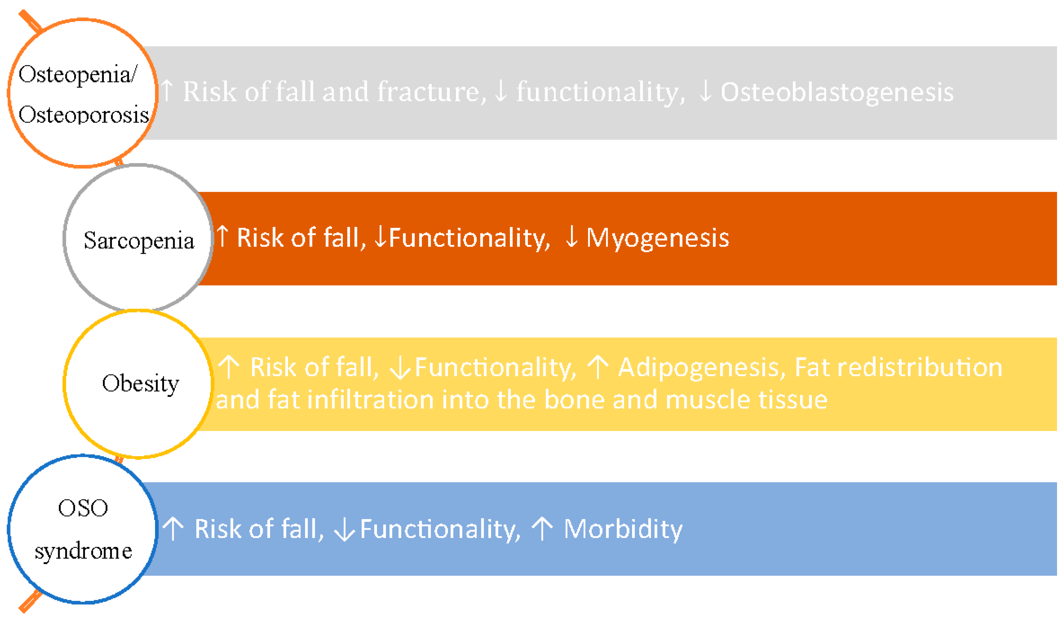
:1. Introduction
2. Diagnostic Criteria
3. Diet and Exercise in OSO Population
4. Physical and Functional Performance Tests to Evaluate OSO Syndrome
5. Fall Prevention in OSO Syndrome
6. Discussion and Summary
Conflicts of Interest
References
- Ilich, J.Z.; Kelly, O.J.; Inglis, J.E.; Panton, L.B.; Duque, G.; Ormsbee, M.J. Interrelationship among muscle, fat, and bone: Connecting the dots on cellular, hormonal, and whole body levels. Ageing Res. Rev. 2014, 15, 51–60. [Google Scholar] [CrossRef] [PubMed]
- Ilich, J.Z.; Kelly, O.J.; Inglis, J.E. Osteosarcopenic obesity syndrome: What is it and how can it be identified and diagnosed? Curr. Gerontol. Geriatr. Res. 2016, 2016, 1–7. [Google Scholar] [CrossRef] [PubMed]
- Stefanaki, C.; Peppa, M.; Boschiero, D.; Chrousos, G.P. Healthy overweight/obese youth: Early osteosarcopenic obesity features. Eur. J. Clin. Investig. 2016, 46, 767–778. [Google Scholar] [CrossRef] [PubMed]
- JafariNasabian, P.; Inglis, J.E.; Reilly, W.; Kelly, O.J.; Ilich, J.Z. Aging human body: Changes in bone, muscle and body fat with consequent changes in nutrient intake. J. Endocrinol. 2017, 234, R37–R51. [Google Scholar] [CrossRef] [PubMed]
- Cunha, P.M.; Ribeiro, A.S.; Tomeleri, C.M.; Schoenfeld, B.J.; Silva, A.M.; Souza, M.F.; Nascimento, M.A.; Sardinha, L.B.; Cyrino, E.S. The effects of resistance training volume on osteosarcopenic obesity in older women. J. Sports Sci. 2018, 36, 1564–1571. [Google Scholar] [CrossRef] [PubMed]
- Hao, J.; Zhang, Y.; Jing, D.; Shen, Y.; Tang, G.; Huang, S.; Zhao, Z. Mechanobiology of mesenchymal stem cells: Perspective into mechanical induction of MSC fate. Acta Biomater. 2015, 20, 1–9. [Google Scholar] [CrossRef] [PubMed]
- Ilich, J.Z.; Kelly, O.J.; Kim, Y.; Spicer, M.T. Low-grade chronic inflammation perpetuated by modern diet as a promoter of obesity and osteoporosis. Arch. Ind. Hyg. Toxicol. 2014, 65, 139–148. [Google Scholar] [CrossRef] [PubMed]
- Deshpande, S.; James, A.W.; Blough, J.; Donneys, A.; Wang, S.C.; Cederna, P.S.; Buchman, S.R.; Levi, B. Reconciling the effects of inflammatory cytokines on mesenchymal cell osteogenic differentiation. J. Surg. Res. 2013, 185, 278–285. [Google Scholar] [CrossRef] [PubMed]
- Gimble, J.M.; Zvonic, S.; Floyd, Z.E.; Kassem, M.; Nuttall, M.E. Playing with bone and fat. J. Cell. Biochem. 2006, 98, 251–266. [Google Scholar] [CrossRef] [PubMed]
- Rosen, C.J.; Bouxsein, M.L. Mechanisms of disease: Is osteoporosis the obesity of bone? Nat. Clin. Pract. Rheumatol. 2006, 2, 35–43. [Google Scholar] [CrossRef] [PubMed]
- Gilsanz, V.; Chalfant, J.; Mo, A.O.; Lee, D.C.; Dorey, F.J.; Mittelman, S.D. Reciprocal relations of subcutaneous and visceral fat to bone structure and strength. J. Clin. Endocrinol. Metab. 2009, 94, 3387–3393. [Google Scholar] [CrossRef] [PubMed]
- Zhang, P.; Peterson, M.; Su, G.L.; Wang, S.C. Visceral adiposity is negatively associated with bone density and muscle attenuation. Am. J. Clin. Nutr. 2015, 101, 337–343. [Google Scholar] [CrossRef] [PubMed]
- Pradhan, A.D.; Manson, J.E.; Rifai, N.; Buring, J.E.; Ridker, P.M. C-reactive protein, interleukin 6, and risk of developing type 2 diabetes mellitus. JAMA 2001, 286, 327–334. [Google Scholar] [CrossRef] [PubMed]
- Liu, P.-Y.; Hornbuckle, L.M.; Panton, L.B.; Kim, J.-S.; Ilich, J.Z. Evidence for the association between abdominal fat and cardiovascular risk factors in overweight and obese African American women. J. Am. Coll. Nutr. 2012, 31, 126–132. [Google Scholar] [CrossRef] [PubMed]
- Anty, R.; Bekri, S.; Luciani, N.; Saint-Paul, M.C.; Dahman, M.; Iannelli, A.; Amor, I.B.; Staccini-Myx, A.; Huet, P.M.; Gugenheim, J.; et al. The inflammatory C-reactive protein is increased in both liver and adipose tissue in severely obese patients independently from metabolic syndrome, type 2 diabetes, and NASH. Am. J. Gastroenterol. 2006, 101, 1824–1833. [Google Scholar] [CrossRef] [PubMed]
- Saijo, Y.; Kiyota, N.; Kawasaki, Y.; Miyazaki, Y.; Kashimura, J.; Fukuda, M.; Kishi, R. Relationship between C-reactive protein and visceral adipose tissue in healthy Japanese subjects. Diabetes Obes. Metab. 2004, 6, 249–258. [Google Scholar] [CrossRef] [PubMed]
- Ormsbee, M.J.; Prado, C.M.; Ilich, J.Z.; Purcell, S.; Siervo, M.; Folsom, A.; Panton, L. Osteosarcopenic obesity: The role of bone, muscle, and fat on health. J. Cachexia Sarcopenia Muscle 2014, 5, 183–192. [Google Scholar] [CrossRef] [PubMed]
- JafariNasabian, P.; Inglis, J.; Kelly, O.; Ilich, J. Osteosarcopenic obesity in women: Impact, prevalence, and management challenges. Int. J. Women Health 2017, 9, 33–42. [Google Scholar] [CrossRef] [PubMed]
- Kelly, O.J.; Gilman, J.C.; Kim, Y.; Ilich, J.Z. Micronutrient intake in the etiology, prevention and treatment of osteosarcopenic obesity. Curr. Aging Sci. 2016, 9, 260–278. [Google Scholar] [CrossRef] [PubMed]
- Kelly, O.J.; Gilman, J.C.; Kim, Y.; Ilich, J.Z. Macronutrient intake and distribution in the etiology, prevention and treatment of osteosarcopenic obesity. Curr. Aging Sci. 2017, 10, 83–105. [Google Scholar] [CrossRef] [PubMed]
- Kelly, O.J.; Gilman, J.C. Can unconventional exercise be helpful in the treatment, management and prevention of osteosarcopenic obesity? Curr. Aging Sci. 2016, 10, 106–121. [Google Scholar] [CrossRef] [PubMed]
- Kanis, J.A.; Melton, L.J.; Christiansen, C.; Johnston, C.C.; Khaltaev, N. The diagnosis of osteoporosis. J. Bone Miner. Res. 1994, 9, 1137–1141. [Google Scholar] [CrossRef] [PubMed]
- Obesity Algorithm: Clinical Guidelines for Obesity Treatment the American Society of Bariatric Physicians (ASBP), Obes. Algorithm. 2015. Available online: http://obesitymedicine.org/obesity-algorithm/ (accessed on 26 January 2017).
- Inglis, J.E. Identifying Osteosarcopenic Obesity in a Group of Older Women. Ph.D. Thesis, The Florida State University, Tallahassee, FL, USA, 2017. [Google Scholar]
- JafariNasabian, P. Analyzing Bone, Muscle and Adipose Tissue Biomarkers to Identify Osteosarcopenic Obesity Syndrome in Older Women. Ph.D. Thesis, The Florida State University, Tallahassee, FL, USA, 2017. [Google Scholar]
- Inglis, J.E.; Jafarinasabian, P.; Hebrock, H.; Ave, M.; Goosby, K.; Beyer, E.; Artese, A.; Panton, L.; Ilich-Ernst, J. Older women with osteosarcopenic obesity have lower handgrip strength and knee extension strength than osteopenic or obese-only women. Adv. Nutr. 2017, 8, 9. [Google Scholar]
- JafariNasabian, P.; Inglis, J.E.; Ave, M.P.; Hall, K.J.; Nieto, S.E.; Kelly, O.J.; Ilich, J.Z. Metabolic profile of osteosarcopenic obesity syndrome: Identifying biomarkers for diagnostic criteria. FASEB J. 2017, 31, 151–155. [Google Scholar]
- Inglis, J.E.; JafariNasabian, P.; Gilman, J.C.; Kelly, O.J.; Ilich, J.Z. Possible nutritional etiology of osteosarcopenic obesity syndrome. FASEB J. 2016, 30, 1156–1158. [Google Scholar]
- Pray, L.; Boon, C.; Ann Miller, E.; Pillsbury, L. Providing Healthy and Safe Foods as We Age: Workshop Summary; National Academies Press: Washington, DC, USA, 2010. [Google Scholar]
- Kim, J.; Lee, Y.; Kye, S.; Chung, Y.-S.; Lee, O. Association of serum vitamin D with osteosarcopenic obesity: Korea National Health and Nutrition Examination Survey 2008–2010. J. Cachexia Sarcopenia Muscle 2016, 8, 259–266. [Google Scholar] [CrossRef] [PubMed]
- Bales, C.W.; Ritchie, C.S. Sarcopenia, weight loss, and nutritional frailty in the elderly. Annu. Rev. Nutr. 2002, 22, 309–323. [Google Scholar] [CrossRef] [PubMed]
- Morley, J.E. Anorexia, sarcopenia, and aging. Nutrition 2001, 17, 660–663. [Google Scholar] [CrossRef]
- Heaney, R.P.; Layman, D.K. Amount and type of protein influences bone health. Am. J. Clin. Nutr. 2008, 87, 1567S–1570S. [Google Scholar] [CrossRef] [PubMed]
- Cuenca-Sánchez, M.; Navas-Carrillo, D.; Orenes-Piñero, E. Controversies surrounding high-protein diet intake: Satiating effect and kidney and bone health. Adv. Nutr. 2015, 6, 260–266. [Google Scholar] [CrossRef] [PubMed]
- Volpi, E.; Campbell, W.W.; Dwyer, J.T.; Johnson, M.A.; Jensen, G.L.; Morley, J.E.; Wolfe, R.R. Is the optimal level of protein intake for older adults greater than the recommended dietary allowance? J. Gerontol. A Biol. Sci. Med. Sci. 2013, 68, 677–681. [Google Scholar] [CrossRef] [PubMed]
- Szychlinska, M.A.; Castrogiovanni, P.; Trovato, F.M.; Nsir, H.; Zarrouk, M.; Furno, D.L.; Di Rosa, M.; Imbesi, R.; Musumeci, G. Physical activity and Mediterranean diet based on olive tree phenolic compounds from two different geographical areas have protective effects on early osteoarthritis, muscle atrophy and hepatic steatosis. Eur. J. Nutr. 2018. [Google Scholar] [CrossRef] [PubMed]
- Trovato, F.M.; Castrogiovanni, P.; Szychlinska, M.A.; Purrello, F.; Musumeci, G. Impact of Western and Mediterranean Diets and Vitamin D on Muscle Fibers of Sedentary Rats. Nutrients 2018, 10, 231. [Google Scholar] [CrossRef] [PubMed]
- Ilich, J.Z.; Inglis, J.E.; Kelly, O.J.; McGee, D.L. Osteosarcopenic obesity is associated with reduced handgrip strength, walking abilities, and balance in postmenopausal women. Osteoporos. Int. 2015, 26, 2587–2595. [Google Scholar] [CrossRef] [PubMed]
- Donnelly, J.E.; Smith, B.K. Is exercise effective for weight loss with ad libitum diet? Energy balance, compensation, and gender differences. Exerc. Sport Sci. Rev. 2005, 33, 169–174. [Google Scholar] [CrossRef] [PubMed]
- Marques, E.A.; Mota, J.; Carvalho, J. Exercise effects on bone mineral density in older adults: A meta-analysis of randomized controlled trials. Age 2012, 34, 1493–1515. [Google Scholar] [CrossRef] [PubMed]
- Turner, C.H.; Robling, A.G. Exercise as an anabolic stimulus for bone. Curr. Pharm. Des. 2004, 10, 2629–2641. [Google Scholar] [CrossRef] [PubMed]
- Aragão, F.R.; Abrantes, C.G.; Gabriel, R.E.; Sousa, M.F.; Castelo-Branco, C.; Moreira, M.H. Effects of a 12-month multi-component exercise program on the body composition of postmenopausal women. Climacteric 2014, 17, 155–163. [Google Scholar] [CrossRef] [PubMed]
- Bethancourt, H.J.; Rosenberg, D.E.; Beatty, T.; Arterburn, D.E. Barriers to and facilitators of physical activity program use among older adults. Clin. Med. Res. 2014, 12, 10–20. [Google Scholar] [CrossRef] [PubMed]
- Dechamps, A.; Gatta, B.; Bourdel-Marchasson, I.; Tabarin, A.; Roger, P. Pilot study of a 10-week multidisciplinary Tai Chi intervention in sedentary obese women. Clin. J. Sport Med. 2009, 19, 49–53. [Google Scholar] [CrossRef] [PubMed]
- Yu, T.-Y.; Pei, Y.-C.; Lau, Y.-C.; Chen, C.-K.; Hsu, H.-C.; Wong, A.M.K. Comparison of the effects of swimming and Tai Chi Chuan on body fat composition in elderly people. Chang Gung Med. J. 2007, 30, 128–134. [Google Scholar] [PubMed]
- Oken, B.S.; Zajdel, D.; Kishiyama, S.; Flegal, K.; Dehen, C.; Haas, M.; Kraemer, D.F.; Lawrence, J.; Leyva, J. Randomized, controlled, six-month trial of yoga in healthy seniors: Effects on cognition and quality of life. Altern. Ther. Health Med. 2006, 12, 40–47. [Google Scholar] [PubMed]
- Fourie, M.; Gildenhuys, G.; Shaw, I.; Shaw, B.; Toriola, A.; Goon, D. Effects of a Mat Pilates Programme on Body Composition in Elderly Women. West Indian Med. J. 2013, 62. [Google Scholar] [CrossRef] [PubMed]
- Hita-Contreras, F.; Martínez-Amat, A.; Cruz-Díaz, D.; Pérez-López, F.R. Osteosarcopenic obesity and fall prevention strategies. Maturitas 2015, 80, 126–132. [Google Scholar] [CrossRef] [PubMed]
- Gloeckl, R.; Heinzelmann, I.; Baeuerle, S.; Damm, E.; Schwedhelm, A.L.; Diril, M.; Buhrow, D.; Jerrentrup, A.; Kenn, K. Effects of whole body vibration in patients with chronic obstructive pulmonary disease—A randomized controlled trial. Respir. Med. 2012, 106, 75–83. [Google Scholar] [CrossRef] [PubMed]
- Lai, C.L.; Chen, H.Y.; Tseng, S.Y.; Liao, W.C.; Liu, B.T.; Lee, M.C.; Chen, H.S. Effect of whole-body vibration for 3 months on arterial stiffness in the middle-aged and elderly. Clin. Interv. Aging 2014, 9, 821–828. [Google Scholar] [CrossRef] [PubMed]
- Corrie, H.; Brooke-Wavell, K.; Mansfield, N.J.; Cowley, A.; Morris, R.; Masud, T. Effects of vertical and side-alternating vibration training on fall risk factors and bone turnover in older people at risk of falls. Age Ageing 2015, 44, 115–122. [Google Scholar] [CrossRef] [PubMed]
- Ma, C.; Liu, A.; Sun, M.; Zhu, H.; Wu, H. Effect of whole-body vibration on reduction of bone loss and fall prevention in postmenopausal women: A meta-analysis and systematic review. J. Orthop. Surg. Res. 2016, 11, 24. [Google Scholar] [CrossRef] [PubMed]
- Slatkovska, L.; Alibhai, S.M.H.; Beyene, J.; Cheung, A.M. Effect of whole-body vibration on BMD: A systematic review and meta-analysis. Osteoporos. Int. 2010, 21, 1969–1980. [Google Scholar] [CrossRef] [PubMed]
- Hughes, V.A.; Frontera, W.R.; Wood, M.; Evans, W.J.; Dallal, G.E.; Roubenoff, R.; Singh, M.A. Longitudinal muscle strength changes in older adults: Influence of muscle mass, physical activity, and health. J. Gerontol. Ser. A Biol. Sci. Med. Sci. 2001, 56, B209–B217. [Google Scholar] [CrossRef]
- Ilich, J.Z.; Brownbill, R.A. Habitual and low-impact activities are associated with better bone outcomes and lower body fat in older women. Calcif. Tissue Int. 2008, 83, 260–271. [Google Scholar] [CrossRef] [PubMed]
- Wolff, I.; van Croonenborg, J.J.; Kemper, H.C.; Kostense, P.J.; Twisk, J.W. The effect of exercise training programs on bone mass: A meta-analysis of published controlled trials in pre- and postmenopausal women. Osteoporos. Int. 1999, 9, 1–12. [Google Scholar] [CrossRef] [PubMed]
- Lanyon, L.E. Functional strain as a determinant for bone remodeling. Calcif. Tissue Int. 1984, 36 (Suppl. 1), S56–S61. [Google Scholar] [CrossRef] [PubMed]
- Deckx, N.; Wens, I.; Nuyts, A.H.; Hens, N.; De Winter, B.Y.; Koppen, G.; Goossens, H.; Van Damme, P.; Berneman, Z.N.; Eijnde, B.O.; et al. 12 weeks of combined endurance and resistance training reduces innate markers of inflammation in a randomized controlled clinical trial in patients with multiple sclerosis. Mediat. Inflamm. 2016, 2016, 6789276. [Google Scholar] [CrossRef] [PubMed]
- Morley, J.E.; Anker, S.D.; von Haehling, S. Prevalence, incidence, and clinical impact of sarcopenia: Facts, numbers, and epidemiology-update 2014. J. Cachexia Sarcopenia Muscle 2014, 5, 253–259. [Google Scholar] [CrossRef] [PubMed]
- Fong, S.S.M.; Choi, A.W.M.; Luk, W.S.; Yam, T.T.T.; Leung, J.C.Y.; Chung, J.W.Y. Bone Mineral Density, Balance Performance, Balance Self-Efficacy, and Falls in Breast Cancer Survivors With and Without Qigong Training: An Observational Study. Integr. Cancer Ther. 2018, 17, 124–130. [Google Scholar] [CrossRef] [PubMed]
- Reuben, D.B.; Magasi, S.; McCreath, H.E.; Bohannon, R.W.; Wang, Y.C.; Bubela, D.J.; Rymer, W.Z.; Beaumont, J.; Rine, R.M.; Lai, J.S.; et al. Motor assessment using the NIH Toolbox. Neurology 2013, 80 (Suppl. 3), S65–S75. [Google Scholar] [CrossRef] [PubMed]
- Kuh, D.; Karunananthan, S.; Bergman, H.; Cooper, R. A life-course approach to healthy ageing: Maintaining physical capability. Proc. Nutr. Soc. 2014, 73, 237–248. [Google Scholar] [CrossRef] [PubMed]
- Cruz-Jentoft, A.J.; Baeyens, J.P.; Bauer, J.M.; Boirie, Y.; Cederholm, T.; Landi, F.; Martin, F.C.; Michel, J.P.; Rolland, Y.; Schneider, S.M.; et al. Sarcopenia: European consensus on definition and diagnosis: Report of the European Working Group on Sarcopenia in Older People. Age Ageing 2010, 39, 412–423. [Google Scholar] [CrossRef] [PubMed]
- Savino, E.; Martini, E.; Lauretani, F.; Pioli, G.; Zagatti, A.M.; Frondini, C.; Pellicciotti, F.; Giordano, A.; Ferrari, A.; Nardelli, A.; et al. Handgrip strength predicts persistent walking recovery after hip fracture surgery. Am. J. Med. 2013, 126, 1068–1075. [Google Scholar] [CrossRef] [PubMed]
- Lindsey, C.; Brownbill, R.A.; Bohannon, R.A.; Ilich, J.Z. Association of Physical Performance Measures with Bone Mineral Density in Postmenopausal Women. Arch. Phys. Med. Rehabil. 2005, 86, 1102–1107. [Google Scholar] [CrossRef] [PubMed]
- Shin, H.; Liu, P.-Y.; Panton, L.B.; Ilich, J.Z. Physical performance in relation to body composition and bone mineral density in healthy, overweight, and obese postmenopausal women. J. Geriatr. Phys. Ther. 2014, 37, 7–16. [Google Scholar] [CrossRef] [PubMed]
- Wang, L.; Song, Y.; Manson, J.E.; Pilz, S.; März, W.; Michaëlsson, K.; Lundqvist, A.; Jassal, S.K.; Barrett-Connor, E.; Zhang, C.; et al. Circulating 25-hydroxy-vitamin D and risk of cardiovascular disease: A meta-analysis of prospective studies. Circ. Cardiovasc. Qual. Outcomes 2012, 5, 819–829. [Google Scholar] [CrossRef] [PubMed]
- Bin, C.M.; Flores, C.; Álvares-da-Silva, M.R.; Francesconi, C.F.M. Comparison between Handgrip Strength, Subjective Global Assessment, Anthropometry, and Biochemical Markers in Assessing Nutritional Status of Patients with Crohn’s Disease in Clinical Remission. Dig. Dis. Sci. 2010, 55, 137–144. [Google Scholar] [CrossRef] [PubMed]
- Brooks, N.; Layne, J.E.; Gordon, P.L.; Roubenoff, R.; Nelson, M.E.; Castaneda-Sceppa, C. Strength training improves muscle quality and insulin sensitivity in Hispanic older adults with type 2 diabetes. Int. J. Med. Sci. 2006, 4, 19–27. [Google Scholar] [CrossRef] [PubMed]
- Wood, R.J.; Gregory, S.; Maneen, J.; Sullivan, R.; Richard, J.; Sawyer, J.; Matthews, T.D.; Headley, S.A. Weight loss and muscle quality in older men with metabolic syndrome. FASEB J. 2011, 25, 982. [Google Scholar]
- Taaffe, D.R.; Henwood, T.R.; Nalls, M.A.; Walker, D.G.; Lang, T.F.; Harris, T.B. Alterations in muscle attenuation following detraining and retraining in resistance-trained older adults. Gerontology 2009, 55, 217–223. [Google Scholar] [CrossRef] [PubMed]
- Nevitt, M.C.; Cummings, S.R.; Kidd, S.; Black, D. Risk factors for recurrent nonsyncopal falls. A prospective study. JAMA 1989, 261, 2663–2668. [Google Scholar] [CrossRef] [PubMed]
- Lord, S.R.; Murray, S.M.; Chapman, K.; Munro, B.; Tiedemann, A. Sit-to-Stand Performance Depends on Sensation, Speed, Balance, and Psychological Status in Addition to Strength in Older People. J. Gerontol. Ser. A Biol. Sci. Med. Sci. 2002, 57, M539–M543. [Google Scholar] [CrossRef]
- Lindemann, U.; Claus, H.; Stuber, M.; Augat, P.; Muche, R.; Nikolaus, T.; Becker, C. Measuring power during the sit-to-stand transfer. Eur. J. Appl. Physiol. 2003, 89, 466–470. [Google Scholar] [CrossRef] [PubMed]
- Centers for Disease Control and Pervention. 30-Second Chair Stand. 2017. Available online: https://www.cdc.gov/steadi/pdf/STEADI-Assessment-30Sec-508.pdf (accessed on 25 April 2018).
- Bernabeu-Mora, R.; Medina-Mirapeix, F.; Llamazares-Herrán, E.; Oliveira-Sousa SL de Sánchez-Martinez, M.P.; Escolar-Reina, P. The accuracy with which the 5 times sit-to-stand test, versus gait speed, can identify poor exercise tolerance in patients with COPD: A cross-sectional study. Medicine 2016, 95, e4740. [Google Scholar] [CrossRef] [PubMed]
- Drey, M.; Sieber, C.C.; Bertsch, T.; Bauer, J.M.; Schmidmaier, R.; FiAT Intervention Group. Osteosarcopenia is more than sarcopenia and osteopenia alone. Aging Clin. Exp. Res. 2016, 28, 895–899. [Google Scholar] [CrossRef] [PubMed]
- Theou, O.; Jones, G.R.; Jakobi, J.M.; Mitnitski, A.; Vandervoort, A.A. A comparison of the relationship of 14 performance-based measures with frailty in older women. Appl. Physiol. Nutr. Metab. 2011, 36, 928–938. [Google Scholar] [CrossRef] [PubMed]
- Lusardi, M.M. Is Walking Speed a Vital Sign? Absolutely! Top. Geriatr. Rehabil. 2012, 28, 67–76. [Google Scholar] [CrossRef]
- Middleton, A.; Fritz, S.L.; Lusardi, M. Walking speed: The functional vital sign. J. Aging Phys. Act. 2015, 23, 314–322. [Google Scholar] [CrossRef] [PubMed]
- Najafi, D.A.; Dahlberg, L.E.; Hansson, E.E. A combination of clinical balance measures and FRAX® to improve identification of high-risk fallers. BMC Geriatr. 2016, 16, 94. [Google Scholar] [CrossRef] [PubMed]
- Peirone, E.; Goria, P.F.; Anselmino, A. A dual-task home-based rehabilitation programme for improving balance control in patients with acquired brain injury: A single-blind, randomized controlled pilot study. Clin. Rehabil. 2014, 28, 329–338. [Google Scholar] [CrossRef] [PubMed]
- Sànchez-Riera, L.; Carnahan, E.; Vos, T.; Veerman, L.; Norman, R.; Lim, S.S.; Hoy, D.; Smith, E.; Wilson, N.; Nolla, J.M.; et al. The global burden attributable to low bone mineral density. Ann. Rheum. Dis. 2014, 73, 1635–1645. [Google Scholar] [CrossRef] [PubMed]
- Falls are Leading Cause of Injury and Death in Older Americans. CDC Online Newsroom. 2016. Available online: https://www.cdc.gov/media/releases/2016/p0922-older-adult-falls.html (accessed on 24 April 2018).
- Falls World Heal. Organ. 2018. Available online: http://www.who.int/en/news-room/fact-sheets/detail/falls (accessed on 24 April 2018).
- Kanis, J.A.; Johnell, O.; Oden, A.; Jonsson, B.; De Laet, C.; Dawson, A. Risk of hip fracture according to the World Health Organization criteria for osteopenia and osteoporosis. Bone 2000, 27, 585–590. [Google Scholar] [CrossRef]
- Eastell, R.; Wahner, H.W.; O'Fallon, W.M.; Amadio, P.C.; Melton, L.J.; Riggs, B.L. Unequal decrease in bone density of lumbar spine and ultradistal radius in Colles’ and vertebral fracture syndromes. J. Clin. Investig. 1989, 83, 168–174. [Google Scholar] [CrossRef] [PubMed]
- Deandrea, S.; Lucenteforte, E.; Bravi, F.; Foschi, R.; La Vecchia, C.; Negri, E. Risk Factors for Falls in Community-dwelling Older People: A Systematic Review and Meta-analysis. Epidemiology 2010, 21, 658–668. [Google Scholar] [CrossRef] [PubMed]
- Valderrama-Hinds, L.M.; Al Snih, S.; Chen, N.; Rodriguez, M.A.; Wong, R. Falls in Mexican older adults aged 60 years and older. Aging Clin. Exp. Res. 2018. [Google Scholar] [CrossRef] [PubMed]
- Murphy, S.L.; Williams, C.S.; Gill, T.M. Characteristics associated with fear of falling and activity restriction in community-living older persons. J. Am. Geriatr. Soc. 2002, 50, 516–520. [Google Scholar] [CrossRef] [PubMed]
- Hita-Contreras, F.; Martínez-Amat, A.; Lomas-Vega, R.; Álvarez, P.; Aránega, A.; Martínez-López, E.; Mendoza, N. Predictive value of stabilometry and fear of falling on falls in postmenopausal women. Climacteric 2013, 16, 584–589. [Google Scholar] [CrossRef] [PubMed]
- Scheffer, A.C.; Schuurmans, M.J.; van Dijk, N.; van der Hooft, T.; de Rooij, S.E. Fear of falling: Measurement strategy, prevalence, risk factors and consequences among older persons. Age Ageing 2008, 37, 19–24. [Google Scholar] [CrossRef] [PubMed]
- Scott, D.; Sanders, K.M.; Aitken, D.; Hayes, A.; Ebeling, P.R.; Jones, G. Sarcopenic obesity and dynapenic obesity: 5-year associations with falls risk in middle-aged and older adults. Obesity 2014, 22, 1568–1574. [Google Scholar] [CrossRef] [PubMed]
- Kaji, H. Linkage between muscle and bone: Common catabolic signals resulting in osteoporosis and sarcopenia. Curr. Opin. Clin. Nutr. Metab. Care 2013, 16, 272–277. [Google Scholar] [CrossRef] [PubMed]
- Hita-Contreras, F.; Martínez-López, E.; González-Matarín, P.; Mendoza, N.; Cruz-Díaz, D.; Ruiz-Ariza, A.; Martínez-Amat, A. Association of bone mineral density with postural stability and the fear of falling in Spanish postmenopausal women. Maturitas 2014, 79, 322–328. [Google Scholar] [CrossRef] [PubMed]
- Beavers, D.P.; Beavers, K.M.; Loeser, R.F.; Walton, N.R.; Lyles, M.F.; Nicklas, B.J.; Shapses, S.A.; Newman, J.J.; Messier, S.P. The independent and combined effects of intensive weight loss and exercise training on bone mineral density in overweight and obese older adults with osteoarthritis. Osteoarthr. Cartil. 2014, 22, 726–733. [Google Scholar] [CrossRef] [PubMed]
- Villareal, D.T.; Apovian, C.M.; Kushner, R.F.; Klein, S. American Society for Nutrition, NAASO, The Obesity Society. Obesity in Older Adults: Technical Review and Position Statement of the American Society for Nutrition and NAASO. Obes. Res. 2005, 13, 1849–1863. [Google Scholar] [CrossRef] [PubMed]
- Morley, J.E.; Argiles, J.M.; Evans, W.J.; Bhasin, S.; Cella, D.; Deutz, N.E.; Doehner, W.; Fearon, K.C.; Ferrucci, L.; Hellerstein, M.K.; et al. Nutritional recommendations for the management of sarcopenia. J. Am. Med. Dir. Assoc. 2010, 11, 391–396. [Google Scholar] [CrossRef] [PubMed]
- Pérez-López, F.R.; Brincat, M.; Erel, C.T.; Tremollieres, F.; Gambacciani, M.; Lambrinoudaki, I.; Moen, M.H.; Schenck-Gustafsson, K.; Vujovic, S.; Rozenberg, S.; et al. EMAS position statement: Vitamin D and postmenopausal health. Maturitas 2012, 71, 83–88. [Google Scholar] [CrossRef] [PubMed]
- Tanaka, K.; Kanazawa, I.; Yamaguchi, T.; Yano, S.; Kaji, H.; Sugimoto, T. Active vitamin D possesses beneficial effects on the interaction between muscle and bone. Biochem. Biophys. Res. Commun. 2014, 450, 482–487. [Google Scholar] [CrossRef] [PubMed]
- Petridou, E.T.; Manti, E.G.; Ntinapogias, A.G.; Negri, E.; Szczerbińska, K. What Works Better for Community-Dwelling Older People at Risk to Fall? J. Aging Health 2009, 21, 713–729. [Google Scholar] [CrossRef] [PubMed]
- Jung, D.; Lee, J.; Lee, S.-M. A Meta-Analysis of Fear of Falling Treatment Programs for the Elderly. West. J. Nurs. Res. 2009, 31, 6–16. [Google Scholar] [CrossRef] [PubMed]
- Madureira, M.M.; Bonfá, E.; Takayama, L.; Pereira, R.M.R. A 12-month randomized controlled trial of balance training in elderly women with osteoporosis: Improvement of quality of life. Maturitas 2010, 66, 206–211. [Google Scholar] [CrossRef] [PubMed]
- Schleicher, M.M.; Wedam, L.; Wu, G. Review of Tai Chi as an effective exercise on falls prevention in elderly. Res. Sports Med. 2012, 20, 37–58. [Google Scholar] [CrossRef] [PubMed]
- Álvarez-Barbosa, F.; del Pozo-Cruz, J.; del Pozo-Cruz, B.; Alfonso-Rosa, R.M.; Rogers, M.E.; Zhang, Y. Effects of supervised whole body vibration exercise on fall risk factors, functional dependence and health-related quality of life in nursing home residents aged 80+. Maturitas 2014, 79, 456–463. [Google Scholar] [CrossRef] [PubMed]
- Ribom, E.L.; Piehl-Aulin, K.; Ljunghall, S.; Ljunggren, O.; Naessén, T. Six months of hormone replacement therapy does not influence muscle strength in postmenopausal women. Maturitas 2002, 42, 225–231. [Google Scholar] [CrossRef]
- Barral, A.B.; Nahas, E.A.; Nahas-Neto, J.; Cangussu, L.M.; de Araujo Buttros, D. Effect of hormone therapy on postural balance in postmenopausal women. Menopause 2012, 19, 768–775. [Google Scholar] [CrossRef] [PubMed]
- Randell, K.M.; Honkanen, R.J.; Komulainen, M.H.; Tuppurainen, M.T.; Kröger, H.; Saarikoski, S. Hormone replacement therapy and risk of falling in early postmenopausal women—A population-based study. Clin. Endocrinol. 2001, 54, 769–774. [Google Scholar] [CrossRef]
- Kawai, M.; de Paula, F.J.A.; Rosen, C.J. New insights into osteoporosis: The bone-fat connection. J. Intern. Med. 2012, 272, 317–329. [Google Scholar] [CrossRef] [PubMed]
- Hamrick, M.W.; McGee-Lawrence, M.E.; Frechette, D.M. Fatty infiltration of skeletal muscle: Mechanisms and comparisons with bone marrow adiposity. Front. Endocrinol. 2016, 7, 69. [Google Scholar] [CrossRef] [PubMed]
- Domiciano, D.S.; Figueiredo, C.P.; Lopes, J.B.; Caparbo, V.F.; Takayama, L.; Menezes, P.R.; Bonfa, E.; Pereira, R.M. Discriminating sarcopenia in community-dwelling older women with high frequency of overweight/obesity. Osteoporos. Int. 2013, 24, 595–603. [Google Scholar] [CrossRef] [PubMed]
- Baumgartner, R.N. Body composition in healthy aging. Ann. N. Y. Acad. Sci. 2000, 904, 437–448. [Google Scholar] [CrossRef] [PubMed]
- Mijnarends, D.M.; Schols, J.M.; Meijers, J.M.; Tan, F.E.; Verlaan, S.; Luiking, Y.C.; Morley, J.E.; Halfens, R.J. Instruments to Assess Sarcopenia and Physical Frailty in Older People Living in a Community (Care) Setting: Similarities and Discrepancies. J. Am. Med. Dir. Assoc. 2015, 16, 301–308. [Google Scholar] [CrossRef] [PubMed]
- Liu, P.-Y.; Ilich, J.Z.; Brummel-Smith, K.; Ghosh, S. New insight into fat, muscle and bone relationship in women: Determining the threshold at which body fat assumes negative relationship with bone mineral density. Int. J. Prev. Med. 2014, 5, 1452–1463. [Google Scholar] [PubMed]


| Intrinsic Factors [4,5,17,18] | Extrinsic Factors [2,18,19,20,21] |
|---|---|
| Age Gender Chronic diseases Muscular strength Skeletal muscle mass Percentage of body fat Bone mineral density | Medication Diet Exercise |
| Condition | T-Score ≤ −1.0 SD at the Femoral Neck, or Lumbar Spine | <20th Percentile of ALM for Women | Fat Mass ≥ 32% for Women |
|---|---|---|---|
| Osteopenia/osteoporosis | Yes | No | No |
| Sarcopenia | No | Yes | No |
| Obesity | No | No | Yes |
| OSO | Yes | Yes | Yes |
© 2018 by the author. Licensee MDPI, Basel, Switzerland. This article is an open access article distributed under the terms and conditions of the Creative Commons Attribution (CC BY) license (http://creativecommons.org/licenses/by/4.0/).
Share and Cite
JafariNasabian, P. How Exercise and Dietary Intervention Affect the Outcome of Osteosarcopenic Obesity Syndrome? J. Funct. Morphol. Kinesiol. 2018, 3, 31. https://doi.org/10.3390/jfmk3020031
JafariNasabian P. How Exercise and Dietary Intervention Affect the Outcome of Osteosarcopenic Obesity Syndrome? Journal of Functional Morphology and Kinesiology. 2018; 3(2):31. https://doi.org/10.3390/jfmk3020031
Chicago/Turabian StyleJafariNasabian, Pegah. 2018. "How Exercise and Dietary Intervention Affect the Outcome of Osteosarcopenic Obesity Syndrome?" Journal of Functional Morphology and Kinesiology 3, no. 2: 31. https://doi.org/10.3390/jfmk3020031
APA StyleJafariNasabian, P. (2018). How Exercise and Dietary Intervention Affect the Outcome of Osteosarcopenic Obesity Syndrome? Journal of Functional Morphology and Kinesiology, 3(2), 31. https://doi.org/10.3390/jfmk3020031