Next Issue
Volume 3, September
Previous Issue
Volume 3, March


Quantum Beam Sci., Volume 3, Issue 2 (June 2019) – 7 articles

Cover Story (view full-size image): 
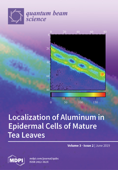
The elemental distribution of epidermal cells of tea leaves was investigated aiming at obtaining information on the mechanism of detoxifying aluminum using micro-beam Particle-Induced X-ray Emission (PIXE) method. The results showed that aluminum is localized to the cell wall, and silicon has a similar distribution. This may indicate that silicon may be involved in aluminum detoxification. View this paper.
  • Issues are regarded as officially published after their release is announced to the table of contents alert mailing list.
  • You may sign up for e-mail alerts to receive table of contents of newly released issues.
  • PDF is the official format for papers published in both, html and pdf forms. To view the papers in pdf format, click on the "PDF Full-text" link, and use the free Adobe Reader to open them.
Order results
Result details
Section
Select all
Export citation of selected articles as:
14 pages, 3409 KiB  
Article
Heavy-Ion Microbeams for Biological Science: Development of System and Utilization for Biological Experiments in QST-Takasaki
by Tomoo Funayama
Quantum Beam Sci. 2019, 3(2), 13; https://doi.org/10.3390/qubs3020013 - 14 Jun 2019
Cited by 13 | Viewed by 5125
Abstract
Target irradiation of biological material with a heavy-ion microbeam is a useful means to analyze the mechanisms underlying the effects of heavy-ion irradiation on cells and individuals. At QST-Takasaki, there are two heavy-ion microbeam systems, one using beam collimation and the other beam [...] Read more.
Target irradiation of biological material with a heavy-ion microbeam is a useful means to analyze the mechanisms underlying the effects of heavy-ion irradiation on cells and individuals. At QST-Takasaki, there are two heavy-ion microbeam systems, one using beam collimation and the other beam focusing. They are installed on the vertical beam lines of the azimuthally-varying-field cyclotron of the TIARA facility for analyzing heavy-ion radiation effects on biological samples. The collimating heavy-ion microbeam system is used in a wide range of biological research not only in regard to cultured cells but also small individuals, such as silkworms, nematode C. elegans, and medaka fish. The focusing microbeam system was designed and developed to perform more precise target irradiation that cannot be achieved through collimation. This review describes recent updates of the collimating heavy ion microbeam system and the research performed using it. In addition, a brief outline of the focusing microbeam system and current development status is described. Full article
(This article belongs to the Special Issue Ion Beams in Biology and Medicine)
Show Figures

Figure 1

14 pages, 4347 KiB  
Review
PIXE and Its Applications to Elemental Analysis
by Keizo Ishii
Quantum Beam Sci. 2019, 3(2), 12; https://doi.org/10.3390/qubs3020012 - 10 Jun 2019
Cited by 33 | Viewed by 13315
Abstract
When charged particles collide with atoms, atomic inner shell electrons become ionized, producing characteristic X-rays. This phenomenon is called particle-induced X-ray emission (PIXE). The characteristic X-ray production cross-sections from PIXE are very large, and the characteristic X-rays of elements contained in a sample [...] Read more.
When charged particles collide with atoms, atomic inner shell electrons become ionized, producing characteristic X-rays. This phenomenon is called particle-induced X-ray emission (PIXE). The characteristic X-ray production cross-sections from PIXE are very large, and the characteristic X-rays of elements contained in a sample are easily measured by a Silicon detector with a high energy resolution. Hence, sodium to uranium can be detected with a sensitivity of ppb~ppm, and PIXE has been applied to trace element analysis. Scanning ion beams can be used to obtain the spatial distributions of elements in a sample. Furthermore, the distributions of elements inside a cell can be investigated using micro ion beams. PIXE analysis is a very useful technique for multi-elemental analysis and is now widely used in many fields and applications, including chemistry, medicine, biology, archaeology, agriculture, materials science, fisheries science, geology, petrology, environmental study, contamination monitoring, resource search, semiconductors, metal, astrophysics, earth science, criminal investigations, and food. Full article
(This article belongs to the Special Issue Ion Beams in Biology and Medicine)
Show Figures

Figure 1

16 pages, 1546 KiB  
Review
Studies on Application of Ion Beam Breeding to Industrial Microorganisms at TIARA
by Katsuya Satoh and Yutaka Oono
Quantum Beam Sci. 2019, 3(2), 11; https://doi.org/10.3390/qubs3020011 - 5 Jun 2019
Cited by 15 | Viewed by 6079
Abstract
Mutation-breeding technologies are useful tools for the development of new biological resources in plants and microorganisms. In Takasaki Ion Accelerators for Advanced Radiation Application (TIARA) at the National Institutes for Quantum and Radiological Science and Technology, Japan, ion beams were explored as novel [...] Read more.
Mutation-breeding technologies are useful tools for the development of new biological resources in plants and microorganisms. In Takasaki Ion Accelerators for Advanced Radiation Application (TIARA) at the National Institutes for Quantum and Radiological Science and Technology, Japan, ion beams were explored as novel mutagens. The mutagenic effects of various ion beams on eukaryotic and prokaryotic microorganisms were described and their application in breeding technology for industrial microorganisms were discussed. Generally, the relative biological effectiveness (RBE) depended on the liner energy transfer (LET) and the highest RBE values were obtained with 12C5+ ion beams. The highest mutation frequencies were obtained at radiation doses that gave 1%–10% of surviving fraction. By using 12C5+ ion beams in this dose range, many microorganisms have been improved successfully at TIARA. Therefore, ion-beam breeding technology for microorganisms will have applications in many industries, including stable food production, sustainable agriculture, environmental conservation, and development of energy resources in the near future. Moreover, genome analyses of the ion-beam-induced mutants are in progress to clear the differences of mutational functions induced by different LET radiations in microorganisms. Further characterizations of mutations induced by different LET radiations will facilitate more effective use of ion beams in microorganisms breeding. Full article
(This article belongs to the Special Issue Ion Beams in Biology and Medicine)
Show Figures

Figure 1

13 pages, 5121 KiB  
Article
Experimental Setup of the Fast Current Controller for the Buenos Aires Heavy Ion Microbeam
by Nahuel Agustín Vega, Nahuel Agustín Müller, Emmanuel de la Fourniére, Emilia Beatriz Halac and Mario Ernesto Debray
Quantum Beam Sci. 2019, 3(2), 10; https://doi.org/10.3390/qubs3020010 - 3 Jun 2019
Viewed by 3079
Abstract
Recently we used the heavy ion microprobe of the Buenos Aires TANDAR Laboratory for Single Event Effects (SEE) and Total Dose (TD) experiments in electronics devices and components, requiring very low beam currents. The facility includes a fast beam switch that allows the [...] Read more.
Recently we used the heavy ion microprobe of the Buenos Aires TANDAR Laboratory for Single Event Effects (SEE) and Total Dose (TD) experiments in electronics devices and components, requiring very low beam currents. The facility includes a fast beam switch that allows the control of the ion beam current and a mobile Si PIN (p-type, intrinsic, n-type) diode that directly measures the number of ions hitting the device. The fast beam deflector was used to reduce the current by producing a pulsed beam or generating a quasi-continuous (Poisson-like distributed) beam with currents ranging from tens to hundreds of ions/s. As an application for this current control method we present a single event effect (SEE) pulses map generated by a 32S8+ beam at 75 MeV on two 0.5 µm technology CMOS digital output buffers where the device was formed by cascading four CMOS inverters with increasing sizes from input to output to drive large loads. Using the same concept of pulse width modulated deflection, we developed a novel gradient scanning method. This system allows to produce in a single irradiation a distribution with a cumulative damage with a difference of two orders of magnitude at constant gradient. To demonstrate the method, we irradiated a lithium niobate monocrystal with 32S8+ beam at 75 MeV energy and later analyzed the produced damage by the micro-Raman technique and an optical profilometer. Full article
(This article belongs to the Collection Facilities)
Show Figures

Figure 1

6 pages, 922 KiB  
Article
Localization of Aluminum in Epidermal Cells of Mature Tea Leaves
by Yoichi Haruyama, Tsuguhisa Fujiwara, Keisuke Yasuda, Manabu Saito and Kohtaku Suzuki
Quantum Beam Sci. 2019, 3(2), 9; https://doi.org/10.3390/qubs3020009 - 29 May 2019
Cited by 8 | Viewed by 3014
Abstract
We have determined the distribution of aluminum in the epidermal cells of mature tea leaves using micro-beam particle-induced X-ray emission. The observed pattern of aluminum distribution in the epidermal cells suggests that aluminum exists in cell walls. Silicon exhibits a distribution that is [...] Read more.
We have determined the distribution of aluminum in the epidermal cells of mature tea leaves using micro-beam particle-induced X-ray emission. The observed pattern of aluminum distribution in the epidermal cells suggests that aluminum exists in cell walls. Silicon exhibits a distribution that is nearly identical to that of aluminum, suggesting co-localization with aluminum. Full article
(This article belongs to the Special Issue Ion Beams in Biology and Medicine)
Show Figures

Figure 1

10 pages, 3478 KiB  
Article
Experimental Study on the Biological Effect of Cluster Ion Beams in Bacillus subtilis Spores
by Yoshihiro Hase, Katsuya Satoh, Atsuya Chiba, Yoshimi Hirano, Shigeo Tomita, Yuichi Saito and Kazumasa Narumi
Quantum Beam Sci. 2019, 3(2), 8; https://doi.org/10.3390/qubs3020008 - 6 May 2019
Cited by 5 | Viewed by 4729
Abstract
Cluster ion beams have unique features in energy deposition, but their biological effects are yet to be examined. In this study, we employed bacterial spores as a model organism, established an irradiation method, and examined the lethal effect of 2 MeV C, 4 [...] Read more.
Cluster ion beams have unique features in energy deposition, but their biological effects are yet to be examined. In this study, we employed bacterial spores as a model organism, established an irradiation method, and examined the lethal effect of 2 MeV C, 4 MeV C2, and 6 MeV C3 ion beams. The lethal effect per particle (per number of molecular ions) was not significantly different between cluster and monomer ion beams. The relative biological effectiveness and inactivation cross section as a function of linear energy transfer (LET) suggested that the single atoms of 2 MeV C deposited enough energy to kill the spores, and, therefore, there was no significant difference between the cluster and monomer ion beams in the cell killing effect under this experimental condition. We also considered the behavior of the atoms of cluster ions in the spores after the dissociation of cluster ions into monomer ions by losing bonding electrons through inelastic collisions with atoms on the surface. To the best of our knowledge, this is the first report to provide a basis for examining the biological effect of cluster ions. Full article
(This article belongs to the Special Issue Ion Beams in Biology and Medicine)
Show Figures

Figure 1

13 pages, 2105 KiB  
Review
Frequency and Spectrum of Radiation-Induced Mutations Revealed by Whole-Genome Sequencing Analyses of Plants
by Yeong Deuk Jo and Jin-Baek Kim
Quantum Beam Sci. 2019, 3(2), 7; https://doi.org/10.3390/qubs3020007 - 30 Apr 2019
Cited by 44 | Viewed by 7402
Abstract
Mutation breeding and functional genomics studies of mutant populations have made important contributions to plant research involving the application of radiation. The frequency and spectrum of induced mutations have long been regarded as the crucial determinants of the efficiency of the development and [...] Read more.
Mutation breeding and functional genomics studies of mutant populations have made important contributions to plant research involving the application of radiation. The frequency and spectrum of induced mutations have long been regarded as the crucial determinants of the efficiency of the development and use of mutant populations. Systematic studies regarding the mutation frequency and spectrum, including genetic and genomic analyses, have recently resulted in considerable advances. These studies have consistently shown that the mutation frequency and spectrum are affected by diverse factors, including radiation type, linear energy transfer, and radiation dose, as well as the plant tissue type and condition. Moreover, the whole-genome sequencing of mutant individuals based on next-generation sequencing technologies has enabled the genome-wide quantification of mutation frequencies according to DNA mutation types as well as the elucidation of mutation mechanisms based on sequence characteristics. These studies will contribute to the development of a highly efficient and more controlled mutagenesis method relevant for the customized research of plants. We herein review the characteristics of radiation-induced mutations in plants, mainly focusing on recent whole-genome sequencing analyses as well as factors affecting the mutation frequency and spectrum. Full article
(This article belongs to the Special Issue Ion Beams in Biology and Medicine)
Show Figures

Figure 1

Previous Issue
Next Issue
Back to TopTop