
How Could Nanomedicine Improve the Safety of Contrast Agents for MRI during Pregnancy?


UTCBS (Chemical and Biological Technologies for Health Group), CNRS, INSERM, Faculté de Pharmacie, Université de Paris Cité, 4 Avenue de L’observatoire, F-75006 Paris, France
*
Author to whom correspondence should be addressed.
Sci 2022, 4(1), 11; https://doi.org/10.3390/sci4010011
Submission received: 29 November 2021
/
Revised: 28 February 2022
/
Accepted: 10 March 2022
/
Published: 15 March 2022
(This article belongs to the Special Issue Feature Papers 2021 Editors Collection)
Abstract
:Pregnancy is a delicate state, during which timely investigation of possible physiological anomalies is essential to reduce the risk of maternal and fetal complications. Medical imaging encompasses different technologies to image the human body for the diagnosis, course of treatment management, and follow-up of diseases. Ultrasound (US) is currently the imaging system of choice for pregnant patients. However, sonographic evaluations can be non-effective or give ambiguous results. Therefore, magnetic resonance imaging (MRI), due to its excellent tissue penetration, the possibility of acquisition of three-dimensional anatomical information, and its high spatial resolution, is considered a valid diagnostical alternative. Nevertheless, currently employed contrast agents to improve the MRI image quality are harmful to the fetus. Because of their ability to cross the placenta, their use on pregnant patients is avoided. This review will firstly recapitulate the most common non-obstetrical, obstetrical, and fetal indications for magnetic resonance imaging on pregnant women. Fetal safety risks, due to the use of strong magnetic fields and exogenous contrast agents, will be presented. Then, possible advantages of nanostructured contrast agents compared to current molecular ones are explored. Nanosystems’ characteristics affecting contrast efficiency, and their potential for improving contrast-enhanced MRI’s safety in pregnant women, are discussed. Lastly, promising examples of nanoparticles as safer alternatives to current MRI contrast agents in pregnancy are discussed.
Keywords:
MRI; magnetic resonance imaging; pregnancy; nanosystems; contrast agents; fetus safety; nanomedicine1. Introduction
Medical imaging encompasses different technologies to image the human body for diagnosis, treatment management, and follow-up of diseases. Imaging examinations may be used in the framework of preventive, curative, or palliative care [1]. The most common imaging techniques include computed tomography (CT), X-ray, positron emission tomography (PET), ultrasound (US), single-positron emission computed tomography (SPECT), and magnetic resonance imaging (MRI).
Pregnancy is a delicate state in which timely investigation of possible physiological anomalies is essential to reduce the risk of maternal and fetal complications [2]. Symptoms such as nausea, vomiting, or abdominal pain are often experienced during gestation. Since these signs are common to different pathologies, their diagnostical interpretation is not always straightforward.
With worldwide improved healthcare policies and the increasing availability of medical equipment, the number of imaging-based procedures is growing considerably [1]. Therefore, nowadays, is always more probable that, recognized or yet unrecognized, pregnant patients may undergo medical imaging evaluations [2].
The debate regarding the safety of various imaging arrangements in pregnant women can result in potential delayed diagnosis linked to the avoidance of useful diagnostic tests, leading to an increased risk of complications [2,3].
Techniques such as X-ray, CT, and PET are based on the utilization of high-energy ionizing radiations. Fetal radiation exposure is hazardous, with the potential to cause spontaneous abortion, teratogenesis, and carcinogenesis [4]. Teratogenic effects are caused by exposure to radiation above a threshold value, while carcinogenic consequences can occur at any radiation dose [4,5].
Ultrasound (US) is seen as the preferred imaging system for pregnant patients because of the lack of ionizing radiation [2]. However, US is limited by poor image resolution, low power of tissue penetration, and dependence on the operator’s skills and experience [6].
Magnetic resonance imaging (MRI), instead, exhibits a particularly high spatial resolution, excellent tissue contrast capabilities, and a high degree of tissue penetration [7]. Moreover, MRI allows the acquisition of three-dimensional anatomical information [8]. These characteristics and the lack of associated ionizing radiation have made MRI a valid alternative to ultrasound during pregnancy, particularly helpful when the latter leads to ambiguous or non-effective evaluations [9].
With a focus on MRI, it is of particular interest to examine the risks that pregnant patients and their fetuses might be exposed to when undergoing a magnetic resonance imaging examination, and what solutions are being studied to potentially limit these risks.
This review aims to investigate the potential of nanoparticulated systems as contrast agents of higher efficacy and safety, for both the pregnant woman and her offspring. To elucidate this concept, three questions should be asked: When does a pregnant woman need an MRI examination? What is the benefit of using a contrast agent? And how could nanomedicine improve the safety of contrast agents? In this regard, this manuscript will firstly summarize the most common indications for magnetic resonance imaging on pregnant women and discuss the safety issues related to its use, both with and without contrast agents. Secondly, characteristics determining contrast agents’ efficiency and examples of nanosystems showing a promising MRI contrast ability will be presented. Lastly, the potential of these nanostructures to improve the safety of contrast-enhanced MRI during pregnancy will be discussed.
2. Magnetic Resonance Imaging on Pregnant Women: Clinical Indications and Safety Considerations
Punctual and appropriate examination of physiological anomalies is essential to reduce the risk of maternal and fetal complications. Symptoms such as nausea, vomiting, or acute abdominal pain are often experienced during gestation. Since these signs are common to various pathologies, their diagnostical interpretation is not always straightforward. If, in these cases, ultrasound, which is the first-line imaging technique for the pregnant patient, fails to lead to an accurate and certain diagnosis, resort to MRI often occurs.
2.1. Indications for MRI during Pregnancy
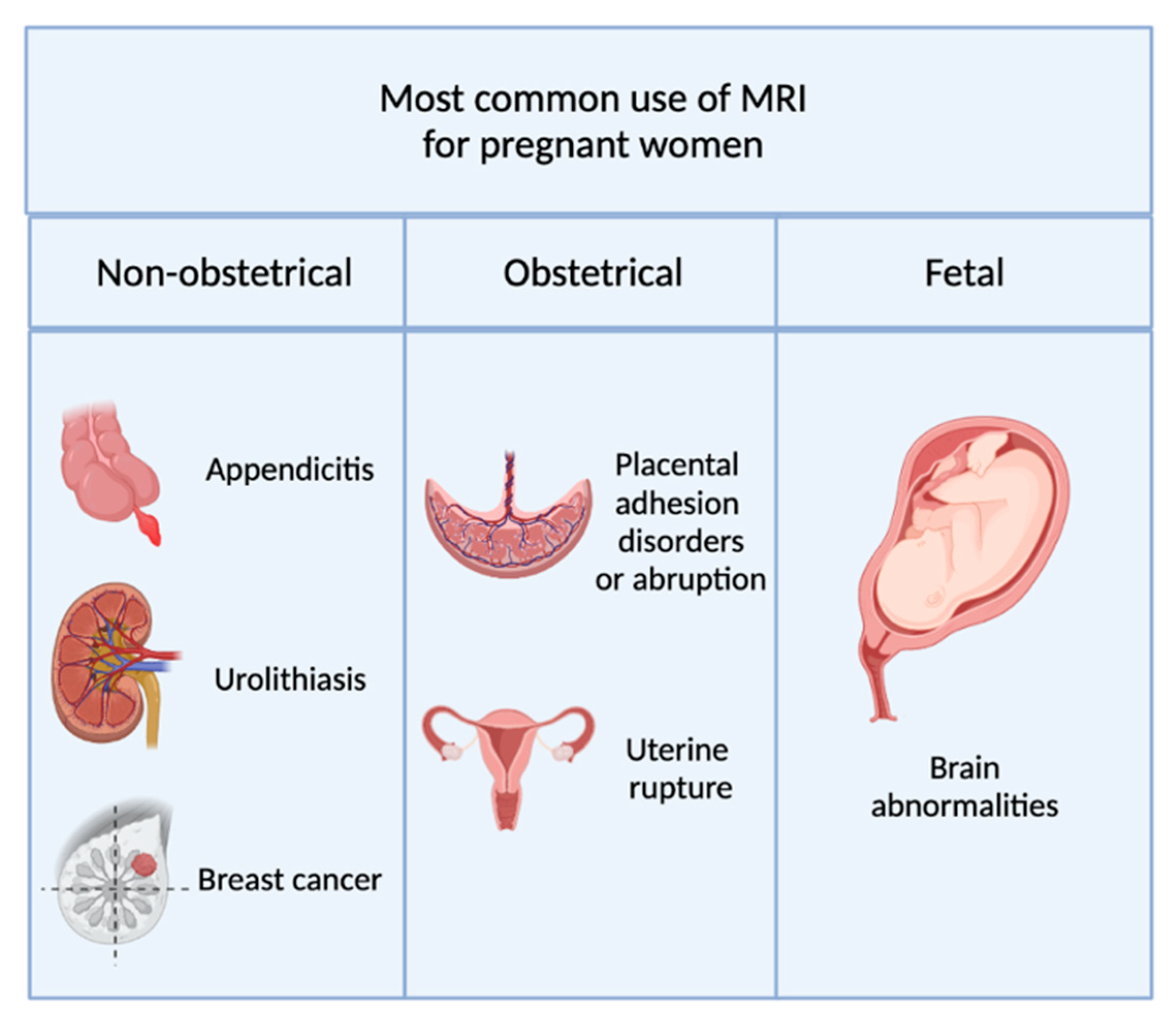
MRI was introduced in clinical practice in the early 1980s, and the first attempts to apply it among pregnant women were already reported in 1983 [10,11,12]. Commonly described indications for MRI during pregnancy can be classified into three main categories (Figure 1): non-obstetric, obstetric or gynecological, and fetal indications [13].
2.1.1. Non-Obstetric Indications
Pregnancy-associated anatomical changes, organ displacement (taking place especially during the second and third trimesters), physiological leukocytosis (elevated white blood cell count), and non-specificity of symptoms such as abdominal pain nausea and vomiting, render the diagnosis of various pathologies on pregnant patients harder [14,15].
Appendicitis is one of the most common non-obstetric causes of abdominal pain in pregnancy [13]. An early diagnosis is paramount to avoid unnecessary or negative surgical interventions and reduce the risk of fetal loss associated with the eventuality of appendiceal rupture or abscess [13,14,16]. MRI has been indicated by many as a powerful diagnostic tool for appendicitis, especially after inconclusive ultrasound examinations [13,14,16,17]. Other gastrointestinal and pancreaticobiliary pathologies that imply diagnosis by MRI are bowel obstruction, cholelithiasis, and pancreatitis [13].
Urologic pathologies such as urolithiasis and renal colic are other common reasons for pregnant women’s hospitalization. Early diagnosis is essential to prevent complications such as pyelonephritis or colic-induced preterm labor [13,15].
MRI can also be used as an alternative to US and CT for the diagnosis of cancer. Breast cancer is the most frequently diagnosed cancer in pregnant women [18,19]; other gynecological malignancies, such as cervical and ovarian cancer, or increasingly common colorectal cancers, may also prompt the need for MRI investigations [16,18,20]. This imaging system can also be applied in the evaluation of the brain, chest maternal diseases, musculoskeletal disorders, cardiovascular post-operative evaluation, and monitoring of congenital or acquired heart pathologies [21,22].
2.1.2. Obstetric and Gynecological Indications
Placenta adhesion disorders are among the most common obstetrical indications for MRI examinations and the cause of most failures in pregnancy [23].
Normally, the placenta is not in contact with the myometrium. In pathological conditions, according to how deeply placental tissues invade the myometrium, three cases can be distinguished:
- Placenta accreta, in which placental villi adhere to the myometrium.
- Placenta increta, where placental villi invade the myometrium.
- Placenta percreta, in which the myometrium is entirely penetrated by the placental villi, that extend even deeper in other uterine tissues [24].
The first-line diagnostic tool for this pathology is US. However, sonographic results are often equivocal and might leave uncertainty about the depth of placental invasion. Ultrasound visualization of this organ is also difficult in cases of posteriorly implanted placenta, where the latter is attached to the back wall of the uterus near the spine. In these situations, additional MRI imaging can be used [25]. The diagnosis of adhesion disorders is essential to plan the surgical management, contain the extent of possible hemorrhage at the moment of delivery, and detect eventual perforation of adjacent organs in case of percreta [14,16,21,26].
Placental abruption (the placenta detached prematurely from the myometrium after 20 weeks of gestation) represents another complication that in extreme cases could lead to fetal death [13]. Ultrasound detection of hematomas related to placental abruption is difficult and often leads to false diagnoses [13,21]. The use of MRI is helpful for correct identification [27].
Early detection of ectopic pregnancies (the fertilized ovum is implanted outside the uterine cavity) is crucial to prevent women’s mortality and preserve their fertility. MRI often allows for a more precise diagnosis compared to the transvaginal US, especially in cases of unusual presentations [24]. The use of MRI has also been reported in the context of the prediction of uterine rupture or individuation of ovarian torsions, adnexal masses, and fibroids [24,27].
2.1.3. Fetal Indications
Fetal MRI is employed in the structural and functional evaluation of the fetus [21,28]. Being mainly used for the individuation of CNS anomalies, MRI has also been implicated in the investigation of the neck, lungs (volumetric estimation), cardiac position and conformation, liver (congenital diaphragmatic hernias), bowel, and urinary tract [21,29].
2.2. Safety Issues of Non-Contrast-Enhanced MRI during Pregnancy
To create images, MRI exploits the interaction between protons of molecules within the body (mainly water) and an external magnetic field. In the absence of a strong magnetic field, protons in our body spin around their axis. The axes of different protons are randomly oriented in space [30]. When the body is put inside a high magnetic field (1.5 T or 3T in clinical diagnostics [31]), all the protons axes align in the direction of the magnetic field, and a net magnetization, called longitudinal magnetization, is generated [32]. During the application of an opportune radiofrequency pulse (RF), the total magnetization is tilted out of alignment, and a new, transverse magnetization component arises. When the RF pulse is removed, both components of the magnetization return, or relax, to their previous states under the influence of the magnetic field. During this relaxation process, the body produces an RF signal, which is detected and processed to produce an image [32].
MRI images include areas of diverse shades of gray, which correspond to different body components. Endogenous contrast of MRI is due to chemical, physical, and proton density differences in tissues [31]. Because of these differences, tissues have different relaxation times, called longitudinal (T1) and transverse (T2), which are the cause of the different shades of gray in the MRI scan (Figure 2).
To date, there are no data that have definitively demonstrated harmful effects on the fetus caused by MRI exposure of the pregnant woman in any trimester of gestation [12,13,33,34,35]. However, prospective and longitudinal studies are lacking; therefore, long-term safety has not yet been established [12,13,33,34,35]. The American College of Radiology (ACR), indeed, recommends pregnancy screening for women before MRI scanning. In the case of pregnancy confirmation, ACR advises the use of MRI only if [34]:
- It is not prudent to delay the examination until the completion of pregnancy;
- The information needed cannot be obtained through other non-ionizing techniques;
- The data gained may influence the management of the patient or the fetus.
The main theoretical risks associated with the use of non-contrast MRI during pregnancy are due to the implication of strong static magnetic fields (1.5 T or 3 T commonly in clinical use), gradient fields, and radiofrequency pulses (RF) [4,12,13,33].
2.2.1. Static Magnetic Fields and Teratogenicity
It has been shown in vitro that cell migration, differentiation, and proliferation can be affected by static magnetic fields [4,12,13,35,36]. Static magnetic field exposure have shown teratogenic effects in animal studies [4,12,13,35]. This suggests potential negative effects on organogenesis during the first trimester of pregnancy. However, direct extrapolation from animal models is not applicable, and no study has shown any MRI-induced teratogenic effect on humans so far [4,12,13,35].
2.2.2. Gradient Fields and Hearing Damage
Rapid time-varying magnetic fields are responsible for the production of acoustic noise [4,12,13]. The American Academy of Pediatrics considers 90 dB to be the limit above which the ear of the fetus can suffer permanent damage [37]. MRI generates acoustic noise ranging between 80 dB and 120 dB [4,38]. Maternal tissues and amniotic fluid attenuates the sound around 30 dB, but since the exact value is still uncertain, MRI evaluations can theoretically represent a risk for fetal hearing damage [4,12,13,38]. Several retrospective studies have not found any evidence of hearing abnormalities due to fetal MRI exposure, but prospective studies are also lacking in this case [12].
2.2.3. Radiofrequency Pulses and Tissue Heating
Radiofrequency pulses energy into the body in the form of heat [4,12,13,35]. In humans, heat-induced abnormalities in the fetus have been associated with an increase in maternal temperature greater than 2–2.5 °C for 30–60 min [4,13,38]. Since the heat gradient decreases from the body surface to the center, fetal heat is usually less than what has been shown to be detrimental [4]. Animal studies on pigs did not find any substantial increase in fetus heat after 3 T MR examinations within 30 min of duration [12,33]. Human studies failed to demonstrate reproducible harmful effects of mother or fetus exposure to clinically used magnetic fields (1.5 T or 3 T) [33,39,40,41]. Therefore, scanning at up to 3 T for less than 30 min is considered safe by the ACR.
In general, the use of non-contrast MRI on pregnant patients is recommended only if potential benefits overcome the still-not-fully established fetal risks, and examinations at the lowest clinical magnetic field of 1.5 T should be privileged [33].
2.3. Safety Issues of Contrast-Enhanced MRI during Pregnancy
The endogenous contrast produced by body components is not always enough to appropriately decipher MRI images. MRI, indeed, suffers from low sensitivity [31]. Contrast-enhanced MRI (CE-MRI) is a magnetic resonance modality in which exogenous substances are administered to increase the quality of images [8].
The most common MRI contrast agents (CA) are either complexes of paramagnetic ions, such as gadolinium Gd3+ and manganese Mn2+, or superparamagnetic iron oxide nanoparticles (SPIONs) [42,43]. Paramagnetic contrast agents usually shorten T1 relaxation time, resulting in the enhanced part being brighter (positive agents), while iron oxide particles shorten T2, producing a darker enhanced area (negative agents) [42]. Different SPIONs have been used in the past in MRI imaging of liver, spleen, and lymph nodes [44]; however, due to poor commercial interest, iron and manganese-based contrast agents are no longer commercially available [43,44,45].
When the administration of contrast to pregnant women is strictly required, gadolinium is the only recommended solution, because no animal or human fetal studies assess the safety of other agents during pregnancy [2].
2.3.1. Safety of Contrast Agents Containing Gadolinium
Currently, clinically used contrast agents are gadolinium-based (GBCA). These substances shorten T1, making the enhanced part of the image brighter [42]. Due to its similar size to calcium (Ca2+) ion, paramagnetic Gd3+ can compete in biological processes in which calcium is involved, resulting in toxicity for the human body [6]. For this reason, agents for clinical use are complexes in which the Gd3+ ion is chelated by a ligand to prevent its release and reduce its toxicity [46]. Agents can be distinguished into linear or macrocyclic, ionic, and non-ionic, depending on the total charge of the complex and the molecular structure of the ligand [6]. Macrocyclic or ionic agents are usually considered safer than linear or non-ionic ones because of the smaller probability of Gd3+ release from the complexes [6,47].
The main safety concerns pertaining to the use of these contrast agents in pregnancy are associated with their potential to be teratogenic, induce neonatal nephrogenic systemic fibrosis (NSF), and bioaccumulate into tissues [12,13,47].
Teratogenicity
Different animal studies have demonstrated that GBCAs can cross the placenta after maternal intravenous administration [12,13,47,48,49]. Once in the fetal circulation, they undergo renal clearance and are excreted in fetal urine, re-entering the amniotic fluid [13,47,48,50]. Prolonged permanence of contrast agents in the amniotic fluid and possible fetal re-absorption may increase the risk of Gd3+dechelation, raising concerns about fetal safety [13,47,50]. Results of animal studies regarding the teratogenicity of GBCAs are controversial [12,47,48].
Different studies on humans have not shown any fetal harm attributable to gadolinium exposure during pregnancy [12,47,48]. However, they are limited by their retrospective data collection and small sample size [12,47,48]. The only cohort, longitudinal, and relatively large sample-size study, by Ray et al., did not demonstrate any increase in congenital anomalies after fetal exposure to gadolinium. However, a slight increase in the risk of stillbirth was reported in previously gadolinium-exposed neonates [51].
Nephrogenic Systemic Fibrosis
Nephrogenic systemic fibrosis (NSF) is a rare condition, mainly characterized by fibrosing skin lesions, which may also imply the fibrosis of internal organs [12,46]. Multiple studies have shown the connection between GBCAs exposure and the occurrence of NSF in subjects with renal impairment [12,46]. Fetal exposure to gadolinium raises concerns about the risk of neonatal NSF development [12,13]. Ray et al. have, indeed, found a higher rate of incidence of inflammatory, rheumatologic, and infiltrative skin conditions in neonates exposed to gadolinium compared to an unexposed group [51]. However, no NSFs have been reported so far in children under 6 years of age [12].
Bioaccumulation
Bioaccumulation of gadolinium in human tissues such as bone, skin, brain, or liver has been demonstrated by many post-mortem studies on subjects that underwent multiple exposures [12,46] Postnatal animal studies confirmed these findings, showing higher deposition degrees for linear agents than for macrocyclic ones [12,46]. Concerns regarding tissue deposition are related to potential gadolinium dechelation over time, resulting in the release of the toxic free ion [12]. However, the long-term consequences of gadolinium chelate bioaccumulation are still unknown.
In conclusion, the latest ACR, ACOG (American College of Obstetricians and Gynecologists), and ESUR (European Society of Urogenital Radiology) guidelines recommend avoiding the use of GBCAs during pregnancy, and their use is restricted only to patients for whom the benefits would considerably outweigh the risks for the fetus [2,33,52]. More specifically, the ESUR recommends, when strictly needed, administrating the lowest dose of the most stable agent [52].
In general, the use of diagnostical MRI on pregnant women is the best choice when ultrasound fails to provide sufficient information for a medical decision. However, the use of MRI during pregnancy should always be assessed through a case-by-case analysis, considering the benefits it could bring to the mother and her offspring against potential risks.
3. Nanostructures as Potential MRI Contrast Agents
In recent years, increasing efforts have been dedicated to the study of nanosystems as drug carriers and diagnostic or theragnostic tools. Nanocarriers have been designed to modulate biodistribution, facilitate barrier crossing and accumulation into the target site, and control the release of even fragile active molecules such as nucleic acids or peptides [53,54]. Nanoprobes have been developed to improve the signal of various diagnostical techniques, such as MRI, SPECT, US, or PET, and include magnetic nanoparticles [55], nanobubbles [56,57], or radio-labeled nanosystems [58]. Such nanotechnologies represent promising game-changers in the treatment and diagnosis of diseases spanning from cancer [59,60] to cardiovascular [61], infective, and inflammatory diseases [62]. In this section, nanosystems with the promising ability for efficient contrast generation in magnetic resonance imagining scanning are described.
3.1. Advantages of Nanostructured MRI Contrast Agents over Conventional Molecular Chelates
Most of the current clinically used MRI gadolinium chelates exhibit drawbacks such as short half-life, non-specific distribution, and potential undesirable toxicities [31,63]. For this reason, much research has been conducted on nanostructured materials, to evaluate them as alternatives to molecular agents. The small size and high surface-to-volume ratio typic of nanosystems provide them with unique properties, different from those of both small molecules and bulk materials [63,64,65].
Nanoparticles’ loadability and tunability of sizes and surfaces may help to create contrast agents with new characteristics.
Nanostructures may, for example, help to reduce the risk of toxic free metals release [63,66], allow better control of their contrast biodistribution, and increase their half-life [6,63,65,67]. Improvement in contrast efficiency and potential minimization of required administration doses may also be obtained [6,63,65,67].
The biodistribution of nanostructured contrasts may be partially controlled by either manipulating nanoparticles’ size or functionalizing their surface [63]. It has been demonstrated that particles smaller than 10 nm undergo rapid renal clearance, while particles larger than 100 nm are recognized by macrophages and accumulate in organs such as the lymph nodes, liver, or spleen [63]. Within this size range, nanoparticles’ dimensions can be tuned to better direct them to the desired region of interest [68]. Blood circulation time may also be improved by surface modification: molecules such as PEG, dextrose, or chitosan may be used to prolong nanoparticles circulation half-life [63,67,69]. An elongated circulation may provide a more appropriate time window for examinations that are difficult to perform with molecular chelates due to their rapid extravasation (e.g.,: angiography) [31,65].
Surface functionalization with ligands, interacting specifically with receptors mainly expressed in certain tissues, can further increase the concentration of nanosystems in specific locations [63]. Antibodies targeting the epidermal growth factor receptor (EGFR), tumor homing peptides, or placental chondroitin sulfate A binding-peptide (plCSA-BP) have been, for example, used to decorate different nanoparticles for the targeted delivery of drugs to the placenta [70,71].
Development of dual-modal contrast agents, suitable for both T1- and T2-weighted analysis, may also be possible using nanoparticles (NPs) [8,31,69]. For example, the combination of manganese-ferrite and Gd2O(CO3)2 in a single core-shell structure has yielded nanoparticles with good longitudinal and transverse relaxation properties [72].
Lastly, nanoparticles may be intrinsically more efficient contrast agents than common molecular chelates. The efficiency of a contrast agent is quantified by a parameter called relaxivity (mM−1 s−1) [31,73,74], which is a function of the concentration C of the contrast-active ion and is defined as:
where Ri is the relaxation rate of the surrounding water. Relaxation rates (s−1) are defined as the reciprocal of T1 and T2:
3.2. Nanoparticles’ Characteristics Affecting Contrast Agent Efficiency
Contrast agents elicit their action by accelerating the water relaxation process. A close interaction between water molecules and T1 agents is required for the T1 shortening process, whereas the T2 shortening occurs on a longer distance range [31].
For T1 contrast agents, the higher the number of water molecules coordinated to the magnetic center (hydration number), the larger the relaxation that the agent will procure [65].
T2 agents, instead, produce small local magnetic fields in their surroundings, subjecting water molecules to field inhomogeneities that accelerate their transverse relaxation process [74]. The ability of some magnetic materials to produce field inhomogeneity is proportional to a property they possess called saturation magnetization. The larger the value of the latter, the bigger the field inhomogeneity that the contrast agent can cause and the stronger its effect [7].
Contrast agents with high hydration numbers and large saturation magnetization can be obtained by designing them in the form of nanosystems. Both these properties may, indeed, be affected by nanoparticles’ size, shape, and surface properties, influencing their efficiency as contrast agents.
3.2.1. Size
In general, decreasing particle size leads to higher surface-to-volume ratios. In the case of T1 contrast agents, smaller-sized particles have a higher number of surface-located magnetic centers available for interactions with surrounding water [73]. Therefore, the smaller the size, the higher the r1 relaxivity [73]. However, decreasing particle size also increases surface energy, which leads to higher reactivity and reduced stability [73]. Therefore, optimized nanoparticles should have a diameter that is a compromise between contrast efficiency and stability.
For superparamagnetic T2 contrast agents, on the contrary, saturation magnetization increases with increasing size, leading to higher r2 relaxivity values, up to a size limit over which the superparamagnetic properties of the nanoparticle are lost [7,73]. Higher saturation magnetization values can, alternatively, be obtained by controlled magnetic nanoparticles clustering [7,74].
3.2.2. Surface Properties
Nanoparticles almost always necessitate a coating (PEG, chitosan, lipids, proteins) to increase their stability, prevent aggregation, improve their biocompatibility, or increase their half-life [74].
Coatings can influence water diffusion and retention around the magnetic center, or affect the magnetic properties of the magnetic material itself [75]. As a consequence, they may impact the T1 and T2 relaxation abilities of contrast agents [75].
Coatings that can increase the magnetic center hydration, or allow an adequate residence time of water molecules around it, should be preferred for T1 contrast agents [75].
For T2 contrast agents, substances such as oleic acid, phosphates, sulfates, and citrates can help to increase the nanoparticle magnetization [75]. In addition, coatings rich in π-electrons (e.g., catechol-PEG) may contribute to enhanced field inhomogeneity, which is crucial to increase r2 [75]. Lastly, even if water contact is not as crucial as for T1 contrast agents, thick, dense coatings may reduce water accessibility, elongating water-nanoparticle distance and reducing the effect of magnetic inhomogeneity [75].
General specifications for an optimal coating to increase relaxivities include:
- Low thickness.
- High hydration.
- Fast water exchange rate.
- Presence of π-electrons.
- Stability and biocompatibility in biologically relevant media [7].
3.3. Examples of Nanostructured MRI Contrast Agents
Almost all MRI contrast agents affect both the longitudinal and transverse relaxation processes; however, their effects are often dominant on either T1 or T2, therefore are categorized them as T1 or T2 contrast agents [31]. The relaxivities ratio r1/r2 is the parameter used to predict if a substance is indicated more as a T1 or as a T2 agent. In general, r1/r2 < 5 are typical of T1 agents, while r1/r2 > 10 characterize promising T2 contrast agents [74,76].
In this section, potential nanostructured contrast agents are presented and categorized as T1, T2, or dual-mode agents.
Examples of these nanostructures along with their characteristics (size, magnetic field, and relaxivities values) are reported in Table 1. For comparison, commercial agents’ relaxivity values are provided: Magnevist® (Gd-DTPA, linear chelate) r1 = 4.3 mM−1·s−1 (1.5 T), Dotarem® (Gd-DOTA, macrocyclic chelate) r1 = 3.4 mM−1·s−1 (3 T) [77], Feridex® (colloidal dextran-coated SPIONs) r2 = 108.0 mM−1·s−1 (4.7 T) [72].
3.3.1. T1 Contrast Agents
T1 contrast agents use paramagnetic materials such as Gd3+, Mn2+, and Fe2+/Fe3+ [8,65], substances in which external magnetic fields induce a weak magnetization, which is not retained upon magnetic field removal [74]. These agents produce a brighter image of the enhanced area compared to unenhanced surroundings and usually result in more effective differentiation of normal tissues from lesions than T2 counterparts [74].
Gadolinium
Currently, gadolinium chelates, such as Gd-DOTA and Gd-DTPA, represent the gold-standard T1 contrast agent for clinical MRI investigations [78]. These substances have relaxivity values (r1) ranging from 3 to 5 mM−1 s−1 [78].
A lot of research has been done about the inclusion of Gd3+ ions in nanoparticles. The packing of a high number of ions on the surface of nano-entities is expected to considerably increase longitudinal relaxation in the vicinity of the nanoparticles, enabling a reduction in administered doses [6,65,79].
A various spectrum of platforms, such as liposomes, silica-based nanostructures, polymeric carriers (micelles, dendrimers), protein conjugates, carbon nanostructures, gold nanoparticles, nanocrystals, or modified carriers of natural origin, such as tobacco mosaic virus (TMV), have been used as scaffolds to increase the amount of Gd3+ ions [8,31,65,67,80].
When incorporated into liposomes, Gd-chelates can be either conjugated to free-floating chains of PEG on the surface, entrapped in the lipophilic bilayer, or loaded into the liposome hydrophilic core [6]. As an example, Pitchaimani et al. (2016) developed a theragnostic nanosystem in which Gd-DOTA chelate was incorporated in the lipidic bilayer of a doxorubicin core-loaded liposome [6,81]. The nanosystem showed a three-fold improved r1 relaxivity compared to those of most of the commercial gadolinium chelates.
The high surface area offered by the pores of mesoporous silica nanoparticles (MSNs) makes these systems an appealing scaffold for the production of potential T1 contrast agents with improved water exchange [6]. MSNs incorporating gadolinium-chelates have been found to have extremely high r1 relaxivity values (20-fold increase) compared to commercial gadolinium chelates at clinically relevant magnetic fields (3 T) [82]. It was also observed that the chelate location on the nanoparticle structure has an influence on the magnetic properties of the final system [82].
Manganese
As alternative paramagnetic agents, manganese oxides, such as MnO, MnO2, or Mn3O4, of different sizes and shapes, have been investigated [8,31,65]. Because of the Mn2+ ion’s ability to enter cells through calcium channels, Mn-based nanosystems are considered potential tools for neuroimaging [31]. Ions of this element have been encapsulated in self-assembling polymeric nanoparticles or used to dope magnetite (Fe3O4) [8,83,84]. The obtained ferrite particles have shown T1-relaxivities increasing proportionally with the extent of surface Mn2+ doping [83].

Table 1.
Size, magnetic field, and relaxivity values of potential nanostructured contrast agents.
| Type of CA | Nanostructured CA | Composition | Mean Diameter (nm) | Magnetic Field (T) | Relaxivity (mM−1·s−1) | Ref. |
|---|---|---|---|---|---|---|
| T1 | Gd-liposomes | DSPG/Gd-DOTA-DSPE/DSPG-PEG-Succinyl/Cholesterol | 156.0 | 14 | 12.3 | [81] |
| Gd-MSNs | Gd-DOTA in porous silica NPs | 138.9 | 3 | 39.3 | [82] | |
| Gd2O3-GO sheets | Gd2O3-decorated GO | 268.9 | 7 | 34.5 | [78] | |
| GlcA-Gd2O3 | (D-glucuronic acid)-coated Gd2O3 | 4.0 | 1.5 | 9.9 | [85] | |
| Mn-polymeric NPs | PEG-coated, Mn-containing, DOPA reverse micelles | 75.8 | 3 | 11.6 | [84] | |
| Mn-ferrite cubes | Mn-doped iron oxide | 16.4 | 0.5 | 57.8 | [83] | |
| USPIONs | PEG-coated iron oxide | 10.0 | 1.4 | 7.3 | [86] | |
| T2 | MLPs | DOPC/Cholesterol/DSPE-PEG2000 MLPs | 156.0 | 9.4 | 575 | [45] |
| UMLs | DPPC/DSPC/DSPE-PEG2000 MLPs | 230.0 | 7 | 225 | [87] | |
| Zn-ferrite | (Zn0.4Fe0.6) Fe2O4 | 15.0 | 4.5 | 687.0 | [88] | |
| Mn-ferrite | PEGylated Mn-Fe2O4 | 68.8 | 1.5 | 217.1 | [89] | |
| SWCNT· | Cylindrical (IO-NPs)-conjugated CNTs | 4–5 (IO-NPs d.) 200–300 (CNTs l.) | 4.7 | 231.3 | [90] | |
| HoF3 NPs | Rhomboidal HoF3 | 110 × 50 | 9.4 | 608.4 | [91] | |
| DyF3 NPs | Rhomboidal DyF3 | 110 × 50 | 9.4 | 380.4 | [91] | |
| T1-T2 | MnFe2O4@SiO2@Gd2O(CO3)2 NPs | Mn-ferrite-silica-Gd2O(CO3)2 (20 nm silica) | 36.5 | 4.7 | r1 = 33.1 r2 = 213 | [72] |
| Gadolinium and Dysprosium oxide NPs | (D-glucuronic acid)-coated (Gd-Dy) oxide | 1.0 | 1.5 | r1 = 6.0 r2 = 40.0 | [92] |
Abbreviations: CA = contrast agent; CNTs = carbon nanotubes; d. = diameter; DOPA = 1,2-Dioleoyl-sn-glycero-3-phosphate; DOPC = 1,2-Dioleoyl-sn-glycero-3-phosphocholine; DOTA = 1,4,7,10-tetraazacyclododecane-1,4,7,10-tetraacetic acid; DPPC = 1,2-Dipalmitoyl-sn-glycero-3-phosphocholine; DSPC = 1,2-Distearoyl-sn-glycero-3-phosphocholine; DSPE = 1,2-distearoyl-sn-glycero-3-phosphoethanolamine; DSPG = 1,2-dioctadecanoyl-sn-glycero-3-phospho-(1′-rac-glycerol); Dy = dysprosium; Gd = gadolinium; GlcA = glucuronic acid; Ho = holmium; GO = graphene oxide; IO = iron oxide; l. = length; MLPs = magneto-liposomes; Mn = manganese; MNSs = mesoporous silica nanoparticles; NPs = nanoparticles; PEG = polyethylene glycol; SWCNT· = single-walled carbon nanotubes; USPIONs = ultra-small iron oxide nanoparticles; Zn = zinc.
Iron
Iron-based contrast agents are commonly used as T2 agents. However, ultra-small iron oxide nanoparticles (USPIONs) with a d < 5 nm [63,74] have been reported to have good T1 relaxation enhancement capabilities and have been proposed as blood pool agents (substances with an extended vascular permanence) [31,65]. PEG-coated USPIONs have been tested to prevent aggregation phenomena and improve their physiological stability [86].
3.3.2. T2 Contrast Agents
Substances commonly employed as T2 contrast agents are often superparamagnetic. [93]. These materials produce contrast-inducing signal loss in the area they enhance. However, the resulting hypointense region may be confused with the dark signal typical of some pathological conditions (e.g., calcification, internal bleeding) or intrinsically low-signal body regions [8,31,74]. This drawback has contributed to substantially limiting their clinical usage [8,31,74]. Most commonly, T2 agents are iron oxide- or ferrite-based substances [31]. More recently, compounds of paramagnetic ions such as dysprosium (Dy3+) or holmium (Ho3+) have also been explored as potential T2 contrast agents [8].
SPIONs and USPIONs
The two most common iron oxides used to produce superparamagnetic iron oxide nanoparticles (SPIONs) are magnetite (Fe3O4) and its oxidized form, maghemite (γ-Fe2O3), which is more stable [31]. To prevent their aggregation, these particles are often covered with polymers or polysaccharides (e.g., PEG, dextran) [31,94].
SPIONs (d~50–150 nm) have been mainly used for liver and spleen imaging, while USPIONs (d < 50 nm), due to their smaller size and longer half-life [31], are used for lymph nodes investigation, bone marrow contrast, brain and kidney perfusion, and angiography [31,63,76]. Both of these compounds are metabolized and included in the body iron pool within a couple of days [31].
SPIONs may be incorporated into liposomes to produce magneto-liposomes (MLPs), platforms with potential theragnostic properties. They may also be conferred with targeting ability, either by surface functionalization or by the influence of an external magnet applied on specific body regions, to induce MLPs accumulation [31]. Some MLPs, even with different phospholipid compositions, showed higher relaxivity values compared to free iron oxide particles [45]. MLPs used for theragnostics allow the magnetic-targeted release of a therapeutic agent and simultaneous MRI imaging [87]. An example of magneto-liposomes, encapsulating an antivascular disrupting agent to “starve” tumoral microenvironments, showed higher r2 relaxivity than those of free iron oxide NPs and iron-based commercial agents [87].
Small iron oxide nanoparticles conjugated to carbon nanotubes have also successfully allowed MRI investigations to track tumor progression and quantify the apparent diffusion coefficient (ADC), an imaging biomarker used to evaluate tumor response to treatment [90].
Ferrites
Doped-iron oxide (ferrites) nanosystems have been fabricated as alternative T2 contrast agents. Ions of different elements, such as Mn, Zn, Co, and Ni, have been used to substitute some iron atoms in iron oxides to obtain ferrites with higher saturation magnetization and, therefore, improved transverse relaxation properties [31].
Another example of iron-oxide-doped nanosystem developed as a T2 contrast agent is PEGylated MnFe2O4 nanocrystals, which present higher r2 relaxivity than commercial Ferumoxide®. This enhanced effect has been hypothesized to be due to the dense clustering of a large amount of MnFe2O4 in the PEGylated system [89].
Dysprosium and Holmium Derivates
Dysprosium (Dy3+) and holmium (Ho3+) paramagnetic ions containing nanoparticles seem to be promising T2 contrast alternatives, especially for ultra-high-field-MRI (>7 T) [8,31]. Dy-DOTA chelates have already been incorporated in mesoporous silica nanoparticles or tobacco mosaic virus, while different dysprosium and holmium oxides and fluorides have been found to have high r2 values at high magnetic field strengths [8,91].
3.3.3. Dual-Mode T1-T2 Contrast Agents
Single-mode contrast agents frequently result in diagnosis uncertainties, especially for small biological targets [31]. Dual-mode contrast agents are slowly emerging. The combination of both contrast effects in a single nanoprobe allows for MRI acquisitions with highly detailed T1- and T2-weighted images [31,74].
Core-shell dual agents with manganese-ferrite (superparamagnetic) and Gd2O(CO3)2 (paramagnetic) separated by a silica layer have been synthesized. The nanoparticles showed higher r1 and r2 relaxivities than commercial iron oxide, whose values could be tuned by modulating the thickness of the silica layer [31,72,74].
Other potential dual-mode systems consist of mixed gadolinium-dysprosium oxide nanoparticles, where T1 contrast is given by the presence of Gd3+ ions, while T2 contrast is given by the doping dysprosium [8,74]. The relaxivities reported for these nanostructures were not shown to be much better than those of conventional T1 and T2 agents [92].
New nanostructured MRI contrast agents will be designed and studied thanks to the advancement of technology that allows their properties to be studied and their efficiency to be evaluated. In parallel, it would be interesting if preclinical and clinical studies of such nanosystems could include safety evaluation for a potential administration to pregnant patients.
4. Potential of Nanostructured Contrasts to Improve the Safety of CE-MRI in Pregnancy
4.1. Possible Advantages of Nanostructured Contrast Agents for MRI on Pregnant Women
The diagnostic power of ultrasound and non-contrast MRI is sometimes limited by insufficient sensitivity and the lack of possibility for functional evaluations [95]. Contrast-enhanced MRI is a potential solution to both of these problems.
However, the effects of gadolinium-based contrast agents on the fetus are still unknown [50,96]. Because of potential safety concerns, gadolinium chelates are traditionally avoided during pregnancy, unless their use is crucial to guarantee the health of the woman or fetus [50]. The toxicity of GBCAs is widely considered to occur only after dissociation of the Gd3+ ion from the chelate in which it was administered [48,50].
Development of safer contrast agents to substitute currently used gadolinium complexes, would allow contrast-enhanced MRI during pregnancy, making a more powerful diagnostic tool available. The risk of the release of toxic ions could be reduced by incorporating metal ions into nanoparticles. For instance, Gd3+ release from the molecular chelate may be avoided, producing nanoparticles (e.g., Gd2O3) in which the paramagnetic ion is covalently bonded to other atoms, preventing its leaching from the structure [65].
The development of contrast nanosystems based on magnetic materials different from gadolinium (e.g., Dy, Ho, Mn, ferrites, or iron oxides) could be another alternative to conventional agents [8,42,97]. However, to represent a realistic solution, this would require the fetal safety evaluation of these substances first.
As completely safe materials for the fetus have not yet been reported, nanostructures may at least allow the reduction of contrast administration doses, decreasing the potential fetal exposure. Some nanoparticles have intrinsically higher contrast efficiency than conventional agents, [8] while others’ loading capacity could be exploited to achieve high contrast payload [81,98]. This could potentially minimize the amount of exogenous agent needed.
4.2. Examples of Evaluation of Potential Nanostructured Contrast Agents on Pregnant Animal Models
Assessment of placental adhesion disorders is currently difficult because of the lack of safe and efficient techniques that could be applied in pregnancy. Examination with ultrasound followed by non-contrast MRI allows the detection of only half of the cases [95].
A group of researchers from the Texas Children’s Hospital, Houston, USA, has developed gadolinium-containing liposomes as a potential alternative to currently employed gadolinium chelates. The nanostructures were evaluated and compared to conventional Gd-chelates on pregnant mouse and rat models [95,100,101]. The experimental plan and transplacental passage results are illustrated in Figure 4.
Magnetic resonance imaging on animals showed no transplacental passage of Gd-liposomes, as shown by the lack of signal enhancement in the fetal compartment [95,100]. Inductively coupled mass spectrometry (ICP-MS) of mouse and rat fetal tissues showed no liposomal-Gd uptake, even 72 h after injection [95,100]. MRI and ICP-MS results were confirmed by the absence of liposomes through a perfused human placenta [100].
The nanostructured contrast agent allowed the visualization of the placental margins [95,100] and a comparatively hypo-enhanced retroplacental space, showing a better diagnostic performance than the conventional Gd-DTPA control, which, instead, did not enable retroplacental space visualization [100]. The difference in the efficiency of the agents could be due to the fast clearance kinetics of the chelate, leading to a non-uniform signal enhancement and an inadequate contrast between the placenta and contiguous structures, which is improved, instead, by the longer-circulating and uniformly distributed liposomal gadolinium [100].
The kinetics of placental perfusion and clearance of Gd-liposomes were also determined with dynamic contrast enhanced-MRI [95].
Based on these results, this liposomal agent appears to be a promising tool for the MRI assessment of placental structure and characterization of conditions that may alter placental perfusion.
After determination of its maternal and fetal safety, this nanostructured contrast agent, with no transplacental passage, could represent a safer alternative to the currently used gadolinium chelates, sparing the fetus from contrast exposure.
5. Conclusions
In general, when designing a nanoparticle for biomedical applications, its biocompatibility and chemical stability in biological environments are indispensable characteristics. For contrast agents as well, these properties need to be controlled and ensured both in vitro and in vivo.
The size and surface properties of an optimal nano-contrast agent should be a compromise between biocompatibility, stability, and improvement of contrast efficiency.
Rational reduction or increase in particle size could be used to create systems with enhanced T1 and T2 contrast power, respectively. Nanostructures’ surfaces may be modified to favor their hydration or ability to produce local magnetic fields, leading to systems with higher contrast capacity.
In the specific case of pregnant women, to prevent unnecessary or undesired fetal exposure to potentially toxic contrast agents, nanosystems design should account for the avoidance of transplacental transfer. The extent of nanoparticles’ placental passage and uptake is determined by their size, surface structure, and composition, and also by integrity, gestational age, and placental maturity [99,102].
The current knowledge of nanoparticles’ interaction and transport across the placenta is still limited [103]. Nanosystems are predominantly thought to enter this organ via pinocytosis/endocytosis and phagocytosis; nevertheless, paracellular mechanisms are also possible [70]. However, published studies about nanoparticle transplacental passage are relatively sparse, and a full understanding of placental transport mechanisms and uptake for all nanomaterials is lacking [99].
To spare the fetus from contrast exposure, low binding of placental tissues should be addressed. This could be reached by employing stealthing moieties (e.g., PEG) and optimizing the surface charge. For example, cationic particles have been found to bind placental trophoblasts, so neutral or anionic particles should be preferred [99,104]. However, knowledge about the influence of nanosystems’ charge on placental transfer is still limited, and results need to be confirmed using several models [103,105].
Different animal studies, in vitro experiments, and ex vivo human placental perfusion studies have shown that nanomaterials can be taken up by the placenta, and even reach the fetus, although this usually occurs at low or negligible amounts, with the exertion of little or no fetal toxicity [99]. However, most of the research investigating nanoparticles’ toxicology in pregnancy has focused on the consequences of environmental exposure, rather than clinically administered particles [106,107].
The many physiological changes occurring during pregnancy, which can modify nanoparticles’ biodistribution [99,107], should be considered in the toxicological studies of a nanosystem designated to obstetric applications.
In conclusion, by using properties that prevent nanoparticles uptake by the placenta, it could be possible to produce “fetal-friendly” contrast agents, with a lower risk of unintended fetal toxic effects compared to currently used ones. However, it should be taken into account that low fetal exposure could still result in undetectable adverse effects, which could pass below the radar in animal models [99].
Therefore, much more research about placental structure and nanoparticles’ properties influencing the interaction and passage across this organ is strongly needed [108]. Indeed, the rational and well-planned design of nanosystems, necessary to ensure their safety for obstetric use, cannot be independent of an in-depth understanding of the aforementioned factors. Researchers are nowadays focusing on the development of innovative nanoparticles to answer the specific needs of pregnancy disorders [109]. To go further, it could be interesting to design new nanosystems such as targeting nanoparticles, such as liposomes, for placental MRI. As liposomes already showed their value as contrast agent delivery systems but also as placental targeting nanocarriers, it seems natural to consider them to ensure a specific delivery of the contrast agent to the placenta to ensure the safety of usage for both the mother and the fetus [110,111,112]. Ultimately, any use of nanomaterials in pregnancy should still require a cautious and extensive risk–benefit evaluation.
Author Contributions
M.D. Conceptualization, investigation, writing—original draft preparation; L.F. Investigation, writing—review, and editing; N.M. resources, writing—review and editing; K.A. (Karine Andrieux) writing—review and editing, funding acquisition and supervision; K.A. (Khair Alhareth) writing—original draft preparation, writing—review, and editing, supervision, project administration. All authors have read and agreed to the published version of the manuscript.
Funding
This research received no external funding.
Institutional Review Board Statement
Not applicable.
Informed Consent Statement
Not applicable.
Data Availability Statement
Not applicable.
Acknowledgments
Authors acknowledge the Erasmus Mundus Joint master’s degree (EMJMD) Nanomed for providing M.D. scholarship.
Conflicts of Interest
The authors declare no conflict of interest.
References
- Smith-Bindman, R.; Kwan, M.L.; Marlow, E.C.; Theis, M.K.; Bolch, W.; Cheng, S.Y.; Bowles, E.J.A.; Duncan, J.R.; Greenlee, R.T.; Kushi, L.H.; et al. Trends in Use of Medical Imaging in US Health Care Systems and in Ontario, Canada, 2000–2016. JAMA-J. Am. Med. Assoc. 2019, 322, 843–856. [Google Scholar] [CrossRef]
- ACOG. ACOG Guidelines for Diagnostic Imaging During Pregnancy and Lactation; ACOG: Washington, DC, USA, 2017. [Google Scholar]
- Bourgioti, C.; Konidari, M.; Gourtsoyianni, S.; Moulopoulos, L.A. Imaging during pregnancy: What the radiologist needs to know. Diagn. Interv. Imaging 2021, 102, 593–603. [Google Scholar] [CrossRef]
- Tirada, N.; Dreizin, D.; Khati, N.J.; Akin, E.A.; Zeman, R.K. Imaging pregnant and lactating patients. Radiographics 2015, 35, 1751–1765. [Google Scholar] [CrossRef]
- Eastwood, K.; Mohan, A.R. Imaging in pregnancy. Obstet. Gynaecol. 2019, 21, 255–262. [Google Scholar] [CrossRef] [Green Version]
- Marasini, R.; Thanh Nguyen, T.D.; Aryal, S. Integration of gadolinium in nanostructure for contrast enhanced-magnetic resonance imaging. Wiley Interdiscip. Rev. Nanomed. Nanobiotechnol. 2020, 12, e1580. [Google Scholar] [CrossRef]
- Kostevšek, N. A Review on the Optimal Design of Magnetic Nanoparticle-Based T2 MRI Contrast Agents. Magnetochemistry 2020, 6, 11. [Google Scholar] [CrossRef] [Green Version]
- Pellico, J.; Ellis, C.M.; Davis, J.J. Nanoparticle-Based Paramagnetic Contrast Agents for Magnetic Resonance Imaging. Contrast Media Mol. Imaging 2019, 2019, 1845637. [Google Scholar] [CrossRef]
- Ramdass, S.; Adam, S.; Lockhat, Z.; Masenge, A.; Suleman, F.E. Foetal magnetic resonance imaging: A necessity or adjunct? A modality comparison of in-utero ultrasound and ultrafast foetal magnetic resonance imaging. S. Afr. J. Radiol. 2021, 25, 6. [Google Scholar] [CrossRef]
- Ladd, M.E.; Bachert, P.; Meyerspeer, M.; Moser, E.; Nagel, A.M.; Norris, D.G.; Schmitter, S.; Speck, O.; Straub, S.; Zaiss, M. Pros and cons of ultra-high-field MRI/MRS for human application. Prog. Nucl. Magn. Reson. Spectrosc. 2018, 109, 1–50. [Google Scholar] [CrossRef]
- Smith, F.; Adam, A.; Philipps, W. NMR Imaging in Pregnancy. Lancet 1983, 321, 61–62. [Google Scholar] [CrossRef]
- Lum, M.; Tsiouris, A.J. MRI safety considerations during pregnancy. Clin. Imaging 2020, 62, 69–75. [Google Scholar] [CrossRef]
- Mervak, B.M.; Altun, E.; McGinty, K.A.; Hyslop, W.B.; Semelka, R.C.; Burke, L.M. MRI in pregnancy: Indications and practical considerations. J. Magn. Reson. Imaging 2019, 49, 621–631. [Google Scholar] [CrossRef]
- Lim, P.S.; Mackey, A.M.; Huang, M.L. MRI of the Placenta and the Pregnant Patient. In Imaging of the Pelvis, Musculoskeletal System, and Special Applications to CAD; Saba, L., Ed.; CRC Press: Boca Raton, FL, USA, 2016; pp. 145–160. ISBN 978-1-4822-1622-6. [Google Scholar]
- Furey, E.A.; Bailey, A.A.; Pedrosa, I. Magnetic Resonance Imaging of Acute Abdominal and Pelvic Pain in Pregnancy. Top. Magn. Reson. Imaging 2014, 23, 225–242. [Google Scholar] [CrossRef]
- Horowitz, J.M.; Hotalen, I.M.; Miller, E.S.; Barber, E.L.; Shahabi, S.; Miller, F.H. How Can Pelvic MRI with Diffusion-Weighted Imaging Help My Pregnant Patient? Am. J. Perinatol. 2020, 37, 577–588. [Google Scholar] [CrossRef]
- Kave, M.; Parooie, F.; Salarzaei, M. Pregnancy and appendicitis: A systematic review and meta-analysis on the clinical use of MRI in diagnosis of appendicitis in pregnant women. World J. Emerg. Surg. 2019, 14, 1–14. [Google Scholar] [CrossRef]
- Gui, B.; Cambi, F.; Micco, M.; Sbarra, M.; Petta, F.; Autorino, R.; De Vincenzo, R.; Valentini, V.; Scambia, G.; Manfredi, R. MRI in pregnant patients with suspected abdominal and pelvic cancer: A practical guide for radiologists. Diagn. Interv. Radiol. 2020, 26, 183–192. [Google Scholar] [CrossRef]
- Vandecaveye, V.; Amant, F.; Lecouvet, F.; Van Calsteren, K.; Dresen, R.C. Imaging modalities in pregnant cancer patients. Int. J. Gynecol. Cancer 2021, 31, 423–431. [Google Scholar] [CrossRef]
- Ishiguro, T.; Nishikawa, N.; Ishii, S.; Yoshihara, K.; Haino, K.; Yamaguchi, M.; Adachi, S.; Watanabe, T.; Soeda, S.; Enomoto, T. PET/MR imaging for the evaluation of cervical cancer during pregnancy. BMC Pregnancy Childbirth 2021, 21, 288. [Google Scholar] [CrossRef]
- Masselli, G. (Ed.) MRI of Fetal and Maternal Diseases in Pregnancy; Springer: Berlin/Heidelberg, Germany, 2016; ISBN 978-3-319-21428-3. [Google Scholar]
- Windram, J.; Grewal, J. Cardiovascular Imaging and Pregnancy. Can. J. Cardiol. 2021, 37, 2080–2082. [Google Scholar] [CrossRef]
- Josephs, S.C. Obstetric and Gynecologic Emergencies: A Review of Indications and Interventional Techniques. Semin. Interv. Radiol. 2008, 25, 337–346. [Google Scholar] [CrossRef] [Green Version]
- Masselli, G.; Derme, M. Mistakes in Emergency Imaging of Pregnant Patients. In Errors in Emergency and Trauma Radiology; Patlas, M.N., Katz, D.S., Scaglione, M., Eds.; Springer Nature: Cham, Switzerland, 2019; pp. 195–206. [Google Scholar]
- Guo, P.; Wu, Y.; Yuan, X.; Wan, Z. Clinical diagnostic value and analysis of MRI combined with ultrasound in prenatal pernicious placenta previa with placenta accreta. Ann. Palliat. Med. 2021, 10, 6753–6759. [Google Scholar] [CrossRef]
- Brown, B.P.; Meyers, M.L. Placental magnetic resonance imaging Part II: Placenta accreta spectrum. Pediatr. Radiol. 2020, 50, 275–284. [Google Scholar] [CrossRef]
- Nagenthran, G.; Rangasami, R.; Chandrasekharan, A.; Soundararajan, P.; Godla, U.R. Role of magnetic resonance imaging in pregnancy-associated obstetric and gynecological complications. Egypt. J. Radiol. Nucl. Med. 2019, 50, 1–7. [Google Scholar] [CrossRef]
- Bulas, D.; Egloff, A. Benefits and risks of MRI in pregnancy. Semin. Perinatol. 2013, 37, 301–304. [Google Scholar] [CrossRef]
- Sakuma, J.; Nakata, M.; Takano, M.; Nagasaki, S.; Hayata, E.; Maemura, T.; Ohtsu, M.; Morita, M. Prenatal evaluation of functional pulmonary hypoplasia via fetal magnetic resonance imaging. J. Obstet. Gynaecol. Res. 2021, 47, 3100–3106. [Google Scholar] [CrossRef]
- Berger, A. Magnetic Resonance Imaging. BMJ 2002, 324, 7328–7335. [Google Scholar] [CrossRef]
- Estelrich, J.; Sánchez-Martín, M.J.; Busquets, M.A. Nanoparticles in magnetic resonance imaging: From simple to dual contrast agents. Int. J. Nanomed. 2015, 10, 1727–1741. [Google Scholar] [CrossRef] [Green Version]
- Peixoto, L.; Magalhães, R.; Navas, D.; Moraes, S.; Redondo, C.; Morales, R.; Araújo, J.P.; Sousa, C.T. Magnetic nanostructures for emerging biomedical applications. Appl. Phys. Rev. 2020, 7, 011310. [Google Scholar] [CrossRef]
- ACR Committee on MR Safety. ACR Manual on MR Safety Version 1.0; ACR Committee on MR Safety: Reston, VA, USA, 2020. [Google Scholar]
- Kanal, E.; Barkovich, A.J.; Bell, C.; Borgstede, J.P.; Bradley, W.G.; Froelich, J.W.; Gimbel, J.R.; Gosbee, J.W.; Kuhni-Kaminski, E.; Larson, P.A.; et al. ACR guidance document on MR safe practices: 2013. J. Magn. Reson. Imaging 2013, 37, 501–530. [Google Scholar] [CrossRef] [Green Version]
- Tsai, L.L.; Grant, A.K.; Mortele, K.J.; Kung, J.W.; Smith, M.P. A practical guide to MR imaging safety: What radiologists need to know. Radiographics 2015, 35, 1722–1737. [Google Scholar] [CrossRef]
- Zablotskii, V.; Polyakova, T.; Lunov, O.; Dejneka, A. How a High-Gradient Magnetic Field Could Affect Cell Life. Sci. Rep. 2016, 6, 37407. [Google Scholar] [CrossRef]
- American Academy of Pediatrics—Committee on Environmental Health. Noise: A Hazard for the Fetus and Newborn. Pediatrics 1997, 100, 724–727. [Google Scholar] [CrossRef] [Green Version]
- Ciet, P.; Litmanovich, D.E. MR Safety Issues Particular to Women. Magn. Reson. Imaging Clin. N. Am. 2015, 23, 59–67. [Google Scholar] [CrossRef]
- Myers, C.; Duncan, K.R.; Gowland, P.A.; Johnson, I.R.; Baker, P.N. Failure to detect intrauterine growth restriction following in utero exposure to MRI. Br. J. Radiol. 2014, 71, 549–551. [Google Scholar] [CrossRef]
- Kok, R.D.; De Vries, M.M.; Heerschap, A.; Van Den Berg, P.P. Absence of harmful effects of magnetic resonance exposure at 1.5 T in utero during the third trimester of pregnancy: A follow-up study. Magn. Reson. Imaging 2004, 22, 851–854. [Google Scholar] [CrossRef]
- Chartier, A.L.; Bouvier, M.J.; McPherson, D.R.; Stepenosky, J.E.; Taysom, D.A.; Marks, R.M. The Safety of Maternal and Fetal MRI at 3 T. Am. J. Roentgenol. 2019, 213, 1170–1173. [Google Scholar] [CrossRef]
- Xiao, Y.D.; Paudel, R.; Liu, J.; Ma, C.; Zhang, Z.S.; Zhou, S.K. MRI contrast agents: Classification and application (Review). Int. J. Mol. Med. 2016, 38, 1319–1326. [Google Scholar] [CrossRef] [Green Version]
- Thomsen, H.S.; Webb, J.A. (Eds.) Contrast Media—Safety Issues and ESUR Guidelines, 3rd ed.; Springer: Berlin/Heidelberg, Germany, 2014; (eBook); ISBN 978-3-642-36724-3. [Google Scholar]
- Bisso, S.; Leroux, J.C. Nanopharmaceuticals: A focus on their clinical translatability. Int. J. Pharm. 2020, 578, 119098. [Google Scholar] [CrossRef]
- Kostevšek, N.; Cheung, C.C.L.; Serša, I.; Kreft, M.E.; Monaco, I.; Franchini, M.C.; Vidmar, J.; Al-Jamal, W.T. Magneto-liposomes as MRI contrast agents: A systematic study of different liposomal formulations. Nanomaterials 2020, 10, 889. [Google Scholar] [CrossRef]
- Do, C.; DeAguero, J.; Brearley, A.; Trejo, X.; Howard, T.; Escobar, G.P.; Wagner, B. Gadolinium-based contrast agent use, their safety, and practice evolution. Kidney360 2020, 1, 561–568. [Google Scholar] [CrossRef] [Green Version]
- Puac, P.; Rodríguez, A.; Vallejo, C.; Zamora, C.A.; Castillo, M. Safety of Contrast Material Use During Pregnancy and Lactation. Magn. Reson. Imaging Clin. N. Am. 2017, 25, 787–797. [Google Scholar] [CrossRef]
- Fraum, T.J.; Ludwig, D.R.; Bashir, M.R.; Fowler, K.J. Gadolinium-based contrast agents: A comprehensive risk assessment. J. Magn. Reson. Imaging 2017, 46, 338–353. [Google Scholar] [CrossRef]
- Prola-Netto, J.; Woods, M.; Roberts, V.H.J.; Sullivan, E.L.; Miller, C.A.; Frias, A.E.; Oh, K.Y. Gadolinium chelate safety in pregnancy: Barely detectable gadolinium levels in the juvenile nonhuman primate after in utero exposure. Radiology 2018, 286, 122–128. [Google Scholar] [CrossRef]
- Oh, K.Y.; Roberts, V.H.J.; Schabel, M.C.; Grove, K.L.; Woods, M.; Frias, A.E. Gadolinium chelate contrast material in pregnancy: Fetal biodistribution in the nonhuman primate. Radiology 2015, 276, 110–118. [Google Scholar] [CrossRef] [Green Version]
- Ray, J.G.; Vermeulen, M.J.; Bharatha, A.; Montanera, W.J.; Park, A.L. Association between MRI exposure during pregnancy and fetal and childhood outcomes. JAMA—J. Am. Med. Assoc. 2016, 316, 952–961. [Google Scholar] [CrossRef]
- Thomsen, H.S. ESUR Guidelines on Contrast Agents; Version 10.0; ESUR: Lisbon, Portugal, 2018. [Google Scholar]
- Do, H.D.; Couillaud, B.M.; Doan, B.T.; Corvis, Y.; Mignet, N. Advances on non-invasive physically triggered nucleic acid delivery from nanocarriers. Adv. Drug Deliv. Rev. 2019, 138, 3–17. [Google Scholar] [CrossRef]
- Carradori, D.; Gaudin, A.; Brambilla, D.; Andrieux, K. Application of Nanomedicine to the CNS Diseases. Int. Rev. Neurobiol. 2016, 130, 73–113. [Google Scholar] [CrossRef]
- Lu, J.; Ni, C.; Huang, J.; Liu, Y.; Tao, Y.; Hu, P.; Wang, Y.; Zheng, S.; Shi, M. Biocompatible Mesoporous Silica–Polydopamine Nanocomplexes as MR/Fluorescence Imaging Agent for Light-Activated Photothermal–Photodynamic Cancer Therapy In Vivo. Front. Bioeng. Biotechnol. 2021, 9, 1029. [Google Scholar] [CrossRef]
- Zhang, C.; Li, Y.; Ma, X.; He, W.; Liu, C.; Liu, Z. Functional micro/nanobubbles for ultrasound medicine and visualizable guidance. Sci. China Chem. 2021, 64, 899–914. [Google Scholar] [CrossRef]
- Li, J.; Xi, A.; Qiao, H.; Liu, Z. Ultrasound-mediated diagnostic imaging and advanced treatment with multifunctional micro/nanobubbles. Cancer Lett. 2020, 475, 92–98. [Google Scholar] [CrossRef]
- Yuan, H.; Wilks, M.Q.; Normandin, M.D.; El Fakhri, G.; Kaittanis, C.; Josephson, L. Heat-induced radiolabeling and fluorescence labeling of Feraheme nanoparticles for PET/SPECT imaging and flow cytometry. Nat. Protoc. 2018, 13, 392–412. [Google Scholar] [CrossRef] [Green Version]
- Baetke, S.C.; Lammers, T.; Kiessling, F. Applications of nanoparticles for diagnosis and therapy of cancer. Br. J. Radiol. 2015, 88, 20150207. [Google Scholar] [CrossRef]
- Barani, M.; Mukhtar, M.; Rahdar, A.; Sargazi, S.; Pandey, S.; Kang, M. Recent Advances in Nanotechnology-Based Diagnosis and Treatments of Human Osteosarcoma. Biosensors 2021, 11, 55. [Google Scholar] [CrossRef]
- Gaurav, C.; Saurav, B.; Goutam, R.; Goyal, A. Nano-Systems for Advanced Therapeutics and Diagnosis of Atherosclerosis. Curr. Pharm. Des. 2015, 21, 4498–4508. [Google Scholar] [CrossRef]
- Brusini, R.; Varna, M.; Couvreur, P. Advanced nanomedicines for the treatment of inflammatory diseases. Adv. Drug Deliv. Rev. 2020, 157, 161. [Google Scholar] [CrossRef]
- Han, X.; Xu, K.; Taratula, O.; Farsad, K. Applications of nanoparticles in biomedical imaging. Nanoscale 2019, 11, 799–819. [Google Scholar] [CrossRef]
- Jacob, L.J.; Deigner, H.P. Nanoparticles and Nanosized Structures in Diagnostics and Therapy. In Precision Medicine: Tools and Quantitative Approaches; Elsevier Inc.: Amsterdam, The Netherlands, 2018; pp. 229–252. ISBN 9780128054338. [Google Scholar]
- Lee, S.H.; Kim, B.H.; Na, H.B.; Hyeon, T. Paramagnetic inorganic nanoparticles as T1 MRI contrast agents. Wiley Interdiscip. Rev. Nanomed. Nanobiotechnol. 2014, 6, 196–209. [Google Scholar] [CrossRef]
- Yadollahpour, A.; Asl, H.M.; Rashidi, S. Applications of nanoparticles in magnetic resonance imaging: A comprehensive review. Asian J. Pharm. 2017, 11, S7–S13. [Google Scholar]
- Makino, A.; Harada, H.; Okada, T.; Kimura, H.; Amano, H.; Saji, H.; Hiraoka, M.; Kimura, S. Effective encapsulation of a new cationic gadolinium chelate into apoferritin and its evaluation as an MRI contrast agent. Nanomed. Nanotechnol. Biol. Med. 2011, 7, 638–646. [Google Scholar] [CrossRef]
- Hoshyar, N.; Gray, S.; Han, H.; Bao, G. The effect of nanoparticle size on in vivo pharmacokinetics and cellular interaction. Nanomedicine 2016, 11, 673–692. [Google Scholar] [CrossRef] [Green Version]
- Mekuria, S.L.; Debele, T.A.; Tsai, H.C. Encapsulation of Gadolinium Oxide Nanoparticle (Gd2O3) Contrasting Agents in PAMAM Dendrimer Templates for Enhanced Magnetic Resonance Imaging in Vivo. ACS Appl. Mater. Interfaces 2017, 9, 6782–6795. [Google Scholar] [CrossRef]
- Zhang, B.; Liang, R.; Zheng, M.; Cai, L.; Fan, X. Surface-functionalized nanoparticles as efficient tools in targeted therapy of pregnancy complications. Int. J. Mol. Sci. 2019, 20, 3642. [Google Scholar] [CrossRef] [Green Version]
- Zhang, B.; Tan, L.; Yu, Y.; Wang, B.; Chen, Z.; Han, J.; Li, M.; Chen, J.; Xiao, T.; Ambati, B.K.; et al. Placenta-specific drug delivery by trophoblast-targeted nanoparticles in mice. Theranostics 2018, 8, 2765–2781. [Google Scholar] [CrossRef]
- Choi, J.; Lee, J.; Shin, T.; Song, H.; Kim, E.Y. Self-Confirming “AND” Logic Nanoparticles for Fault-Free MRI. JACS 2010, 132, 11015–11017. [Google Scholar] [CrossRef] [Green Version]
- Zhou, Z.; Yang, L.; Gao, J.; Chen, X. Structure–Relaxivity Relationships of Magnetic Nanoparticles for Magnetic Resonance Imaging. Adv. Mater. 2019, 31, 1804567. [Google Scholar] [CrossRef]
- Wu, K.; Su, D.; Liu, J.; Saha, R.; Wang, J.P. Magnetic nanoparticles in nanomedicine: A review of recent advances. Nanotechnology 2019, 30, 502003. [Google Scholar] [CrossRef] [Green Version]
- Zhang, W.; Liu, L.; Chen, H.; Hu, K.; Delahunty, I.; Gao, S.; Xie, J. Surface impact on nanoparticle-based magnetic resonance imaging contrast agents. Theranostics 2018, 8, 2521–2548. [Google Scholar] [CrossRef]
- Caspani, S.; Magalhães, R.; Araújo, J.P.; Sousa, C.T. Magnetic Nanomaterials as MRI Contrast Agents. Materials 2020, 13, 2586. [Google Scholar] [CrossRef]
- Shen, Y.; Goerner, F.L.; Snyder, C.; Morelli, J.N.; Hao, D.; Hu, D.; Li, X.; Runge, V.M. T1 relaxivities of gadolinium-based magnetic resonance contrast agents in human whole blood at 1.5, 3, and 7T. Investig. Radiol. 2015, 50, 330–338. [Google Scholar] [CrossRef] [Green Version]
- Wang, F.; Peng, E.; Zheng, B.; Li, S.F.Y.; Xue, J.M. Synthesis of Water-Dispersible Gd2O3/GO Nanocomposites with Enhanced MRI T1 Relaxivity. J. Phys. Chem. C 2015, 119, 23735–23742. [Google Scholar] [CrossRef]
- Ulanova, M.; Poljak, A.; Wen, W.; Bongers, A.; Gloag, L.; Gooding, J.; Tilley, R.; Sachdev, P.; Braidy, N. Nanoparticles as contrast agents for the diagnosis of Alzheimer’s disease: A systematic review. Nanomedicine 2020, 15, 725–743. [Google Scholar] [CrossRef]
- Aouidat, F.; Boumati, S.; Khan, M.; Tielens, F.; Doan, B.T.; Spadavecchia, J. Design and synthesis of gold-gadolinium-coreshell nanoparticles as contrast agent: A smart way to future nanomaterials for nanomedicine applications. Int. J. Nanomed. 2019, 14, 9309–9324. [Google Scholar] [CrossRef] [Green Version]
- Pitchaimani, A.; Thanh Nguyen, T.D.; Wang, H.; Bossmann, S.H.; Aryal, S. Design and characterization of gadolinium infused theranostic liposomes. RSC Adv. 2016, 6, 36898–36905. [Google Scholar] [CrossRef]
- Davis, J.J.; Huang, W.; Davies, G. Location-tuned relaxivity in Gd-doped mesoporous silica nanoparticles. J. Mater. Chem. 2012, 22, 22848–22850. [Google Scholar] [CrossRef] [Green Version]
- Zhao, Z.; Sun, C.; Bao, J.; Yang, L.; Wei, R.; Cheng, J.; Lin, H.; Gao, J. Surface manganese substitution in magnetite nanocrystals enhances: T1 contrast ability by increasing electron spin relaxation. J. Mater. Chem. B 2018, 6, 401–413. [Google Scholar] [CrossRef]
- Liu, D.; He, C.; Poon, C.; Lin, W. Theranostic nanoscale coordination polymers for magnetic resonance imaging and bisphosphonate delivery. J. Mater. Chem. B 2014, 2, 8249–8255. [Google Scholar] [CrossRef]
- Park, J.Y.; Baek, M.J.; Choi, E.S.; Woo, S.; Kim, J.H.; Kim, T.J.; Jung, J.C.; Chae, K.S.; Chang, Y.; Lee, G.H. Paramagnetic ultrasmall gadolinium oxide nanoparticles as advanced T 1 MRI contrast agent: Account for large longitudinal relaxivity, optimal particle diameter, and in Vivo T1 MR images. ACS Nano 2009, 3, 3663–3669. [Google Scholar] [CrossRef]
- Tromsdorf, U.I.; Bruns, O.T.; Salmen, S.C.; Beisiegel, U.; Weller, H. A Highly Effective, Nontoxic T 1 MR Contrast Agent Based on Ultrasmall PEGylated Iron Oxide Nanoparticles. Nano Lett. 2009, 9, 4434–4440. [Google Scholar] [CrossRef]
- Thébault, C.J.; Ramniceanu, G.; Boumati, S.; Michel, A.; Seguin, J.; Larrat, B.; Mignet, N.; Ménager, C.; Doan, B.T. Theranostic MRI liposomes for magnetic targeting and ultrasound triggered release of the antivascular CA4P. J. Control. Release 2020, 322, 137–148. [Google Scholar] [CrossRef]
- Jang, J.; Nah, H.; Lee, J.; Moon, S.H.; Kim, M.G.; Cheon, J. Critical Enhancements of MRI Contrast and Hyperthermic Effects by Dopant-Controlled Magnetic Nanoparticles. Angew. Chem. 2009, 4, 1234–1238. [Google Scholar] [CrossRef]
- Kang, B.; Lim, J.; Son, H.; Choi, Y.; Kang, T.; Jung, J.; Huh, Y.-M.; Haam, S.; Lim, E.-K. PEGylated Magnetic Nano-Assemblies as Contrast Agents for Effective T2-Weighted MR Imaging. Nanomaterials 2019, 9, 410. [Google Scholar] [CrossRef] [PubMed] [Green Version]
- Al Faraj, A.; Shaik, A.P.; Shaik, A.S. Magnetic single-walled carbon nanotubes as efficient drug delivery nanocarriers in breast cancer murine model: Noninvasive monitoring using diffusion-weighted magnetic resonance imaging as sensitive imaging biomarker. Int. J. Nanomed. 2014, 10, 157–168. [Google Scholar] [CrossRef] [PubMed] [Green Version]
- González-Mancebo, D.; Becerro, A.I.; Rojas, T.C.; García-martín, M.L.; Fuente, J.M.; De Ocaña, M. HoF3 and DyF3 Nanoparticles as Contrast Agents for High-Field Magnetic Resonance Imaging. Part. Part. Syst. Charact. 2017, 34, 1–10. [Google Scholar] [CrossRef]
- Tegafaw, T.; Xu, W.; Ahmad, M.W.; Baeck, J.S.; Chang, Y.; Bae, J.E.; Chae, K.S.; Kim, T.J.; Lee, G.H. Dual-mode T1 and T2 magnetic resonance imaging contrast agent based on ultrasmall mixed gadolinium-dysprosium oxide nanoparticles: Synthesis, characterization, and in vivo application. Nanotechnology 2015, 26, 365102. [Google Scholar] [CrossRef] [PubMed]
- Murphy, D.J.; Kwong, R.Y. Contrast Agents in Cardiovascular Magnetic Resonance Imaging. In Cardiovascular Magnetic Resonance Imaging. Contemporary Cardiology; Kwong, R.Y., Ed.; Springer Science+Business Media: Berlin/Heidelberg, Germany, 2019; pp. 127–143. ISBN 978-1-4939-8841-9. [Google Scholar]
- Issa, B.; Obaidat, I.M. Magnetic Nanoparticles as MRI Contrast Agents. In Magnetic Resonance Imaging; Manchev, L., Ed.; IntechOpen: Rijeka, Croatia, 2019; ISBN 978-1-83880-173-1. [Google Scholar]
- Shetty, A.N.; Pautler, R.; Ghagahda, K.; Rendon, D.; Gao, H.; Starosolski, Z.; Bhavane, R.; Patel, C.; Annapragada, A.; Yallampalli, C.; et al. A liposomal Gd contrast agent does not cross the mouse placental barrier. Sci. Rep. 2016, 6, 27863. [Google Scholar] [CrossRef] [PubMed]
- Bird, S.T.; Gelperin, K.; Sahin, L.; Bleich, K.B.; Fazio-Eynullayeva, E.; Woods, C.; Radden, E.; Greene, P.; McCloskey, C.; Johnson, T.; et al. First-trimester exposure to gadolinium-based contrast agents: A utilization study of 4.6 Million U.S. Pregnancies. Radiology 2019, 293, 193–200. [Google Scholar] [CrossRef] [PubMed]
- Park, M.; Lee, N.; Choi, S.H.; An, K.; Yu, S.H.; Kim, J.H.; Kwon, S.H.; Kim, D.; Kim, H.; Baek, S.I.; et al. Large-scale synthesis of ultrathin manganese oxide nanoplates and their applications to T1 MRI contrast agents. Chem. Mater. 2011, 23, 3318–3324. [Google Scholar] [CrossRef]
- Guo, C.; Hu, J.; Bains, A.; Pan, D.; Luo, K.; Li, N.; Gu, Z. The potential of peptide dendron functionalized and gadolinium loaded mesoporous silica nanoparticles as magnetic resonance imaging contrast agents. J. Mater. Chem. B 2016, 4, 2322–2331. [Google Scholar] [CrossRef]
- Keelan, J.A.; Leong, J.W.; Ho, D.; Iyer, K.S. Therapeutic and safety considerations of nanoparticle-mediated drug delivery in pregnancy. Nanomedicine 2015, 10, 2229–2247. [Google Scholar] [CrossRef] [PubMed]
- Ghaghada, K.B.; Starosolski, Z.A.; Bhayana, S.; Stupin, I.; Patel, C.V.; Bhavane, R.C.; Gao, H.; Bednov, A.; Yallampalli, C.; Belfort, M.; et al. Pre-clinical evaluation of a nanoparticle-based blood-pool contrast agent for MR imaging of the placenta. Placenta 2017, 57, 60–70. [Google Scholar] [CrossRef]
- Badachhape, A.A.; Kumar, A.; Ghaghada, K.B.; Stupin, I.V.; Srivastava, M.; Devkota, L.; Starosolski, Z.; Tanifum, E.A.; George, V.; Fox, K.A.; et al. Pre-clinical magnetic resonance imaging of retroplacental clear space throughout gestation. Placenta 2019, 77, 1–7. [Google Scholar] [CrossRef] [PubMed]
- Valero, L.; Alhareth, K.; Gil, S.; Lecarpentier, E.; Tsatsaris, V.; Mignet, N.; Fournier, T.; Andrieux, K. Nanomedicine as a potential approach to empower the new strategies for the treatment of preeclampsia. Drug Discov. Today 2018, 23, 1099–1107. [Google Scholar] [CrossRef] [PubMed]
- Saunders, M. Transplacental transport of nanomaterials. Wiley Interdiscip. Rev. Nanomed. Nanobiotechnol. 2009, 1, 671–684. [Google Scholar] [CrossRef] [PubMed]
- Di Bona, K.R.; Xu, Y.; Ramirez, P.A.; DeLaine, J.; Parker, C.; Bao, Y.; Rasco, J.F. Surface charge and dosage dependent potential developmental toxicity and biodistribution of iron oxide nanoparticles in pregnant CD-1 mice. Reprod. Toxicol. 2014, 50, 36–42. [Google Scholar] [CrossRef] [PubMed]
- Valero, L.; Alhareth, K.; Gil, S.; Simasotchi, C.; Roques, C.; Scherman, D.; Mignet, N.; Fournier, T.; Andrieux, K. Assessment of dually labelled PEGylated liposomes transplacental passage and placental penetration using a combination of two ex-vivo human models: The dually perfused placenta and the suspended villous explants. Int. J. Pharm. 2017, 532, 729–737. [Google Scholar] [CrossRef] [PubMed]
- Abdelkhaliq, A.; Van Der Zande, M.; Peters, R.J.B.; Bouwmeester, H. Combination of the BeWo b30 placental transport model and the embryonic stem cell test to assess the potential developmental toxicity of silver nanoparticles. Part. Fibre Toxicol. 2020, 17, 11–16. [Google Scholar] [CrossRef] [PubMed]
- Aengenheister, L.; Dugershaw, B.B.; Manser, P.; Wichser, A.; Schoenenberger, R.; Wick, P.; Hesler, M.; Kohl, Y.; Straskraba, S.; Suter, M.J.F.; et al. Investigating the accumulation and translocation of titanium dioxide nanoparticles with different surface modifications in static and dynamic human placental transfer models. Eur. J. Pharm. Biopharm. 2019, 142, 488–497. [Google Scholar] [CrossRef] [PubMed]
- Correia Carreira, S.; Walker, L.; Paul, K.; Saunders, M. The toxicity, transport and uptake of nanoparticles in the in vitro BeWo b30 placental cell barrier model used within NanoTEST. Nanotoxicology 2015, 9, 66–78. [Google Scholar] [CrossRef]
- Pereira, K.V.; Giacomeli, R.; Gomes de Gomes, M.; Haas, S.E. The challenge of using nanotherapy during pregnancy: Technological aspects and biomedical implications. Placenta 2020, 100, 75–80. [Google Scholar] [CrossRef] [PubMed]
- Šimečková, P.; Hubatka, F.; Kotouček, J.; Turánek Knötigová, P.; Mašek, J.; Slavík, J.; Kováč, O.; Neča, J.; Kulich, P.; Hrebík, D.; et al. Gadolinium labelled nanoliposomes as the platform for MRI theranostics: In vitro safety study in liver cells and macrophages. Sci. Rep. 2020, 10, 4780. [Google Scholar] [CrossRef]
- Alfaifi, A.A.; Heyder, R.S.; Bielski, E.R.; Almuqbil, R.M.; Kavdia, M.; Gerk, P.M.; da Rocha, S.R.P. Megalin-targeting liposomes for placental drug delivery. J. Control. Release 2020, 324, 366–378. [Google Scholar] [CrossRef] [PubMed]
- Renshall, L.J.; Beards, F.; Evangelinos, A.; Greenwood, S.L.; Brownbill, P.; Stevens, A.; Sibley, C.P.; Aplin, J.D.; Johnstone, E.D.; Teesalu, T.; et al. Targeted Delivery of Epidermal Growth Factor to the Human Placenta to Treat Fetal Growth Restriction. Pharmaceutics 2021, 13, 1778. [Google Scholar] [CrossRef] [PubMed]
Figure 1.
Summary of the most common indications for MRI during pregnancy.

Figure 2.
Representation of the longitudinal (T1) and transverse (T2) magnetization vectors after the radiofrequency pulse (RF) is turned off.
Figure 2.
Representation of the longitudinal (T1) and transverse (T2) magnetization vectors after the radiofrequency pulse (RF) is turned off.

Figure 3.
Effect of relaxivity, relaxation time, and relaxation rate on the MRI image.


Publisher’s Note: MDPI stays neutral with regard to jurisdictional claims in published maps and institutional affiliations. |
© 2022 by the authors. Licensee MDPI, Basel, Switzerland. This article is an open access article distributed under the terms and conditions of the Creative Commons Attribution (CC BY) license (https://creativecommons.org/licenses/by/4.0/).
Share and Cite
MDPI and ACS Style
Difonzo, M.; Fliedel, L.; Mignet, N.; Andrieux, K.; Alhareth, K. How Could Nanomedicine Improve the Safety of Contrast Agents for MRI during Pregnancy? Sci 2022, 4, 11. https://doi.org/10.3390/sci4010011
AMA Style
Difonzo M, Fliedel L, Mignet N, Andrieux K, Alhareth K. How Could Nanomedicine Improve the Safety of Contrast Agents for MRI during Pregnancy? Sci. 2022; 4(1):11. https://doi.org/10.3390/sci4010011
Chicago/Turabian StyleDifonzo, Marinella, Louise Fliedel, Nathalie Mignet, Karine Andrieux, and Khair Alhareth. 2022. "How Could Nanomedicine Improve the Safety of Contrast Agents for MRI during Pregnancy?" Sci 4, no. 1: 11. https://doi.org/10.3390/sci4010011