Microfabrication of Embedding a Flexible Parylene-Based Microelectrode Array within Body-on-a-Chip †
by
 , and
, and
Tatsuya Omaki
1, 
Yoshikazu Hirai
1,*,
Ken-ichiro Kamei
2,
Toshiyuki Tsuchiya
1 and
Osamu Tabata
1 1
Department of Micro Engineering, Kyoto University, Kyoto, Japan
2
Institute for Integrated Cell-Material Sciences, Kyoto University, Kyoto, Japan
*
Author to whom correspondence should be addressed.
†
Presented at the Eurosensors 2017 Conference, Paris, France, 3–6 September 2017.
Proceedings 2017, 1(4), 302; https://doi.org/10.3390/proceedings1040302
Published: 24 August 2017
(This article belongs to the Proceedings of Proceedings of Eurosensors 2017, Paris, France, 3–6 September 2017)
Abstract
:To study drug response on human heart cells and predict drug induced cardiotoxicity, a microfluidic cell culture device with an integrated microelectrode array (MEA) is a promising approach. Here we integrate flexible MEA into microengineered and microfluidic in vitro human models, known as “Body-on-a-Chip”, during its fabrication. In this work, Au electrodes are covered by two layers of parylene C films, and then embedded in a polydimethylsiloxane (PDMS) layer, resulting in an easy-to-integrate process and compatible with soft-lithography. For a proof of fabrication concept, the impedance of individual electrode-electrolyte interfaces are measured to show a potential for network electrophysiology.
1. Introduction
In the early stages of drug development, a microfluidic cell culture device having multiple organs and a circulatory system, known as Body-on-a-Chip (BoC) [1], will allow predicting the potential efficacy and toxicity of drug candidates. Previously, we developed a microfluidic device—Integrated Heart/Cancer on a Chip (Figure 1)—with human healthy heart cells (hCMs) and liver cancer cells (HepG2) to recapitulate the side effects of an anti-cancer drug. Then, we showed the potential of BoC by evaluating the side effect with a dead cell assay [2]. On the other hand, it becomes increasingly important to integrate sensors into the device to study drug response on cells and predict drug-induced cardiotoxicity. This is because real-time read-out of in vitro systems can provide temporally resolved high-content information on pharmacodynamic drug responses, compared to the dead cell assay [3].
Among a variety of real-time living cell assays, electrophysiology of living heart cells will provide deep insights for the pharmaceutical testing. The challenge is now to integrate microelectrode array (MEA) into the cell chamber of BoC and record drug-induced electrophysiological alterations without affecting the cells activity. A flexible MEA fabricated directly onto polydimethylsiloxane (PDMS) can be utilized for a cell culture substrate; however, it poses serious process challenges [4], and the use of molecular adhesive [5] may not compatible with BoC fabrication. Furthermore the flexible parylene-based MEAs [6] are still not easy/simple to integrate for BoC. Here, we propose a simple fabrication procedure for a flexible parylene-based MEA embedded in PDMS, which is compatible with soft-lithography during BoC fabrication.
2. Materials and Methods
2.1. Device Design
A schematic illustration of the flexible parylene-based MEA is shown in Figure 2. A gold (Au) electrodes were designed to have a size of 50 μm in diameter. To measure network electrophysiology of heart cells, all electrodes were designed to align with a pitch of 240 μm along the length of the heart cell chamber. The Au electrodes were covered by two layers of parylene C film due to its high biocompatibility, transparency and flexibility [7]. The electrodes were also connected to contact pads. The reference electrode was placed close the heart cell chamber.
2.2. MEA Fabrication
The fabrication of flexible parylene-based MEA is summarized in Figure 3. A parylene C film with 5-μm thickness was deposited on a Si substrate using vacuum deposition system (PDS 2010, Specialty Coating Systems, Inc., Indianapolis, IN USA) (Figure 3a). A positive photoresist (OFPR-800, Tokyo Ohka Kogyo Co., LTD., Kawasaki, Japan) was patterned (Figure 3b), then parylene C was patterned using O2 plasma etching with photoresist as a mask (Figure 3c). Following an Au layer with 200-nm thickness as an electrode-site and contacts were formed on the parylene C by electron beam (EB) deposition without any adhesion metal layer, and wet etching (Figure 3d). After the Au electrode patterning, the Au layer was then covered with another a parylene C layer with 5-μm thickness, followed by parylene C patterning (i.e., a patterned Au electrode layer were sandwiched between two parylene C films) (Figure 3e). A PDMS layer with 50-μm thickness (Sylgard 184, Dow Corning Toray Co., Ltd., Tokyo, Japan) pre-mixed in 1:10 ratio with the curing agent was spin-coated on the substrate with the defined MEA and cured at 80 °C for 120 min (Figure 3f). The MEA embedded in PDMS was gently peeled off from the substrate, keeping their planar shape (Figure 3g). The electrode-site and the contacts were opened by O2 plasma etching of a parylene C with 3-μm thickness (Figure 3h), and finally this component was permanently bonded between the control and perfusion layer of a BoC device (Figure 2).
2.3. MEA Characterization
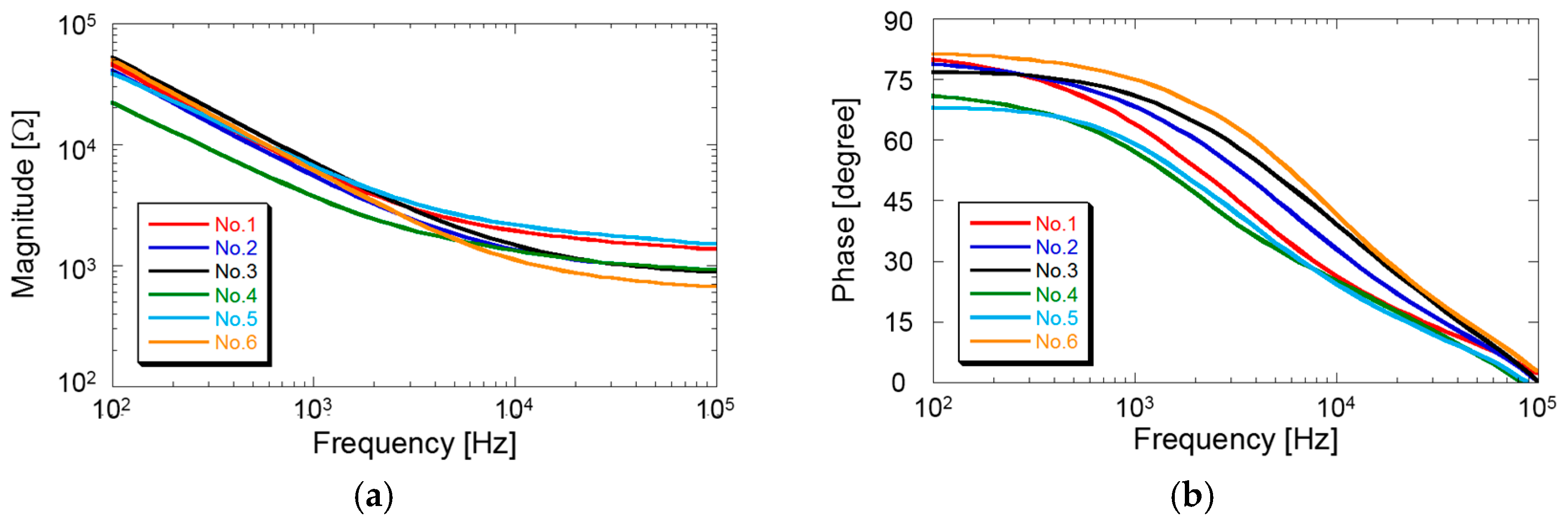
Electrochemical impedance spectroscopy of the fabricated MEA was used to evaluate the electrical properties of individual electrodes, to validate the fabrication concept. The impedance of magnitudes and phases of individual electrode-electrolyte interfaces of the Au electrodes were characterized using a CellTest system (1470E Multichannel Potentiostat/Galvanostat instruments and 1260 Impedance Analyzer, AMETEK Inc., Berwyn, PA, USA) (Figure 4a). A solution of phosphate-buffered saline (PBS, pH 7.2, Sigma-Aldrich Co. LLC., Tokyo, Japan) was used, which had similar electrical properties as a physiological solution. The impedance spectra were recorded by applying 0.05 V RMS sine wave with frequencies varying in a frequency range 100 Hz–100 kHz.
3. Results and Discussion
3.1. Device Fabrication
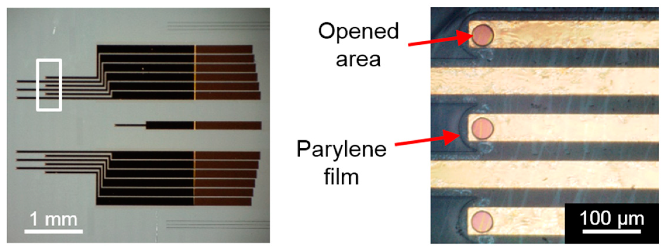
As shown in Figure 5, Au electrodes covered by two layers of parylene C films was successfully peeled off from the Si substrate without the use of any sacrificial layer, and electrode-site and the contacts were opened. Because OFPR-800 is based on a diazonaphthoquinone chemistry, this positive photoresist is not ideal in terms of plasma-resistance; however, for the etching of thin layers of parylene, a 2-μm-thick photoresist showed promise as a mask in this process. Figure 4b shows an optical image of the fabricated MEA bonded to the control layer, and this device was used for the impedance measurements.
3.2. Electrical Properties
Electrical impedance was determined by using electrochemical impedance spectroscopy in a three-electrode measurement setup (Figure 4b). Figure 6 show the impedance magnitude of the fabricated MEA (Figure 6a) and phase (Figure 6b) measured in PBS. The average impedance of the six each electrode at 1 kHz were found to be 5.9 kΩ. The measured impedance of individual electrode-electrolyte interfaces reveals a potential for network electrophysiology within the BoC.
4. Conclusions
We have demonstrated the microfabrication and preliminary characterization of the flexible parylene-based MEA integrated in the BoC. The experimental results showed that the fabricated MEA can be used for network electrophysiological activity. Compared to other existing BoC, this device is the first design that has the flexible MEAs for a mapping of cardiac electrophysiology, leading to investigate how the heart cells are responding to the drugs.
Acknowledgments
The present study was supported in part by the Toyota Physical & Chemical Research Institute Scholars. Part of the present research was conducted at the Kyoto University Nano Technology Hub as part of the “Nanotechnology Platform Project” sponsored by MEXT, Japan.
Conflicts of Interest
The authors declare no conflict of interest. The founding sponsors had no role in the design of the study; in the collection, analyses, or interpretation of data; in the writing of the manuscript, and in the decision to publish the results.
References
- Bhatia, S.N.; Ingber, D.E. Microfluidic organs-on-chips. Nat. Biotechnol. 2014, 32, 760–772. [Google Scholar] [CrossRef] [PubMed]
- Kato, Y.; Hirai, Y.; Kamei, K.; Tsuchiya, T.; Tabata, O. Microfluidic device to interconnect multiple organs via fluidic circulation: Towards Body-on-a-Chip. In Proceedings of the 18th International Conference on Solid-State Sensors, Actuators and Microsystems (Transducers 2015), Anchorage, AK, USA, 21–25 June 2015; pp. 1549–1552. [Google Scholar]
- Maoz, B.M.; Herland, A.; Henry, O.Y.F.; Leineweber, W.D.; Yadid, M.; Doyle, J.; Mannix, R.; Kujala, V.J.; FitzGerald, E.A.; Parker, K.K.; et al. Organs-on-Chips with combined multi-electrode array and transepithelial electrical resistance measurement capabilities. Lab Chip 2017, 7, 2294–2302. [Google Scholar] [CrossRef] [PubMed]
- Adrega, T.; Lacour, S.P. Stretchable gold conductors embedded in PDMS and patterned by photolithography: Fabrication and electromechanical characterization. J. Micromech. Microeng. 2010, 20, 055025. [Google Scholar] [CrossRef]
- Byun, I.; Coleman, A.W.; Kim, B. Transfer of thin Au films to polydimethylsiloxane (PDMS) with reliable bonding using (3-mercaptopropyl)trimethoxysilane (MPTMS) as a molecular adhesive. J. Micromech. Microeng. 2013, 23, 085016. [Google Scholar] [CrossRef]
- Pakazad, S.K.; Savov, A.; van de Stolpe, A.; Dekker, R. A novel stretchable micro-electrode array (SMEA) design for directional stretching of cells. J. Micromech. Microeng. 2014, 24, 034003. [Google Scholar] [CrossRef]
- Chang, T.Y.; Yadav, V.G.; Leo, S.D.; Mohedas, A.; Rajalingam, B.; Chen, C.L.; Selvarasah, S.; Dokmeci, M.R.; Khademhosseini, A. Cell and protein compatibility of Parylene-C surfaces. Langmuir 2007, 23, 11718–11725. [Google Scholar] [CrossRef] [PubMed]
Figure 1.
(a) Illustration of side effects of the drug on heart. (b) Schematic cross-sectional view of a unit of the device and its optical image (top view).
Figure 1.
(a) Illustration of side effects of the drug on heart. (b) Schematic cross-sectional view of a unit of the device and its optical image (top view).
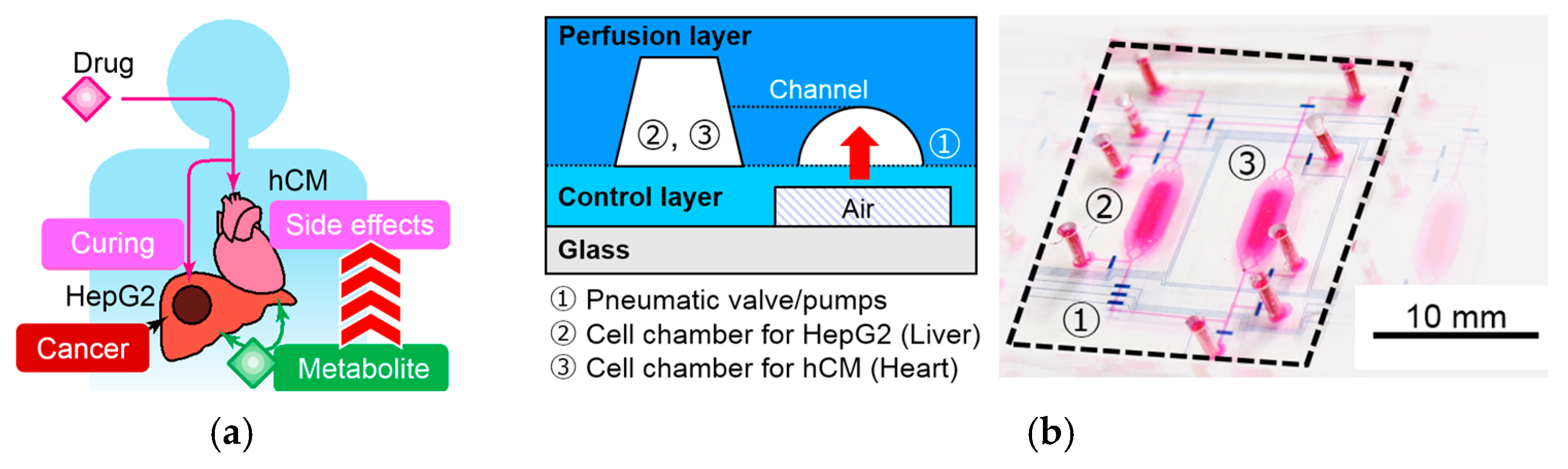
Figure 2.
Schematic illustrations of the flexible parylene-based MEA embedded in PDMS and close-up view of the electrodes part.
Figure 2.
Schematic illustrations of the flexible parylene-based MEA embedded in PDMS and close-up view of the electrodes part.

Figure 3.
Process flow of the parylene-based MEA embedded in PDMS.
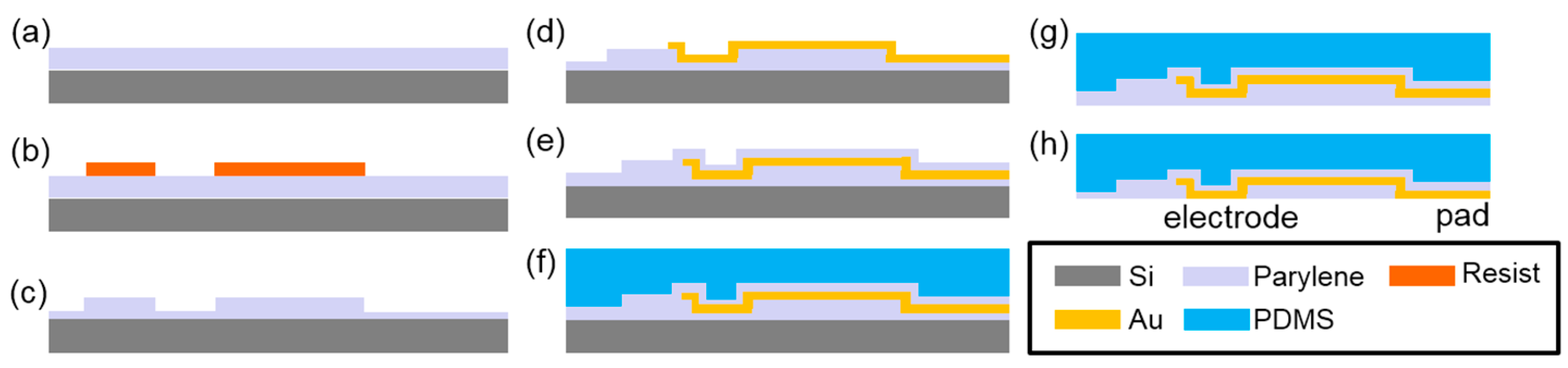
Figure 4.
(a) Schematic illustration of the electrochemical impedance spectroscopy in a three-electrode measurement setup. (b) Setup for the fabricated MEA embedded in the control layer.
Figure 4.
(a) Schematic illustration of the electrochemical impedance spectroscopy in a three-electrode measurement setup. (b) Setup for the fabricated MEA embedded in the control layer.

Figure 5.
Photograph of the fabricated MEA bonded to the control layer of the Body-on-a-Chip device and close-up view of the Au electrode arrays. (12 Au electrodes each 50 μm in diameter).
Figure 5.
Photograph of the fabricated MEA bonded to the control layer of the Body-on-a-Chip device and close-up view of the Au electrode arrays. (12 Au electrodes each 50 μm in diameter).

Figure 6.
The impedance magnitude of the fabricated MEA (a) and phase (b).
Publisher’s Note: MDPI stays neutral with regard to jurisdictional claims in published maps and institutional affiliations. |
© 2017 by the authors. Licensee MDPI, Basel, Switzerland. This article is an open access article distributed under the terms and conditions of the Creative Commons Attribution (CC BY) license (https://creativecommons.org/licenses/by/4.0/).
Share and Cite
MDPI and ACS Style
Omaki, T.; Hirai, Y.; Kamei, K.-i.; Tsuchiya, T.; Tabata, O. Microfabrication of Embedding a Flexible Parylene-Based Microelectrode Array within Body-on-a-Chip. Proceedings 2017, 1, 302. https://doi.org/10.3390/proceedings1040302
AMA Style
Omaki T, Hirai Y, Kamei K-i, Tsuchiya T, Tabata O. Microfabrication of Embedding a Flexible Parylene-Based Microelectrode Array within Body-on-a-Chip. Proceedings. 2017; 1(4):302. https://doi.org/10.3390/proceedings1040302
Chicago/Turabian StyleOmaki, Tatsuya, Yoshikazu Hirai, Ken-ichiro Kamei, Toshiyuki Tsuchiya, and Osamu Tabata. 2017. "Microfabrication of Embedding a Flexible Parylene-Based Microelectrode Array within Body-on-a-Chip" Proceedings 1, no. 4: 302. https://doi.org/10.3390/proceedings1040302