1. Introduction
The need of a rapid, low cost and sensitive pathogen sensing systems is urgent because of the increase of spreading diseases and epidemics. This is due to the increasing movement of goods and people especially by flight. There are many approaches to develop such in vitro diagnosis systems specially Lab-On-Chip (LOC) one. Most of these approaches use electrochemical and optical methods. However, they have several drawbacks like nonlinear detection range, lack of sensitivity, non-specific detection and complexity of integration [
1,
2,
3]. The other approaches use mechanical and magnetic based methods. The systems based on magnetic approaches hold promising perspectives due to their miniaturization possibility [
4,
5]. Some attempts have been made in order to embed magnetic sensors such as GMR, TMR, and planar Hall Effect sensors [
6]. The LOC pathogen sensing system should be sensitive with a low limit of detection, cost effective, size selective, simple to use and with a large dynamic range. Having low cost and very sensitive detection system is rather challenging.
The miniaturized system that we aim to develop tends to overcome these challenges. In our case, we take advantage of the nonlinearity of superparamagnetic nanoparticles (SPN). Because of their biocompatibility, SPN are particularly well suited for the biomedical domain. They are commonly used in numerous applications such as magnetic resonance imaging (MRI), information storage, and medical diagnostics using magnetic immunoassays. In fact, the particles can be used both for magnetic actuation and detection of biomolecules. In the case of immunoassays, the SPN can become markers by bounding them to the biological target substance using the highly specific antibody-antigen interaction [
7]. The challenge is to detect the magnetic marker particles with high accuracy and selectivity. These particles are described by two characteristics: magnetization curve and frequency-dependent relaxation effects. The magnetization curve is nonlinear, non-hysteretic and it saturates at high fields [
8]. To exploit this nonlinearity, we use the frequency mixing method in order to detect the presence of particles in a microfluidic channel. In this method, two magnetic excitation signals are applied to the sample reservoir at distinct frequencies (
f1,
f2) and the amplitude of the response signal is measured at a specific frequency (
) that allows detecting and quantifying the amount of SPN [
9]. The magnetic frequency mixing technique is very well suited for this purpose, thus allowing its use for immunoquantification.
The development of SPN detection techniques in a microfluidic platform has been investigated. To achieve this, a magnetic micro-detection unit is combined with a microfluidic structure for the first time. The microfluidic allows us to use small quantities of analyte and reagents, and will enable also quick analysis. The goal is to develop a simple and portable analysis platform for magnetic immunoquantification which may be applied for diagnosis of illnesses, prevention of pandemia, food monitoring, environmental protection, etc.
2. Multiphysics Simulations and Analytical Calculation
In the scope of the design and realization of a microstructure aimed for multipathogen immunoassays using superparamagnetic beads, we have achieved multiphysics simulations in order to get a good compromise between the various important parameters. These simulations allow to optimize the microstructure, a proper magnetization of the nanoparticles, the detection sensitivity, minimize the heat transfer effects, and thus the viability of the biological samples. Also, we should consider the diffusion in the microfluidic structure.
In a first step towards using the magnetic frequency mixing technique in microfluidic structures, planar coils have to be designed and simulated. COMSOL electrical and magnetic simulations along with an analytical calculation have been used in order to optimize the dimensions, shape, and positions of the coils so that a proper distribution and density of magnetic flux can be obtained at the sample area.
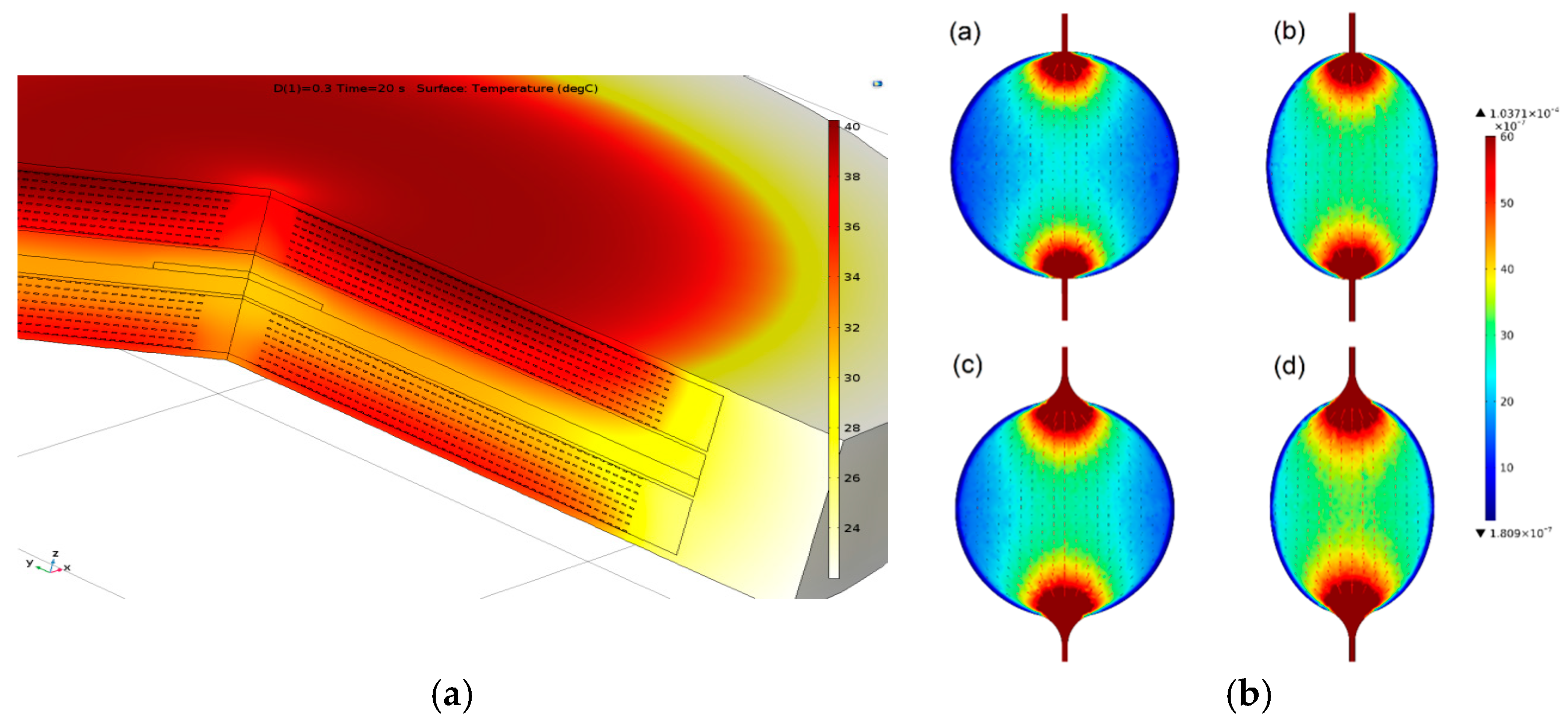
Actually, the most important goal using multiphysics simulations is finding the best parameters that ensure an uniform distribution of the sample flow, sufficiently low temperatures in the microfluidic reservoir (below 37 °C), a homogeneous magnetic field distribution and optimum coil sensitivity. These entities are sensitive to heat resulting from the bias current of the coils. The variation of the voltage and magnetic flux as a function of the relative magnetic permeability of the sample is presented in
Figure 1. Besides, heat transfer and microfluidic simulations (
Figure 2a,b) have been achieved to optimize the microstructures for their usage with biological entities.
3. Experimental Structure and Preliminary Results
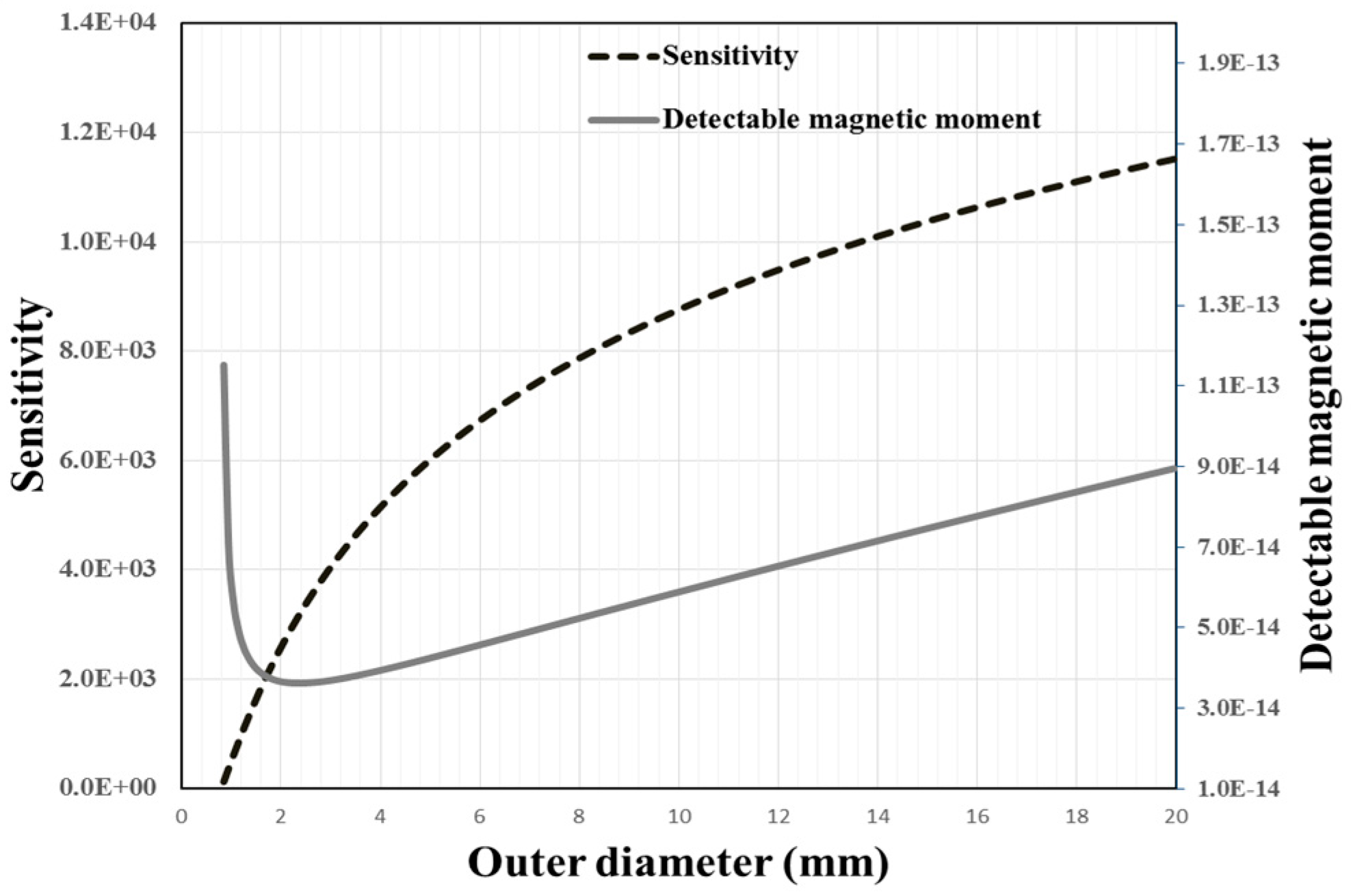
The miniaturized experimental structure is composed of planar copper coils as excitation and detection coils. For the detection coil, defining the inner and outer radius by choosing a good compromise between sensitivity and minimum detectable moment is important (
Figure 3). These coils surround the microfluidic channel, optimized with the shape of meander (
Figure 4), which can contain 14 μL of SPN solution, equivalent to a droplet of blood.
The magnetic response curve for different concentrations of SNP in the microfluidic channel is shown in
Figure 5.
A very good linearity of detected signal versus SNP concentration has been observed for 3 orders of magnitude. Furthermore, the test was repeated in day’s intervals and proved to be very reproducible. With an accepted error of less than 15%, our limit of detection is about 15 ng/μL for 20 nm core sized SPN which is very promising preliminary result for a LOC (
Figure 5). A small quantity of solution sample was used which is very convenient for future cost effective biological tests. These preliminary results validate the magnetic detection of nanoparticles and further improvements are expected regarding the limit of detection, especially with noise reduction. With our magnetic detection system validated, this structure will be used for implementing immunoassays for testing C-reactive protein samples with different concentrations.
4. Conclusions
In this work, a miniaturized system with planar excitation and detection coils was designed and fabricated to achieve magnetic immunoassay using superparamagnetic nanoparticles (SPN) with frequency mixing technique. Both analytical and multiphysics simulation tools have been done in order to optimize different electrical and geometrical parameters of the system. Consequently, PDMS microfluidic structures along with planar coils have been realized in accordance. The variation of the voltage and magnetic flux versus relative magnetic permeability of the sample has been calculated analytically. Heat transfer and microfluidic simulations allowed to optimize the microstructures for their usage with biological entities. The meander shape of the optimized microfluidic reservoir can contain 14 µL of SPN solution, equivalent to a droplet of blood. Based on the preliminary experimental results, our limit of detection is about 15 ng/µL for 20 nm core sized SPN which is very promising for a LOC. The simulations have been also in correlation with the experiments. Moreover, the device should be validated for the detection of biological substances such as C-reactive protein which indicates the general level of inflammation in the body and allows identifying and keeping track of infections and diseases that cause inflammation.


