Proceeding Paper

Integration and Bio-Functionalization of Vertically Aligned Carbon Nanotube Forests on High Frequency AlN Gravimetric Sensors †

1
GMME-CEMDATIC, ETSI de Telecomunicación, Universidad Politécnica de Madrid, 28040 Madrid, Spain
2
Confocal Microscopy Unit, Spanish National Cancer Research Centre (CNIO), 28029 Madrid, Spain
*
Author to whom correspondence should be addressed.
Presented at the Eurosensors 2017 Conference, Paris, France, 3–6 September 2017.
Proceedings 2017, 1(4), 537; https://doi.org/10.3390/proceedings1040537
Published: 7 August 2017
(This article belongs to the Proceedings of Proceedings of Eurosensors 2017, Paris, France, 3–6 September 2017)

Abstract

:
In this work, we grow different types of carbon nanotube (CNT) forests directly on AlN-based electroacoustic biosensors and study their functionalization efficiency. CNTs are used to increase the effective surface area of the sensor, pursuing a better sensitivity, without increasing the sensor capacitance. Here we use a general method for CNT bio-functionalization with specific receptors for targeted species. First, we make them hydrophilic. Next, we cover them with alternating polymers. Finally, we incubate their surface in an N-hydroxysuccinimide biotin solution. To study the functionalization homogeneity along the CNT length, Streptavidin conjugated with fluorescein isothiocyanate is used as the targeted species for confocal fluorescence microscopy characterization.

1. Introduction

Gravimetric biosensors are ideal candidates for in-field sensing applications such as point-of-care diagnosis, water analysis and environmental monitoring. This is because they are label free, fast to analyze and their high frequency operation results in a high sensitivity [1]. In particular, thin film electroacoustic resonators based on AlN piezoelectric layers have demonstrated good performance as biosensors [2,3]. Increasing the effective surface area of a resonator without modifying the electrical contact area increases its sensitivity and reduces its response time. The side walls of nanomaterials, such as those of CNT forests, are ideally suited for this. CNT forests of controlled quality and surface density can be directly integrated on resonators, thus obtaining a platform with a large area for bio-functionalization. The functionalization chemistry of CNTs has been widely studied [4], but carrying it out directly on the resonators without compromising their performance is not straightforward. In this work we directly grow CNTs over the top electrode of AlN-based resonators. We use a custom designed microfluidic system to functionalize the CNTs, enabling the liquid containing the targeted species to properly flow between them. The final aim is to study the functionalization efficiency of different types of forests directly integrated on AlN-based resonators.

2. Materials and Methods

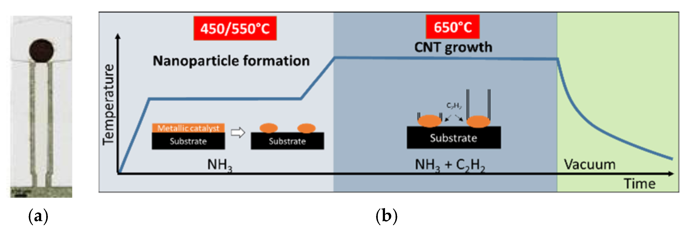
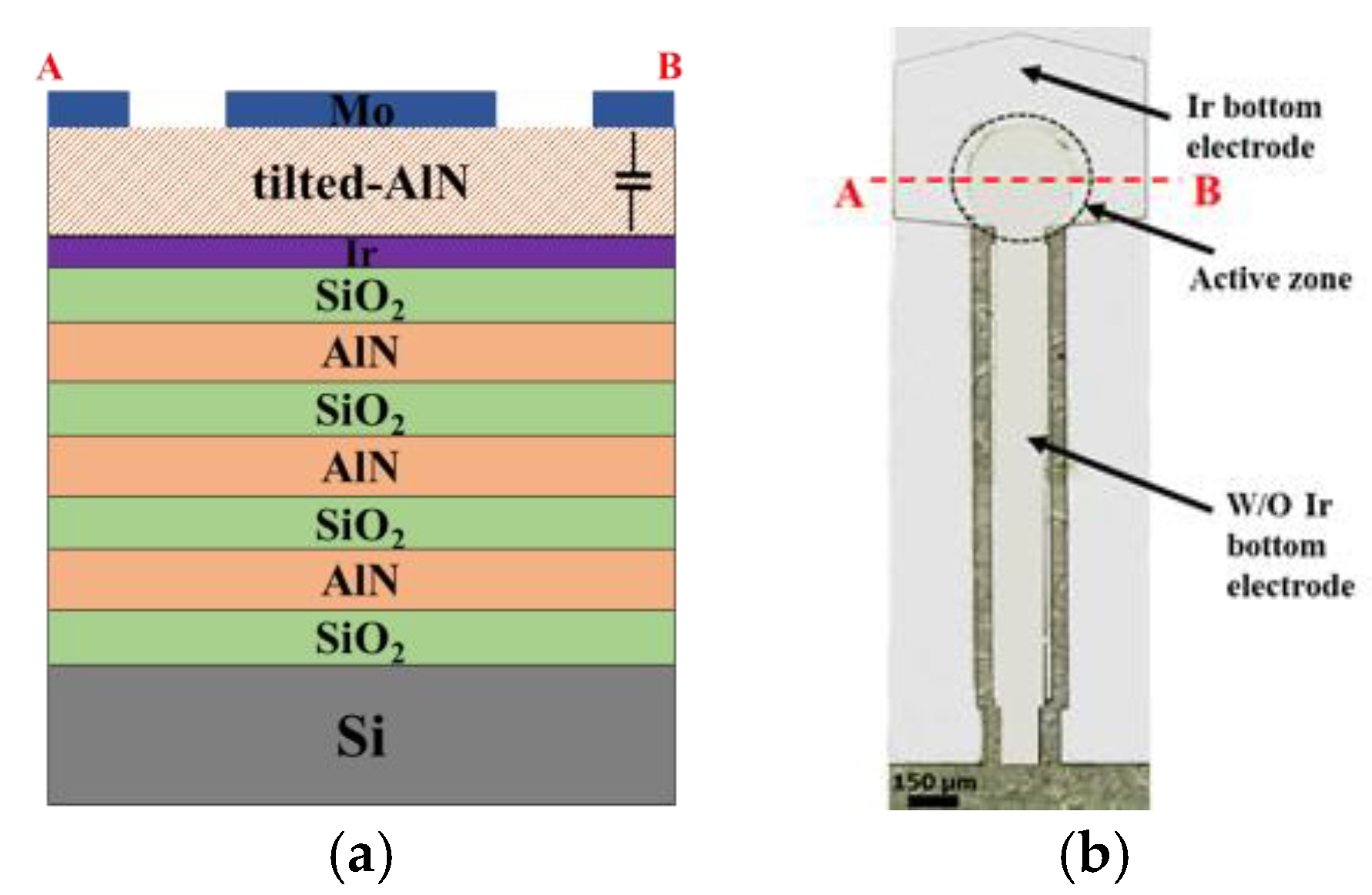
2.1. Device Fabrication

The shear mode 1.4 GHz SMRs consist of a thin AlN piezoelectric film sandwiched between two metallic electrodes mounted on a fully-insulating acoustic reflector used to confine the acoustic energy within the piezoelectric layer and prevent it from radiating to the Si substrate. The acoustic reflector is made of five alternating layers of low and high acoustic impedance (SiO2 and AlN, respectively) (Figure 1a). All layers are deposited by pulsed-DC sputtering, except for the Ir bottom electrode which is e-beam evaporated to reduce substrate roughness and promote the growth of highly piezoelectric AlN films. AlN films with tilted grains are deposited in order to excite the shear modes required for the in-liquid operation of the SMRs. These are sputtered though a two-step process [3] in an ultra-high-vacuum system by bombarding a high purity 150 mm Al target with an Ar/N2 (40:60) gas mixture. First an AlN seed layer is grown at high pressure (0.66 Pa). Subsequently, the piezoelectric AlN film is deposited at a lower pressure (0.27 Pa) using a substrate temperature of 400 °C and a substrate bias of −55 V. The deposition conditions of all the films are tuned to obtain low residual stress to prevent delamination at the high temperatures needed for the direct growth of the CNT forests (650 °C) [5]. To ward off the electrical contact pads from the active area where the CNT forests are integrated, electrical extensions of 1.7 mm are used, below which the Ir bottom electrode is etched (Figure 1b).

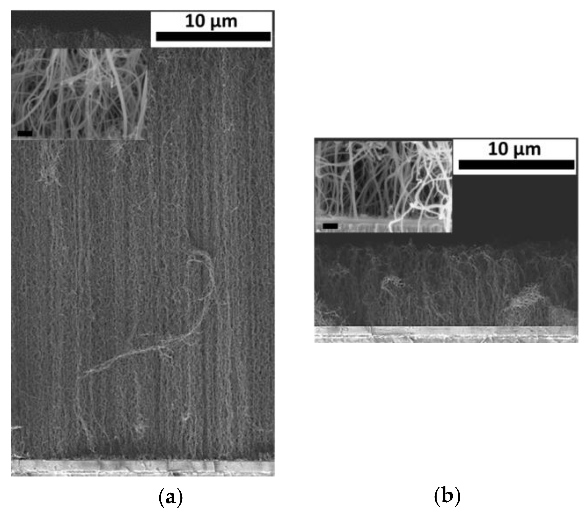
2.2. CNT Forests Integration

CNT forests are directly integrated on the SMRs active area (Figure 2a). They are grown by a two-step low pressure CVD process using a Fe evaporated film as catalyst. First, the catalyst film is restructured into nanoparticles (NPs) at high temperature in a reducing atmosphere (NH3). Then, a carbon-containing gas (C2H2) is flowed and CNTs start growing on each NP (Figure 2b). Growing CNTs on metallic substrates as Mo is a challenging task since it is essential to prevent the catalyst NPs from diffusing into the metallic substrate. This is achieved by placing a thin Al film between the Mo electrode and the Fe catalyst; the Al film acts as a stabilizer preventing the NPs from surface diffusion and lowering their temperature formation [6] (Figure 2b). The growth of the CNT forests only on the active area is achieved by patterning the Al/Fe films by a lift-off process. We used a 12 nm-thick Al stabilizer and Fe catalyst layers of two different thickness (4 and 10 nm) to explore the influence of the CNTs areal density on the functionalization efficiency.

2.3. Functionalization of CNTs

To carry out the functionalization protocol, the device is placed in a microfluidic system, connected to a peristaltic pump, to flow the buffers through. In order to ensure the proper functionalization of the hydrophobic inner side of the forest, the side walls of each tube need to become hydrophilic, which we achieve following a specific protocol [7]. First, we incubate the devices with the anionic surfactant sodium dodecyl benzene sulfonate (SDBS). Next, we deposit 3.5 bilayers alternating positively charged polyallylamine hydrochloride (PAH) and negatively charged sodium polystyrene sulfonate (SPS) polymers. After an acid treatment used to activate the primary amines on PAH, CNTs are incubated with N-Hydroxysuccinimide-biotin 5 mM solution in PBS. Biotin functionalized CNTs are blocked with 6% bovine serum albumin (BSA) in PBS (10 nM phosphate buffer saline). Then, the surface receptors are ready for target detection, in this case streptavidin fluorescein isothiocyanate (FITC) conjugated. Confocal fluorescence microscopy is used to analyze the efficiency of the functionalization throughout the CNT forest.

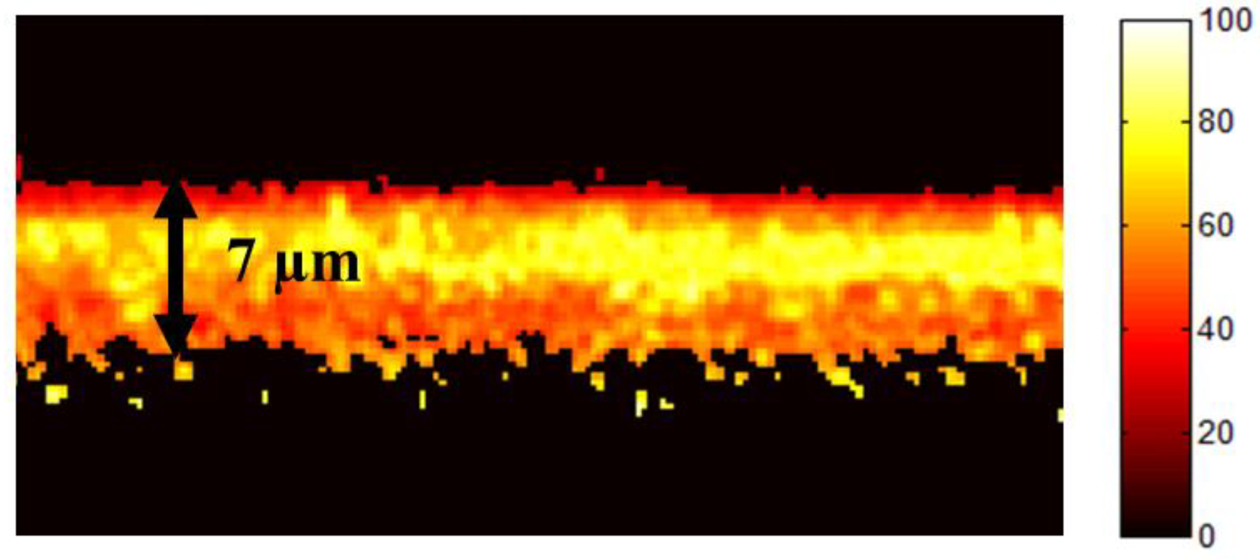
3. Results and Discussion

We study the functionalization efficiency of integrated CNT forests with two different types of forests. The first one is obtained with 4 nm-thick Fe over the 12 nm-thick Al stabilizer and allowing CNT growth for 10 min (Figure 3a). The second type is grown over a 10 nm-thick Fe and allowing CNT growth for 3 min (Figure 3b). In both cases NPs are formed during 5 min. As observed, increasing catalyst thickness and lowering the CNT growth time reduces the tubes density (insets from Figure 3) and the forest height (from 40 µm to 7 µm).
The first functionalization test was performed on the taller and denser forest (Figure 3a). We use a positive and negative control, achieving this last by eliminating the biotin from the incubation process. After inspecting the samples by confocal fluorescence microscopy we confirm that, with the positive control, the forest is functionalized with streptavidin FITC. However, this only occurs in the upper third of the 40 µm height of the forest. We attribute this to the small tubes separation and an excessive height of the forest (Figure 3a). In contrast, the functionalization performed on the second type of forest (Figure 3b) appears to be more uniform. In this case confocal fluorescence microscopy confirms that the tubes are functionalized along their entire length (Figure 4).

4. Conclusions

CNT forests can be used in bio-sensors as active sensing layers to improve sensitivity and selectivity by increasing the active surface area and with specific bio-functionalization, respectively. In this work we demonstrate: (1) that the direct integration of CNT forests on the metallic top electrode of AlN-based electroacoustic sensors is possible by using and Al/Fe catalytic system and reducing the temperature for NP formation, and (2) that the specific bio-functionalization of the tubes is only efficient (homogeneous) when forests with low tube density and small height are grown, which is achieved by using a thicker Fe and allowing less time for CNT growth.

Acknowledgments

This work was partially supported by the Ministerio de Economía y Competitividad del Gobierno de España through the project MAT2013-45957-R.

Conflicts of Interest

The authors declare no conflict of interest.

References

  1. Sauerbrey, G. Verwendung von Schwingquarzen zur Wagungdiinner Schichten und zur Mikrowagung. Zeitschrift fur Phys. 1959, 155, 206–222. [Google Scholar] [CrossRef]
  2. Wingqvist, G.; Bjurstrom, J.; Hellgren, A.; Katardjiev, I. Immunosensor utilizing a shear mode thin film bulk acoustic sensor. Sens. Actuators B Chem. 2007, 127, 248–252. [Google Scholar] [CrossRef]
  3. DeMiguel-Ramos, M.; Díaz-Durán, B.; Escolano, J.-M.; Barba, M.; Mirea, T.; Olivares, J.; Clement, M.; Iborra, E. Gravimetric biosensor based on a 1.3GHz AlN shear-mode solidly mounted resonator. Sens. Actuators B Chem. 2017, 239, 1282–1288. [Google Scholar] [CrossRef]
  4. Van Hooijdonk, E.; Bittencourt, C.; Snyders, R.; Colomer, J.-F. Functionalization of vertically aligned carbon nanotubes. Beilstein J. Nanotechnol. 2013, 4, 129–152. [Google Scholar] [CrossRef] [PubMed]
  5. Mirea, T.; Olivares, J.; DeMiguel-Ramos, M.; Clement, M.; Sangrador, J.; Iborra, E.; Esconjauregui, S. Carbon nanotube forests as top electrodes for AlN-based electroacoustic resonators. In Proceedings of the 2014 IEEE International Ultrasonics Symposium, Chicago, IL, USA, 3–6 September 2014; IEEE: Piscataway, NJ, USA, 2014; pp. 1476–1479. [Google Scholar]
  6. Olivares, J.; Mirea, T.; Díaz-Durán, B.; Clement, M.; DeMiguel-Ramos, M.; Sangrador, J.; de Frutos, J.; Iborra, E. Growth of carbon nanotube forests on metallic thin films. Carbon 2015, 90, 9–15. [Google Scholar] [CrossRef]
  7. Yost, A.L.; Shahsavari, S.; Bradwell, G.M.; Polak, R.; Fachin, F.; Cohen, R.E.; McKinley, G.H.; Toner, M.; Rubner, M.F.; Wardle, B.L. Layer-by-layer functionalized nanotube arrays: A versatile microfluidic platform for biodetection. Microsyst. Nanoeng. 2015, 1, 15037. [Google Scholar] [CrossRef]
Figure 1. SMR structure (a) AB cross section scheme of the device active area; (b) Optical image of the device top view where electrical extensions can be observed.
Proceedings 01 00537 g001
Figure 2. Growth of CNT forest on the metallic top electrode of SMRs (a) Optical top view of a device with CNTs grown on its active area; (b) Steps of the CVD growth process.
Proceedings 01 00537 g002
Figure 3. SEM image of CNT forests grown under different conditions giving rise to different heights and tubes densities (a) with 4 nm-thick Fe catalyst for 10 min; (b) with 10 nm-thick Fe catalyst for 3 min. The insets show the difference in tubes separation. Scale bars in both cases are 200 nm.
Proceedings 01 00537 g003
Figure 4. Confocal fluorescence microscopy image of a 7 µm tall CNT forest after functionalization with biotin-streptavidin FITC.
Proceedings 01 00537 g004
Publisher’s Note: MDPI stays neutral with regard to jurisdictional claims in published maps and institutional affiliations.

