Proceeding Paper

Biomolecular Detection Based on the Rotational Dynamics of Magneto-Plasmonic Nanoparticles †

1
Center for Integrated Sensor Systems, Danube University Krems, 2700 Wr. Neustadt, Austria
2
PROFACTOR GmbH, 4407 Steyr-Gleink, Austria
*
Author to whom correspondence should be addressed.
Presented at the Eurosensors 2017 Conference, Paris, France, 3–6 September 2017.
Proceedings 2017, 1(4), 541; https://doi.org/10.3390/proceedings1040541
Published: 7 August 2017
(This article belongs to the Proceedings of Proceedings of Eurosensors 2017, Paris, France, 3–6 September 2017)

Abstract

:
We report on a nanoparticle-based biosensor that represents a label-free homogeneous bioassay suitable for in-vitro biomolecular diagnostics. The underlying detection principle is based on the optical observation of the rotational dynamics of multicomponent nanoparticles utilizing magnetic and plasmonic properties. The plasmon-optical properties of the anisotropic nanoparticles depending on their material composition and geometrical design were investigated by numerical simulations. Based on such an analysis, monodisperse magneto-plasmonic nanoparticles were fabricated using physical fabrication methods. Rotational dynamics measurements revealed the lowest detectable particle concentration in the picomolar (ng/mL) regime, which is very promising in reaching the biomolecular limit of detection which is relevant for routine clinical diagnostics.

1. Introduction

Heterogeneous biosensor assays such as enzyme-linked immunosorbent assays (ELISA) show a high sensitivity and wide dynamic range. However, they still face intrinsic problems such as steric hindrance [1], restricted diffusion [2] and extensive sample preparations that limit their applicability at point-of-care [3]. These issues can be circumvented in homogeneous immunoassay principles, where the signal generating probes are mixed with the analyte sample followed by a target molecule detection directly within the probe-analyte mixture. The presented “Magnetic Lab-on-a-Bead” (MLoB) follows a homogeneous sensing principle which relies on changes in the rotational dynamics of optically anisotropic nanoparticles [4]. The feasibility and great potential of nanoparticle-based homogeneous immunodiagnostics for simple, fast and sensitive biomarker detection has been demonstrated by Schrittwieser et al. [5]. Although their chemically synthesized core-shell nanoprobes were of high quality, they still show a relevant distribution in size, which leads to a spread in the measured signal. Moreover, no plasmonic excitations could be observed in chemically synthesized core-shell nanorods, which would increase the sensitivity.
The intrinsic problems encountered in chemically synthesized nanoparticles may be overcome by using Nanoimprint Lithography (NIL). In 2015, Kwon et al. [6] reported on the feasibility in fabricating large amounts of highly uniform sombrero-shaped magnetite nanoparticles using NIL.
In this study, we combine NIL and thin film technology to fabricate monodisperse magneto-plasmonic nanoparticles with high reproducibility. The optical properties of the aspired magneto-plasmonic nanoparticles are investigated by numerical calculations. A redesigned experimental setup enabling measurements in transmission and scattering geometry has been validated by determining the lowest detectable nanoparticle concentration.

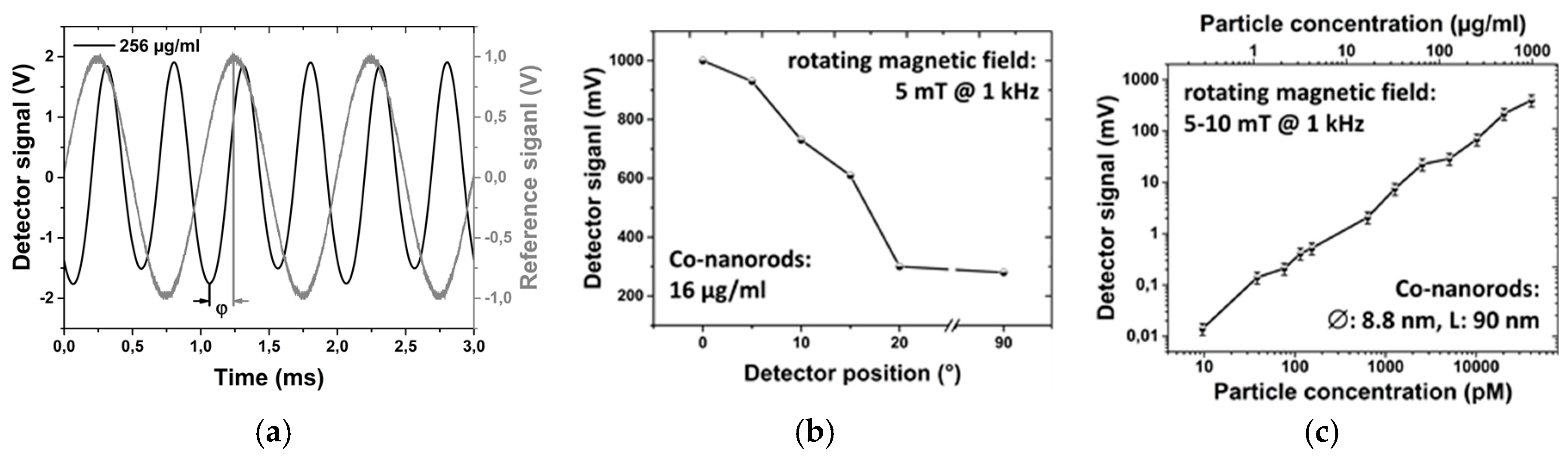
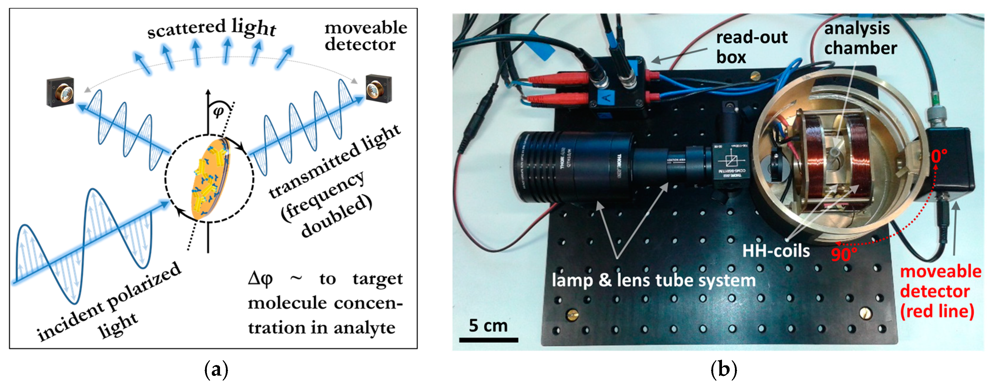
2. MLoB Detection Principle, Measurement Setup and Validation

The MLoB biosensing principle [4] is based on the optical detection of changes in the rotational dynamics of optically anisotropic nanoparticles immersed in the analyte sample such as whole-blood (Figure 1a). Briefly, functionalized nanoparticles rotating in a time-varying magnetic field specifically bind target molecules on their surface, which leads to an increase in their hydrodynamic volume. This increase is detected by measuring the phase lag change φ between the actual particle alignment relative to the direction of the rotating magnetic field. The phase lag signal as a result of the scattering of linearly polarized light at the optically anisotropic nanoparticles directly quantifies the target molecule concentration in the analyte.
The optical polarizability of magnetic nanorods is sufficient to detect the average nanorod orientation in the analyte solution. However, the scattering cross section of noble metal covered nanorods supporting plasmonic resonances is by a factor of 100 higher [4]. This allows for lower particle concentrations in the analyte sample and leads to a significant sensitivity increase.
All rotational dynamics measurements are carried out with our redesigned setup. It consists of two Helmholtz coils arranged perpendicular to each other (Figure 1b), which are sourced by an audio amplifier providing two sinusoidal currents of adjustable amplitude and phase. A rotating magnetic field is created when the two currents are 90° phase shifted. Two Hall-sensors monitor the electrical current through the individual coils, which serve as a reference signal for the actual magnetic field orientation. Magnetic field strengths up to 20 mT are possible. A collimated, linearly polarized, polychromatic light source with a beam diameter of ≤4 mm is accomplished by a lens tube system. The 600–1150 nm detection window fits the spectral range where blood shows the lowest absorption. A photodetector is equidistantly moveable around the cuvette situated at the center of the Helmholz coils, thus allowing scattering and transmission measurements. A Lock-in amplifier compares the reference signal of the rotating magnetic field with the signal provided by the detector. Due to the elongated symmetry of the nanoparticles (Figure 1a), the detector signal is frequency-doubled and the Lock-in amplifier is set to measure the second harmonic of the reference signal. For rotational dynamics observations, phase and amplitude are recorded in dependence of the rotation frequency and magnetic field amplitude.
The validation of the principle and setup is carried out with chemically synthesized plain Co-nanorods dissolved in ultra-pure water (kindly provided by J. Schotter and S. Schrittwieser from AIT Austrian Institute of Technology) [4]. The characteristic frequency-doubled detector signal and the reference signal are shown in Figure 2a. Extinction measurements for varying detector position showed the highest and lowest signal amplitudes for transmission (0°) and scattering (90°) geometry, respectively (Figure 2b). Transmission measurements depending on the nanorod concentrations revealed a limit of detection of about 10 pM or 250 ng/mL (Figure 2c). A remarkable concentration range of four orders of magnitude is covered.

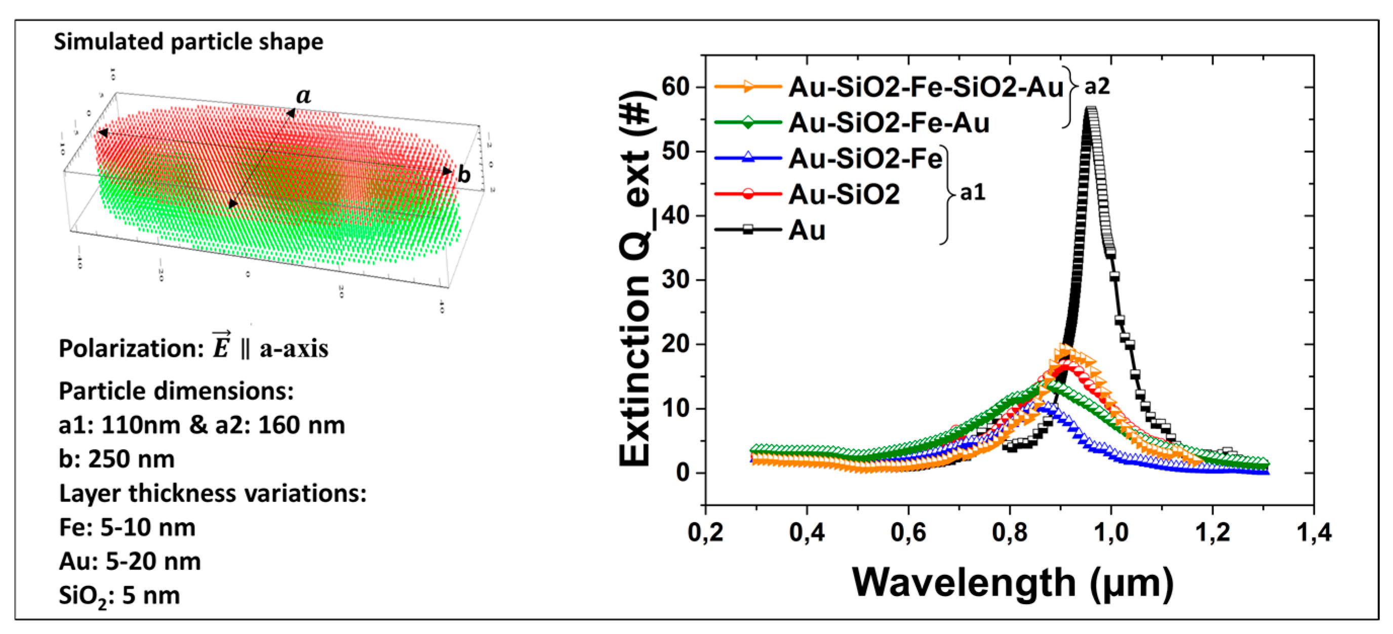
3. Numerical Simulations of the Optical Properties of Multicomponent Nanoparticles

A discrete-dipole approximation code has been established to study the extinction cross section of hybrid nanoparticles [7]. We focus on disc-like nanoparticles of elliptically shaped base area. The magnetic and plasmonic materials are iron (Fe) and gold (Au), respectively. Further oxide protection layers are optional. The complex refractive index values of the involved materials were taken from experimental data [8]. The particles are suspended in water with a real refractive index of 1.333.
The plasmonic peaks of different particle architectures for polarization oriented along the semi-minor a-axis are optimized to fit the detection window (Figure 3), while the extinction peaks of b-axis response are red-shifted beyond the detection window. In order to minimize quenching effects of the plasmonic resonances, the Fe-layer is separated from the adjacent Au-layers by an intermediate oxide layer. The difference in extinction between polarization orientations along the a- and b-axis reaches a factor of 10 and defines the signal amplitude for phase detection.

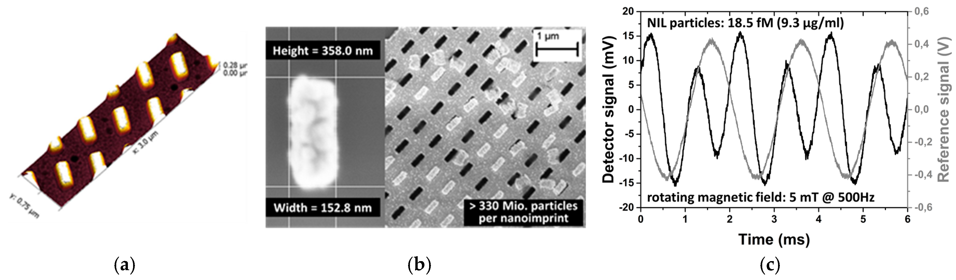
4. Nanoparticle Fabrication and Rotational Dynamics Measurements

Soft Nanoimprint Lithography is an inexpensive technique to fabricate high resolution nanopatterns on large areas with good reproducibility [6,9]. A flexible hybrid-PDMS working stamp comprising a 1 × 1 cm2 array of 200 × 400 nm rectangular shaped pillars of 250 nm height is imprinted in a bilayer resist system consisting of a lift-off and nanoimprint layer. The imprint step is followed by an oxygen plasma etching and developing step, thin film deposition and a subsequent lift-off process resulting in a transfer of the particles in liquid solution. AMF and SEM images after metal layer deposition (Figure 4a) and during/after lift-off (Figure 4b), respectively, illustrate the low roughness and monodispersity of NIL-fabricated nanoparticles. First optical extinction measurements demonstrated a detectability of Au/Fe/Au-multilayer nanoparticles of 18.5 fM (9.3 µg/mL). A significant decrease in the limit of detection of NIL-nanoparticles is expected after a comprehensive optimization procedure and more detailed measurements.

5. Conclusions

Our nanoparticle-based MLoB-biosensor suitable for immunodiagnostics has been validated using plain Co-nanorods resulting in a lowest detectable nanorod concentration in the sub-pM (ng/mL) regime. The plasmonic resonances of the aspired multicomponent nanoparticles are tailored by numerical calculations to the detection window of the measurement setup, thus, ensuring a high signal output. First nanoimprinted multicomponent nanoparticles are highly monodisperse. The extinction measurements show their high potential as highly sensitive nanoprobes for diagnostics.

Acknowledgments

The authors acknowledge funding by the Lower Austria Forschungs-und Bildungsges.m.b.H. (NFB) within the Life Science Call and partly from the Federal Ministry of Transport, Innovation and Technology (BMVIT) supported by the Austrian Research and Promotion Agency (project LAMPION, #861414).

Conflicts of Interest

The authors declare no conflict of interest.

References

  1. Edwards, D.A. Steric hindrance effects in thin reaction zones: Applications to BIAcore. IMA J. Appl. Math. 2007, 72, 865–893. [Google Scholar] [CrossRef]
  2. Jennissen, H.P.; Zumbrink, T. A novel nanolayer biosensor principle. Biosens. Bioelectron. 2004, 19, 987–997. [Google Scholar] [CrossRef] [PubMed]
  3. Liu, X.; Dai, Q.; Austin, L.; Coutts, J.; Knowles, G.; Zou, J.; Chen, H.; Huo, Q. A one-step homogeneous immunoassay for cancer biomarker detection using gold nanoparticle probes coupled with dynamic light scattering. J. Am. Chem. Soc. 2008, 130, 2780–2782. [Google Scholar] [CrossRef] [PubMed]
  4. Schrittwieser, S.; Schotter, J.; Maier, T.; Bruck, R.; Muellner, P.; Kataeva, N.; Soulantika, K.; Ludwig, F.; Huetten, A.; Brueckl, H. Homogeneous biosensor based on optical detection of the rotational dynamics of anisotropic nanoparticles. Procedia Eng. 2010, 5, 1107–1110. [Google Scholar] [CrossRef]
  5. Schrittwieser, S.; Pelaz, B.; Parak, W.J.; Lentijo-Mozo, S.; Soulantica, K.; Dieckhoff, J.; Ludwig, F.; Guenther, A.; Tschöpe, A.; Schotter, J. Homogeneous Biosensing Based on Magnetic Particle Labels. Sensors 2016, 16, 828. [Google Scholar] [CrossRef] [PubMed]
  6. Kwon, B.S.; Zhang, W.; Li, Z.; Krishnan, K.M. Direct Release of Sombrero-Shaped Magnetite Nanoparticles via Nanoimprint Lithography. Adv. Mater. Interfaces 2015, 2, 1400511. [Google Scholar] [CrossRef]
  7. Draine, B.T.; Flatau, P.J. User Guide for the Discrete Dipole Approximation Code DDSCAT 6.1. Available online: http://arxiv.org/abs/astro-ph/0409262v2 (accessed on 1 August 2017).
  8. Palik, E.D. Handbook of Optical Constants of Solids; Academic Press: San Diego, CA, USA, 1998. [Google Scholar]
  9. Li, Z.; Gu, Y.; Wang, L.; Ge, H.; Wu, W.; Xia, Q.; Yuan, C.; Chen, Y.; Cui, B.; Williams, R.S. Hybrid Nanoimprint−Soft Lithography with Sub-15 nm Resolution. Nano Lett. 2009, 9, 2306–2310. [Google Scholar] [CrossRef] [PubMed]
Figure 1. (a) Detection principle: The nanoprobe follows a rotating magnetic field with phase lag ϕ, which changes upon molecular binding; (b) MLoB-setup suitable for all-angle detection.
Proceedings 01 00541 g001
Figure 2. (a) Frequency-doubled phase lag detector signal and reference coil signal; Detector signal amplitude for different (b) detector positions at adjusted gains and (c) nanoparticle concentrations.
Proceedings 01 00541 g002
Figure 3. Simulated extinction factor Q_ext for varying geometry and material composition for light polarization parallel to the particles a-axis.
Proceedings 01 00541 g003
Figure 4. AMF and SEM image of nanoimprinted Au-particles (40 nm) before (a) and (b) during/after lift-off, respectively; (c) Typical rotational dynamics measurements for nanoimprinted hybrid nanoparticles consisting of Au (25 nm)/Fe (8 nm)/Au (25 nm) multilayers and a size of 150 × 360 nm.
Proceedings 01 00541 g004
Publisher’s Note: MDPI stays neutral with regard to jurisdictional claims in published maps and institutional affiliations.

