Article

In Vitro Electrochemical Corrosion Assessment of Magnesium Nanocomposites Reinforced with Samarium(III) Oxide and Silicon Dioxide Nanoparticles

1
Mechanical Engineering, University of Tennessee at Chattanooga, 615 McCallie Ave., Chattanooga, TN 37403, USA
2
Mechanical Design & Production, Faculty of Engineering, Cairo University, Cairo 12613, Egypt
3
Department of Mechanical, Industrial and Manufacturing Engineering, University of Toledo, Toledo, OH 43606, USA
4
Department of Mechanical Engineering, National University of Singapore, Singapore 119077, Singapore
*
Author to whom correspondence should be addressed.
J. Compos. Sci. 2022, 6(6), 154; https://doi.org/10.3390/jcs6060154
Submission received: 19 March 2022 / Revised: 16 May 2022 / Accepted: 23 May 2022 / Published: 25 May 2022
(This article belongs to the Special Issue Feature Papers in Journal of Composites Science in 2022)

Abstract

:
Recent research on biodegradable magnesium-based implants has been focusing on increasing their mechanical strength and controlling their corrosion rate. One promising approach to significantly improve the mechanical properties of magnesium is the addition of nanoparticles to the magnesium matrix. However, there is limited research on the corrosion behavior of these new magnesium nanocomposites. In this study, the electrochemical corrosion characteristics of this new class of biomaterials are investigated. Two magnesium nanocomposites reinforced with nanoparticles (0.5, 1.0, and 1.5 Vol%) of samarium oxide (Sm2O3), and silicon dioxide (SiO2), were fabricated and tested. Corrosion behavior was assessed in comparison with high-purity magnesium samples as the control group. The addition of the nanoparticles to the magnesium matrix strengthened the materials, which was represented in an increase in the microhardness. However, the fabricated nanocomposite samples exhibited a slightly reduced corrosion resistance compared to the high-purity magnesium control due to the differences in the purity level and fabrication methods. Both nanocomposites showed the highest corrosion resistance, represented in the slowest corrosion rates, at the 1.0 Vol% content. Hence, the developed nanocomposites are still promising candidates as biodegradable materials for bone-fixation application owing to their superior mechanical properties and acceptable corrosion characteristics.

1. Introduction

Osteosynthesis treatment is a common surgical procedure aimed at fixing fractured bones by joining adjacent bone fragments until fusion and complete healing is achieved. Osteosynthesis treatment is receiving more interest due to the increase in car and sporting accidents, which can surpass 20,000 cases in merely one hospital [1]. The fixation of the broken bones is currently achieved by using mostly stiff and bioinert (permanent) metallic hardware (e.g., titanium, stainless steel, and CoCr alloys) in the form of screws, pins, and plates [2]. The permanent existence of stiff metallic parts in the body after bone healing may result in unfavorable reactions such as stress shielding and bone resorption [3,4]. In addition, leaving the fixation hardware inside the body after healing may cause future bone fractures due to physical activities, especially in children, teenagers, and athletes [5,6]. Hence, osteosynthesis may highly benefit from biodegradable implants to eliminate the pain and cost of subsequent implant-removal surgeries.
Unlike bioinert materials, a biodegradable material is expected to slowly degrade and get replaced by natural bone tissue. A successful biodegradable implant would exhibit a combination of biocompatibility, mechanical strength, and controlled degradation behavior [7,8]. Lightweight magnesium is a widely investigated candidate for osteosynthesis treatment due to its high biocompatibility and superior mechanical properties compared to biodegradable polymers [9,10]. For instance, the elastic modulus, compressive yield strength, and density of magnesium are comparable to those of natural bone, thereby reducing the risk of osteopenia and avoiding osteolysis [11,12,13]. As per Staiger et al. [14], in addition to being an irreplaceable cofactor to constitute enzymes for metabolism, magnesium is one of the natural elements of orthopedic tissue and is entirely non-toxic to human cells and innoxiously excreted through the urinal system. Despite its high corrosion resistance, pure magnesium exhibits low yield strength of 69–100 MPa, which is considered insufficient for the intended bone-fixation application [15]. Thus, several alloys and metallic composite systems have been investigated in the literature to improve their mechanical and corrosion properties. It is worth mentioning that the conventional definition of “corrosion” usually refers to an undesired phenomenon. In the case of magnesium implants, they are expected to completely dissolve in bodily fluids directly after bone healing. In this context implant dissolving is a highly desirable, if necessary, phenomenon. However, the corrosion rates of magnesium produced by conventional casting techniques corrode (dissolve) rapidly in physiological environments, which leads to a loss of the mechanical integrity of the implant before the completion of the bone-healing process. In addition, the fast corrosion possibly occurs with a high release of corrosion byproducts, which in the case of pure Mg is associated with hydrogen gas that may be detrimental to the healing process [16].
The addition of alloying elements, especially rare-earth (RE) elements such as Yttrium (Y), Cerium (Ce), and Lanthanum (La), has been widely investigated. When alloying elements were added to magnesium, they were reported to improve the tensile strength and creep properties of magnesium in addition to the corrosion resistance [17,18,19,20]. For instance, Huang et al. [21] investigated the RE effect on mechanical properties of magnesium alloys and reported that ductility increased up to 25–30% with lower anisotropy, and the interpretation behind that was the elimination of large precipitates and grain refinement after introducing RE. For medical applications, RE elements generally exhibit an antimicrobial property against bacteria and fungi/yeast [22], which led to their usage in topical treatments of burns [23], as well as in investigating antibacterial alloys [24,25]. For implant use, however, RE elements carry the risk of toxicity, which limits their percentages in the used alloys [26].
For this reason, research has been directed toward manufacturing nanocomposites of magnesium using nanoparticles in attempt to improve the mechanical properties with the least volume content of the added particles (less than 3%) [27]. It has been shown in the literature that the addition of nano-length scale reinforcements into the magnesium matrix can significantly improve the strength, ductility, and corrosion resistance of the material [28,29,30,31]. In particular, samarium oxide (Sm2O3) and silicon dioxide (SiO2) nanoparticles have shown promising mechanical, high temperature creep, and corrosion properties [32,33]. In addition, both nanoparticles show a high degree of biocompatibility represented in low cytotoxicity and favorable bone-cell response, qualifying them for various biomedical applications. The insoluble compounds of samarium, such as (Sm2O3), are found in general to be nontoxic and are secreted out of the bloodstream [34]. Research on the toxicology of silicon dioxide nanoparticles is still ongoing to determine the safe dosage for the different types of administration [35].
In our previous work, we successfully added nano-size (less than 100 nm) nanoparticles to the magnesium matrix to create magnesium-based nanocomposites with improved mechanical (higher strength and ductility), damping, and creep properties, without any tangible weight penalty [27,36]. To the best of the authors’ knowledge, only two studies in the literature reported the influence of the addition of samarium oxide (Sm2O3) [31] and silicon dioxide (SiO2) [29] nanoparticles on the corrosion behavior of magnesium-based nanocomposites for the bone-implant applications. Both studies found that the addition of the nanoparticles resulted in an improvement in the corrosion behavior following the increase of the particles’ volume content as compared to pure magnesium samples manufactured using the same powder metallurgy method. However, both studies used different approaches and testing conditions to assess the corrosion behavior. In addition, both studies did not investigate the electrochemical corrosion properties of the developed nanocomposites and did not compare the corrosion results with an unbiased control group.
The main objective of this work is to assess and compare the effect of adding two different types of nanoparticles (Sm2O3 and SiO2) on the corrosion behavior of the resulting magnesium nanocomposite using the same testing method and conditions. Magnesium nanocomposites containing Sm2O3 and SiO2 nanoparticles are assessed at 0, 0.5, 1.0, and 1.5 Vol% levels and compared to high-purity magnesium (HP-Mg) control samples. High-purity magnesium is known to have a superior corrosion resistance due to its high purity; however, it has poor mechanical properties.

2. Materials and Methods

Pure magnesium and magnesium-based nanocomposites of (0.5, 1.0, and 1.5 Vol%) of Sm2O3 and SiO2 nanoparticles were manufactured using powder metallurgy and hybrid microwave sintering, followed by hot extrusion as described by Kujur et al. [27]. Magnesium powder with >98.5% purity and 60–300 µm particle size (Merck, Darmstadt, Germany) was used as the base metal. The Sm2O3 nanoparticles (20–30 nm) provided by United States Research Nanomaterials and hollow silica (SiO2) nanoparticles (10–20 nm) provided by Sigma Aldrich were used as the reinforcement. Mechanical alloying machine RETSCH PM-400 (Retsch, Haan, Germany) was used for blending the weighed samples of pure Mg powder and the nanoparticles. Billets of the mixture were cold compacted uniaxially under the pressure of 1000 psi. A hybrid microwave sintering process at 630 °C in a 2.45 GHz, 900 W Sharp microwave oven was used to sinter the 35-mm diameter and 40-mm height billets. The billets were soaked at 450 °C for 2 h prior to hot extrusion. A 150 T hydraulic press was used to extrude the billets at a die temperature of 400 °C at an extrusion ratio of 20.25:1 to get cylindrical rods of 8 mm diameter. Extruded rods were reduced to 7 mm to eliminate the surface imperfections. The prepared samples were cut and characterized for their microstructural, mechanical, and corrosion properties.

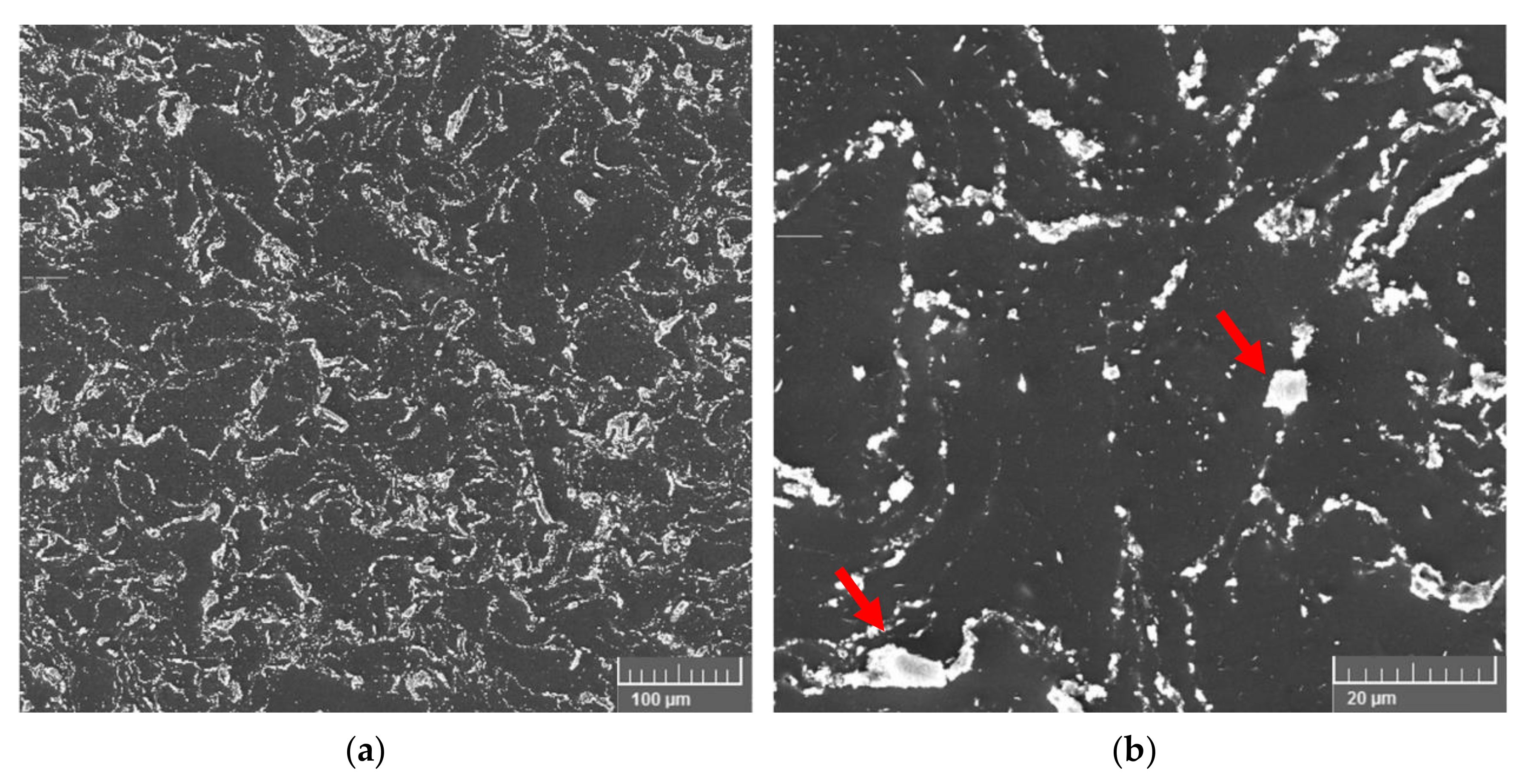
2.1. SEM Investigation

SEM imaging of the 1 Vo1% Mg-Sm2O3 nanocomposite was selected to verify the success of the followed fabrication methods to distribute the nanoparticles within the magnesium matrix phase. To this end, the nanocomposite samples were mechanically polished using standard metallographic techniques, with the final step using 0.05 µm polycrystalline diamond solution until a mirror surface was obtained. Just before the investigation, samples were etched in acetic-nitric solution (10 mL nitric acid, 5 mL acetic acid, 20 mL water, and 60 mL ethanol) for 3 s. The microstructure investigation was conducted using scanning electron microscopy (SEM) under a high vacuum by using an FEI Quanta 650 ESEM system manufactured by Thermo Fisher Scientific Company (Hillsboro, OR, USA). The Everhart-Thornley detector was used with a high acceleration voltage of 10 kV. The working distance used was about 10 mm.

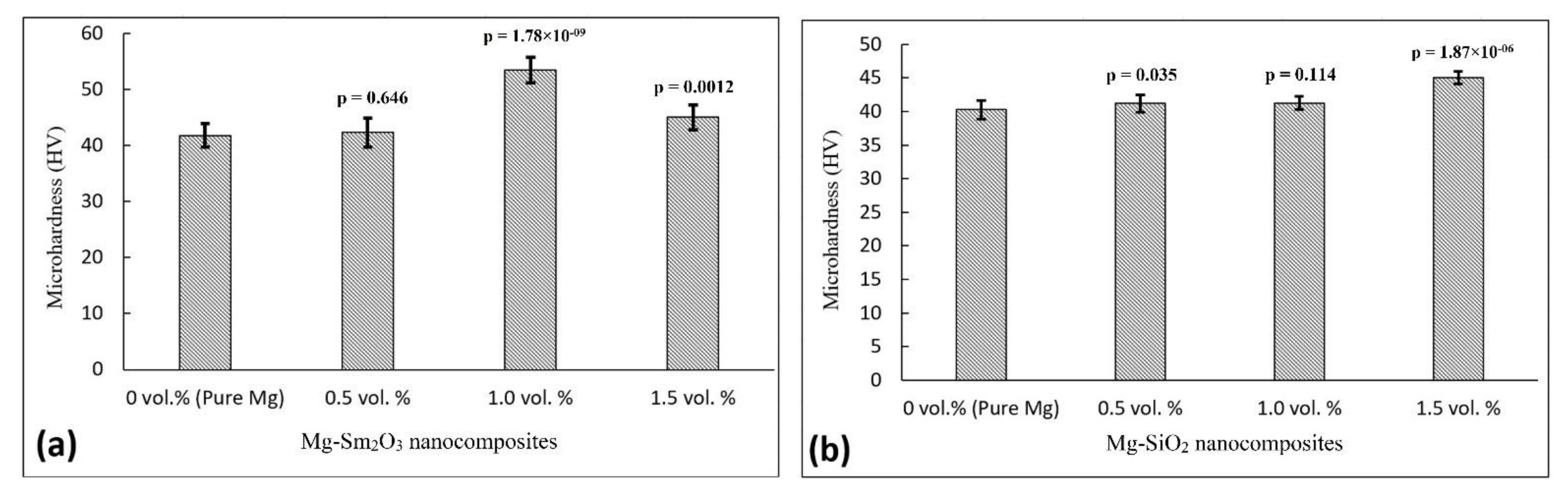
2.2. Microhardness Test

Microhardness test samples were mounted in polymeric material and then polished using 180–2000 grit SiC papers in 90° parallel lines. The Vickers’ hardness of the prepared samples was measured with a Shimadzu microhardness tester HMV-G Series (Kyoto, Japan) using a 1 kg load cell and 15 s dwell time. There were 10 measures taken for each tested sample. The significance difference was performed by calculating the p-values using the t-test with the pure magnesium as the basic control group.

2.3. Electrochemical Corrosion Tests

A pin of 6 mm thickness was electrically connected by a copper wire through a radial notch of 1.5 mm for each composition. High purity magnesium (99.9%) as rolled rods (Goodfellow, Pittsburgh, PA, USA) were used to make the control samples. The elemental analysis of the high-purity magnesium as provided by the supplier was as follows (Fe: 280 PPM, Mn: 70 PPM, Al: 70 PPM, Si: 50 PPM, Cu: 20 PPM, Zn: <20 PPM, and Ni: <10 PPM). All samples were then insulated using nonconductive epoxy except for one of the flat surfaces. Exposed surfaces were then ground using 400–2000 grit SiC sandpaper. Samples were then cleaned in an ethanol bath and dried in warm air. Electrochemical corrosion tests were conducted using Gamry potentiostat, interface 1010e model with Gamry framework software. A conventional three-electrode cell was used with the magnesium samples as the working electrode, a graphite rod as the counter electrode, and a saturated calomel electrode (SCE) as the reference electrode. Although immersion test data would have been beneficial to determine the corrosion rates, the conducted electrochemical tests still can serve as a reliable approach that provides comparative values that reflect the effect of adding the nanoparticles to the magnesium matrix, which is the main purpose of this work.

2.3.1. Simulated Body Fluid (SBF) Preparation

The bioactivity of artificial implants is customarily tested in vitro in a simulated body environment that comprises a solution containing similar inorganic ions’ levels to blood plasma, controlled temperature, and pH level with a certain buffer. Drawbacks of conventional SBF were found to be a lack of stability and different Cl and HCO3− levels [37]. Oyane et al. [38] developed an m-SBF that better matches the blood plasma and was found to be stable for up to 8 weeks without changing the pH level and ion levels at room temperature. The composition of the m-SBF is shown in Table 1. The solution was buffered using HEPES to keep the pH of the solution in the range of 7.4 ± 0.05. HEPES (4-(2-hydroxyethyl)-1-piperazineethanesulfonic acid) is a zwitterionic sulfonic acid buffering agent that is widely used to maintain the physiological pH in cell cultures. The solution was prepared using ultrapure water and reagent grade chemicals (Sigma Aldrich, Milwaukee, WI, USA).

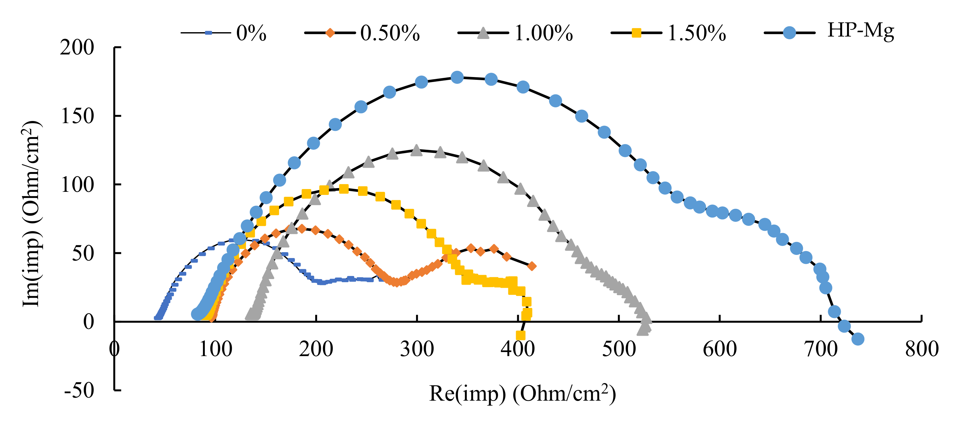
2.3.2. Electrical Impedance Spectroscopy (EIS)

The test was conducted in the modified simulated body fluid electrolyte (m-SBF) prepared according to the procedure in [38] at room temperature. Samples were left to stabilize in the m-SBF to establish open-circuit voltage (OCV) for 10 min to eliminate the effect of the corrosion products. A low AC voltage of 10 mV (rms) was applied at a frequency range of 105–0.1 Hz and 10 points per decade. The developed Nyquist plot of the real vs. imaginary parts of the impedance was used to estimate the total corrosion resistance of the different samples. As shown in Figure 1, the electric circuit found to best fit the measured data where Rs represents the electrolyte resistance, Rp is the polarization resistance through the metal, and Rt is the charge transfer resistance at the metal electrolyte interface. Qc is a constant phase element (CPE) that represents the capacitance due to the formation of the corrosion products layer, and Qdl is another CPE representing the double-layer capacitance.

2.3.3. Potentiodynamic Polarization Test (PDP)

In the PDP test, the current density was monitored while applying a DC voltage varying from −0.25 to 0.25 V relative to the measured OCV at a scan rate of 2 mV/s. Tafel extrapolation and linear fit were used to establish the corrosion current density icorr.

3. Results and Discussion

3.1. SEM Investigation

The SEM imaging of the microstructure of the 1.0 Vol% nanocomposite, Figure 2, shows a uniform distribution of the nanoparticles in the magnesium matrix. This indicates the success of the fabrication method presented in this study to generate a homogenous nanocomposite. However, it is important to emphasize that some localized agglomeration (10–20 µm) can be seen on the grain boundaries. This suggests that at volume content higher than 1.0 Vol%, more of these agglomeration sites will present, which may directly lead to accelerated corrosion rates and more corrosion pits on the surface of the nanocomposites.

3.2. Microhardness

Figure 3 shows the microhardness results of the prepared nanocomposites at different nanoparticle contents. The microhardness of the Mg-Sm2O3 nanocomposite (Figure 3a) increased from 40.7 ± 2.1 HV for pure magnesium (without the addition of any nanoparticles) to reach a mean value of 53.4 ± 2.8 HV for the 1.0 Vol% Sm2O3 nanocomposite. This represents 1.3 times increase in the microhardness after the addition of the Sm2O3 nanoparticles. The increase in the microhardness can be attributed to the presence of the hard Sm2O3 particles along the interfaces of the magnesium matrix particles. This limits the deformation of the matrix grains caused by dislocation movement and twining [39]. In addition, the mismatch between the coefficient of thermal expansion of the matrix and the nanoparticles contributes to the strengthening of the Mg-Sm2O3 nanocomposites [40,41]. Similarly, the addition of the SiO2 nanoparticles increased the microhardness value compared to the pure magnesium material due to similar strengthening mechanisms. For instance, the microhardness of pure magnesium increased from 40.3 ± 1.4 HV to 45.1 ± 0.9 HV for the Mg-1.5Vol%SiO2 nanocomposite. In the case of the Mg-Sm2O3 nanocomposites, further increase in the Sm2O3 nanoparticles resulted in less increase in the microhardness. This can be attributed to the tendency of the Sm2O3 nanoparticles to agglomerate and coarsen (clustering effect), to reduce the internal free energy of the system, due to large van der Waals forces between the nanoparticles [27,36,41]. This phenomenon seems to not occur in the case of the Mg-SiO2 nanocomposites as the microhardness kept increasing with the addition of more SiO2 nanoparticles.

3.3. Electrochemical Corrosion Behavior

Figure 4 shows the Nyquist plot of the Mg-Sm2O3 nanocomposites against the sintered pure magnesium and cold-rolled HP-Mg. The summary of fitted resistances is given in Table 2. In the equivalent circuit for fitting EIS data, Rtot is considered the summation of the metal polarization resistance Rp and the charge transfer resistance Rt. The metal polarization resistance Rp was used for comparison between the different nanocomposites similar to Feliu et al. [42]. As depicted from the fitted data, it was found that the nanocomposites at all the particle volume fractions and the pure magnesium fabricated in the lab had a lower total corrosion resistance than HP-Mg. Among the different volume fractions, the nanocomposite containing 1.0 Vol% showed the highest total corrosion resistance of ~387 Ohm/cm2. Further corrosion rates, Table 2, calculated from the potentiodynamic polarization data, as seen in Figure 5, showed a good agreement with the EIS data. All nanocomposites showed a negative reduction in the corrosion potential (Ecorr) and an increase in the corrosion current density (icorr) as compared to HP-Mg. The 1.0 Vol% again showed the least icorr of 490 µA/cm2, which is expected to result in the lowest corrosion rate of all the Mg-Sm2O3 nanocomposites. The enhancement in the corrosion behavior of the 1.0 Vol% nanocomposite can be attributed to the observed grain refinement of the magnesium matrix accompanying the increase in the nanoparticles Vol% due to the grain boundary pinning mechanism, where the added nanoparticles pinned the recrystallized grains of Mg, hence restraining its growth, without any nucleation added [27]. However, past the 1.0 Vol%, the chances of the agglomeration sites of the impurities and the particles have increased, increasing the impact of the internal galvanic corrosion (causing galvanic coupling) due to localized agglomerations and leading to a deteriorated corrosion resistance [43]. It is worth mentioning that the noticed high Tafel slopes are intrinsic to the magnesium corrosion process and can be attributed to the formation of an unstable layer of magnesium hydroxide that quickly dissolves in chloride-containing solutions such as the simulated body fluid solution.
Similar to the case of samarium oxides (Sm2O3) nanocomposites, the addition of silicon dioxide (SiO2) yielded a general reduction in corrosion resistance as compared to HP-Mg. Nyquist plots from the EIS test shown in Figure 6 summarize the comparison between the different compositions in terms of corrosion resistance. Corrosion resistance as described earlier as the summation of the Rp and Rt is the diameter of the capacitive loops shown in Figure 6. Hence, the bigger the radii of the capacitive loops, the higher is the corrosion resistance. Again, the nanocomposite of a 1.0 Vol% was found to have the highest corrosion resistance of ~416 Ohm/cm2 out of all the as-sintered and extruded pure magnesium and its nanocomposites. Corrosion current densities (icorr) from the PDP test also agree with the trend observed in corrosion resistance from the EIS test. The measured Tafel curves of the Mg-SiO2 nanocomposites vs. the HP-Mg curve are shown in Figure 7. It can be seen that the 1.0 Vol% Mg-SiO2 nanocomposite possesses the least corrosion current density (icorr) of 153 µA/cm2, as well as the least negative corrosion potential (Ecorr) of −1.702 V. Out of the two nanocomposite groups, Mg-1.0% SiO2 is the closest in corrosion behavior to the HP-Mg. Table 3 includes a summary of the measured electrochemical corrosion characteristics of the Mg-SiO2 nanocomposites. Similarly, the enhancement in the corrosion behavior is attributed to the grain boundary pinning mechanism and the grain refinement of the magnesium matrix with the addition of the SiO2 nanoparticles [27]. Unlike the microhardness, which increased after adding more than 1.0 Vol% SiO2 nanoparticles, the corrosion properties started to slightly degrade past the 1.0 Vol% SiO2 nanoparticle content. Again, the chances of the agglomeration sites of the impurities and the particles have increased, hence increasing the possibility of galvanic coupling due to localized agglomerations and leading to a deteriorated corrosion resistance.
It is worth mentioning that there are differences in the electrochemical corrosion values of magnesium that are usually noticed in the literature. The discrepancies, and sometimes the contradiction, between the results stems from the variety of the test conditions that different groups follow [44]. In the case of this study, the consistency between the PDP and the EIS test results suggests the validity of the noticed trend for each of the tested Mg-Sm2O3 and Mg-SiO2 nanocomposites. For instance, the in vitro electrochemical corrosion data in this study reveals that the strengthening benefits of adding the nanoparticles to the magnesium matrix have come with the cost of slightly reducing the corrosion resistance as compared to HP-Mg. However, the strengthening of the material outweighs the reduction in the corrosion resistance, especially in the case of 1.0 Vol% nanocomposites as the reduction was minimum, keeping in mind that the corrosion resistance of the rolled HP-Mg, tested in this study, is known to be superior to other Mg alloys and nanocomposites. For example, this study and our previous work show that the 1.0 Vol% Mg-Sm2O3 nanocomposite has about a 31% increase in microhardness over pure sintered and hot extruded magnesium [27] and ~100% over the as-rolled HP-Mg used in this study [15]. In addition, the Mg-SiO2 nanocomposites were shown to have about a 12% increase in the microhardness and a 10% increase in the yield strength [15,45]. It is important to note that the obtained corrosion current density for the tested samples in this study (based on the Tafel extrapolation method) can be used as an indicator for the expected corrosion rates and a benchmark of corrosion behavior. However, caution should be given as in vitro electrochemical corrosion tests do not take into account the effect of the corrosion products and pH evolution at the corroded surface. In addition, it is noteworthy that the buffer HEPES used in this study was found to affect the corrosion data measured in electrochemical tests as compared to CO2 [46]. However, since all sample groups and the control were subject to the same conditions, the trend in the results shall stay valid.
In a more general tribocorrosion test, this increase in the mechanical strength and hardness is hypothesized to make the developed nanocomposites outperform the high-purity magnesium, although it is not included in this study. To compare the corrosion properties of the two nanocomposites, Figure 8 represents a summary of the measured microhardness and corrosion rates of selected cases for both nanocomposites vs. the HP-Mg. It is shown that the Mg-1.0%Sm2O3 and Mg-1.5%SiO2 nanocomposites are promising cases included in this study showing a good combination of improved strength and corrosion behavior.
Another key observation is that the corrosion resistance and corrosion current density of the pure magnesium samples, manufactured using the 98.5% purity magnesium powder and by using the proposed microwave sintering and hot extrusion process, have fallen below the corresponding values of HP-Mg samples produced by cold rolling. A lower purity magnesium powder can be a major factor resulting in less corrosion resistance for all the produced nanocomposites. Another factor for the reduction in the corrosion resistance could be the possibility of defects (e.g., micro voids) that might have occurred in any of the manufacturing process steps. Hence, we anticipate that the prepared magnesium-based nanocomposites show evident enhancement in the mechanical and corrosion properties. This suggests that a magnesium nanocomposite fabricated using the proposed blending, sintering, and extrusion processes in this work with better fabrication quality would have a significantly higher mechanical strength and a slower corrosion rate than these reported values.

4. Conclusions

In this study, the electrochemical corrosion behavior of two magnesium-based nanocomposites, reinforced with nanoparticles of samarium oxide (Sm2O3) and silicon dioxide (SiO2), was assessed at three different volume fractions (0.5, 1.0, and 1.5 Vol%), and the conclusions were:
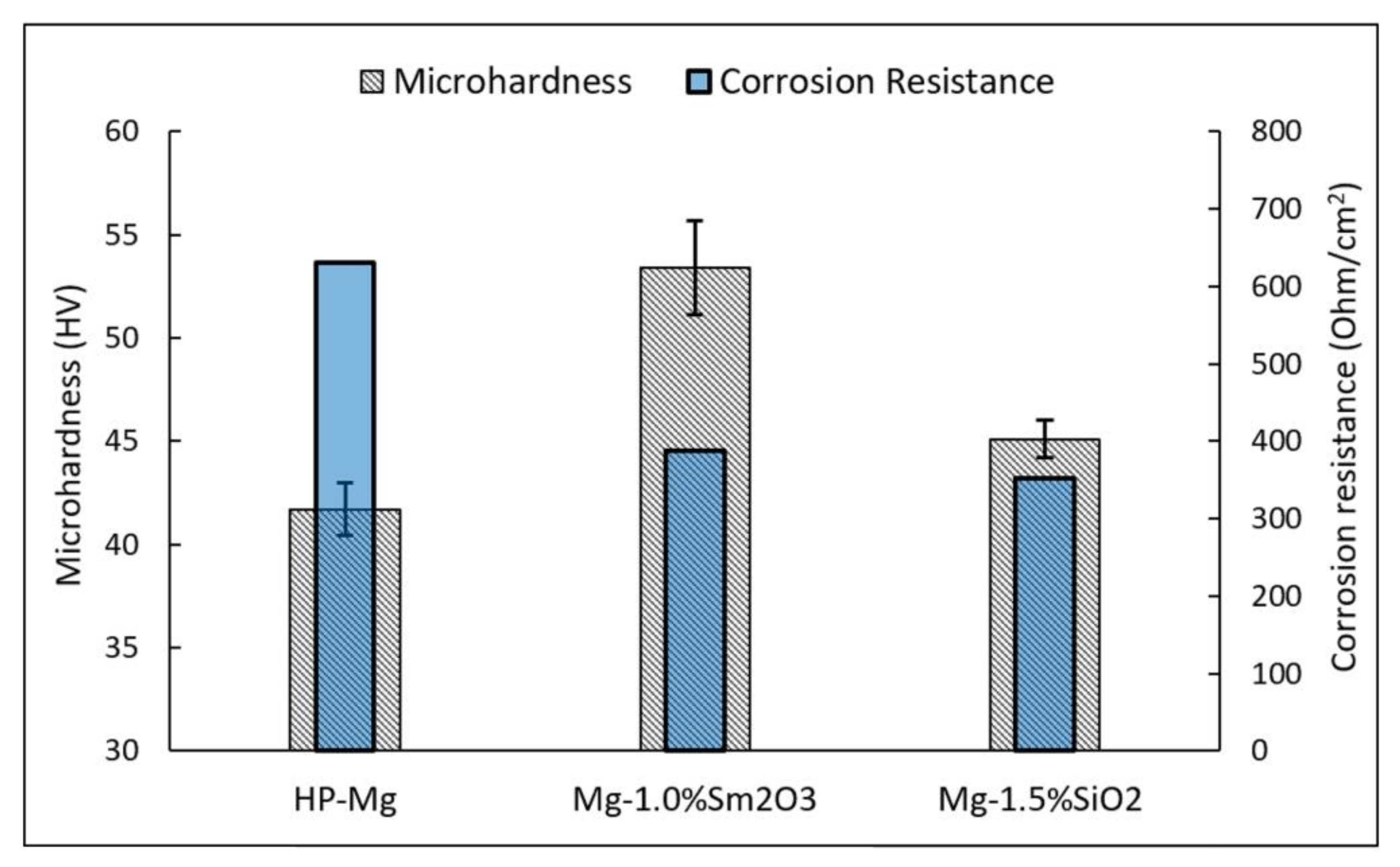
  • The microhardness increased 31% for the 1.0 Vol% Sm2O3 nanocomposite and 12% for the 1.5 vol.% SiO2 nanocomposite compared to pure Mg.
  • Both nanocomposites showed the highest corrosion resistance and lowest corrosion current density at the 1.0 Vol% content. Increasing the nanoparticles content above 1.0% Vol resulted in a reduction in corrosion resistance due to localized agglomerations of the nanoparticles at higher contents.
  • The corrosion resistance of the pure magnesium samples was less than that for the HP-Mg. These pure magnesium samples were manufactured using the same fabrication methods used to make the nanocomposites.
  • Therefore, a tribocorrosion behavior of the developed nanocomposites is hypothesized to be better than HP-Mg due to the improved wear resistance. Future work on tribocorrosion behavior is required to assess this hypothesis.
Biodegradable materials with sufficient mechanical strength and controlled degradation rates are needed to improve osteosynthesis treatments. As the investigated nanocomposites offer superior mechanical strength over HP-Mg, more future work, focusing on the development of magnesium-based nanocomposites with higher purity as well as protective coatings, is expected to result in strong biodegradable materials with controlled corrosion rates needed for bone implant applications. For instance, a further increase of the added SiO2 nanoparticles may result in a greater strengthening effect. In addition, the corrosion rates of such stronger materials can be controlled using biocompatible protective coatings [7,8]. Moreover, the development of more efficient fabrication methods that can be utilized to include a higher volume fraction of the nanoparticles may lead to a less localized agglomeration effect and hence even stronger and more corrosion-resistant materials.

Author Contributions

Writing—original draft preparation, M.A., A.S. and H.I.; writing—review and editing, M.A., S.M. and H.I.; visualization, H.I., M.H. and M.G.; supervision, H.I. and M.G.; funding acquisition, H.I. All authors have read and agreed to the published version of the manuscript.

Funding

This research received no external funding.

Acknowledgments

The authors acknowledge the support of the University of Tennessee at Chattanooga. Research reported in this publication was supported by the FY2021 and FY2022 Center of Excellence for Applied Computational Science competition.

Conflicts of Interest

The authors declare no conflict of interest.

References

  1. Bicsák, Á.; Abel, D.; Tack, L.; Smponias, V.; Hassfeld, S.; Bonitz, L. Complications after osteosynthesis of craniofacial fractures—An analysis from the years 2015–2017. Oral Maxillofac. Surg. 2021, 25, 199–206. [Google Scholar] [CrossRef] [PubMed]
  2. Planell, J.; Navarro, M. Challenges of bone repair. In Bone Repair Biomaterials; Woodhead Publishing: Sawston, UK, 2009; pp. 3–24. [Google Scholar]
  3. Johansson, B.; Grepe, A.; Wannfors, K.; Hirsch, J.M. A clinical study of changes in the volume of bone grafts in the atrophic maxilla. Dento Maxillo Facial Radiol. 2001, 30, 157–161. [Google Scholar] [CrossRef]
  4. Ibrahim, H.; Esfahani, S.N.; Poorganji, B.; Dean, D.; Elahinia, M. Resorbable bone fixation alloys, forming, and post-fabrication treatments. Mater. Sci. Eng. C 2017, 70, 870–888. [Google Scholar] [CrossRef] [PubMed] [Green Version]
  5. Huang, Z.-M.; Fujihara, K. Stiffness and strength design of composite bone plates. Compos. Sci. Technol. 2005, 65, 73–85. [Google Scholar] [CrossRef]
  6. Rahmanian, R.; Shayesteh Moghaddam, N.; Haberland, C.; Dean, D.; Miller, M.; Elahinia, M. Load bearing and stiffness tailored NiTi implants produced by additive manufacturing: A simulation study. In Proceedings of the International Society for Optical Engineering, San Diego, CA, USA, 5–7 July 1995. [Google Scholar]
  7. Abdalla, M.; Joplin, A.; Elahinia, M.; Ibrahim, H. Corrosion Modeling of Magnesium and Its Alloys for Biomedical Applications. Corros. Mater. Degrad. 2020, 1, 219–248. [Google Scholar] [CrossRef]
  8. Abdalla, M.; Ibrahim, H. A Physical Approach to Simulate the Corrosion of Ceramic-Coated Magnesium Implants. Appl. Sci. 2021, 11, 6724. [Google Scholar] [CrossRef]
  9. Lesz, S.; Hrapkowicz, B.; Karolus, M.; Gołombek, K. Characteristics of the Mg-Zn-Ca-Gd alloy after mechanical alloying. Materials 2021, 14, 226. [Google Scholar] [CrossRef]
  10. Cesarz-Andraczke, K.; Kazek-Kęsik, A. PEO layers on Mg-based metallic glass to control hydrogen evolution rate. Bull. Pol. Acad. Sci. Tech. Sci. 2020, 68, 119–124. [Google Scholar]
  11. Witte, F. The history of biodegradable magnesium implants: A review. Acta Biomater. 2010, 6, 1680–1692. [Google Scholar] [CrossRef]
  12. Persaud-Sharma, D.; McGoron, A. Biodegradable magnesium alloys: A review of material development and applications. J. Biomim. Biomater. Tissue Eng. 2011, 12, 25–39. [Google Scholar] [CrossRef] [Green Version]
  13. Datta, M.K.; Chou, D.-T.; Hong, D.; Saha, P.; Chung, S.J.; Lee, B.; Sirinterlikci, A.; Ramanathan, M.; Roy, A.; Kumta, P.N. Structure and thermal stability of biodegradable Mg-Zn-Ca based amorphous alloys synthesized by mechanical alloying. Mater. Sci. Eng. B 2011, 176, 1637–1643. [Google Scholar] [CrossRef]
  14. Staiger, M.P.; Pietak, A.M.; Huadmai, J.; Dias, G. Magnesium and its alloys as orthopedic biomaterials: A review. Biomaterials 2006, 27, 1728–1734. [Google Scholar] [CrossRef]
  15. Goodfellow. Magnesium-Material Properties. Available online: http://www.goodfellow.com/E/Magnesium.html (accessed on 7 March 2021).
  16. Lee, J.-W.; Han, H.-S.; Han, K.-J.; Park, J.; Jeon, H.; Ok, M.-R.; Seok, H.-K.; Ahn, J.-P.; Lee, K.E.; Lee, D.-H. Long-term clinical study and multiscale analysis of in vivo biodegradation mechanism of Mg alloy. Proc. Natl. Acad. Sci. USA 2016, 113, 716–721. [Google Scholar] [CrossRef] [Green Version]
  17. Nakatsugawa, I.; Kamado, S.; Kojima, Y.; Ninomiya, R.; Kubota, K. Corrosion of magnesium alloys containing rare earth elements. Corros. Rev. 1998, 16, 139–158. [Google Scholar] [CrossRef]
  18. Ibrahim, H.; Klarner, A.D.; Poorganji, B.; Dean, D.; Luo, A.A.; Elahinia, M. Microstructural, mechanical and corrosion characteristics of heat-treated Mg-1.2 Zn-0.5 Ca (wt%) alloy for use as resorbable bone fixation material. J. Mech. Behav. Biomed. Mater. 2017, 69, 203–212. [Google Scholar] [CrossRef]
  19. Chmielewska, A.; MacDonald, T.; Ibrahim, H.; McManus, T.; Lindemann, J.L.; Smith, P.; Rong, L.; Luo, A.; Advincula, R.; Swieszkowski, W. Biocompatibility of a novel heat-treated and ceramic-coated magnesium alloy (Mg-1.2 Zn-0.5 Ca-0.5 Mn) for resorbable skeletal fixation devices. MRS Commun. 2020, 10, 467–474. [Google Scholar] [CrossRef]
  20. Ibrahim, H.; Luo, A.; Dean, D.; Elahinia, M. Effect of Zn content and aging temperature on the in-vitro properties of heat-treated and Ca/P ceramic-coated Mg-0.5% Ca-x% Zn alloys. Mater. Sci. Eng. C 2019, 103, 109700. [Google Scholar] [CrossRef]
  21. Huang, Y.; Gan, W.; Kainer, K.U.; Hort, N. Role of multi-microalloying by rare earth elements in ductilization of magnesium alloys. J. Magnes. Alloy. 2014, 2, 1–7. [Google Scholar] [CrossRef] [Green Version]
  22. Evans, C. Toxicology and pharmacology of the lanthanides. In Biochemistry of the Lanthanides; Springer: Boston, MA, USA, 1990; pp. 339–389. [Google Scholar]
  23. Evans, C. Past, Present, and Possible Future Clinical Applications of the Lanthanides. In Biochemistry of the Lanthanides; Springer: Boston, MA, USA, 1990; pp. 391–425. [Google Scholar]
  24. Jing, H.; Wu, X.; Liu, Y.; Lu, M.; Yang, K.; Yao, Z.; Ke, W. Antibacterial property of Ce-bearing stainless steels. J. Mater. Sci. 2007, 42, 5118–5122. [Google Scholar] [CrossRef]
  25. Yuan, J.; Li, W.; Wang, C. Effect of the La alloying addition on the antibacterial capability of 316L stainless steel. Mater. Sci. Eng. C 2013, 33, 446–452. [Google Scholar] [CrossRef]
  26. Weng, W.; Biesiekierski, A.; Li, Y.; Dargusch, M.; Wen, C. A review of the physiological impact of rare earth elements and their uses in biomedical Mg alloys. Acta Biomater. 2021, 130, 80–97. [Google Scholar] [CrossRef]
  27. Kujur, M.S.; Mallick, A.; Manakari, V.; Parande, G.; Tun, K.S.; Gupta, M. Significantly enhancing the ignition/compression/damping response of monolithic magnesium by addition of Sm2O3 nanoparticles. Metals 2017, 7, 357. [Google Scholar] [CrossRef] [Green Version]
  28. Swain, S.; Gotman, I.; Unger, R.; Gutmanas, E. Bioresorbable β-TCP-FeAg nanocomposites for load bearing bone implants: High pressure processing, properties and cell compatibility. Mater. Sci. Eng. C 2017, 78, 88–95. [Google Scholar] [CrossRef]
  29. Prasadh, S.; Manakari, V.; Parande, G.; Wong, R.C.W.; Gupta, M. Hollow silica reinforced magnesium nanocomposites with enhanced mechanical and biological properties with computational modeling analysis for mandibular reconstruction. Int. J. Oral Sci. 2020, 12, 1–11. [Google Scholar] [CrossRef]
  30. Ali, M.; Hussein, M.; Al-Aqeeli, N. Magnesium-based composites and alloys for medical applications: A review of mechanical and corrosion properties. J. Alloys Compd. 2019, 792, 1162–1190. [Google Scholar] [CrossRef]
  31. Kujur, M.S.; Manakari, V.; Parande, G.; Prasadh, S.; Wong, R.; Mallick, A.; Gupta, M. Effect of samarium oxide nanoparticles on degradation and invitro biocompatibility of magnesium. Mater. Today Commun. 2021, 26, 102171. [Google Scholar] [CrossRef]
  32. Herath, H.; Di Silvio, L.; Evans, J. In vitro evaluation of samarium (III) oxide as a bone substituting material. J. Biomed. Mater. Res. Part A 2010, 94, 130–136. [Google Scholar] [CrossRef]
  33. Krishnan, V.; Lakshmi, T. Bioglass: A novel biocompatible innovation. J. Adv. Pharm. Technol. Res. 2013, 4, 78. [Google Scholar] [CrossRef]
  34. Rim, K.T.; Koo, K.H.; Park, J.S. Toxicological evaluations of rare earths and their health impacts to workers: A literature review. Saf. Health Work. 2013, 4, 12–26. [Google Scholar] [CrossRef] [Green Version]
  35. Guo, C.; Liu, Y.; Li, Y. Adverse effects of amorphous silica nanoparticles: Focus on human cardiovascular health. J. Hazard. Mater. 2021, 406, 124626. [Google Scholar] [CrossRef]
  36. Haghshenas, M.; Gupta, M. Magnesium nanocomposites reinforced with rare earth element nanoparticles: Nanoindentation-driven response. Nanocomposites 2020, 6, 22–30. [Google Scholar] [CrossRef] [Green Version]
  37. Kim, H.M.; Kishimoto, K.; Miyaji, F.; Kokubo, T.; Yao, T.; Suetsugu, Y.; Tanaka, J.; Nakamura, T. Composition and structure of the apatite formed on PET substrates in SBF modified with various ionic activity products. J. Biomed. Mater. Res. 1999, 46, 228–235. [Google Scholar] [CrossRef]
  38. Oyane, A.; Kim, H.M.; Furuya, T.; Kokubo, T.; Miyazaki, T.; Nakamura, T. Preparation and assessment of revised simulated body fluids. J. Biomed. Mater. Res. Part A 2003, 65, 188–195. [Google Scholar] [CrossRef] [PubMed]
  39. Razavi, M.; Fathi, M.; Meratian, M. Microstructure, mechanical properties and bio-corrosion evaluation of biodegradable AZ91-FA nanocomposites for biomedical applications. Mater. Sci. Eng. A 2010, 527, 6938–6944. [Google Scholar] [CrossRef]
  40. Lin, G.; Liu, D.; Chen, M.; You, C.; Li, Z.; Wang, Y.; Li, W. Preparation and characterization of biodegradable Mg-Zn-Ca/MgO nanocomposites for biomedical applications. Mater. Charact. 2018, 144, 120–130. [Google Scholar] [CrossRef]
  41. Haghshenas, M.; Muhammad, M.; Hasannaeimi, V.; Mukherjee, S.; Gupta, M. Ambient and non-ambient temperature depth-sensing indentation of Mg-Sm2O3 nanocomposites. Int. J. Adv. Manuf. Technol. 2019, 105, 2947–2956. [Google Scholar] [CrossRef]
  42. Feliu, S. Electrochemical impedance spectroscopy for the measurement of the corrosion rate of magnesium alloys: Brief review and challenges. Metals 2020, 10, 775. [Google Scholar] [CrossRef]
  43. Song, G.L.; Atrens, A. Corrosion mechanisms of magnesium alloys. Adv. Eng. Mater. 1999, 1, 11–33. [Google Scholar] [CrossRef]
  44. Kirkland, N.; Birbilis, N.; Staiger, M. Assessing the corrosion of biodegradable magnesium implants: A critical review of current methodologies and their limitations. Acta Biomater. 2012, 8, 925–936. [Google Scholar] [CrossRef]
  45. Manakari, V.; Kannan, S.; Parande, G.; Doddamani, M.; Columbus, S.; Vincent, S.; Gupta, M. In-vitro degradation of hollow silica reinforced magnesium syntactic foams in different simulated body fluids for biomedical applications. Metals 2020, 10, 1583. [Google Scholar] [CrossRef]
  46. Kannan, M.B.; Khakbaz, H.; Yamamoto, A. Understanding the influence of HEPES buffer concentration on the biodegradation of pure magnesium: An electrochemical study. Mater. Chem. Phys. 2017, 197, 47–56. [Google Scholar] [CrossRef]
Figure 1. Equivalent circuit for fitting EIS data.
Jcs 06 00154 g001
Figure 2. (a) SEM micrographs of the microstructure of 1 Vo1% Mg-Sm2O3 nanocomposite; (b) An enlargement fragment of the area in (a), arrows indicating localized agglomeration of the nanoparticles.
Jcs 06 00154 g002
Figure 3. Microhardness measurements of the prepared nanocomposites. (a) Mg-Sm2O3 nanocomposite with maximum microhardness achieved at 1 Vol% (p = 1.78 × 10−09, n = 13, compared to pure Mg), and (b) Mg-SiO2 nanocomposite with maximum microhardness achieved at 1.5 Vol% (p = 1.87 × 10−06, n = 10, compared to pure Mg).
Jcs 06 00154 g003
Figure 4. Nyquist plots of the measured impedances for the Mg-Sm2O3 nanocomposites group.
Jcs 06 00154 g004
Figure 5. Tafel curves of Mg-Sm2O3 nanocomposites vs. pure magnesium and HP-Mg.
Jcs 06 00154 g005
Figure 6. Nyquist plots of the measured impedances for the Mg-SiO2 nanocomposites group.
Jcs 06 00154 g006
Figure 7. Tafel curves of Mg-SiO2 nanocomposites vs. pure magnesium and HP-Mg.
Jcs 06 00154 g007
Figure 8. Comparison of the measured microhardness and corrosion rates of the Mg-1.0%Sm2O3 and Mg-1.5%SiO2 Nanocomposites compared to the HP-Mg samples.
Jcs 06 00154 g008
Table 1. Chemical composition of 1 L of m-SBF.
ChemicalConcentration (g/L)
NaCl5.403
NaHCO30.504
Na2CO30.426
KCl0.225
K2HPO4·3H2O0.23
MgCl2·6H2O0.311
HEPES17.892
CaCl20.293
Na2SO40.072
1 M NaOH (mL)15
Table 2. Summary of electrochemical corrosion characteristics of Mg-Sm2O3 nanocomposites.
EIS Test ResultsPDP Test Results
MaterialRp (Ohm/cm2)Rt (Ohm/cm2)Rtot (Ohm/cm2)Ecorr (V)icorr (µA/cm2)
HP-Mg (as rolled)54487631−1.6248
Pure Mg17483256−1.87770
Mg-0.5% Sm2O3184151334−1.82623
Mg-1% Sm2O333948387−1.96490
Mg-1.5% Sm2O313523158−1.94812
Table 3. Summary of electrochemical corrosion characteristics of Mg-SiO2 nanocomposites.
EIS Test ResultsPDP Test Results
MaterialRp (Ohm/cm2)Rt (Ohm/cm2)Rtot (Ohm/cm2)Ecorr (V)icorr (µA/cm2)
HP-Mg (as rolled)54487631−1.6248
Pure Mg17483256−1.87770
Mg-0.5% SiO223782318−1.748320
Mg-1% SiO2312104416−1.702153
Mg-1.5% SiO226389352−1.747250
Publisher’s Note: MDPI stays neutral with regard to jurisdictional claims in published maps and institutional affiliations.

