1. Introduction
The amnion rupture sequence is an uncommon condition that occurs very early in pregnancy, and it leads to variable and complex fetal anomalies. Amnion parts constrict embryonic mass and interrupt the normal development of the anatomical structures [
1]. The prevalence of this condition is 1.16 in 10,000 live births [
2]. The anatomical parts affected depend on the time of amnion rupture [
3]. Most of the cases described involve cranio-facial defects and abdominal defects [
4]. There is no increased risk of recurrence in subsequent pregnancies [
5].
2. Detailed Case Description
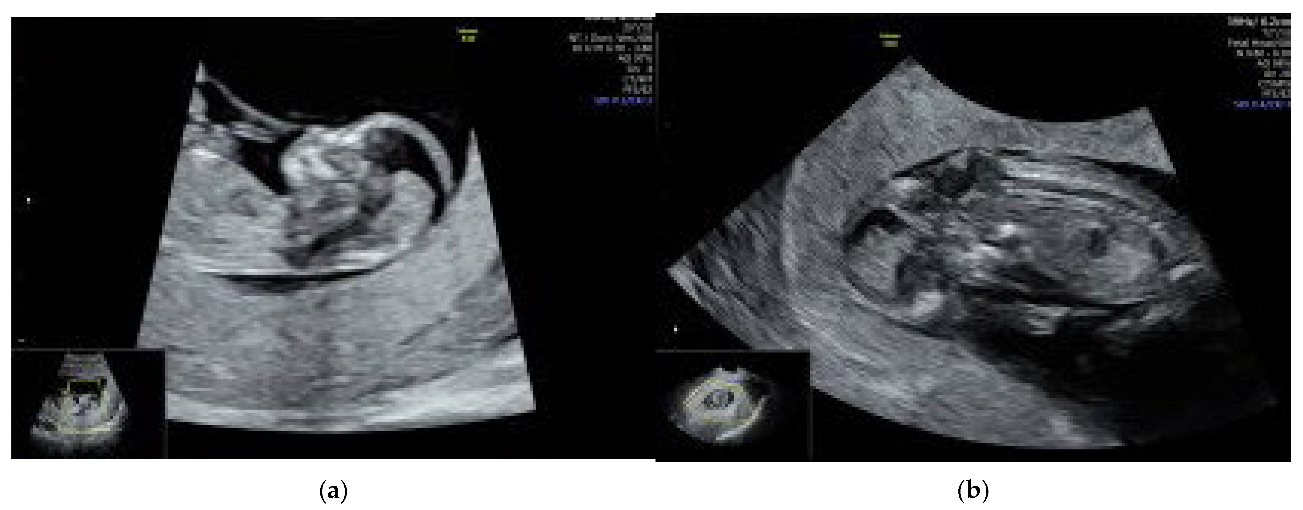
We present a case of a 35 year old woman who presented for her first-trimester ultrasound examination at 12 weeks and 1 day of gestation. This was her first pregnancy following spontaneous conception, and she had no history of miscarriages or any other medical issues. She was taking folic acid. Her first trimester was complicated by severe vaginal bleeding. The amniotic membrane wrapped the fetus, and the retro-amniotic space was largely increased. These findings were suggestive of an open neural tube defect due to an early amnion rupture. The most important sign suggesting the cause of the anomaly was the presence of the amniotic membrane attached to the fetus and the amniotic cavity being reduced in size.
We performed a transvaginal ultrasound to better visualize the spine and the brain. On the TV scan, we could demonstrate the occipital bone defect and the herniation of the fetal brain tissue throughout it (see
Figure 1b). There were no other fetal anomalies detected on a thorough ultrasound examination.
A first trimester screening for chromosomal abnormalities and preeclampsia was made using a combination of maternal age, maternal characteristics and history, ultrasound markers such as nuchal translucency, tricuspid regurgitation, ductus venosus and nasal bone, and biochemistry (Beta-HCG and PAPP-A). The results showed a low chance for the most common trisomies (trisomy 21, 13 and 18) and a high chance for developing preeclampsia and fetal growth restriction. An invasive genetic (Whole Exome Sequencing) testing was performed, and it was negative. She was counselled for the outcome of this defect regardless of the genetic testing results, and she opted for the termination of pregnancy. She was admitted to the hospital. She was started on Mifepristone 200 mg per os and Misoprostol 0.2 mgx4 vaginally 36 h after the administration of Mifepristone. She aborted the fetus and the placenta in a few hours after the administration of Misoprostol. The anatomo-pathological results confirmed an occipital encephalocele measuring 1 cm and also described abnormally implanted ears, an abnormal shape of the chin and dysmorphic face features, with no other thoracic or abdominal anomalies. The histology confirmed our suspicion of amnion rupture, with thin amniotic strands being visible around the fetal cranium.
3. Discussion
The malformative syndrome known as congenital band sequence, amniotic rupture sequence, congenital ring constriction, amniotic band disruption complex, or congenital transverse defect is identified by various synonyms. These terms reflect the clinical diversity of the condition or the uncertainties regarding its cause [
6]. The term amniotic constriction band encompasses a spectrum of congenital anomalies known for the significant variability in how they manifest clinically, from minor ring constrictions to severe defects that are not compatible with life [
7].
Craniofacial defects are thought to arise from a vascular disruption sequence, which may or may not involve cephaloamniotic adhesion. However, certain chronological and topographical evidence does not align with the patterns of embryonic vascular development, casting doubt on the vascular hypothesis as the sole explanation. Nonetheless, a disruption in blood flow to the affected area, along with mechanical factors, could play a key role in the pathogenesis, particularly in cases of morphological disruption [
6].
Amniotic rupture sequence is a highly uncommon condition. A widely accepted explanation for the disease’s development suggests that it starts with a rupture of the amniotic sac in the first trimester. This event leads to amniotic fluid leakage, which compresses the fetus and leads to the formation of fibrous bands. These bands can then directly harm fetal parts, causing amputation, deformation, or constriction. Another hypothesis suggests that the condition may arise from a disruption in the vascular supply to the embryo, serving as a potential cause [
8].
The encephalocele is a cranial bone defect, with its most frequent location being in the occipital region. It can be associated with chromosomal or genetic abnormalities such as trisomy 13 and 18, Meckel–Gruber syndrome, Walker–Warburg syndrome and amniotic band syndrome [
9]. The prognosis depends on the extension of the lesion. However, the mortality rate is about 50% for posterior encephalocele, and more than 50% of survivors have a neurological handicap [
10].
The literature describes new-borns with encephalocele due to amniotic band syndromes [
11,
12,
13,
14]. There were also reported cases of encephaloceles diagnosed in the first trimester due to amniotic bands [
15]. A study performed by Ushakov on 28 cases described the ultrasound features of amniotic band syndrome in the first trimester. He concluded that amniotic bands disappear with the progression of the pregnancy and that in many cases the bands could not be evaluated [
15].
The significance of this case lies in its rarity and the insight it provides into the etiology of encephaloceles. Traditionally, the literature has focused on genetic mutations, nutritional deficiencies (such as folic acid), and environmental factors as primary causes of neural tube defects. However, this case underscores the mechanical disruptions that amniotic bands can cause, leading to significant cranial and neural anomalies. This case contributes to the medical literature by emphasizing the need for awareness and understanding of less common causes of neural tube defects such as encephalocele. It suggests that in cases of cranial anomalies detected prenatally, clinicians should consider the possibility of amniotic band syndrome as a contributing factor.
In conclusion, encephaloceles can be diagnosed in the first trimester of pregnancy during the 11–14 week scan [
13,
15]. The etiology should be investigated whenever possible in order to counsel the patient for current and future pregnancies.
Author Contributions
Conceptualization, N.G. (Nicoleta Gana) and N.G. (Nicolae Gică); methodology, A.M.P., A.M.V. and R.B.; resources, L.M.A. and N.G. (Nicolae Gică); writing—original draft preparation, L.M.A. and I.H. writing—review and editing, N.G. (Nicoleta Gana), A.M.V., F.M.N. and R.B. All authors have read and agreed to the published version of the manuscript.
Funding
This research received no external funding.
Institutional Review Board Statement
The study was conducted in accordance with the Declaration of Helsinki. The ethical review and approval of this study were waived by the Ethics Committee of Carol Davila University of Medicine and Pharmacy, because it is a single case report.
Informed Consent Statement
Written informed consent has been obtained from the patient to publish this paper.
Data Availability Statement
Data are contained within the article.
Conflicts of Interest
The authors declare no conflicts of interest.
References
- Ferris, N.J.; Tien, R.D. Amnion rupture sequence with ‘exencephaly’: MR findings in a surviving infant. AJNR Am. J. Neuroradiol. 1994, 15, 1030–1033. [Google Scholar] [PubMed]
- Garza, A.; Crodero, J.F.; Mulinare, J. Epidemiology of the early amnion rupture spectrum of defects. Am. J. Dis. Child. 1988, 142, 541–544. [Google Scholar] [CrossRef] [PubMed]
- Higginbottom, M.D.; Jones, K.L.; Hall, B.D.; Smith, D.W. The amniotic disruption complex: Timing of amniotic rupture and variable spectra of consequent defects. J. Pediatr. 1979, 95, 544–549. [Google Scholar] [CrossRef] [PubMed]
- Ossipoff, V.; Hall, B.D. Etiologic factors in the amniotic band syndrome: A study of 24 patients. Birth Defects 1977, 13, 117–132. [Google Scholar] [PubMed]
- Kalousek, D.K.; Bamforth, S. Amnion rupture sequence in previable fetuses. Am. J. Med. Genet. 1988, 31, 63–73. [Google Scholar] [CrossRef] [PubMed]
- Atiyeh, B.S.; Moucharafieh, R. An unusual amniotic rupture sequence with thoracoabdominal restricting band, low-set posterior hairline, and trapezius contracture. J. Craniofac. Surg. 2010, 21, 1400–1403. [Google Scholar] [CrossRef] [PubMed]
- Basha, M.J.; Nagnur, M.I.; Mohiuddin, M.S.; Mohiuddin, M.J.; Salman, S.; CSunder, S. Streeter’s Syndrome of Lower Limb Associated with CTEV. J. Orthop. Case Rep. 2022, 12, 50–53. [Google Scholar] [CrossRef] [PubMed]
- Jamsheer, A.; Materna-Kiryluk, A.; Badura-Stronka, M.; Wiśniewska, K.; Wieckowska, B.; Mejnartowicz, J.; Balcar-Boroń, A.; Borszewska-Kornacka, M.; Czerwionka-Szaflarska, M.; Gajewska, E.; et al. Comparative study of clinical characteristics of amniotic rupture sequence with and without body wall defect: Further evidence for separation. Birth Defects Res. A Clin. Mol. Teratol. 2009, 85, 211–215. [Google Scholar] [CrossRef] [PubMed]
- Markovic, I.; Bosnjakovic, P.; Milenkovic, Z. Occipital Encephalocele: Cause, Incidence, Neuroimaging and Surgical Management. Curr. Pediatr. Rev. 2020, 16, 200–205. [Google Scholar] [CrossRef] [PubMed]
- Kiymaz, N.; Yilmaz, N.; Demir, I.; Keskin, S. Prognostic factors in patients with occipital encephalocele. Pediatr. Neurosurg. 2010, 46, 6–11. [Google Scholar] [CrossRef] [PubMed]
- Yengo-Kahn, A.M.; Plackis, A.C.; Bonfield, C.M.; Reddy, S.K. Correction of a vertex encephalocele related to amniotic band syndrome. BMJ Case Rep. 2020, 13, e234735. [Google Scholar] [CrossRef] [PubMed]
- da Silva, A.J.F.; Silva, C.S.M.E.; Mariano, S.C.R. Amniotic band syndrome with double encephalocele: A case report. Surg. Neurol. Int. 2020, 11, 448. [Google Scholar] [CrossRef] [PubMed]
- Ushakov, F.; Lia, C. P10.04: Amniotic band syndrome: First trimester diagnosis and classification. Ultrasound Obstet. Gynecol. 2017, 50, 186. [Google Scholar] [CrossRef]
- Yazawa, O.; Hirokawa, D.; Okamoto, K.; Tanaka, M.; Shibasaki, J.; Sato, H. A new phenotype of amniotic band syndrome with occipital encephalocele-like morphology: A case report. Childs Nerv. Syst. 2022, 38, 1405–1408. [Google Scholar] [CrossRef] [PubMed]
- Sepulveda, W.; Wong, A.E.; Andreeva, E.; Odegova, N.; Martinez-Ten, P.; Meagher, S. Sonographic spectrum of first-trimester fetal cephalocele: Review of 35 cases. Ultrasound Obstet. Gynecol. 2015, 46, 29–33. [Google Scholar] [CrossRef] [PubMed]
| Disclaimer/Publisher’s Note: The statements, opinions and data contained in all publications are solely those of the individual author(s) and contributor(s) and not of MDPI and/or the editor(s). MDPI and/or the editor(s) disclaim responsibility for any injury to people or property resulting from any ideas, methods, instructions or products referred to in the content. |
© 2024 by the authors. Licensee MDPI, Basel, Switzerland. This article is an open access article distributed under the terms and conditions of the Creative Commons Attribution (CC BY) license (https://creativecommons.org/licenses/by/4.0/).


 ,
, 


