
Formation of Calcium Oxalate Patinas as Protective Layer on Basaltic Stone Surfaces of 17th Century Raigad Hill Fort, India


1
Research Associate, Department of Tourism Administration, Dr. Babasaheb Ambedkar Marathwada University, Chhatrapati Sambhaji Nagar 431004, India
2
Archeological Survey of India, Sion Fort, Mumbai 400022, India
*
Author to whom correspondence should be addressed.
Heritage 2023, 6(7), 5374-5392; https://doi.org/10.3390/heritage6070283
Submission received: 29 May 2023
/
Revised: 29 June 2023
/
Accepted: 11 July 2023
/
Published: 14 July 2023
(This article belongs to the Special Issue Decay and Conservation Studies of Building Mortars and Stones)
Abstract
:This work reports calcium oxalate film formation on basaltic stone surfaces of the 17th-century western India Raigad Hill Fort. Nine stone samples extracted from the exterior surfaces of different historical structures of the fort were investigated under FTIR, optical microscopy, XRD, and SEM-EDX. The FTIR spectroscopy revealed intense peaks for Ca-oxalate patinas on basaltic stone surfaces. Observation under optical microscopy clearly showed milky white oxalate films, and peaks for crystalline calcium oxalate, including rock silicates, were prominently observed through XRD investigations. The surface morphology, the origin of the oxalate film, and the state of conservation of the basalt rock were investigated through SEM-EDX. The massive structures at Raigad, at a height of about 800 m, have hardly been chemically cleaned or coated with preservatives in the past. The presence of organic filaments in SEM photomicrographs indicated the biological origin of the oxalate patina due to the thick growth of microbiota on the monument stone during very heavy monsoons. The oxalic acid secreted by microbes dislodged the Ca-rich plagioclase of the stone, ensuring Ca-ions’ availability for film formation. The optical and mineralogical analyses suggest that the film is not the result of simple deposition but of the surface transformation of basaltic stone.
1. Introduction
In most of the published research, calcium oxalate film formation or patina has been reported on calcareous stones such as marble, limestone, etc., mainly from Mediterranean regions. The literature occasionally reports the Ca-oxalate film on basaltic stone surfaces in natural outcrops due to lichen colonisation (Pertusaria corolla) [1,2]. The oxalate films on the basalt surfaces of Romanesque monuments have been studied for the characterization of volcanic rocks and surface deposits [3]. When the basaltic stone surfaces of the Raigad hill fort were observed under the brightly illuminated sunlight, all the stone surfaces gave off a glittering sheen and reflected shine. Through the naked eye, the patina appeared such as a lustrous, uneven layer of whitish, homogeneous colour. As the monument stone surfaces have hardly been chemically cleaned or coated in the past [4], we ascribed this to the formation of a calcium oxalate patina probably generated due to the very thick microvegetation growth for hundreds of years on the basaltic stone surfaces of the monument. India’s west coast receives heavy rainfall during the monsoon period (June to September-October), and the thick biological growth completely covers the stone surfaces during the rainy season (Figure 1a). The microvegetation covering survives up to February or March every year until the microbes no longer find nutrients and the start of hot weather. Previous research has documented the formation of similar films on the Colosseum of Rome linked to lichen that covered all the monument surfaces [5].
In the literature, oxalate film formation has mainly been reported on substrates such as granite stone, limestone, marble, wall and easel paintings, lime mortar, glass, and written materials [6,7,8,9,10,11,12]. Abdon A.O.D. et al. have reported microbial deterioration of sandstone from the Osirian’s Sarcophagus chamber due to rising dampness [13]. The oxalate covering is vital for preserving historical surfaces from deterioration and needs special care. In the present time, concerning the preservation of historical surfaces, the preservation of oxalate patina is of prime importance [14]. At Raigad Fort, the stonework has yet to be chemically coated. Despite the very oppressive climatic conditions, the basaltic stone surfaces of the fort are free from deterioration, probably due to the natural covering of the oxalate patina formed over the years. We attribute the formation of the Ca-oxalate film on the basalt surfaces of Raigad Fort to the growth of thick vegetation over hundreds of years. The calcium oxalate has formed directly on the stone surfaces of the fort walls, and the contribution from atmospheric deposits seems to be negligible due to the very low pollution level in the vicinity. The colour of the film at Raigad is naturally white and does not seem to be dependent on the environment. The film is compact, homogeneous, and adheres perfectly to the substrate, and the stone surface is well protected. The wind erosion and/or mechanical abrasion caused during conservation work were not noticed. However, wherever a small part of the monument has been chemically cleaned previously, we have noticed accelerated deterioration due to the loss of natural covering and the opening of the stone pores.
In most cases, the naturally occurring calcium oxalate is colourless or sometimes gives a milky appearance with a bright, glittering shine. Previous work has revealed that in a temperate environment, the monohydrate of calcium oxalate is the most stable phase. However, numerous finds of dihydrate have also been reported, and there is hardly any clear-cut distinction between the formation of one phase or another [15]. In many cases, the two forms co-exist in the same layer with a range of relative abundance. Cariate et al. have concluded that environmental pH is the sole factor on which the stability of the film depends [16]. It has also been reported that whewellite is most stable at room temperature, and weddelite, on hydration, transforms into whewellite in one step [17,18]. A study suggests that the stability of Ca-oxalate phases depends on internal water molecules, while external water molecules lead to the transformation of weddelite to whewellite [19].
The basaltic stone surfaces of the Raigad Fort experienced varying chemical, physical, and biological processes during about 400 years of their exposure to the local atmospheric conditions. The microbial interaction with the stone over the period leads to the formation of a Ca-oxalate patina. This paper aims to document the climatic conditions favourable to patina formation. The surface oxalate films on the fort wall have been characterised using instrumental techniques such as FTIR, optical microscopy, and XRD. The optical and morphological observations of the film were carried out by SEM-EDX in this work.
Raigad Hill Fort, Its Structure and Environment
Spread over an area of more than three kilometres, the Raigad Fort is located in the Sahayadri hill range of Konkan, Maharashtra state (Figure 1a). The great Maratha warrior Chhatrapati Shivaji Maharaj established his capital at Raigad on his coronation day, 6 June 1674. During this period, the Raigad was adorned with many residential mansions, army buildings, barracks, granaries, temples, royal palaces, and ministerial houses (Figure 1b). Situated at about 800 m above sea level, the fort has many water reservoirs and rock-cut tanks, which were the perennial sources of water supply for its occupants. The main Sahayadri range stretches into several traversed subsidiary hills that run parallel to the west coast, and the Arabian Sea is 60–80 km from the fort. Many rivers and streams originate in the Sahayadri and flow westward into the Arabian Sea. The higher slopes and spurs of these hills are covered with very dense forests.
The Sahayadri hills consist of dark-coloured volcanic lava flows and isolated laterite capping. The lava is spread out in horizontal sheets or beds, forming ridges, lofty peaks, and plateaus that form India’s famous western Ghats and are recognised under the UNSECO World Heritage Natural Site. The Deccan trap attains a thickness of 750–810 mts. at the Raigad plateau. Basalt is an extrusive volcanic rock that has a low silica content, is dark grey to black in colour, and is very rich in iron and magnesium. The chemical composition of basalt rock from the Mahad area (very close to Raigad Fort) has been studied [20], and the average composition reported is as below (Table 1).
The basalt at Raigad Fort is composed of abundant plagioclase, feldspars, quartz, enstatite, augite, magnetite, olivine, pyroxene, hornblend, muscovite, etc. The presence of 10–12% Ca-plagioclase is the major source of calcium ions during the formation of the surface oxalate film. The basalt is dark grey to grey in colour and is hard, compact, tough, and fine to medium-grained in texture, with a density of 2.8 to 3.01 and a pH varying between 7 and 8.5. Beds of laterite, 5–50 m in thickness, mainly formed by mechanical and physical disintegration of the underlying stone, cap several peaks and lofty ridges in the Sahayadri range.
Figure 2a–h shows a close view of the stone surfaces of Raigad Fort, depicting various stages towards the formation of an oxalate patina. Figure 2a shows the Jagdishwara temple in the fort complex with calcium oxalate covering the entire basaltic wall. A close-up view of the same stone surface with oxalate covering can be seen in Figure 2b. In Figure 2c, a surface covering of Ca-oxalate with a glittering shine in the sun is observed. Figure 2d shows the growth of microvegetation on basaltic wall surfaces and binding lime at the start of the monsoon season. In Figure 2e, exposed stone surfaces with oxalate covering as well as the growth of microvegetation on some parts of the surface stone are observed. At the start of the summer season in the month of February every year, the vegetation growth on the surface of the stone starts to dry. Figure 2g depicts a close view of the drying of vegetation from the fort’s basaltic wall. No major chemical cleaning work has been carried out at Raigad Fort so far. In Figure 2h, enhanced porosity of the surface stone due to cleaning and removal of the Ca-oxalate patina is observed.
The climate at Raigad is similar to that on the west coast of India, with excessive and regular monsoon rainfall, a very oppressive summer, and high humidity throughout the year. The summer season at Raigad from March to May is followed by the southwest monsoon from June to September-October, and November to February is the post-monsoon cold season.
The meteogram of the Raigad is shown in Figure 3. The maximum temperature (upper thick red line) for the year 2022 is in the range of 25 °C to 43.5 °C (Figure 3a). The average minimum temperature (lower, thicker red line) for the year varies between 10 °C and 22 °C. The annual relative humidity is in the range of 25% to 90%, as shown in blue. The precipitation at Raigad (Figure 3b) shows the precipitation level during the months of January to December 2022. Most precipitation occurs from June to November. The cloudy days are shown in the grey background; the denser the cloud cover, In tropical and monsoon climates, the amounts may be underestimated. The average rainfall in and around the fort is about 3884 mm, and nearly 95% of the rain is received during the monsoon period. July receives the heaviest rainfall. Figure 3c shows wind speed and wind direction. The purple points represent the wind direction, and the maximum wind flow direction is in the order of south, west, and north, respectively. The three lines represent the maximum, minimum, and average wind speeds. The thick vegetation growth in and around the monument is noticed in every rainy season (Figure 1a). The air pollution level at Raigad is always very fair, with low to very low particulate matter.
Lower cryptograms, such as ferns, pteridophytes, and bryophytes, are non-vascular plants lacking roots, stems, and leaves. They are typically found growing in damp and shaded areas and often colonise hard surfaces such as rocks, tree trunks, and buildings [21,22]. The presence of these plants on the stone surfaces of Raigad Fort has had an unexpected positive impact on the preservation of the basaltic stone. One of the critical factors responsible for the preservation of the stone is the oxalic acid produced by these lower cryptograms. Oxalic acid is a naturally occurring organic acid found in various plants and is known to be a potent chelating agent, meaning it can bind to metal ions such as calcium, magnesium, and iron. When the organic acid produced by these lower cryptograms comes into contact with the calcium of the stone, it forms a calcium oxalate layer on the stone’s surface.
The calcium oxalate layer is protective, restricting the stone from further deterioration. The layer also makes the stone surface more resistant to weathering and erosion, protecting it from acid rain, extreme temperatures, and wind erosion. The Ca-oxalate layer is highly insoluble, making it resistant to dissolution and erosion by water and other solvents. Wherever big cavities on the stone surfaces were noticed, they were filled with aerial lime that accelerated oxalate film formation over time. A homogeneous covering of patina was observed on lime surfaces in continuation of stone structures (Figure 2d). A comparative study on nanocalcium carbonate, natural calcium carbonate, and converted calcium hydroxide for better consolidation has recently been investigated [23] for filling gaps and cavities and consolidating loose stone structures.
The plants that produce oxalic acids, such as Selaginella delicatula, Adiantum philippens, Drynaria quercifolia, Pteris pellucida, Riccia, and Funaria hygrometrica, are known as bioindicators of environmental pollution. The presence of these plants on the stone surfaces of the Raigad Fort indicates that the surrounding environment is relatively free from pollution, as they thrive in clean and unpolluted conditions.
2. Materials and Methods
The nine basalt stone samples representing most of the massive structures at Raigad Fort were extracted from the exterior surfaces of the fort wall along with the substrate. The location of the samples, along with their designation, is listed in Table 1. The samples were mostly extracted from a height of 4–5 feet above ground level. The patina was found homogeneously covering the stone surfaces all over the fort complex and gave off a bright, glittering shine in the sun. The sample extraction was taken from a major structural block representing the entire fort complex. A large number of nine samples were extracted for investigations to prove that patina has universally formed on the stone walls of the entire fort complex. As the fort was mainly constructed in ashlar masonry interlocking the stone blocks and only 5% of the structure has lime or mud binder, the samples were primarily collected from the major stone blocks. However, thick vegetation growth was noticed on the lime surfaces, along with stone structures (Figure 2d). All the external surfaces were giving off a glittering appearance, including the lime areas. The stone surfaces were cleaned with soft, dry brushes to remove any adhering dust, and samples were extracted along with the substrate. The samples were immediately covered with Japanese tissue paper and sealed in an airtight container for laboratory investigations. The details of the samples extracted for the scientific investigations are shown in Table 2.
The Fourier Transform Infrared spectroscopy (FTIR) is a compassionate technique frequently applied to identify calcium oxalate patina. FTIR also identifies the calcium phosphate, calcite, silica, and organic matters generally associated with oxalate film and helps to gain knowledge about the formation mechanism of the patina. Therefore, to understand the mineral composition present in the samples, structure, bonding, etc., the functional group analysis of the surface stone was carried out using ATR-FTIR (IR Spirit make Shimaju) on 50 °C oven-dried samples in absorbance mode. The powdered samples of the exterior stone passing through 425 μm were placed on the instrument’s base. The diamond ATR accessories were placed on the top of the sample to obtain FTIR spectra in the 400 to 4000 cm−1 regions. The resolution of the recorded spectra was 2 cm−1, and the number of scans performed was 32 within the wave number studied.
The oxalate films on the surface stone were also investigated through an Olympus optical microscope fitted in a Horiba Scientific France Raman Spectrophotometer (Model Xplora Plus). For photographic detection, the mercury arc became the principal light source. Six stone samples (SS-1, SS-2, SS-4, SS-5, SS-6, and SS-7) were observed under optical microscopy in this study. The interface of the basaltic substrate and patina layer was also observed under the optical microscope for stone samples SS-1 to SS-4.
The X-ray diffractograms (XRD) identify the occurrence of crystalline components such as calcium oxalate, calcite, silicates, etc., present on the stone’s surface. The identification of silicates is related to the colour observed for the oxalate patina. However, the colour of oxalate films may also be linked to iron-base minerals or influenced by the organic component trapped in the patina. The XRD analysis of the samples of oxalate patina was carried out to understand the mineralogical composition. The surface stone samples were dried at 50 °C, and their outer surfaces were manually ground using an augite mortar. The XRD analysis was performed using an X-ray diffractogram (Xpert Phillips Pantalytical) on 0.2 g of each sample. CuKα radiation source in a 2θ range of 10–70° at 2°/min was used for this analysis. Further, Match! 3 software was employed to analyse the diffractogram of the samples.
The SEM-EDX analysis was performed to understand the surface stone’s morphological features. Firstly, the stone samples were cut into 1 cm cubes and wrapped in aluminium foil from all sides except the top exterior surface. A platinum coating was applied to the samples to improve their conductivity. The coated sample was placed in an Environmental Scanning Electron Microscope (ESEM) (EFI, Quanta, 200) chamber. An incident electron beam of 15 Kv Voltage under high vacuum conditions was applied. The micrographs formed by the backscattered electrons were captured at different magnifications. Energy-dispersive X-rays (EDX) subsequently analysed all the samples to determine the elemental composition.
3. Results
The FTIR interferograms of stone samples SS-1 to SS-9 are depicted in Figure 4 and Figure 5, and the relevant functional group data in respect of all the samples is listed in Table 3.
The fundamental peak of calcium oxalate is observed at 1620 cm−1 (OC=O asymmetrical stretching) in all the FTIR spectra of stone samples. The gypsum peak also give a signal in the same range at around 1629 cm−1 in FTIR spectra. The Raigad Fort is located away from the city in a forested area, and the place is devoid of any significant industrial set-up. In the past, we have not observed any gypsum deposition on the stone surfaces of the Raigad Fort. Hence, calcium oxalate has invariably formed on the basaltic stone surfaces due to the release of oxalic acid by microorganisms during its respiration process, leading to the formation of calcium oxalate films [10,24,25]. The other peaks of calcium oxalate monohydrate at 1650 cm−1 (C=stretching), 2330 cm−1 (O=C-O stretching), 1215 cm−1 (C-C (=O)-O stretching), 935 cm−1 (C=C stretching) are present in the spectra. At 3600–3900 cm−1 (O-H stretching peak is also observed in the FTIR spectra of all the samples. The FTIR spectra are also marked for peaks of muscovite at 490, 714, 815,1060, and 1080 cm−1 in the spectra. The peaks for orthoclase of basaltic rock are observed at 580, 555,615, 715, 815, 1060 and 1080 cm−1 in the spectra. The asymmetric C-H group at 3000 cm−1 and water molecules at 660 cm−1 are observed in the FTIR spectra. The prominent C-H peaks indicate the biological origin of the oxalate patina on basaltic stone surfaces. The FTIR spectra clearly indicate the formation of oxalate patina on basaltic stone surfaces, which is also supported by the samples’ XRD and SEM-EDX analysis results. The FTIR peaks belong to the monohydrate phase (whewellite) of Ca-oxalate. In the hot tropical Indian climatic conditions, whewellite has preferentially formed on the stone surface, which is also the most stable phase.
3.1. Optical Analysis
The optical microscopic images of six stone samples are shown in Figure 6. In all the samples, milky white crystals of calcium oxalate are seen on the surface stone with a bright, glittering shine. This confirms the formation of a Ca-oxalate patina on the historical surfaces of Raigad Fort. On observation, the clear crystalline structure of the film is seen on most of the surface’s stone, wherein a diffuse image is observed in some samples. However, the Ca-oxalate has invariably formed on all the surfaces of the stone protecting the historical structures. The thickness of the film was measured using a thickness gauge, and the thickness found was in the range of 20 to 30 microns for all the samples. From the thickness of the film, it may be certified that a uniform, homogenous layer of calcium oxalate covering has formed on the stone surface of the entire fort complex, protecting the underlying stone of the fort.
The patina along with the substrate was observed under an optical microscope for samples numbers SS-1 to SS-4. The optical images obtained are shown in Figure 7 for the interface of the substrate and calcium oxalate patina layer. The bipyramid images of whewellite crystals on the stone surfaces are clearly visible under an optical microscope. The substrate layer represents the basaltic rock appearance in the optical images. The patina adheres very well to the stone surfaces, which protects it from further deterioration. The thickness of the observed patina was around 20–30 microns.
3.2. Mineralogical Analysis
The mineralogical analysis was performed through the XRD of the stone samples. The XRD spectrum obtained is shown in Figure 8 for samples SS-1 to SS-9. On the stone surfaces, the major minerals observed are muscovite and orthoclase, with traces of quartz in some samples, in agreement with the FTIR and SEM-EDX data of the samples. The stone samples SS-3, SS-6, and SS-9 show traces of calcite on the stone surfaces. However, no calcite peak was observed in the FTIR spectra of the stone samples. The XRD diffractograms do not show any gypsum peak on the stone surface that supports the FTIR results of the samples.
Interestingly, peaks corresponding to Ca-oxalate were observed in the diffractogram images of all the stone samples [26]. The intensity of the Ca-oxalate peaks clearly denotes considerable crystallisation of calcium oxalate monohydrate on the stone surface in humid environments and under suitable pH conditions [27]. This substantiates the reason for the glittering sheen on the surface of the Raigad Fort during visual observation. The XRD analysis is in agreement with the SEM-EDX data.
3.3. Morphological Analysis
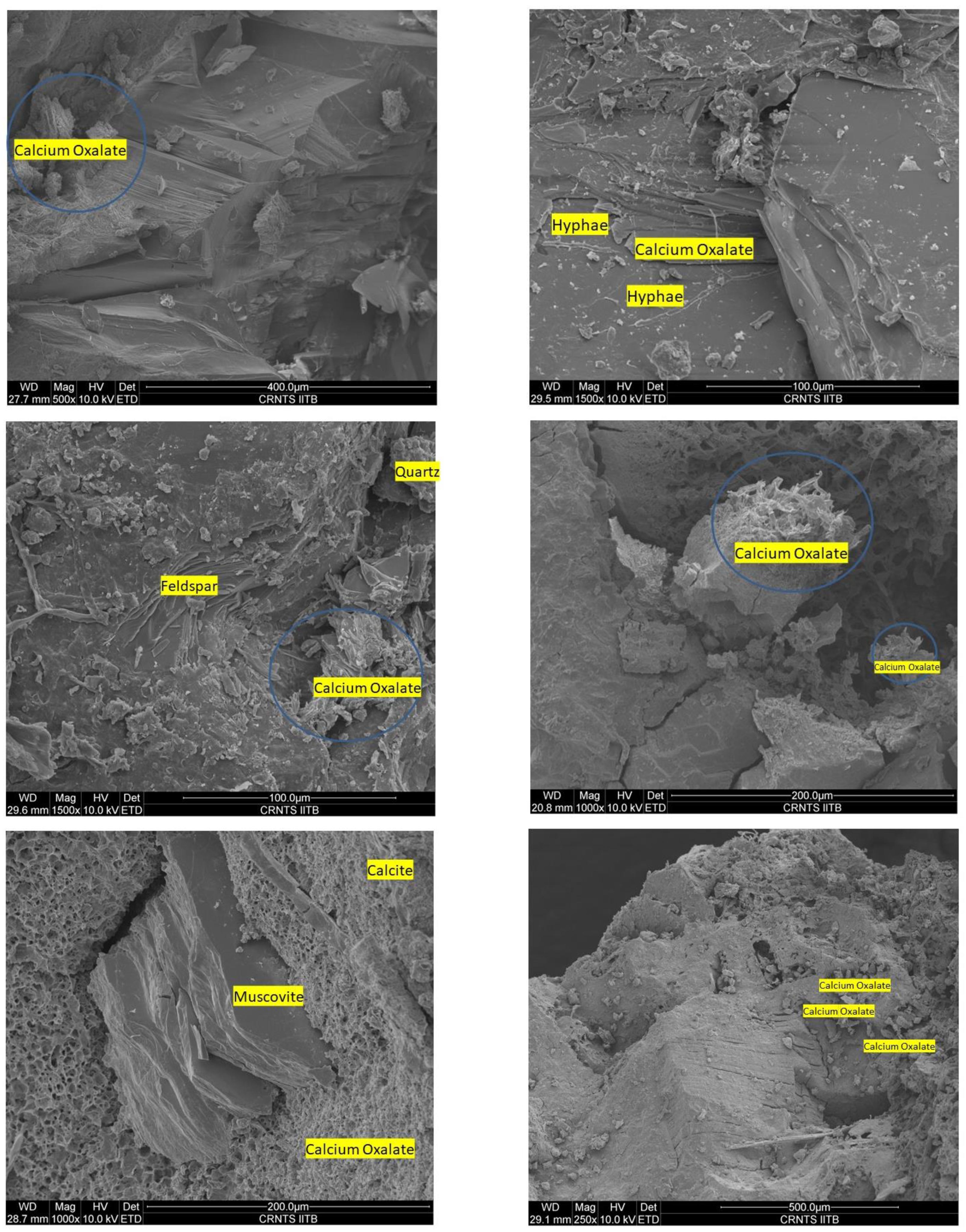
The SEM analysis was performed on six stone samples (SS-1, SS-3, SS-4, SS-5, SS-6, and SS-7) with oxalate patina to visualise their morphology at various magnifications ranging from 250× to 1500×. The photo micrograph images obtained for the stone samples are shown in Figure 9. The elemental composition of the oxalate film was analysed under the EDX probe, and the data obtained is shown in (Table 4).
The patina of all the stone samples investigated is mainly composed of homogeneous microcrystalline Ca-oxalate with diffuse porosity. The photomicrograph observes feldspar, muscovite, and quartz at higher magnification (Figure 8b,c). In Figure 8d, calcium oxalate crystals and the hyphae of the dead microvegetation are seen, indicating the biological origin of the patina. All the silicate minerals observed under SEM are part of the basaltic substrate of the monument rock and are now embedded in a newly formed oxalate film. They do not show any chemical alterations. From the SEM images (Figure 8a–f), no significant deterioration of the stone surfaces is observed except for some micro-fractures of oxalate films. The Ca-oxalate was identified on the basis of the envelope shape (tetrahedral-bipyramidal, 3D) structure of whewellite formed due to the oversaturation of oxalate in a longitudinal/biconcave barbell shape.
This also indicates the protective nature of the oxalate films that saved the heritage structure of Raigad Fort. The SEM photomicrographs also highlight that the possible mechanism of film formation may be due to the initial colonisation of microorganisms that first produce Ca-oxalate biominerals. With subsequent colonization, the deposition thickness of the film has improved on the stone surfaces. The EDX analysis of the stone surfaces of samples SS-1, SS-3, SS-4, SS-6, and SS-7 showed that they are mainly composed of carbon, oxygen, and calcium (Table 4), and other elements such as sodium, sulphur, chlorides, potassium, etc. are present in traces. The elements silica, alumina, iron, etc., that were observed in significant quantities in the EDX data are parts of substrate rock. The higher percentage of phosphorus highlights the biological origin of the oxalate patina.
4. Discussion
The origin of oxalate films on stone surfaces is still under debate due to various controversies and varying reports, mainly from the Mediterranean basin. Two postulates—the natural biological origin of oxalate patina and/or degradation of previously applied organic-based surface coatings—are mostly suggested for film formation [28,29,30]. Another hypothesis suggested by some researchers is the combination of anthropogenic and natural factors with a possible organic origin of oxalic acid due to polluted atmospheres worldwide [31,32]. The reason being offered is that the pollutants in the air and the organic acids present in the aerosol may react with the lime and marble stone surfaces to form oxalic acid [33]. Despite various possibilities reported about the formation of calcium oxalate patina, most researchers have considered oxalate film formation due to microorganisms inhabiting the monument surfaces such as algae, fungi, lichen, bacteria, etc. [34,35,36]. For the Janjira and Murud Sea forts in the Arabian Sea on the west coast of India, situated in proximity to Raigad fort, the formation of Ca-oxalate and Ca-phosphate films on the surfaces of the binding lime mortars has already been assigned to bacterial colonisation in saline conditions protecting the ancient lime works [37,38]. In south India’s excavated monument of Salvankuppam (4–5th CE), previously submerged and now exposed by the 2004 December 26 tsunami, evidence of the formation of amorphous Ca-phosphate precipitate caused by bio-mineralization in saline conditions has been investigated [39]. The microorganisms inhabiting the monument stone should be capable of producing oxalic acids both in vitro and in vivo for film formation. However, according to some researchers, the role of oxalate-forming microorganisms is still controversial, as these organisms also play a significant role in stone deterioration [40,41]. Many transformations of ancient materials into calcium oxalate due to microbes have been reported in the literature. The FTIR spectroscopy revealed a minor transformation of proteinaceous binding material into calcium oxalate in the paint layer of an 18th-century Hindu temple in New Delhi [42]. The evidence presented by many investigators from the interface between lichen and rock substrate indicates that weathering of the minerals is accelerated due to some species of lichen. The impact of lichen on stone substrates is attributed to both physical and chemical processes [43]. Raman spectroscopy played a vital role in the interpretation and characterization of both the physical and chemical nature of lichens. The extensive damage caused to the Italian fresco is attributed to encrustation mainly composed of calcium oxalate, as identified under Raman spectroscopy [44]. The FTIR and Raman spectroscopy revealed the formation of calcium oxalate on the painted surface of India’s Orchha fort due to biological colonisation that led to deterioration and sometimes masking of the paint layer [10]. Besides oxalic acid, the microorganisms also secrete other acids that may have the capacity to solubilize the calcium carbonate of the stones and may prove detrimental to the survival of historic structures [45,46,47]. Results show that a combination of microorganisms such as phototropic, chemotropic, organotrophic bacteria, etc., inhabiting the monument surfaces take part in the secretions, and it is impossible to separate them from the whole community [48,49].
The oxalic acid released from the microorganism acts on the Ca-rich plagioclase of the basaltic rock, dislodging the Ca-ions that form Ca-ions [1,2]. The oxalates formed by microbes slowly accumulate on the stone surfaces as an insoluble patina. Depending on the surrounding conditions, the Ca-oxalate patina may have a homogeneous layered structure or a stratified one, but its mineralogical composition in both conditions remains the same. The two most common crystalline phases of Ca-oxalate occurring in nature are monohydrate (whewellite) and dehydrate (weddelite). These phases are associated with feldspars and quartz in different proportions and ratios on basaltic stone surfaces. Whewellite and weddelite are characterised by their low solubility, and they commonly protect the underlying stone from atmospheric degradation [50]. The calcium oxalate film also imparts an aesthetic appearance to the monument, with some degree of chromatic alterations to the stone surfaces. The oxalate film formation by a natural biological process first occurs at the microbe-substrate layer interface in the presence of a suitable level of pH, humidity, and reduction potential [51]. The chemical and mineralogical transformations of the substrate at the interface are a function of the intrinsic compositional and physical characteristics of the material and ancient treatments on the surface of geominerals [52]. Monte, in his published research, has suggested that the formation of Ca-oxalate depends on the “formation condition” and not on the “formation causes” [53]. Moreover, the naturally occurring ca-oxalate films widely vary in colour and thickness, as reported in many instances [54]. On basaltic stone surfaces, it is reported that the film or patina primarily consists of Ca-oxalate in association with Ca-phosphates, silicates, calcite, and organic materials from microbes [55].
5. Conclusions
Analytical results indicated the calcium oxalate patina on historical basalt stone surfaces has occurred due to microbial colonization. The thick microvegetation growth on monument surfaces, the high humidity, and the appropriate pH created specific environmental conditions for the formation of a calcium oxalate patina. The high humidity and tropical conditions favoured the formation of the most stable whewellite phase on the stone surfaces. In around 400 years of its deposition, the calcium oxalate has led to the transformation of surface stone, and the patina has been found protecting the underlying surfaces. The patina was found homogeneously covering the external surfaces of the basalt stone as well as mud and lime layers, giving a glittering sheen to the entire fort complex. The presence of hyphe in the SEM photomicrograph indicated the biological origin of the film. This is probably the first report on oxalate film formation on basaltic stone surfaces of heritage structures in the Indian subcontinent.
Author Contributions
M.R.S.: Conceptualization, formal analysis, writing—original draft, writing—review and editing; R.Y.: data curation. Both the authors have contributed equally. All authors have read and agreed to the published version of the manuscript.
Funding
This research received no external funding.
Data Availability Statement
Not applicable.
Acknowledgments
This work is communicated towards celebrations of 350 years of coronation of India’s great Maratha warrior, the Chhatrapati Shivaji Maharaj. The authors are grateful to D. N. Singh, IIT Bombay and Lijjit P. for their extensive help in the analytical investigations. The help extended by Shubham Patil and Remi Thomas of Babasaheb Ambedkar Marathwada University, Chhatrapati Sambhajinagar is gratefully acknowledged.
Conflicts of Interest
The authors declare no conflict of interest.
References
- Jones, D.; Wilson, M.J.; Tait, J.M. Weathering of a basalt by Pertusaria corallina. Lichenologist 1980, 12, 277–290. [Google Scholar] [CrossRef]
- Arocena, J.M.; Siddique, T.; Thring, R.W.; Kapur, S. Investigation of lichens using molecular techniques and associated mineral accumulations on a basaltic flow in a Mediterranean environment. Catena 2007, 70, 356–365. [Google Scholar] [CrossRef]
- Columbu, S.; Piras, G.; Sitzia, F.; Pagnotta, S.; Raneri, S.; Legnaioli, S.; Palleschi, V.; Lezzerini, M.; Giamello, M. Petrographic and mineralogical characterization of volcanic rocks and surface-depositions on romanesque monuments. Mediterr. Archaeol. Archaeom. 2018, 18, 37–64. [Google Scholar] [CrossRef]
- Liebig, J. XI.—Note on thierschite. Q. J. Chem. Soc. Lond. 1854, 6, 112–113. [Google Scholar] [CrossRef] [Green Version]
- Cipriani, C.; Franchi, L. The presence of whewellite in alteration crust of Roman monuments. Boll. Serv. Geol. Ital. 1958, 79, 555–556. [Google Scholar]
- Arocena, J.M.; Zhu, L.P.; Hall, K. Mineral accumulations induced by biological activity on granitic rocks in Qinghai Plateau, China. Earth Surf. Process. Landf. J. Br. Geomorphol. Res. Group 2003, 28, 1429–1437. [Google Scholar] [CrossRef]
- Bonazza, A.; Messina, P.; Sabbioni, C.; Grossi, C.M.; Brimblecombe, P. Mapping the impact of climate change on surface recession of carbonate buildings in Europe. Sci. Total Environ. 2009, 407, 2039–2050. [Google Scholar] [CrossRef]
- Del Monte, M.; Sabbioni, C. Weddellite on limestone in the Venice [Italy] environment. Environ. Sci. Technol. 1983, 17, 518–522. [Google Scholar] [CrossRef]
- Gómez-Bolea, A.; Llop, E.; Ariño, X.; Saiz-Jimenez, C.; Bonazza, A.; Messina, P.; Sabbioni, C. Mapping the impact of climate change on biomass accumulation on stone. J. Cult. Herit. 2012, 13, 254–258. [Google Scholar] [CrossRef]
- Kanth, A.P.; Singh, M.R. Vibrational spectroscopy and SEM-EDX analysis of wall painted surfaces, Orchha Fort, India. J. Archaeol. Sci. Rep. 2019, 24, 434–444. [Google Scholar] [CrossRef]
- Carmona, N.; Villegas, M.A.; Navarro, J.F. Characterisation of an intermediate decay phenomenon of historical glasses. J. Mater. Sci. 2006, 41, 2339–2346. [Google Scholar] [CrossRef]
- Bonazza, A.; Natali, C.; Ghedini, N.; Vaccaro, C.; Sabbioni, C. Oxalate patinas on stone monuments in the Venetian Lagoon: Characterization and origin. Int. J. Archit. Herit. 2015, 9, 542–552. [Google Scholar] [CrossRef]
- El-Derby, A.A.O.D.; Mansour, M.M.A.; Salem, M.Z.M. Investigation the microbial deterioration of sandstone from the osirion’s sarcophagus chamber as affected by rising ground water level. Mediterr. Archaeol. Archaeom. 2016, 16, 273–281. [Google Scholar] [CrossRef]
- Alessandrini, G.; Amicarelli, V.; Bellini, A.; Biscontin, G.; Cecchi, R.; Fassina, V. The Oxalate Films: Origin and Significance in the Conservation of Works of Art; Centro Congressi Cariplo: Milan, Italy, 1989. [Google Scholar]
- Manganelli, C.; Del, F.; Camaiti, M.; Borselli, G.; Maravalaki, P.; Taino, P. The Oxalate Films: Origin and Significance in the Conservation of Works of Art; Centro Congressi Cariplo: Milan, Italy, 1989. [Google Scholar]
- Cariati, F.; Rampazzi, L.; Toniolo, L.; Pozzi, A. Calcium oxalate films on stone surfaces: Experimental assessment of the chemical formation. Stud. Conserv. 2000, 45, 180–188. [Google Scholar]
- Aulinas, M.; Garcia-Valles, M.; Gimeno, D.; Fernandez-Turiel, J.L.; Ruggieri, F.; Pugès, M. Weathering patinas on the medieval (S. XIV) stained glass windows of the Pedralbes Monastery (Barcelona, Spain). Environ. Sci. Pollut. Res. 2009, 16, 443–452. [Google Scholar] [CrossRef]
- Conti, C.; Brambilla, L.; Colombo, C.; Dellasega, D.; Gatta, G.D.; Realini, M.; Zerbi, G. Stability and transformation mechanism of weddellite nanocrystals studied by X-ray diffraction and infrared spectroscopy. Phys. Chem. Chem. Phys. 2010, 12, 14560–14566. [Google Scholar] [CrossRef]
- Conti, C.; Casati, M.; Colombo, C.; Realini, M.; Brambilla, L.; Zerbi, G. Phase transformation of calcium oxalate dihydrate–monohydrate: Effects of relative humidity and new spectroscopic data. Spectrochim. Acta Part A Mol. Biomol. Spectrosc. 2014, 128, 413–419. [Google Scholar] [CrossRef]
- Najafi, S.J.; Cox, K.G.; Sukheswala, R.N. Geology and geochemistry of the basalt flows (Deccan Traps) of the Mahad-Mahabaleshwar section, India. In Deccan Volcanism and Related Provinces in Other Parts of the World; Subbaro, K.V., Sukheswala, R.N., Eds.; Geological Society of India: Bangalore, India, 1981; Volume 3, pp. 300–315. [Google Scholar]
- Kothari, M.J.; Moorthy, S. Flora of Raigad District Maharashtra State; Flora of India Series 3; Botanical Survey of India: Calcutta, India, 1993. [Google Scholar]
- Fraser-Jenkins, C.R.; Gandhi, K.N.; Kholia, B.S.; Benniamin, A. An Annotated Checklist of Indian Pteridophytes; Bishen Singh Mahendra Pal Singh: Dehra Dun, India, 2017. [Google Scholar]
- Salama, K.K.; Ali, M.F.; El Sheikh, S.M. A comparison between nano calcium carbonate, natural calcium carbonate and converted calcium hydroxide for consolidation. Sci. Cult. 2019, 5, 35–40. [Google Scholar] [CrossRef]
- King, H.E.; Mattner, D.C.; Plümper, O.; Geisler, T.; Putnis, A. Forming cohesive calcium oxalate layers on marble surfaces for stone conservation. Cryst. Growth Des. 2014, 14, 3910–3917. [Google Scholar] [CrossRef]
- La Russa, M.F.; Ruffolo, S.A.; Barone, G.; Crisci, G.M.; Mazzoleni, P.; Pezzino, A. The use of FTIR and micro-FTIR spectroscopy: An example of application to cultural heritage. Int. J. Spectrosc. 2009, 2009, 893528. [Google Scholar] [CrossRef] [Green Version]
- Pavía, S.; Caro, S. Origin of films on monumental stone. Stud. Conserv. 2006, 51, 177–188. [Google Scholar]
- Horner, H.T.L.; Tiffany, H.; Cody, A.M. Calcium oxalate bipyramidal crystals on the basidiocarps of Geastrum minus (Lycoperdales). Proc. Iowa Acad. Sci. 1985, 92, 70–77. [Google Scholar]
- Gadd, G.M.; Bahri-Esfahani, J.; Li, Q.; Rhee, Y.J.; Wei, Z.; Fomina, M.; Liang, X. Oxalate production by fungi: Significance in geomycology, biodeterioration and bioremediation. Fungal Biol. Rev. 2014, 28, 36–55. [Google Scholar]
- Del Monte, M.; Sabbioni, C.; Zappia, G. The origin of calcium oxalates on historical buildings, monuments and natural outcrops. Sci. Total Environ. 1987, 67, 17–39. [Google Scholar]
- Fassina, V.; Molteni, C. Problemi di conservazione connessi all’umidita delle murature: La diagnostica e le tecnologie conservative applicate al restauro della cripta di S. Marco in Venezia. In Proceedings of the 3rd International Symposium on the Conservation of Monuments in the Mediterranean Basin, Venice, Italy, 22–25 June 1994; pp. 803–813. [Google Scholar]
- Kawamura, K.; Kaplan, I.R. Motor exhaust emissions as a primary source for dicarboxylic acids in Los Angeles ambient air. Environ. Sci. Technol. 1987, 21, 105–110. [Google Scholar] [CrossRef]
- Martinelango, P.K.; Dasgupta, P.K.; Al-Horr, R.S. Atmospheric production of oxalic acid/oxalate and nitric acid/nitrate in the Tampa Bay airshed: Parallel pathways. Atmos. Environ. 2007, 41, 4258–4269. [Google Scholar] [CrossRef]
- Saiz-Jimenez, C. Biogenic vs. anthropogenic oxalic acid in the environment. In Le Pellicole ad Ossalati: Origine e Significato Nella Conservazione delle Opere d’Arte; Centro CNR “Gino Bozza”: Milano, Italy, 1989; pp. 207–214. [Google Scholar]
- Ciccarone, C.; Pinna, D. Calcium oxalate films on stone monuments—Microbiological investigations. Aerobiologia 1993, 9, 33–37. [Google Scholar] [CrossRef]
- Del Monte, M.; Sabbioni, C. A study of the patina called ‘scialbatura’ on imperial Roman marbles. Stud. Conserv. 1987, 32, 114–121. [Google Scholar] [CrossRef]
- Hernanz, A.; Gavira-Vallejo, J.M.; Ruiz-López, J.F. Calcium oxalates and prehistoric paintings. The usefulness of these biomaterials. J. Optoelectron. Adv. Mater. 2007, 9, 512–521. [Google Scholar]
- Karche, T.; Singh, M.R. Biologically induced calcium oxalate mineralization on 15th century lime mortar, Murud Sea fort, India. J. Archaeol. Sci. Rep. 2021, 39, 103178. [Google Scholar] [CrossRef]
- Singh, M.R.; Ganaraj, K.; Sable, P.D. Surface mediated Ca-phosphate biomineralization and characterization of the historic lime mortar, Janjira Sea Fort, India. J. Cult. Herit. 2020, 44, 110–119. [Google Scholar] [CrossRef]
- Singh, M.R.; Vinodh, K.S.; Ganaraj, K. Evidence of amorphous Ca-phosphate precipitate caused by bio mineralisation in 4–5th CE lime plasters of the previously submerged east coastal monument of Salvankuppam. Mineralogia 2022, 52, 19–30. [Google Scholar] [CrossRef]
- Ariño, X.; Ortega-Calvo, J.J.; Gomez-Bolea, A.; Saiz-Jimenez, C. Lichen colonization of the Roman pavement at Baelo Claudia (Cadiz, Spain): Biodeterioration vs. bioprotection. Sci. Total Environ. 1995, 167, 353–363. [Google Scholar] [CrossRef] [Green Version]
- Adamo, P.; Violante, P. Weathering of rocks and neogenesis of minerals associated with lichen activity. Appl. Clay Sci. 2000, 16, 229–256. [Google Scholar] [CrossRef]
- Kanth, A.P.; Singh, M.R. Spectroscopic and chromatographic investigation of the wall painted surfaces of an 18th century Indian temple, New Delhi. Vib. Spectrosc. 2019, 104, 102947. [Google Scholar] [CrossRef]
- Chen, J.; Blume, H.P.; Beyer, L. Weathering of rocks induced by lichen colonization—A review. Catena 2000, 39, 121–146. [Google Scholar] [CrossRef]
- Edwards, H.G.M.; Farwell, D.W.; Seaward, M.R.D.; Giacobini, C. Preliminary Raman microscopic analyses of a lichen encrustation involved in the biodeterioration of Renaissance frescoes in central Italy. Int. Biodeterior. 1991, 27, 1–9. [Google Scholar] [CrossRef]
- Krumbein, W.E.; Ciabach, J. Biology of stone and minerals in buildings—Biodeterioration, biotransfer, bioprotection. In Proceedings of the VIth International Congress on Deterioration and Conservation of Stone, Torun, Poland, 12–14 September 1988; Nicholas Copernicus University: Torun, Poland, 1988; pp. 1–12. [Google Scholar]
- Krumbein, W.E.; Petersen, K.; Schellnhuber, H.J. On the geomicrobiology of yellow, orange, red, brown and black films and crusts developing on several different types of stone and objects of art. In Le Pellicole ad Ossalati: Origine e Significato Nella Conservazione Delle Opere d’Arte; Centro CNR “Gino Bozza”: Milano, Italy, 1989; pp. 337–348. [Google Scholar]
- Anagnostidis, K.; Gehrmann, C.K.; Gross, M.; Krumbein, W.E.; Lisi, S.; Pantazidou, A.; Urzì, C.; Zagari, M. Biodeterioration of marbles of the Parthenon and Propylaea, Acropolis, Athens-associated organisms, decay and treatment suggestions. In La Conservation des Monuments Dans le Bassin Méditerranéen: Actes du 2ème Symposium International; Musée d’Art et d’Histoire: Geneva, Switzerland, 1991; pp. 305–325. [Google Scholar]
- Warscheid, T.; Petersen, K.; Krumbein, W.E.; Ciabach, J. Physiological characterization of chemoorganotrophic bacteria isolated from sandstones. In Proceedings of the VIth International Congress on Deterioration and Conservation of Stone, Torun, Poland, 12–14 September 1988; Nicholas Copernicus University: Torun, Poland, 1988; pp. 26–32. [Google Scholar]
- Palmer, R.J., Jr.; Hirsch, P. Photosynthesis-based microbial communities on two churches in northern Germany: Weathering of granite and glazed brick. Geomicrobiol. J. 1991, 9, 103–118. [Google Scholar] [CrossRef]
- Alessandrini, G.; Bonecchi, R.; Peruzzi, R.; Toniolo, L. Caratteristiche composizionali e morfologiche di pellicole ad ossalato: Studio comparato su substrati lapidei di diversa natura. In Le Pellicole ad Ossalati: Origine e Significato Nella Conservazione delle Opere d’Arte; Centro CNR “Gino Bozza”: Milano, Italy, 1989; pp. 137–150. [Google Scholar]
- Adamo, P.; Colombo, C.; Violante, P. Iron oxides and hydroxides in the weathering interface between Stereocaulon vesuvianum and volcanic rock. Clay Miner. 1997, 32, 453–461. [Google Scholar] [CrossRef]
- Abeer, F. Elhagrassy and Amira Hakeem Comparative study of biological cleaning and laser techniques for conservation of weathered stone in Failaka Island, Kuwait. Sci. Cult. 2018, 4, 43–50. [Google Scholar] [CrossRef]
- Monte, M. Biogenesis of oxalate patinas on marble specimens in fungal culture. Aerobiologia 2003, 19, 271–275. [Google Scholar] [CrossRef]
- Franzini, M.; Gratziu, C.; Wicks, E. Calcium oxalate films on marble monuments. Rend. Soc. Ital. Mineral. Petrol. 1984, 39, 59–70. [Google Scholar]
- Lazzarini, L.; Salvadori, O. A reassessment of the formation of the patina called scialbatura. Stud. Conserv. 1989, 34, 20–26. [Google Scholar] [CrossRef]
Figure 1.
(a)—Showing thick vegetation growth on the monument surface of Raigad fort. (b)—General vew of Raigad fort.
Figure 1.
(a)—Showing thick vegetation growth on the monument surface of Raigad fort. (b)—General vew of Raigad fort.
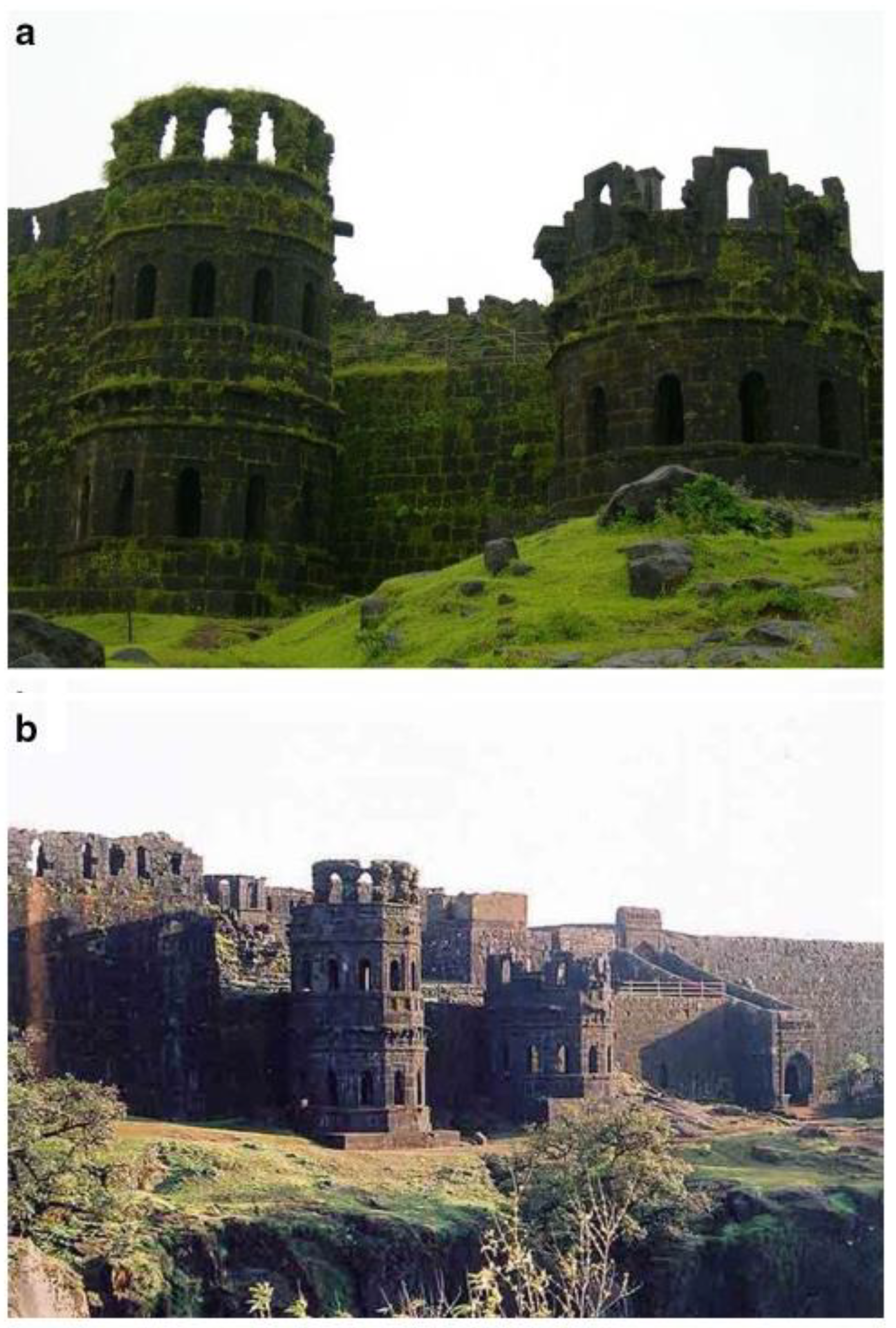
Figure 2.
(a–h) showing stages of Ca-oxalate patina and their formation on basaltic stone surfaces, Raigad Fort.
Figure 2.
(a–h) showing stages of Ca-oxalate patina and their formation on basaltic stone surfaces, Raigad Fort.
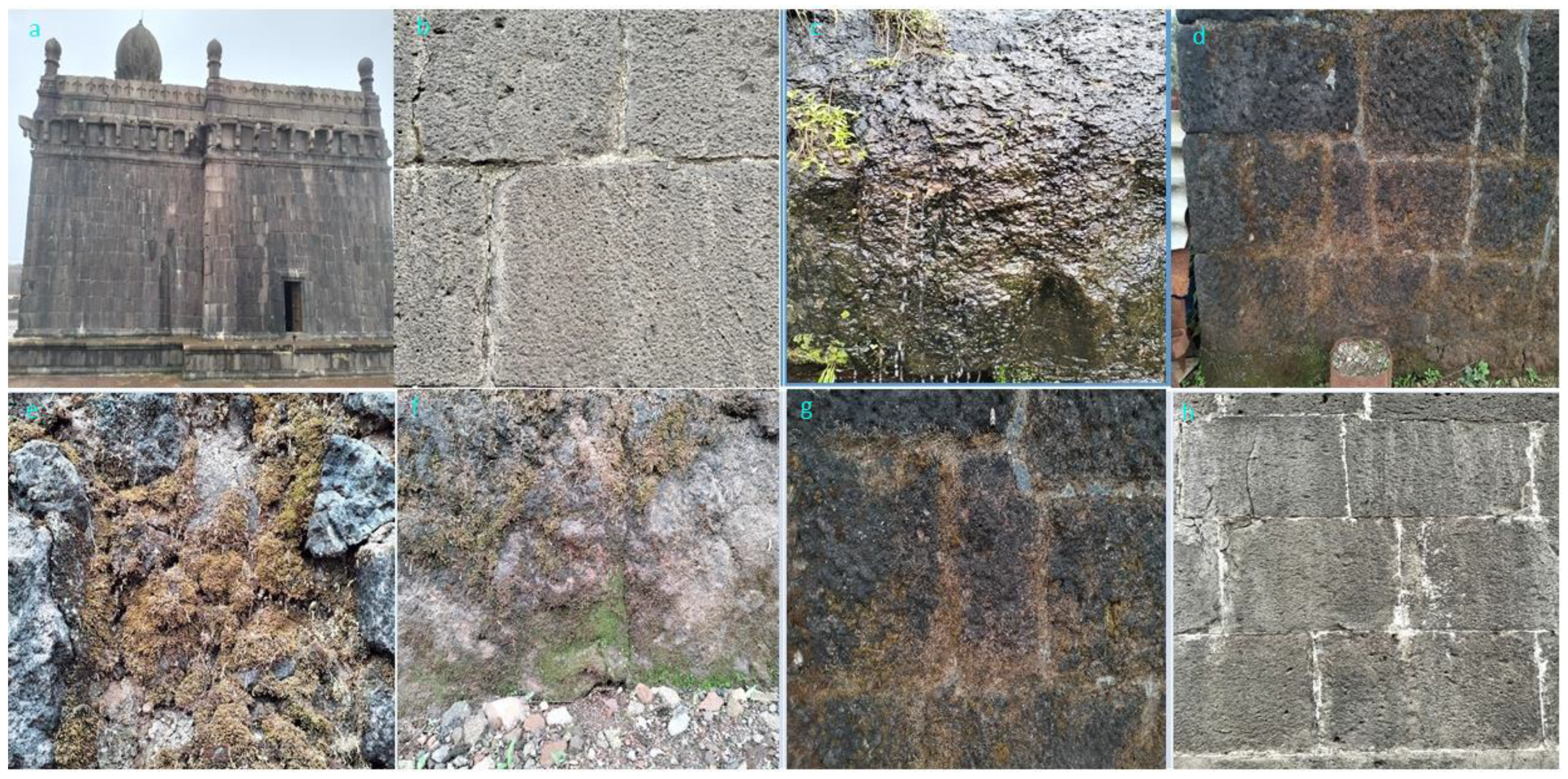
Figure 3.
(a). Temperature and relative humidity Vs time graph of Raigad. (b). Precipitation diagram of Raigad. (c). graph showing the wind direction and wind speed at Raigad. (derived from www.meteoblue.com (accessed on 28 May 2023)).
Figure 3.
(a). Temperature and relative humidity Vs time graph of Raigad. (b). Precipitation diagram of Raigad. (c). graph showing the wind direction and wind speed at Raigad. (derived from www.meteoblue.com (accessed on 28 May 2023)).

Figure 4.
FTIR spectra of surface stone samples of Raigad hill fort.
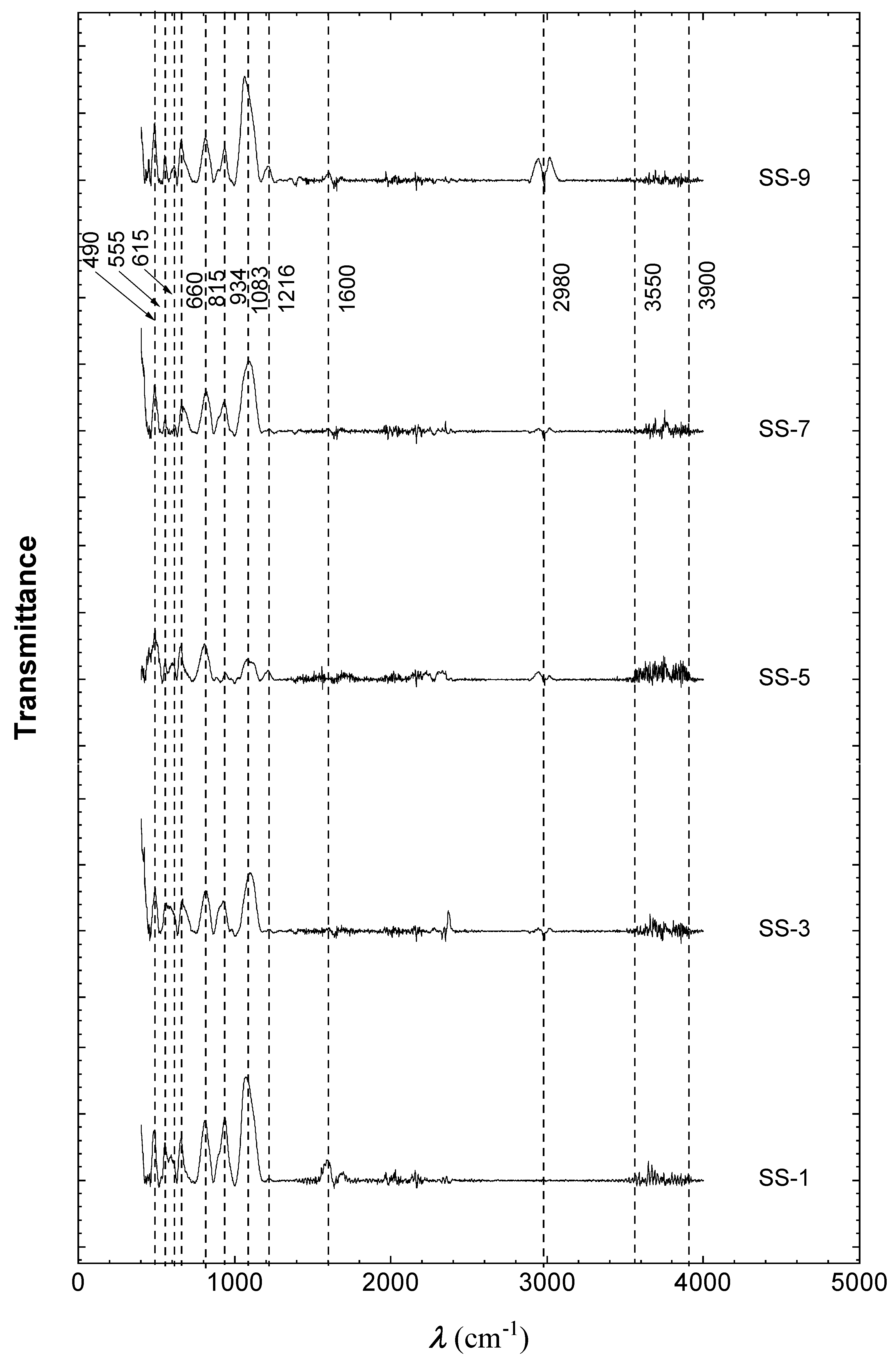
Figure 5.
FTIR spectra of surface stone samples of Raigad hill fort.
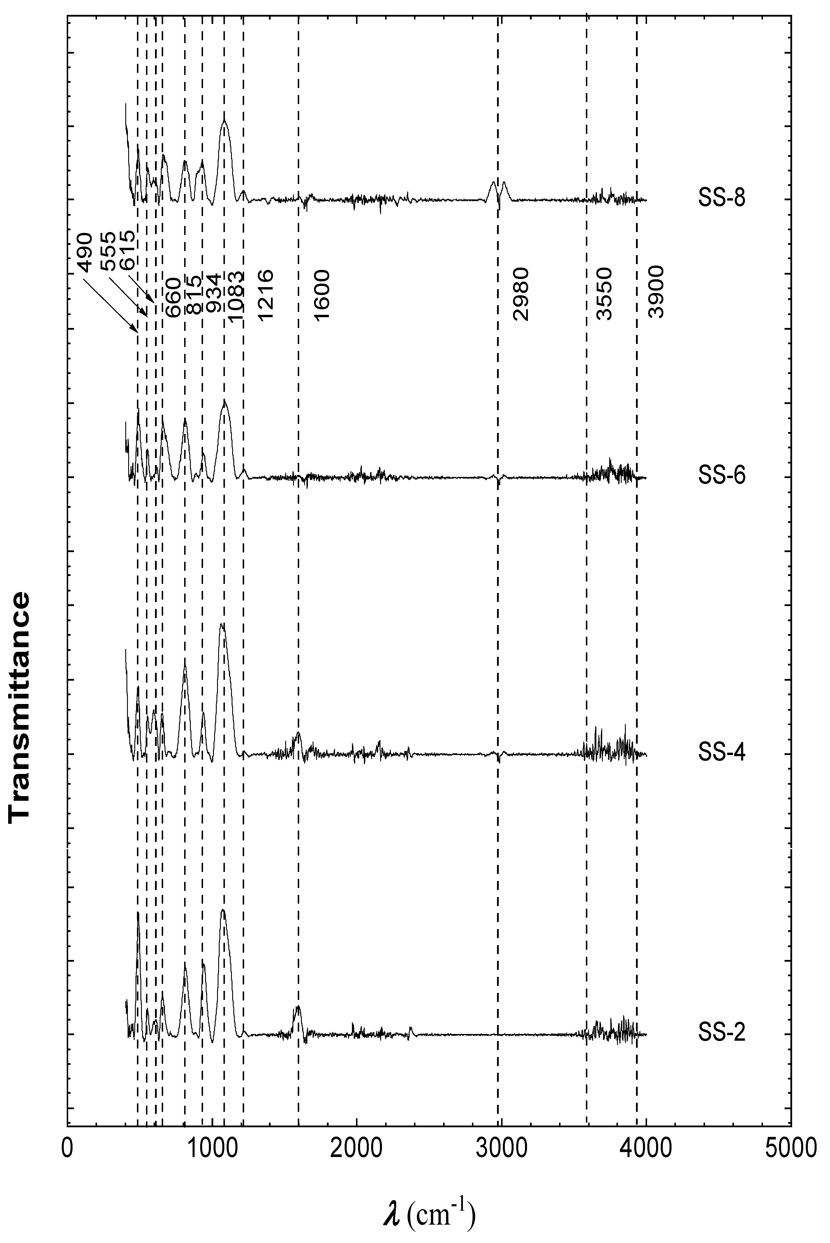
Figure 6.
Optical microscopic images of Ca-oxalate on stone surfaces of Raigad Fort.

Figure 7.
Showing optical images of patina along with substrate layer for samples SS-1 to SS-4.
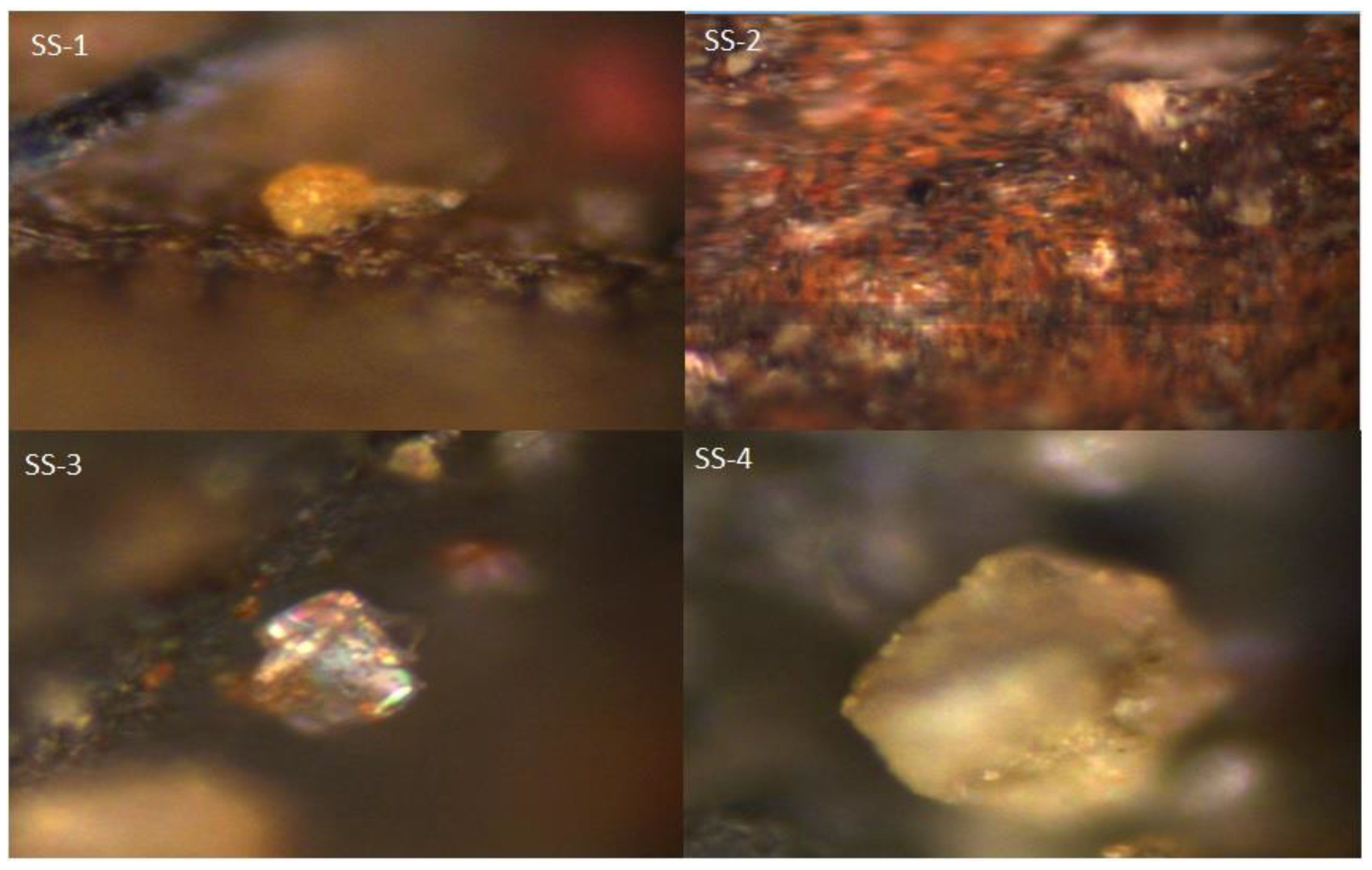
Figure 8.
XRD diffractogram of stone sample SS-1 to SS-9 from Raigad Fort.
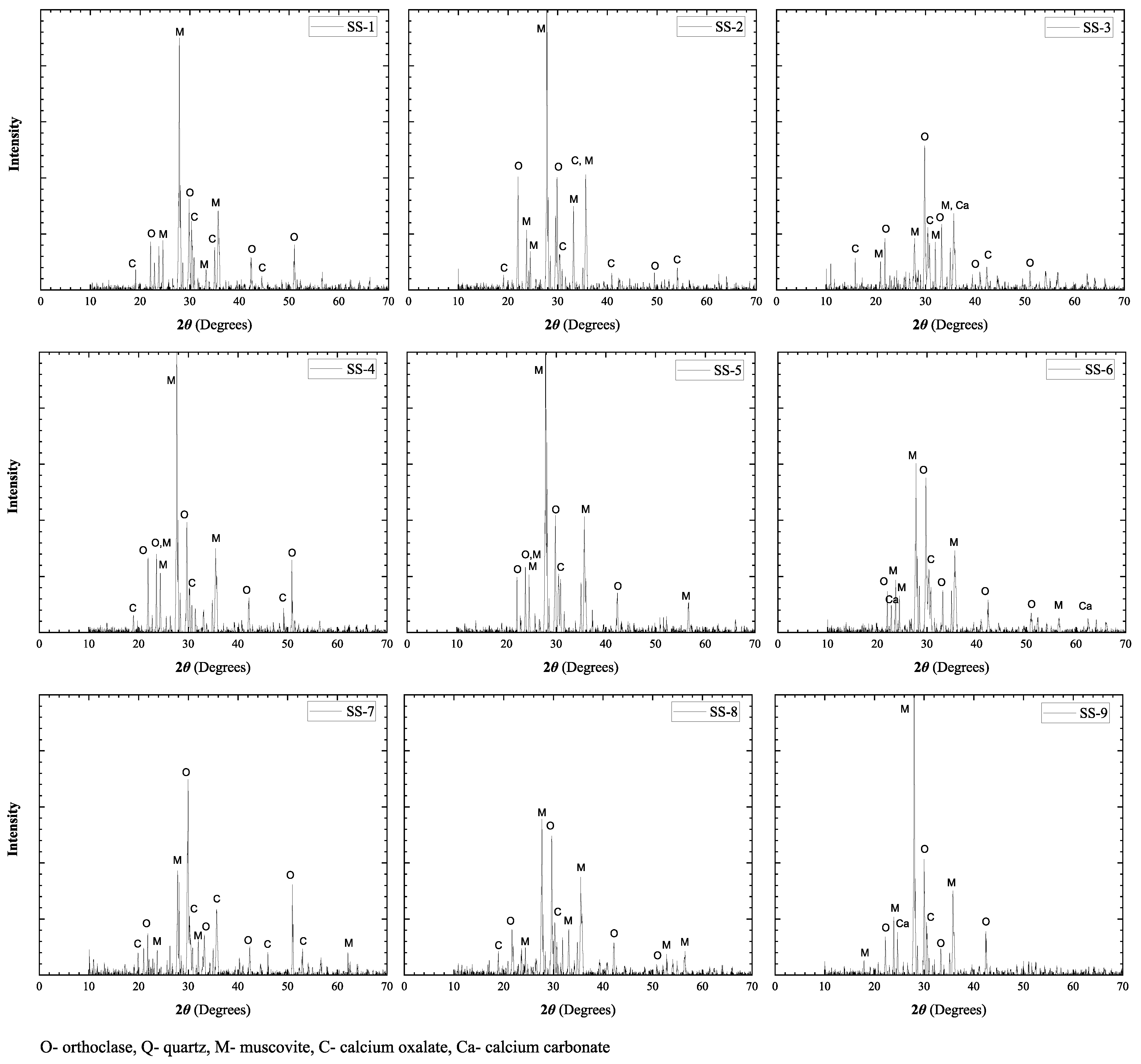
Figure 9.
Showing SEM photo micrograph of surface stone samples of Raigad Hill fort.

Table 1.
Average chemical composition of basalt rock near Raigad Fort.
| Major Components | Weight Percentage |
|---|---|
| SiO2 | 45–52% |
| K2O and Na2O | 2–5% |
| TiO2 | 0.5–2% |
| Fe2O3 | 5–14% |
| Al2O3 | about 14% |
| MgO | 5–12% |
| CaO | 10–12% |
Table 2.
Details of the basalt stone surface samples extracted from Raigad Fort.
| Designation | Sample | Additional Details | Location |
|---|---|---|---|
| SS-1 | Stone sample | East Facing Wall (Stone) | Haathi Khana |
| SS-2 | Stone sample | West Facing Wall | Back Side of Bazaar Peth |
| SS-3 | Stone sample | East Facing wall near Samadhi (Stone) | Jagdishwar Temple |
| SS-4 | Stone sample | Peth Structure-11 (Right) | Inside of Bazaar |
| SS-5 | Stone sample | North Facing Wall | Near Nagar Khana |
| SS-6 | Stone sample | South Facing Wall | Outer Wall of Nagar Khana |
| SS-7 | Stone sample | East Facing Wall | Outer wall of Nagar Khana |
| SS-8 | Stone sample | Burning Patch from Back Plinth | Inside of Simhasan |
| SS-9 | Stone sample | East Facing | Outer Wall of Queen Palace |
Table 3.
The functional groups present in basaltic stone samples.
| Sample | Frequency (cm−1) | Mineral | Assignment |
|---|---|---|---|
| Stone Samples | 490 | Muscovite | Al-O Bending Vibration |
| 555 | Orthoclase | Si-O Stretch | |
| 615 | Orthoclase | Al-O Coordination Vibration | |
| 660 | Water molecule | Wagging of Water Molecule | |
| 815 | Orthoclase, Muscovite | Si-O stretch, Si-O-Al Stretch, | |
| 935 | Calcium Oxalate | C=C Stretching | |
| 1080 | Orthoclase, Muscovite | Si-O-Si Stretch | |
| 1215 | Calcium Oxalate | C-C(=O)-O Stretching | |
| 1620 | Calcium Oxalate | OC=O Asymmetrical Stretching | |
| 1650 | Calcium Oxalate | Stretching | |
| 2330 | Calcium Oxalate | O=C=O Stretching | |
| 3000 | Aliphatic Group | Asymmetric C-H Group | |
| 3600–3900 | Calcium Oxalate Monohydrate | O-H Stretching |
Table 4.
EDX data of the surface stone samples, Raigad fort.
| Sample Designation/Elements | SS-1 | SS-3 | SS-4 | SS-6 | SS-7 |
|---|---|---|---|---|---|
| C | 15.6 | 14.5 | 14.0 | 45.3 | 32.8 |
| N | 3.4 | 2.8 | 1.7 | 1.6 | 4.3 |
| O | 43.8 | 45.8 | 46.9 | 40.3 | 44.4 |
| F | 2.1 | 0.5 | 0.7 | 0.0 | 0.3 |
| Na | 0.8 | 0.1 | 0.7 | 0.0 | 0.1 |
| Mg | 1.4 | 1.3 | 2.7 | 0.3 | 1.3 |
| Al | 4.6 | 4.5 | 4.1 | 1.8 | 0.0 |
| Si | 12.4 | 14.5 | 14.5 | 4.4 | 8.4 |
| P | 4.4 | 5.8 | 2.6 | 1.4 | 2.2 |
| S | 0.2 | 0.2 | 0.1 | 0.1 | 0.1 |
| Cl | 0.1 | 0.1 | 0.0 | 0.0 | 0.0 |
| K | 0.6 | 0.5 | 0.4 | 0.1 | 0.0 |
| Ca | 3.6 | 3.4 | 3.4 | 2.0 | 2.1 |
| Fe | 7.1 | 5.9 | 7.8 | 2.7 | 4.0 |
Disclaimer/Publisher’s Note: The statements, opinions and data contained in all publications are solely those of the individual author(s) and contributor(s) and not of MDPI and/or the editor(s). MDPI and/or the editor(s) disclaim responsibility for any injury to people or property resulting from any ideas, methods, instructions or products referred to in the content. |
© 2023 by the authors. Licensee MDPI, Basel, Switzerland. This article is an open access article distributed under the terms and conditions of the Creative Commons Attribution (CC BY) license (https://creativecommons.org/licenses/by/4.0/).
Share and Cite
MDPI and ACS Style
Singh, M.R.; Yadav, R. Formation of Calcium Oxalate Patinas as Protective Layer on Basaltic Stone Surfaces of 17th Century Raigad Hill Fort, India. Heritage 2023, 6, 5374-5392. https://doi.org/10.3390/heritage6070283
AMA Style
Singh MR, Yadav R. Formation of Calcium Oxalate Patinas as Protective Layer on Basaltic Stone Surfaces of 17th Century Raigad Hill Fort, India. Heritage. 2023; 6(7):5374-5392. https://doi.org/10.3390/heritage6070283
Chicago/Turabian StyleSingh, Manager Rajdeo, and Rajendra Yadav. 2023. "Formation of Calcium Oxalate Patinas as Protective Layer on Basaltic Stone Surfaces of 17th Century Raigad Hill Fort, India" Heritage 6, no. 7: 5374-5392. https://doi.org/10.3390/heritage6070283