Review

An Introduction and Recent Advances in the Analytical Study of Early Synthetic Dyes and Organic Pigments in Cultural Heritage

by
Diego Tamburini
1,*,
Francesca Sabatini
2,†,
Sanne Berbers
3,4,
Maarten R. van Bommel
4,5 and
Ilaria Degano
2
1
Department of Scientific Research, The British Museum, Great Russell Street, London WC1B 3DG, UK
2
Department of Chemistry and Industrial Chemistry, University of Pisa, Via G. Moruzzi 13, 56124 Pisa, Italy
3
Cultural Heritage Laboratory, Cultural Heritage Agency of The Netherlands, Hobbemastraat 22, 1071 ZC Amsterdam, The Netherlands
4
Analytical Sciences, van ‘t Hoff Institute for Molecular Sciences, Faculty of Sciences, University of Amsterdam, Science Park 904, 1098 XH Amsterdam, The Netherlands
5
Conservation and Restoration of Cultural Heritage, Amsterdam School for Heritage, Memory and Material Culture (AHM), Faculty of Humanities, University of Amsterdam, P.O. Box 94552, 1090 GN Amsterdam, The Netherlands
*
Author to whom correspondence should be addressed.
Current address: Department of Earth and Environmental Sciences, University of Milano-Bicocca, Piazza della Scienza 1, 20126 Milan, Italy.
Heritage 2024, 7(4), 1969-2010; https://doi.org/10.3390/heritage7040094
Submission received: 8 February 2024 / Revised: 7 March 2024 / Accepted: 8 March 2024 / Published: 28 March 2024
(This article belongs to the Section Cultural Heritage)

Abstract

:
This article reviews the research recently undertaken to characterise and identify early synthetic dyes (ESDs) and synthetic organic pigments (SOPs) as well as study their degradation pathways with a focus on cultural heritage applications. Since the invention of the first fully synthetic dye in 1856, these materials have been used in a variety of historic objects and artworks, such as textiles, furniture, prints, paintings, drawings, etc. The synthesis of new dyes and their implementation into pigment formulations were rapid phenomena, so that by the beginning of the 20th century, thousands of new molecules were patented and commercialised. This review will focus on these early formulations synthesised up to approximately 1914 and the outbreak of World War I. ESDs and SOPs represent a challenge from an analytical point of view. Molecular databases are fundamental for the unequivocal identification of these compounds, but reference materials are not always readily available. A combination of analytical techniques is often needed for characterisation, especially in the case of SOPs, which have both organic and inorganic components. Furthermore, the degradation of these molecules can jeopardise their detection in historic objects that have been exposed to light, humidity and temperature fluctuations. Nevertheless, ESDs and SOPs are important research tools. As the dates of their first synthesis are often known with precision, based on patent information, their detection can be used to refine the production date of objects. Additionally, their trade from Europe to the world and their adoption in artistic practices around the globe is an active area of research.

1. Introduction

Since prehistoric times, humankind has used organic colourants extracted from some plants and animals to dye textiles, stain wood and paper, apply as ink or pigment in paintings. The first example of a textile dyed with indigo is more than 6000 years old and was found in the Preceramic site of Huaca Prieta on the north coast of Peru [1]. A drastic change occurred in the second half of the 19th century, when synthetic colourants started being developed in Europe. This had a dramatic impact on the natural colourant industry in Europe, which saw a drastic decline in just a few decades. In other parts of the world, natural dyes were also gradually replaced by synthetic ones. At the end of the 19th century, almost 1000 different synthetic colourants were available, which were more brilliant, cheaper and easier to dye compared to most natural dyes and covered the full-colour palette [2]. These colourants found applications mostly as textile dyes, but also as wood stains, inks, synthetic organic pigments (SOPs) and more. In the case of SOPs, the soluble dye is often converted into an insoluble pigment by precipitation on an inorganic substrate, similar to traditional organic lake pigments production [3].
Many consider 1856 as the start of the synthetic colourant industry, the year when the young Sir William Henry Perkin (1838–1907) invented mauveine, also known as mauve [4]. However, several earlier attempts to modify natural colourants and synthesise new ones had already been made. The first semi-synthetic colourant was indigo carmine, developed in 1743 by Johann Christian Barth (c. 1700–1759) [5] by treating indigo with concentrated sulphuric acid, so that one or two sulphonic acid groups were attached to indigotin. A water-soluble blue dye was therefore formed, which could dye fibres such as wool, silk, linen and cotton, bypassing the traditional, laborious vat process needed for indigoid natural dyes. It was used both as a single dye and in mixtures to obtain green and violet colours. However, indigo carmine had poor light and wash fastness. Despite its poor properties, it was found in objects from the early 20th century [6] and it is currently still in use as a food colourant, in cosmetics and for colouring pharmaceuticals [5].
In 1771, Peter Woulfe (1727–1803) treated indigo with nitric acid, forming the yellow dye picric acid [7]. Although derived from indigotin, the chemical structure of picric acid is very different from the blue molecule. In fact, picric acid was the trade name of 2,4,6-trinitrophenol (TNP), which has a close relation to 2,4,6-trinitrotoluene (TNT) [8]. Indeed, it was often used as an explosive, but also to dye wool, silk and leather, whereas it does not have good affinity to cotton or linen. It provides a yellow colour with a greenish hue, which makes it suitable to dye green in combination with other dyes, such as indigo carmine. The wash and light fastness are poor. However, it was still in use at least until the early 20th century [9].
In 1776, the violet mordant dye murexide was invented by Carl Wilhelm Scheele (1742–1786) [10]. It has never been detected in historical artifacts but was identified in the early 20th century Lefranc archives of SOPs [11]. In 1832, Karl Ludwig Reichenbach (1788–1869) accidentally discovered the blue acid dye pittacal [12].
The synthesis of mauveine in 1856 was indeed an accidental result of Perkin’s attempts to synthesise the anti-malarial drug quinine [4]. He noted that violet crystals were present in the reaction flask and decided to try to dye silk with it, which resulted in a beautiful violet colour. In those days, violet and purple shades were obtained by overdyeing red natural dyes, such as madder, redwood and cochineal, with indigo, thus requiring a mordant dyeing followed by a vat dyeing, i.e., a rather laborious procedure. Using mauveine, dyeing could be performed in a single step, without mordanting. Perkin patented the process, and just one year later, mauveine-dyed dresses appeared in Parisian fashion [13]. The dye was only used for about a decade before it was replaced by dyes from the triarylmethane class. Fuchsine is one of the most famous dyes of this class and was synthesised by Jacob Natanson just a few months after mauveine was patented [14]. Different ssynthetic strategies were developed to produce fuchsine, all starting with aniline (phenylamine); hence, these ESDs are often referred to as aniline dyes. Interestingly, Nathanson did not readily recognise the potential to use fuchsine as a textile dye until May 1859, when it was finally brought onto the market [15]. Fuchsine provides a pinkish-red colour and is also an important building block for other dyes of the triarylmethane class.
In 1858, Johann Peter Griess was the first to succeed in the diazotisation reaction, which produces the characteristic azo group (-N=N-) that gives the name to this dye class. The first azo dyes include Aniline Yellow, produced by Charles Mène in 1862, Bismarck Brown, developed by Carl Alexander Martius in 1863, and Chrysoidine, invented in 1875 by Heinrich Caro. A range of orange azo dyes was developed during the period 1875–1877, followed by a series of Scarlet Reds (also referred to as Ponceau Reds) in 1878. At the end of the 19th century, about 70% of the dyes belonged to the azo dye class, showing a wide range of colours.
The xanthene dye class started with the invention of Fluorescein in 1871 by Adolf Baeyer [16]. It is a yellow, strong fluorescent colourant that was not used as a textile dye. However, after bromination, the red dye Eosin is formed, as discovered by Caro in 1873. Rhodamines also belong to this class.
In addition to the development of new dyes, there were also attempts to reproduce natural molecules via synthetic pathways. The development of synthetic alizarin in 1868 paved the way to produce a whole new anthraquinone dye class [17], whereas the synthesis of indigotin in 1869 started the decline of natural indigo production, although it took until the early 20th century before indigotin could be synthesised in an economically profitable manner [18].
Several additional classes of synthetic colourants exist, including, for example, disperse and reactive dyes, but these were mostly synthesised after 1914 and the outbreak of World War I, while this review only focuses on early formulations.
The analysis and identification of the synthetic colourants used in cultural heritage objects offer many analytical challenges. First, the number of possible formulations and the diversity of chemical structures are very high. The commercial nomenclature developed in the 19th and early 20th century can be very confusing as well [19]. Some of these historic materials are no longer commercialised today and have been in use for short periods, and hence, accessing reference materials for their identification is not straightforward [20]. Nevertheless, reference materials and databases built by analysing them are essential for the identification and study of synthetic colourants. Researchers interested in this historic period tend to exploit original sources of reference materials, such as dye sample books [20,21,22,23,24,25], or historic collections of labelled dyes and pigments in powder form [11,26]. However, these historic sources of reference samples can lack full reliability, as discrepancies between the dyes reported in the labels and the ones used to produce the samples were observed [20,21]. Moreover, colourant formulations are often composed of complex mixtures, including synthesis by-products, or as a result of intentional mixing of different dyes to obtain a certain shade [27]. Finally, these molecules may undergo degradation, especially when exposed to light [28]. Although the main component might still be present, the degradation products can interfere with the analysis and/or data interpretation.
Different non-invasive and micro-invasive approaches have been developed and optimised in recent decades. UV–Vis-NIR reflectance [29], fluorescence [30] and Raman spectroscopies [31] are the most suitable spectroscopic approaches, but these have their limitations. When applied directly on the area of interest, for example via optical fibres, a combined spectrum is obtained consisting of the response of the substrate (i.e., the textile or wood), the mixture of dyes, their potential degradation products and other constituents, e.g., the binding medium in the case of paintings [6]. UV–Vis-NIR spectroscopy is therefore more suitable as a screening technique to indicate the dye class present [32], whereas Raman spectroscopy can provide straightforward identification, especially for SOPs [33]. The Raman signal-to-noise ratio is greatly improved if the colourant is extracted and mixed with a silver colloid solution [34]. The technique is referred to as surface-enhanced Raman spectroscopy (SERS) and often requires a small sample. When sampling is allowed, liquid chromatography (LC) coupled with photo diode array (PDA) detection and/or mass spectrometry (MS) can be accessed. The chromatographic separation and the detection at a molecular level make this technique the most accurate one in terms of identification [35]. However, the colourants of interest in cultural heritage objects are quite different in solubility and chromatographic behaviour. Thus, sample treatment must be carefully optimised to maximise recovery and information from a unique micro-sample [6]. Furthermore, the development of LC methods that can analyse a wide range of colourants is often preferred to LC systems dedicated to one dye class [36]. High-resolution tandem mass spectrometry is powerful to distinguish isomers and the study of tandem mass spectra is a valuable tool to elucidate the structure of unknown compounds [20,27,37]. Most recently, the potential applications of MS imaging have also been recorded to map the distribution of ESDs or SOPs [38,39].
As dyes often require mordants to enhance their affinity to fibres, and SOPs are commonly precipitated on inorganic substrates, techniques that can characterise inorganic components, such as X-ray fluorescence (XRF) and scanning electron microscopy with energy dispersive X-ray spectroscopy (SEM-EDX), become essential to provide this complementary information [40].
As a result of this complexity, heritage scientists tend to use multi-analytical approaches to the study and identification of these materials, exploiting the complementarity of the techniques and trying to gather information non-invasively that can be used for strategic and minimal sampling, when needed.

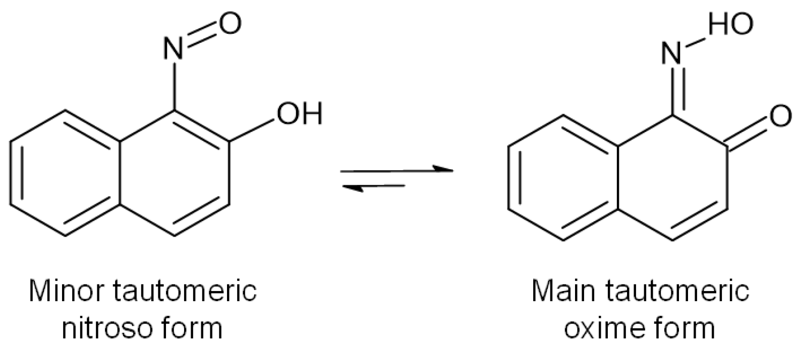
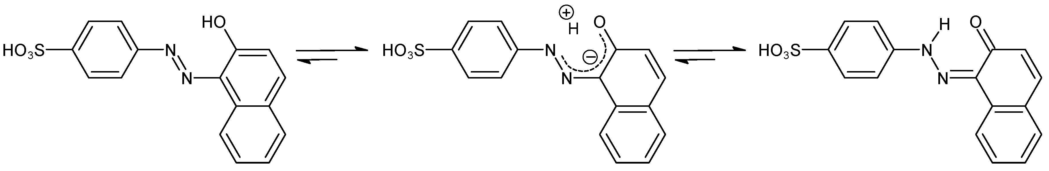
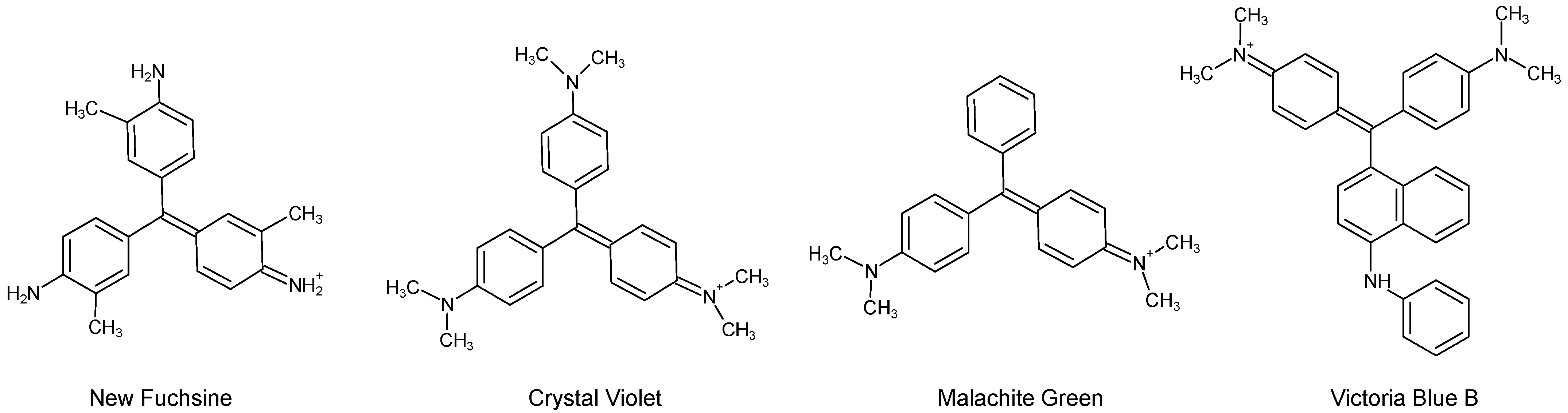
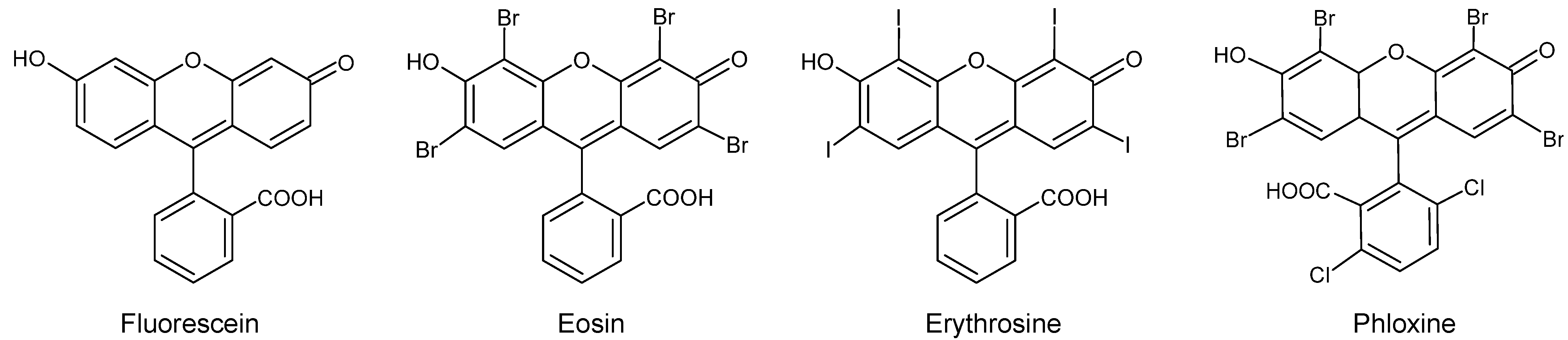
2. Categorisation and Chemistry of Early Synthetic Dyes

The rapid progress of ESDs in the second half of the 19th century resulted in their number growing exponentially in a few decades. In 1924, the Colour Index was created by the Society of Dyers and Colourists with the aim of clarifying and cataloguing the complex world of colourants [41]. The Index included chemical formula, chemical structures, manufacturer names, synthetic pathways, application guidelines and more [42]. The third edition of the Colour Index was printed in 1971 and contains around 27,000 colourants [43]. But, most importantly, the Colour Index assigned numbers to all colourants, so that a universal reference system was created, solving the problem of subjective commercial names being used.
However, synthetic colourants can be categorised in various ways. One classification considers the application of these molecules used as dyes on fibres [44,45]. Acid dyes refer to anionic, water-soluble molecules that are generally applied in an acidic bath. They contain acid groups, such as SO3H and COOH, and they establish bonds with protonated amino groups present on proteinaceous fibres. Basic dyes are cationic molecules and most of the first synthetic dyes belong to this category. The basic centre is often a nitrogen atom that can accommodate protonation and these dyes react with acid groups on the fibres. While proteinaceous fibres naturally contain such acid groups, cellulosic fibres do not, and hence, they need mordanting. To address this limitation, direct dyes were specifically designed to dye cotton without the aid of mordants. They are mostly anionic but their interaction with the cotton fibres is not ionic and is referred to as substantivity [46]. Most of these dyes have symmetric molecular structures with at least two azo bonds and they generally need an electrolyte in the dye bath, such as NaCl or Na2SO4. Ingrain dyes form a special category that was developed in the 1880s with the idea of forming the dyes directly on the fibres to overcome solubility problems. Two soluble components are introduced into the fibre and suitable conditions for their azo-coupling are used [45]. For example, the fibres can be saturated with an alkaline solution of a suitably substituted naphthol and, after drying, they are passed into a neutral solution of a diazotised base. Mordant dyes require metallic mordants and encompass a large variety of chemical structures, including anthraquinones, but the Colour Index reports that this category does not have a strict definition, and its make-up is more conventional. Other categories, such as solvent dyes, disperse dyes and vat dyes, are reported in the Colour Index, but the classification can be less precise, as in the case of mordant dyes. These categories were adopted by the Colour Index to create the Colour Index (C.I.) Generic Names, which are composed of the dye’s recognised usage class, its hue and a serial number, e.g., Acid Red 88, Basic Violet 14, Solvent Red 2, etc. [47], as exemplified in Figure 1.
In some cases, the same colourant can be present in more than one application category. For example, a disperse dye can often be applied as a solvent dye, and a vat dye can sometimes be used as a pigment, etc. In these cases, the Colour Index generally assigns two different Generic Names. However, a chemical categorisation based on the molecular structures is also provided in the Colour Index, for which the C.I. Constitution Numbers were created, as summarised in Table 1 [47]. These five-figure numbers represent the conventional way to unambiguously refer to colourant molecules.
Nitroso dyes are characterised by the presence of the NO group on aromatic rings, which establishes a tautomeric equilibrium with the oxime form [48] (Figure 2). These dyes are historically prepared by the action of nitrous acid on phenols or naphthols and form green iron complexes [45], which represent their main application.
Nitro dyes are characterised by the NO2 chromophore. Although picric acid (C.I. 10305), was obtained in 1771, as previously discussed [45], Martius Yellow (2,4-dinitronaphthol; Acid Yellow 24, C.I. 10315) was the first nitro yellow obtained in 1864 by the deliberate synthetic process of using nitric acid on phenols, naphthols, diphenylamines and other compounds to introduce nitro groups at the ortho- and para-positions [45]. The popular Naphthol Yellow S (2,4-dinitronaphthol sulphonic acid; Acid Yellow 1, C.I. 10316) appeared in 1879 (Figure 3).
The azo bond -N=N- is the chromophore in azo dyes. The diazotisation reaction converts aromatic amines into diazonium salts by using nitrous acid and mineral acid [49]. The resulting diazonium salts are unstable and can be easily combined with an amine or a phenol to form azo dyes. However, only a few azo dyes (notably Bismark Brown—Basic Brown 1, C.I. 21000) were synthetised before 1875, when it was understood that the final products could be obtained in the same reaction flask where the diazonium salts were formed, without a need to isolate them [45]. Chrysoidine (Basic Orange 2, C.I. 11270) was the first of these dyes and many molecules were synthetised in the following years. Monoazo dyes contain one diazo bond, but diazotisation and azo-coupling can be repeated. Biebrich Scarlet (Acid Red 66, C.I. 26905) was the first disazo dye synthetised as early as 1879. Congo Red (Direct Red 28, C.I. 22120) is also a disazo dye in which benzidine was used as diazotisation centre. When it was first synthesised by Paul Böttinger in 1884, the direct cotton dye class was born (Figure 4). As diazotisation can potentially be repeated on amino functionalities, trisazo and polyazo dyes also exist.
However, when the azo group is adjacent to a hydroxyl group in the molecular structure, a tautomeric equilibrium is established between the azoenol and the ketohydrazone forms [50] (Figure 5). This is the case for all β-naphthol dyes [37,51]. Several studies have shown that, in these systems, the azoenol form is the minor tautomer in both solution and solid state and that the substituents on the aromatic rings modulate the equilibrium [52,53]. Thus, these molecules are more correctly referred to as “hydrazone dyes” and are represented as such in this review.
Among the earliest dyes to be synthesised, triarylmethane dyes were not immediately understood from a chemical point of view. The triarylmethane core used to be empirically obtained by mixing aniline derivatives with oxidising agents that would favour the coupling of the molecules, as in the early processes of obtaining rosaniline or magenta (Basic Violet 14, C.I. 42510), which started as early as 1856. It took almost 20 years of research to understand the chemical nature of these dyes, thus enabling their synthetic processes to be controlled more effectively [45]. In addition to the popular formulations of Methyl Violet (Basic Violet 1, C.I. 42535) and Crystal Violet (Basic Violet 3, C.I. 42555), the use of different reagents producing different substituents on the aromatic rings led to the exponential expansion of this dye class, with Victoria Blue dyes (Basic Blue 26, C.I. 44045 and Basic Blue 11, C.I. 44040) and Diamond Green dyes (Basic Green 4, C.I. 42000 and Basic Green 1, C.I. 42040) among the most popular ones [28,54] (Figure 6).
As early as 1862, the sulphonation of triarylmethane dyes was introduced by treatment with sulphuric acid and the so-called aniline blue or Nicholson Blue (Acid Blue 93, C.I. 42780) became popular due to its increased water-solubility compared to non-sulphonated triarylmethane dyes [44,45]. Additional classes of sulphonated dyes, such as Patent Blues (Acid Blue 3, C.I. 42051 and Acid Blue 5, C.I. 42052), which are hydroxylated diaminotriphenylmethanes, were quickly developed (Figure 7).
Diarylmethane dyes represent a small category, with Auramine (Basic Yellow 2, C.I. 41000) being the main dye. Its synthesis from Michler’s ketone (bis[4-(dimethylamino)phenyl]methanone) represents an insightful reaction (Figure 8), as it appears to revert during ageing [54,55]. A similar mechanism has been observed as the degradation pathway of other triarylmethane dyes [28], as discussed in Section 5.2.
The xanthene ring is the chromophore of the xanthene dyes, sometimes referred to as resorcinol dyes in old literature, as they were obtained by the initial condensation of resorcinol and phthalic anhydride to form a xanthene ring substituted with two hydroxy groups and one carboxyphenyl group [44,45]. The simplest molecule of this dye class is Fluorescein (Acid Yellow 73, C.I. 45350), which takes the name from the fluorescent properties of this dye class. Modifications of Fluorescein yielded several popular red/pink dyes. Eosin (Acid Red 87, C.I. 45380) is the tetrabromo derivative of fluorescein; Erythrosine (Acid Red 51, C.I. 45430) is the tetraiodo derivative; Rose Bengale (Acid Red 93, C.I. 45435) corresponds to the tetraiododichloro derivative; Phloxine (Acid Red 98, C.I. 45405) corresponds to the tetrabromodichloro derivative of Fluorescein (Figure 9).
Rhodamines are another important class of xanthene dyes. Their main feature is their basic character, which is provided by the presence of amino groups. Rhodamine B (Basic Violet 10, C.I. 45170) was probably the most popular dye of this class, followed by its isomer Rhodamine 6G (Basic Red 1, C.I. 45160). The addition of sulpho groups to rhodamines brings back the acid character and gives a deeper violet colour. Acid rhodamines include the popular Violamines (Figure 10), such as Violamine B (also known as Fast Acid Violet B, Acid Violet 30, C.I. 45186), Violamine R (also referred to as Fast Acid Violet A2R, Acid Violet 9, C.I. 45190), Violamine G (C.I. 45200) and Violamine 3B (also named Fast Acid Blue R; Acid Blue 19, C.I. 45205), which differ for the different N-substituents on the amino groups.
Galleine (Mordant Violet 25, C.I. 45445) and Coeruleine (Mordant Green 22, C.I. 45510) represent a special class of xanthene dyes. Obtained by coupling phthalic anhydride and pyrolgallol (1,2,3-trihydroxyphenol) in an oxidative atmosphere, the high degree of hydroxylation makes them quite similar to anthraquinone dyes (mordant dyes) for application purposes (Figure 11).
The category of synthetic anthraquinones was born in 1868 with the discovery that the natural molecule alizarin (1,2-dihydroxyanthraquinone, Mordant Red 11, C.I. 58000) could be synthesised from anthracene. The synthesis of tri-, tetra-, penta- and hexahydroxyanthraquinones quickly followed, yielding dyes of all shades. Different substituents on the anthraquinone rings produced dyes with different characteristics, e.g., Alizarin Orange (1,2-dihydroxy-3-nitroanthraquinone, Mordant Orange 14, C.I. 58015), although all these dyes require a mordant. The addition of another condensed aromatic ring containing a nitrogen atom produced the popular dyes Alizarin Blue R (C.I. 67410), Alizarin Green S (C.I. 67405) and Alizarin Black P (C.I. 67425/67430), as shown in Figure 12. Mordanting with chromium was often used in combination with these dyes to obtain dark shades and blacks.
The azine dyes represent another broad category (Figure 13). Mauveine (C.I. 50245) itself was found to be an azine dye after substantial effort from various chemists, who tried to unveil its chemical formula and structure. The azine aromatic structure is represented by two aromatic rings connected by two nitrogen atoms in a phenazine configuration. Two amino groups are commonly positioned on the aromatic rings and various N- and C-substituents are introduced. Safranine (Basic Red 2, C.I. 50240) is among the earliest and simplest of these dyes. The introduction of additional N-phenyl groups produced the Indamine Blues (C.I. 50204 and following) family and the Nigrosines (C.I. 50420 and following). The easy introduction of condensed aromatic rings further expands this category, with molecules such as the Rosindulines (C.I. 50080–50095). In the acridine family, the phenazine ring is replaced by two aromatic rings connected by one nitrogen and one carbon atom. Phosphine (Basic Orange 15, C.I. 46045) is reported as a mixture of methylated derivatives of 4-amino-7-aminophenylacridine. Thiazines contain a nitrogen and a sulphur atom connecting the two aromatic rings. Methylene Blue (Basic Blue 9, C.I. 52015) is by far the most known dye in this category and contains two fully N-methylated amino groups. An additional nitro group is present in Methylene Green (Basic Green 5, C.I. 52020), whereas Thiocarmine R (C.I. 52035) is a sulphonated thiazine with N-benzyl groups. Oxazines contain an oxygen atom instead of a sulphur atom when compared to thiazines, and thus, the phenoxazine ring is the central nucleus. Meldola Blue (Basic Blue 6, C.I. 51175) is the most famous dye in this category and takes the name from the chemist who synthesised it first, starting with nitrosodimethylaniline and β-naphthol. In fact, this molecule has an additional condensed aromatic ring.
Classes and categories have kept expanding during the 20th century and this topic has been often reviewed [48,56].

3. Synthetic Organic Pigments

Unlike ESDs, SOPs exhibit insolubility in their intended medium, sometimes retaining their crystal and particle structure. Certain organic colourants are inherently insoluble in the most common solvents. However, in most cases, synthetic dyes require a substrate or a precipitating agent, depending on their chemical structure and intended use [3]. Therefore, some of the molecules described in Section 2 constitute the organic component of SOPs. Various precipitation processes, chemical treatments and the incorporation of extenders and coatings were developed, creating complex systems, about which a considerable amount is yet to be understood. Common applications of SOPs encompass decorative and protective coatings, oil- and resin-based paints, emulsion paints, distempers, lime and other aqueous-based paints, pigment finishes on leather and textiles, printing inks on paper and textiles, photography, colouration of plastics, etc.
The complexity of these materials is directly related to the five crucial stages affecting the composition of the SOPs found in heritage objects: (i) initial synthesis of the colourant; (ii) subsequent processing of the synthetic colourant into SOP with possible precipitation on a substrate, coatings and changing the particle size; (iii) integration of the SOP into a medium alongside various components such as additional pigments, fillers, additives and extenders; (iv) application of the resulting paint; and (v) ageing processes. The available contemporary literature on the manufacturing and characteristics of SOPs is scarce and predominantly in German [2,57,58]. Moreover, sources detailing certain precipitation methods may lack accessible samples to verify the existence and use of these SOPs. It must also be noted that the number of SOPs developed and used up to World War I was relatively limited. In 1887, a list of 21 pigments was published, all derived from synthetic alizarin. These could be safely used according to the German Society for the Promotion of Rational Painting Techniques. In 1930, Martin Henry Fischer (1879–1962) warned his fellow artists: ‘It is a good rule for the painter to eliminate everything from the manufacturer’s catalogue which bears a name unfamiliar to him or the content of which is not wholly disclosed to him’ thereby referring to new SOPs, which should be avoided in his opinion [59].
SOPs can be categorised according to their dye counterparts, whose characteristics determine how the dye can be precipitated onto a substrate. Before precipitation the dye is brought into solution, then a solution or suspension of the substrate or precipitating agent is added under varying conditions, e.g., high temperature, high pressure and stirring. Different solvents and chemicals were used to ensure the dissolution of the dyes. When the precipitation is complete, the SOP particles are washed, filtered, and dried. Then, the particles could be optionally coated, milled and further processed [3].
As the classification of the main groups of organic colourants is previously discussed, this section covers some categorisation based on the precipitation method.
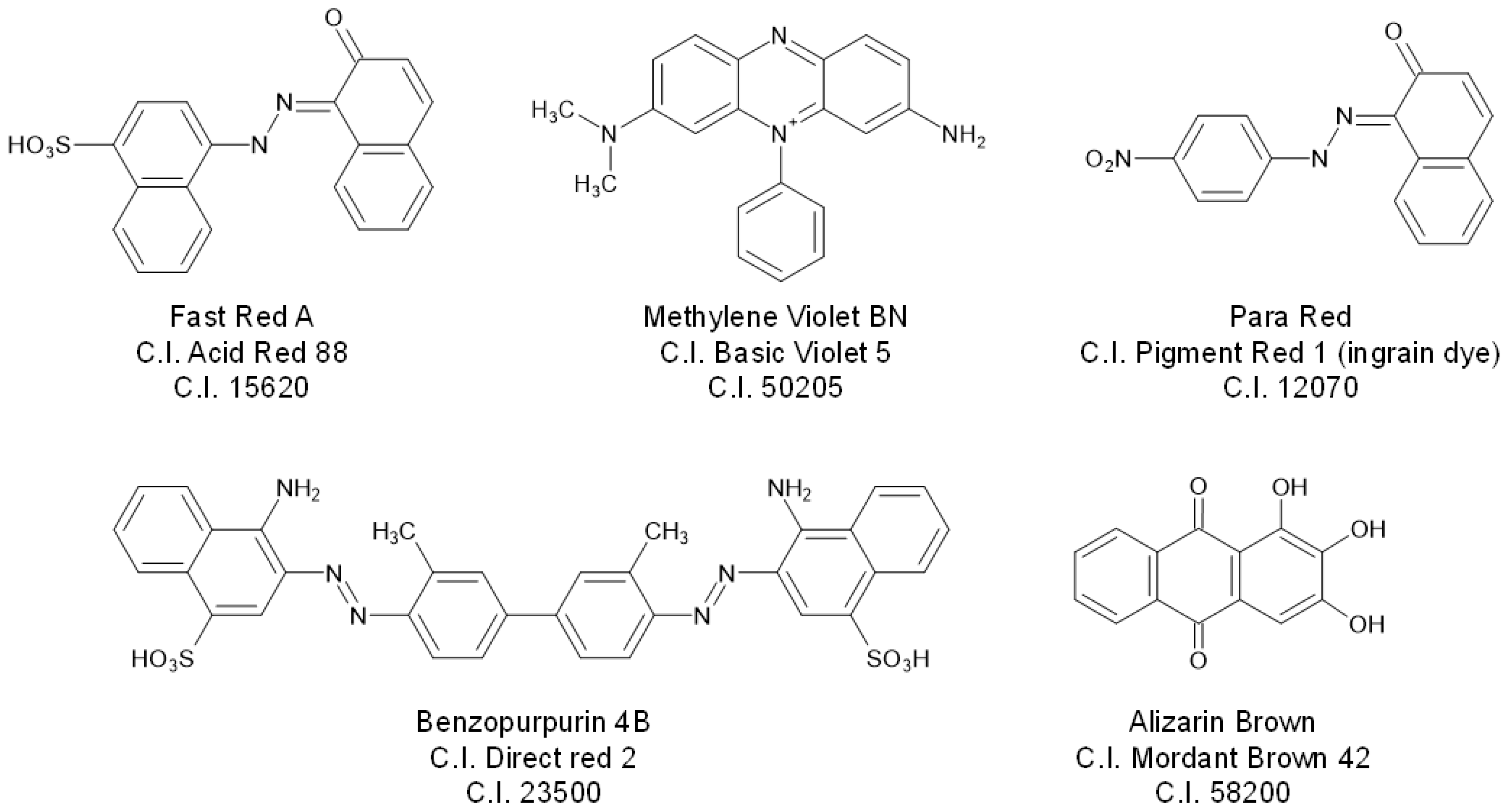
  • Organic pigments: these are the organic colourants that are insoluble in a medium as they are. Not all of these have a corresponding dye, e.g., Pigment Red 3, a β-naphthol dye (C.I. 12120) and Vat Blue 4/Pigment Blue 60/indanthrene blue (C.I. 69800).
  • Toners: although this term is outdated, it was used to indicate a concentrated SOP. These are insoluble salts of water-soluble acid dyes with an appropriate counterion that acts as a precipitation agent, e.g., Ca, Ba, Mn, Sr inorganic salts, or the salts of water-soluble basic dyes complexed with acids, such as tannic acid. The C.I. records the different precipitation agents, e.g., Pigment Red 57:1 (C.I. 15850:1) is the calcium salt of PR57 and Pigment Red 57:2 (C.I.15850:2) is the barium salt of Pigment Red 57.
  • Lakes: these are similar to toners but precipitated with a substrate of a light and neutral colour, e.g., alumina, blanc-fix, to form a metal complex, in analogy with historical lake-pigments prepared with natural dyes. Their tinctorial strength is lower than toners or organic pigments due to the lower organic dye content and the presence of a neutral-coloured substrate, hence, this class came into decline after WWI. Generally, dyes with only basic characteristics do not form lakes, whereas acid dyes do, e.g., Pigment Red 60 (C.I. 16105:1) is the barium lake of Mordant Red 9 (C.I. 16105)
  • Extended pigments: these are pigments or toners (rarely lakes) diluted with a light and neutral-coloured extender (e.g., alumina, blanc-fix, calcium carbonate). The extender is not an integral part of the precipitated pigment but is incorporated in the pigment particles. Also, these are called ‘reduced’ pigments as the colour is reduced in intensity by the colour of the extender.
The toner and lake SOPs can be precipitated using a wide variety of substrates. Selecting an appropriate substrate is challenging and must be tailored to the intended use of the SOP. Moreover, the lightfastness and solubility of the pigment are affected by the substrate and incomplete precipitation can result in bleeding into the medium. Certain precipitation agents/substrates were used for specific dyes for brief periods, before becoming obsolete. Some organic colourants can be precipitated together as combined toners or lakes, e.g., two or more dyes are brought into a solution together and then precipitated simultaneously on the same precipitation agent/substrate [60]. An overview of some of the common precipitation agents/substrates used for the precipitation of SOPs is given in Table 2 [3,57,58].

4. Analytical Techniques

The preservation of the integrity of valuable works of art has always been a crucial point when investigating their physical and chemical composition. Thus, non-invasive analytical techniques have been developed and are often applied to the study of synthetic colourants. These techniques are generally rapid, present high versatility and enable information to be obtained from multiple areas of an object without the need to sample. The application of non-invasive techniques can also provide key information to guide selective and strategic sampling for subsequent invasive approaches, which are often more accurate or sensitive to investigate the molecular composition of ESDs and SOPs as well as degradation products and synthesis by-products. Invasive techniques can be non-destructive when the sample is not modified during analysis and can be potentially re-analysed multiple times, or destructive when the sample is treated or destroyed to extract the information. Some spectroscopic techniques can be used in both non-invasive and invasive modalities, as summarised in the following sections. When sample collection is required, the amount is generally very low (usually less than 0.5 mg and down to a few fibres in some cases). Therefore, the sampling from the artwork is often unnoticeable and the approaches are referred to as micro-invasive. Nevertheless, multi-analytical approaches are often used nowadays in these types of investigations, combining non-invasive and micro-invasive techniques. The most frequently and successfully applied techniques in this field are discussed in the following sections along with their advantages and limitations as well as examples of some relevant applications. An overview of the analytical workflow is presented in Table 3.

4.1. Spectroscopic Techniques

Reflectance spectroscopy is a well-established technique for the characterisation of dyes and pigments. Three areas of application can be outlined: material identification, colour change measurements and colour matching. Colour appearance is not an objective property, as it depends on the light source, the observer, the object dimensions and the background [61]. Colourimetry exploits reflectance spectroscopy and enables colour to be measured and systematically defined based on the CIE L*a*b* colour system. The ΔE value is calculated as the square root of (ΔL*)2 + (Δa*)2 + (Δb*)2 and is the main tool to evaluate colour differences. Colourimetry has been applied to evaluate the possible fading of synthetic colourants in aged paint mock-ups [62,63,64,65], felt-tip pen inks on papers [66], historical plastic objects coloured with β-naphthols [67] and dyes on textiles [23,24].
The most common way to use reflectance spectroscopy for non-invasive material identification is by coupling a spectrophotometer with a fibre-optic probe. The resulting system, referred to as FORS (fibre optics reflectance spectroscopy), is utilisable in situ and enables many reflectance spectra to be quickly recorded and visualised in real-time [68]. The instrumentation is relatively inexpensive and safe both for the operator (no exposure to heat or high-energy beams) and for the artwork (no contact, mechanical or physical stress). Databases containing a wide range of FORS spectra of SOPs are available [29] and the Istituto di Fisica Applicata Nello Carrara of the Italian National Research Council (IFAC-CNR) created an online database of reflectance spectra of traditional and synthetic pigments [69]. FORS has been applied to identify ESDs and SOPs in powders and paint tubes belonging to Fernando Melani (1907–1985) and Lucio Fontana (1899–1968) atelier materials [70,71], in oil paintings [72,73], prints [74,75] and historical textiles [25,32,76,77,78,79]. The technique has limitations due to a relatively poor wavelength resolution (ca. 2–5 nm) and a limited “fingerprinting” ability compared to other spectroscopic approaches. This was evident in the analysis of the miscellaneous pigments contained in “The Italian Flag” (Fernando Melani; 1955–1960, acrylic resin on textiles), in which only phthalocyanines were detected, although several β-naphthols and xanthenes were present [80]. Similarly, in “Tuttomondo” (Keith Haring, 1989, styrene resin on wall), only the general classes of the SOPs were assigned by FORS [81]. Another limitation is given by the dimension of the probe aperture, which is usually larger than 1 mm, thus preventing artwork details smaller than the probe to be investigated without spectral contributions from surrounding areas. For textiles, the weaving structure can also be problematic, as threads coloured with different dyes can be present in the same area; moreover, the contribution due to scattering related to the thread structure cannot be overcome. To summarise, FORS more commonly identifies the chemical classes of these molecules (as proven for trialrylmethanes, xanthenes, phthalocyanines, Hansa Yellows, quinolines, etc.) [29], whereas other techniques are required to confirm the identifications at the molecular level and check for the possible presence of mixtures [32,74,75,77].
Recently, reflectance spectroscopy has also been configured as an imaging technique, commonly referred to as hyperspectral imaging (HSI). By using special cameras, reflectance spectra are recorded for each pixel of an image, and, upon suitable data treatment, false-colour images are created, showing the distribution of materials on the surface [82]. In order to derive a reliable interpretation, data must be compared with those obtained for reference materials [83]. While numerous applications of HSI for the identification of mineral pigments in paintings are available, applications to synthetic organic pigments and dyes remain scarce [83,84,85,86,87], again owing to the limited “fingerprinting” capabilities of the technique towards these materials. Although missing the spectral information, multiband imaging (MBI or MSI), a photographic technique that exploits the absorbance, reflectance and luminescence properties of materials in different ranges of the electromagnetic spectrum, has also been used to map the distribution of synthetic colourants in textiles [78].
Fluorescence spectroscopy exploits the characteristic of several ESDs and SOPs to exhibit high fluorescence. Micro-spectrofluorimetry has been successfully applied to detect SOPs in atelier materials [71] and in van Gogh’s and Lucien Pissarro’s paintings [30]. Confocal µ-fluorescence spectroscopy shows high spatial resolution similar to 3D fluorescence spectroscopy, and it was successfully applied to the analysis of eosin lake paint model systems [88]. Several reference SOPs were analysed by 3D fluorescence spectroscopy and the method was applied to study an Emile Bernard (1868–1941) painting [6]. This in situ technique is very efficient in performing a first scanning of the surface allowing to select representative areas for minimising the subsequent sampling step. Unfortunately, the results are strongly affected by the substrate, pH and other materials present in the artwork: this issue is common to most non-invasive techniques, which generally suffer from matrix effects [6]. Moreover, when a mixture of dyes is used, the low fluorescent dyes can remain completely undetected. Time-resolved fluorescence (TRF) spectroscopy enables the fluorescence decay of fluorophores to be registered after excitation or the fluorescence anisotropy decay due to the reorientation of the emission dipole of the fluorophores to be registered during excitation [89]. The technique enables the quick differentiation of the painting materials, mapping their distribution and, in some cases, detecting chemical changes. Measures can be performed in situ, without any requirement in terms of the darkness of the surrounding environment and in a quantitative way. Nevertheless, it has been noticed that the emission of different natural and synthetic dyes (e.g., disazo, phtalocyanines) exhibit similar lifetimes, and that the same dye can be characterised by different lifetimes depending on its manufacture, degradation state and sample heterogeneity [90]. Thus, complementary analyses are required to avoid deriving misleading interpretations of different time-resolved emissions.
Fourier-transform infrared spectroscopy (FTIR) can be applied in situ thanks to portable devices, or non-destructively on micro-samples with benchtop instruments. It has been applied for the detection of ESDs and SOPs in reference materials, felt-tip pen inks, drawings, paintings and textiles in both transmission [63,66,70,71,81,91,92] and attenuated total reflection (ATR) modes [63,66,71,93,94,95,96,97,98]. Lakes prepared with the same dye but complexed with different substrates produce different FTIR spectra due to their different coordination geometry. Furthermore, FTIR may allow the identification of fillers and extenders and provide details on binding media in paint samples [95,97]. Synthesis conditions also affect the structure and composition of lakes, and consequently their FTIR spectra [99]. Diffuse reflectance infrared Fourier-transform (DRIFT) and external reflectance FTIR spectroscopies were applied for the analysis of Turkey Red in historical textiles [100] and Henry Matisse gouaches [33], respectively. Recently, metal underlayer attenuated total reflection Fourier-transform infrared spectroscopy (MU-ATR) has been proposed as a new methodology to enhance the FTIR signal by depositing a micro-extract on a gold-coated glass; a commercial FTIR microscope with a fixed incident angle is then used [101]. This technique was successfully used on the extracts of reference pigments, colour charts and dyed fibres [22,101,102], but it proved to be much less efficient when directly applied on a gold-coated sample. Finally, the brilliance, monochromaticity and high focalisation of synchrotron radiation have been recently exploited in SR-μ-FTIR imaging techniques for the determination of organic pigments in historical artworks [103]. Thus, FTIR-based techniques are a good choice to sensitively identify dye classes without being destructive (except for MU-ATR) or, in some set-ups, invasive, but they can be limited in differentiating similar molecules and detecting specific degradation processes. Moreover, diagnostic signals can be hidden by the sample matrix.
Raman spectroscopy has become one of the most important analytical techniques for the investigation of ESDs and SOPs. Although Raman and FTIR spectroscopies are considered complementary techniques for the characterisation of organic materials, Raman has shown some advantages over FTIR, such as the versatility of the irradiation source (different kinds of laser can be chosen), the higher spatial resolution and higher sensitivity to detect the weak vibrational frequencies of specific chemical groups, and the lower interference from the matrix signals. This clarifies why Raman has been extensively used for SOP characterisation and identification within artworks and exhaustive databases were produced, allowing the quick and effective interpretation of the results [104,105,106,107,108,109,110]. A Raman database, containing nearly 300 reference spectra of synthetic organic pigments (SOPrano), was built by several research groups coordinated by the Royal Institute for Cultural Heritage (KIK/IRPA, Brussels) [31] and is available in digital format [111]. Both benchtop and portable instrumentation are available, the latter being valuable when pigments must be pinpointed in artworks characterised by wide chromatic areas [112,113]. Raman spectroscopy performs very well for the identification of SOPs in reference and historical atelier material [91,110,114,115], in drawings and paintings of modern artists [33,74,80,92,93,97,106,110,116,117,118], in plastic design objects [119] and sculptures [120]. The main limitation of the Raman spectroscopy is the background fluorescence signal produced by the organic matrix or support. In the case of most natural dyes and several synthetic ones, the background fluorescence makes the detection of Raman signals often impossible. To overcome this issue, some advanced protocols were developed. The use of surface-enhanced Raman spectroscopy (SERS) enables the Raman signal to be enhanced by more than seven orders of magnitude, thanks to the surface plasmonic resonance phenomenon obtainable by using silver or gold nanoparticles [34,121]. It can be applied deposing the silver nanoparticles directly on the artwork, or on a micro-sample (as-is or on an extract), resulting in a micro-invasive/micro-destructive technique. Examples of successful use of SERS encompass the identification of SOPs in commercial artists’ colour charts [22], paintings [92], inks [66,104,118], dyes on textiles [94,122] and colourants in Japanese woodblock prints from the Meiji period [75,123,124]. Additionally, an interesting study has been recently carried out on the photostability of acrylic and alkyd paints, showing that Raman is effective in revealing the degradation phenomena occurring in the binder but not those relative to the pigment [125].
When dealing with spectroscopic data, given the rapidity of data collection and the consequently high amount of information obtained, along with the relatively low specificity of the profiles, post-processing elaboration and multivariate statistical data analysis is fundamental to enhance the relevant spectroscopic features and to evidence discrimination among samples within the dataset [126,127].

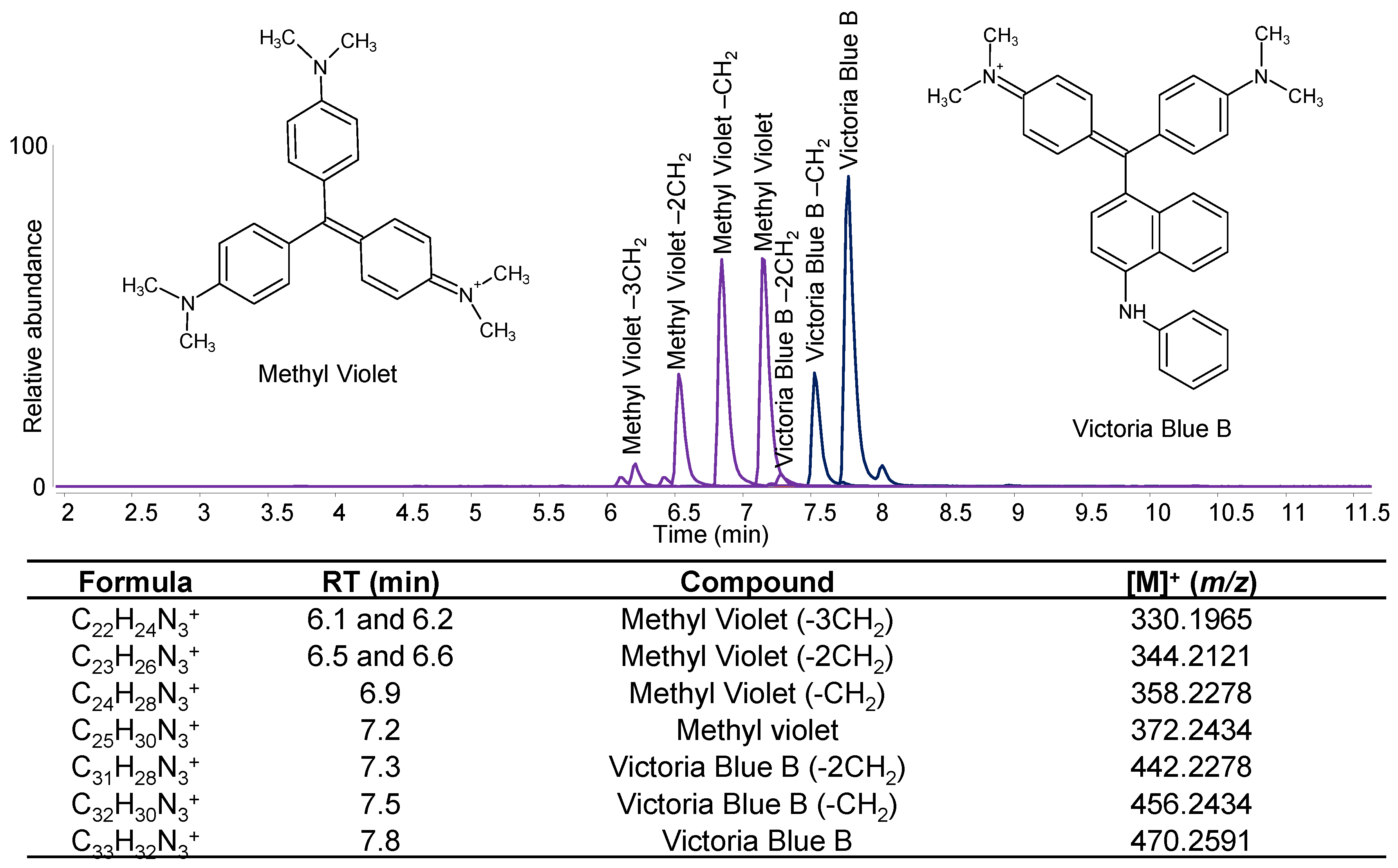
4.2. Mass Spectrometry Techniques (No or Minimal Sample Treatment)

Colourant molecules are generally too polar and non-volatile enough to be analysed by gas chromatography–mass spectrometry (GC-MS). However, some studies have focused on the use of derivatising agents to detect some natural dyes and some diagnostic hydrolysis products by GC-MS [128,129]. Although no specific application to synthetic dyes has been attempted with these analytical protocols, some overlap exists between the chemical classes of natural and synthetic dyes. Conversely, direct mass spectrometry techniques have been explored more consistently with specific application to synthetic colourant identification. Direct temperature resolved–mass spectrometry (DT-MS) is a micro-analytical technique that enables the direct analysis, without any pre-treatment, of the desorbed products from a platinum/rhodium filament by MS or MS/MS, which was successfully applied to the analysis of pigments in powder form [130]. On the one hand, the technique is promising for the analysis of aged paint tubes or paint samples, which can become insoluble upon ageing. On the other hand, the binding media contained in paint formulations may produce fragment ions which could overlap with those related to the dye/pigment. Thus, the ionisation and desorption conditions need to be optimised to enhance the selectivity towards the colourants. It was proven that the application of the electron impact (EI) at 16 eV or chemical ionisation (CI) are suitable for the analysis of SOPs in acrylic emulsion paint both in the case of commercial formulations [131] and in a sample collected from a painting by Sam Francis [132].
Pyrolysis gas chromatography coupled to mass spectrometry (Py-GC/MS) is a widely employed technique that exploits the thermal degradation of the analyte in an inert atmosphere to reduce its chemical complexity. The technique enables the rapid identification of some synthetic colourants without sample preparation or derivatisation steps by identifying characteristic pyrolysis products, which have been listed in various libraries [64,133,134]. The technique is very useful in the case of pigment formulations insoluble in the most common extraction solvents. However, many classes of colourants have a common molecular skeleton, thus often incurring in common fragmentation patterns, but the characteristic pyrolysis products of individual colourants have been ascertained in some cases [64,135]. Py-GC/MS has been widely applied for the analysis of felt-tip pens [63,66,93], pigments in Clyfford Still, Jackson Pollock and Francis Bacon paintings [64,134], and contemporary plastic objects and artworks [67,136]. The analysis of SOPs in commercial powders and in acrylic emulsion paints revealed the formation of different pyrolysis fragments suggesting that the compounds incorporated within the paint could affect the thermal degradation pathways [137]. Although pyrolysis is not able to provide detailed information on photo-ageing pathways, differences between the pyrograms of unaged and artificially aged paint model systems can provide useful information [64].
Laser desorption ionisation–mass spectrometry (LDI-MS) and matrix-assisted LDI-MS (MALDI-MS) are soft ionisation techniques and represent versatile, rapid and sensitive methods for the analysis of ESDs and SOPs, as they are not significantly affected by interferences from the matrix. Following minimal sample treatment and exploiting a laser to desorb the analytes from the matrix, positive and negative ion spectra are rapidly acquired and colourants are distinguished and characterised according to the m/z values of the corresponding ions. LDI-MS and MALDI-MS were used for the analysis of SOPs in reference materials [97,115], historical textiles [138] and contemporary paintings [98,106,139]. MALDI-MS/MS was also employed for detecting tattoo organic pigments, generally poorly soluble, in ink and tissue samples [140]. These techniques were also employed to study the photodegradation of rhodamine B and crystal violet in pen inks on paper and the role played by the matrix on the yield of ionisation was compared [141,142,143]. Even if the characterisation of the various degradation compounds is hampered by the lack of a separation step, LDI-MS has been applied to the study of artificially aged inks [144].
Ambient ionisation mass spectrometry (AMS) encompasses a series of newly designed instruments based on a novel ionisation technique developed in 2004. The definition of “ambient ionisation sources” includes different kinds of techniques such as paper spray (PS), direct analysis in real-time (DART), desorption electrospray ionisation (DESI), direct infusion-electrospray ionisation (DI-ESI), and surface acoustic wave nebulisation (SAWN). AMS rapidly ionises samples with only minimal pre-treatment or can be used on the samples/objects in situ. PS-MS allows the direct analysis of untreated samples from paper or other porous media, and it requires neither elevated temperatures nor gas, and thus it is particularly appropriate for portable MS. It was applied for the detection of dyes in foods and beverages [145,146]. DART is highly versatile, and a sample may be analysed in various states (gas, liquid and solid). DART-MS and DI-ESI-MS were applied to investigate the photodegradation pathway of eosin in linseed oil; DI-ESI-MS proved to be more informative [147]. A DESI source has recently been built, optimised and applied to the identification of historical early synthetic dyestuffs from a dye pattern book [148]. SAWN is a softer ionisation method than ESI, in which no voltage is applied to the sample before or during analysis, thus avoiding the possible electrochemical oxidation of the analytes. SAWN was used for the analysis of SOPs and ESDs in reference powder materials and historical textiles [149].
In recent times, mass spectrometry techniques, such as MALDI, DESI and SIMS (secondary ion mass spectrometry) have been implemented in imaging mode. DESI, mounted on a 2D stage, enables MS spectra to be collected by rastering the spray across the sample surface. DESI-MSI was used to map the distribution of synthetic dyes in printed and dyed fabrics in the late 19th century [38,150]. MeV-SIMS is a technique where primary ions of MeV energies induce the desorption of molecules from the sample surface by electronic sputtering. The relative release of secondary molecular ions is then detected by a time-of-flight (ToF) detector. A spectral database of some SOPs belonging to different chemical classes has been developed by analysing alkyd and acrylic commercial paints, proving the capability of the technique to identify simultaneously both the pigment and the binder in a short analysis time [151]. The first use of MALDI-MSI was recently published, highlighting the potentialities of the developed protocol for the study of the spatial−temporal chemical changes of Eosin in oil mock-ups as well as samples from Van Gogh artworks, correlating the chemical changes with the discolouration of the painting [39].

4.3. Chromatographic Techniques

Following the extraction of the chromophore-containing molecules from the matrix, chromatographic techniques enable their separation and quantitation. Due to the very broad range of dye classes, differing in terms of solubility and stability in acid and alkaline media, the selection of one comprehensive extraction method applicable to all the classes appears as a virtually impossible task. The main extraction methods, mostly developed for natural dyes but applicable to synthetic dyes as well, are listed below.
  • Boiling the sample in a mixture of organic solvents and strong acids is the strongest and most aggressive method, which results in good molecular recovery but may be responsible for the cleavage of labile bonds and oxidation of moieties. HCl 0.5 M and MeOH (1:1) or HCl/MeOH/H2O (2:1:1) are the most used extraction solutions [152].
  • Formic acid is a weak acid, which is used in solution HCOOH: MeOH (5:95 v/v) and allows one to perform a mild extraction [153].
  • Ethylenediaminetetracetic acid (EDTA) is a complexation agent used because it is a strong chelator of aluminium and of other ions, thus releasing molecules from complexes without decomposing them. To improve the extraction yields, EDTA can be used in a mixture with an organic solvent such as dimethylformamide (DMF) [154].
  • Oxalic acid is also used for its mild acidic and chelating properties. It is mixed in an oxalic acid/MeOH/acetone/H2O (1:30:30:40, v/v/v/v) solution and is mainly used for mordant dyes and organic lakes [155]; it is also used in a mixture pyridine/H2O/1.0 M oxalic acid (45:45:10 v/v/v) [32].
  • Ammonia-EDTA treatment of the sample can be followed by an ion pair dispersive liquid–liquid microextraction in chloroform using tetra-n-butylammonium bromide (TBAB) as ion pair reagent and methanol as disperser [156].
  • Hydrofluoric acid (HF) is used for the mild extraction of unstable colourants belonging to a wide range of classes [157], but needs to be handled with special glassware and extra care.
  • Polar organic solvents such as pyridine, dimethylsulphoxide (DMSO) and dimethylformamide (DMF) are commonly used upon heating at 80–100 °C mostly for the direct extraction of indigoids [21,78].
Different extraction techniques can often be combined, for example, by first using an organic solvent and, in a second step, an acid or complexing agent. Heating can be provided by water baths, heated metallic blocks, or oven, including the aid of microwaves or ultrasounds. Once the colourant molecules are in solution, their separation becomes fundamental to study the mixtures of original molecules, synthesis by-products and/or degradation products. UV–Vis spectroscopy is coupled with separative techniques. Databases of UV–Vis absorption spectra of synthetic dyes have been created [158,159,160,161]. This technique also provides a quick and inexpensive way to monitor dye degradation, although the identification of the intermediates and the characterisation of minor final products remain challenging [62,162,163,164]. UV–Vis spectroscopy can in principle be used without coupling with a separation technique for the analysis of colourants in solution, but overall spectra are obtained in the case of mixtures.
Thin-layer chromatography (TLC) has been employed for decades to separate the mixtures of dyes in different matrices by exploiting the chromatographic principles of adsorption and partition of the dye molecules between a stationary and a mobile phase. The molecular components separated on TLC plates can then be studied separately directly on the plate or collected for further analysis. For example, the reflectance spectra of the TLC spots can be acquired and used in the preliminary identification of the dye, allowing for a rapid screening [165]. TLC can also be coupled to FTIR [63,166] or Raman spectroscopy [104]. Recently, an innovative TLC plate made of silver iodide applied onto a gold-coated glass slide was combined with FTIR and SERS. The dye identification of Perkin’s mauveine, daylight fluorescent pigments, felt-tip pen inks and 19th–early 20th century French writing ink was performed by TLC-SERS directly on the silica plate [63,166,167,168,169].
Although a certain level of separation is obtainable by TLC, molecules with similar structures are not easily separated, and the information obtained lacks insight into the molecular level. High-performance liquid chromatography (HPLC) coupled with diode array detector (DAD) and/or mass spectrometry (MS) is therefore the method of choice for colourant characterisation, due to its high selectivity and sensitivity, excellent chromatographic efficiency, and the possibility of semi-quantitative or quantitative analysis, if analytical standards are available. Reversed-phase chromatography (RP-HPLC) is the most suitable method for the analysis of organic dyes and pigments, as most molecules of interest are polar and water-soluble [35]. The most used columns contain a C18 or C8 (or even C4) stationary phase. Water, acetonitrile and methanol are favoured eluents. Acetonitrile is preferred over methanol due to its lower absorbance in the UV range and lower viscosity, resulting in lower-back pressure [170]. The typical phase modifiers are formic acid, trifluoroacetic acid and phosphate buffers [35], but only formic acid is suitable for LC-MS systems. When dealing with acid dyes, ion-pair reagents such as tetrabutylammonium hydroxide or triethylamine can be admixed to the mobile phase to increase separation and peak resolution [6,36].
HPLC-DAD has been extensively used for the characterisation of synthetic colourants in archive materials and artworks [5,11,40,62,171,172,173,174,175,176,177,178,179], whereas HPLC-MS is slightly less common due to the higher cost of the instrument and higher complexity of data interpretation [11,21,32,36,62,80,156,160,171,172,173,174,180,181,182,183]. On the other hand, the straightforward identification of a compound can be more easily achieved by high-resolution tandem mass spectrometry (HR-MS/MS), whose applications have increased in heritage science in the last decade. In most cases, the chromatographic and mass spectrometric systems are coupled by an electrospray ionisation source and HR-MS/MS is mostly obtained by Q-ToF [37,184] or, more recently, orbitrap mass analysers [185,186]. HPLC-MS/MS has been applied to artworks, including archive and reference paint materials [11,27,37,65,70,187], historical inks [168,181], 19th–20th century textiles [78,79,86,184], modern and contemporary paintings [188,189,190,191,192] and street art murals [193,194].
In the last decade, the development of ultra-high-pressure liquid chromatography (UHPLC), capable of operating at a higher pressure than HPLC (maximum pressure 1300–1500 bar and 400–600 bar, respectively), has permitted to achieve better chromatographic resolution and lower detection limits in shorter times by exploiting stationary phases with smaller particle size [195]. This setup was used for the analysis of dyes in historical textiles [196]. An innovative two-dimensional liquid chromatography (LC × LC) system, with ion-exchange chromatography in the first dimension and fast ion-pair liquid chromatography in the second, was used for the separation of 54 dyes extracted from archive materials [183]. The method was also used to disclose the composition of dye extracts from 17th and 19th century heritage objects [26]. The potentiality of 2D chromatography was exploited in a recent approach developed for the study of dye photodegradation. This methodology utilises a novel light-exposure technology based on a liquid-core-waveguide (LCW) cell embedded in a multiple-heart-cut LC × LC system by coupling the cell to the heart-cut valve. The sample is flushed from the heart-cut loops into the cell by an isocratic pump. It is then irradiated using different time intervals and subsequently transferred by the same isocratic pump to a second-dimension sample loop to be separated in the second dimension [197]. The analytical performance of the new system was validated [198] and tested on dye references [199,200].

4.4. X-ray-Based Techniques

X-ray fluorescence (XRF) is rarely used for SOP identification, as the technique is only effective for elemental characterisation, and thus it is mostly applied for inorganic pigments. However, the detection of inorganic substrates such as tin or aluminium can be used as an indication that an SOP might be present. XRF can be used complementarily with micro-Raman spectroscopy to determine which paints most likely contain SOPs. Elemental analysis may also be used to ascribe the type of metal ions used in dye precipitation, as achieved by laser-induced breakdown spectroscopy (LIBS) on standard materials and textiles [201,202]. Additionally, for colourants containing halogen atoms, the presence of their signals in the elemental map can be a precious hint. Based on this, the presence of eosin was hypothesised thanks to the detection of bromine by XRF [40,75,179,203] and, when sampling was allowed, scanning electron microscopy-energy-dispersive X-ray spectroscopy (SEM-EDX) [30,40,179,203,204]. However, the small particle size of SOPs can make the localisation of the inorganic fraction of the substrate/precipitation agent in a cross-section difficult. Distinguishing lake, toner and extender pigments within a paint has not yet been successfully achieved, except when the substrate is highly specific for a particular type of precipitation.
SOPs may be crystalline and some of them have been characterised in powder form by X-ray powder diffraction (XRD) [205,206,207]. Their analysis in paintings is complicated due to presence of high concentration of fillers and extenders, hindering the pigments. Nevertheless, XRD has performed well in the identification of pigments in acrylic and alkyd commercial paints, while is nearly ineffective in oil and gum formulations [205].

5. Degradation and Synthetic Pathways

Organic materials are known to degrade due to the relative lack of molecular stability when exposed to various environmental factors such as light, humidity and temperature. Many synthetic colourants are renowned for being particularly light sensitive [6,23,24]. Studies on degradation mechanisms have elucidated some pathways that lead to the molecular degradation of ESDs and SOPs [11,62,65,96,147,200,208,209,210,211,212]. Separation techniques are often required for such studies, as degradation products and their parent molecules often show similar spectral features. Although bulk techniques can assess chemical changes related to ageing [28], these cannot identify the chemical mechanisms and the individual degradation products formed.
However, when it comes to identifying synthetic colourants and their degradation products, one of the main difficulties is to distinguish between synthesis by-products and degradation products, as these can easily overlap. An additional problem is that the parent molecule, synthetic by-products and degradation products can be potentially altered by intensive sample preparation. The knowledge of the precise chemical formulation of reference materials and the creation of exhaustive databases is fundamental for the correct description of dye degradation pathways [20,21]. Several studies on dye degradation and the identification of the photo-oxidation products have been conducted by HPLC-DAD-MS/MS under photo-accelerated conditions in solution [26,62,173,197,200,211,213] or in painting/textile matrices [65,208,210] as well as with attention to other mechanisms not related to light exposure [212]. Some of the observations gathered are summarised in the following sections.

5.1. Distinguishing Synthetic Pathways

During the initial period of synthetic colourant production, the use of pure starting materials as well as a high control over the synthetic pathway were not always easy to achieve. For example, in the early synthetic process of Fuchsine (C.I. 42510, Basic Violet 14), a mixture of aniline and toluidine was oxidised, thus producing a mixture of pararosaniline and its mono-, bis- and tri-C-methylated derivatives [79,214]. Therefore, although fuchsin is often described as trimethylpararosaniline, according to this early synthetic process, a mixture of molecules should be expected. In fact, the above-mentioned four molecular components are often detected in historical samples and usually exhibit a bell-shaped profile, probably reflecting the ratios of the starting materials used in the synthetic process [20,21,27,32,79,214]. However, in 1889, a different synthetic process for Fuchsine started being used, aiming to produce pure trimethylpararosaniline by condensing 4,4′-methylene-di-o-toluidine and o-toluidine [20,214]. The historical samples of fuchsin produced with this process tend to show trimethylpararosaniline as the most abundant component. [20,214]. As a result, the molecular profile of fuchsin is indicative of the synthetic pathway used for its production, as shown in Figure 14.

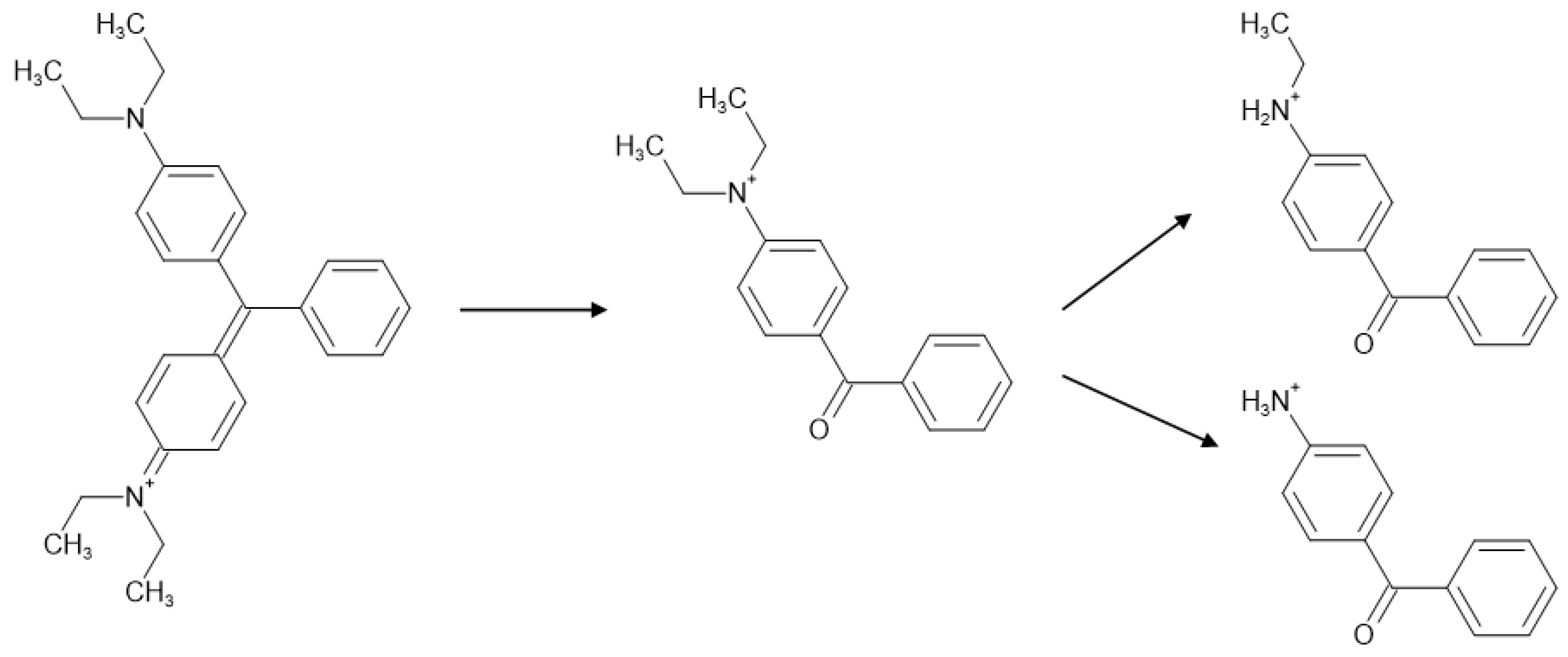
5.2. Dealkylation, Desulphonation and Loss of Other Functional Groups

Dealkylation is a known photodegradation pathway of various classes of synthetic colourants [28], especially those containing N-alkylated functionalities, such as some triarylmethane dyes (Methyl Violets, Diamond Greens, Patent Blues, etc.) [20,173,197,211,213], as well as Rhodamines [187,208] and some azines [20,21]. These colourants are often detected as mixtures of differently alkylated molecules.
However, it is not always straightforward to attribute the presence of dealkylated components to the degradation of the original molecule or to the presence of synthesis by-products. Thus, when reference materials are not available, it might be more reliable to evaluate the degradation of a sample with respect to another area of the artwork (e.g., uncovered artwork/areas under the frame, parts exposed to heat sources, recto/verso, etc) [173,189,203]. Similar considerations can be drawn for sulphonated colourants. For example, Aniline Blue (C.I. 42780 Acid Blue 93) is a triarylmethane dye that started being synthesised in 1862 with the intention of introducing three sulphonic acid groups into the structure. Alas, the results of early synthetic processes led to mixtures of mono-, bis- and trisulphonated derivatives [20,79]. However, the trisulphonated molecule can easily desulphonate upon ageing [27].

5.3. Other Photochemical Reactions

Dealkylation, desulphonation and other losses of functional groups may lead to a colour shift of the colourant, and in any case, to a change in the extinction coefficient of the molecule at the different wavelengths (i.e., when auxochrome groups are lost) [173,200]. However, these degradation reactions do not entail the alteration of the chromophore core, hence the fading or discolouration can be considered minor, compared to other reactions that lead to more drastic structural changes. The photooxidation and photoreduction of triarylmethane dyes are well-known phenomena [28]. Starting with the ejection of an electron, radical reactions can result in the disruption of the conjugated electron system and can proceed to the actual cleavage of the methane core and the formation of benzophenone derivatives [28]. For example, this reaction is common for Malachite Green (C.I. 42000; Basic Green 4) and Brilliant Green (C.I. 42040; Basic Green 1). The corresponding benzophenone derivatives are yellow compounds, so their presence implies a drastic colour change in these green dyes [20] (Figure 15). Additionally, these compounds can also form as a result of biodegradation of triarylmethane dyes [55].
Also, in this case, it is not always straightforward to assign these products to degradation or to the presence of synthesis by-products in historic samples. As an example, Auramine (C.I. 41000, Basic Yellow 2) used to be produced by reducing Michler’s ketone (bis[4-(dimethylamino)phenyl]methanone), which has been detected alongside its demethylated derivatives in samples from historic textiles and dye pattern books containing auramine [20,78]. However, it is difficult to assess whether these molecules are the sole result of Auramine oxidation or are residual starting materials from the synthesis.
Similarly, Rhodamines undergo a clear pattern of dealkylation due to ageing, as shown in several studies [21,78,171,174,194,208]. Nonetheless, all reference materials found in historical collections contain partially dealkylated impurities (i.e., synthesis by-products). Eosin Y (Acid Red 87, C.I. 45380), as a dye and as a pigment, poses some specific challenges. Both the analytical standard and historical reference material come with a significant amount of debrominated eosin [190,209]. Additionally, different studies performed in solution, on dyed yarns and paint mock-ups subjected to artificial ageing proved that different effects can be observed based on the physical state of the dye molecule and its environment [39,62,147,186,200,209]. Debromination was observed in several cases, while a consensus has only been reached on the nature of the final product of degradation, i.e., (3,5-dibromo-2,4-dihydroxybenzoyl)benzoic acid, a colourless benzophenone [65,200,209,210].

5.4. Chemical Fading

Although light is probably the most dangerous factor in synthetic colourant preservation, other degradation mechanisms can affect the colour stability of these molecules. The exposure of Congo Red (C.I. 22120, Direct Red 28) and other direct dyes of the disazo benzidine class to formaldehyde has recently been shown to cause the addition of formaldehyde to the amino group conjugated with the azo bond with subsequent formation of an imine and further cyclisation [212]. As Congo Red is symmetric, the reaction can occur on one or both sides of the molecule. In the latter case, the resulting compound has been shown to be colourless, and hence being directly responsible for the chemical fading of Congo Red [215] (Figure 16). Formaldehyde is a common pollutant and was also used as a fixative since 1899 [212]. The presence of the degradation products described by Chen et al. [212] has been assessed for Benzopurpurin 4B (C.I. 23500, Direct Red 2) in historic textile samples [78] and the reaction has been observed to occur in Bismark Brown (C.I. 21000; Basic Brown 1 and C.I. 21020; Basic Brown 4) as well [20].

6. Case Studies

6.1. Burnished Indigo in Modern Textile-Making

Burnishing is a finishing process historically applied to indigo-dyed textiles to impart them a worn effect and metallic sheen as well as give them a deep blue-purple shade. Probably first recorded in the 12th century, the process involves the addition of materials on the surface of the textile, such as egg white, blood and fruit juices and the beating of the dyed fabric with stones or wooden mallets. Although the process is scarcely documented and remains quite mysterious, the use of the indigo dye is a fundamental component and appears in various textile making practices, from China to Africa to Arabia. In the framework of a British Museum temporary exhibition “Making their mark: women silversmiths from Oman” (19 October 2023–17 December 2023), a group of Dhofari textiles (two dresses and two head-shawls) were investigated. The textiles were produced in India in 1990 and were considered to have been made using the traditional burnished indigo process (Figure 17). The scientific investigation was aimed at evaluating the actual use of indigo as a dye in the textile making.
Indigo has a characteristic reflectance/absorbance spectrum [216]. FORS was therefore initially used to possibly identify the presence of indigo non-invasively. The reflectance spectra obtained from the four textiles were very similar, suggesting that the same dye or mixture of dyes was probably used. However, the results did not match with indigo, as the inflection point of the spectra obtained from the textiles was significantly shifted towards higher wavelengths compared to indigo, which shows an inflection point at ca. 700–720 nm [216] (Figure 18). Additionally, although indigo can be easily distinguished from other natural sources of blue, some synthetic colourants produce relatively similar reflectance spectra and, especially if dye mixtures are considered, the identification becomes less straightforward (Figure 18).
Due to the inconclusive results obtained by FORS, a sample (2–3 mm of a single thread) was taken from one of the textiles and analysed by HPLC-DAD-ESI-Q-ToF after the extraction of the dyes using a double extraction method published in [20]. The results revealed the presence of the molecular components of Methyl Violet (Basic Violet 1, C.I. 42535) and Victoria Blue B (Basic Blue 26, C.I. 44045) (Figure 19). The identification was obtained by comparing the retention times, DAD spectra, high-resolution masses and MS/MS fragmentation spectra of the molecules present in the sample with those obtained from the analysis of historic reference samples [20,21], resulting in a perfect match for the identified synthetic dyes. This confirmed that these textiles are not dyed with indigo and enabled the information about their production to be corrected.

6.2. Naphthol Green—A Forgotten Artists’ Pigment of the Early 20th Century

Naphthol Green B (Acid Green 1, C.I. 10020) is a nitroso textile dye, but little was known about its use as lake pigment (Pigment Green 12, C.I. 10020:1) until an extended historical source research combined with the analytical identification of this colourant was carried out [94]. The Colour Index mentions Pigment Green 12 as a barium lake, starting in 1956. However, by that time, it would have been obsolete, as more stable green SOPs were available. Hence, more research into its historical production and use was needed. Interestingly, this colourant is still commercially available today.
An extensive review of primarily German literature from the beginning of the 20th century resulted in an overview of all the commercial names and producers of both Acid Green 1 and Pigment Green 12. This highlights the variety of production of a relatively unknown pigment. Naphthol Green B was detected in artists’ quality historical colour charts from different manufacturers ranging from 1910 to 1932. Using a combination of non-invasive techniques (Raman spectroscopy, UV/VIS/NIR reflectance spectroscopy, optical microscopy and XRF) both the dye and likely substrate were identified, sometimes in combination with unidentified yellow SOPs (Figure 20). The evidence showed the potential of these non-invasive approaches for the identification of Naphthol Green B in works of art as well as their limitations when it comes to mixtures of colourants (Figure 20c).
The authors proceeded by mapping a chronological overview of the commercial names and manufacturers of both the dye and the pigment, looking at contemporary sources describing its use and lightfastness. The earliest sources describing Naphthol Green B as an SOP (between 1892 and 1914) indicate the use of a lead-based substrate. After 1914, most sources indicate a barium- and/or aluminium salt precipitation, with adjustments being made according to the intended use of the pigment. This combined knowledge of application, production and chronology highlights the complexity of early SOP production. Despite the collected evidence and the extensive use of Naphthol Green B in artists’ paint, this pigment has not been identified in a work of art yet.

6.3. SOPs as Printing Inks

Some studies have recently explored the composition of historical inks derived from SOPs, which are known to be extremely prone to fading, being dispersed onto paper in an aqueous medium. Besides writing and drawing, inks have also been widely applied for printing. Artworks on paper deserve particular attention in terms of lightfastness in order to properly inform conservation and display strategies. Moreover, sampling is often not an option for these objects, and several studies have highlighted state-of-the-art non-invasive techniques for diagnostic campaigns applied to drawings, Japanese woodblock prints and gouaches. The most recent literature highlights promising results obtained with Raman spectroscopy-related techniques.
Martins and co-workers have explored the advantages of a multi-analytical spectroscopic approach for the characterisation of the gouaches used in printing the Jazz book illustrated by Henri Matisse and published by Tériade in 1947 [33]. The research questions were related to study different copies of the book, some of which are currently housed in collections and repositories all around the world along with the original maquettes. Differences in the materials used and their photostability were of particular interest.
Diffused reflectance spectroscopy, transmitted and reflectance infrared spectroscopy, confocal and portable Raman spectroscopy, SERS (after sample collection and treatment) and portable X-ray fluorescence spectroscopies were applied with minimal invasiveness to various copies of the printed book (Figure 21A) and the lightfastness of the gouaches was evaluated by microfadeometry. Principal component analysis (PCA) was used as a data exploratory method to identify gouaches with similar spectral signatures and help establish the number of gouaches used for the reproductions—i.e., if the same inks were used for printing all the different copies of Jazz. The grouping of the yellow areas accordingly to PCA results is provided in Figure 21B, highlighting different substrates used in combination with different yellow colourants. This information was employed to plan further non-invasive analyses by portable Raman and microfadeometry. It also guided micro-sampling for non-destructive analyses by micro-FTIR and confocal Raman, and finally for micro-destructive SERS.
The multi-analytical approach was extremely successful in highlighting that, by comparing the data obtained on the copy from the Museum of Modern Art in New York (MoMA) and the Musée National d’Art Moderne de Paris (MNAM), thirty-nine gouaches were used to print Jazz copies, and that the composition of the inks was consistent across the three copies investigated. The information on the light stability obtained by microfadeometry allowed the authors to group the prints based on their proneness to colour change and new recommendations for the exhibition of the Jazz plates at MoMA were produced. From an analytical point of view, the approach was successful in identifying white fillers as barium sulphate, zinc and lead white, titanium white, calcium carbonate and sulphate, and inorganic pigments as iron oxide, lead chromate, vermilion, Prussian blue and synthetic ultramarine blue. The presence of gum Arabic was tentatively hypothesised based on the traditional preparation of gouache paints and on the detection of specific infrared bands. Finally, all the SOPs were detected non-invasively by combining p-XRF, r-FTIR and p-Raman analysis, except for methyl violet and/or crystal violet (MV/CV) and Rhodamine 3B and 6G pigments, which could only be identified by confocal Raman or SERS (Figure 21C). Portable XRF also provided relevant information on the possible use of complex ions as precipitating agents (copper ferrocyanide or phosphotungstic acid).
A similar approach was applied in a recent study of Japanese woodblock prints from the Meiji era (1868–1912) conducted by Reggio and co-workers [124]. The study describes the identification of the colourants used in six sheets cut from a Japanese print produced by the artist Kokunimasa Utagawa in 1896. A novel micro-extractive SERS-based protocol was applied, aided by macro X-Ray fluorescence imaging (MA-XRF) and ATR-FTIR spectroscopy. Colour measurements were also collected to obtain diffuse reflectance spectra. In addition to several inorganic pigments (crystalline sulphur, Prussian blue, amorphous arsenic sulphides, vermilion, iron oxide and carbon black), several synthetic organic pigments were detected, including azo β-naphthols, triarylmethanes and barium sulphonic lakes.
The sampling strategy proposed entails the touch-dry removal of small colouring material weakly bound to the surface of the artwork, transferred through a silicone sampler (i.e., avoiding incision or wetting of the printed material) to planar SERS substrates with selected solvents in a one-step procedure. The development of the SERS substrate includes several steps, whose detailed discussion goes beyond the scope of this review. The SERS spectra show an increased signal-to-noise ratio with respect to the one obtained by Raman, which allows for detecting a wide range of SOPs with high sensitivity. The selectivity of the approach is improved using different solvents (e.g., water, acetone) for transferring the analytes onto the SERS support (Figure 22).
The adoption of a multi-analytical approach entailing micro-sampling was key to describing the composition of a yellow lake, whose spectra match with those of PR60:1. Although PR60:1 was only patented by BASF in 1902, the authors hypothesise that the addition of barium sulphate as a filler to a naphthalene sulphonic acid toner might have resulted in the formation of the corresponding SOP. Although further experiments would be needed to confirm this hypothesis (e.g., XRD analyses or XANES/EXAFS), the application of the multi-analytical micro-invasive approach presented in the paper is promising.
These two case studies highlight the analytical challenges posed by delicate printed or painted surfaces, for which sampling must be reduced to nanograms of sample, and the mapping of organic and inorganic components is needed to obtain significant and robust data.

7. Conclusions and Future Perspectives

Early synthetic colourants represent a wide and complex research field that stimulated technical advances and inter-disciplinary research in recent decades. Analytical techniques suitable for the identification of these materials in cultural heritage objects and works of art are available, with Raman spectroscopy and HPLC-DAD-MS among the most powerful ones. Nevertheless, an ideal approach to the study of synthetic colourants often includes the application of a multi-analytical protocol starting with non-invasive spectroscopic and imaging techniques and moving into micro-destructive analysis in a strategic way, in order to minimise damage to the objects. Synthetic organic pigments (SOPs) present specific challenges. Information about their manufacture is scarce and scattered, and their organic and inorganic components make specific characterisation strategies needed. This review highlights the potentialities and limits of all analytical approaches developed so far and the advantages of applying them in an integrated way. Future research avenues include the further development of non-invasive techniques for the identification of these materials, with hyperspectral imaging (HSI) attracting particular attention. Coupling molecular identification and mapping capabilities makes mass spectrometry imaging (MSI) particularly interesting for future applications as well. Sophisticated imaging techniques tend to produce high amounts of data that require powerful data treatment tools. Therefore, the integration of advanced data treatment methods will be tackled in the future, including artificial intelligence-related algorithms, to aid and accelerate the interpretation of large datasets.
Nevertheless, the interpretation of the data acquired by different techniques ultimately relies on the comparison with information gathered on reference materials, possibly subjected to natural or artificial ageing, for successful identifications. Thus, not only an appropriate knowledge of the literature is necessary but also the availability, widening and updating of the existing databases are paramount for an accurate interpretation of the data and to enhance our chances of identifying synthetic colourants. Further research into historic reference materials is pivotal and more studies are needed to expand our knowledge about synthetic dyes that have been used for specific periods and are no longer available on the market.
Further studies into the degradation of synthetic colourants at a molecular level are still needed to keep understanding the impact of environmental factors on the fading and discolouration of these materials, so that conservation and display strategies can be tailored accordingly. This research is also crucial for tackling questions related to the reconstruction of original colours in historic objects, a topic that is attracting considerable attention in museums and galleries.
Finally, the invention and diffusion of synthetic colourants are part of an entangled history chapter involving strands of industrial chemistry, economic, fashion, colour history and more. Integrating the information that can be drawn from analytical chemistry into a broader historical and archival research context has high potential to revisit and expand what we know about the synthetic colour revolution and its implications that we still experience today.

Author Contributions

The authors contributed equally to the conceptualisation of this review. Original draft preparation was carried out by D.T. with contributions from all co-authors. Review and editing were carried out by all authors. Visualisation and supervision were carried out by D.T. All authors have read and agreed to the published version of the manuscript.

Funding

This research received no external funding.

Data Availability Statement

No new data were created or analysed in this study. Data sharing is not applicable to this article.

Acknowledgments

The authors would like to thank Zeina Klink-Hoppe, Aude Mongiatti and Monique Pullan for useful discussions and for facilitating access to the Omani textiles presented in Section 6.1.

Conflicts of Interest

The authors declare no conflicts of interest.

References

  1. Splitstoser, J.C.; Dillehay, T.D.; Wouters, J.; Claro, A. Early Pre-Hispanic Use of Indigo Blue in Peru. Sci. Adv. 2016, 2, e1501623. [Google Scholar] [CrossRef]
  2. Lehne, A. Tabellarische Uebersicht Uber Die Kunstlichen Organische Frabstoffe und Ihre Anwendung in Farbeirei und Zeugdruck; Springer: Berlin/Heidelberg, Germany, 1893. [Google Scholar]
  3. Jennison, F.H. The Manufacture of Lake Pigments from Artificial Colorus; Scott, Greenwood & Co.: London, UK, 1901. [Google Scholar]
  4. Bäumler, E. Die Rotfabriker, Familiengeschichte Eines Weltunternehmens; R. Piper GmbH & Co., KG: München, Germany, 1988. [Google Scholar]
  5. de Keijzer, M.; van Bommel, M.R.; Keijzer, R.H.; Knaller, R.; Oberhumer, E. Indigo Carmine: Understanding a Problematic Blue Dye. Stud. Conserv. 2012, 57, S87–S95. [Google Scholar] [CrossRef]
  6. van Bommel, M.R.; Vanden, I.; Wallert, A.M.; Boitelle, R.; Wouters, J.; Vanden Berghe, I.; Wallert, A.M. High-Performance Liquid Chromatography and Non-Destructive Three-Dimensional Fluorescence Analysis of Early Synthetic Dyes. J. Chromatogr. A 2007, 1157, 260–272. [Google Scholar]
  7. Woulfe, P. A Method of Dying Wool and Silk, of a Yellow Colour, with Indigo; and also with Several Other Blue and Red Colouring Substances and Receipt for Making the Yellow Dye. Philos. Trans. R. Soc. 1771, 61, 127–130. [Google Scholar]
  8. Sprengel, H. On a New Class of Explosives Which Are Non-Explosive during Their Manufacture, Storage, and Transport. J. Chem. Soc. 1873, 26, 796–808. [Google Scholar]
  9. Van Bommel, M.R.; Wallert, A.M.; Vanden Berghe, I.; Wouters, J.; Barnett, J.; Boitelle, R. Analysis of Synthetic Dyes in an Embroidery of Emile Bernard (c. 1892). In Proceedings of the ICOM-CC International Committee for Conservation, 14th Triennial Meeting, Hague, The Netherlands, 12–16 September 2005; James & James/Earthscan: London, UK, 2005. [Google Scholar]
  10. Cooksey, C.A.; Dronsfield, A. Pre-Perkin Synthetic Routes to Purple. Dye. Hist. Archaeol. 2003, 19, 118–124. [Google Scholar]
  11. Degano, I.; Tognotti, P.; Kunzelman, D.; Modugno, F. HPLC-DAD and HPLC-ESI-Q-ToF Characterisation of Early 20th Century Lake and Organic Pigments from Lefranc Archives. Herit. Sci. 2017, 5, 7. [Google Scholar] [CrossRef]
  12. Kauffman, G.B. Pittacal—The First Synthetic Dyestuff. J. Chem. Educ. 1977, 54, 753. [Google Scholar] [CrossRef]
  13. Nagendrappa, G. Sir William Henry Perkin: The Man and His ‘Mauve’. Resonance 2010, 15, 779–793. [Google Scholar] [CrossRef]
  14. Cooksey, C.; Dronsfield, A. Fuchsine or Magenta: The Second Most Famous Aniline Dye. A Short Memoir on the 150th Anniversary of the First Commercial Production of This Well Known Dye. Biotech. Histochem. 2009, 84, 179–183. [Google Scholar] [CrossRef]
  15. Van Den Belt, H. Why Monopoly Failed: The Rise and Fall of Société La Fuchsine. Br. J. Hist. Sci. 1992, 25, 45–63. [Google Scholar]
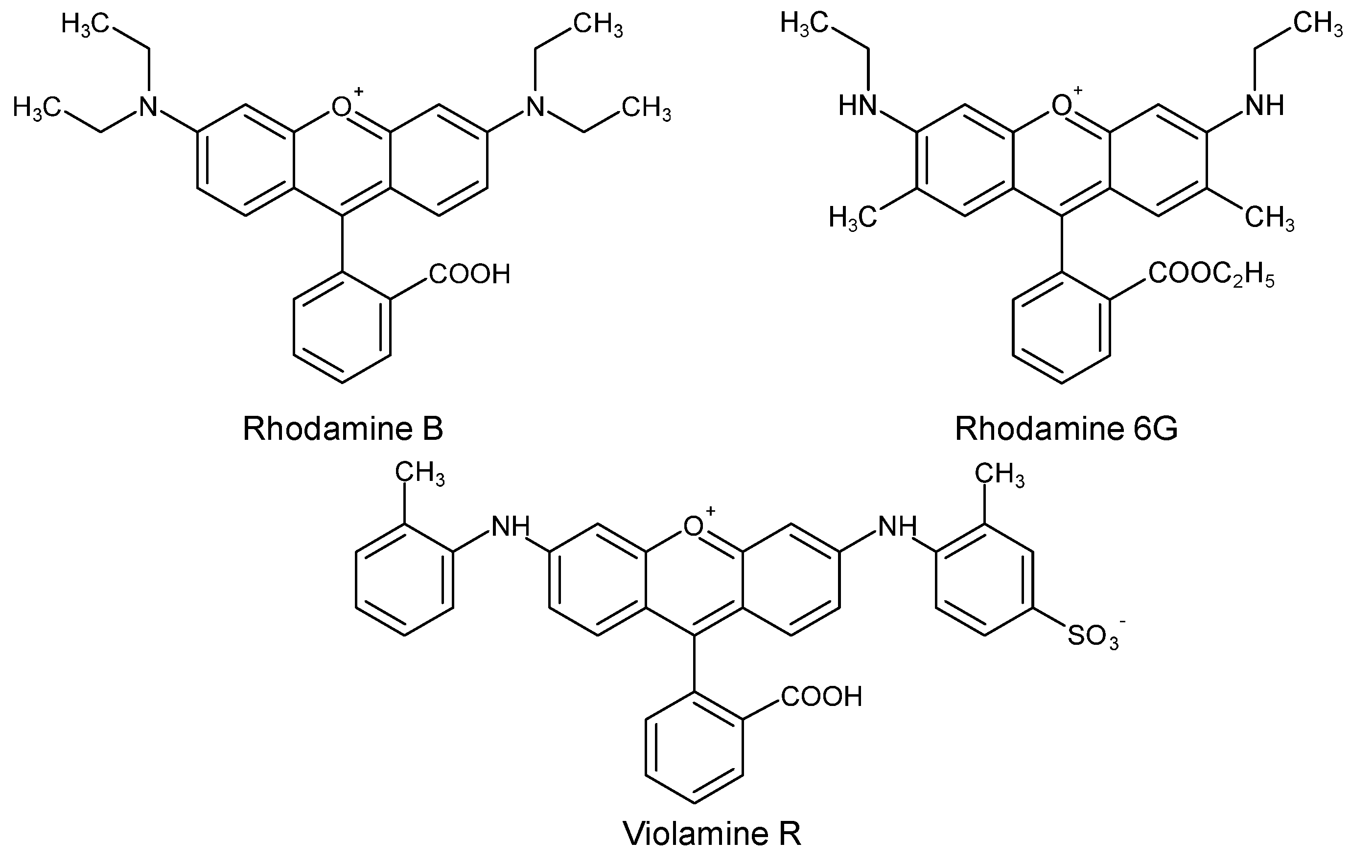
  16. de Keijzer, M.; van Bommel, M.R. Bright New Colours: The History and Analysis of Fluorescein, Eosin, Erythrosine, Rhodamine and Some of Their Derivatives. In The Diversity of Dyes in History and Archaeology; Kirby, J., Ed.; Archetype Publications: London, UK, 2017; pp. 326–338. [Google Scholar]
  17. Fieser, L.F. The Discovery of Synthetic Alizarin. J. Chem. Educ. 1930, 7, 2609. [Google Scholar] [CrossRef]
  18. Schmidt, H. Indigo—100 Jahre Industrielle Synthese. Chem. Unserer Zeit 1997, 31, 121–128. [Google Scholar] [CrossRef]
  19. Bird, C.L. Dyestuff Nomenclature*. J. Soc. Dye. Colour. 1945, 61, 321–328. [Google Scholar] [CrossRef]
  20. Tamburini, D. On the Reliability of Historic Books as Sources of Reference Samples of Early Synthetic Dyes—The Case of “The Coal Tar Colours of the Farbwerke Vorm. Meister, Lucius & Brüning, Höchst on the Main, Germany—A General Part” (1896). Dye. Pigment. 2024, 221, 111796. [Google Scholar] [CrossRef]
  21. Tamburini, D.; Shimada, C.M.; McCarthy, B. The Molecular Characterization of Early Synthetic Dyes in E. Knecht et al’s Textile Sample Book “A Manual of Dyeing” (1893) by High Performance Liquid Chromatography—Diode Array Detector—Mass Spectrometry (HPLC-DAD-MS). Dye. Pigment. 2021, 190, 109286. [Google Scholar] [CrossRef]
  22. Sessa, C.; Steuer, C.; Quintero Balbas, D.; Sciutto, G.; Prati, S.; Stege, H. Analytical Studies on Commercial Artists’ Colour Charts from Das Deutsche Farbenbuch (1925)—Identification of Synthetic and Natural Organic Colourants by Raman Microscopy, Surface-Enhanced Raman Spectroscopy and Metal Underlayer ATR-FTIR Spectroscopy. Herit. Sci. 2022, 10, 109. [Google Scholar] [CrossRef]
  23. Hagan, E.; Poulin, J. The Effect of Prior Exposure on the Lightfastness of Early Synthetic Dyes on Textiles. Herit. Sci. 2022, 10, 138. [Google Scholar] [CrossRef]
  24. Hagan, E.; Castro-Soto, I.; Breault, M.; Poulin, J. The Lightfastness of Early Synthetic Organic Dyes. Herit. Sci. 2022, 10, 50. [Google Scholar] [CrossRef]
  25. Caggiani, M.C.; Forleo, T.; Pojana, G.; Lagioia, G.; Mangone, A.; Giannossa, L.C. Characterization of Silk-Cotton and Wool-Cotton Blends Pattern Books by Fibre Optic Reflectance Spectroscopy. The Booming Market of First Synthetic Textile Dyes in Early 20th Century. Microchem. J. 2022, 175, 107178. [Google Scholar] [CrossRef]
  26. Pirok, B.W.J.; den Uijl, M.J.; Moro, G.; Berbers, S.V.J.; Croes, C.J.M.; van Bommel, M.R.; Schoenmakers, P.J. Characterization of Dye Extracts from Historical Cultural-Heritage Objects Using State-of-the-Art Comprehensive Two-Dimensional Liquid Chromatography and Mass Spectrometry with Active Modulation and Optimized Shifting Gradients. Anal. Chem. 2019, 91, 3062–3069. [Google Scholar] [CrossRef]
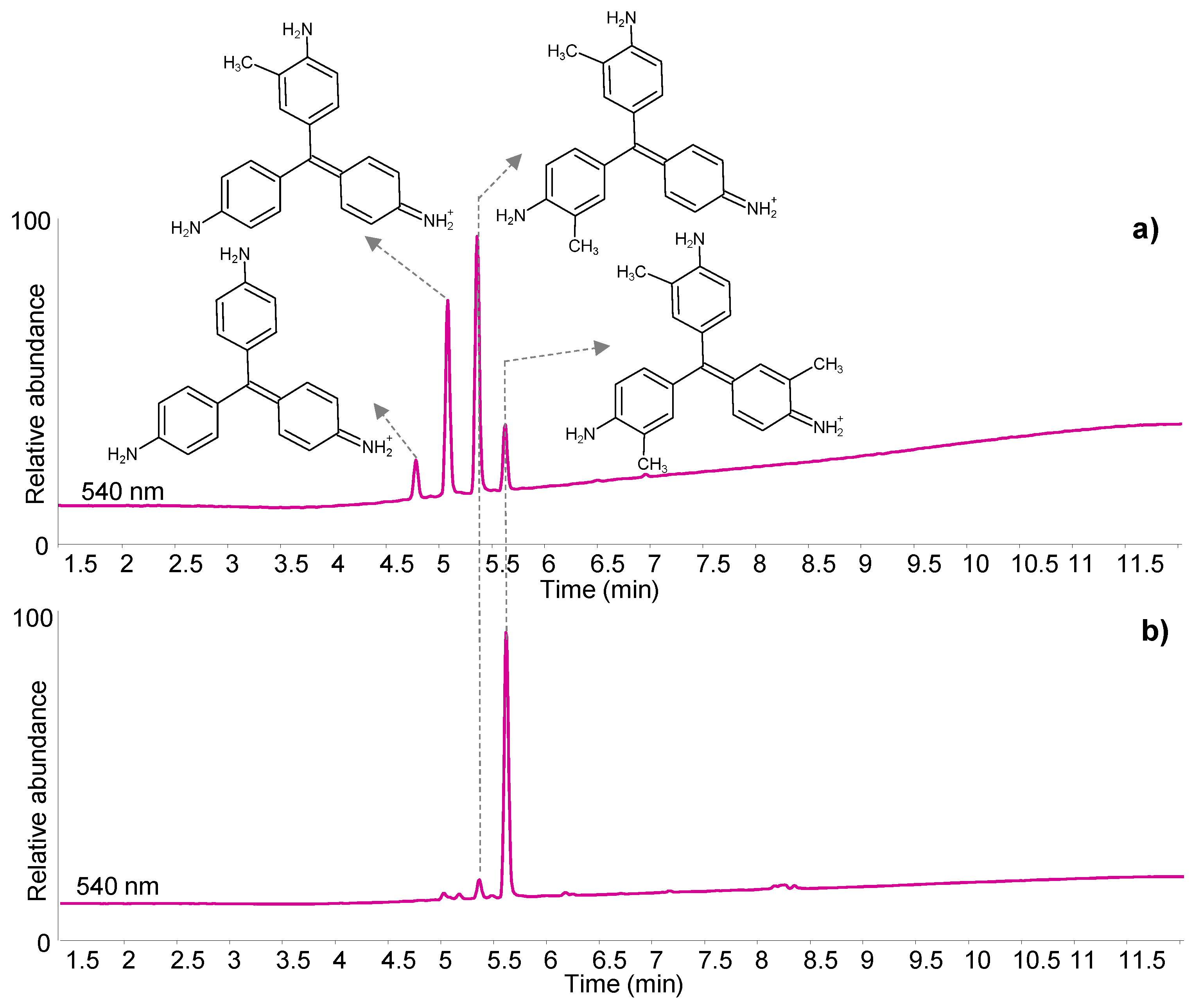
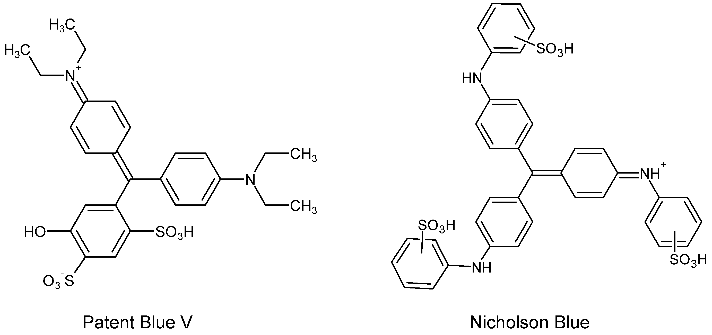
  27. Degano, I.; Sabatini, F.; Braccini, C.; Colombini, M.P. Triarylmethine Dyes: Characterization of Isomers Using Integrated Mass Spectrometry. Dye. Pigment. 2019, 160, 587–596. [Google Scholar] [CrossRef]
  28. Duxbury, D.F. The Photochemistry and Photophysics of Triphenylmethane Dyes in Solid and Liquid Media. Chem. Rev. 1993, 93, 381–433. [Google Scholar]
  29. Montagner, C.; Bacci, M.; Bracci, S.; Freeman, R.; Picollo, M. Library of UV–Vis–NIR Reflectance Spectra of Modern Organic Dyes from Historic Pattern-Card Coloured Papers. Spectrochim. Acta A Mol. Biomol. Spectrosc. 2011, 79, 1669–1680. [Google Scholar] [CrossRef]
  30. Claro, A.; Melo, M.J.; Seixas de Melo, J.S.; van den Berg, K.J.; Burnstock, A.; Montague, M.; Newman, R. Identification of Red Colorants in van Gogh Paintings and Ancient Andean Textiles by Microspectrofluorimetry. J. Cult. Herit. 2010, 11, 27–34. [Google Scholar] [CrossRef]
  31. Fremout, W.; Saverwyns, S. Identification of Synthetic Organic Pigments: The Role of a Comprehensive Digital Raman Spectral Library. J. Raman Spectrosc. 2012, 43, 1536–1544. [Google Scholar] [CrossRef]
  32. Tamburini, D.; Breitung, E.; Mori, C.; Kotajima, T.; Clarke, M.L.; McCarthy, B. Exploring the Transition from Natural to Synthetic Dyes in the Production of 19th-Century Central Asian Ikat Textiles. Herit. Sci. 2020, 8, 114. [Google Scholar] [CrossRef]
  33. Martins, A.; Prud’hom, A.C.; Duranton, M.; Haddad, A.; Daher, C.; Genachte-Le Bail, A.; Tang, T. Jazz Colors: Pigment Identification in the Gouaches Used by Henri Matisse. Heritage 2021, 4, 4205–4221. [Google Scholar] [CrossRef]
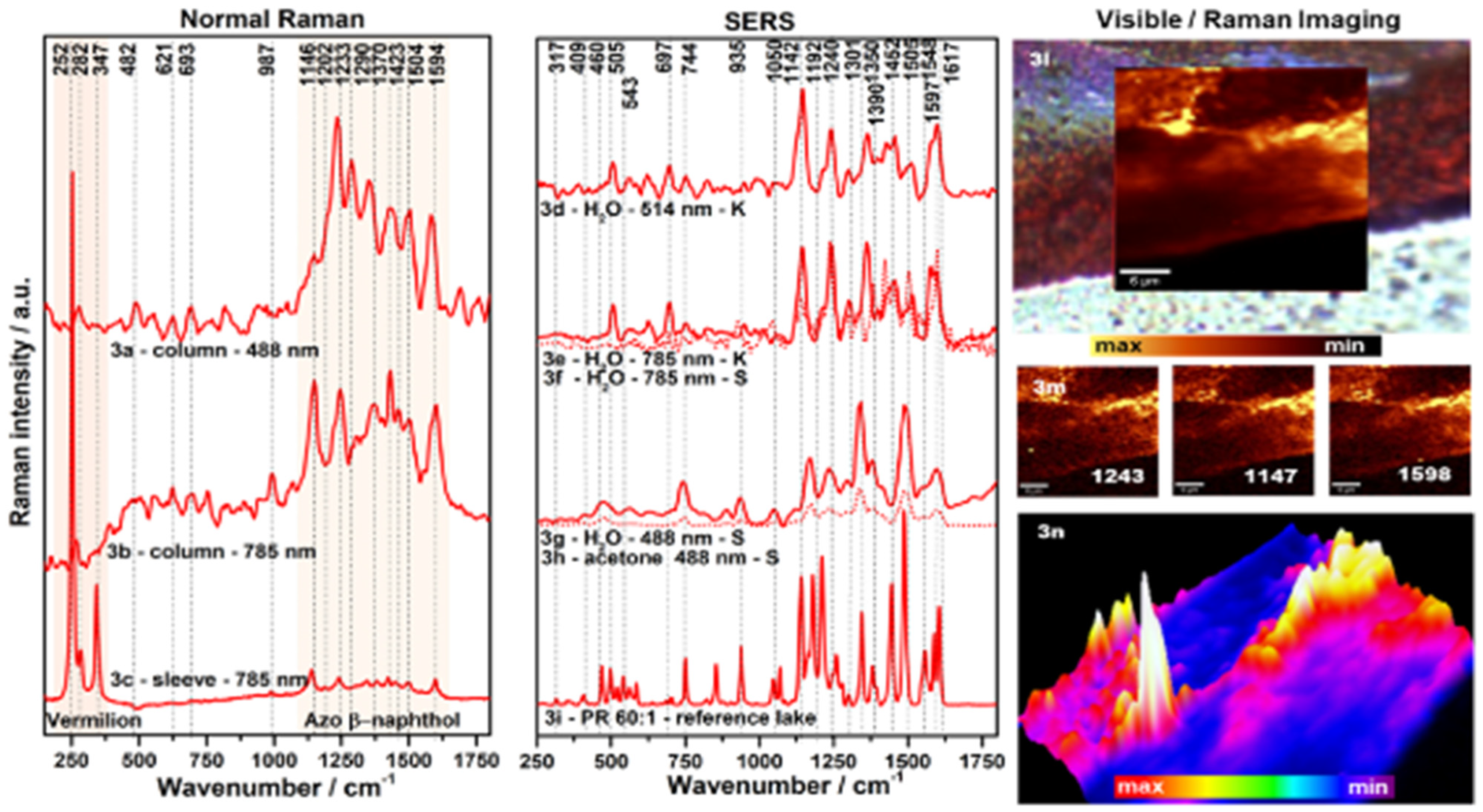
  34. Sessa, C.; Weiss, R.; Niessner, R.; Ivleva, N.P.; Stege, H. Towards a Surface Enhanced Raman Scattering (SERS) Spectra Database for Synthetic Organic Colourants in Cultural Heritage. The Effect of Using Different Metal Substrates on the Spectra. Microchem. J. 2018, 138, 209–225. [Google Scholar] [CrossRef]
  35. Degano, I.; La Nasa, J. Trends in High Performance Liquid Chromatography for Cultural Heritage. Top. Curr. Chem. 2016, 374, 20. [Google Scholar] [CrossRef]
  36. Groeneveld, I.; Pirok, B.W.J.; Molenaar, S.R.A.; Schoenmakers, P.J.; van Bommel, M.R. The Development of a Generic Analysis Method for Natural and Synthetic Dyes by Ultra-High-Pressure Liquid Chromatography with Photo-Diode-Array Detection and Triethylamine as an Ion-Pairing Agent. J. Chromatogr. A 2022, 1673, 463038. [Google Scholar] [CrossRef]
  37. Sabatini, F.; Degano, I. Investigating the Fragmentation Pathways of β-Naphthol Pigments Using Liquid Chromatography/Electrospray Ionization Quadrupole Time-of-Flight Mass Spectrometry. Rapid Commun. Mass Spectrom. 2020, 34, e8789. [Google Scholar] [CrossRef]
  38. Sandström, E.; Vettorazzo, C.; Mackay, C.L.; Troalen, L.G.; Hulme, A.N. Historical Textile Dye Analysis Using DESI-MS. Heritage 2023, 6, 4042–4053. [Google Scholar] [CrossRef]
  39. Alvarez-Martin, A.; Quanico, J.; Scovacricchi, T.; Avranovich Clerici, E.; Baggerman, G.; Janssens, K. Chemical Mapping of the Degradation of Geranium Lake in Paint Cross Sections by MALDI-MSI. Anal. Chem. 2023, 95, 18215–18223. [Google Scholar] [CrossRef]
  40. Centeno, S.A.; Hale, C.; Carò, F.; Cesaratto, A.; Shibayama, N.; Delaney, J.; Dooley, K.; van der Snickt, G.; Janssens, K.; Stein, S.A. Van Gogh’s Irises and Roses: The Contribution of Chemical Analyses and Imaging to the Assessment of Color Changes in the Red Lake Pigments. Herit. Sci. 2017, 5, 18. [Google Scholar] [CrossRef]
  41. Rowe, F.M. (Ed.) The Society of Dyers and Colourists Colour Index, 1st ed.; Society of Dyers and Colourists: Bradford, UK, 1924. [Google Scholar]
  42. Wich, E. The Colour Index. Color Res. Appl. 1977, 2, 77–80. [Google Scholar] [CrossRef]
  43. Society of Dyers and Colourists. American Association of Textile Chemists and Colorists. In Colour Index, 3rd ed.; Society of Dyers and Colourists: Bradford, UK, 1971. [Google Scholar]
  44. Knecht, E.; Rawson, C.; Loewenthal, R. A Manual of Dyeing: For the Use of Practical Dyers, Manufacturers, Students, and All Interested in the Art of Dyeing, 1st ed.; Charles Griffin and Company: London, UK, 1893. [Google Scholar]
  45. Fay, I.W. The Chemistry of the Coal-Tar Dyes, 2nd ed.; D. Van Nostrand Company: New York, NY, USA, 1919. [Google Scholar]
  46. Sekar, N. Direct Dyes. In Handbook of Textile and Industrial Dyeing; Elsevier: Amsterdam, The Netherlands, 2011; pp. 425–445. [Google Scholar]
  47. The Colour IndexTM Colour-Index.Com Published Online by Society of Dyers and Colourists and American Association of Textile Chemists and Colorists. Available online: https://colour-index.com/ (accessed on 7 February 2024).
  48. Kiernan, J. Classification and Naming of Dyes, Stains and Fluorochromes. Biotech. Histochem. 2001, 76, 261–278. [Google Scholar] [CrossRef]
  49. Griess, P. Preliminary Notice of the Reaction of Nitrous Acid with Picramic Acid and Aminonitrophenol. Ann. Chem. Pharm. 1858, 106, 123–125. [Google Scholar]
  50. Hunger, K.; Schmidt, M.U. Hydrazone Pigments (Formerly Called Azo Pigments). In Industrial Organic Pigments; Wiley: Hoboken, NJ, USA, 2018; pp. 193–424. [Google Scholar]
  51. Angelin, E.M.; Oliveira, M.C.; Nevin, A.; Picollo, M.; Melo, M.J. To Be or Not to Be an Azo Pigment: Chemistry for the Preservation of Historical β-Naphthol Reds in Cultural Heritage. Dye. Pigment. 2021, 190, 109244. [Google Scholar] [CrossRef]
  52. Vannucci, G.; Cañamares, M.V.; Prati, S.; Sanchez-Cortes, S. Study of the Azo-hydrazone Tautomerism of Acid Orange 20 by Spectroscopic Techniques: UV–Visible, Raman, and Surface-enhanced Raman Scattering. J. Raman Spectrosc. 2020, 51, 1295–1304. [Google Scholar] [CrossRef]
  53. Aiken, S.; Gabbutt, C.D.; Gillie, L.J.; Heywood, J.D.; Jacquemin, D.; Rice, C.R.; Heron, B.M. The Remarkable Hyperchromicity of Ketohydrazone Dyes and Pigment Lakes Derived from 4-Morpholino-2-naphthol. Eur. J. Org. Chem. 2013, 2013, 8097–8107. [Google Scholar] [CrossRef]
  54. Gessner, T.; Mayer, U. Triarylmethane and Diarylmethane Dyes. In Ullmann’s Encyclopedia of Industrial Chemistry; Wiley: Hoboken, NJ, USA, 2000. [Google Scholar]
  55. Wanyonyi, W.C.; Onyari, J.M.; Shiundu, P.M.; Mulaa, F.J. Biodegradation and Detoxification of Malachite Green Dye Using Novel Enzymes from Bacillus Cereus Strain KM201428: Kinetic and Metabolite Analysis. Energy Procedia 2017, 119, 38–51. [Google Scholar] [CrossRef]
  56. Gürses, A.; Açıkyıldız, M.; Güneş, K.; Gürses, M.S. Classification of Dye and Pigments. In Dyes and Pigments; Springer: Cham, Switzerland, 2016; pp. 31–45. [Google Scholar]
  57. Wagner, H. Die Korperfarben; Wissenschaftliche Verlagsgesellschaft: Stuttgart, Germany, 1938. [Google Scholar]
  58. Zerr, G.; Rubencamp, R. Handbuch Der Farben-Fabrikation. Lehrbuch Der Fabrikation, Unterscuchung Unf Verwendung Aller in Der Praxis Vorkommenden Korperfarben, 1st ed.; Springer: Dresdeb, Germany, 1906. [Google Scholar]
  59. Fischer, M. The Permanent Palette; National Publishing Society: New York, NY, USA, 1930. [Google Scholar]
  60. Sabatini, F.; Eis, E.; Magini, F.; Degano, I.; Rickert, T. Bright Orange and Scarlet Red: Disclosing the Composition and Degradation Mechanisms of ‘Combined Lake’ Formulations. Dye. Hist. Archaeol. 2023, 37/40. Available online: https://www.researchgate.net/publication/369227280_Bright_Orange_and_Scarlet_Red_Disclosing_the_Composition_and_Degradation_Mechanisms_of_%27Combined_Lake%27_Formulations (accessed on 6 March 2024).
  61. León, K.; Mery, D.; Pedreschi, F.; León, J. Color Measurement in L∗a∗b∗ Units from RGB Digital Images. Food Res. Int. 2006, 39, 1084–1091. [Google Scholar] [CrossRef]
  62. Alvarez-Martin, A.; Trashin, S.; Cuykx, M.; Covaci, A.; De Wael, K.; Janssens, K. Photodegradation Mechanisms and Kinetics of Eosin-Y in Oxic and Anoxic Conditions. Dye. Pigment. 2017, 145, 376–384. [Google Scholar] [CrossRef]
  63. Izzo, F.C.; Vitale, V.; Fabbro, C.; Van Keulen, H. Multi-Analytical Investigation on Felt-Tip Pen Inks: Formulation and Preliminary Photo-Degradation Study. Microchem. J. 2016, 124, 919–928. [Google Scholar] [CrossRef]
  64. Ghelardi, E.; Degano, I.; Colombini, M.P.; Mazurek, J.; Schilling, M.; Khanjian, H.; Learner, T. A Multi-Analytical Study on the Photochemical Degradation of Synthetic Organic Pigments. Dye. Pigment. 2015, 123, 396–403. [Google Scholar] [CrossRef]
  65. Sabatini, F.; Eis, E.; Degano, I.; Thoury, M.; Bonaduce, I.; Lluveras-Tenorio, A. The Issue of Eosin Fading: A Combined Spectroscopic and Mass Spectrometric Approach Applied to Historical Lakes. Dye. Pigment. 2020, 180, 108436. [Google Scholar] [CrossRef]
  66. Germinario, G.; Garrappa, S.; D’Ambrosio, V.; van der Werf, I.D.; Sabbatini, L. Chemical Composition of Felt-Tip Pen Inks. Anal. Bioanal. Chem. 2018, 410, 1079–1094. [Google Scholar] [CrossRef]
  67. Micheluz, A.; Angelin, E.M.; Lopes, J.A.; Melo, M.J.; Pamplona, M. Discoloration of Historical Plastic Objects: New Insight into the Degradation of β-Naphthol Pigment Lakes. Polymers 2021, 13, 2278. [Google Scholar] [CrossRef]
  68. Picollo, M.; Bacci, M.; Casini, A.; Lotti, F.; Porcinai, S.; Radicati, B.; Stefani, L. Fiber Optics Reflectance Spectroscopy: A Non-Destructive Technique for the Analysis of Works of Art. In Optical Sensors and Microsystems: New Concepts, Materials, Technologies; Martellucci, S., Chester, A.N., Mignani, A.G., Eds.; Springer: Boston, MA, USA, 2000; pp. 259–265. ISBN 978-0-306-47099-8. [Google Scholar]
  69. IFAC. Fiber Optics Reflectance Spectra (FORS) of Pictorial Materials in the 270–1700 Nm Range; Institute for Applied Physics “Nello Carrara” of the National Research Council (IFAC-CNR): Florence, Italy, 2020. [Google Scholar]
  70. Carlesi, S.; Bartolozzi, G.; Cucci, C.; Marchiafava, V.; Picollo, M. The Artists’ Materials of Fernando Melani: A Precursor of the Poor Art Artistic Movement in Italy. Spectrochim. Acta A Mol. Biomol. Spectrosc. 2013, 104, 527–537. [Google Scholar] [CrossRef]
  71. Zaffino, C.; Passaretti, A.; Poldi, G.; Fratelli, M.; Tibiletti, A.; Bestetti, R.; Saccani, I.; Guglielmi, V.; Bruni, S. A Multi-Technique Approach to the Chemical Characterization of Colored Inks in Contemporary Art: The Materials of Lucio Fontana. J. Cult. Herit. 2017, 23, 87–97. [Google Scholar] [CrossRef]
  72. Bacci, M.; Picollo, M.; Trumpy, G.; Tsukada, M.; Kunzelman, D. Non-Invasive Identification of White Pigments on 20Th-Century Oil Paintings by Using Fiber Optic Reflectance Spectroscopy. J. Am. Inst. Conserv. 2007, 46, 27–37. [Google Scholar] [CrossRef]
  73. Bacci, M.; Magrini, D.; Picollo, M.; Vervat, M. A Study of the Blue Colors Used by Telemaco Signorini (1835–1901). J. Cult. Herit. 2009, 10, 275–280. [Google Scholar] [CrossRef]
  74. Haddad, A.; Nakie-Miller, T.; Jenks, J.B.; Kowach, G. Andy Warhol and His Amazing Technicolor Shoes: Characterizing the Synthetic Dyes Found in Dr. Ph. Martin’s Synchromatic Transparent Watercolors and Used in À La Recherche Du Shoe Perdu. Colorants 2022, 2, 1–21. [Google Scholar] [CrossRef]
  75. Cesaratto, A.; Luo, Y.-B.; Smith, H.D.; Leona, M. A Timeline for the Introduction of Synthetic Dyestuffs in Japan during the Late Edo and Meiji Periods. Herit. Sci. 2018, 6, 22. [Google Scholar] [CrossRef]
  76. Chavanne, C.; Verney, A.; Paquier-Berthelot, C.; Bostal, M.; Buléon, P.; Walter, P. Bayeux Tapestry: First Use of Early Synthetic Dyes for the Restoration of a Masterpiece. Dye. Pigment. 2023, 208, 110798. [Google Scholar] [CrossRef]
  77. Ciccola, A.; Serafini, I.; D’Agostino, G.; Giambra, B.; Bosi, A.; Ripanti, F.; Nucara, A.; Postorino, P.; Curini, R.; Bruno, M. Dyes of a Shadow Theatre: Investigating Tholu Bommalu Indian Puppets through a Highly Sensitive Multi-Spectroscopic Approach. Heritage 2021, 4, 1807–1820. [Google Scholar] [CrossRef]
  78. Tamburini, D.; Dyer, J.; Cartwright, C.; Green, A. Changes in the Production Materials of Burmese Textiles in the Nineteenth Century—Dyes, Mordants and Fibres of Karen Garments from the British Museum’s Collection. Herit. Sci. 2023, 11, 150. [Google Scholar] [CrossRef]
  79. Tamburini, D.; Kim-Marandet, M.; Kim, S.A. Dye Identification in Mounting Textiles of Traditional Korean Paintings from the Late Joseon Dynasty. Heritage 2023, 6, 44–66. [Google Scholar] [CrossRef]
  80. Carlesi, S.; Bartolozzi, G.; Cucci, C.; Marchiafava, V.; Picollo, M.; La Nasa, J.; Di Girolamo, F.; Dilillo, M.; Modugno, F.; Degano, I.; et al. Discovering “the Italian Flag” by Fernando Melani (1907–1985). Spectrochim. Acta A Mol. Biomol. Spectrosc. 2016, 168, 52–59. [Google Scholar] [CrossRef]
  81. Cucci, C.; Bartolozzi, G.; De Vita, M.; Marchiafava, V.; Picollo, M.; Casadio, F. The Colors of Keith Haring: A Spectroscopic Study on the Materials of the Mural Painting Tuttomondo and on Reference Contemporary Outdoor Paints. Appl. Spectrosc. 2016, 70, 186–196. [Google Scholar] [CrossRef]
  82. Vermeulen, M.; Smith, K.; Eremin, K.; Rayner, G.; Walton, M. Application of Uniform Manifold Approximation and Projection (UMAP) in Spectral Imaging of Artworks. Spectrochim. Acta A Mol. Biomol. Spectrosc. 2021, 252, 119547. [Google Scholar] [CrossRef]
  83. Pérez-Arantegui, J.; Rupérez, D.; Almazán, D.; Díez-de-Pinos, N. Colours and Pigments in Late Ukiyo-e Art Works: A Preliminary Non-Invasive Study of Japanese Woodblock Prints to Interpret Hyperspectral Images Using in-Situ Point-by-Point Diffuse Reflectance Spectroscopy. Microchem. J. 2018, 139, 94–109. [Google Scholar] [CrossRef]
  84. Biron, C.; Mounier, A.; Le Bourdon, G.; Servant, L.; Chapoulie, R.; Daniel, F. Revealing the Colours of Ukiyo-e Prints by Short Wave Infrared Range Hyperspectral Imaging (SWIR). Microchem. J. 2020, 155, 104782. [Google Scholar] [CrossRef]
  85. De La Codre, H.; Daniel, F.; Chapoulie, R.; Servant, L.; Mounier, A. Investigating the Materials Used in Eighteenth-Century Tapestries from the Three French Royal Manufactories: Inputs of Hyperspectral Approaches. Eur. Phys. J. Plus 2021, 136, 1193. [Google Scholar] [CrossRef]
  86. Vermeulen, M.; Tamburini, D.; McGeachy, A.C.; Meyers, R.D.; Walton, M.S. Multiscale Characterization of Shellfish Purple and Other Organic Colorants in 20th-Century Traditional Enredos from Oaxaca, Mexico. Dye. Pigment. 2022, 206, 110663. [Google Scholar] [CrossRef]
  87. Vlachou-Mogire, C.; Danskin, J.; Gilchrist, J.R.; Hallett, K. Mapping Materials and Dyes on Historic Tapestries Using Hyperspectral Imaging. Heritage 2023, 6, 3159–3182. [Google Scholar] [CrossRef]
  88. Claro, A.; Melo, M.J.; Schäfer, S.; de Melo, J.S.S.; Pina, F.; van den Berg, K.J.; Burnstock, A. The Use of Microspectrofluorimetry for the Characterization of Lake Pigments. Talanta 2008, 74, 922–929. [Google Scholar] [CrossRef]
  89. Nevin, A.; Comelli, D.; Valentini, G.; Anglos, D.; Burnstock, A.; Cather, S.; Cubeddu, R. Time-Resolved Fluorescence Spectroscopy and Imaging of Proteinaceous Binders Used in Paintings. Anal. Bioanal. Chem. 2007, 338, 1897–1905. [Google Scholar]
  90. Nevin, A.; Cesaratto, A.; Bellei, S.; D’Andrea, C.; Toniolo, L.; Valentini, G.; Comelli, D. Time-Resolved Photoluminescence Spectroscopy and Imaging: New Approaches to the Analysis of Cultural Heritage and Its Degradation. Sensors 2014, 14, 6338–6355. [Google Scholar] [CrossRef]
  91. Doherty, B.; Vagnini, M.; Dufourmantelle, K.; Sgamellotti, A.; Brunetti, B.; Miliani, C. A Vibrational Spectroscopic and Principal Component Analysis of Triarylmethane Dyes by Comparative Laboratory and Portable Instrumentation. Spectrochim. Acta A Mol. Biomol. Spectrosc. 2014, 121, 292–305. [Google Scholar] [CrossRef]
  92. Haddad, A.; Rogge, C.E. Is There an International Klein Pink? Colorants 2023, 2, 194–208. [Google Scholar] [CrossRef]
  93. Moretti, P.; Germinario, G.; Doherty, B.; van der Werf, I.D.; Sabbatini, L.; Mirabile, A.; Sgamellotti, A.; Miliani, C. Disclosing the Composition of Historical Commercial Felt-Tip Pens Used in Art by Integrated Vibrational Spectroscopy and Pyrolysis-Gas Chromatography/Mass Spectrometry. J. Cult. Herit. 2019, 35, 242–253. [Google Scholar] [CrossRef]
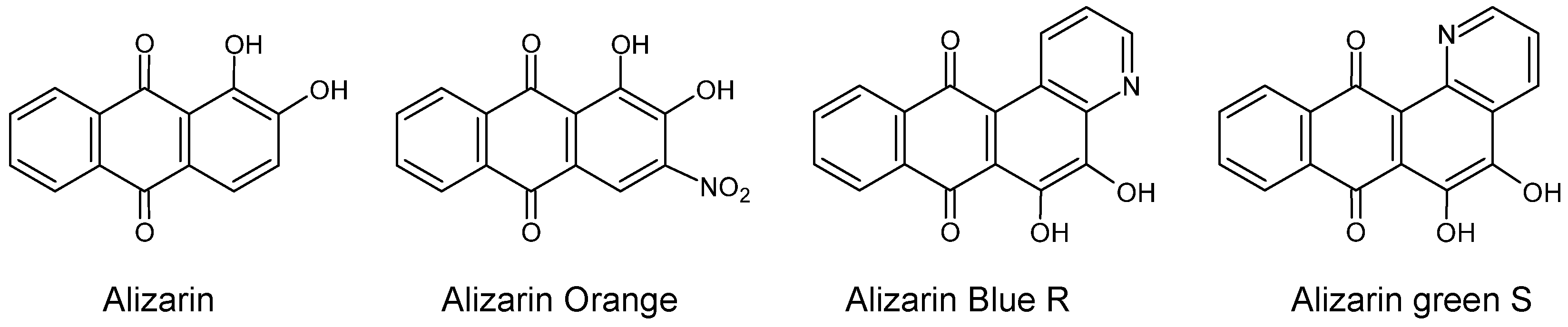
  94. Neugebauer, W.; Sessa, C.; Steuer, C.; Allscher, T.; Stege, H. Naphthol Green—A Forgotten Artists’ Pigment of the Early 20th Century. History, Chemistry and Analytical Identification. J. Cult. Herit. 2018, 36, 153–165. [Google Scholar] [CrossRef]
  95. Anselmi, C.; Capitani, D.; Tintaru, A.; Doherty, B.; Sgamellotti, A.; Miliani, C. Beyond the Color: A Structural Insight to Eosin-Based Lakes. Dye. Pigment. 2017, 140, 297–311. [Google Scholar] [CrossRef]
  96. Alvarez-Martin, A.; Janssens, K. Protecting and Stimulating Effect on the Degradation of Eosin Lakes. Part 1: Lead White and Cobalt Blue. Microchem. J. 2018, 141, 51–63. [Google Scholar] [CrossRef]
  97. Stenger, J.; Kwan, E.E.; Eremin, K.; Speakman, S.; Kirby, D.; Stewart, H.; Huang, S.G.; Kennedy, A.R.; Newman, R.; Khandekar, N. Lithol Red Salts: Characterization and Deterioration. e-Preservation Sci. 2010, 7, 147–157. [Google Scholar]
  98. Lomax, S.Q.; Lomax, J.F. The Synthesis Characterization of Historical Novel Azo Pigments: Implications for Conservation Science. Herit. Sci. 2019, 7, 101. [Google Scholar] [CrossRef]
  99. Beltran, V.; Marchetti, A.; De Meyer, S.; Nuyts, G.; De Wael, K. Geranium Lake Pigments: The Role of the Synthesis on the Structure and Composition. Dye. Pigment. 2021, 189, 109260. [Google Scholar] [CrossRef]
  100. Wertz, J.H.; Tang, P.L.; Quye, A.; France, D.J. Characterisation of Oil and Aluminium Complex on Replica and Historical 19th c. Turkey Red Textiles by Non-Destructive Diffuse Reflectance FTIR Spectroscopy. Spectrochim. Acta A Mol. Biomol. Spectrosc. 2018, 204, 267–275. [Google Scholar] [CrossRef]
  101. Prati, S.; Milosevic, M.; Sciutto, G.; Bonacini, I.; Kazarian, S.G.; Mazzeo, R. Analyses of Trace Amounts of Dyes with a New Enhanced Sensitivity FTIR Spectroscopic Technique: MU-ATR (Metal Underlayer ATR Spectroscopy). Anal. Chim. Acta 2016, 941, 67–79. [Google Scholar] [CrossRef]
  102. Quintero Balbas, D.; Prati, S.; Sciutto, G.; Catelli, E.; Mazzeo, R. Thin-Layer Chromatography/Metal Underlayer-ATR FTIR Methodology for the Study of Synthetic Dyes Extracted from Degraded Wool Fibres. New J. Chem. 2019, 43, 9411–9419. [Google Scholar] [CrossRef]
  103. Beltran, V.; Salvadó, N.; Butí, S.; Cinque, G. Micro Infrared Spectroscopy Discrimination Capability of Compounds in Complex Matrices of Thin Layers in Real Sample Coatings from Artworks. Microchem. J. 2015, 118, 115–123. [Google Scholar] [CrossRef]
  104. Geiman, I.; Leona, M.; Lombardi, J.R. Application of Raman Spectroscopy and Surface-Enhanced Raman Scattering to the Analysis of Synthetic Dyes Found in Ballpoint Pen Inks. J. Forensic Sci. 2009, 54, 947–952. [Google Scholar] [CrossRef]
  105. Vandenabeele, P.; Moens, L.; Edwards, H.G.M.; Dams, R. Raman Spectroscopic Database of Azo Pigments and Application to Modern Art Studies. J. Raman Spectrosc. 2000, 31, 509–517. [Google Scholar]
  106. Lomax, S.Q.; Lomax, J.F.; De Luca-Westrate, A. The Use of Raman Microscopy and Laser Desorption Ionization Mass Spectrometry in the Examination of Synthetic Organic Pigments in Modern Works of Art. J. Raman Spectrosc. 2014, 45, 448–455. [Google Scholar] [CrossRef]
  107. Scherrer, N.C.; Stefan, Z.; Francoise, D.; Annette, F.; Renate, K. Synthetic Organic Pigments of the 20th and 21st Century Relevant to Artist’s Paints: Raman Spectra Reference Collection. Spectrochim. Acta A Mol. Biomol. Spectrosc. 2009, 73, 505–524. [Google Scholar] [CrossRef]
  108. Colombini, A.; Kaifas, D. Characterization of Some Orange and Yellow Organic and Fluorescent Pigments by Raman Spectroscopy. e-Preservation Sci. 2010, 7, 14–21. [Google Scholar]
  109. Ropret, P.; Centeno, S.A.; Bukovec, P. Raman Identification of Yellow Synthetic Organic Pigments in Modern and Contemporary Paintings: Reference Spectra and Case Studies. Spectrochim. Acta A Mol. Biomol. Spectrosc. 2008, 69, 486–497. [Google Scholar] [CrossRef]
  110. Pause, R.; van der Werf, I.D.; van den Berg, K.J. Identification of Pre-1950 Synthetic Organic Pigments in Artists’ Paints. A Non-Invasive Approach Using Handheld Raman Spectroscopy. Heritage 2021, 4, 1348–1365. [Google Scholar] [CrossRef]
  111. Royal Institute for Cultural Heritage (KIK/IRPA) Modern and Contemporary Art in the Laboratory. Available online: https://modern.kikirpa.be (accessed on 6 March 2024).
  112. Rousaki, A.; Vandenabeele, P. In Situ Raman Spectroscopy for Cultural Heritage Studies. J. Raman Spectrosc. 2021, 52, 2178–2189. [Google Scholar] [CrossRef]
  113. Ciccola, A.; Tozzi, L.; Romani, M.; Serafini, I.; Ripanti, F.; Curini, R.; Vitucci, F.; Cestelli Guidi, M.; Postorino, P. Lucio Fontana and the Light: Spectroscopic Analysis of the Artist’s Collection at the National Gallery of Modern and Contemporary Art. Spectrochim. Acta A Mol. Biomol. Spectrosc. 2020, 236, 118319. [Google Scholar] [CrossRef]
  114. Schulte, F.; Brzezinka, K.-W.; Lutzenberger, K.; Stege, H.; Panne, U. Raman Spectroscopy of Synthetic Organic Pigments Used in 20th Century Works of Art. J. Raman Spectrosc. 2008, 39, 1455–1463. [Google Scholar] [CrossRef]
  115. Lomax, S.Q.; Lomax, J.F.; Graham, T.K.; Moore, T.J.T.; Knapp, C.G. Historical Azo Pigments: Synthesis and Characterization. J. Cult. Herit. 2018, 35, 218–224. [Google Scholar] [CrossRef]
  116. Sodo, A.; Bicchieri, M.; Guiso, M.; Ricci, M.A.; Ricci, G. Raman Investigations on Marker Pen Inks. J. Raman Spectrosc. 2012, 43, 1781–1787. [Google Scholar] [CrossRef]
  117. Rosi, F.; Grazia, C.; Fontana, R.; Gabrieli, F.; Pensabene Buemi, L.; Pampaloni, E.; Romani, A.; Stringari, C.; Miliani, C. Disclosing Jackson Pollock’s Palette in Alchemy (1947) by Non-Invasive Spectroscopies. Herit. Sci. 2016, 4, 18. [Google Scholar] [CrossRef]
  118. Haddad, A.; Neufeld, L.; Martins, A. Realizing Sensations: Analyzing Paul Cezanne’s Watercolors and Assessing Their Light Sensitivity with Microfade Testing. Herit. Sci. 2023, 11, 39. [Google Scholar] [CrossRef]
  119. Angelin, E.M.; de Sá, S.; Picollo, M.; Nevin, A.; Callapez, M.E.; Melo, M.J. The Identification of Synthetic Organic Red Pigments in Historical Plastics: Developing an in Situ Analytical Protocol Based on Raman Microscopy. J. Raman Spectrosc. 2021, 52, 145–158. [Google Scholar] [CrossRef]
  120. Haddad, A.; Randall, M.; Zycherman, L.; Martins, A. Reviving Alexander Calder’s Man-Eater with Pennants: A Technical Examination of the Original Paint Palette. Heritage 2021, 4, 1920–1937. [Google Scholar] [CrossRef]
  121. Amato, F.; Micciche’, C.; Cannas, M.; Gelardi, F.M.; Pignataro, B.; Li Vigni, M.; Agnello, S. Ag Nanoparticles Agargel Nanocomposites for SERS Detection of Cultural Heritage Interest Pigments. Eur. Phys. J. Plus 2018, 133, 74. [Google Scholar] [CrossRef]
  122. Longoni, M.; Gavazzi, M.; Monti, D.; Bruni, S. Early Synthetic Textile Dyes of the Late 19th Century from the “Primo Levi” Chemistry Museum (Rome): A Multi-Technique Analytical Investigation. J. Cult. Herit. 2023, 59, 131–139. [Google Scholar] [CrossRef]
  123. Leona, M.; Decuzzi, P.; Kubic, T.A.; Gates, G.; Lombardi, J.R. Non Destructive Identification of Natural and Synthetic Organic Colorants in Works of Art by Surface Enhanced Raman Scattering. Anal. Chem. 2011, 83, 3990–3993. [Google Scholar] [CrossRef]
  124. Reggio, D.; Mirabile, A.; Lazzari, M. Sensing Soluble Molecules through SERS Substrates in One-Step Procedure: Unrevealing the Meiji Woodblock Printing Materials. Talanta 2023, 254, 124177. [Google Scholar] [CrossRef]
  125. Anghelone, M.; Stoytschew, V.; Jembrih-Simbürger, D.; Schreiner, M. Spectroscopic Methods for the Identification and Photostability Study of Red Synthetic Organic Pigments in Alkyd and Acrylic Paints. Microchem. J. 2018, 139, 155–163. [Google Scholar] [CrossRef]
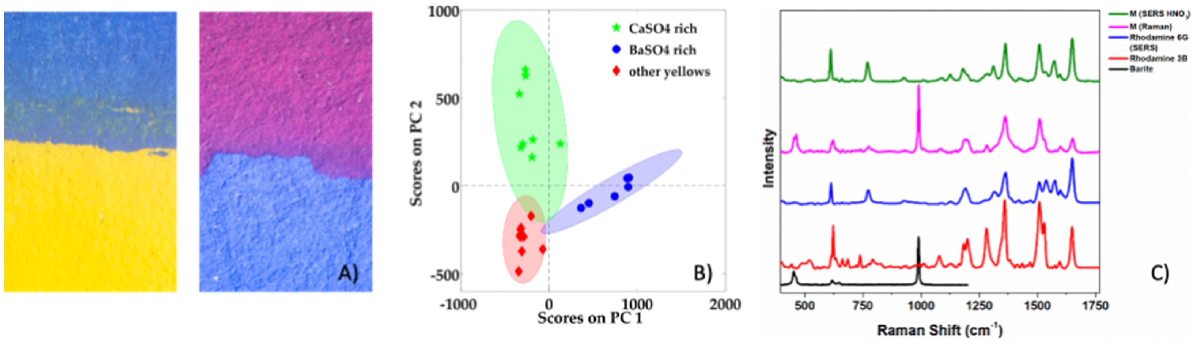
  126. Coccato, A.; Caggiani, M.C. An Overview of Principal Components Analysis Approaches in Raman Studies of Cultural Heritage Materials. J. Raman Spectrosc. 2023, 55, 125–147. [Google Scholar] [CrossRef]
  127. Manfredi, M.; Barberis, E.; Aceto, M.; Marengo, E. Non-Invasive Characterization of Colorants by Portable Diffuse Reflectance Infrared Fourier Transform (DRIFT) Spectroscopy and Chemometrics. Spectrochim. Acta A Mol. Biomol. Spectrosc. 2017, 181, 171–179. [Google Scholar] [CrossRef]
  128. Poulin, J. A New Methodology for the Characterisation of Natural Dyes on Museum Objects Using Gas Chromatography–Mass Spectrometry. Stud. Conserv. 2018, 63, 36–61. [Google Scholar] [CrossRef]
  129. Degani, L.; Riedo, C.; Gulmini, M.; Chiantore, O. From Plant Extracts to Historical Textiles: Characterization of Dyestuffs by GC–MS. Chromatographia 2014, 77, 1683–1696. [Google Scholar] [CrossRef]
  130. Lomax, S.; Schilling, M.; Learner, T. The Identification of Synthetic Organic Pigments by FTIR and DTMS. In Modern Paints Uncovered; Getty Publications: Los Angeles, CA, USA, 2007. [Google Scholar]
  131. Boon, J.J.; Learner, T. Analytical Mass Spectrometry of Artists’ Acrylic Emulsion Paints by Direct Temperature Resolved Mass Spectrometry and Laser Desorption Ionisation Mass Spectrometry. J. Anal. Appl. Pyrolysis 2002, 64, 327–344. [Google Scholar] [CrossRef]
  132. Menke, C.A.; Rivenc, R.; Learner, T. The Use of Direct Temperature-Resolved Mass Spectrometry (DTMS) in the Detection of Organic Pigments Found in Acrylic Paints Used by Sam Francis. Int. J. Mass Spectrom. 2009, 284, 2–11. [Google Scholar] [CrossRef]
  133. Rehorek, A.; Plum, A. Characterization of Sulfonated Azo Dyes and Aromatic Amines by Pyrolysis Gas Chromatography/Mass Spectrometry. Anal. Bioanal. Chem. 2007, 388, 1653–1662. [Google Scholar] [CrossRef]
  134. Russell, J.; Singer, B.W.; Perry, J.J.; Bacon, A. The Identification of Synthetic Organic Pigments in Modern Paints and Modern Paintings Using Pyrolysis-Gas Chromatography--Mass Spectrometry. Anal. Bioanal. Chem. 2011, 400, 1473. [Google Scholar] [CrossRef]
  135. Germinario, G.; Rigante, E.C.L.; van der Werf, I.D.; Sabbatini, L. Pyrolysis Gas Chromatography–Mass Spectrometry of Triarylmethane Dyes. J. Anal. Appl. Pyrolysis 2017, 127, 229–239. [Google Scholar] [CrossRef]
  136. La Nasa, J.; Biale, G.; Sabatini, F.; Degano, I.; Colombini, M.P.M.P.; Modugno, F. Synthetic Materials in Art: A New Comprehensive Approach for the Characterization of Multi-Material Artworks by Analytical Pyrolysis. Herit. Sci. 2019, 7, 8. [Google Scholar] [CrossRef]
  137. Fardi, T.; Pintus, V.; Kampasakali, E.; Pavlidou, E.; Schreiner, M.; Kyriacou, G. Analytical Characterization of Artist’s Paint Systems Based on Emulsion Polymers and Synthetic Organic Pigments. J. Anal. Appl. Pyrolysis 2018, 135, 231–241. [Google Scholar] [CrossRef]
  138. Soltzberg, L.J.; Hagar, A.; Kridaratikorn, S.; Mattson, A.; Newman, R. MALDI-TOF Mass Spectrometric Identification of Dyes and Pigments. J. Am. Soc. Mass Spectrom. 2007, 18, 2001–2006. [Google Scholar] [CrossRef]
  139. Kirby, D.P.; Khandekar, N.; Sutherland, K.; Price, B.A. Applications of Laser Desorption Mass Spectrometry for the Study of Synthetic Organic Pigments in Works of Art. Int. J. Mass Spectrom. 2009, 284, 115–122. [Google Scholar] [CrossRef]
  140. Schreiver, I.; Eschner, L.-M.; Luch, A. Matrix-Assisted Laser Desorption/Ionization Tandem Mass Spectrometry for Identification of Organic Tattoo Pigments in Inks and Tissue Samples. Analyst 2018, 143, 3941–3950. [Google Scholar]
  141. Dunn, J.; Siegel, J.; Allison, J. Photodegradation and Laser Desorption Mass Spectrometry for the Characterization of Dyes Used in Red Pen Inks. Sci. J. Forensic 2003, 48, 652–657. [Google Scholar]
  142. Weyermann, C.; Kirsch, D.; Costa-Vera, C.; Spengler, B. Photofading of Ballpoint Dyes Studied on Paper by LDI and MALDI MS. J. Am. Soc. Mass Spectrom. 2006, 17, 297–306. [Google Scholar] [CrossRef]
  143. Weyermann, C.; Kirsch, D.; Vera, C.C.; Spengler, B. Evaluation of the Photodegradation of Crystal Violet upon Light Exposure by Mass Spectrometric and Spectroscopic Methods. J. Forensic Sci. 2009, 54, 339–345. [Google Scholar] [CrossRef]
  144. Weyermann, C.; Spengler, B. The Potential of Artificial Aging for Modelling of Natural Aging Processes of Ballpoint Ink. Forensic Sci. Int. 2008, 180, 23–31. [Google Scholar] [CrossRef]
  145. Taverna, D.; Di Donna, L.; Mazzotti, F.; Policicchio, B.; Sindona, G. High-Throughput Determination of Sudan Azo-Dyes within Powdered Chili Pepper by Paper Spray Mass Spectrometry. J. Mass Spectrom. 2013, 48, 544–547. [Google Scholar] [CrossRef]
  146. Guo, T.; Zhang, Z.; Yannell, K.E.; Dongb, Y.; Cooks, R.G. Paper Spray Ionization Mass Spectrometry for Rapid Quantification of Illegal Beverage Dyes. Anal. Methods 2017, 9, 6273–6279. [Google Scholar]
  147. Alvarez-Martin, A.; Cleland, T.P.; Kavich, G.M.; Janssens, K.; Newsome, G.A. Rapid Evaluation of the Debromination Mechanism of Eosin in Oil Paint by Direct Analysis in Real Time and Direct Infusion-Electrospray Ionization Mass Spectrometry. Anal. Chem. 2019, 91, 10856–10863. [Google Scholar] [CrossRef]
  148. Sandström, E.; Vettorazzo, C.; Mackay, C.L.; Troalen, L.G.; Hulme, A.N. Development and Application of Desorption Electrospray Ionization Mass Spectrometry for Historical Dye Analysis. Anal. Chem. 2023, 95, 4846–4854. [Google Scholar] [CrossRef]
  149. Astefanei, A.; van Bommel, M.R.; Corthals, G.L. Surface Acoustic Wave Nebulisation Mass Spectrometry for the Fast and Highly Sensitive Characterisation of Synthetic Dyes in Textile Samples. J. Am. Soc. Mass Spectrom. 2017, 28, 2108–2116. [Google Scholar] [CrossRef]
  150. Wei, L.; Ma, Y.; Guo, Z.; Ding, J.; Jin, G.; Gu, A.; Lei, Y. Application of Advanced Analytical Techniques in Organic Cultural Heritage: A Case Study of Ancient Architecture Relics in the Palace Museum (Beijing). Coatings 2022, 12, 636. [Google Scholar] [CrossRef]
  151. Krmpotić, M.; Jembrih-Simbürger, D.; Siketić, Z.; Marković, N.; Anghelone, M.; Tadić, T.; Plavčić, D.; Malloy, M.; Radović, I.B. Identification of Synthetic Organic Pigments (SOPs) Used in Modern Artist’s Paints with Secondary Ion Mass Spectrometry with MeV Ions. Anal. Chem. 2020, 92, 9287–9294. [Google Scholar] [CrossRef]
  152. Degano, I.; Ribechini, E.; Modugno, F.; Colombini, M.P. Analytical Methods for the Characterization of Organic Dyes in Artworks and in Historical Textiles. Appl. Spectrosc. Rev. 2009, 44, 363–410. [Google Scholar] [CrossRef]
  153. Zhang, X.; Laursen, R.A. Development of Mild Extraction Methods for the Analysis of Natural Dyes in Textiles of Historical Interest Using LC-Diode Array Detector-MS. Anal. Chem. 2005, 77, 2022–2025. [Google Scholar] [CrossRef]
  154. Surowiec, I. Application of High-Performance Separation Techniques in Archaeometry. Microchim. Acta 2008, 162, 289–302. [Google Scholar] [CrossRef]
  155. Manhita, A.; Ferreira, T.; Candeias, A.; Barrocas Dias, C. Extracting Natural Dyes from Wool---an Evaluation of Extraction Methods. Anal. Bioanal. Chem. 2011, 400, 1501. [Google Scholar] [CrossRef]
  156. Serafini, I.; McClure, K.R.; Ciccola, A.; Vincenti, F.; Bosi, A.; Peruzzi, G.; Montesano, C.; Sergi, M.; Favero, G.; Curini, R. Inside the History of Italian Coloring Industries: An Investigation of ACNA Dyes through a Novel Analytical Protocol for Synthetic Dye Extraction and Characterization. Molecules 2023, 28, 5331. [Google Scholar] [CrossRef]
  157. Sanyova, J.; Reisse, J. Development of a Mild Method for the Extraction of Anthraquinones from Their Aluminum Complexes in Madder Lakes Prior to HPLC Analysis. J. Cult. Herit. 2006, 7, 229–235. [Google Scholar] [CrossRef]
  158. Christie, R.M.; Standring, P.N. Colour and Constitution Relationships in Organic Pigments. Part 2—Disazoacetoacetanilides11Part 1-Monoazoacetoacetanilides: Dyes and Pigments, 9(1). Dye. Pigment. 1989, 11, 109–121. [Google Scholar] [CrossRef]
  159. Christie, R.M.; Standring, P.N.; Griffiths, J. Colour and Constitution Relationships in Organic Pigments. Part 1—Monoazoacetoacetanilides. Dye. Pigment. 1988, 9, 37–56. [Google Scholar] [CrossRef]
  160. Souto, C.S.C.N. Analysis of Early Synthetic Dyes with HPLC-DAD-MS. Master’s Thesis, Universidade de Lisboa, Lisboa, Portugal, 2010. [Google Scholar]
  161. Talsky, G.; Ristić-Šolajić, M. High-Resolution/Higher-Order Derivative Spectrophotometry for Identification and Estimation of Synthetic Organic Pigments in Artists’ Paints. Anal. Chim. Acta 1987, 196, 123–134. [Google Scholar] [CrossRef]
  162. Deomartins, A.; Canalli, V.M.; Azevedo, C.M.N.; Pires, M. Degradation of Pararosaniline (C.I. Basic Red 9 Monohydrochloride) Dye by Ozonation and Sonolysis. Dye. Pigment. 2006, 68, 227–234. [Google Scholar] [CrossRef]
  163. Jain, R.; Sikarwar, S. Photodegradation of Hazardous Dye Naphthol Yellow S Over Titanium Dioxide. J. Dispers. Sci. Technol. 2011, 32, 1345–1352. [Google Scholar] [CrossRef]
  164. Zaied, M.; Chutet, E.; Peulon, S.; Bellakhal, N.; Desmazières, B.; Dachraoui, M.; Chaussé, A. Spontaneous Oxidative Degradation of Indigo Carmine by Thin Films of Birnessite Electrodeposited onto SnO2. Appl. Catal. B 2011, 107, 42–51. [Google Scholar] [CrossRef]
  165. Sherma, J. Advances in the Thin-Layer Chromatographic Forensic Analysis of Inks. J. Liq. Chromatogr. Relat. Technol. 2016, 39, 549–557. [Google Scholar] [CrossRef]
  166. Causin, V.; Casamassima, R.; Marega, C.; Maida, P.; Schiavone, S.; Marigo, A.; Villari, A. The Discrimination Potential of Ultraviolet-Visible Spectrophotometry, Thin Layer Chromatography, and Fourier Transform Infrared Spectroscopy for the Forensic Analysis of Black and Blue Ballpoint Inks. J. Forensic Sci. 2008, 53, 1468–1473. [Google Scholar] [CrossRef]
  167. Cañamares, M.V.; Reagan, D.A.; Lombardi, J.R.; Leona, M. TLC-SERS of Mauve, the First Synthetic Dye. J. Raman Spectrosc. 2014, 45, 1147–1152. [Google Scholar] [CrossRef]
  168. Ferretti, A.; Degano, I.; Legnaioli, S.; Campanella, B.; Sainati, A.; Colombini, M.P. Shedding Light on the Composition and Degradation Mechanism of Dyes in Historical Ink’s Collection (19th–20th Century). Dye. Pigment. 2023, 220, 111672. [Google Scholar] [CrossRef]
  169. Campanella, B.; Botti, J.; Cavaleri, T.; Cicogna, F.; Legnaioli, S.; Pagnotta, S.; Poggialini, F.; Poli, T.; Scalarone, D.; Palleschi, V. The Shining Brightness of Daylight Fluorescent Pigments: Raman and SERS Study of a Modern Class of Painting Materials. Microchem. J. 2020, 152, 104292. [Google Scholar] [CrossRef]
  170. Troalen, L.G.; Phillips, A.S.; Peggie, D.A.; Barran, P.E.; Hulme, A.N. Historical Textile Dyeing with Genista tinctoria L.: A Comprehensive Study by UPLC-MS/MS Analysis. Anal. Methods 2014, 6, 8915–8923. [Google Scholar] [CrossRef]
  171. Douglas, J.G.; Kavich, G.; Mori, C.; Wallace, D.; Barden, R. Materials Characterization of the Ruby Slippers from the 1939 Classic Film, The Wizard of Oz. Herit. Sci. 2018, 6, 49. [Google Scholar] [CrossRef]
  172. Liu, J.; Zhou, Y.; Zhao, P.; Peng, Z.; Wang, S. Identification of Early Synthetic Dyes in Historical Chinese Textiles of the Late Nineteenth Century by High-Performance Liquid Chromatography Coupled with Diode Array Detection and Mass Spectrometry. Color. Technol. 2016, 132, 177–185. [Google Scholar] [CrossRef]
  173. Confortin, D.; Neevel, H.; Brustolon, M.; Franco, L.; Kettelarij, A.J.A.J.; Williams, R.M.R.M.; van Bommel, M.R. Crystal Violet: Study of the Photo-Fading of an Early Synthetic Dye in Aqueous Solution and on Paper with HPLC-PDA, LC-MS and FORS. J. Phys. Conf. Ser. 2010, 231, 012011. [Google Scholar] [CrossRef]
  174. Chen, V.J.; Smith, G.D.; Holden, A.; Paydar, N.; Kiefer, K. Chemical Analysis of Dyes on an Uzbek Ceremonial Coat: Objective Evidence for Artifact Dating and the Chemistry of Early Synthetic Dyes. Dye. Pigment. 2016, 131, 320–332. [Google Scholar] [CrossRef]
  175. de Melo, J.S.; Takato, S.; Sousa, M.; Melo, M.J.; Parola, A.J. Revisiting Perkin’s Dye(s): The Spectroscopy and Photophysics of Two New Mauveine Compounds (B2 and C). Chem. Commun. 2007, 25, 2624–2626. [Google Scholar]
  176. Kucharska, M.; Grabka, J. A Review of Chromatographic Methods for Determination of Synthetic Food Dyes. Talanta 2010, 80, 1045–1051. [Google Scholar] [CrossRef]
  177. Cai, M.; Jin, M.; Weavers, L.K. Analysis of Sonolytic Degradation Products of Azo Dye Orange G Using Liquid Chromatography–Diode Array Detection-Mass Spectrometry. Ultrason. Sonochemistry 2011, 18, 1068–1076. [Google Scholar] [CrossRef]
  178. Petroviciu, I.; Teodorescu, I.C.; Vasilca, S.; Albu, F. Transition from Natural to Early Synthetic Dyes in the Romanian Traditional Shirts Decoration. Heritage 2023, 6, 505–523. [Google Scholar] [CrossRef]
  179. Geldof, M.; de Keijzer, M.; van Bommel, M.; Pilz, K.; Salvant, J.; van Keulen, H.; Megens, L. Van Gogh’s Geranium Lake. In Van Gogh’s Studio Practice; Mercatorfonds: Brussels, Belgium, 2013. [Google Scholar]
  180. Plater, M.J.; Raab, A. Mauveine and the Mauve Shade Six Pence Stamp. J. Chem. Res. 2016, 40, 648–651. [Google Scholar]
  181. Sousa, M.M.; Melo, M.J.; Parola, A.J.; Morris, P.J.T.; Rzepa, H.S.; de Melo, J.S.S. A Study in Mauve: Unveiling Perkin’s Dye in Historic Samples. Chem.–A Eur. J. 2008, 14, 8507–8513. [Google Scholar] [CrossRef]
  182. Lech, K.; Wilicka, E.; Witowska-Jarosz, J.; Jarosz, M. Early Synthetic Dyes—A Challenge for Tandem Mass Spectrometry. J. Mass Spectrom. 2012, 48, 141–147. [Google Scholar] [CrossRef]
  183. Pirok, B.W.J.; Knip, J.; van Bommel, M.R.; Schoenmakers, P.J. Characterization of Synthetic Dyes by Comprehensive Two-Dimensional Liquid Chromatography Combining Ion-Exchange Chromatography and Fast Ion-Pair Reversed-Phase Chromatography. J. Chromatogr. A 2016, 1436, 141–146. [Google Scholar] [CrossRef]
  184. Tamburini, D.; Dyer, J.; Cartwright, C. First Evidence and Characterisation of Rare Chrome-Based Colourants Used on 19th-Century Textiles from Myanmar. Dye. Pigment. 2023, 218, 111472. [Google Scholar] [CrossRef]
  185. Sundberg, B.N.; Pause, R.; van der Werf, I.D.; Astefanei, A.; van den Berg, K.J.; van Bommel, M.R. Analytical Approaches for the Characterization of Early Synthetic Organic Pigments for Artists’ Paints. Microchem. J. 2021, 170, 106708. [Google Scholar] [CrossRef]
  186. Alvarez-Martin, A.; Newsome, G.A.; Janssens, K. High-Resolution Mass Spectrometry and Nontraditional Mass Defect Analysis of Brominated Historical Pigments. Anal. Chem. 2021, 93, 14851–14858. [Google Scholar] [CrossRef]
  187. Ferreira, B.R.V.; Correa, D.N.; Eberlin, M.N.; Vendramini, P.H. Fragmentation Reactions of Rhodamine B and 6G as Revealed by High Accuracy Orbitrap Tandem Mass Spectrometry. J. Braz. Chem. Soc. 2017, 28, 136–142. [Google Scholar]
  188. Vermeulen, M.; Miranda, A.S.O.; Tamburini, D.; Delgado, S.E.R.; Walton, M. A Multi-Analytical Study of the Palette of Impressionist and Post-Impressionist Puerto Rican Artists. Herit. Sci. 2022, 10, 44. [Google Scholar] [CrossRef]
  189. Sabatini, F.; Manariti, A.; di Girolamo, F.; Bonaduce, I.; Tozzi, L.; Rava, A.; Colombini, M.P.; Lluveras-Tenorio, A. Painting on Polyurethane Foam: “Composizione-Superficie Lunare” by Giulio Turcato. Microchem. J. 2020, 156, 104872. [Google Scholar] [CrossRef]
  190. Sabatini, F.; Degano, I.; Colombini, M.P. Development of a Method Based on High-Performance Liquid Chromatography Coupled with Diode Array, Fluorescence, and Mass Spectrometric Detectors for the Analysis of Eosin at Trace Levels. Sep. Sci. Plus 2020, 3, 207–215. [Google Scholar] [CrossRef]
  191. La Nasa, J.; Nodari, L.; Nardella, F.; Sabatini, F.; Degano, I.; Modugno, F.; Legnaioli, S.; Campanella, B.; Tufano, M.K.K.; Zuena, M.; et al. Chemistry of Modern Paint Media: The Strained and Collapsed Painting by Alexis Harding. Microchem. J. 2020, 155, 104659. [Google Scholar] [CrossRef]
  192. Reiß, L.; Machill, S.; Lübken, T.; Herm, C. HPLC–HR-ESI–MS/MS Identification of Fluorescent Dyes and Optical Brighteners and Their Degradation Products in Daylight Fluorescent Paints. Herit. Sci. 2023, 11, 146. [Google Scholar] [CrossRef]
  193. La Nasa, J.; Campanella, B.; Sabatini, F.; Rava, A.; Shank, W.; Lucero-Gomez, P.; De Luca, D.; Legnaioli, S.; Palleschi, V.; Colombini, M.P.; et al. 60 Years of Street Art: A Comparative Study of the Artists’ Materials through Spectroscopic and Mass Spectrometric Approaches. J. Cult. Herit. 2021, 48, 129–140. [Google Scholar] [CrossRef]
  194. Sabatini, F.; La Nasa, J.; Degano, I.; Campanella, B.; Legnaioli, S.; Saccani, I.; Modugno, F. Fluorescent Paints in Contemporary Murals: A Case Study. Heritage 2023, 6, 5689–5699. [Google Scholar] [CrossRef]
  195. Serrano, A.; van Bommel, M.R.; Hallett, J. Evaluation between Ultrahigh Pressure Liquid Chromatography and High-Performance Liquid Chromatography Analytical Methods for Characterizing Natural Dyestuffs. J. Chromatogr. A 2013, 1318, 102–111. [Google Scholar] [CrossRef]
  196. Wertz, J.H.; Quye, A.; France, D.; Tang, P.L.; Richmond, L. Authenticating Turkey Red Textiles through Material Investigations by FTIR and UHPLC. In Proceedings of the ICOM-CC 18th Triennial Meeting, Copenhagen, Denmark, 4–8 September 2017. [Google Scholar]
  197. den Uijl, M.J.; van der Wijst, Y.J.H.L.; Groeneveld, I.; Schoenmakers, P.J.; Pirok, B.W.J.; van Bommel, M.R. Combining Photodegradation in a Liquid-Core-Waveguide Cell with Multiple-Heart-Cut Two-Dimensional Liquid Chromatography. Anal. Chem. 2022, 94, 11055–11061. [Google Scholar] [CrossRef]
  198. Groeneveld, I.; Bagdonaite, I.; Beekwilder, E.; Ariese, F.; Somsen, G.W.; van Bommel, M.R. Liquid Core Waveguide Cell with In Situ Absorbance Spectroscopy and Coupled to Liquid Chromatography for Studying Light-Induced Degradation. Anal. Chem. 2022, 94, 7647–7654. [Google Scholar] [CrossRef]
  199. Groeneveld, I.; Schoemaker, S.E.; Somsen, G.W.; Ariese, F.; Van Bommel, M.R. Characterization of a Liquid-Core Waveguide Cell for Studying the Chemistry of Light-Induced Degradation. Analyst 2021, 146, 3197–3207. [Google Scholar]
  200. den Uijl, M.J.; Lokker, A.; van Dooren, B.; Schoenmakers, P.J.; Pirok, B.W.J.; van Bommel, M.R. Comparing Different Light-Degradation Approaches for the Degradation of Crystal Violet and Eosin Y. Dye. Pigment. 2022, 197, 109882. [Google Scholar] [CrossRef]
  201. Campanella, B.; Degano, I.; Grifoni, E.; Legnaioli, S.; Lorenzetti, G.; Pagnotta, S.; Poggialini, F.; Palleschi, V. Identification of Inorganic Dyeing Mordant in Textiles by Surface-Enhanced Laser-Induced Breakdown Spectroscopy. Microchem. J. 2018, 139, 230–235. [Google Scholar] [CrossRef]
  202. Campanella, B.; Grifoni, E.; Hidalgo, M.; Legnaioli, S.; Lorenzetti, G.; Pagnotta, S.; Poggialini, F.; Ripoll-Seguer, L.; Palleschi, V. Multi-Technique Characterization of Madder Lakes: A Comparison between Non- and Micro-Destructive Methods. J. Cult. Herit. 2018, 33, 208–212. [Google Scholar] [CrossRef]
  203. Fieberg, J.E.; Knutås, P.; Hostettler, K.; Smith, G.D. “Paintings Fade Like Flowers”: Pigment Analysis and Digital Reconstruction of a Faded Pink Lake Pigment in Vincent van Gogh’s Undergrowth with Two Figures. Appl. Spectrosc. 2017, 71, 794–808. [Google Scholar]
  204. Hendriks, E.; Jansen, L.; Salvant, J.; Ravaud, E.; Eveno, M.; Menu, M.; Fiedler, I.; Geldof, M.; Megens, L.; van Bommel, M.R.; et al. A Comparative Study of Vincent van Gogh’s Bedroom Series. In Studying Old Master Paintings: Technology and Practice-The National Gallery Technical Bulletin; Spring, M., Ed.; Archetype Publications: London, UK, 2011; pp. 237–243. [Google Scholar]
  205. Lomax, S.Q. The Application of X-Ray Powder Diffraction for the Analysis of Synthetic Organic Pigments. Part 2: Artists’ Paints. J. Coat. Technol. Res. 2010, 7, 325–330. [Google Scholar]
  206. Whitaker, A. Crystal Structures of Azo Pigments Derived from Acetoacetanilide. J. Soc. Dye. Colour. 1988, 104, 294–300. [Google Scholar] [CrossRef]
  207. Whitaker, A. X-ray Powder Diffraction of Synthetic Organic Colorants. In Analytical Chemistry of Synthetic Colorants; Peters, A., Freeman, H., Eds.; Blackie: London, UK, 1995. [Google Scholar]
  208. Sabatini, F.; Giugliano, R.; Degano, I. Photo-Oxidation Processes of Rhodamine B: A Chromatographic and Mass Spectrometric Approach. Microchem. J. 2018, 140, 114–122. [Google Scholar] [CrossRef]
  209. Pirok, B.W.J.; Moro, G.; Meekel, N.; Berbers, S.V.J.; Schoenmakers, P.J.; van Bommel, M.R. Mapping Degradation Pathways of Natural and Synthetic Dyes with LC-MS: Influence of Solvent on Degradation Mechanisms. J. Cult. Herit. 2019, 38, 29–36. [Google Scholar] [CrossRef]
  210. Chieli, A.; Romani, A.; Degano, I.; Sabatini, F.; Tognotti, P.; Miliani, C. New Insights into the Fading Mechanism of Geranium Lake in Painting Matrix. Dye. Pigment. 2020, 181, 108600. [Google Scholar]
  211. Sabatini, F.; Degano, I.; van Bommel, M. Investigating the In-Solution Photodegradation Pathway of Diamond Green G by Chromatography and Mass Spectrometry. Color. Technol. 2021, 137, 456–467. [Google Scholar] [CrossRef]
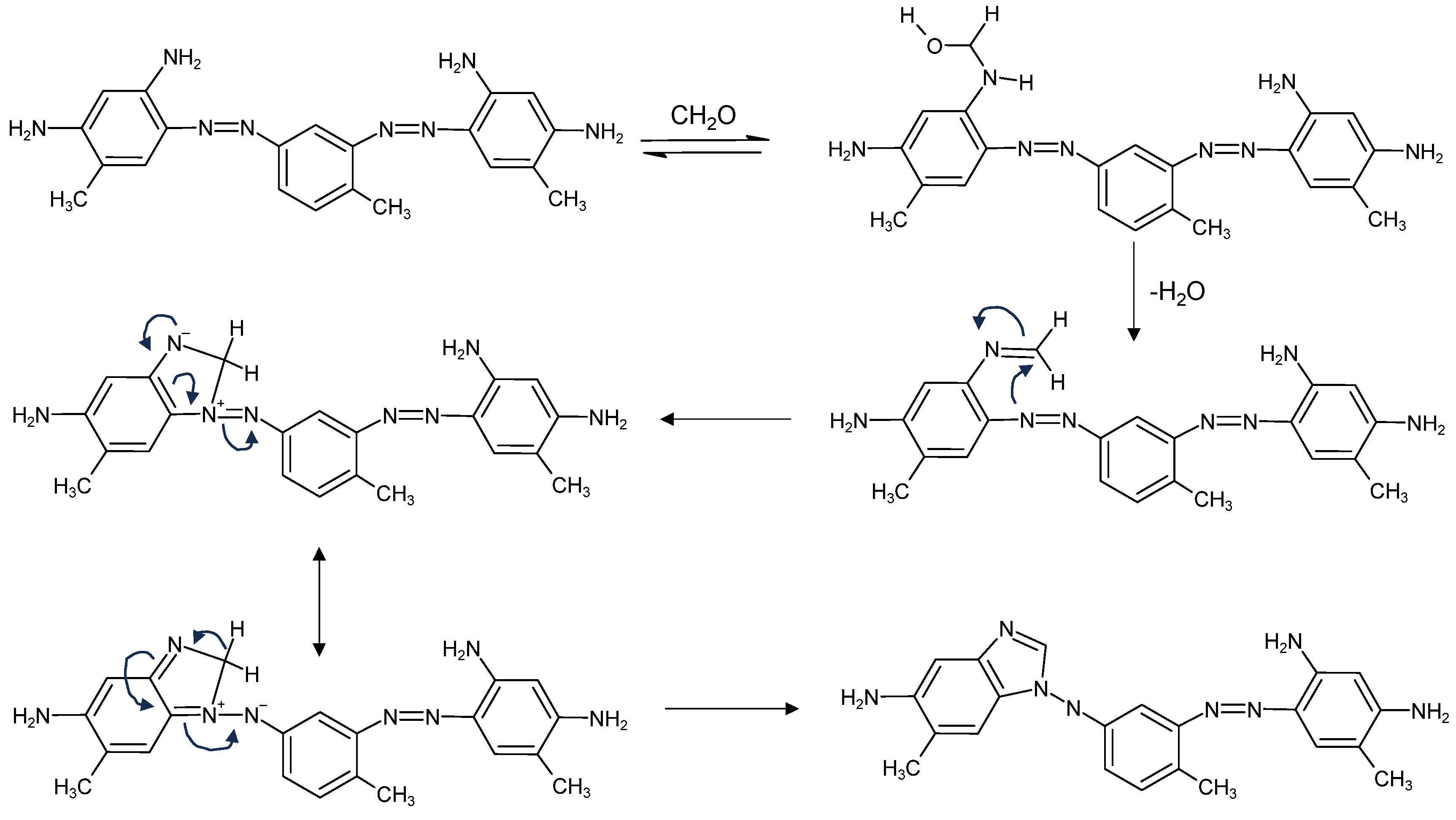
  212. Chen, V.J.; Minto, R.E.; Manicke, N.; Smith, G.D. Structural Elucidation of Two Congo Red Derivatives on Dyed Historical Objects Indicative of Formaldehyde Exposure and the Potential for Chemical Fading. Dye. Pigment. 2022, 201, 110173. [Google Scholar] [CrossRef]
  213. Favaro, G.; Confortin, D.; Pastore, P.; Brustolon, M. Application of LC-MS and LC-MS-MS to the Analysis of Photo-Decomposed Crystal Violet in the Investigation of Cultural Heritage Materials Aging. J. Mass Spectrom. 2012, 47, 1660–1670. [Google Scholar] [CrossRef]
  214. Chen, V.J.; Smith, G.D.; Whitaker, M.R.; von Rabenau, B. Identification of Red Dyes in Selected Textiles from Chin and Karen Ethnic Groups of Myanmar by LC-DAD-ESI-MS. In Dyes in History and Archaeology 33/34; Kirby, J., Ed.; Archetype Publications: London, UK, 2021; pp. 92–101. [Google Scholar]
  215. Smith, G.D.; Esson, J.M.; Chen, V.J.; Hanson, R.M. Forensic Dye Analysis in Cultural Heritage: Unraveling the Authenticity of the Earliest Persian Knotted-Pile Silk Carpet. Forensic Sci. Int. 2021, 3, 100130. [Google Scholar] [CrossRef]
  216. Aceto, M.; Agostino, A.; Fenoglio, G.; Idone, A.; Gulmini, M.; Picollo, M.; Ricciardi, P.; Delaney, J.K. Characterisation of Colourants on Illuminated Manuscripts by Portable Fibre Optic UV-Visible-NIR Reflectance Spectrophotometry. Anal. Methods 2014, 6, 1488. [Google Scholar] [CrossRef]
Figure 1. Examples of colourants belonging to different application categories with their respective most common commercial names, C.I. Generic Names and C.I. Constitution Numbers.
Heritage 07 00094 g001
Figure 2. Tautomeric forms of the nitroso dye Mordant Green 4 (C.I. 10005).
Heritage 07 00094 g002
Figure 3. Molecular structures of the nitro dyes Martius Yellow (Acid Yellow 24, C.I. 10315) and Naphthol Yellow S (Acid Yellow 1, C.I. 10316).
Heritage 07 00094 g003
Figure 4. Molecular structures of the monoazo dye Chrysoidine (Basic Orange 2, C.I. 11270) and the disazo dyes Biebrich Scarlet (Acid Red 66, C.I. 26905) and Congo Red (Direct Red 28, C.I. 22120).
Heritage 07 00094 g004
Figure 5. Tautomeric equilibrium between the azoenol form and ketohydrazone form of orange II (Acid Orange 7, C.I. 15510).
Heritage 07 00094 g005
Figure 6. Molecular structures of the triarylmethane dyes New Fuchsine (Basic Violet 2, C.I. 42520), Crystal Violet (Basic Violet 3, C.I. 42555), Malachite Green (Basic Green 4, C.I. 42000) and Victoria Blue B (Basic Blue 26, C.I. 44045).
Heritage 07 00094 g006
Figure 7. Molecular structures of the sulphonated triarylmethane dyes Patent Blue V (Acid Blue 3, C.I. 42051) and Nicholson Blue (Acid Blue 93, C.I. 42780).
Heritage 07 00094 g007
Figure 8. Schematic reaction of the reduction of Michler’s ketone to Auramine (Basic Yellow 2, C.I. 41000).
Heritage 07 00094 g008
Figure 9. Molecular structures of the xanthene dyes Fluorescein (Acid Yellow 73, C.I. 45350), Eosin (Acid Red 87, C.I. 45380), Erythrosine (Acid Red 51, C.I. 45430) and Phloxine (Acid Red 98, C.I. 45405).
Heritage 07 00094 g009
Figure 10. Molecular structures of the xanthene dyes Rhodamine B (Basic Violet 10, C.I. 45170), Rhodamine 6G (Basic Red 1, C.I. 45160) and Violamine R (Acid Violet 9, C.I. 45190).
Heritage 07 00094 g010
Figure 11. Molecular structures of the xanthene dyes Galleine (Mordant Violet 25, C.I. 45445) and Coeruleine (Mordant Green 22, C.I. 45510).
Heritage 07 00094 g011
Figure 12. Molecular structures of the anthraquinone dyes Alizarin (Mordant Red 11, C.I. 58000), Alizarin Orange (Mordant Orange 14, C.I. 58015), Alizarin Blue R (C.I. 67410) and Alizarin Green S (C.I. 67405).
Heritage 07 00094 g012
Figure 13. Molecular structures of the dyes Phosphine (Basic Orange 15, C.I. 46045), Safranine (Basic Red 2, C.I. 50240), Methylene Blue (Basic Blue 9, C.I. 52015) and Meldola Blue (Basic Blue 6, C.I. 51175).
Heritage 07 00094 g013
Figure 14. Chromatographic profiles obtained by HPLC-DAD at 540 nm of Fuchsine samples produced with (a) the early synthetic process and (b) the later synthetic process.
Heritage 07 00094 g014
Figure 15. Degradation pathway observed for Brilliant Green (C.I. 42040; Basic Green 1), leading to the formation of benzophenone degradation products.
Heritage 07 00094 g015
Figure 16. Reaction scheme showing the addition of formaldehyde to Bismark Brown R (C.I. 21020; Basic Brown 4) and leading to the formation of degradation products. The reaction can occur on both sides of the molecule.
Heritage 07 00094 g016
Figure 17. Images of a Dhofari woman’s dress (abuthail; accession number 2011,6003.46). @The Trustees of the British Museum. Shared under a Creative Commons Attribution-NonCommercial-ShareAlike 4.0 International (CC BY-NC-SA 4.0) licence.
Heritage 07 00094 g017
Figure 18. Reflectance spectra acquired by FORS for one of the Omani textiles (representative of the four textiles investigated) and the reference samples of fabrics dyed with indigo, Methyl Violet (Basic Violet 1, C.I. 42535), Victoria Blue B (Basic Blue 26, C.I. 44045) and Victoria Blue R (Basic Blue 11, C.I. 44040). @The Trustees of the British Museum. Shared under a Creative Commons Attribution-NonCommercial-ShareAlike 4.0 International (CC BY-NC-SA 4.0) licence.
Heritage 07 00094 g018
Figure 19. Extracted ion chromatograms obtained by HPLC-ESI-Q-ToF analysis (positive ionisation mode) of a sample taken from textile 2011,6003.46 from the British Museum’s collection. The molecular components of Methyl Violet (Basic Violet 1, C.I. 42535) and Victoria Blue B (Basic Blue 26, C.I. 44045) are listed in the legend along with their chemical formula, retention times (RTs) and accurate masses of the corresponding molecular ions [M]+. @The Trustees of the British Museum. Shared under a Creative Commons Attribution-NonCommercial-ShareAlike 4.0 International (CC BY-NC-SA 4.0) licence.
Heritage 07 00094 g019
Figure 20. (a) Raman spectra of a modern Napthol Green B powder (top), a naturally aged powder dye dated 1900 and labelled ’Naphtholgrün B’ (middle) and a naturally aged lake pigment on paper dated 1893 labelled ’Naphtholgrün B’ (bottom). (b) UV/VIS/NIR reflectance spectra of different green hues of commercial colour charts containing Pigment Green 12 dated 1893–1932. (c) UV/VIS/NIR reflectance spectra of ten different green colour references. Reproduced from [94]. © Elsevier Masson SAS. All rights reserved.
Heritage 07 00094 g020
Figure 21. (A) Details of Jazz plates examined under the optical microscope (10× magnification); (B) results of PCA analysis carried out on the p-XRF spectra taken on 23 yellow areas across the 20 MoMA Jazz pates; (C) confocal Raman and SERS spectra of the “magenta” colour (M) alongside reference spectra for Rhodamine 6G (SERS; 612, 775, 1090, 1179, 1310, 1363, 1510, 1574 and 1679 cm−1), Rhodamine 3B (confocal Raman; 271, 365, 620, 633, 662, 1082, 1187, 1199, 1362, 1510, 1530, 1599 and 1650 cm−1) and barite (sulphate band at 988 cm−1). Image rearranged from [33].
Heritage 07 00094 g021
Figure 22. Raman spectra (3a–3c); SERS micro-extracts (3d–3h) with water and acetone transferred to KLARITE® and SOKEN® replicas. Reference spectrum (3i) from SOPRANO Spectral library, PR 60:1, Sunbrite Red, C.I. 16105, naphthalene sulphonic acid (Ba-salt); visible and 3D mapping micrographs (3l–3n) on SOKEN® replica, inset overall Raman map (3l), band extraction mode with main bands of an azo β-naphthol dye (3m) and 3D SERS mapping (3n) with WITec software (https://raman.oxinst.com/products/software/WITec-software-suite). Reprinted from [124] with permissions from Elsevier.
Heritage 07 00094 g022
Table 1. Main chemical categories of synthetic colourants and their C.I. Constitution Numbers.
C.I. Numbers C.I. Numbers
Nitroso10000–10299Quinoline47000–47999
Nitro10300–10999Thiazole49000–49399
Monoazo11000–19999Azine50000–50999
Disazo20000–29999Oxazine51000–51999
Trisazo30000–34999Thiazine52000–52999
Polyazo35000–36999Anthraquinone58000–72999
Diarylmethane41000–41999Indigoid73000–73999
Triarylmethane42000–44999Phthalocyanine74000–74999
Xanthene45000–45999Natural75000–75999
Acridine46000–46999Inorganic77000–77999
Table 2. An overview of the most used precipitation agents/substrates for SOPs until approximately WWI.
DyeTypePrecipitation Agent/Substrate
Acid dyes *TonersBarium chloride, lead nitrate, lead acetate, zinc sulphate, aluminium sulphate, aluminium acetate, manganese sulphate, tin chloride, antimony chloride, calcium nitrate, calcium acetate
Basic dyes *TonersTannic acid, phosphoric acid, arsenic acid, antimonious acid, stannic acid, resinic acid, palmitic acid, stearic acid (and other similar fatty acids)
Intended use/dye dependent *Lakes and extendersBarium sulphate (natural or blanc-fix), kaolin (China clay), calcium sulphate (Paris white, gypsum, satin white), potassium aluminium sulphate, alumina, diatomaceous earth (Kiezelguhr), red lead, zinc oxide, lead sulphate, aluminium hydrate, calcium carbonate (chalk), barium phosphate, lead carbonate (white lead), calcium phosphate, carbon (lamp and vegetable black), green earth
* Some dyes have both basic and acidic characteristics or different functional groups that allow them to be precipitated in multiple ways.
Table 3. Analytical steps for the analysis of synthetic colourants.
CategoryTechniqueExpected InformationAdvantages (+)/Disadvantages (−)
Non-invasive Analysis

Heritage 07 00094 i001

Preliminary indications of the presence of ESDs and SOPs and their distribution + decision on sampling areas
Visual examinationOptical microscopyTechnical examination of production processes, paint application, ink strokes, fibre classification
Electronic spectroscopyColourimetryColour change measurements and colour matching+ Utilisable in situ; enables colour to be measured and systematically defined
− No information useful for identification
Fibre optic reflectance/Fluorescence spectroscopyPreliminary indications of the colourants present and their spatial distribution, colour matching+ Utilisable in situ; enables large numbers of spectra to be quickly recorded and visualised in real time; inexpensive; safe; database of spectra available
− Relatively poor spatial (probes with aperture diameters > 1 mm) and wavelength resolution (ca. 2–5 nm); limited “fingerprinting” ability; results strongly affected by substrate, pH and other materials present
ImagingBroadband multispectral imaging (MSI)/Hyperspectral imaging (HSI)False-colour images are created, showing the distribution of materials on the surface+ Utilisable in situ; capability to scan wide surfaces; some dye identification possible
− Few applications due to limited fingerprinting ability; complex data elaboration; sensitivity to external illumination conditions
X-ray-based techniquesX-ray fluorescence spectroscopy (XRF)Investigation of the inorganic components (elemental analysis and distribution)+ Utilisable in situ; sensitive and selective
− Poor sensitivity to light elements
Vibrational spectroscopiesPortable FTIR spectroscopyInformation on ESDs, SOPs and other materials present in the sample+ Utilisable in situ; provides information on other materials present in the sample and on the recipe used for SOPs
− Spectra affected by the matrix; reference spectra are needed for interpretation; poor spatial resolution; surface scattering effect
Portable Raman spectroscopyInformation on ESDs and SOPs in several different matrices and on their spatial distribution+ Utilisable in situ; versatile irradiation source; high sensitivity; databases available
− Background fluorescence signal produced by the organic matrix or support
Invasive/non-destructive analyses

Heritage 07 00094 i001

Detailed information related to the samples
MicroscopyOptical microscopy (possibly on cross-sections)Indication of dye technology, removal of dirt/contaminants on the fibre surface, assessment of the stratigraphy for paint cross-sections+ Relatively easy to use
− Only preliminary information; if the sample is treated, it cannot be used for further analysis
Benchtop vibrational spectroscopiesFTIR/Raman spectroscopyInformation on ESDs, SOPs and other materials present in the sampleSee above for portable FTIR
+ higher spectral and spatial resolution compared to the portable equipment
− possible signal overlap; minor components often non-detectable
Surface-enhanced Raman spectroscopy (SERS)Information on ESDs and SOPs in several different matricesSee above for portable Raman
+ Signal enhanced by several orders of magnitude; reduced problem of background fluorescence
− The chromophore-containing molecule can be unavailable for the desired SERS effect due to the matrix; uneven signal enhancement; minor compounds not always detectable
X-ray-based techniquesScanning electron microscopy Energy dispersive X-ray spectroscopy (SEM-EDX)Identification of fibres and mordant analysis/identification of precipitating agents in SOPs or additives+ Combined visual and elemental analysis; high magnification and spatial resolution; mapping capabilities
− Sample cannot be used for further analysis; morphological information difficult to be interpreted
Laser-induced breakdown spectroscopy (LIBS)Investigation of the inorganic components (elemental analysis)+ Almost non-destructive (the laser beam might leave a small mark on the fibre); quick; relatively inexpensive
− No information about molecular structure of the inorganic components
X-ray powder diffraction (XRD)Identification of the crystalline structure of SOPs+ Suitable for studying the interaction between the dye and its substrate
− Only applicable to crystalline phases
X-ray fluorescence spectroscopy (XRF)Investigation of the inorganic components (elemental analysis)+ Sensitive and selective;
− Detection of light atoms is difficult; no information about molecular structure of the inorganic components
Invasive/micro-destructive analyses

Heritage 07 00094 i001

Highly detailed molecular information
Spectroscopies entailing modification of the sampleSurface-enhanced Raman spectroscopy (SERS) applied to a sample extractDetailed information on ESDs and SOPs in several different matrices+ Very high sensitivity
− Uneven signal enhancement; spectra dominated by the main component of a mixture
Fourier Transform infrared spectroscopy (FTIR) in transmission mode (KBr pellet, diamond cell, etc.)Detailed information on some ESDs and SOPs as well as substrates and other materials+ High sensitivity; particle localisation with microscope
− Overlapping signals; low specificity
Mass spectrometry techniques (no or minimal sample treatment)Direct temperature resolved-mass spectrometry (DT-MS)Molecular information on ESDs, SOPs and other organic material in the sample (e.g., binding medium, additives, etc.)+ Quick acquisition; no sample treatment
− Matrix ionisation can produce fragments preventing SOPs or ESDs detection/identification
Pyrolysis gas chromatography coupled to mass spectrometry (Py-GC/MS) *Rapid identification of some synthetic colourants; information on binding media and any other organic material+ No sample treatment; useful for insoluble pigments; libraries available for SOPs
− Pigments of the same chemical class cannot often be distinguished; interferences due to textile matrices
Laser desorption ionisation coupled to mass spectrometry (LDI-MS) and matrix-assisted laser desorption ionisation (MALDI-MS)Information at molecular level of ESDs and SOPs; in imaging mode information on the spatial distribution of the analytes is provided+ Minimal or no sample treatment
− No separation of the analytes; scarce insight on minor/degradation products; complex spectral interpretation
Ambient ionisation mass spectrometry (AMS)Information at molecular level of ESDs and SOPs; in imaging mode information on the spatial distribution of the analytes is provided+ Minimal pre-treatment; possibility to be used in situ;
DESI can be implemented in imaging mode (DESI-MSI)
− No separation of the analytes; scarce insight on minor/degradation products; complex spectral interpretation
Secondary ions mass spectrometry (SIMS)Information at molecular level of SOPs and binding media; in imaging mode information on the spatial distribution+ Identification of both the pigment and the binder in a short analysis time.
− No separation of the analytes; complex spectral interpretation
Chromatographic techniquesThin-layer chromatography (TLC)Pre-screening for main dye components; separation for further spectroscopic analyses+ Possibility to be directly combined with FTIR and SERS; fraction recollection
− Separation not highly precise
High-pressure liquid chromatography—diode array detector—tandem mass spectrometry (HPLC-DAD-MS/MS)Identification of colourants and dyestuff sources at a molecular level; identification of synthetic by-products and unknown degradation products+ Ultimate sensitivity and selectivity; highly efficient chromatographic separation; high-resolution mass information
− Expensive equipment; time-consuming sample treatment; possible modifications of the analytes induced by sample treatment; difficult data interpretation
* Py-GC/MS is a chromatographic technique as well.
Disclaimer/Publisher’s Note: The statements, opinions and data contained in all publications are solely those of the individual author(s) and contributor(s) and not of MDPI and/or the editor(s). MDPI and/or the editor(s) disclaim responsibility for any injury to people or property resulting from any ideas, methods, instructions or products referred to in the content.

