
The Antibacterial Performance of Implant Coating Made of Vancomycin-Loaded Polymer Material: An In Vitro Study

Department of Prosthodontics, College of Dentistry, Qassim University, Buraydah 52571, Saudi Arabia
Surfaces 2023, 6(3), 304-315; https://doi.org/10.3390/surfaces6030022
Submission received: 14 August 2023
/
Revised: 3 September 2023
/
Accepted: 7 September 2023
/
Published: 13 September 2023
(This article belongs to the Special Issue Applications of Nanotechnology in Diagnosis and Therapy)
Abstract
:Bacterial adhesion and biofilm formation on the surface of titanium implants are the main causes of implant-associated infection. An antibacterial coating on the implant surface can reduce the risk of biofilm formation. The aim of this study was to investigate the bactericidal effects of a van-comycin-loaded polymer coated on an implant surface. For this purpose, poly(N-isopropylacrylamide) (PNIPAAm) was first synthesized as a homopolymer or by co-polymerization with acrylamide (PNIPAAm-AAm) at a 5% weight ratio. Then, thin and uniform polymer coatings were prepared using the spin coating technique. The degree of surface hydro-philicity of the polymer coatings was evaluated by measuring the water contact angle (CA). For the antibacterial tests, the polymer-coated surfaces were loaded with vancomycin. The tests were performed in three conditions: on a glass surface (control), on a PNIPAAm-AAm-coated surface, and on a PNIPAAm-AAm-coated surface loaded with vancomycin. The death rates of the bacteria in contact with the coated surfaces were evaluated at different temperatures with fluorescence microscopy. A scanning electron microscopy (SEM) analysis of cross sections of the polymer coatings revealed a uniform thin film of approximately 200 nm in thickness. The water contact angle analysis performed at different temperatures revealed that the polymer-coated surfaces were more hydrophobic (CAs ranging between 53° and 63°) than the uncoated glass surface (CA ranging between 15° and 35°). The bacterial death rate, measured at 40 °C or while continuously switching the temperature between 37 °C and 40 °C, was higher in the presence of the surface coated with vancomycin-loaded PNIPAAm-AAm than when using the other surfaces (p-value ≤ 0.001). The vancomycin-loaded polymer coating evaluated in this study exhibited effective antibacterial properties when the polymer reached the phase transition temperature.
1. Introduction
Implantable devices are used extensively in the orthopedic and dental fields. In orthopedics, implants are utilized mainly for hip and knee joint replacements, while in dentistry, they are applied to replace missing teeth. In both fields, the use of these implants may greatly improve patients’ quality of life. Titanium (Ti) and its alloys are the most commonly used implant materials due to their excellent biocompatibility and good mechanical properties [1]. However, bacterial infection at the implant site can lead to peri-implant mucositis or peri-implantitis, which are difficult to treat and may cause implant failure [2]. Bacterial adhesion to an implant surface can be followed by biofilm formation, which promotes bacterial resistance to antimicrobial treatments [3]. Some investigations revealed that bacterial colonization can occur shortly after implant placement and may take place at the tissue–implant interface [4].
As such, preventing bacterial attachment to the implant could be the most effective strategy to reduce the risk of biofilm formation on its surface [3]. To that end, a systemic antibiotic prophylaxis is routinely administered soon after implant placement and can help avoid postsurgical infection [5]. Nevertheless, the systemic administration of antibiotics has some limitations, e.g., the drug concentration reached at the treatment site may be low, and the drugs may have toxic effects. Furthermore, some reports showed that biofilms can work as a mechanical barrier to certain drugs, which can limit the effect of systemic antibiotics in preventing implant-associated infections [6].
Thus, the localized application of antibiotics at the implant site has been suggested as an alternative approach to prevent bacterial infection. This approach requires coating the implant surface with the drug, incorporating the drug into the implant surface, or using a carrier material to release the antibacterial agent at the bone–implant interface [7]. This method is often preferable to the systemic treatment because it allows for achieving effective antibacterial doses using small amounts of the therapeutic agent, thus reducing the potential risks associated with systemic administration [8]. Moreover, the local release of antibiotics allows for specific peri-implantitis pathogens to be targeted, helping to control possible antibiotic resistance.
Various techniques can be used to incorporate therapeutic agents into an implant surface, including plasma spray and dip coating [9,10,11]. It is believed that to achieve effective drug release at the implant site, both the coating layer and the antibiotics must meet specific requirements. In fact, the coating layer should be able to control and sustain drug release for the determined treatment period, while the selected antibiotic agents should be effective against a broad bacterial spectrum and be stable at high body temperatures. Several antibiotics have been examined to treat implant-associated infections. Among these, vancomycin has been investigated by many researchers after local release around implants because of its efficacy against Gram-positive microorganisms such as methicillin-resistant Staphylococcus aureus (MRSA) [12]. Another commonly used antibiotic for such applications is gentamicin, as it has a broad antibacterial spectrum [13]. In an in vitro study, Li et al. found that gentamicin loaded into a Ti implant coating and subjected to gamma radiation maintained its antibacterial effect [14]. Other antibiotics that are commonly used in implant coating include amoxicillin, cephalothin, and tobramycin [15].
The proper release of antibiotics from the coating layer is essential for their effectiveness and is affected by the method of implant coating and drug incorporation. Some studies reported that the use of nanotechnologies in drug delivery applications can provide the sustained release of drugs and enhance the bioavailability of small molecules [16,17]. Many researchers investigated the application of polymeric materials as carriers of therapeutic agents within implants. The advantage of these polymeric materials is that they can absorb and carry large amounts of therapeutic agents, which could facilitate the sustained release of a wide range of drugs. Furthermore, some studies reported that implants coated with a polymer layer can allow for limited burst release, improving the drug release characteristics and prolonging the overall release [14]. According to some reports, implant integration with the bone can be improved using polymer coatings, as this can result in good osteoblast adhesion to the implant and cell proliferation [18,19]. However, there is a lack of information about the possible risks of local toxicity at the release site, which requires further investigation.
Many polymeric materials have been evaluated for their ability to sustain local drug release around implant surfaces [19,20]. Chitosan is now frequently used, as it has good biocompatibility and antibacterial properties [21,22]. Gulati et al. reported that chitosan coatings on Ti surfaces are useful for delivering antibiotics around implants for a prolonged period, with predictable release kinetics [20]. They also suggested that they might be clinically used with different types of drugs and implants [20]. Other popular polymers are thermoresponsive polymers, which show unique changes in their physical properties associated with temperature variations [23,24]. One such polymer, poly(N-isopropylacrylamide) (PNIPAAm), has been frequently investigated for drug delivery applications [23,25,26]. PNIPAAm has unique thermal properties, showing transitional changes at temperatures that are close to physiological body temperatures. Specifically, these transitional changes occur at the temperature known as the lower critical solution temperature (LCST), which is from 32 to 34 °C [25]. In this temperature range, PNIPAAm undergoes a conversion from a hydrophilic and swollen form (below the LCST) to a hydrophobic and shrunken form (above the LCST) [27]. Because of this property, PNIPAAm has been investigated extensively for drug delivery purposes. In the dental field, the used materials have to demonstrate excellent biocompatibility to be suitable for both patients and the staff [28].
The changes in the surface hydrophobicity of polymer coatings have also attracted attention for their possible relevance to osseointegration [29,30]. Some studies reported that surface hydrophilicity (also known as wettability) can affect protein adsorption and cell adhesion to implants [31]. Other researchers found that some cells adhere preferentially to hydrophilic surfaces, while other cells are attracted to hydrophobic surfaces [32]. Hydrophobic surfaces can establish strong hydrophobic interactions, and as such, they are believed to be more protein-absorbent than hydrophilic surfaces [33]. In an in vitro instigation, Milleret et al. reported that blood clot formation and platelet activation, which are essential for implant integration with the bone, could be improved by hydrophilic surfaces [34]. In another study, a sandblasted and acid-etched (SLA) Ti implant surface modified to achieve high surface hydrophilicity promoted better osteogenic responses compared to a hydrophobic SLA surface [35].
Although hydrophobicity is considered an important property of the surface of an implant, there is a limited number of reports investigating its role in osseointegration [36,37,38,39]. Surface hydrophobicity is generally defined by the value of the contact angle with water (CA). This value is determined by measuring the angle between the tangent line of a liquid’s surface and that of a horizontal solid’s surface; it can range from 0° to 180°. In general, a CA of less than 90° is typical of hydrophilic surfaces; the closer to 0° a CA is, the more hydrophilic the surface will be. In contrast, surfaces with a CA greater than 90° are considered hydrophobic, and their hydrophobicity increases as the CA approaches 180°.
In the present study, a drug delivery system using a thin PNIPAAm coating was developed. When the temperature increased to 40 °C, the drug could be released from the coating. The aim of this study was to investigate the bactericidal effect of an implant surface coated with vancomycin-loaded polymer layers.
2. Materials and Methods
2.1. Polymer Synthesis
For polymer synthesis, the single monomer N-isopropylacrylamide (NIPAAm) was polymerized and used to synthesize conventional PNIPAAm. In addition, NIPAAm was copolymerized with acrylamide (AAm) at a 5% weight ratio to form the PNIPAAm-AAm polymer. The synthesis process was carried out as previously described [40]. In detail, the NIPAAm monomer was first refined by dissolving it in a hexane solution before it was polymerized. The synthesis of the PNIPAAm polymer was performed with free-radical polymerization using 1 g of NIPAAm, 0.0354 g of azobisisobutyronitrile (AIBN), and 20 mg of 2-hydroxy-4-(methacryloyloxy)benzophenone. The mixture was dissolved in 1 mL of ethanol and gently stirred for 15 min to form a clear solution. The obtained solution was moved to a flask with a septum and was purged with nitrogen for 5–10 min to remove all the oxygen. Later, the flask was immersed in a warm oil bath (at 60°C) for 60 minutes and then opened to the air to terminate the polymerization reaction. Finally, the flask was left in a fume hood overnight at room temperature. The next day, the formed polymer was washed with Milli-Q water several times to remove all unreacted and excess reagents.
Meanwhile, 50 mg of acrylamide (5% weight ratio to NIPAM) was added to the same polymer mixture to copolymerize PNIPAAm with acrylamide, obtaining PNIPAAm-AAm. The idea behind copolymerizing PNIPAAm with 5% acrylamide was to obtain a polymer with a transition temperature that was slightly above the body temperature. In a previous study, the product obtained by copolymerizing PNIPAAm with 5% acrylamide showed a transition temperature around 40 °C [40]. The synthesis procedures for PNIPAAm-AAm were the same as those mentioned above for pure PNIPAAm.
2.2. Polymer Coating
The spin coating technique was used to form a thin and uniform polymer coating on the chosen substrate. For this procedure, glass slides, i.e., the coating substrate, were first left in a sealed beaker with hexamethyldisilazane for 2 h to improve the polymer adhesion to the surface. Then, 0.5 g of synthesized PNIPAAm polymer was dissolved in 5 mL of ethanol, obtaining a polymer/ethanol solution. Last, 60 μl of the polymer/ethanol solution was spin-coated on the glass substrates for 60 s at 3000 rpm.
2.3. Scanning Electron Microscopy (SEM)
The polymer coating was evaluated at a high magnification with scanning electron microscopy (SEM), using a Leo Ultra 55 electron microscope operated at 2–5 kV (Carl Zeiss Meditec AG, Oberkochen, Germany). The SEM evaluation was performed to visualize the cross-sectional structure of the polymer coatings on the coated glass slides. For all samples, gold sputtering was used to obtain images with adequate contrast, using gold (20 nm) in an ion sputtering device (JFC-1100E, JEOL, Tokyo, Japan).
2.4. CA Measurement
The degree of the surface hydrophilicity of the polymer coatings was evaluated with CA measurement. The measurements were performed using the sessile drop technique, with a goniometer (DSA100, Kruss, Hamburg, Germany). First, the control surfaces (i.e., glass slides with no polymer coating) were cleaned by washing in a surfactant solution and rinsing with Milli-Q water. The test groups consisted of the glass surfaces coated with the PNIPAAm and PNIPAAm-AAm polymers. The CA measurements were performed for all groups at different temperatures, ranging from 25 °C to 50 °C. During each measurement, the temperature of the glass substrate and polymer coating was controlled by placing the surfaces on top of an aluminum stage in a water bath with a programmed temperature. All surfaces were kept at the desired temperature for at least 5 min before performing the CA measurement. Then, water droplets were released using an automated unit, and the CA measurements were conducted 10 s after the water drops reached the surface. The measurements (n = 8 for each group) were performed in air, using Milli-Q water as the probe liquid.
2.5. Loading of Vancomycin on the Coated Layers
Vancomycin was selected to investigate its antibacterial characteristics upon loading on the polymer coatings. The procedures performed for vancomycin incorporation into the polymer coatings were previously described [40]. In summary, all coated surfaces in the test group were loaded with vancomycin by completely immersing them in a vancomycin solution (0.2 mg/mL) for a full day. Later, the surfaces were removed from the solution and gently dried in nitrogen. Figure 1 illustrates the drug release system designed in this study, consisting of the vancomycin-loaded polymer coating.
2.6. Bacterial Live/Dead Analysis
The growth of Staphylococcus epidermidis on the glass surfaces coated with polymer layers and loaded with vancomycin as well as on control surfaces was evaluated. The experiment was conducted on the following surfaces:
Group 1: Glass surface (control);
Group 2: PNIPAAm-AAm-coated surface (test);
Group 3: PNIPAAm-AAm-coated surface loaded with vancomycin (test).
For this experiment, a tube containing 5 mL of tryptic soy broth (30 g/L, Sigma–Aldrich, Eschenstr, Germany) was inoculated one day before the analysis with a bacterial colony of S. epidermidis picked from agar culture plates. The inoculated cells were cultured at 37 °C in a shaking incubator at 200 rpm until they reached the stationary phase. After incubating the bacteria with the coated surfaces for 24 h, the samples were subjected to predetermined heat conditions for 3 h. All samples were evaluated at 3 temperature settings, i.e., 37 °C, 40 °C, and at temperatures fluctuating between 37 °C and 40 °C (30 min each). Later, bacterial viability was evaluated using the live/dead staining method (LIVE/DEAD® BacLightTM Bacterial Viability Kit L7007, New York, NY, USA); the death rate of the bacterial populations in contact with the different surfaces was determined using fluorescent microscopy. Upon live/dead staining, live bacteria appeared green, while dead bacteria appeared red. The mean percentages, with standard deviations, of live/dead bacteria were determined using the fluorescent microscopy images.
2.7. Analysis
For the statistical analysis of these results, the non-parametrical Kruskal–Wallis test (SPSS Statistics version 22, IBM Corp., New York, NY, USA) was selected to evaluate the differences in bacterial death rates among the all groups at the three temperature set points. In addition, post hoc testing was used to perform multiple comparisons within each temperature set point. The significance level was set at p = 0.05. All data were plotted as mean ± standard error.
3. Results
3.1. SEM
The SEM analysis of cross sections of the polymer coatings revealed a uniform thin film of approximately 200 nm in thickness (Figure 2).
3.2. CA Measurements
The images of the live CA measurements are shown in Figure 3. The CA values of all the evaluated surfaces are summarized in Figure 4. The glass substrate showed a hydrophilic surface, with CA ranging between 15° and 35° at different temperatures. The surfaces coated with a polymer layer were more hydrophobic compared to the glass surface when evaluated at temperatures ranging from 25 °C to 50 °C, as the CAs of the surfaces coated with a polymer layer ranged between 53° and 63° at different temperatures. Overall, for the surfaces coated with a polymer layer, the CA tended to increase slightly as the temperature increased, which indicated increased hydrophobicity.
3.3. Live/Dead Bacterial Analysis
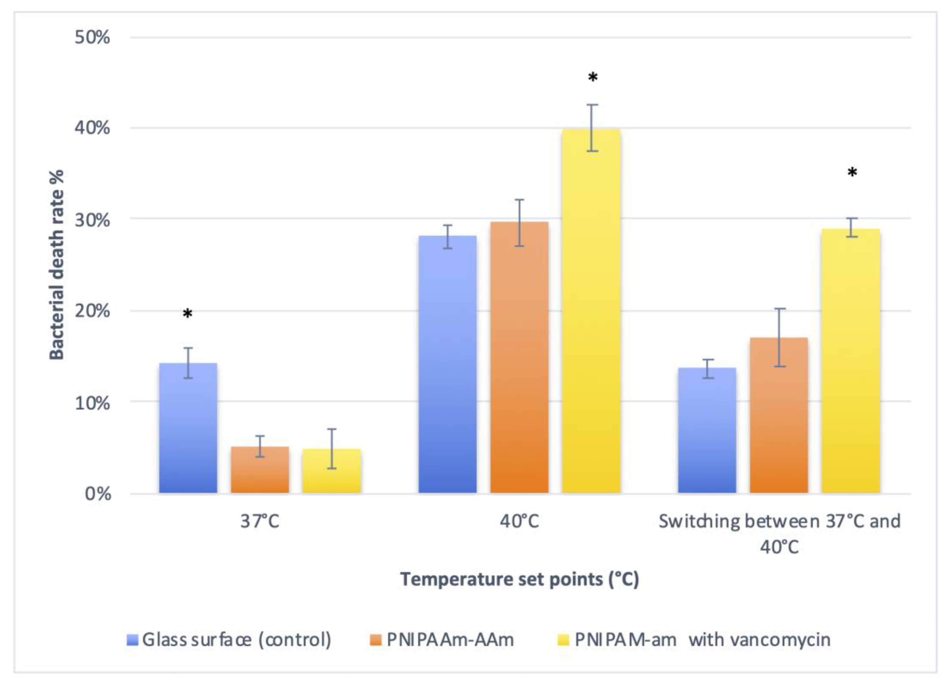
The antimicrobial effect of the PNIPAAm-AAm coating with and without vancomycin was evaluated at different temperatures. Figure 5 shows images taken using a fluorescent microscope of the live and dead bacteria at different temperatures. The analysis of the bacterial death rate revealed a significantly higher death rate in the control group compared to the polymer groups at 37 °C (p ≤ 0.001) (Figure 6). No significant differences in bacterial death rate were found between the two polymer groups at this temperature (p = 0.147). In contrast, a significantly higher level of bacterial death was observed on surfaces coated with PNIPAAm-AAm and vancomycin when the temperature was raised to 40 °C compared to the other groups (p ≤ 0.001). Increased bacterial death in the presence of the PNIPAAm-AAm–vancomycin coating was also observed when the temperature was continuously switched between 37 °C and 40 °C (p ≤ 0.001). In these temperature conditions, no significant differences were found between the control group and the polymer group without vancomycin (p = 0.254).
4. Discussion
The localized antibiotic release at the bone–implant interface is an alternative to invasive treatment modalities used for implant-associated infections [41]. This in vitro study evaluated the antibacterial properties of an implant coating consisting of a vancomycin-loaded polymer. The PNIPAAm polymer was synthesized by free-radical co-polymerization at a 5% weight ratio with acrylamide. It is recommended to conduct material syntheses through reliable and eco-friendly processes [42]. Some reports revealed that PNIPAAm demonstrated good biocompatibility [43,44]. The PNIPAAm polymer material was designed as a thin coating using the spin coating technique. This technique is commonly used to obtain coating layers with a uniform thickness. One of the advantages of this technique is that the layer thickness can be easily controlled by changing the concentration of the solution used. In this study, an adequate amount of antibiotic solution was incorporated into a thin polymer layer of around 200 nm in thickness. In a previous study, vancomycin was incorporated in a polymer coating of 300 nm in thickness, which resulted in the release of the antibiotic and the killing of all bacteria in a range of 2 mm around the coated surface [40]. It is essential that the polymers used to coat implants have good biocompatibility and do not interfere with bone integration.
The CA test showed that the glass surface coated with polymer layers was more hydrophobic than the uncoated glass surface, particularly when the temperature was increased. However, the polymer coating was still classified as hydrophilic because the water CA on it was less than 90° in all tests. Several studies of bone cells showed that the surface hydrophilicity of an implant can influence cell responses such as adhesion, proliferation, and differentiation (44, 61). Other reports suggested that surface hydrophilicity can also affect soft tissue cells such as fibroblasts (78, 79). Furthermore, an in vitro study published by Hong et al. found that the hydrophilic modification of Ti dental implants improved the thrombogenic properties of Ti, which might enhance the bone healing process around the implant [45].
Numerous publications suggested that hydrophilic implant surfaces can improve the differentiation of mesenchymal stem cells into bone cells [35,46]. In a minipig model, Buser et al. evaluated bone formation when using a sandblasted, large-grit, and acid-etched SLA surface (i.e., a hydrophilic surface) in comparison with a standard SLA surface [47]. In the presence of the modified SLA surface, significantly greater bone formation was observed compared with the control implants after 2 and 4 weeks of healing [47]. Similar findings were reported by Schwarz et al., who found that hydrophilic implants could improve angiogenesis during the early stages of osseointegration in dogs [48]. Furthermore, some clinical investigations showed that hydrophilic implants could influence gene expression after implant placement, which might enhance the implants’ osseointegrative properties [46].
To test the efficacy of the drug delivery system evaluated in the current study, we assessed the growth of S. epidermidis around PNIPAAm-coated surfaces loaded with vancomycin. Staphylococcus is one of the pathogens most frequently associated with implanted medical devices [49,50]. The results of the bacterial tests suggest that the PNIPAAm-coated surfaces were better able to preserve the antibiotic before the temperature increase, as the control group showed more bacterial death compared to the group of polymer-coated surfaces at 37 °C. Both polymer-coated groups (i.e., with and without the antibiotic) showed similar rates of bacterial death at 37 °C. This may indicate that the polymer did not reach the transition phase, which would cause polymer shrinkage and, subsequently, drug release. Differences in bacterial death between the two polymer groups were observed when the temperature was increased to 40 °C. In these conditions, significantly higher rates of bacterial death were observed in the presence of the polymer containing the antibiotic. This increase was attributed to the release of the antibiotic from the polymer coating after polymer shrinkage. These findings suggest that polymer coatings (as drug carriers) may not affect or change drug properties. We also observed higher rates of bacterial death in the presence of the polymer containing vancomycin when the temperature was continuously changed between 37 °C and 40 °C, but bacterial death was less extensive than that seen at the constant temperature of 40 °C.
Drug release from the antibiotic-loaded polymer coating described here could be initiated by a temperature increase. The temperature range tested in this study was chosen to resemble the increase in body temperature that occurs during inflammation; although an increase in body temperature does not always occur in the case of implant-associated infection, the body temperature can reach 40 °C in some cases. With implant infection, the actual changes in temperature at the implant site may be minimal; this has not yet been sufficiently investigated. However, the sustained drug release we observed as the temperature increased could be valuable in some clinical cases of implant-associated infection after implant insertion.
Vancomycin was used in this study against S. epidermidis. Vancomycin is a narrow-spectrum antibiotic active against Gram-positive bacteria that is often used when treatment with other antibiotics has failed [51]. Several other antibiotics have been investigated for treating implant-associated infections [14,52]. However, some reports suggested that routine antibiotic treatments may not be effective against bacteria in biofilms because they are more resistant to antimicrobial agents compared to planktonic bacteria [53,54]. It is believed that it is difficult to eliminate an infection after a bacterial biofilm is formed; from a clinical perspective, implant removal could be the only available solution. As such, one of the most effective strategies to reduce the risk of infection around dental implants is to prevent biofilm formation. Many studies have thus been conducted on antibacterial coatings as an alternative strategy to prevent biofilm formation on implants [52,55].
The idea behind copolymerizing PNIPAAm with 5% acrylamide was to obtain a polymer with a transition temperature slightly above the body temperature, to be able to mimic the polymer shrinking and drug release that could occur in the body during a fever. In a previous study, PNIPAAm copolymerized with 5% acrylamide showed a transition temperature of around 40 °C. The copolymerization of PNAPAAm with acrylamide was reported to have no effect on the thermal sensitivity of PNIPAAm [56]. Because the phase transition of this polymer occurs at around 40 °C, this polymer could be suitable for future clinical experiments. In fact, the transition occurs a few degrees above the normal body temperature, which allows for avoiding an uncontrolled drug release from the polymer. However, the high temperature needed for phase transition could pose risks to the living tissues, as some reports showed that cortical bone necrosis can be induced at 47 °C [57]. Overall, polymer materials synthesized using PNIPAAm have revealed good biocompatibility and have been investigated extensively for drug delivery applications [58]. In addition, some researchers have evaluated the use of these polymers in different forms for tissue and bone regeneration [43].
In previous studies, various polymer materials were evaluated for localized drug release around the surface of an implant. For instance, Gollwitzer et al. investigated the antibacterial properties of a coating consisting of poly(D,L-lactic acid) (PDLLA) and the antibiotic gentamicin applied on the surface of Ti K-wires [59]. They concluded that coating implants with PDLLA could provide new strategies for reducing the risk of biomaterial-associated infections. Furthermore, some researchers reported that coating an implant with PDLLA could offer good mechanical stability and present high osteoinductive potential in vivo [60]. Lv et al. constructed antibacterial coatings loaded with minocycline on Ti substrates using chitosan and alginate and reported long-term antibacterial effects [55]. Regardless of the polymers used as drug carriers, it has been recommended to allow for an initial burst release of the antibiotic [61]. This initial burst release aims to avoid the development of bacterial resistance to the antibiotic [14].
The vancomycin-loaded polymer coating described in this study showed the ability to release antibiotics in response to a temperature change. Although this study revealed good antibacterial properties of this coating system, it has limitations. One of these limitations is that the experiments were conducted using only one pathogen. In addition, the characteristics of the release of vancomycin from the polymer coating were not investigated. Future investigations should evaluate the drug release features in the long term. In addition, bacterial death after the temperature increase was evaluated for a relatively short time. In this test, the temperature was increased to 40 °C to enable the polymer to reach its transition phase and release the drug. This temperature conditions may not resemble the clinical situation, as implant-associated infection does not always result in an increase in body temperature. It is believed that the localized antibiotic release around an implant surface can be a good alternative to other treatment modalities for implant-associated infections [41].
5. Conclusions
The vancomycin-loaded polymer coating evaluated in this study exhibited effective antibacterial properties when the polymer reached the phase transition temperature. This study describes a promising implant coating strategy based on coatings with antibacterial properties that could be effective in treating implant-associated infections. It remains to be tested if these in vitro results can be replicated in the clinical setting. Therefore, more in vitro and in vivo studies are necessary to confirm these findings.
Funding
This research received no external funding.
Data Availability Statement
Data collected and interpreted in this study are maintained by the authors and can be made available upon request.
Acknowledgments
The author would like to thank the Deanship of Scientific Research, Qassim University for funding the publication of this project.
Conflicts of Interest
The author declares no conflict of interest.
References
- Neoh, K.G.; Hu, X.; Zheng, D.; Kang, E.T. Balancing osteoblast functions and bacterial adhesion on functionalized titanium surfaces. Biomaterials 2012, 33, 2813–2822. [Google Scholar] [CrossRef] [PubMed]
- Darouiche, R.O. Treatment of infections associated with surgical implants. N. Engl. J. Med. 2004, 350, 1422–1429. [Google Scholar] [CrossRef] [PubMed]
- Al-Radha, A.S.D.; Dymock, D.; Younes, C.; O’Sullivan, D. Surface properties of titanium and zirconia dental implant materials and their effect on bacterial adhesion. J. Dent. 2012, 40, 146–153. [Google Scholar] [CrossRef] [PubMed]
- Fürst, M.M.; Salvi, G.E.; Lang, N.P.; Persson, G.R. Bacterial colonization immediately after installation on oral titanium implants. Clin. Oral. Implant. Res. 2007, 18, 501–508. [Google Scholar] [CrossRef]
- Jahoda, D.; Nyc, O.; Pokorný, D.; Landor, I.; Sosna, A. Antibiotic treatment for prevention of infectious complications in joint replacement. Acta Chir. Orthop. Traumatol. Cech. 2006, 73, 108–114. [Google Scholar] [CrossRef]
- Zimmerli, W.; Moser, C. Pathogenesis and treatment concepts of orthopaedic biofilm infections. FEMS Immunol. Med. Microbiol. 2012, 65, 158–168. [Google Scholar] [CrossRef]
- Niu, S.; Cao, X.; Zhang, Y.; Zhu, Q.; Zhu, J.; Zhen, P. Peri-implant and systemic effects of high-/low-affinity bisphosphonate-hydroxyapatite composite coatings in a rabbit model with peri-implant high bone turnover. BMC Musculoskelet. Disord. 2012, 13, 97. [Google Scholar] [CrossRef]
- Norton, L.W.; Koschwanez, H.E.; Wisniewski, N.A.; Klitzman, B.; Reichert, W.M. Vascular endothelial growth factor and dexamethasone release from nonfouling sensor coatings affect the foreign body response. J. Biomed. Mater. Res. Part A 2007, 81, 858–869. [Google Scholar] [CrossRef]
- Decker, J.F.; Lee, J.; Cortella, C.A.; Polimeni, G.; Rohrer, M.D.; Wozney, J.M.; Hall, J.; Susin, C.; Wikesjo, U.M. Evaluation of implants coated with recombinant human bone morphogenetic protein-2 and vacuum-dried using the critical-size supraalveolar peri-implant defect model in dogs. J. Periodontol. 2010, 81, 1839–1849. [Google Scholar] [CrossRef]
- Li, Y.; Li, Q.; Zhu, S.; Luo, E.; Li, J.; Feng, G.; Liao, Y.; Hu, J. The effect of strontium-substituted hydroxyapatite coating on implant fixation in ovariectomized rats. Biomaterials 2010, 31, 9006–9014. [Google Scholar] [CrossRef]
- Offermanns, V.; Andersen, O.Z.; Falkensammer, G.; Andersen, I.H.; Almtoft, K.P.; Sorensen, S.; Sillassen, M.; Jeppesen, C.S.; Rasse, M.; Foss, M.; et al. Enhanced osseointegration of endosseous implants by predictable sustained release properties of strontium. J. Biomed. Mater. Res. Part B Appl. Biomater. 2015, 103, 1099–1106. [Google Scholar] [CrossRef] [PubMed]
- Adams, C.S.; Antoci, V., Jr.; Harrison, G.; Patal, P.; Freeman, T.A.; Shapiro, I.M.; Parvizi, J.; Hickok, N.J.; Radin, S.; Ducheyne, P. Controlled release of vancomycin from thin sol-gel films on implant surfaces successfully controls osteomyelitis. J. Orthop. Res. 2009, 27, 701–709. [Google Scholar] [CrossRef] [PubMed]
- Romanò, C.L.; Scarponi, S.; Gallazzi, E.; Romanò, D.; Drago, L. Antibacterial coating of implants in orthopaedics and trauma: A classification proposal in an evolving panorama. J. Orthop. Surg. Res. 2015, 10, 157. [Google Scholar] [CrossRef]
- Li, L.L.; Wang, L.M.; Xu, Y.; Lv, L.X. Preparation of gentamicin-loaded electrospun coating on titanium implants and a study of their properties in vitro. Arch. Orthop. Trauma Surg. 2012, 132, 897–903. [Google Scholar] [CrossRef] [PubMed]
- Goodman, S.B.; Yao, Z.; Keeney, M.; Yang, F. The future of biologic coatings for orthopaedic implants. Biomaterials 2013, 34, 3174–3183. [Google Scholar] [CrossRef] [PubMed]
- Aberoumandi, S.M.; Khalilov, R.; Davaran, S.; Nasibova, A.; Abbasi, E.; Saghfi, S.; Akbarzadeh, A. An update on clinical applications of nanoparticles in brain and retinal disease (CNS). Adv. Biol. Earth Sci. 2017, 2, 125–142. [Google Scholar]
- Nasibova, A. Generation of nanoparticles in biological systems and their application prospects. Adv. Biol. Earth Sci. 2023, 8, 140–146. [Google Scholar]
- Hu, Y.; Cai, K.; Luo, Z.; Xu, D.; Xie, D.; Huang, Y.; Yang, W.; Liu, P. TiO2 nanotubes as drug nanoreservoirs for the regulation of mobility and differentiation of mesenchymal stem cells. Acta Biomater. 2012, 8, 439–448. [Google Scholar] [CrossRef]
- Kung, S.; Devlin, H.; Fu, E.; Ho, K.Y.; Liang, S.Y.; Hsieh, Y.D. The osteoinductive effect of chitosan-collagen composites around pure titanium implant surfaces in rats. J. Periodontal Res. 2011, 46, 126–133. [Google Scholar] [CrossRef]
- Gulati, K.; Ramakrishnan, S.; Aw, M.S.; Atkins, G.J.; Findlay, D.M.; Losic, D. Biocompatible polymer coating of titania nanotube arrays for improved drug elution and osteoblast adhesion. Acta Biomater. 2012, 8, 449–456. [Google Scholar] [CrossRef]
- Chua, P.-H.; Neoh, K.-G.; Kang, E.-T.; Wang, W. Surface functionalization of titanium with hyaluronic acid/chitosan polyelectrolyte multilayers and RGD for promoting osteoblast functions and inhibiting bacterial adhesion. Biomaterials 2008, 29, 1412–1421. [Google Scholar] [CrossRef] [PubMed]
- Jou, C.-H.; Yuan, L.; Lin, S.-M.; Hwang, M.-C.; Chou, W.-L.; Yu, D.-G.; Yang, M.-C. Biocompatibility and antibacterial activity of chitosan and hyaluronic acid immobilized polyester fibers. J. Appl. Polym. Sci. 2007, 104, 220–225. [Google Scholar] [CrossRef]
- Kavanagh, C.A.; Rochev, Y.A.; Gallagher, W.M.; Dawson, K.A.; Keenan, A.K. Local drug delivery in restenosis injury: Thermoresponsive co-polymers as potential drug delivery systems. Pharmacol. Ther. 2004, 102, 1–15. [Google Scholar] [CrossRef] [PubMed]
- Ward, M.A.; Georgiou, T.K. Thermoresponsive Polymers for Biomedical Applications. Polymers 2011, 3, 1215. [Google Scholar] [CrossRef]
- Ramanan, R.M.; Chellamuthu, P.; Tang, L.; Nguyen, K.T. Development of a temperature-sensitive composite hydrogel for drug delivery applications. Biotechnol. Prog. 2006, 22, 118–125. [Google Scholar] [CrossRef]
- Pişskin, E.; Dinçer, S.; Türk, M. Gene delivery: Intelligent but just at the beginning. J. Biomater. Sci. Polym. Ed. 2004, 15, 1181–1202. [Google Scholar] [CrossRef]
- Stile, R.A.; Burghardt, W.R.; Healy, K.E. Synthesis and Characterization of Injectable Poly(N-isopropylacrylamide)-Based Hydrogels That Support Tissue Formation in Vitro. Macromolecules 1999, 32, 7370–7379. [Google Scholar] [CrossRef]
- Shahi, S.; Özcan, M.; Maleki Dizaj, S.; Sharifi, S.; Al-Haj Husain, N.; Eftekhari, A.; Ahmadian, E. A review on potential toxicity of dental material and screening their biocompatibility. Toxicol. Mech. Methods 2019, 29, 368–377. [Google Scholar] [CrossRef]
- Schwarz, F.; Wieland, M.; Schwartz, Z.; Zhao, G.; Rupp, F.; Geis-Gerstorfer, J.; Schedle, A.; Broggini, N.; Bornstein, M.M.; Buser, D.; et al. Potential of chemically modified hydrophilic surface characteristics to support tissue integration of titanium dental implants. J. Biomed. Mater. Res. B Appl. Biomater. 2009, 88, 544–557. [Google Scholar] [CrossRef]
- Tugulu, S.; Löwe, K.; Scharnweber, D.; Schlottig, F. Preparation of superhydrophilic microrough titanium implant surfaces by alkali treatment. J. Mater. Sci. Mater. Med. 2010, 21, 2751–2763. [Google Scholar] [CrossRef]
- Ventre, M.; Causa, F.; Netti, P.A. Determinants of cell-material crosstalk at the interface: Towards engineering of cell instructive materials. J. R. Soc. Interface 2012, 9, 2017–2032. [Google Scholar] [CrossRef] [PubMed]
- García, A.J. Interfaces to Control Cell-Biomaterial Adhesive Interactions. In Polymers for Regenerative Medicine; Werner, C., Ed.; Springer: Berlin/Heidelberg, Germany, 2006; pp. 171–190. [Google Scholar] [CrossRef]
- Xu, L.-C.; Siedlecki, C.A. Effects of surface wettability and contact time on protein adhesion to biomaterial surfaces. Biomaterials 2007, 28, 3273–3283. [Google Scholar] [CrossRef] [PubMed]
- Milleret, V.; Tugulu, S.; Schlottig, F.; Hall, H. Alkali treatment of microrough titanium surfaces affects macrophage/monocyte adhesion, platelet activation and architecture of blood clot formation. Eur. Cell Mater. 2011, 21, 430–444; discussion 444. [Google Scholar] [CrossRef]
- Wall, I.; Donos, N.; Carlqvist, K.; Jones, F.; Brett, P. Modified titanium surfaces promote accelerated osteogenic differentiation of mesenchymal stromal cells in vitro. Bone 2009, 45, 17–26. [Google Scholar] [CrossRef] [PubMed]
- Palmquist, A.; Omar, O.M.; Esposito, M.; Lausmaa, J.; Thomsen, P. Titanium oral implants: Surface characteristics, interface biology and clinical outcome. J. R. Soc. Interface 2010, 7 (Suppl. S5), S515–S527. [Google Scholar] [CrossRef] [PubMed]
- Wiącek, A.E.; Terpiłowski, K.; Jurak, M.; Worzakowska, M. Low-temperature air plasma modification of chitosan-coated PEEK biomaterials. Polym. Test. 2016, 50, 325–334. [Google Scholar] [CrossRef]
- Wiącek, A.E.; Terpiłowski, K.; Jurak, M.; Worzakowska, M. Effect of low-temperature plasma on chitosan-coated PEEK polymer characteristics. Eur. Polym. J. 2016, 78, 1–13. [Google Scholar] [CrossRef]
- Terpiłowski, K.; Wiącek, A.E.; Jurak, M. Influence of nitrogen plasma treatment on the wettability of polyetheretherketone and deposited chitosan layers. Adv. Polym. Technol. 2018, 37, 1557–1569. [Google Scholar] [CrossRef]
- Alenezi, A.; Hulander, M.; Atefyekta, S.; Andersson, M. Development of a photon induced drug-delivery implant coating. Mater. Sci. Eng. C 2019, 98, 619–627. [Google Scholar] [CrossRef]
- Chen, S.; Darby, I. Dental implants: Maintenance, care and treatment of peri-implant infection. Aust. Dent. J. 2003, 48, 212–220; quiz 263. [Google Scholar] [CrossRef]
- Ahmadov, I.; Bandaliyeva, A.; Nasibova, A.; Hasanova, F.; Khalilov, R. The synthesis of the silver nanodrugs in the medicinal plant baikal skullcap (scutellaria baicalensis georgi) and their antioxidant, antibacterial activity. Adv. Biol. Earth Sci. 2020, 5, 103–118. [Google Scholar]
- Ren, Z.; Wang, Y.; Ma, S.; Duan, S.; Yang, X.; Gao, P.; Zhang, X.; Cai, Q. Effective bone regeneration using thermosensitive poly (N-isopropylacrylamide) grafted gelatin as injectable carrier for bone mesenchymal stem cells. ACS Appl. Mater. Interfaces 2015, 7, 19006–19015. [Google Scholar] [CrossRef] [PubMed]
- Meenach, S.A.; Anderson, A.A.; Suthar, M.; Anderson, K.W.; Hilt, J.Z. Biocompatibility analysis of magnetic hydrogel nanocomposites based on poly (N-isopropylacrylamide) and iron oxide. J. Biomed. Mater. Res. Part. A Off. J. Soc. Biomater. Jpn. Soc. Biomater. Aust. Soc. Biomater. Korean Soc. Biomater. 2009, 91, 903–909. [Google Scholar] [CrossRef] [PubMed]
- Hong, J.; Kurt, S.; Thor, A. A hydrophilic dental implant surface exhibits thrombogenic properties in vitro. Clin. Implant. Dent. Relat. Res. 2013, 15, 105–112. [Google Scholar] [CrossRef] [PubMed]
- Donos, N.; Hamlet, S.; Lang, N.P.; Salvi, G.E.; Huynh-Ba, G.; Bosshardt, D.D.; Ivanovski, S. Gene expression profile of osseointegration of a hydrophilic compared with a hydrophobic microrough implant surface. Clin. Oral. Implant. Res. 2011, 22, 365–372. [Google Scholar] [CrossRef]
- Buser, D.; Broggini, N.; Wieland, M.; Schenk, R.K.; Denzer, A.J.; Cochran, D.L.; Hoffmann, B.; Lussi, A.; Steinemann, S.G. Enhanced bone apposition to a chemically modified SLA titanium surface. J. Dent. Res. 2004, 83, 529–533. [Google Scholar] [CrossRef]
- Schwarz, F.; Herten, M.; Wieland, M.; Dard, M.; Becker, J. Chemisch modifizierte, ultra-hydrophile Titanimplantatoberflächen. Mund Kiefer Gesichtschirurgie 2007, 11, 11–17. [Google Scholar] [CrossRef]
- Von Eiff, C.; Peters, G.; Heilmann, C. Pathogenesis of infections due to coagulase-negative staphylococci. Lancet Infect. Dis. 2002, 2, 677–685. [Google Scholar] [CrossRef]
- Vuong, C.; Otto, M. Staphylococcus epidermidis infections. Microbes Infect. 2002, 4, 481–489. [Google Scholar] [CrossRef]
- Loll, P.J.; Axelsen, P.H. The structural biology of molecular recognition by vancomycin. Annu. Rev. Biophys. Biomol. Struct. 2000, 29, 265–289. [Google Scholar] [CrossRef]
- Li, Y.; Wang, S.; Li, S.; Fei, J. Clindamycin-loaded titanium prevents implant-related infection through blocking biofilm formation. J. Biomater. Appl. 2022, 36, 1231–1242. [Google Scholar] [CrossRef] [PubMed]
- Lindsay, D.; Von Holy, A. Bacterial biofilms within the clinical setting: What healthcare professionals should know. J. Hosp. Infect. 2006, 64, 313–325. [Google Scholar] [CrossRef] [PubMed]
- Stewart, P.S.; Costerton, J.W. Antibiotic resistance of bacteria in biofilms. Lancet 2001, 358, 135–138. [Google Scholar] [CrossRef] [PubMed]
- Lv, H.; Chen, Z.; Yang, X.; Cen, L.; Zhang, X.; Gao, P. Layer-by-layer self-assembly of minocycline-loaded chitosan/alginate multilayer on titanium substrates to inhibit biofilm formation. J. Dent. 2014, 42, 1464–1472. [Google Scholar] [CrossRef]
- Caykara, T.; Kiper, S.; Demirel, G. Thermosensitive poly(N-isopropylacrylamide-co-acrylamide) hydrogels: Synthesis, swelling and interaction with ionic surfactants. Eur. Polym. J. 2006, 42, 348–355. [Google Scholar] [CrossRef]
- Eriksson, A.R.; Albrektsson, T. Temperature threshold levels for heat-induced bone tissue injury: A vital-microscopic study in the rabbit. J. Prosthet. Dent. 1983, 50, 101–107. [Google Scholar] [CrossRef]
- Lewis, G.; Coughlan, D.; Lane, M.; Corrigan, O. Preparation and release of model drugs from thermally sensitive poly (N-isopropylacrylamide) based macrospheres. J. Microencapsul. 2006, 23, 677–685. [Google Scholar] [CrossRef]
- Gollwitzer, H.; Ibrahim, K.; Meyer, H.; Mittelmeier, W.; Busch, R.; Stemberger, A. Antibacterial poly(D,L-lactic acid) coating of medical implants using a biodegradable drug delivery technology. J. Antimicrob. Chemother. 2003, 51, 585–591. [Google Scholar] [CrossRef]
- Schmidmaier, G.; Wildemann, B.; Stemberger, A.; Haas, N.P.; Raschke, M. Biodegradable poly(D,L-lactide) coating of implants for continuous release of growth factors. J. Biomed. Mater. Res. 2001, 58, 449–455. [Google Scholar] [CrossRef]
- Schmidmaier, G.; Lucke, M.; Wildemann, B.; Haas, N.P.; Raschke, M. Prophylaxis and treatment of implant-related infections by antibiotic-coated implants: A review. Injury 2006, 37 (Suppl. S2), S105–S112. [Google Scholar] [CrossRef]
Figure 1.
Illustration for the drug release system designed in this study based on vancomycin-loaded polymer coating.
Figure 1.
Illustration for the drug release system designed in this study based on vancomycin-loaded polymer coating.

Figure 2.
The SEM image for polymer coating at cross section showed thin polymer layer of around 200 nm in thickness.
Figure 2.
The SEM image for polymer coating at cross section showed thin polymer layer of around 200 nm in thickness.

Figure 3.
The images of live Contact angle with water measurements for the evaluated surfaces: (a) glass surface (control), (b) glass surface coated with PNIPAM, and (c) glass surface coated with PNIPAM-am.
Figure 3.
The images of live Contact angle with water measurements for the evaluated surfaces: (a) glass surface (control), (b) glass surface coated with PNIPAM, and (c) glass surface coated with PNIPAM-am.

Figure 4.
The contact angle with water values of all evaluated surfaces.

Figure 5.
BacLightTM staining images showing differences in bacterial death rates between the evaluated groups at different temperature set points.
Figure 5.
BacLightTM staining images showing differences in bacterial death rates between the evaluated groups at different temperature set points.

Figure 6.
The percentage of cell death among remaining adhered cells. * = significant differences between surfaces.
Figure 6.
The percentage of cell death among remaining adhered cells. * = significant differences between surfaces.

Disclaimer/Publisher’s Note: The statements, opinions and data contained in all publications are solely those of the individual author(s) and contributor(s) and not of MDPI and/or the editor(s). MDPI and/or the editor(s) disclaim responsibility for any injury to people or property resulting from any ideas, methods, instructions or products referred to in the content. |
© 2023 by the author. Licensee MDPI, Basel, Switzerland. This article is an open access article distributed under the terms and conditions of the Creative Commons Attribution (CC BY) license (https://creativecommons.org/licenses/by/4.0/).
Share and Cite
MDPI and ACS Style
Alenezi, A. The Antibacterial Performance of Implant Coating Made of Vancomycin-Loaded Polymer Material: An In Vitro Study. Surfaces 2023, 6, 304-315. https://doi.org/10.3390/surfaces6030022
AMA Style
Alenezi A. The Antibacterial Performance of Implant Coating Made of Vancomycin-Loaded Polymer Material: An In Vitro Study. Surfaces. 2023; 6(3):304-315. https://doi.org/10.3390/surfaces6030022
Chicago/Turabian StyleAlenezi, Ali. 2023. "The Antibacterial Performance of Implant Coating Made of Vancomycin-Loaded Polymer Material: An In Vitro Study" Surfaces 6, no. 3: 304-315. https://doi.org/10.3390/surfaces6030022