
Review on Corrosion, Tribocorrosion and Osseointegration of Titanium Alloys as Biomaterials

Institut FEMTO-ST, Université Bourgogne Franche-Comté, CNRS, SUPMICROTECH-ENSMM, 25000 Besancon, France
Corros. Mater. Degrad. 2023, 4(4), 644-658; https://doi.org/10.3390/cmd4040033
Submission received: 11 July 2023
/
Revised: 24 September 2023
/
Accepted: 27 October 2023
/
Published: 28 November 2023
Abstract
:When introduced into the body, the implant interacts with biological environment and may suffer corrosion. In addition, when this implant is submitted to friction, it may degrade by tribocorrosion due to the simultaneous action of corrosion by the body liquid and mechanical wear. Both corrosion and tribocorrosion are connected to the presence of proteins that cover the surface implant. The latter plays an ambiguous role on corrosion since dozens of contradictory papers pointed out their beneficial or detrimental effect. After its introduction into the body, the implant should form a direct interface with bone through structural and functional connection. The osseointegration and the strength of interfacial bond depend on surface properties of the implant, namely, its topographical and physico-chemical properties. In addition, since bone cells are sensitive to the species produced during the implant corrosion, when corrosion occurs, this may lead to impact osseointegration and to cause implant loosening. There is a strong connection between corrosion and osseointegration, both of which are worth discussion. That is the object of the present narrative review where we will discuss: (1) corrosion and tribocorrosion of titanium alloys used as biomaterials paying particular attention to the influence of proteins, (2) the effect of implant roughness and surface energy on osseointegration.
1. Introduction
Biomaterials are known to exhibit high biocompatibility, good resistance to corrosion and suitable mechanical properties. Many of these materials including metals, ceramics, polymers and composites are used in medical applications to reduce pain and to enhance quality and longevity of human life. Biomaterials are mainly used in different parts of human body as cranial, chin and jaw implants, interbody fusion cages, dental implant, knee and hip replacement implants, spinal implants, stent, and artificial skin and cartilage. Metals are the most used family of biomaterials and can be grouped in five categories (Table 1).
Iron, gold, platinum or silver devices to fix fractures have been used since late 18th-19th century. The use of stainless steel, cobalt alloys and titanium alloys is more recent and dates back to the 1950s [1,2]. Magnesium and some of its biocompatible alloys have been used as biomaterials these last years due their biodegradability, their low density (1.74 g/cm3) and low elastic modulus (41–45 GPa), which are similar to those of human bone.
In comparison with the other metallic biomaterials, magnesium shows poor resistance to corrosion due to its low standard electrode potential (−2.37 V). This drawback becomes an advantage when magnesium is used as biodegradable material. This was the case for magnesium screws used to fix femoral neck fractures. Magnesium degraded in compatible rate with that of bon reconstruction leading to achieve successful healing [3,4]. Magnesium alloys containing proper amounts of Li, Ca, Zn or Mn, with better mechanical properties and better resistance to corrosion than pure magnesium have been developed and successfully used for cardiovascular stent (Mg-6Li-1Zn [5], Mg-2.2Nb-0.1Zn-0.4Zr [6]) or orthopedic applications (Mg-3Sn-1Zn-0.5Mn [7], Mg-3.5Li-0.5Ca [8]). Mechanical performance and corrosion resistance of Mg alloys may also be greatly enhanced by using suited surface treatments [9].
Titanium and titanium-based alloys are the two most common implant biomaterials. They are widely used in dental and orthopedic surgery. These materials are also used in other medical applications such cardiac valve prostheses, vascular stents, pacemakers and artificial hearts.
Titanium alloys have unique combination of an excellent resistance to corrosion, low density, high tensile strength, low elastic modulus and good biocompatibility. Pure titanium presents a hexagonal close packed (HCP) α-phase at ambient temperature, but undergoes a phase transformation to body centered cubic (BCC) β-phase over a temperature of 882 °C. This temperature may be lowered or raised when certain alloying elements are added to titanium. Depending on their microstructure, titanium alloys are classified on five categories: near α, α, α + β, near β and β. For a given chemical composition, various α + β alloys with different ratio α/β and different microstructures may be obtained after heat treatment and cooling in appropriate conditions [10,11].
When used in dentistry, as well as in orthopedic applications, biomaterial must have high strength and an elastic modulus as close as possible to the bone’s one to avoid stress shielding. Indeed, an important mismatch of elastic modulus between the implant and the bone will cause stress transfer to bone leading to bone resorption around the implant with a probable consequence to provoke implant loosening [12].
Elastic modulus of the bone is in the range of 15–30 GPa, whereas titanium has a modulus of 110 GPa, about five times higher. In addition, titanium has a relatively poor shear strength and may suffer severe wear when sliding against metals or ceramics [13,14]. To overcome the shortcomings of pure titanium many works have been carried out in order to elaborate new titanium alloys, combining low elastic modulus, high strength and high wear resistance. In the 1960s, new α + β titanium-based alloys (Ti-6Al-4V and Ti-6Al-7Nb) were developed. These materials have quite a similar elastic modulus than pure titanium (100 GPa), but a higher ultimate tensile strength (1000 MPa instead of 300 MPa). Nevertheless, due to the toxicity of aluminum and vanadium, these alloys are gradually replaced by new Al and V free β titanium alloys. The new materials contain β stabilizers non-toxic elements such Nb, Zr and Ta and combine high resistance to corrosion and high strength with low elastic modulus that situated between 50 and 80 GPa. Since the 2000s, the development of β titanium alloys is an active research area and numerous binary (e.g., Ti-Nb, Ti-Ta), ternary (e.g., Ti-Nb-Ta, Ti-Nb-Sn) or quaternary (e.g., Ti-Nb-Zr-Ta, Ti-Nb-Zr-Sn) titanium alloys have been proposed as biomedical implants [15]. Indeed, these biomaterials have lower Young’s modulus and better biocompatibility compared to the widely used Ti–6Al–4V alloy. Nevertheless, these materials present lower ultimate tensile strength than Ti–6Al–4V alloy (≈700 MPa instead of 1000 MPa). This drawback has been overcome by submitting the materials to appropriate thermomechanical treatments in order to improve their mechanical properties. Cold deformation followed by heat treatment strengthening allows tailoring β titanium alloys with a wide variety of mechanical properties. Cold rolling followed by annealing treatment at suitable temperature may lead to grain refinement that is beneficial to enhance significantly mechanical properties of the material [16]. It is worth noting that heat treatment of β titanium alloys leads to the formation of pure β phase that is usually combined with the martensitic brittle ω phase that is responsible in causing ductility reduction. For a given chemical composition, high performance β titanium alloys without precipitation of this undesired martensitic phase may be achieved by careful choosing of treatment temperature and cooling rate [17]. The addition of certain interstitial elements such oxygen may also lead to the suppression of the formation of this phase [15]. Ti–32Nb–2Ta–3Zr–0.5O with Young’s modulus of 55 GPa and ultimate tensile strength of 1370 MPa and Ti–23Nb–0.7Ta–2Zr–1.2O with Young’s modulus of 60 GPa and ultimate tensile strength 880 MPa are two examples of high performance β titanium alloys containing oxygen [18].
Shape Memory Alloys (SMA) is a particular family of materials that have been widely used as biomaterials. SMA can transform from martensite to austenite by heating and from austenite to martensite by cooling. Theses transformations are the key factor of the shape recovery in these materials that can recover from large pseudoplastic deformation when subjected to mechanical loading and/or heating. Among the materials that present the shape memory effect, Ti-Ni alloys are the most used ones in medicine. One can mention applications such orthodontic, bone and spine fixation, or self-expanding stents [19,20,21]. Ti-Ni alloys show good resistance to corrosion and high biocompatibility. They also have a lower Young’s modulus than stainless steel and titanium alloys (30–80 GPa) [22]. In addition, these alloys present better resistance to corrosion and biocompatibility than stainless steel and pure titanium [23].
The presence of a high content of nickel in Ti-Ni alloys (30–60%), is a drawback since nickel that is known as a toxic material release from the alloy and may induce inflammations and immunological response [24]. To overcome this drawback free nickel SMA containing only safe elements such a Nb, Ta and Zr have been developed [25,26]. In [26], the authors reported that Ti-19Zr-11Nb-4Ta shape memory alloy shows better resistance to corrosion than titanium and Ti-6Al-4V and a good biocompatibility that makes it a good candidate for biomedical applications.
Porous titanium-based alloys are another type of titanium alloys that present numerous advantages for using as biomaterials. The presence of porosities makes it possible for bone ingrowth into an implant, leading to improve osseointegration. In addition, by choosing a suited size and structure of pores, it becomes possible to adjust density and mechanical properties of the implant. This reduces the stiffness mismatch between bone and implant and leads to increase reliability and lifetime of the fixations.
The use of additive manufacturing technologies to fabricate customized porous titanium alloys implants has been developed in the last three decades. These technologies, namely, Selective Laser Melting (SLM), Directed Energy Deposition (DED) and Electron Beam Melting (EBM), permit to fabricate implants and bone substitute with customized shape, graded porosity, complex geometry and suited mechanical properties [27,28]. Porous titanium identical to human bone with various size of porosities has been obtained by SLM method [29,30,31]. In [32], the SLM method was used to fabricate a Ti-6Al-4V implant with porosities enabling vessel ingrowth while a Ti-6Al-4V dense dental implant with porous surface was obtained by SLS method [33]. Recently, Ti-Ni alloy with an elastic modulus of 0.8 GPa has been obtained by SLM method [34]. This low value of Young’s modulus situates in the range of values for cancellous bone (0.1–5 GPa). Using such material with stiffness identical to bone’s one avoids the stress shielding effect that leads to wear of bone.
2. Corrosion of Titanium Alloys as Biomaterials
When implanted in the body, an implant may suffer corrosion due to the presence of chloride ions and proteins. Corrosion resistance of Ti-6Al-4V and Ti-6Al-7Nb in saline solution has been investigated in [35]. It was found that replacing vanadium by niobium leads to increase titanium resistance to corrosion by stabilizing the oxide layer and improving passivation. In [36], the authors studied the passivation of Ti–6Al–4V, Ti–6Al–7Nb and Ti–13Nb–13Zr in presence of proteins. They reported that proteins interact with the repassivation film and pointed out that corrosion reduced the hardness of the materials surfaces. In another comparative study between Ti-6Al-4V and Ti-13Nb-13Zr, the authors reported that the latter material displays better resistance to corrosion than the former due to more protective qualities of zirconium and niobium oxides that form at the surface [37]. A similar result was reported in two other studies where the authors showed that Ti-10Zr-10Nb-5Ta [38] and Ti-42.5Zr-5Nb-10Ta [39] displayed a better resistance to corrosion than Ti-6Al-4V. In [40], the authors the studied corrosion behavior of several titanium alloys in phosphate buffered saline solution. They found that pure titanium, near-beta Ti-13Nb-13Zr and beta Ti-15Mo alloys displayed better corrosion resistant properties than the mixed phase alpha-beta alloys Ti-6AI-4V and Ti-6AI-7Nb. In two other papers, the authors studied corrosion behavior of titanium [41] and niobium [42] in phosphate buffered saline solution (PBS) with and without proteins (bovine serum albumin: BSA). The obtained results indicated a reduction of protective quality of the passivating layer in presence of proteins. In [38] the authors reported that Ti-10Zr-10Nb-5Ta showed better corrosion resistance than Ti-6Al-4V and 316 L steel when tested in various electrolytes: NaCl, Ringer’s solution and phosphate buffered saline solution. In the case of corrosion tests conducted in phosphate buffered saline solution with addition of proteins the results showed that 316L steel had better corrosion resistance than the two other materials. The authors reported also that in presence of proteins the passive current density increases for the three materials indicating a lowering of resistance to corrosion.
The detrimental effect of proteins on corrosion behavior has also been reported in [43]. In this study, the authors investigated the effect of proteins on corrosion rates of 316 L stainless steel, pure titanium and Ti-6Al-4V. The obtained results showed that proteins increased corrosion rate of stainless steel and titanium but did not have any effect on the Ti-6Al-4V alloy. This last result is in agreement with another study where corrosion resistance of Ti-6Al-4V has been conducted in phosphate buffered saline solution (PBS) alone and with an addition of proteins (bovine serum albumin: BSA). The authors reported that the corroded surfaces in PBS and PBS + BSA do not show any significant difference in comparison with the original surface [44].
The enhancement of corrosion in presence of proteins has been attributed to the interaction between proteins that are negatively charged with positively charged metal ions in the body. This interaction leads to the formation of colloidal organometallic complexes that change the pH solutions and may enhance the rate of metal dissolution and the corrosion rate [45]. It is worth noting that even though most papers reported a detrimental effect of proteins on corrosion, some authors pointed out that an addition of proteins may have no effect or beneficial effect on corrosion. The corrosion behavior of AISI 316 L, wrought Co-28Cr-6Mo and Ti-6Al-4V has been studied in aerated PBS at various concentrations of BSA. The authors reported that the Ti–6Al–4V alloy had the lowest corrosion rate and the highest breakdown potential. It was also shown that BSA enhanced the alloy passive film stability at higher concentrations [45]. A beneficial effect of proteins has also been obtained during a study of corrosion behavior of Ti–6Al–4V in saliva biological [46], whereas no effect of proteins on corrosion of this material has been seen in another work [43]. Corrosion behavior of titanium, Ti-6Al-4V and Ti-6Al-7Nb alloys have been investigated in 2 M sulfuric acid and 2 M sodium hydroxide with and without additions of albumine [47]. The authors reported that albumine inhibits the hydrogen evolution as well as the anodic dissolution reactions in both electrolytes used.
From the results mentioned above it appears that the effect of proteins on corrosion resistance of titanium-based alloys is controversial since it seems to depend on the compositions of both electrolyte and material. The presence of proteins is mentioned as beneficial by some authors and detrimental by others. In addition, it is worth mentioning that in certain environments and depending on the nature of metal–protein complexes that form, after protein adsorption, one can see a transition from an initially beneficial effect of proteins to a detrimental effect for material degradation. Since this transition occurs after several hours, it might be missed in short-term studies [48].
3. Tribocorrosion of Titanium Alloys as Biomaterials
Biomaterial implanted in the body may suffer mechanical wear and corrosion by the body liquid. The synergistic effect of corrosion and wear may lead to a catastrophic degradation of the implant [49,50,51]. In the mouth, teeth are subjected to a tribocorrosion cycle due to chemical and mechanical actions. In addition, electrolytic cells between stressed and unstressed areas may form and lead to accelerate implant deterioration [50].
In the field of orthopedics, total hip replacement is one of the most successful medical operations. Hip prostheses are constituted from a metallic stem and a metallic or ceramic femoral head in a polymeric or ceramic acetabular cup (Figure 1 and Table 2). Lifetime and reliability of the prosthesis depend strongly on the resistance of contacting materials to tribocorrosion [52]. Implants’ failure is mainly due to asceptic loosening that is generally caused by a bacterial infection of the prosthesis, a poor osseointegration of the implant or stress shielding due an important mismatch of elastic modulus between the implant and the bone. The presence of wear particles from the implant can also play a critical role in periprosthetic inflammatory reactions and osteolysis leading to implant loosening [53].
In [44], corrosion and tribocorrosion tests under reciprocating sliding have been conducted on Ti-6Al-4V alloy in phosphate buffered saline solution (PBS) alone and with an addition of proteins (bovine serum albumin: BSA). The authors reported that the corroded surfaces in PBS and PBS + BSA do not show any significant difference in comparison with the original surface. On the contrary, BSA plays an important role affecting the tribocorrosion behavior of the alloy depending on the stage of the passive film. It was pointed out that when a potential was applied to form a passive film in its steady state, proteins integrate the stable passive film formed creating a tribolayer that may provide high protection of the surface. In [54], tribocorrosion propreties of different types of titanium alloys in simulated body fluid were investigated. The studied materials were Ti (CP), Ti-6Al-4V, Ti-13Nb-13Zr, Ti-15Mo and Ti-45Nb.The authors reported that in open circuit potential tests, Ti (CP) showed the noblest behavior, whereas Ti-6Al-4V and Ti-13Nb-13Zr showed the lowest volume loss and friction coefficient values when sliding against an alumina ball. On the contrary, Ti-15Mo and Ti-45Nb showed higher wear rate. The authors concluded that Ti-13Nb-13Zr is the best candidate for implants applications due to its relatively elastic modulus, less material loss and nontoxic-alloying element. In [38], a comparative study of tribocorrosion behavior of 316 L stainless steel, Ti-6Al-4V and Ti-10Zr-10Nb-5Ta has been conducted. The materials have been submitted to friction against an alumina ball in ringer’s solution and in phosphate buffer solution (PBS) containing proteins (BSA). Figure 2 shows examples of wear tracks after tests conducted in NaCl, PBS and PBS + 1 g/L BSA. It appears clearly that the volume of material removal is higher for the two titanium-based alloys in comparison with 316 L steel. In addition, one can see that Ti-10Zr-10Nb-5Ta suffers more material removal than Ti-6Al-4V (Figure 2 and Figure 3). It is worth noting that even though 316 L steel is softer than the two other materials it shows a better resistance to wear probably due to a better protective quality of the passivating film that forms on its surface. Hardness values of the materials are as follows: 195 HV, 410 HV and 425 HV respectively for 316 L steel, Ti-6Al-4V and 10Zr-10Nb-5Ta. TiO2, the principal oxide that form at the surface of titanium alloys, has weak mechanical properties and degrades easier than passivating film that forms on the surface of stainless steel. TiO2 film degrades under friction and exposes the naked surface of the material, which, in turn, oxidizes again when in contact with the electrolyte [49]. This process leads to an important material removal (Figure 2 and Figure 3).
The effect of proteins on 316 L stainless steel, pure titanium and Ti-6Al-4V during fretting corrosion tests has been studied in [43]. A decrease in steel corrosion has been reported, whereas no effect has been found on the other metals. In another fretting corrosion study of Ti-6Al-4V alloy, the authors pointed out that wear was not affected by the presence of collagen or albumin [55]. It was reported in [56] that the presence of protein in saline solution resulted in a significant decrease in the fretting corrosion of stainless steel, an increase in fretting corrosion of titanium and no effect on Ti-6Al-4V.
From the numerous published works, it appears that the effect of proteins on corrosion degradation under friction is inconsistent and depends on the nature of the metal, the composition of the electrolyte and the type of proteins used [57]. Finally, let us offer a reminder that test duration is an often-neglected factor in corrosion and tribocorrosion studies, although it may drastically alter the results. Indeed, it has been pointed out that during short-term studies (<6 h), proteins may protect material from degradation, whereas in the case of longer tests, a detrimental effect of proteins is observed [48].
4. Osseointegration of Titanium Alloys as Biomaterials
Osseointegration is the formation of a direct interface between the implant and bone through structural and functional connection. The strength of the interfacial bond depends on adhesion of cells to the surface implant. After adhesion, cells spread out and proliferate leading to achieve osseointegration. Cells must adhere to the surface implant for their survival. If they do not adhere, they will not proliferate and the implant is consequently rejected from the body.
The behavior of cells (i.e., adhesion, spreading and proliferation) on surface implant depends on: (1) the physico-chemical characteristics of the material (charge, wettability, hydrophilic or hydrophobic nature, specific functional groups, etc.), (2) the topography of the surface of the material (roughness and waviness).
There exist many techniques of surface treatment to accelerate and strengthen adhesion of cells to the surface implant, e.g., sandblasting and acid etching [58,59,60,61,62], laser nanostructuring [63,64,65] dry or wet process film deposition [66,67,68,69,70], ion implantation and plasma immersion ion implantation [71,72,73]. These techniques are intended to modify physico-chemical properties of the surface, its topography or both.
After the biomaterial is implanted into the body, proteins adsorb on its surface and form an extracellular matrix (ECM) that creates an environment in which cells can migrate and differentiate. The synthesis of the ECM, as well as adhesion, proliferation or differentiation of cells, is greatly influenced by implant surface topography whose arithmetic mean deviation Ra ranges from nano-roughness (Ra < 0.1 µm) to micro-roughness (Ra = 0.1–100 µm) [59,74,75,76,77].
The beneficial effect of roughness on cell adhesion, pointed out in numerous studies, is attributed to the fact that large topographical irregularities on the surface mechanically improve the anchorage of the implant in the surrounding bone tissue. In [78], the authors reported that grit-blasted screw made from Ti-6Al-4V alloy show a higher bone-to-implant contact percentage and stronger adhesion to bone than machined smooth one. A beneficial effect of roughness has also been reported in [79], where the authors showed that osteoblast accelerates collagen production and calcification on rough surfaces rather than on smooth ones. In another work the effect of titanium surface etched with mixed solution of three acid containing HCl, HF and H3PO4 in various concentrations has been studied. The authors reported that the greatest obtained value of roughness (Ra = 53.25 µm) attributed to the composition 80%HCl-10%HF-10%H3PO4 is the value that leads to the formation of the samples with the higher ability in cell proliferation and cell attachment [80]. Other studies reported that titanium with micro-scale Ra exhibited greater cell attachment, proliferation and differentiation than titanium with smoother surface [81,82,83,84]. In contrast, with the results mentioned above, other works reported that osseointegration is better with smooth surfaces in comparison with rough ones. In [75], the authors conducted a comparative study between titanium surfaces with two different values of Ra: 108.9 nm and 3.3 nm. The ~100 nm scale surface roughness was obtained by silicon carbide (SiC) polishing, whereas the second surface was prepared using a silica suspension (mirror-polished Ti). The morphology of osteoblast at initial adhesion on the surfaces has been examined by the authors who concluded that the rougher surface induces poor cell adhesion and contribute to reduce cell survival in comparison with the smoother one.
Other studies pointed out that when Ra is in the order of 100 nm, there was a marked reduction in both fibroblast [85] and osteoblast [86] attachment and proliferation in comparison with smoother surface with a Ra of 2 nm. Finally, numerous studies cited in [75] reported that cells cultured on titanium and titanium alloys with a surface roughness (Ra) between 370 nm and 700 nm show higher cellular function than those cultured with micro-scale indicating that cells are healthy and function optimally when cultured on smoother surface.
The results mentioned above show that Ra is a controversial factor affecting cell behavior since different studies have found that a high value of Ra may be either beneficial or detrimental for osseointegration. In fact, Ra do not fully describe the topography of the surface since it does not provide information on the either shape or on the features of the surface (e.g., shape of peaks and the distance between them, the presence of grooves or pores on the surface). Instead of Ra, certain authors have used the statistical parameters of surface roughness to describe the surface topography. In [87], the authors showed that the surface profile with positive kurtosis exhibited better bioactivity compared to the surface profile with negative kurtosis coefficient.
Other roughness parameters have been used. Instead of Ra (the 2D roughness parameter), Sa, which is the extension of Ra to a surface (3D average roughness), has been used to characterize surface of dental implants. The results showed stronger bone responses with values between 1.0 μm and 2.0 μm in comparison with smoother or rougher surfaces [88]. In a previous work, 100 research papers studying the effect of titanium surface topography on osseointegration were compared [89]. The authors pointed out that large number of the published results showed that surfaces with an Sa < 1 μm present less strong bone responses than rougher surfaces while other works reported that moderate surface roughness of Sa = 1–2 μm showed stronger bone responses than surfaces with Sa higher than 2 μm.
In another study, a roughness ratio, r, which is the ratio of actual contact area to the projected area parallel to the plane of the surface has been used as a better controlling parameter for characterizing cells behavior [77]. Surfaces with a roughness ratio in the range r = 1.05–3 were tested, and it was found that the best results in terms of cell adhesion, growth and proliferation were obtained with the intermediate roughness ratio (r = 2).
Interactions between cells and surface take place at multiple length scales and, consequently, both nano and micro roughness must be taken into account [90]. This approach has been used in [83], where the authors showed and improvement of implant osseointegration thanks to the introduction of nanoscale structures in combination with micro-/submicro-scale roughness. In another study, the authors reported that the multi-scaled morphologies enhanced the osseointegration period [91].
From the various techniques used to accelerate and strengthen adhesion of cells to the implant, nanostructuration of surface appears to be a suited and powerful one. Nanostructuration may be conducted using both dry/wet process deposition or a laser beam [62,63,68,71,72,92]. A comparative study has been conducted to investigate the behavior of osteoblastic cells on mirror-polished surfaces and anodized stainless steel ones with regular array of pores [64]. The authors reported a more rapid spreading of osteoblastic cells on the anodized surface in comparison with the mirror-polished one. This finding was confirmed by another study where the authors showed that the anodized titanium surface with nanopores of 30 and 50 nm promoted the human mesenchymal stem cells osseointegration [69]. In [93], titanium surfaces with nanopores of 30, 150 and 300 nm in diameter were prepared by physical vapor deposition and used as substrates for human mesenchymal stem cell culture. The best behavior was found with nanopores of 30 nm that promote early osteoblastic differentiation and, consequently, rapid osseointegration of titanium implant.
In contrast with anodization or vapor deposition, laser texturing surface is a powerful technique that allows to nanostructure surface implants with a high precision and repeatability. It allows the creation of nano-holes, nano-grooves or nano-grills on the surface nano-pillars, which enhance the surface of contact with cells and positively affect cell adhesion and osseointegration [65,94,95,96]. In [97], the authors reported that laser nanostructured Ti-6Al-4V implant enhanced cell spreading in comparison with sandblasted implant. In the same way a better osseointegration has been found on laser textured titanium surface in comparison with the untreated one [98]. In [99], the authors showed that laser nanostructuration of Ti-6Al-4V implant significantly increases bone integration compared with machine-finished and grit-blasted implants.
The effect of laser texturing has been investigated in the case of stainless steel and titanium. It has been found that in comparison with untreated surfaces, nano-pillar and nano-hole topographies led to enhanced chondrogenic differentiation, whereas nano-grill topographies inhibited this differentiation [65]. In another study, it has been found that structures with spatial periods of 3, 5, 10 and 17 μm prepared by Direct Laser Interference Patterning (DLIP) on dental implants allowed to show that the amount of cells was up to 2.5 times higher after 7 days on DLIP-structured surface than on grit-blasted and acid-etched surfaces [100]. Beneficial effect of DLIP treatment has also been reported in [101], where the authors pointed out that cell viability of human osteoblasts on line-like patterned surfaces is higher than the grit-blasted and acid-etched ones.
In addition to roughness, surface hydrophilicity is another factor that affects cells attachment and spreading. Previous studies reported better osseointegration on high surface energy hydrophilic implant surfaces in comparison with hydrophobic ones [77,102,103,104]. However, in [77], the authors reported that above a certain value of surface energy cell growth and proliferation degrade. Finally, note that surface hydrophilicity and roughness may influence cell adhesion and osseointegration via combined actions and synergistic effect [85].
5. Conclusions and Future Perspectives
Even though titanium alloys are widely used as biomaterials, the mechanisms of their corrosion and tribocorrosion in presence of proteins are not fully understood and contradictory results are still published. While numerous publications suggest a detrimental effect of proteins that lead to accelerate material loss many others argue that the opposite occurs. Disagreements found between the results reported are probably due to differences in microstructure of the materials studied (same composition but not same microstructure, same phases), the nature of proteins used, experimental conditions and procedures (temperature, pH, the number of proteins used, etc.). In addition, the effect of a specific protein varies on different metals and alloys [105]. On the other hand, since there exist different chemical compositions (notably oxygen concentration), different pH values and different biological environment in the body, the implant will be submitted to more or less severe corrosion/tribocorrosion conditions depending on which part in the body it is implanted. Disagreements found between the results reported concerning the effect of proteins may also be due to the tests duration since it has been reported that beneficial effect of proteins may turn detrimental after some hours leading to increase material degradation. This transition does not have time to occur in short-term studies and may be missed [48].
Even though β titanium alloys are used as implants, on the basis of existing studies, their reliability and lifespan should be increased thanks to future research particularly in the following directions:
- Development of corrosion resistant porous β titanium alloys by 3D printing technique. This technique allows conducting controllable and precise fabrication to obtain materials with a suitable Young’s modulus and controlled porosity that provide channels for bone growth [106,107]. Nevertheless, due to their inhomogeneity and the presence of cavities, β titanium alloys may be less resistant to corrosion in the same way as porous titanium alloys fabricated by the powder metallurgy [108,109,110,111]. Overcoming this drawback and fabricating corrosion resistant β titanium alloys by 3D printing techniques could be the subject of further research;
- Development of soft computing techniques to design new β titanium alloys with suitable properties. Using soft computing techniques is the way to accelerate the development of new materials and reduce experimental studies [33]. CALPHAD (Calculation of PHAse Diagrams) approach that is a computational method to design new materials has been successfully used to design Ti-Zr-Ta alloys with high yield strength and low elastic modulus [112]. Other systems have been investigated by CALPHAD approach, among which include the two alloys Ti-Ni-Sn [113] and Ti-Zr-Sn [114]. In another work, the CALPHAD approach was combined with artificial intelligence algorithms to explore novel composition of the Ti-Nb-Zr-Sn system. This approach allowed the authors to determine suitable chemical compositions and temperatures for heat treatments that allow the formation of β phase while avoiding/minimizing formation of ω-phase [115];
- Concerning osseointegration, there exist many techniques using additive or substrative mater processes (physical and chemical deposition methods, laser texturing, chemical and mechanical etching, etc.) that allow modifying topography and/or wettability of the implant material. Despite numerous studies and publications, one can say that, currently, the effect of roughness on osseointegration is not clear and contradictory results are still published even though a beneficial effect of nanostructuring is widely reported.
Multiscale structuration by combining micro and nano texturing or micro-texturation combined with sinusoidal form structure at the bottom of the grooves allowed positive results to be obtained in terms of cell behavior and osseointegration [77,85,116]. Multiscale structuration taking into account the size of cells is a promising avenue for future research that could lead to a marked improvement in osseointegration.
Enhancement of osseointegration may also be achieved by biochemical methods. These methods that have been developed during the last decade consist in immobilizing active molecules onto the surfaces in order to simulate the interaction between the implant and the biological tissue and consequently to favor osseointegration [117,118]. Since this treatment does not modify the roughness, when it is coupled with micro/nanostructuration, it might lead to obtain high quality of osseointegration especially when the molecules used inhibit bacterial adhesion [115].
Funding
This research received no external funding.
Data Availability Statement
No new data were created or analyzed in this study. Data sharing is not applicable to this article.
Conflicts of Interest
The author declares no conflict of interest.
References
- Park, J.; Lakes, R.S. Biomaterials: An Introduction; Springer: New York, NY, USA, 2007; p. 11. [Google Scholar]
- Oldani, C.; Dominguez, A. Titanium as a Biometal for Implants. In Recent Advences in Arthroplasty; IntechOpen Book Series; Fokter, S., Ed.; IntechOpen: London, UK, 2012; pp. 149–161. [Google Scholar]
- Amukarimi, S.; Masoud Mozafari, M. Biodegradable magnesium-based biomaterials: An overview of challenges and opportunities. Med. Comm. 2021, 2, 123–144. [Google Scholar] [CrossRef]
- Huang, S.; Wang, B.; Zhang, X.; Lu, F.; Wang, Z.; Tian, S.; Li, D.; Yang, J.; Cao, F.; Cheng, L.; et al. High-purity weight-bearing magnesium screw: Translational application in the healing of femoral neck fracture. Biomaterials 2020, 238, 119829. [Google Scholar] [CrossRef]
- Wu, J.; Zhao, D.; Ohodnicki, J.; Lee, B.; Roy, A.; Yao, R.; Chen, S.; Dong, Z.; Heineman, W.R.; Kumta, P.N. In vitro and in vivo evaluation of multi-phase ultra-high ductility Mg-Li-Zn alloys for cardiovascular stent applications. ACS Biomater. Sci. Eng. 2018, 4, 919–932. [Google Scholar] [CrossRef] [PubMed]
- Mao, L.; Shen, L.; Chen, J.; Zhang, X.; Kwak, M.; Wu, Y.; Fan, R.; Zhang, L.; Pei, J.; Yuan, G.; et al. A promising biodegradable magnesium alloy suitable for clinical vascular stent application. Sci. Rep. 2017, 7, 46343. [Google Scholar] [CrossRef] [PubMed]
- Hou, L.; Li, Z.; Zhao, H.; Pan, Y.; Pavlinich, S.; Liu, X.; Li, X.; Zheng, Y.; Li, L. Microstructure, mechanical properties, corrosion behavior and biocompatibility of as-extruded biodegradable Mg–3Sn–1Zn–0.5Mn alloy. J. Mater. Sci. Technol. 2016, 32, 874–882. [Google Scholar] [CrossRef]
- Xia, D.; Liu, Y.; Wang, S.; Zeng, R.-C.; Liu, Y.; Zheng, Y.; Zhou, Y. In vitro and in vivo investigation on biodegradable Mg-Li-Ca alloys for bone implant application. Sci. China Mater. 2019, 62, 256–272. [Google Scholar] [CrossRef]
- Zhang, T.; Wang, W.; Liu, J.; Wang, L.; Tang, Y.; Wang, K. A review on magnesium alloys for biomedical applications. Front. Bioeng. Biotechnol. 2022, 10, 953344. [Google Scholar] [CrossRef] [PubMed]
- Wang, W.; Zhang, M.; Liu, Y.; Xie, J.; Wu, Y.; Yang, S. Effect of Heat Treatment on Microstructure and Properties of Ti-6Al-4V-0.5Si alloy. Proc. Manufact. 2019, 37, 592–598. [Google Scholar] [CrossRef]
- Zhao, Z.-Y.; Li, L.; Bai, P.K.; Jin, Y.; Wu, L.Y.; Li, J.; Guan, R.G.; Qu, H.Q. The Heat Treatment Influence on the Microstructure and Hardness of TC4 Titanium Alloy Manufactured via Selective Laser Melting. Materials 2018, 11, 1318. [Google Scholar] [CrossRef]
- Geetha, M.; Singh, A.; Asokamania, R.; Gogia, A.K. Ti based biomaterials the ultimate choice for orthopaedic implants—A review. Prog. Mat. Sci. 2009, 54, 397–425. [Google Scholar] [CrossRef]
- Jaber Razavi Arab, S.; Aghajani, H. Wear Behavior of Pure Titanium Coated With WC-Co by the Use of Electrospark Deposition Method. J. Tribol. 2019, 141, 051605. [Google Scholar] [CrossRef]
- Takadoum, J. Tribological behaviour of alumina sliding on several kinds of materials. Wear 1993, 170, 285–290. [Google Scholar] [CrossRef]
- Weng, W.; Biesiekierski, A.; Li, Y.; Wen, C. Effects of selected metallic and interstitial elements on the microstructure and mechanical properties of beta titanium alloys for orthopedic applications. Materialia 2019, 6, 100323. [Google Scholar] [CrossRef]
- Maa, Y.; Du, Z.; Cui, X.; Cheng, G.; Liu, T.; Liu, G.; Gong, T.; Liu, H.; Wang, X.; Chen, Y. Effect of cold rolling process on microstructure and mechanical properties of high strength β titanium alloy thin sheets. Prog. Nat. Sci. Mater. Int. 2018, 28, 711–717. [Google Scholar] [CrossRef]
- Sidhu, S.S.; Singh, H.; Gepreel, M.A.H. A review on alloy design, biological response, and strengthening of β-titanium alloys as biomaterials. Mater. Sci. Eng. C 2021, 121, 111661. [Google Scholar] [CrossRef] [PubMed]
- Furuta, T.; Kuramoto, S.; Hwang, J.; Nishino, K.; Saito, T.; Niinomi, M. Mechanical properties and phase stability of Ti-Nb-Ta-Zr-O alloys. Mater. Trans. 2007, 48, 1124–1130. [Google Scholar] [CrossRef]
- Zhu, J.; Zeng, Q.; Fu, T. An updated review on TiNi alloy for biomedical applications. Corros. Rev. 2019, 37, 539–552. [Google Scholar] [CrossRef]
- Hu, T.; Chu, C.; Xin, Y.; Wu, S.; Yeung, K.W.; Chu, P.K. Corrosion products and mechanism on TiNi shape memory alloy in physiological environment. J. Mater. Res. 2010, 25, 350–358. [Google Scholar] [CrossRef]
- Teixeira, R.L.P.; De Lacerda, J.C.; Conceicao, I.C.; Da Silva, S.N.; Siqueira, G.O.; Moura Filha, F. The effects of niobium on the bioactivity of Ni-Ti-Al-Nb shape memory alloys. Arch. Metall. Mater. 2021, 66, 437–442. [Google Scholar]
- Jani, J.M.; Leary, M.; Subic, A.; Gibson, M. A review of shape memory alloy research, applications and opportunities. Mater. Des. 2014, 56, 1078–1113. [Google Scholar] [CrossRef]
- Wever, D.; Veldhuizen, A.; Sanders, M.; Schakenraad, J.; Van Horn, J. Cytotoxic, allergic and genotoxic activity of a nickel-titanium alloy. Biomaterials 1997, 18, 1115e20. [Google Scholar] [CrossRef] [PubMed]
- Ng, C.H.; Chan, C.W.; Man, H.C.; Waugh, D.G.; Lawrence, J. NiTi shape memory alloy with enhanced wear performance by laser selective area nitriding for orthpeadic applications. Surf. Coat. Technol. 2017, 309, 1015–1022. [Google Scholar] [CrossRef]
- Nasakina, E.O.; Konushkin, S.V.; Baskakova, M.I.; Fedyuk, I.M.; Sergienko, K.V.; Baikin, A.S.; Kaplan, M.A.; Sevostyanov, M.A.; Kolmakov, A.G. The production of Thin wire of Ti-Nb-Ta-Zr shape memory alloy for medical devices. J. Mat. SC Eng. 2018, 7, 100403. [Google Scholar] [CrossRef]
- Li, Q.; Kong, L.; Xu, S.; Gong, H.; Li, Y. Corrosion resistance and cytocompatibility alloy for biomedical applications. J. Mat. Res. Technol. 2023, 26, 2352–2357. [Google Scholar] [CrossRef]
- Dzogbewu, T. Additive manufacturing of porrous Ti-based alloys for biomedical applications- A review. J. New Gener. Sci. 2017, 15, 277–294. [Google Scholar]
- Cheng, A.; Humayun, A.; Cohen, D.J.; Boyan, B.D.; Schwart, Z. Additively manufactured 3D porous Ti-6Al-4V constructs mimic trabecular bone structure and regulate osteoblast proliferation, differentiation and local factor production in a porosity and surface roughness dependent manner. Biofabrication 2014, 6, 045007. [Google Scholar] [CrossRef]
- Pattanayak, D.K.; Fukuda, A.; Matsushita, t.; Takemoto, M.; Fujibayashi, S.; Sasaki, K.; Nishida, N.; Nakamura, T.; Kokubo, T. Bioactive Ti metal analogous to human cancellous bone: Fabrication by selective laser melting and chemical treatments. Acta Bimateriala 2011, 7, 1398–1406. [Google Scholar] [CrossRef]
- Barbas, A.; Bonnet, A.S.; Lipinski, P.; Pesci, R.; Dubois, G. Development and mechanical characterization of porous substitutes. J. Mech. Beh. Biomed. Mat. 2012, 9, 34–44. [Google Scholar] [CrossRef]
- Ferraris, S.; Spriano, S. Porous titanium by additive manufacturing: A focus on surfaces for bone integration. Metals 2021, 11, 1343. [Google Scholar] [CrossRef]
- Matena, J.; Peterson, S.; Gieseke, M.; Kampmann, A.; Tesk, M.; Beyerbach, M.; Escobar, H.M.; Haferkamp, H.; Gellrich, N.-C.; Nolte, I. SLM produced porous titanium implant improvements for enhanced vascularization and osteoblast seeding. Int. J. Mol. Sci. 2015, 16, 7478–7492. [Google Scholar] [CrossRef]
- Traini, T.; Mangano, C.; Sammons, R.L.; Mangano, F.; Macchi, A.; Piatelli, A. Direct laser metal sintering as a new approach to fabrication of an isoelastic functionally graded material for manufacture of porous titanium dental implants. Materials 2008, 24, 1525–1533. [Google Scholar] [CrossRef] [PubMed]
- Farber, E.; Orlov, A.; Borisov, E.; Repnin, A.; Kuzin, S.; Golubkov, N.; Popovich, A. TiNi alloy lattice Structures with negative poisson’s ratio: Computer simulation and experimental results. Metals 2022, 12, 1476. [Google Scholar] [CrossRef]
- Kobayashi, E.; Wang, T.J.; Doi, H.; Yoneyama, T.; H Hamanaka, H. Mechanical properties and corrosion resistance of Ti-6Al-7Nb alloy dental castings. J. Mater Sci. Mater Med. 1998, 9, 567–574. [Google Scholar] [CrossRef]
- Kaur, M.; Singh, K. Review on titanium and titanium based alloys as biomaterials for orthopaedec applications. Mater. Sci. Eng. C 2019, 102, 844–862. [Google Scholar] [CrossRef] [PubMed]
- Yu, S.Y.; Scully, J.R. Corrosion and passivity of Ti-13%Nb-13%Cr in comparison to other biomedical implants alloys. Corrosion 1997, 53, 965–976. [Google Scholar] [CrossRef]
- Carquigny, S.; Takadoum, J.; Ivanescu, S. Corrosion and tribocorrosion study of 316L steel, Ti–6Al–4V and Ti–10Zr–10Nb–5Ta. Tribol. Mater. Surf. Interfaces 2019, 13, 112–119. [Google Scholar] [CrossRef]
- Li, Z.; Lai, W.; Wang, B.; Tong, X.; You, D.; Li, W.; Wang, X. A novel Ti42.5Zr42.5Nb5Ta10 multi-principal element alloy with excellent properties for biomedical applications. Intermettalics 2022, 151, 107731. [Google Scholar] [CrossRef]
- Khan, M.A.; Williams, R.L.; Williams, D.F. In-vitro corrosion and wear of titanium alloys in the biological environment. Biomaterials 1996, 17, 2117–2126. [Google Scholar] [CrossRef]
- Cheng, X.; Roscoe, S.G. Corrosion behavior of titanium in the presence of calcium phosphate and serum proteins. Biomaterials 2005, 26, 7350–7356. [Google Scholar] [CrossRef]
- Wang, W.; Mohammadi, F.; Alfantazi, A. Corrosion behaviour of niobium in phosphate buffered saline solutions with different concentrations of bovine serum albumin. Corros. Sci. 2012, 57, 11–21. [Google Scholar] [CrossRef]
- Williams, R.L.; Brown, S.A.; Merritt, K. Electrochemical studies on the influence of proteins on the corrosion of implant alloys. Biomaterials 1988, 9, 181–186. [Google Scholar] [CrossRef] [PubMed]
- Runa, M.J.; Mathew, M.T.; Rocha, L.A. Tribocorrosion response of Ti6Al4V alloys commonly used in femoral stems. Tribol. Int. 2013, 68, 85–93. [Google Scholar] [CrossRef]
- Karimi, S.; Nickchi, T.; Alfantazi, A. Effects of bovine serum albumin on the corrosion behaviour of AISI 316L, Co–28Cr–6Mo, and Ti–6Al–4V alloys in phosphate buffered saline solutions. Cor. Sci. 2011, 53, 3262–3272. [Google Scholar] [CrossRef]
- Dragus, L.; Benea, L.; Simionescu, N.; Ravoiu, A.; Neaga, V. Effect of the inflammatory conditions and albumin presence on the corrosion behavior of grade 5 Titanium alloy in saliva biological solution. IOP Conf. Ser. Mater. Sci. Eng. 2019, 572, 012005. [Google Scholar] [CrossRef]
- Contu, F.; Elsener, B.; Böhni, H. Serum effect on the electrochemical behaviour of titanium, Ti-6A-14V and Ti-6Al-7Nb alloys in sulphuric acid and sodium hydroxide. Corros. Sci. 2004, 46, 2241–2254. [Google Scholar] [CrossRef]
- Hedberg, Y.S. Role of proteins in the degradation of relatively inert alloys in the human body. NPJ Mater Degrad. 2018, 2, 26. [Google Scholar] [CrossRef]
- Takadoum, J. Materials and Surface Engineering in Tribology; Willey: New York, NY, USA, 2008; pp. 98–108. [Google Scholar]
- Da Silva Vieira Marques, I.; Fernanda Alfaro, M.; da Cruz, N.C.; Mesquita, M.F.; Takoudis, C.; Sukotjo, C.; Mathew, M.T.; Barao, V.A.R. Tribocorrosion behavior of biofunctional titanium oxide films produced by micro-arc oxidation: Synergism and mechanisms. J. Mech. Behav. Biomed. Mater. 2016, 60, 8–21. [Google Scholar] [CrossRef]
- Henry, P.; Takadoum, J.; Bercot, P. Corrosion of 316L stainless steel and TA6V4 alloy in H2SO4 media. Corros. Sci. 2009, 51, 1308–1314. [Google Scholar] [CrossRef]
- Takadoum, J.; Igartua, A. Phenomena of tribocorrosion in medical and industrial sectors. In Testing Tribocorrosion of Passivating Materials Supporting Research and Industrial Innovation; Celis, J.P., Ponthiaux, P., Eds.; Routledge: Leeds, UK, 2012; pp. 14–28. [Google Scholar]
- Longhofer, L.K.; Alexander Chong, A.; Strong, N.M.; Wooley, P.H.; Yang, S.Y. Specific material effects of wear-particle-induced inflammation and osteolysis at the boneeimplant interface: A rat model. J. Orthop. Transl. 2017, 8, 5–11. [Google Scholar]
- Hacisalihoglu, I.; Samancioglu, A.; Yildiz, F.; Purcek, G.; Alsaran, A. Tribocorrosion properties of different type titanium alloys in simulated body fluid. Wear 2015, 332–333, 679–686. [Google Scholar] [CrossRef]
- Hiromoto, S.; Mishler, S. The influence of proteins on the fretting–corrosion behaviour of a Ti6Al4V alloy. Wear 2006, 261, 1002–1011. [Google Scholar] [CrossRef]
- Brown, S.A.; Merritt, K. Fretting corrosion of plates and screws: An in vitro test method. In Corrosion and Degradation of Implant Materials: Second Symposium; Griffing, C.D., Ed.; ASTM: Philadelphia, PA, USA, 1985; pp. 105–116. [Google Scholar]
- Liamas, E.; Thomas, O.R.T.; Muñoz, A.I.; Zhang, Z.J. Tribocorrosion behaviour of pure titanium in bovine serum albumin solution: A multiscale study. J. Mech. Behav. Biomed. Mater. 2020, 102, 103511. [Google Scholar] [CrossRef] [PubMed]
- Balza, I.C.; Zujur, D.; Gil, L.; Subero, R.; Dominguez, E.; Delvasto, P.; Alvarez, J. Sandblasting as a surface modification technique on titanium alloys for biomedical applications: Abrasive particle behaviour. IOP Conf. Ser. Mater. Sci. Eng. 2013, 45, 012004. [Google Scholar] [CrossRef]
- Nicolas-Silvente, A.I.; Velasco-Ortega, E.; Ortiz-Garcia, I.; Monsalve-Guil, L.; Gil, J.; Jimenez-Guerra, A. Influence of the Titanium Implant Surface Treatment on the Surface Roughness and Chemical Composition. Materials 2020, 13, 314. [Google Scholar] [CrossRef]
- Velasco-Ortega, E.; Ortiz-Garcia, I.; Jimenez-Guerra, A.; Numez-Marquez, E.; Moreno-Munoz, J.; Rondon-Romero, J.-L.; Cabanillas-Balsera, D.; Mnsalve-Guil, L. Osseointegration of Sandblasted and Acid-Etched Implant Surfaces. A Histological and Histomorphometric Study in the Rabbit. Int. J. Mol. Sci. 2021, 22, 8507. [Google Scholar] [CrossRef]
- Velasco-Ortega, E.; Ortiz-García, I.; Jiménez-Guerra, A.; Loreto Monsalve-Guil, L.; Fernando Munoz-Guzón, F.; Perez, R.A.; Gil, F.J. Comparison between Sandblasted Acid-Etched and Oxidized Titanium Dental Implants: In Vivo Study. Int. J. Mol. Sci. 2019, 20, 3267. [Google Scholar] [CrossRef]
- Li, J.; Zhou, P.; Shokouh Attarilar, S.; Hongyuan Shi, H. Innovative Surface Modification Procedures to Achieve Micro/Nano-Graded Ti-Based Biomedical Alloys and Implants. Coatings 2021, 11, 647. [Google Scholar] [CrossRef]
- Bressan, E.; Sbricoli, L.; Guazzo, R.; Ilaria Tocco, I.; Roman, M.; Vindigni, V.; Edoardo Stellini, E.; Chiara Gardin, C.; Ferroni, L.; Sivolella, S.; et al. Nanostructured Surfaces of Dental Implants. Int. J. Mol. Sci. 2013, 14, 1918–1931. [Google Scholar] [CrossRef]
- Le Guehennec, L.; Martin, F.; Lopez Heredia, M.A.; Louarn, G.; Amouriq, Y.; Jacques Cousty, J.; Layrolle, P. Osteoblastic cell behavior on nanostructured metal implants. Nanomedicine 2008, 3, 61–71. [Google Scholar] [CrossRef]
- Böker, K.O.; Kleinwort, F.; Klein-Wiele, J.H.; Simon, P.; Jäckle, K.; Taheri, S.; Lehmann, W.; Schilling, A.F. Laser Ablated Periodic Nanostructures on Titanium and Steel Implants Influence Adhesion and Osteogenic Differentiation of Mesenchymal Stem Cells. Materials 2020, 13, 3526. [Google Scholar] [CrossRef]
- Dominguez-trujillo, C.; Peon, E.; Chicardi, E.; Perz, H.; Ridriguez-Ortiz, J.A.; Pavon, J.J.; Garcia-Couce, J.; Galvin, J.C.; Garcia-Moreno, F.; Torres, Y. Sol-gel deposition of hydroxyapatite coatings on porous titanium for biomedical applications. Surf. Coat. Technol. 2018, 333, 158–162. [Google Scholar] [CrossRef]
- Urbaanski, W.; Marycz, K.; Krzak, J.; Pezowicz, C.; Dragan, S.F. Cytokine induction of sol-gel derived TiO2 and SiO2 coatings on metallic substrates after implantation to rat femur. Int. J. Nanomed. 2017, 12, 1639–1645. [Google Scholar] [CrossRef] [PubMed]
- Goller, G. The effect of bond coat on mechanical properties of plasma sprayed bioglass-titanium coatings. Ceram. Int. 2004, 30, 351–355. [Google Scholar] [CrossRef]
- Lavenus, S.; Trichet, V.; Hoornartet, A.; Le Chevalier, S.; Louarn, G.; Layrolle, P. Cell differentiation and osseointegration influenced by nanoscale anodized titanium surfaces. Nanomedecine 2012, 7, 967–980. [Google Scholar] [CrossRef] [PubMed]
- Gabor, R.; Cvrcek, L.; Doubkova, M.; Nehasil, V.; Hlinka, J.; Unucka, P.; Buril, M.; Podeprelova, A.; Seidlerova, J.; Bacakova, L. Hybrid coatings for orthopaedic implants formed by physical vapour deposition and microarc oxidation. Mater. Des. 2022, 219, 110811. [Google Scholar] [CrossRef]
- Braceras, I.; Alava, J.I.; Onate, J.I.; Brizuela, M.; Garcia-Luis, A.; Garagorri, N.; Viviente, J.L.; Maeztu, M.A. Improved osseointegration in ion implantation treated dental implants. Surf. Coat. Technol. 2002, 158–159, 28–32. [Google Scholar] [CrossRef]
- Rautray, T.R.; Narayanan, R.; Kwon, T.Y.; Kim, K.H. Surface Modification of Titanium and Titanium Alloys by Ion Implantation. J. Biomed. Mater. Res. B Appl. Biomater. 2010, 93, 581–591. [Google Scholar] [CrossRef] [PubMed]
- You, L.; Wang, T.; Zhang, X.; Li, Z.; Ding, L.; Li, J.; Xiao, C.; Han, F.; Li, B. Enhanced osseointegration of titanium implants by surface modification with silicon doped titania nanotubes. Int. J. Nanomed. 2020, 15, 8583–8594. [Google Scholar]
- Lange, R.; Luthen, F.; Beck, U.; Baumann, A.; Nebel, B. Cell extracellular matrix interactions and physicochemical characteristics of titanium surfaces depend on the roughness of the material. Biomol. Eng. 2002, 19, 255–261. [Google Scholar] [CrossRef]
- Migita, S.; Yamaguchi, T. Initial adhesion behavior of osteoblast on titanium with sub-micron scale roughness. Recent Prog. Mater. 2020, 2. [Google Scholar] [CrossRef]
- Boyen, B.D.; Lossdorfer, S.; Wang, L.; Zhao, G.; Lohmann, C.H.; Cochran, D.L.; Schwartz, Z. Osteoblasts generate an osteogenic microenvironment when gown on surface with rough microtopographies. Eur. Cell Mat. 2003, 6, 22–27. [Google Scholar] [CrossRef] [PubMed]
- Majhy, B.; Priyadarshini, P.; Sen, A.K. Effect of surface energy and roughness on cell adhesion and growth- facile surface modification for enhance cell culture. RSC Adv. 2021, 11, 15467. [Google Scholar] [CrossRef] [PubMed]
- Schwartz, Z.; Raz, P.; Zhao, G.; Barak, Y.; Tauber, M.; Yao, H.; Boyan, B.D. Effect of Micrometer-Scale Roughness of the Surface of Ti6Al4V Pedicle Screws in Vitro and in Vivo. J. Bone Jt. Surg. Am. 2008, 90, 2485–2498. [Google Scholar] [CrossRef] [PubMed]
- Keller, J.C.; Stanford, C.M.; Wightman, J.P.; Draughn, R.A.; Zaharias, R. Characterizations of titanium implant surfaces. III. J. Biomed. Mater. Res. 1994, 28, 939–946. [Google Scholar] [CrossRef] [PubMed]
- Zareidoost, A.; Yousef, M.; Ghaseme, B.; Amanzadeh, A. The relationship of surface roughness and cell response of chemical surface modification of titanium. J. Mater. Sci. Med. 2012, 23, 1479–1488. [Google Scholar] [CrossRef] [PubMed]
- Sammons, R.L.; Lumbikanonda, N.; Gross, M.; Cantzler, P. Comparison of osteoblast spreading on microstructured dental implant surfaces and cell behaviour in an explant model of osseointegration. A scanning electron microscopic study. Clin. Oral. Implan. Res. 2005, 16, 657–666. [Google Scholar] [CrossRef] [PubMed]
- Gittens, R.A.; McLachlan, T.; Olivares-Navarette, R.; Cai, Y.; Berner, S.; Tannenbaum, R.; Schwartz, Z.; Sandhage, K.H.; Boyan, B.D. The effects of combined on micron-/submicron-scale surface roughness and nanoscale features on cell profileration and differentiation. J. Biomater. 2011, 32, 3395–3403. [Google Scholar] [CrossRef]
- Rupp, F.; Scheideler, L.; Olshanska, N.; de Wild, M.; Wieland, M.; Geis-Gerstorfer, J. Enhancing surface free energy and hydrophilicity through chemical modification of microstructured titanium implant surfaces. J. Biomed. Mater. Res. A 2006, 72, 323–334. [Google Scholar] [CrossRef]
- Klein, M.O.; Bijelic, A.; Ziebart, T.; Koch, F.; Kammerer, P.W.; Wielend, M.; Konerding, M.A.; Al-Nawas, B. Submicron scale- structured hydrophilic titanium surfaces promote early osteogenic gene response for cell adhesion and cell differentiation. Clin. Implant. Dent. Relat. Res. 2013, 15, 166–175. [Google Scholar] [CrossRef]
- Migita, S.; Okuyama, S.; Araki, K. Sub-micrometer scale surface roughness of titanium reduces fibroblast function. J. Appl. Biomat. Funct. Mater. 2016, 14, 65–69. [Google Scholar] [CrossRef]
- Migita, S.; Araki, K. Effect of nanometer scale surface roughness of titanium for osteoblast function. Bioengineering 2017, 4, 162–170. [Google Scholar] [CrossRef]
- Sugar, P.; Ludrovcova, B.; Hubalek Kalbacova, M.; Sugarova, J.; Sahul, M.; Jaroslav Kovacik, J. Laser Surface Modification of Powder Metallurgy-Processed Ti-Graphite Composite Which Can Enhance Cells’ Osteo-Differentiation. Materials 2021, 14, 6067. [Google Scholar] [CrossRef] [PubMed]
- Albrektsson, T.; Wennerberg, A. Oral implant surfaces: Part 1-review focusing on topographic and chemical properties of different surfaces and in vivo responses to them. Int. J. Prosthodont. 2000, 17, 536–543. [Google Scholar]
- Wennerberg, A.; Albrektsson, T. Effects of titanium surface topography on bone integration: A systematic review. Clin. Oral Implant. Res. 2009, 20, 172–184. [Google Scholar] [CrossRef] [PubMed]
- Ranella, A.; Barberoglou, M.; Bakogianni, S.; Fotakis, C.; Stratakis, E. Tuning cell adhesion by controlling the roughness and wettability of 3D micro/nano silicon structures. Acta Biomater. 2010, 6, 2711–2720. [Google Scholar] [CrossRef] [PubMed]
- Nyoung, D.; Ko, W.; Rae, H.; Jin, S.; Lee, D.; Ho, S.; Haeng, J.; Woo, Y.; Grace, L.; Lee, D.; et al. Titanium dental implants surface-immobilized with gold nanoparticles as osteoinductive agents for rapid osseointegration. J. Colloid Interface Sci. 2016, 469, 129–137. [Google Scholar]
- Salou, L.; Hoornaert, A.; Stanovici, J.; Briand, S.; Louarn, G.; Layrolle, P. Comparative bone tissue integration of nanostructured and microroughened dental implants. Nonomedicine 2015, 10, 741–751. [Google Scholar] [CrossRef] [PubMed]
- Lavenu, S.; Berreur, M.; Trichet, V.; Pilet, P.; Louarn, G.; Layolle, P. Adhesion and osteogenic differentiation of human Mesenchymal stem cells on titanium nanopores. Eur. Cel. Mater. 2011, 22, 84–96. [Google Scholar] [CrossRef]
- Gittens, R.A.; Olivares-Navarrete, R.; Schwartz, Z.; Barbara, D.; Boyan, B.D. Implant Osseointegration and the Role of Microroughness and Nanostructures: Lessons for Spine Implants. Acta Biomater. 2014, 10, 3363–3371. [Google Scholar] [CrossRef]
- Trueba, P.; Giner, M.; Rodríguez, A.; Beltran, A.M.; Amado, J.M.; Montoya-García, M.J.; Rodríguez-Albelo, L.M.; Torres, Y. Tribo-mechanical and cellular behavior of superficially modified porous titanium samples using femtosecond laser. Surf. Coat. Technol. 2021, 422, 127555. [Google Scholar] [CrossRef]
- Wu, Y.N.; Law, J.B.K.; He, A.Y.; Low, H.Y.; Hui, J.H.P.; Lim, C.T.; Yang, Z.; Lee, E.H. Substrate topography determines the fate of chondrogenesis from human mesenchymal stem cells resulting in specific cartilage phenotype formation. Nanomedicine 2014, 10, 1507–1516. [Google Scholar] [CrossRef] [PubMed]
- Lackington, W.-A.; Schweizer, P.; Khokhlova, M.; Cancellieri, C.; Guimond, S.; Chopard-Lallier, A.L.; Hofstetter, J.; Schmutz, P.; Maeder, X.; Rottmar, M. Femtosecond Laser-Texturing the Surface of Ti-Based Implants to Improve Their Osseointegration Capacity. Adv. Mater. Interfaces 2022, 9, 2201164. [Google Scholar] [CrossRef]
- Marticorena, M.; Corti, G.; Olmedo, D.; Guglielmotti, M.B.; Duhalde, S. Laser surface modification of Ti implants to improve osseointegration. J. Phys. Conf. Ser. 2007, 59, 662–665. [Google Scholar] [CrossRef]
- Coathup, M.J.; Blunn, G.W.; Mirhosseini, N.; Erskine, K.; Liu, Z.; Garrod, D.R.; Li, L. Controlled Laser Texturing of Titanium Results in Reliable Osteointegration. J Orthop. Res. 2017, 35, 820–828. [Google Scholar] [CrossRef] [PubMed]
- Zwahr, C.; Welle, A.; Weingärtner, T.; Heinemann, C.; Kruppke, B.; Gulow, N.; Holthaus, M.G.; Andrés Fabián Lasagni, A.F. Ultrashort Pulsed Laser Surface Patterning of Titanium to Improve Osseointegration of Dental Implants. Adv. Eng. Mater. 2019, 21, 1900639. [Google Scholar] [CrossRef]
- Zwahr, C.; Günther, D.; Brinkmann, T.; Gulow, N.; Oswald, S.; Holthaus, M.G.; Lasagni, A.F. Laser Surface Pattering of Titanium for Improving the Biological Performance of Dental Implants. Adv. Healthc. Mater. 2017, 6, 201600858. [Google Scholar] [CrossRef]
- Lang, N.P.; Salvi, G.E.; Huynha-Ba, G.; Ivanovski, S.; Donos, N.; Bosshardt, D.D. Early osseointegration to hydrophilic and hydrophobic implant surfaces in humans. Cli. Oral Implant. Res. 2011, 22, 349–356. [Google Scholar] [CrossRef]
- Sartoretto, S.C.; Calasans-Maia, J.; da Costa, Y.O.; Louro, R.S.; Monica, J.M.G.; Calasans-Maia, D. Accelerated healing period with hydrophilic implant placed in sheep tibia. Braz. Dent. J. 2017, 28, 559–565. [Google Scholar] [CrossRef]
- Arima, Y.; Iwata, H. Effect of wettability and surface functional groups on protein adsorption and cell adhesion using well-defined mixed self-assembled monolayers. Biomaterials 2007, 28, 3074–3082. [Google Scholar] [CrossRef]
- Eliaz, N. Corrosion of metallic biomaterials: A review. Materials 2019, 12, 407. [Google Scholar] [CrossRef]
- Shen, Y.W.; Tsai, Y.S.; Hsu, J.T.; Shie, M.Y.; Huang, H.L.; Fuh, L.J. Biomechanical Analyses of Porous Designs of 3D-Printed Titanium Implant for Mandibular Segmental Osteotomy Defects. Materials 2022, 15, 576. [Google Scholar] [CrossRef] [PubMed]
- Yang, S.; Jiang, W.; Ma, X.; Wang, Z.; Sah, R.L.; Wang, J.; Sun, Y. Nanoscale Morphologies on the Surface of 3D-Printed Titanium Implants for Improved Osseointegration: A Systematic Review of the Literature. Int. J. Nanomed. 2023, 18, 4171–4191. [Google Scholar] [CrossRef] [PubMed]
- Gai, X.; Bai, Y.; Li, S.; Wang, L.; Ai, S.; Hao, Y.; Yang, R.; Dai, K. Review on Corrosion Characteristics of Porous Titanium Alloys Fabricated by Additive Manufacturing. J. Shanghai Jiaotong Univ. 2021, 26, 416–430. [Google Scholar] [CrossRef]
- Fojt, J.; Joska, L.; Málek, J. Corrosion behaviour of porous Ti–39Nb alloy for biomedical applications. Corros. Sci. 2013, 71, 78–83. [Google Scholar] [CrossRef]
- Xu, W.; Lu, X.; Zhang, B.; Liu, C.; Lv, S.; Yang, S.; Qu, X. Effects of Porosity on Mechanical Properties and Corrosion Resistances of PM-Fabricated Porous Ti-10Mo Alloy. Metals 2018, 8, 188. [Google Scholar] [CrossRef]
- Tjandra, J.; Alabort, E.; Barba, D.; Pedrazzini, S. Corrosion, fatigue and wear of additively manufactured Ti alloys for orthopaedic implants. Mater. Sci. Technol. 2023, 39, 2951–2965. [Google Scholar] [CrossRef]
- Wu, R.; Yi, Q.; Lei, S.; Dai, Y.; Lin, J. Design of Ti-Zr-Ta Alloys with Low Elastic Modulus Reinforced by Spinodal Decomposition. Coatings 2022, 12, 756. [Google Scholar] [CrossRef]
- Gürth, M.; Grytsiv, A.; Vrestal, J.; Romaka, V.V.; Giester, G.; Bauer, E.; Rogel, P. On the constitution and thermodynamic modelling of the system Ti–Ni–Sn. RSC Adv. 2015, 5, 92270–92291. [Google Scholar] [CrossRef]
- Tan, J.; Xu, G.; Tao, X.; Chen, F.; Cui, Y.; Zhou, L. CALPHAD assessment of bio-oriented Ti–Zr–Sn system and experimental validation in Ti/Zr-rich alloys. Calphad 2019, 67, 101686. [Google Scholar] [CrossRef]
- Jha, R.; Dulikravich, G.S. Discovery of New Ti-Based Alloys Aimed at Avoiding/Minimizing Formation of α” and ω-Phase Using CALPHAD and Artificial Intelligence. Metals 2021, 11, 15. [Google Scholar] [CrossRef]
- Raimbault, O.; Benayoun, S.; Karine Anselme, K.; Mauclair, C.; Bourgade, T.; Kietzig, A.-M.; Girard-Lauriault, P.-L.; Valette, S.; Donnet, C. The effects of femtosecond laser-textured Ti-6Al-4V on wettability and cell response. Mater. Sci. Eng. C 2016, 69, 311–320. [Google Scholar] [CrossRef] [PubMed]
- Jiang, P.; Zhang, Y.; Hu, R.; Shi, B.; Zhang, L.; Huang, Q.; Yang, Y.; Tang, P.; Lin, C. Advanced surface engineering of titanium materials for biomedical applications: From static modification to dynamic responsive regulation. Bioact. Mater. 2023, 27, 15–57. [Google Scholar] [CrossRef] [PubMed]
- Lupi, S.M.; Torchia, M.; Rizzo, S. Biochemical Modification of Titanium Oral Implants: Evidence from In Vivo Studies. Materials 2021, 14, 2798. [Google Scholar] [CrossRef] [PubMed]
Figure 1.
Complete hip joint prothesis. Reprinted with permission from [49]. Copyright 2008, ISTE Ltd and John Wiley&Sons, Inc.
Figure 1.
Complete hip joint prothesis. Reprinted with permission from [49]. Copyright 2008, ISTE Ltd and John Wiley&Sons, Inc.

Figure 2.
Wear tracks recorded on 316 L (A), Ti-6Al-4V (B) and Ti-10Zr-10Nb-5Ta (C) after tribocorrosion tests in NaCl solution (1), PBS (2) and PBS + 1 g/L BSA (3). Adapted with permission from [38]. Copyright 2019 Taylor & Francis Ltd.
Figure 2.
Wear tracks recorded on 316 L (A), Ti-6Al-4V (B) and Ti-10Zr-10Nb-5Ta (C) after tribocorrosion tests in NaCl solution (1), PBS (2) and PBS + 1 g/L BSA (3). Adapted with permission from [38]. Copyright 2019 Taylor & Francis Ltd.
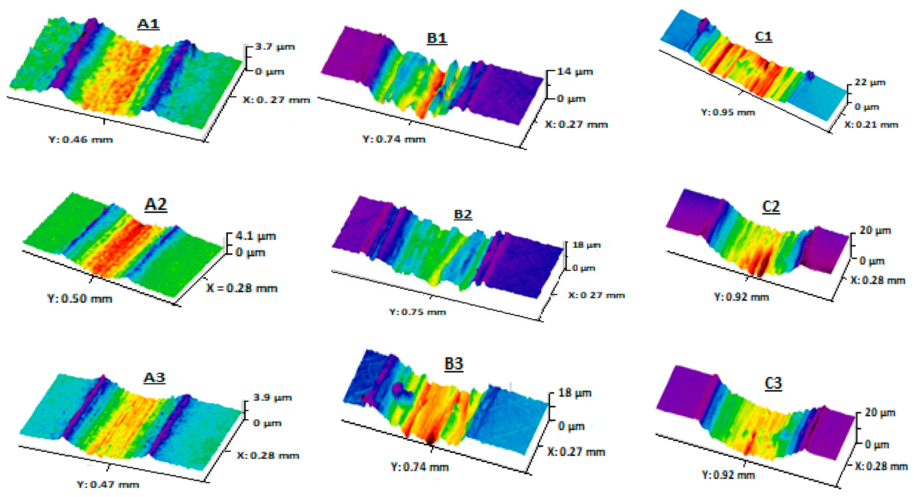
Figure 3.
Wear rate measured at open circuit potential in various electrolytes after sliding against an alumina ball under an applied load of 5 N in (NaCl: ![Cmd 04 00033 i001]() ), (Ringer’s solution:
), (Ringer’s solution: ![Cmd 04 00033 i002]() ), (PBS:
), (PBS: ![Cmd 04 00033 i003]() ), (PBS + 1 g/L BSA:
), (PBS + 1 g/L BSA: ![Cmd 04 00033 i004]() ), (PBS + 5 g/L BSA:
), (PBS + 5 g/L BSA: ![Cmd 04 00033 i005]() ). 1: 316 L, 2: Ti-6Al-4V, 3: Ti-10Zr-10Nb-5Ta. Adapted with permission from [38]. Copyright 2019 Taylor & Francis Ltd.
). 1: 316 L, 2: Ti-6Al-4V, 3: Ti-10Zr-10Nb-5Ta. Adapted with permission from [38]. Copyright 2019 Taylor & Francis Ltd.
 ), (Ringer’s solution:
  ), (PBS:
  ), (PBS + 1 g/L BSA:
  ), (PBS + 5 g/L BSA:
  ). 1: 316 L, 2: Ti-6Al-4V, 3: Ti-10Zr-10Nb-5Ta. Adapted with permission from [38]. Copyright 2019 Taylor & Francis Ltd.
Figure 3.
Wear rate measured at open circuit potential in various electrolytes after sliding against an alumina ball under an applied load of 5 N in (NaCl: ![Cmd 04 00033 i001]() ), (Ringer’s solution:
), (Ringer’s solution: ![Cmd 04 00033 i002]() ), (PBS:
), (PBS: ![Cmd 04 00033 i003]() ), (PBS + 1 g/L BSA:
), (PBS + 1 g/L BSA: ![Cmd 04 00033 i004]() ), (PBS + 5 g/L BSA:
), (PBS + 5 g/L BSA: ![Cmd 04 00033 i005]() ). 1: 316 L, 2: Ti-6Al-4V, 3: Ti-10Zr-10Nb-5Ta. Adapted with permission from [38]. Copyright 2019 Taylor & Francis Ltd.
). 1: 316 L, 2: Ti-6Al-4V, 3: Ti-10Zr-10Nb-5Ta. Adapted with permission from [38]. Copyright 2019 Taylor & Francis Ltd.
), (Ringer’s solution: ), (PBS: ), (PBS + 1 g/L BSA: ), (PBS + 5 g/L BSA: ). 1: 316 L, 2: Ti-6Al-4V, 3: Ti-10Zr-10Nb-5Ta. Adapted with permission from [38]. Copyright 2019 Taylor & Francis Ltd.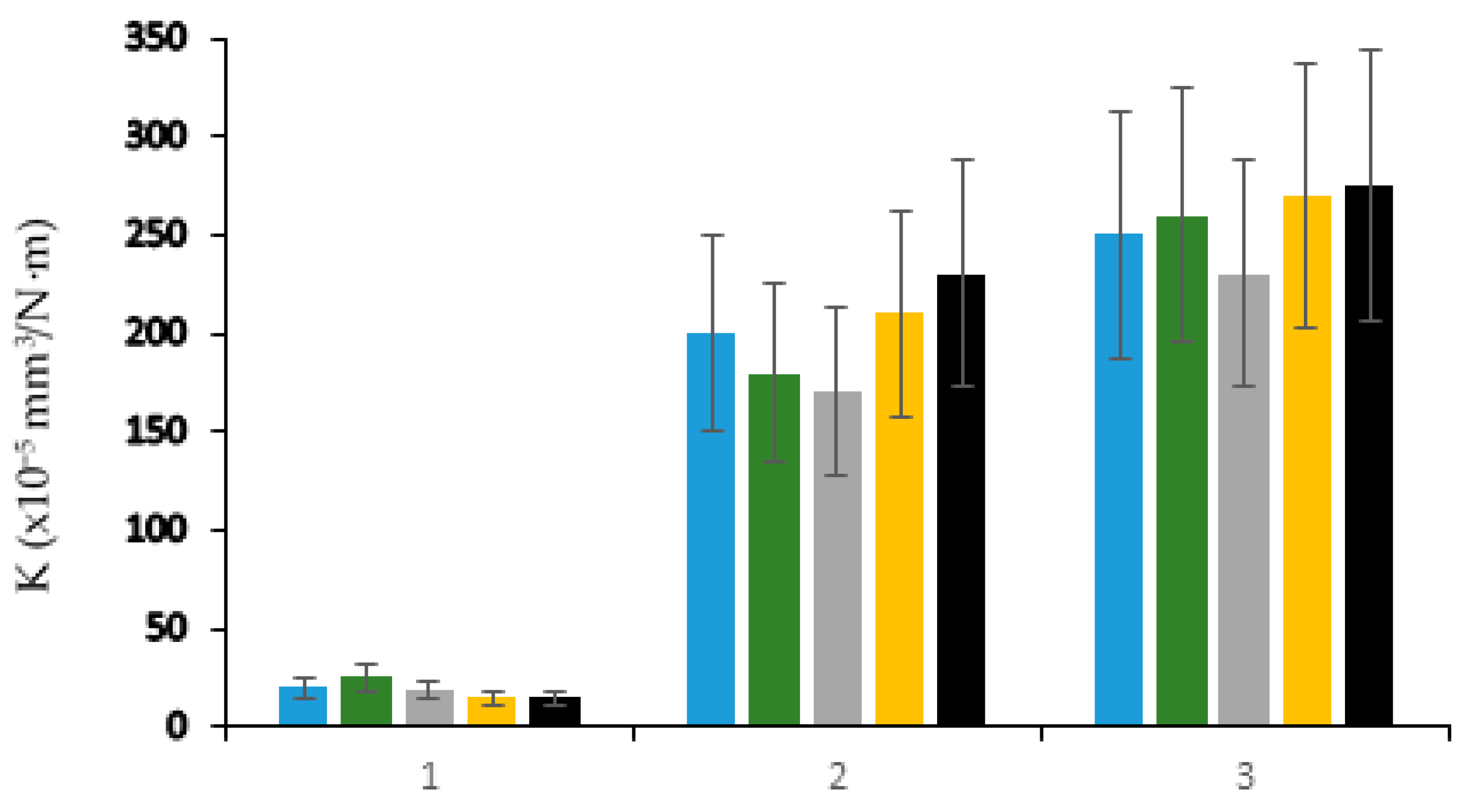

Table 1.
Examples of metals used as biomaterials.
| Stainless Steel | Fe-18Cr-14Ni-2.5Mo Fe-21Cr-10Ni-3.5Mn-2.5Mo Fe-23Mn-21Cr-1Mo-1N |
| Cobalt-Based Alloys | Co-28Cr-6Mo Co-20Cr-15W-10Ni-1.5Mn Co-20Cr-15Ni-15Fe-7Mo-2Mn |
| Titanium and Titanium-Based Alloys | Ti Ti-6Al-4V Ti-6Al-7Nb Ti-13Nb-13Zr Ti-19Zr-11Nb-4Ta Ti-10Zr-10Nb-5Ta Ti-42.5Zr-5Nb-10Ta Ti-23Nb-0.7Ta-2Zr-1.2O Ti-32Nb-2Ta-3Zr-0.5O |
| Special Alloys | Zr-2.5Nb Ni-45Ti |
| Magnesium and Magnesium-Based Alloys | Mg Mg-3Zn-0.5Ca Mg-2.2Nd-0.1Zn-0.4Zr Mg-9Li-1Zn |
Table 2.
Main combinations of materials used for the femoral head and the acetabular cup in hip prostheses.
Table 2.
Main combinations of materials used for the femoral head and the acetabular cup in hip prostheses.
| Femoral Head | Acetabular Cup |
|---|---|
| CoCrMo alloy | Ultra-high molecular weight polyethylene (UHMWPE) |
| Partially stabilized zirconia | UHMWPE |
| Alumina | UHMWPE |
| CoCrMo alloy | CoCrMo alloy |
| Alumina | Alumina |
Disclaimer/Publisher’s Note: The statements, opinions and data contained in all publications are solely those of the individual author(s) and contributor(s) and not of MDPI and/or the editor(s). MDPI and/or the editor(s) disclaim responsibility for any injury to people or property resulting from any ideas, methods, instructions or products referred to in the content. |
© 2023 by the author. Licensee MDPI, Basel, Switzerland. This article is an open access article distributed under the terms and conditions of the Creative Commons Attribution (CC BY) license (https://creativecommons.org/licenses/by/4.0/).
Share and Cite
MDPI and ACS Style
Takadoum, J. Review on Corrosion, Tribocorrosion and Osseointegration of Titanium Alloys as Biomaterials. Corros. Mater. Degrad. 2023, 4, 644-658. https://doi.org/10.3390/cmd4040033
AMA Style
Takadoum J. Review on Corrosion, Tribocorrosion and Osseointegration of Titanium Alloys as Biomaterials. Corrosion and Materials Degradation. 2023; 4(4):644-658. https://doi.org/10.3390/cmd4040033
Chicago/Turabian StyleTakadoum, Jamal. 2023. "Review on Corrosion, Tribocorrosion and Osseointegration of Titanium Alloys as Biomaterials" Corrosion and Materials Degradation 4, no. 4: 644-658. https://doi.org/10.3390/cmd4040033