
Polymeric Theragnostic Nanoplatforms for Bone Tissue Engineering


1
School of Biosciences and Technology, Vellore Institute of Technology, Vellore 632014, India
2
Division of Cardiovascular Physiology, Department of Medical Sciences, University of Miyazaki, Miyazaki 889-2155, Japan
*
Author to whom correspondence should be addressed.
J. Nanotheranostics 2023, 4(3), 280-298; https://doi.org/10.3390/jnt4030013
Submission received: 21 April 2023
/
Revised: 1 July 2023
/
Accepted: 18 July 2023
/
Published: 20 July 2023
(This article belongs to the Topic Applications of Polymers and Polymer Nanomaterials in Drug Delivery and Nanomedicine)
Abstract
:Nanomaterial-based tissue engineering strategies are precisely designed and tweaked to contest specific patient needs and their end applications. Though theragnostic is a radical term very eminent in cancer prognosis, of late, theragnostic approaches have been explored in the fields of tissue remodulation and reparation. The engineering of theragnostic nanomaterials has opened up avenues for disease diagnosis, imaging, and therapeutic treatments. The instantaneous monitoring of therapeutic strategy is expected to co-deliver imaging and pharmaceutical agents at the same time, and nanoscale carrier moieties are convenient and efficient platforms in theragnostic applications, especially in soft and hard tissue regeneration. Furthermore, imaging modalities have extensively contributed to the signal-to-noise ratio. Simultaneously, there is an accumulation of high concentrations of therapeutic mediators at the defect site. Given the confines of contemporary bone diagnostic systems, the clinical rationale demands nano/biomaterials that can localize to bone-diseased sites to enhance the precision and prognostic value for osteoporosis, non-healing fractures, and/or infections, etc. Furthermore, bone theragnostics may have an even greater clinical impact and multimodal imaging procedures can overcome the restrictions of individual modalities. The present review introduces representative theragnostic polymeric nanomaterials and their advantages and disadvantages in practical use as well as their unique properties.
1. Introduction
In the recent past, innovation in and research into tissue engineering and regenerative approaches, especially for soft and hard tissue regeneration, has noticeably increased. Tissue engineering and regenerative medicine aim to generate functionally alternative constructs for damaged and diseased tissues by merging an interconnected assortment of suitable platforms, cells/stem cells, and growth factors [1,2]. For regenerating bone tissue, mesenchymal progenitors (expressing extracellular tissue matrix) come together with scaffolds (acting as a supportive framework) and growth factors (inducing cell expansion and differentiation) to accomplish the whole process [3]. Approximately three decades ago, a new paradigm emerged as an alternative approach to tissue and organ reconstruction, tissue engineering, which aimed to regenerate a patient’s own tissues and organs that are biocompatible and bifunctional, and can overcome immune rejection. The initial clinical application of human cells in tissue engineering was in the early 1980s using skin tissue and cells fibroblasts, keratinocytes, along with a template matrix [2]. Later, periodontal and alveolar bone tissues were used for regeneration using site-specific membranes, the process known as guided tissue regeneration (GTR) and guided bone regeneration (GBR). With the increasing scrutiny of tissue engineering and regenerative medicine in diverse fields of research, the public and medical communities are looking to widen its prospects, although human clinical trials on tissue engineering are still scarce [3,4,5]. While several studies on strategies for tissue regeneration have been reported, regenerating bone tissue remains a challenge. The complex hierarchical anatomic architecture of bone related to the high mechanical tension to which it is exposed makes it difficult to replicate. High vascularization of the bone-tissue junction also hinders its engineering, especially in large bone anomalies [4,5]. Regeneration strategies have restrictions, such as the poor mechanics of scaffold materials, inadequate cell ingrowth for bone healing, and low expression of osteogenic growth factors for cell proliferation and differentiation. The term nanomaterials, specifically nanoparticles, gained worldwide importance after 1959, and after half a century, remarkable accomplishments related to these have been made by the human race in different arenas. The suitability of nanomaterials in the field of biomedicine is not new [6]. Their micro sizes facilitate easy circulation in the body, ready migration to the desired tissues, and target-specific binding [7]. The fundamental design of nanomaterials aims to control particle size, surface functionalization, and the effectual release and transfer of active therapeutics during medical prognosis. The use of nanomaterials has also changed the magnetic and molecular imaging modalities due to their optical and magnetic features [8].
The existence of nanomaterials can be simultaneously both therapeutic and diagnostic (theragnostic nanomaterials/nanoplatforms) and cater to the manufacturing of multi-functional nanosystems [9]. Hence, the current aim is designing and building multifunctional nanosystems (polymeric materials) for diagnosis and therapeutics [10]. Even though abundant theragnostic materials, both organic and inorganic, in the form of scaffolds, nanoparticles, hydrogels, colloids, microspheres, etc., have been engineered and investigated for cancer treatment, all the said criteria do not fit into a single system [11,12,13]. Most of the design processes used to develop a particular theragnostic nanomaterial are by: (a) a combination/binding of target drugs/stimulants to nanomaterial; (b) a merger with an image contrast enhancer; (c) the effective incorporation of two or more therapeutic imaging agents in one nanomaterial [14]. Over the last few years, research in healthcare sectors has progressively concentrated on altering traditional treatment methods into a more individualized prognosis model. One such attempt to fast-track this archetype shift is by introducing a merger of therapeutic and diagnostic manifestos, i.e., theragnostic agents or diapeutic (diagnostic cum therapeutic) agents with the aim of a faster, highly precise, and responsive diapeutic system [15].
Bone nanomaterials improve, repair, and facilitate new bone formation and have many promising clinical approaches ranging from the prognosis of non-healing long bone fractures to spondylosyndesis [16]. The exploitation of porous scaffold matrices from polymers to ceramics to support bone cell proliferation and differentiation is an enduring area of relevance. Some of the existing challenges comprise the obtaining of nanomaterials that can mimic the mechanical as well as biological environment of the original bone tissue matrix and aid in the vascularization of sizable tissue assemblies. Nanoplatforms with newer bio-functionalities have emerged to re-form nanoscale topological and biomechanical cues from the extracellular matrix as potential entrants for biomimetic materials [17,18]. Bone is a dynamic and rigid hard tissue comprising cells rooted in an abundant hard intercellular matrix. The highly vascularized tissue present in the bone continues to transform throughout the lifespan of an individual. It plays an important role in movement, acts as a supportive framework for sufficient load bearing, and maintains a protective covering for the delicate internal tissues of the body [19,20]. Bone is associated with maintaining a homeostatic balance in the body via its deposition of calcium and phosphate ions and in controlling the absorption of key electrolytes in the blood barrier.
This review discusses the role of various polymeric nanoplatforms concerning therapeutic and diagnostic insight for bone tissue repair and development and highlights the challenges in providing theragnostics.
2. Necessity to Regenerate the Complex Bone Structure
All of the bones that constitute the skeleton, from long bones in the limbs to short bones in the wrists, flat bones in the sternum to random bones in the vertebrae, possess a discrete loading environment that directs the in vivo regeneration of varied micro/macroscopic bony constructs with precise structures, mechanical properties, and spatial dissemination [21]. Bone tissue is compact or trabecular with an ordered arrangement of macroscale to nanoscale components (Figure 1). The nanocomposite structure (firm and elastic collagen fibers strengthened by hydroxyapatite crystals delivers the necessary compressive intensity and robustness to the bone. The prominent potential of bone tissue to remarkably regenerate in adolescent people is indicative of the effortless recovery of most fractures without the interference of major mediation. Regardless of this, larger bone imperfections post bone tumor resections and nonunion bony fractures require the prototype for a coordinated restoration of bone and sometime may require surgery [21,22]. At present, autografts are the gold standard prognosis for donor bone tissue from a non-load-bearing site and its transplantation into the deficient site [22]. Spondylosyndesis procedures also indicate the unmet requirement for massive autologous bone grafting procedures, which has augmented the most conventional in-patient treatment over the last two decades [23,24]. Nonetheless, its value is severely compromised by its limited availability and donor site morbidity [25]. Reviewing new bone restoration strategies is the need of the hour, the significance of which is driven by the excruciating pain correlated with bone injury and the rising therapeutic and socio-economic shortcomings. The hurdles in the field of tissue engineering, especially in monitoring the treatment methods, can be overcome by personalized medicine technologies that have been proven to be useful in monitoring and tracking treatment efficacy and diseases. By combining up-to-date diagnostic modalities with a therapeutic biomaterial, theragnostic biomaterials (herein bone theragnostic biomaterials) aid in integrating precision medicine with tissue engineering and, more specifically, these nanoplatforms can offer non-invasive monitoring of engineered tissues of interest in real time while delivering therapeutic cues to promote bone regeneration and repair [2,26].
3. Polymeric Nanoplatforms for Bone Diagnostics and Theragnostics
Over the last decade, advances in nanotechnology have enabled accessibility to structural mimics that facilitate the engineering of various tissues. Subsequently, and more recently, there has been an unmet need to develop personalized medicine for custom-made therapies toward individual patients’ prerequisites [27]. These nano-theragnostic strategies for tissue engineering and regenerative medicine can thus be used to direct tailor-made requirements through the articulate design to endorse tissue-specific functions in the body [28]. Such strategies can initiate in situ tissue formation by utilizing the body’s natural cellular populations to drive this renewal without ex vivo cell manipulation [29]. Furthermore, effective diagnosis and prognosis of bone-related disorders are essential to restrict morbidity and mortality in a population. Engineering tissues can be challenging, as well as the difficulty in precisely examining the prognosis, personalized medicine technologies, and monitoring treatment efficacy and disease conditions [30]. Integrating diagnostic modalities with a therapeutic biomaterial platform permits non-invasive surveillance of the engineered tissue of interest while providing therapeutic signals to aid regeneration and repair [30,31].
Systemic biomaterials used for the non-invasive administering of bone theranostics have offered several advantages in terms of their exclusive target-specific administration, imaging, and functionalization efficacy. Among them, nanoparticles (NPs), owing to their targeted tissue specificity recompense for localization to disease sites in vivo. Surface-functionalized NPs can facilitate improved blood circulation time over standard chemotherapeutics [31,32]. Intrinsically multifunctional, these NPs aid tissue targeting and sustained responsive drug release and prevent degradation of complex drug payloads [33,34]. Metallic NPs characteristically exhibit magnetism or fluorescence, which makes imaging probe loading, targeting ligands, and therapeutic moiety targeting easy [35]. Such multifunctional systems can significantly assist precision medicine in screening and detecting the specific molecular makeup of highly variable diseases, optimizing therapy strategies and delivery, and monitoring the integrative therapy effects. These also enhance cellular characteristics such as osteointegration, osteoconduction, osteoinduction, cell fate, cell binding, labeling, and tracking [36,37,38].
At present, the fabrication of polymer-drug conjugates (PDCs) and nanomedicines (otherwise called polymeric prodrugs or polymer nanotherapeutics) have gained great value over the past decade [39]. Polymer-drug conjugates (PDCs) are macromolecules comprising a covalent bond-allied drug-polymer-targeting moiety backbone with three major components: a hydrophilic solubilizer, a drug-linked-polymer, and a targeting moiety directing the polymer-drug conjugate to a desired functional destination [39,40]. A polymer conjugated with a drug can enhance its pharmacokinetics, increase its stability and loading capacity, and regulate sustained and premature drug release patterns. Moreover, this approach can deliver a variety of polar and nonpolar molecules that are otherwise tough with drug-loaded nanoparticles prepared via physical encapsulation [41].
Polymeric materials based on their functional groups have been used to develop nanoparticle-based theragnostics for imaging/diagnosis and treatment, mainly for cancer. Although not much has been explored for bone tissue engineering applications, some in vitro and in vivo studies with polymeric nanoparticles presently in the market have revealed the boundless potential of this technology [41]. Polymeric biomaterials have been the most clinically exploited nanoplatform in cancer theragnostics and are now slowly emerging in bone repair applications [42]. Both synthetic and natural polymers have been explored as polymer-drug conjugate-based nano-theragnostics. Some synthetic polymers include polyvinyl pyrrolidone (PVP), polyethylene glycol (PEG), polylactic acid (PLA), poly(vinyl alcohol), polylactic acid (PCL), poly(lactic-co-glycolic acid) (PLGA), etc. Natural polymers, such as collagen, keratin, silk, cellulose, gelatin, alginate, chitosan, hyaluronic acid, etc., are also a part of the nanoconjugate formulation [42,43]. Herein, a few different types of polymers and polymeric platforms utilized as theragnostic nanotools in bone tissue engineering and regenerative medicine, converging on the preparation, in vitro and in vivo behavior, use in scaffold improvement, delivery carriers, and cell labelling are discussed.
3.1. Natural Polymers
The most abundant explored extensively polymers in nature with bulk properties, biodegradability, alterable structural features, and flexible side chains for easy functionalization are that of natural origin. Owing to their facile synthesis method, natural polymers can be tuned to cater to several end applications [43]. For naturally occurring polymers, desolvation, gelation, solvent evaporation, nanoprecipitation, etc., are the most convenient and preferred processes that aid in self-assembly [44].
3.1.1. Gelatin
Gelatin is a nontoxic, cost-compliant, and biodegradable polymer that is synthesized using alkaline or acidic hydrolysis of collagen. Gelatin exhibits a covalent surface-bound interaction with carboxylic acid and amino groups that extends gelatin’s ability to fit into many therapeutic applications. While the use of conjugated gelatin nanoplatforms as cancer therapeutic agents and in antimicrobial photodynamic therapy has been reported, nano-theragnostic tools for bone tissue engineering remain comparatively new. Celikkin and co-workers (2019) reported an enhancing X-ray attenuation technique for bone tissue regeneration via three-dimensionally printed gelatin methacrylate hydrogels doped with gold nanoparticles. Methacrylate hydrogels induced extracellular matrix formation and, in combination with gold nanoparticles (used as contrast agents due to their high X-ray attenuation), these three-dimensionally printed biocompatible matrices were evaluated for new bone regeneration in mesenchymal stem cells (MSCs) [45]. In another study, multilayered iron oxide/polycaprolactone and gelatin nanofibrous membrane constructs were fabricated for tissue engineering theranostics. The fabricated nanofibrous membrane was combined with a non-contact magnetic force to balance the mechanical integrity of the 3D structures. PCL/gelatin nanofibers and their cell biocompatibility were assessed in MSCs-seeded fiber constructs. The in vivo behavior of the magnetic membranes was evaluated using magnetic resonance imaging in animal models [46]. Iron and hydroxyapatite-based alginate/gelatin 3D matrices were developed as compatible magnetic resonance imaging biomaterial for studying the non-invasive bone tissue formation in a rat cranial defect model. This theragnostic tool for bone tissue engineering exhibited osteoconduction and assisted non-invasive tracking of cell migration; inflammatory stimuli and matrix accumulation via magnetic resonance-contrast enabled matrices and helped assess the non-invasive bone-regeneration between the native and implanted bone, demonstrating a promising theragnostic nanotool to review the healing of bone fractures [47].
3.1.2. Alginates
Alginates are naturally occurring polysaccharide-polyanions that possess high biocompatibility and biodegradability. With their non-toxic and highly hydrophilic properties, alginates can fit into several cost-compliant biotechnological applications [48,49]. Alginate-based biomaterials fit into the mold of cell-homing materials utilized for bone-tissue engineering and regenerative medicine [50]. These have been fabricated into diverse forms such as hydrogels, foams, capsules, sponges, fibers, microspheres, etc. [50,51,52]. Nonetheless, alginate-based hydrogel systems are a supporting matrix and delivery platform for bone-tissue. A wide range of synthetic/natural polymers in combination with alginates has been optimized to provide the required hygroscopicity, biodegradability, mechanical strength, and tenacity to make multifunctional alginate nanomaterials [50].
Zhao et al. demonstrated that an active nano-construct scaffold, through external stimuli, can influence and regulate the properties of the nano-construct, facilitating the delivery of biological agents. The study depicted iron oxide nanoparticles in an RGD-coupled alginate matrix significantly enhanced the delivery rate of the cell-coupled bioactive agents when subjected to magnetic stimulus [53]. An experimental design was proposed by Coluccino et al. for stem cell differentiation and to obtain a tissue-substitute bioactive environment for orthopedic repair. An alginate-based matrix associated with transforming growth factor-β1 and hydroxyapatite was developed that controlled the release of chemical signals, required for an effectual and selective differentiation of mesenchymal stem cells into bony-chondral lineages. This osteochondral nanoplatform demonstrated high cellular adhesion, proliferation, and in vivo compatibility with highly functional biomechanical cues, illustrating a potential osteochondral tissue-engineering biomaterial [54]. A nano-biocomposite bead was fabricated using a conglomeration of chitosan, gelatin, alginatem, and nano-hydroxyapatite that demonstrated high mechanical strength and biological characteristics of natural bone. The nano-biocomposite bead exhibited a high porosity with excellent hydrophobicity and biodegradability under physiological environments. In vitro seeding of osteoblast cells over the nano-biocomposite bead showed favorable viability and gene expressions, inferring its importance in bone tissue engineering and reformative therapies [55]. Angili and co-workers fabricated a 3D-printed poly l-lactic acid scaffold coated with alginate and magnesium oxide nanoparticles with different cellular topologies that not only showed excellent biological biocompatibility, antibacterial activity, and cell survival, but also indicated increasing MgO concentrations enhanced compressive strength and the elastic modulus of the biomaterial, mimicking the process of efficient bone repair and refurbishment [56]. Bone-related diseases, such as osteogenesis imperfecta, arthralgia, congenital malformation, osteoporosis, etc., are often associated with insufficient self-repair and necessitate medical intervention as conventional orthopedic therapeutics suffer shortcomings, such as graft rejection and implant failure.
3.1.3. Cellulose
Cellulose, the most profuse polysaccharide accessible, and its derivatives have been extensively used in the fabrication of bio-composites for the theragnosis of bone diseases, such as bone and cartilage damage, osteomyelitis, osteoporosis, etc. The effectiveness of cellulose and its composites on bone engineering applications is noteworthy. However, shortcomings, such as a poor degradation rate and a well-knit, packed nanofiber layer with a small pore size, may hinder the penetration of cells into the derived matrices [57]. Hence, it is necessary to upgrade the structural and functional suitability of cellulose for its applications in treating bone disorders. Nanocomposite scaffolds with tunable pore sizes and mechanical strength using cellulose, chitosan, and silver nanoparticles were fabricated that exhibited substantial antimicrobial effect, favorable adhesion, and viability of human osteosarcoma cell lines [58]. The incorporation of cellulose also enhanced biomineralization for bone development. The highly crystalline surface topology of silver nanoparticles on the cellulose-chitosan scaffold also facilitated attaining the desirable porosity with nano-pore diameters and mechanical strength similar to that of cancellous bone [59].
Of late, cellulose-based biomaterials have been explored in bone tissue engineering as highly structured and porous microspheres. BMP-2-laden bacterial cellulose and type-I collagen microspheres exhibited biocompatibility and effectively promoted MC3T3-EL cell adhesion, proliferation, and osteogenic differentiation. Additionally, the bacterial cellulose scaffolds layered with porous silk fibroin presented a significant removal of osteosarcoma and favorable bone defect repair under near-infrared radiation [60]. An experimental investigation on carboxymethyl cellulose merged with phenol-molecule-based microcapsules was loaded with rat bone marrow mesenchymal stem cells, which showed higher osteogenic activity both in vitro and in vivo. The upregulation of mRNA and protein expression levels of osteogenesis-related genes was also observed [61]. In contrast, in situ hydrogels fabricated with calcium-enriched nano-fibrillated cellulose exhibited improved biocompatibility, cell attachment, alkaline phosphatase, and calcium deposition activity [62]. Hydroxyapatite, being one of the most biocompatible agents, combined with cellulose to develop into nanocomposites for bone cell proliferation, were three-dimensionally printed and evaluated for the sustained release of strontium and calcium ions and substantial proliferation, bone marrow stem cell differentiation, and bone tissue revival [63]. In another study, cellulose nanocrystals doped with gelatin and alginate developed into 3D-printed hydrogel scaffolds presented considerable matrix mineralization in the bone tissue [64]. Therefore, cellulose biomaterials are multifunctional in promoting bone repair and development.
3.1.4. Chitosan
Among the other naturally derived polysaccharides, chitosan, for pharmaceutical and biomedical applications, has been advantageous due to its favorable biocompatible, biodegradable, and non-toxic features [65]. Chitosan forms stable ionic complexes with multivalent ions or polymers for the fabrication of various biomaterials such as gels, sponges, beads, micro/nano particles, etc. Owing to its hydrophilic and mucoadhesive properties, chitosan-based materials have been studied as therapeutic particles and, in combination with various nucleic acids, these have been observed to accelerate peptide and protein transfer across the mucosal membrane [66]. Chitosan, in the form of nanoparticles, has been investigated as a potential delivery vehicle in the fields of tissue engineering and regenerative medicine [67].
Nanoparticles of sodium triphosphate and chondroitin sulfate with chitosan polymer prepared via an ionic gelation method were studied to evaluate the nanoparticle-controlled association and release kinetics of the fused proteins. Higher concentrations of the polymer when combined with crosslinkers in the nanoparticle aided in sustained protein release, demonstrating their probability as potential competent protein carriers in bone tissue repair [68]. In another study, Bastami and co-workers demonstrated three-dimensional chitosan nanoparticles with β-tricalcium-phosphate and gelatin for the sustained release of bone morphogenetic protein-2 that was dispersed into collagen hydrogel constructs to evaluate the bone tissue renewal in vitro in human buccal fat pad-derived stem cells [69]. Nanocomposites prepared with different degrees of deacetylation, molecular weights, and concentrations of chitosan armored with mesoporous silica nanoparticles were studied to observe bone regeneration. The nanocomposites had high mechanical compression resistance and good biocompatibility in human primary osteoblasts. In vivo implantation of the nanocomposites in a mice calvaria defect model showed their appropriateness for bone tissue regeneration with the mineralization and vascularization of osteoblasts [70]. Natural regeneration of bone is limited to bone defects, and external intervention is essential to augment bone tissue revival. Amidst the various scaffolds, hydrogels are suited platforms for repair and chitosan has recently drawn significant attention as a compatible graft material to form thermal and pH-responsive injectable gel grafts. Preparations of injectable matrices of chitosan with high-water imbibing ability, nominal invasiveness, porosity, and tunability into bone defects highlights the use of such stimuli responsive injectable hydrogel matrices for future perceptions in bone engineering [71].
3.1.5. Gellan Gum
Polysaccharides of microbial origin have been extensively explored owing to the promising physiochemical features they possess. Structurally diverse, these biomolecules fit into several biomedical, food function applications. Currently, gellan gum, a linear polysaccharide comprising high molecular weight chains of rhamnose, glucose, and glucuronate, has been widely investigated for its excellent properties as a food ingredient [72,73]. Nonetheless, because of its unique structural confirmation and gelling capacity, gellan gum is used as one of the multifunctional additives in many pharmaceutical outcomes and as a drug delivery system [74]. Preparations of gellan-based biomaterials have been significantly investigated in the field of cancer therapy, but its role in bone tissue theragnostics with a focus on imaging modalities still continues to be very new and needs more detailed study and established perceptions.
Gellan gum has materialized as a remarkable surface-modifying, biocompatible gelling biomaterial in the application of bone tissue engineering by stabilizing gold nanorods. In a study reported by Vieira et al. (2015), a low-acyl gellan gum was coated onto the surface of gold nanorods via a layer-by-layer method to form a gellan-based hydrogel shell around individual nanorods. The gellan gum hydrogel shell around the gold nanorods showed reversible pH-responsive features, improved cytocompatibility, and osteogenic ability with human osteosarcoma cells SaOS-2, with enhanced mineralization. Thus, this hydrogel system indicates further insights that are relevant for drug delivery, tissue engineering, and regenerative medicine applications [75]. An injectable gellan gum/chlorhexidine/nanohydroxyapatite hydrogel matrix was developed and assessed for its osteogenic potential to treat infectious bony defects. Bone marrow mesenchymal stem cells seeded into the hydrogel network exhibited good cell proliferation, as well as improved mechanics, biodegradation, and osteogenesis. Furthermore, the gellan gum hydrogel showed a concentration-dependent inhibition of Enterococcus faecalis, thus indicating its potential in treating refractory periradicular periodontitis infections [76]. Treatment for osteochondral defects with gellan-gum-based functional coatings and porous composites incorporated with osteoconductive materials, such as demineralized bone particle (DBP) or bone derivatives, are currently being expansively reviewed as an emerging candidate for OD in small animal models for soft and hard tissue engineering [77].
3.2. Synthetic Polymers
Synthetically derived polymers are highly suitable for changing the physicochemical properties of a nanomaterial and have a precise high drug-loading capacity. As carriers, these can home various organic and inorganic moieties via dissolution/covalent interaction/dispersion mechanisms for the effective delivery of drugs.
3.2.1. Polylactic Acid (PLA)
Among the synthetic biodegradable polymers explored for tissue regeneration, polylactic-acid-based nanomaterials find their place in various biological applications, i.e., regulated drug release, gene therapy, implant coatings, etc. [78,79,80]. Wiglusz et al. (2020) analytically characterized beta-calcium diphosphate-modified poly(l-lactide) nanocomposites with a tunable chain length and varied phosphate-to-polymer ratios via self-assembly. The nanocomposites were analyzed in vitro to verify their potential for osteochondral defect reparation [81]. Recently, nano-hydroxyapatite, investigated for critical-sized bone defects, has shown improved vascularization, indicating the formation of new bone structures. Furthermore, nanocomposites from poly(l-lactide) and nano-hydroxyapatite were functionalized with various concentrations of a rare earth element, europium (III), to study the acceleration in the bioactivity of the developed nanocomposite scaffolds. The effect of europium ions when tested for cell eidonomy, proliferation, differentiation, and metabolism of progenitor adipose forming tissue, bone, and cartilage cells, was shown to promote enhanced osteogenesis and chondrogenesis that was linked with enhanced extracellular matrix protein secretion (proteins specific to bone and articular cartilage tissue) [82].
3.2.2. Poly(ε-Caprolactone) (PCL)
The time span for the degradation of biomaterials should be well-compatible with the rate of regeneration of a particular tissue for it to present ideal specifications during the restoration period. Biodegradable polymers such as poly(ε-caprolactone) (PCL) are extensively used in tissue engineering and regenerative medicine. PCL is used in absorbable medical equipment such as surgical sutures, stents, or biomaterials devoted to the targeted delivery of a particular drug [83]. PCL, being biocompatible and biodegradable, can accommodate a variety of cell carriers and support matrices to develop PCL-based nanomaterial methods of polymerization, porogen leaching, electrospinning, 3D printing, etc. [83,84]. Furthermore, hybrid nanoparticles of hydroxylated multi-walled carbon nanotubes functionalized with magnetic iron oxide nanoparticles were synthesized to fabricate a biocompatible porous poly(ε-caprolactone) matrix for regenerative medicine application. Cytotoxic studies of the matrices evaluated in a SAOS-2 human cell line exhibited concentration-dependent cytotoxicity and had a positive impact on the activity of SAOS-2 cells with improved cell attachment [85].
In another study, 3D-printed PCL scaffolds coated with CuS nanoparticle-PEG soft hydrogel were evaluated for bone tissue repair; good photothermal properties and stable viscoelasticity and mechanical function were observed in the scaffolds. Near-infrared (NIR) light irradiation exposure mediated the release of dexamethasone sodium phosphate incorporated in the CuS-PEG-PCL scaffold, competently stimulated the osteogenic differentiation of bone mesenchymal stem cells, and accelerated bone formation in the tibial defect of rats [86]. The concept of guided bone regeneration (GBR) has evolved with the barrier membrane to prevent non-functional scar tissue layer development on the defect site. A bilayer membrane consisting of silk fibroin/PCL-PEG-PCL incorporated with nano-calcium phosphate and a PCL membrane was fabricated to understand the GBR potential. This sandwiched two-layered scaffold with calcium phosphate nanoparticles exhibited enhanced osteoconductivity and biomineralization in human dental pulp stem cells, supporting its excellent biocompatibility and possible pertinence of membranes for GBR prognosis [87].
Graphene nanoparticles containing poly-caprolactone solution were used to coat borate-based bioactive glass material to prepare nanocomposites acquiring electrical conductivity. When analyzed under simulated body fluid conditions, an increased concentration of graphene nanoparticles revealed better electrical conductivity and higher percentages of viability in pre-osteoblastic MC3T3-E1 cells post treatment with higher activity of alkaline phosphatase in cells [88].
3.2.3. Poly(Lactic-Co-Glycolic Acid) (PLGA)
The existing diagnosis and treatment of critical bone ailment remains difficult in orthopedic surgeries. Biomaterials and growth factors have materialized as a new therapy replacement in bone regeneration. Amongst them is the copolymer PLGA, which, owing to its biodegradability and biocompatibility, has been introduced among the Food and Drug Administration (FDA)-approved therapeutic devices. Wang and co-workers set up an experimental investigation where the encapsulation of bone morphogenetic protein-2 (BMP-2) into a polymeric matrix was evaluated in a nude mouse model. PLGA and hydroxyapatite biocomposite nanofiber constructs were laden with BMP-2 through electrospinning. The constructs were investigated in nude mice experiments and parameters such as serum BMP-2 concentration, ALP activity, X-ray qualification, and H&E tissue staining. The experimental results revealed the competent morphology and mechanical strength of the matrix. In vivo experiments confirmed the improved bioactivity of BMP-2, thus improving the formation of new bone and the healing of segmental defects in vivo [89]. Akermanite, a fascinating melilite mineral of the sorosilicate group, has recently been used for effective bone regeneration. Yet, owing to their weak fracture resistance, ceramics, when combined with polymeric moieties, can surpass the limitations of low biodegradation rates, weak mechanical properties, and decreased apatite-forming capacity. Akermanite-supplemented ions, such as calcium and magnesium, as well as copper and strontium ions, were doped into PLGA-moxifloxacin scaffolds to induce angiogenesis and osteogenesis and improve bone repair [90]. PLGA nanoparticles laden with human parathyroid hormones were experimentally prepared by a modified double emulsion-solvent diffusion-evaporation method and integrated into porous freeze-dried chitosan-gelatin matrices to assess their cytocompatibility on clonal human osteoblast cells. The enhanced expressions of alkaline phosphatase levels in human osteoblast cells endorsed the activity of sporadically released hormones from the matrix, supporting the growth of new bone-like cells [91].
4. Theragnostic Approaches for Bone Tissue Engineering
Bone tissue engineering with nano/biomaterials can be precisely modulated to cater to specific end-application requirements. Nonetheless, several limitations persist in the field, especially in regenerating hard tissues. The merger of diagnostic approaches to design theragnostic (combined therapy and diagnostic) nano/biomaterials offers an exclusive manifestation to deliver dual monitoring and treatment competencies to simultaneously uplift personalized medicine [9]. Herein, we abridge some of the recent progresses to have occurred in the field of hard tissue (bone) regeneration theragnostics and the clinical potentials with respect to polymeric nanoplatforms (Figure 2). Recently, many therapeutic systems have been developed that can have a substantial effect by mediating the bone extracellular matrix environment [10]. Some of these systems take into consideration the use of combination therapy based on nanoparticles, which, unlike conventional delivery systems, have unique physicochemical properties and high specificity. These are also tiny enough to easily circulate within the blood barrier, migrate to the looked-for tissues, and unambiguously bind to the target cell [11,12,26]. Theragnostic nanoplatforms, owing to their optical and magnetic properties, have shown enhanced molecular imaging for engineering multi-functional delivery systems.
Today, in vivo molecular investigation relies on non-invasive imaging techniques, in particular opto-acoustic systems, magnetic resonance enterography, and nucleology imaging. These can be differentiated and categorized into molecular, morphological, and anatomical imaging sensory systems [92]. Moreover, highly complex imaging modalities can be attained using techniques such as optical photon emission computed tomography, optical imaging, positron emission tomography, and single-photon emission computed tomography. These theragnostic tools can sense molecular biomarkers targets and readily detect molecular biomarker targets. In comparison, high spatial resolution can resolve computed tomography optoacoustic systems and magnetic resonance enterography [93]. Theragnostic nanomaterials have opened up avenues for favorable and efficient disease tomography and treatments that hold great capacity to be employed from the laboratory to the clinic (Figure 2). Table 1 shows the various molecular imaging modalities, their specifications, along with their advantages and disadvantages [10,92].
5. Challenges in the Current Bone Theragnostic System and the Potential of Nanoplatforms to Overcome Them
Regardless of the prevalent use of conventional diagnostic techniques and strategies in orthopedic diagnostics and theragnostics, shortcomings persist in the critical detection and diagnosis of many pathologies, especially in the early stages of bone disease when clinical intervention is most perilous [94]. Orthopedic disorders are the second leading cause of global frailty, resulting in a sharp increase in direct health care costs each year. Bone-related diseases are becoming prevalent, with major impacts on conditions such as osteoporosis, which affects more than two-thirds of the total morbidity and mortality of the population. Hence, early and competent detection and treatment of modality for such bone disorders is the need of the hour [95,96].
Conventional theragnostic biomaterials are amalgamations of the existing approaches and, with newer modular techniques for therapy, diagnosis, and moiety targeting, a high degree of specificity and tunability can be achieved at the cost of complexity [96,97]. One such example is contrasting or diagnostic agents embedded within biomaterials for local applications that facilitate tunable scaffold characteristics and detection modalities. Implantable nanofibers of PCL embedding porphyrin sensors for efficient bone regeneration show decreased sensitivity in humans due to the higher implantation depths compared to murine models [98]. Multiple challenges persist for most of the systemically administered biomaterials. Nanoparticles, for instance, show high specificity in the detection of bone-related conditions, such as osteomyelitis, osteoporosis, bone tumors, etc., but their activity is hindered by the unique structural characteristics of bone, limiting vascularization. Bone can also exhibit disease-specific restrictions to delivery, including sub-micrometer canaliculi, which aid in bacterial infection [99]. Nanomaterial-protein adsorption lowers the targeting ability and alters biodistribution to the reticuloendothelium, which in turn leads to poor bone accumulation [100]. Enhancements in surface chemistry to amend protein adsorption and maintain targeting specificity are important to improve theragnostic purposes. Alternatively, the use of magnetic resonance imaging techniques was implemented for imaging gadolinium-doped hydroxyapatite nanoparticles absorbed into PCL scaffolds to track in vitro bone development [101]; furthermore, nano-hydroxyapatite-alginate-gelatin scaffolds incorporating SPIONs have been used to detect mineralization [102]. Nevertheless, there are quite a number of limitations that the current clinical theragnostics for bone repair lack, such as imaging microvessels and cells in vivo [103], the detection of implant-associated bacterial infections, postsurgical inflammation, etc. [104].
To conclude, the diagnostic needs are different for research and clinical scenarios. Most biomaterials used for imaging modalities have fluorescent detection sense modality, which can collate some unique diagnostic information, including bacteria-triggered infection detection, metabolites, or protein sensing [105]. Fluorescence, however, is not a standard clinical detection modality and is restricted by tissue penetration; as alternatives, current clinical approaches use X-rays/CT, magnetic imaging, and ultrasound, which can be amplified to enhance contrasting specificity for orthopedic applications through the development of diagnostic and theragnostic biomaterials [106]. Hence, the precise engineering of these technologies to further competently and locally deliver theragnostics indicate a prominent area of forthcoming exploration.
6. Recent Theragnostic Approaches for Bone-Linked Disorders
Although bone-linked disorders are not new, the application of theragnostics to treat such disorders persists at a very undeveloped stage and much research is necessary with high precision and technical improvements. The incidence of bone-linked disorders has radically soared in recent times. Disorders such as osteoporosis, osteoarthritis, bone fractures, non-healing bone wounds, and bone-related bacterial and fungal infections, are projected to impact over 200 million people worldwide [107]. Moreover, the frequency of osteoarthritic cases exceeds 20% of the mid-aged and aging population. However, cases of bone cancer are uncommon, leading to mortality in the affected patients [107]. Treatments and therapies for bone infections are extremely difficult, with high failure rates of about 30% [108]. Generally, the increased prevalence of bone-related disorders poses a tremendous clinical and socioeconomic burden worldwide. Hence, early detection and diagnosis are critical for enhancement in therapeutic and theragnostic efficacy and survival. Most diagnosis methodologies for bone disorders mainly rely on intensive radiologic imaging and biomarker examinations [107]. Magnetic resonance imaging (MRI) is a beneficial instrument for the detection of altered joint signals in the primary stages of osteoarthritis. For bone fractures and cancer, different approaches, such as X-ray, computed tomography (CT), MRI, positron emission tomography (PET)-CT scan, dual-energy X-ray absorptiometry (DXA), and biopsy, have been used [109].
Diverse emerging applications of multivariate nanomedicine in bone theragnostics have been extensively explored, including biological tumor tomography, microcrack detection, arthritis biomarker sensors, targeted fluoro-delivery, etc. Chen et al. (2017) developed targeted SPIONs coated with polyethylene glycol for the in vivo labeling and tracking of T cells in a collagen-induced arthritis model of rheumatoid arthritis [110]. MRI imaging implied a sharp increase in the signal-to-noise ratio of the femoral growth plates infused with the SPIONS, showing high transverse reflexivity, good selectivity, and bioavailability as a potent MRI probe in clinical applications. For the detection of bone metastases, a trastuzumab-functionalized SPION polymersomes was fabricated for its assessment against human endothelial receptor 2 (HER2) as a novel MRI contrast agent [111].
Polyethylene glycol glycosylated gold nanorods were conjugated with two tumor-specific oligopeptides, PT6 and PT7, to target an osteosarcoma cell line via specific photoacoustic imaging [112]. Bone-targeting self-assembled polymeric vesicles of Poly(ε-caprolactone)67-b-poly[(l-glutamic acid)6-stat-(l-glutamic acid-alendronic acid)16] were competent as a single photon emission computed tomography/computed tomography (SPECT/CT) imaging enhancer for early detection of bone cancer [113]. Chitosan has been widely researched as a drug carrier due to the presence of polycations; its good solubility, functionalization, and biodegradability were combined with a biomolecule kartogenin to fabricate nanoparticles that exhibited a fluoresced regeneration effect on cartilage degeneration [114]. The development of berberine-loaded chitosan nanoparticles via ionic cross-linking possessed a high anti-apoptotic effect by lowering Bax and caspase-3 expressions and upregulating Bcl-2 (major bone-related markers) [115]. An alginate-enclosed chitosan-calcium phosphate nanocarrier incorporating iron-saturated bovine lactoferrin was prepared for effective oral administration to suppress the progression of osteoarthritis [116]. In addition, nanosheets of graphene oxide were combined with photopolymerizable poly-d, l-lactic acid/polyethylene glycol and transforming growth factor-ß for localized delivery in cartilage tissue restoration [117]. An amalgamation of thermoresponsive triblock polymers of monomethoxy PEG (mPEG) and a PLGA-based carrier for treating osteoporosis was developed. This copolymer-based delivery system showed superior biocompatibility and a controlled and sustained release of the drug calcitonin to augment the therapeutic efficacy of the drug against osteoporosis [118]. Ma et al. (2016) combined GO-modified ß-tricalcium phosphate (GO-TCP) to form composite matrices with a high photothermal effect for osteoporosis that, under irradiation NIR, showed significant improvement in bone-forming ability [119].
7. Conclusions and Future Directions
Engineered nanoplatforms with diagnostic and reformatory abilities are cutting-edge technological miracles that not only facilitate the reparation of tissues of interest but also decide the suitability of the biomaterial and their cargo. However, challenges in successfully engineering tissue persist. Over the years, there has been a transition in the process of development of innovative theragnostics, such as nanoparticles, scaffolds, cell-based matrices, etc. The current investigations are largely directed toward the preparation and analytical characterization of these nanoplatforms, with very little light being shed on their ability to fit in with genetic diversity, long-term toxicity, and in vivo functionality. Hence, advancements that ensure that these systems are translatable from the laboratory to the clinic need special attention (Figure 3).
Theragnostic manifestos require thorough preclinical screening and extended investigations of toxicity, engraftment, bone regeneration, and adequate safety profiles. Other technological expansions are necessary to instrument customized biomaterial theragnostic platforms at collective levels of organizational and operational complexity for tissue engineering, especially bone applications. At a greater level, we picture that regenerative bone theragnostics may have huge potential as personalized and tailored medicine platforms, aiding patient-specific treatment plans based on diagnostic output and upgrading patient outcomes.
Author Contributions
Conceptualization, K.B. and H.M.; Writing—original draft preparation, K.B. and H.M.; Writing—review and editing, K.B. and H.M. All authors have read and agreed to the published version of the manuscript.
Funding
This research received no external funding.
Informed Consent Statement
Not applicable.
Data Availability Statement
Not applicable.
Acknowledgments
The authors thank the management of Vellore Institute of Technology, Tamil Nadu, India, and University of Miyazaki, Japan for their administrative assistance.
Conflicts of Interest
The authors declare no conflict of interests.
References
- Badylak, S.F.; Nerem, R.M. Progress in tissue engineering and regenerative medicine. Proc. Natl. Acad. Sci. USA 2010, 107, 3285–3286. [Google Scholar] [CrossRef] [PubMed]
- Fisher, M.B.; Mauck, R.L. Tissue engineering and regenerative medicine: Recent innovations and the transition to translation. Tissue Eng. Part B Rev. 2013, 19, 1–13. [Google Scholar] [CrossRef] [PubMed] [Green Version]
- Pina, S.; Oliveira, J.M.; Reis, R.L. Natural-based nanocomposites for bone tissue engineering and regenerative medicine: A review. Adv. Mater. 2015, 27, 1143–1169. [Google Scholar] [CrossRef] [PubMed] [Green Version]
- Ansari, M. Bone tissue regeneration: Biology, strategies and interface studies. Prog. Biomater. 2019, 8, 223–237. [Google Scholar] [CrossRef] [Green Version]
- Anjum, S.; Rahman, F.; Pandey, P.; Arya, D.K.; Alam, M.; Rajinikanth, P.S.; Ao, Q. Electrospun biomimetic nanofibrous scaffolds: A promising prospect for bone tissue engineering and regenerative medicine. Int. J. Mol. Sci. 2022, 23, 9206. [Google Scholar] [CrossRef]
- Black, C.R.; Goriainov, V.; Gibbs, D.; Kanczler, J.; Tare, R.S.; Oreffo, R.O. Bone tissue engineering. Curr. Mol. Biol. Rep. 2015, 1, 132–140. [Google Scholar] [CrossRef]
- Lalwani, G.; Patel, S.C.; Sitharaman, B. Two-and three-dimensional all-carbon nanomaterial assemblies for tissue engineering and regenerative medicine. Ann. Biomed. Eng. 2016, 44, 2020–2035. [Google Scholar] [CrossRef]
- Liu, X.L.; Chen, S.; Zhang, H.; Zhou, J.; Fan, H.M.; Liang, X.J. Magnetic nanomaterials for advanced regenerative medicine: The promise and challenges. Adv. Mater. 2019, 31, 1804922. [Google Scholar] [CrossRef]
- Jiang, Z.; He, J.; Wang, X.; Zhu, D.; Li, N.; Ren, L.; Yang, G. Nanomaterial-based cell sheet technology for regenerative medicine and tissue engineering. Colloids Surf. B Biointerfaces 2022, 217, 112661. [Google Scholar] [CrossRef]
- Gambhir, R.P.; Rohiwal, S.S.; Tiwari, A.P. Multifunctional surface functionalized magnetic iron oxide nanoparticles for biomedical applications: A review. Appl. Surf. Sci. Adv. 2022, 11, 100303. [Google Scholar] [CrossRef]
- Kurian, A.G.; Singh, R.K.; Patel, K.D.; Lee, J.H.; Kim, H.W. Multifunctional GelMA platforms with nanomaterials for advanced tissue therapeutics. Bioact. Mater. 2022, 8, 267–295. [Google Scholar] [CrossRef] [PubMed]
- Gupta, A.; Singh, S. Multimodal potentials of gold nanoparticles for bone tissue engineering and regenerative medicine: Avenues and prospects. Small 2022, 18, 2201462. [Google Scholar] [CrossRef] [PubMed]
- Kargozar, S.; Nazarnezhad, S.; Kermani, F.; Baino, F. Nanomaterials and scaffolds for tissue engineering and regenerative medicine. In Nanomaterials and Nanotechnology in Medicine; Visakh, P.M., Ed.; John Wiley & Sons Ltd.: Hoboken, NJ, USA, 2022; pp. 279–302. [Google Scholar]
- Hosseini, S.M.; Mohammadnejad, J.; Salamat, S.; Zadeh, Z.B.; Tanhaei, M.; Ramakrishna, S. Theranostic polymeric nanoparticles as a new approach in cancer therapy and diagnosis: A review. Mater. Today Chem. 2023, 29, 101400. [Google Scholar] [CrossRef]
- Zhang, P.; Li, Y.; Tang, W.; Zhao, J.; Jing, L.; McHugh, K.J. Theranostic nanoparticles with disease-specific administration strategies. Nano Today 2022, 42, 101335. [Google Scholar] [CrossRef]
- Gong, T.; Xie, J.; Liao, J.; Zhang, T.; Lin, S.; Lin, Y. Nanomaterials and bone regeneration. Bone Res. 2021, 3, 15029. [Google Scholar] [CrossRef] [Green Version]
- Yao, Z.; Xu, J.; Shen, J.; Qin, L.; Yuan, W. Biomimetic hierarchical nanocomposite hydrogels: From design to biomedical applications. J. Compos. Sci. 2022, 6, 340. [Google Scholar] [CrossRef]
- Caballero, D.; Abreu, C.M.; Lima, A.C.; Neves, N.N.; Reis, R.L.; Kundu, S.C. Precision biomaterials in cancer theranostics and modelling. Biomaterials 2022, 280, 121299. [Google Scholar] [CrossRef]
- Messina, P.V.; Pistonessi, B.D.; Belen, F. Nanophase ceramic particles in bone regenerative therapeutics: Theranostic platforms for bone disorders. In Inorganic Nanosystems; Academic Press: Cambridge, MA, USA, 2023; pp. 437–471. [Google Scholar]
- Ferreira, A.M.; Gentile, P.; Chiono, V.; Ciardelli, G. Collagen for bone tissue regeneration. Acta Biomater. 2012, 8, 3191–3200. [Google Scholar] [CrossRef]
- Stevens, M.M. Biomaterials for bone tissue engineering. Mater. Today 2008, 11, 18–25. [Google Scholar] [CrossRef]
- Stevens, M.M.; George, J.H. Exploring and engineering the cell surface interface. Science 2005, 310, 1135–1138. [Google Scholar] [CrossRef]
- Dimitriou, R.; Jones, E.; McGonagle, D.; Giannoudis, P.V. Bone regeneration: Current concepts and future directions. BMC Med. 2011, 9, 66. [Google Scholar] [CrossRef] [PubMed] [Green Version]
- Retzepi, M.A.R.I.; Donos, N. Guided bone regeneration: Biological principle and therapeutic applications. Clin. Oral Implant. Res. 2010, 21, 567–576. [Google Scholar] [CrossRef] [PubMed]
- Murugan, R.; Ramakrishna, S. Development of nanocomposites for bone grafting. Compos. Sci. Technol. 2005, 65, 2385–2406. [Google Scholar] [CrossRef]
- Kharbikar, B.N.; Zhong, J.X.; Cuylear, D.L.; Perez, C.A.; Desai, T.A. Theranostic biomaterials for tissue engineering. Curr. Opin. Biomed. Eng. 2021, 19, 100299. [Google Scholar] [CrossRef]
- Kidpun, P.; Ruanglertboon, W.; Chalongsuk, R. State-of-the-art knowledge on the regulation of advanced therapy medicinal products. Pers. Med. 2022, 19, 251–261. [Google Scholar] [CrossRef]
- Kundu, B.; Rajkhowa, R.; Kundu, S.C.; Wang, X. Silk fibroin biomaterials for tissue regenerations. Adv. Drug Deliv. Rev. 2013, 65, 457–470. [Google Scholar] [CrossRef]
- Raut, M.P.; Asare, E.; Syed Mohamed, S.M.D.; Amadi, E.N.; Roy, I. Bacterial cellulose-based blends and composites: Versatile biomaterials for tissue engineering applications. Int. J. Mol. Sci. 2023, 24, 986. [Google Scholar] [CrossRef]
- Billiet, T.; Vandenhaute, M.; Schelfhout, J.; Van Vlierberghe, S.; Dubruel, P. A review of trends and limitations in hydrogel-rapid prototyping for tissue engineering. Biomaterials 2012, 33, 6020–6041. [Google Scholar] [CrossRef]
- Galler, K.M.; Akamp, T.; Knüttel, H.; Widbiller, M. A critical analysis of clinical research methods to study regenerative endodontics. Int. Endod. J. 2022, 55, 456–470. [Google Scholar] [CrossRef]
- Fathi-Achachelouei, M.; Knopf-Marques, H.; Ribeiro da Silva, C.E.; Barthès, J.; Bat, E.; Tezcaner, A.; Vrana, N.E. Use of nanoparticles in tissue engineering and regenerative medicine. Front. Bioeng. Biotechnol. 2019, 7, 113. [Google Scholar] [CrossRef] [Green Version]
- Gu, M.; Wang, X.; Toh, T.B.; Chow, E.K.H. Applications of stimuli-responsive nanoscale drug delivery systems in translational research. Drug Discov. Today 2018, 23, 1043–1052. [Google Scholar] [CrossRef] [PubMed]
- Van Vlerken, L.E.; Amiji, M.M. Multi-functional polymeric nanoparticles for tumour-targeted drug delivery. Expert. Opin. Drug Deliv. 2006, 3, 205–216. [Google Scholar] [CrossRef] [PubMed]
- Siafaka, P.I.; Okur, N.Ü.; Karantas, I.D.; Okur, M.E.; Gündoğdu, E. A Current update on nanoplatforms as therapeutic and diagnostic tools: A review for the materials used as nanotheranostics and imaging modalities. Asian J. Pharm. Sci. 2021, 16, 24–46. [Google Scholar] [CrossRef]
- Lin, Z.; Goswami, N.; Xue, T.; Chai, O.J.H.; Xu, H.; Liu, Y.; Su, Y.; Xie, J. Engineering metal nanoclusters for targeted therapeutics: From targeting strategies to therapeutic applications. Adv. Funct. Mater. 2021, 31, 2105662. [Google Scholar] [CrossRef]
- Vanrijt, S.; Habibvoic, P. Enhancing regenerative approaches with nanoparticles. J. R. Soc. Interface 2017, 14, 20170093. [Google Scholar] [CrossRef] [PubMed] [Green Version]
- Barik, A.; Kirtania, M.D. In-Vitro and In-Vivo Tracking of Cell-Biomaterial Interaction to Monitor the Process of Bone Regeneration. In Regenerative Medicine: Emerging Techniques to Translation Approaches; Springer Nature: Singapore, 2023; pp. 305–329. [Google Scholar]
- Gupta, C.; Naik, I.; Menon, M.; Ambre, P.; Coutinho, E. A Review on Exploring the Opportunities of Polymer Drug Conjugated Systems for Targeted Cancer Treatment. Curr. Drug Deliv. 2023, 20, 8–30. [Google Scholar]
- Thakor, P.; Bhavana, V.; Sharma, R.; Srivastava, S.; Singh, S.B.; Mehra, N.K. Polymer–drug conjugates: Recent advances and future perspectives. Drug Discov. Today 2020, 25, 1718–1726. [Google Scholar] [CrossRef]
- Natfji, A.A.; Osborn, H.M.; Greco, F. Feasibility of polymer-drug conjugates for non-cancer applications. Curr. Opin. Colloid Interface Sci. 2017, 31, 51–66. [Google Scholar] [CrossRef]
- Bao, G.; Mitragotri, S.; Tong, S. Multifunctional nanoparticles for drug delivery and molecular imaging. Annu. Rev. Biomed. Eng. 2013, 15, 253–282. [Google Scholar] [CrossRef]
- Lombardo, D.; Kiselev, M.A.; Caccamo, M.T. Smart nanoparticles for drug delivery application: Development of versatile nanocarrier platforms in biotechnology and nanomedicine. J. Nanomater. 2019, 2019, e3702518. [Google Scholar] [CrossRef]
- You, K.; Gao, B.; Wang, M.; Wang, X.; Okoro, K.C.; Rakhimbekzoda, A.; Feng, Y. Versatile polymer-based strategies for antibacterial drug delivery systems and antibacterial coatings. J. Mater. Chem. B 2022, 10, 1005–1018. [Google Scholar] [CrossRef] [PubMed]
- Celikkin, N.; Mastrogiacomo, S.; Walboomers, X.F.; Swieszkowski, W. Enhancing X-ray attenuation of 3D printed gelatin methacrylate (GelMA) hydrogels utilizing gold nanoparticles for bone tissue engineering applications. Polymers 2019, 11, 367. [Google Scholar] [CrossRef] [PubMed] [Green Version]
- Li, Q.; Ge, L.; Wan, W.; Jiang, J.; Zhong, W.; Ouyang, J.; Xing, M. Magnetically guided fabrication of multilayered iron oxide/polycaprolactone/gelatin nanofibrous structures for tissue engineering and theranostic application. Tissue Eng. Part C Methods 2015, 12, 1015–1024. [Google Scholar] [CrossRef]
- Lodi, M.B.; Makridis, A.; Carboni, N.M.; Kazeli, K.; Curreli, N.; Samaras, T.; Angelakeris, M.; Mazzarella, G.; Fanti, A. Design and characterization of magnetic scaffolds for bone tumor hyperthermia. IEEE Access. 2022, 10, 19768–19779. [Google Scholar] [CrossRef]
- Farshidfar, N.; Iravani, S.; Varma, R.S. Alginate-based biomaterials in tissue engineering and regenerative medicine. Mar. Drugs 2023, 21, 189. [Google Scholar] [CrossRef] [PubMed]
- He, L.; Shang, Z.; Liu, H.; Yuan, Z.X. Alginate-based platforms for cancer-targeted drug delivery. BioMed Res. Int. 2020, 2020, 1487259. [Google Scholar] [CrossRef] [PubMed]
- He, Q.; Tong, T.; Yu, C.; Wang, Q. Advances in Algin and Alginate-Hybrid Materials for Drug Delivery and Tissue Engineering. Mar. Drugs 2023, 21, 14. [Google Scholar] [CrossRef]
- Bidarra, S.J.; Barrias, C.C.; Granja, P.L. Injectable alginate hydrogels for cell delivery in tissue engineering. Acta Biomater. 2014, 10, 1646–1662. [Google Scholar] [CrossRef]
- Kolambkar, Y.M.; Dupont, K.M.; Boerckel, J.D.; Huebsch, N.; Mooney, D.J.; Hutmacher, D.W.; Guldberg, R.E. An alginate-based hybrid system for growth factor delivery in the functional repair of large bone defects. Biomaterials 2011, 32, 65–74. [Google Scholar] [CrossRef] [Green Version]
- Wei, Q.; Zhou, J.; An, Y.; Li, M.; Zhang, J.; Yang, S. Modification, 3D printing process and application of sodium alginate-based hydrogels in soft tissue engineering: A review. Int. J. Biol. Macromol. 2023, 232, 123450. [Google Scholar] [CrossRef]
- Coluccino, L.; Stagnaro, P.; Vassalli, M.; Scaglione, S. Bioactive TGF-β1/HA alginate-based scaffolds for osteochondral tissue repair: Design, realization and multilevel characterization. J. Appl. Biomater. Funct. Mater. 2016, 14, 42–52. [Google Scholar] [CrossRef] [PubMed]
- Sharma, C.; Dinda, A.K.; Potdar, P.D.; Chou, C.F.; Mishra, N.C. Fabrication and characterization of novel nano-biocomposite scaffold of chitosan–gelatin–alginate–hydroxyapatite for bone tissue engineering. Mater. Sci. Eng. C Mater. Biol. Appl. 2016, 64, 416–427. [Google Scholar] [CrossRef] [PubMed]
- Angili, S.N.; Morovvati, M.R.; Kardan, H.M.; Saber-Samandari, S.; Razmjooee, K.; Abed, A.M.; Toghraie, D.; Khandan, A. Fabrication and finite element simulation of antibacterial 3D printed Poly L-lactic acid scaffolds coated with alginate/magnesium oxide for bone tissue regeneration. Int. J. Biol. Macromol. 2023, 24, 1152–1165. [Google Scholar] [CrossRef] [PubMed]
- Joseph, B.; Sagarika, V.K.; Sabu, C.; Kalarikkal, N.; Thomas, S. Cellulose nanocomposites: Fabrication and biomedical applications. J. Bioresour. Bioprod. 2020, 5, 223–237. [Google Scholar] [CrossRef]
- Hasan, A.; Waibhaw, G.; Saxena, V.; Pandey, L.M. Nano-biocomposite scaffolds of chitosan, carboxymethyl cellulose and silver nanoparticle modified cellulose nanowhiskers for bone tissue engineering applications. Int. J. Biol. Macromol. 2018, 111, 923–934. [Google Scholar] [CrossRef] [PubMed]
- Deng, J.; Song, Q.; Liu, S.; Pei, W.; Wang, P.; Zheng, L.; Huang, C.; Ma, M.; Jiang, Q.; Zhang, K. Advanced applications of cellulose-based composites in fighting bone diseases. Compos. B Eng. 2022, 16, 110221. [Google Scholar] [CrossRef]
- Zhang, W.; Wang, X.C.; Li, X.Y.; Jiang, F. A 3D porous microsphere with multistage structure and component based on bacterial cellulose and collagen for bone tissue engineering. Carbohydr. Polym. 2020, 236, 116043. [Google Scholar] [CrossRef]
- Du, X.; Huang, F.; Zhang, S.; Yao, Y.; Chen, Y.; Chen, Y.; Huang, H.; Bai, B. Carboxymethylcellulose with phenolic hydroxyl microcapsules enclosinggene-modified BMSCs for controlled BMP-2 release in vitro. Artif. Cells Nanomed. Biotechnol. 2017, 45, 1710–1720. [Google Scholar] [CrossRef] [Green Version]
- Kamel, R.; El-Wakil, N.A.; Elkasabgy, N.A. Calcium-enriched nanofibrillated cellulose/poloxamer in-situ forming hydrogel scaffolds as a controlled delivery system of raloxifene HCl for bone engineering. Int. J. Nanomed. 2021, 16, 6807. [Google Scholar] [CrossRef]
- Xu, D.; Gan, K.; Wang, Y.; Wu, Z.; Wang, Y.; Zhang, S.; Peng, Y.; Fang, X.; Wei, H.; Zhang, Y.; et al. A composite deferoxamine/black phosphorus nanosheet/gelatin hydrogel scaffold for ischemic tibial bone repair. Int. J. Nanomed. 2022, 17, 1015–1030. [Google Scholar] [CrossRef]
- Hou, Y.; Hu, J.; Park, H.; Lee, M. Chitosan-based nanoparticles as a sustained protein release carrier for tissue engineering applications. J. Biomed. Mater. Res. A 2012, 100, 939–947. [Google Scholar] [CrossRef] [PubMed] [Green Version]
- Bastami, F.; Paknejad, Z.; Jafari, M.; Salehi, M.; Rad, M.R.; Khojasteh, A. Fabrication of a three-dimensional β-tricalcium-phosphate/gelatin containing chitosan-based nanoparticles for sustained release of bone morphogenetic protein-2: Implication for bone tissue engineering. Mater. Sci. Eng. C Mater. Biol. Appl. 2017, 72, 481–491. [Google Scholar] [CrossRef] [PubMed]
- Chopra, S.; Mahdi, S.; Kaur, J.; Iqbal, Z.; Talegaonkar, S.; Ahmad, F.J. Advances and potential applications of chitosan derivatives as mucoadhesive biomaterials in modern drug delivery. J. Pharm. Pharmacol. 2006, 58, 1021–1032. [Google Scholar] [CrossRef] [PubMed]
- Nagpal, K.; Singh, S.K.; Mishra, D.N. Chitosan nanoparticles: A promising system in novel drug delivery. Chem. Pharm. Bull. 2010, 58, 1423–1430. [Google Scholar] [CrossRef] [Green Version]
- Liu, Z.; Wang, K.; Peng, X.; Zhang, L. Chitosan-based drug delivery systems: Current strategic design and potential application in human hard tissue repair. Eur. Polym. J. 2022, 20, 110979. [Google Scholar] [CrossRef]
- Keller, L.; Regiel-Futyra, A.; Gimeno, M.; Eap, S.; Mendoza, G.; Andreu, V.; Wagner, Q.; Kyzioł, A.; Sebastian, V.; Stochel, G.; et al. Chitosan-based nanocomposites for the repair of bone defects. Nanomedicine 2017, 13, 2231–2240. [Google Scholar] [CrossRef]
- Lavanya, K.; Chandran, S.V.; Balagangadharan, K.; Selvamurugan, N.J. Temperature-and pH-responsive chitosan-based injectable hydrogels for bone tissue engineering. Mater. Sci. Eng. C Mater. Biol. Appl. 2020, 111, 110862. [Google Scholar] [CrossRef]
- Mao, D.; Li, Q.; Li, D.; Chen, Y.; Chen, X.; Xu, X. Fabrication of 3D porous poly (lactic acid)-based composite scaffolds with tunable biodegradation for bone tissue engineering. Mater. Des. 2018, 142, 1–10. [Google Scholar] [CrossRef]
- Osmałek, T.; Froelich, A.; Tasarek, S. Application of gellan gum in pharmacy and medicine. Int. J. Pharm. 2014, 15, 328–340. [Google Scholar] [CrossRef]
- Das, M.; Giri, T.K. Hydrogels based on gellan gum in cell delivery and drug delivery. J. Drug Deliv. Sci. Technol. 2020, 56, 101586. [Google Scholar] [CrossRef]
- Oliveira, J.T.; Martins, L.; Picciochi, R.; Malafaya, P.B.; Sousa, R.A.; Neves, N.M.; Mano, J.F.; Reis, R.L. Gellan gum: A new biomaterial for cartilage tissue engineering applications. J. Biomed. Mater. Res. A 2010, 93, 852–863. [Google Scholar] [CrossRef] [PubMed] [Green Version]
- Vieira, S.; Vial, S.; Maia, F.R.; Carvalho, M.; Reis, R.L.; Granja, P.L.; Oliveira, J.M. Gellan gum-coated gold nanorods: An intracellular nanosystem for bone tissue engineering. RSC Adv. 2015, 5, 77996–78005. [Google Scholar] [CrossRef]
- Xu, L.; Bai, X.; Yang, J.; Li, J.; Xing, J.; Yuan, H.; Xie, J.; Li, J. Preparation and characterisation of a gellan gum-based hydrogel enabling osteogenesis and inhibiting Enterococcus faecalis. Int. J. Biol. Macromol. 2020, 165, 2964–2973. [Google Scholar] [CrossRef] [PubMed]
- Thangavelu, M.; Kim, D.; Jeong, Y.W.; Lee, W.; Jung, J.J.; Song, J.E.; Reis, R.L.; Khang, G. Enhancing osteochondral tissue regeneration of gellan gum by incorporating Gallus gallus var Domesticus-derived demineralized bone particle. In Biomimicked Biomaterials: Advances in Tissue Engineering and Regenerative Medicine; Springer: Berlin/Heidelberg, Germany, 2020; pp. 79–93. [Google Scholar]
- Hwang, C.; Park, S.; Kang, I.G.; Kim, H.E.; Han, C.M. Tantalum-coated polylactic acid fibrous membranes for guided bone regeneration. Mater. Sci. Eng. C Mater. Biol. Appl. 2020, 115, 111112. [Google Scholar] [CrossRef]
- Elmowafy, E.M.; Tiboni, M.; Soliman, M.E. Biocompatibility, biodegradation and biomedical applications of poly (lactic acid)/poly (lactic-co-glycolic acid) micro and nanoparticles. J. Pharm. Investig. 2019, 49, 347–380. [Google Scholar] [CrossRef]
- Zienkiewicz, J.A.; Strzep, A.; Jedrzkiewicz, D.; Nowak, N.; Rewak-Soroczynska, J.; Watras, A.; Jolanta, E.; Wiglusz, R.J. Preparation and characterization of self-assembled poly(l-Lactide) on the surface of β-tricalcium diphosphate (V) for bone tissue theranostics. Nanomaterials 2020, 10, 331. [Google Scholar] [CrossRef] [Green Version]
- Marycz, K.; Smieszek, A.; Targonska, S.; Walsh, S.A.; Szustakiewicz, K.; Wiglusz, R.J. Three dimensional (3D) printed polylactic acid with nano-hydroxyapatite doped with europium (III) ions (nHAp/PLLA@ Eu3+) composite for osteochondral defect regeneration and theranostics. Mater. Sci. Eng. C Mater. Biol. Appl. 2020, 110, 110634. [Google Scholar] [CrossRef]
- Anju, S.; Prajitha, N.; Sukanya, V.S.; Mohanan, P.V. Complicity of degradable polymers in health-care applications. Mater. Today Chem. 2020, 16, 100236. [Google Scholar] [CrossRef]
- Joseph, B.; Ninan, N.; Visalakshan, R.M.; Denoual, C.; Bright, R.; Kalarikkal, N.; Yves, G.; Krasimir, V.; Thomas, S. Insights into the biomechanical properties of plasma treated 3D printed PCL scaffolds decorated with gold nanoparticles. Compos. Sci. Technol. 2021, 202, 108544. [Google Scholar] [CrossRef]
- Montanheiro, T.L.D.A.; Schatkoski, V.M.; de Menezes, B.R.C.; Pereira, R.M.; Ribas, R.G. Recent progress on polymer scaffolds production: Methods, main results, advantages and disadvantages. Express Polym. Lett. 2022, 16, 197–219. [Google Scholar] [CrossRef]
- Świętek, M.; Brož, A.; Tarasiuk, J.; Wroński, S.; Tokarz, W.; Kozieł, A.; Błażewicz, M.; Bačáková, L. Carbon nanotube/iron oxide hybrid particles and their PCL-based 3D composites for potential bone regeneration. Mater. Sci. Eng. C Mater. Biol. Appl. 2019, 104, 109913. [Google Scholar] [CrossRef] [PubMed]
- Xue, X.; Zhang, H.; Liu, H.; Wang, S.; Li, J.; Zhou, Q.; Xiao, C.; Xiaoxian, R.; Yingying, J.; Yonghui, D.; et al. Rational Design of Multifunctional CuS Nanoparticle-PEG Composite Soft Hydrogel-Coated 3D Hard Polycaprolactone Scaffolds for Efficient Bone Regeneration. Adv. Funct. Mater. 2022, 32, 2202470. [Google Scholar] [CrossRef]
- Fu, Y.C.; Nie, H.; Ho, M.L.; Wang, C.K.; Wang, C.H. Optimized bone regeneration based on sustained release from three-dimensional fibrous PLGA/HAp composite scaffolds loaded with BMP-2. Bioeng. Biotechnol. 2008, 99, 996–1006. [Google Scholar] [CrossRef]
- Türk, M.; Deliormanlı, A.M. Graphene-containing PCL-coated porous 13-93B3 bioactive glass scaffolds for bone regeneration. Mater. Res. Express 2018, 5, 045406. [Google Scholar] [CrossRef]
- Nie, H.; Ho, M.L.; Wang, C.K.; Wang, C.H.; Fu, Y.C. BMP-2 plasmid loaded PLGA/HAp composite scaffolds for treatment of bone defects in nude mice. Biomaterials 2009, 30, 892–901. [Google Scholar] [CrossRef]
- Pouroutzidou, G.K.; Papadopoulou, L.; Lazaridou, M.; Tsachouridis, K.; Papoulia, C.; Patsiaoura, D.; Ioannis, T.; Konstantinos, C.; George, V.; Konstantinos, M.P.; et al. Composite PLGA–Nanobioceramic Coating on Moxifloxacin-Loaded Akermanite 3D Porous Scaffolds for Bone Tissue Regeneration. Pharmaceutics 2023, 15, 819. [Google Scholar] [CrossRef]
- Gentile, P.; Nandagiri, V.K.; Pabari, R.; Daly, J.; Tonda-Turo, C.; Ciardelli, G.; Ramtoola, Z. Influence of parathyroid hormone-loaded PLGA nanoparticles in porous scaffolds for bone regeneration. Int. J. Mol. Med. Sci. 2015, 16, 20492–20510. [Google Scholar] [CrossRef] [Green Version]
- Willmann, J.K.; Van, B.N.; Dinkelborg, L.M.; Gambhir, S.S. Molecular imaging in drug development. Nat. Rev. Drug Discov. 2008, 7, 591–607. [Google Scholar] [CrossRef]
- Wei, H.; Cui, J.; Lin, K.; Xie, J.; Wang, X. Recent advances in smart stimuli-responsive biomaterials for bone therapeutics and regeneration. Bone Res. 2022, 10, 17. [Google Scholar] [CrossRef]
- Ackun-Farmmer, M.A.; Overby, C.T.; Haws, B.E.; Choe, R.; Benoit, D.S. Biomaterials for orthopedic diagnostics and theranostics. Curr. Opin. Biomed. Eng. 2021, 19, 100308. [Google Scholar] [CrossRef]
- Lambers, F.M.; Kuhn, G.; Müller, R. Advances in multimodality molecular imaging of bone structure and function. Bone Key Rep. 2012, 1, 37. [Google Scholar] [CrossRef] [PubMed] [Green Version]
- Kumar, Y.; Hayashi, D. Role of Imaging in musculoskeletal care. Curr. Phys. Med. Rehabil. Rep. 2016, 4, 28–36. [Google Scholar] [CrossRef]
- Liu, T.W.; MacDonald, T.D.; Jin, C.S.; Gold, J.M.; Bristow, R.G.; Wilson, B.C.; Zheng, G. Inherently multimodal nanoparticle-driven tracking and real-time delineation of orthotopic prostate tumors and micrometastases. ACS Nano 2013, 7, 4221–4232. [Google Scholar] [CrossRef] [PubMed]
- Choi, J.; Burns, A.A.; Williams, R.M.; Zhou, Z.; Flesken-Nikitin, A.; Zipfel, W.R.; Wiesner, U.; Nikitin, A.Y. Core-shell silica nanoparticles as fluorescent labels for nanomedicine. J. Biomed. Opt. 2007, 12, 064007. [Google Scholar] [CrossRef] [Green Version]
- de Mesy Bentley, K.L.; Trombetta, R.; Nishitani, K.; Bello-Irizarry, S.N.; Ninomiya, M.; Zhang, L.; Chung, H.L.; McGrath, J.L.; Daiss, J.L.; Awad, H.A.; et al. Evidence of Staphylococcus aureus deformation, proliferation, and migration in canaliculi of live cortical bone in murine models of osteomyelitis. J. Bone Miner. Res. 2017, 32, 985–990. [Google Scholar] [CrossRef] [Green Version]
- Lavrador, P.; Gaspar, V.M.; Mano, J.F. Stimuli-responsive nanocarriers for delivery of bone therapeutics—Barriers and progresses. J. Control Release 2018, 273, 51–67. [Google Scholar] [CrossRef]
- Ganesh, N.; Ashokan, A.; Rajeshkannan, R.; Chennazhi, K.; Koyakutty, M.; Nair, S.V. Magnetic resonance functional nano-hydroxyapatite incorporated poly (caprolactone) composite scaffolds for in situ monitoring of bone tissue regeneration by MRI. Tissue Eng. A 2014, 20, 2783–2794. [Google Scholar] [CrossRef] [Green Version]
- Sajesh, K.M.; Ashokan, A.; Gowd, G.S.; Sivanarayanan, T.B.; Unni, A.K.K.; Nair, S.V.; Koyakutty, M. Magnetic 3D scaffold: A theranostic tool for tissue regeneration and non-invasive imaging in vivo. Nanomedicine 2019, 18, 179–188. [Google Scholar] [CrossRef]
- Ladd, M.E.; Bachert, P.; Meyerspeer, M.; Moser, E.; Nagel, A.M.; Norris, D.G.; Schmitter, S.; Speck, O.; Straub, S.; Zaiss, M. Pros and cons of ultra-high-field MRI/MRS for human application. Prog. Nucl. Magn. Reson. Spectrosc. 2018, 109, 1–50. [Google Scholar] [CrossRef]
- Upputuri, P.K.; Sivasubramanian, K.; Mark, C.S.K.; Pramanik, M. Recent developments in vascular imaging techniques in tissue engineering and regenerative medicine. BioMed Res. Int. 2015, 2015, 783983. [Google Scholar] [CrossRef] [Green Version]
- Park, C.S.; Ha, T.H.; Kim, M.; Raja, N.; Yun, H.S.; Sung, M.J.; Kwon, O.S.; Yoon, H.; Lee, C.S. Fast and sensitive near-infrared fluorescent probes for ALP detection and 3d printed calcium phosphate scaffold imaging in vivo. Biosens. Bioelectron. 2018, 105, 151–158. [Google Scholar] [CrossRef] [PubMed]
- Wyss, P.P.; Herrera, L.C.; Bouteghmes, N.S.; Sarem, M.; Reichardt, W.; Leupold, J.; Hennig, J.; Shastri, V.P. Nanoprobes for multimodal visualization of bone mineral phase in magnetic resonance and near-infrared optical imaging. ACS Omega 2016, 1, 182–192. [Google Scholar] [CrossRef] [PubMed] [Green Version]
- Zheng, K.; Bai, J.; Yang, H.; Xu, Y.; Pan, G.; Wang, H.; Geng, D. Nanomaterial-assisted theranosis of bone diseases. Bioact. Mater. 2023, 24, 263–312. [Google Scholar] [CrossRef] [PubMed]
- Ryan, E.J.; Ryan, A.J.; González-Vázquez, A.; Philippart, A.; Ciraldo, F.E.; Hobbs, C.; Nicolosi, V.; Boccaccini, A.R.; Kearney, C.J.; O’Brien, F.J. Collagen scaffolds functionalised with copper-eluting bioactive glass reduce infection and enhance osteogenesis and angiogenesis both in vitro and in vivo. Biomaterials 2019, 197, 405–416. [Google Scholar] [CrossRef] [PubMed]
- Meltzer, P.S.; Helman, L.J. New horizons in the treatment of osteosarcoma. N. Engl. J. Med. 2021, 385, 2066–2076. [Google Scholar] [CrossRef]
- Chen, C.L.; Siow, T.Y.; Chou, C.H.; Lin, C.H.; Lin, M.H.; Chen, Y.C.; Hsieh, W.Y.; Wang, S.J.; Chang, C. Targeted superparamagnetic iron oxide nanoparticles for in vivo magnetic resonance imaging of T-cells in rheumatoid arthritis. Mol. Imaging Biol. 2017, 19, 233–244. [Google Scholar] [CrossRef]
- Pourtau, L.; de Oliveira, H.; Thevenot, J.; Brisson, A.R.; Sandre, O.; Miraux, S.; Thiaudière, E.; Lecommandoux, S. Antibody-functionalized magnetic polymersomes: In vivo targeting and imaging of bone metastases using high resolution MRI. Adv. Healthc. Mater. 2013, 2, 1420–1424. [Google Scholar] [CrossRef] [Green Version]
- Ma, Z.; Qin, H.; Chen, H.; Yang, H.; Xu, J.; Yang, S.; Hu, J.; Xing, D. Phage display-derived oligopeptide-functionalized probes for in vivo specific photoacoustic imaging of osteosarcoma. Nanomedicine 2017, 13, 111–121. [Google Scholar] [CrossRef]
- Zhou, X.; Yan, N.; Cornel, E.J.; Cai, H.; Xue, S.; Xi, H.; Fan, Z.; He, S.; Du, J. Bone-targeting polymer vesicles for simultaneous imaging and effective malignant bone tumor treatment. Biomaterials 2021, 269, 120345. [Google Scholar] [CrossRef]
- Kang, M.L.; Ko, J.Y.; Kim, J.E.; Im, G.I. Intra-articular delivery of kartogenin-conjugated chitosan nano/microparticles for cartilage regeneration. Biomaterials 2014, 35, 9984–9994. [Google Scholar] [CrossRef]
- Zhou, Y.; Liu, S.Q.; Peng, H.; Yu, L.; He, B.; Zhao, Q. In vivo anti-apoptosis activity of novel berberine-loaded chitosan nanoparticles effectively ameliorates osteoarthritis. Int. Immunopharm. 2015, 28, 34–43. [Google Scholar] [CrossRef] [PubMed]
- Samarasinghe, R.M.; Kanwar, R.K.; Kanwar, J.R. The effect of oral administration of iron saturated-bovine lactoferrin encapsulated chitosan-nanocarriers on osteoarthritis. Biomaterials 2014, 35, 7522–7534. [Google Scholar] [CrossRef] [PubMed]
- Shen, H.; Lin, H.; Sun, A.X.; Song, S.; Wang, B.; Yang, Y.; Dai, J.; Tuan, R.S. Acceleration of chondrogenic differentiation of human mesenchymal stem cells by sustained growth factor release in 3D graphene oxide incorporated hydrogels. Acta Biomater. 2020, 105, 44–55. [Google Scholar] [CrossRef] [PubMed]
- Lipp, L.; Sharma, D.; Banerjee, A.; Singh, J. Controlled delivery of Salmon calcitonin using thermosensitive triblock copolymer depot for treatment of osteoporosis. ACS Omega 2019, 4, 1157–1166. [Google Scholar] [CrossRef] [Green Version]
- Ma, H.; Jiang, C.; Zhai, D.; Luo, Y.; Chen, Y.; Lv, F.; Yi, Z.; Deng, Y.; Wang, J.; Chang, J.; et al. A bifunctional biomaterial with photothermal effect for tumor therapy and bone regeneration. Adv. Funct. Mater. 2016, 26, 1197–1208. [Google Scholar] [CrossRef]
Figure 1.
Ordered classification of bone tissue over their magnitude and occurrence ranging from calcified outer compact layer to resident cells coated with cell membrane receptors and precise nanoarchitecture of the surrounding extracellular matrix. Reproduced with permission from Reference [22] © 2005 American Society for the Advancement of Science.

Figure 2.
Nanoplatforms loaded with targeting and therapeutic moieties that serve as contrast agents for several imaging techniques to accomplish theragnostic objectives. AuNP: gold nanoparticle; CT: micro-computed tomography; MRI: magnetic resonance imaging [26]. Reproduced from open access source.

Figure 3.
Comprehensive design of the contemporary research advances and the future expectations in biomaterials with numerous functions of bone therapeutics and regeneration [93]. Reproduced from an open access source.
Figure 3.
Comprehensive design of the contemporary research advances and the future expectations in biomaterials with numerous functions of bone therapeutics and regeneration [93]. Reproduced from an open access source.


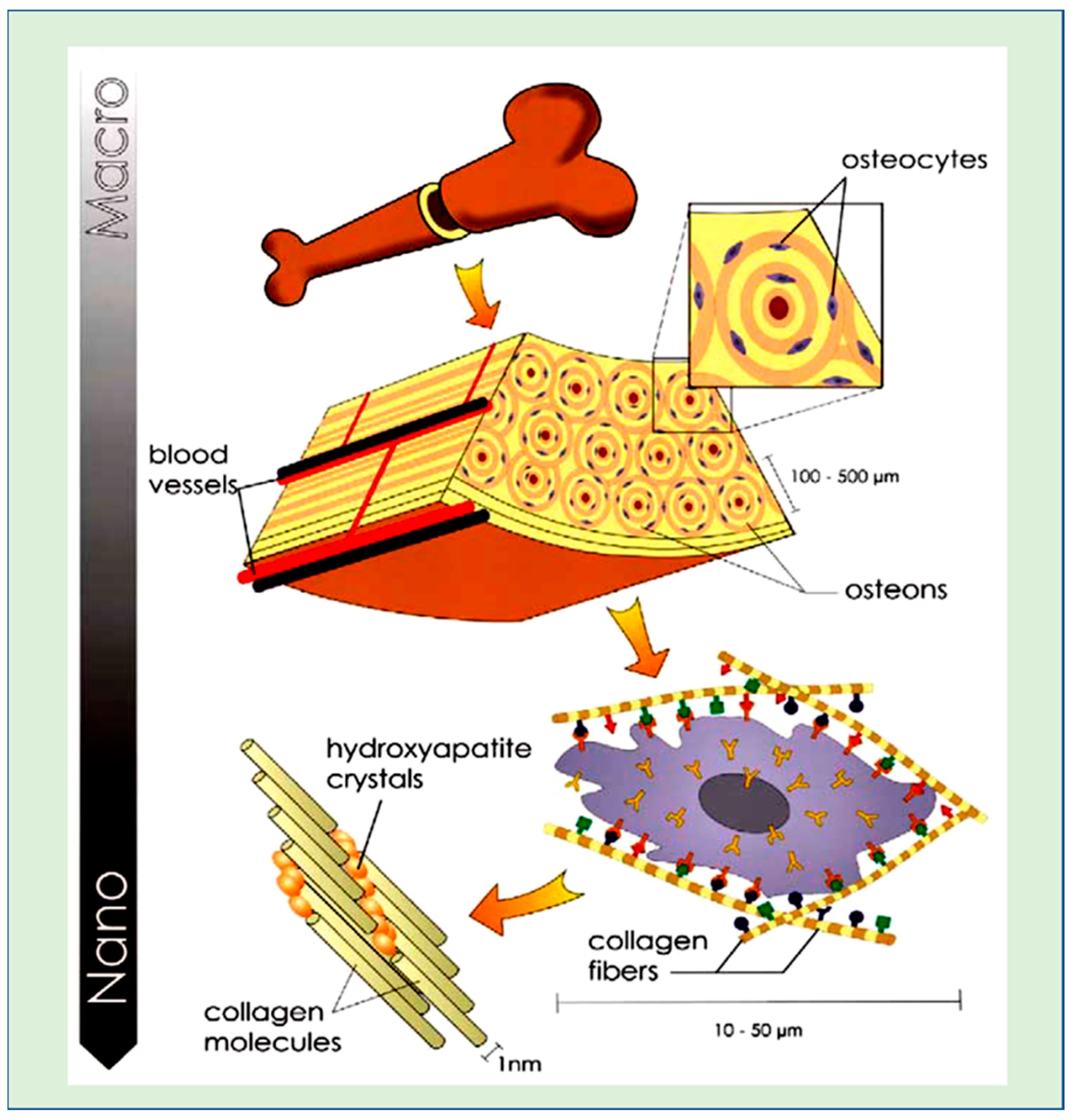
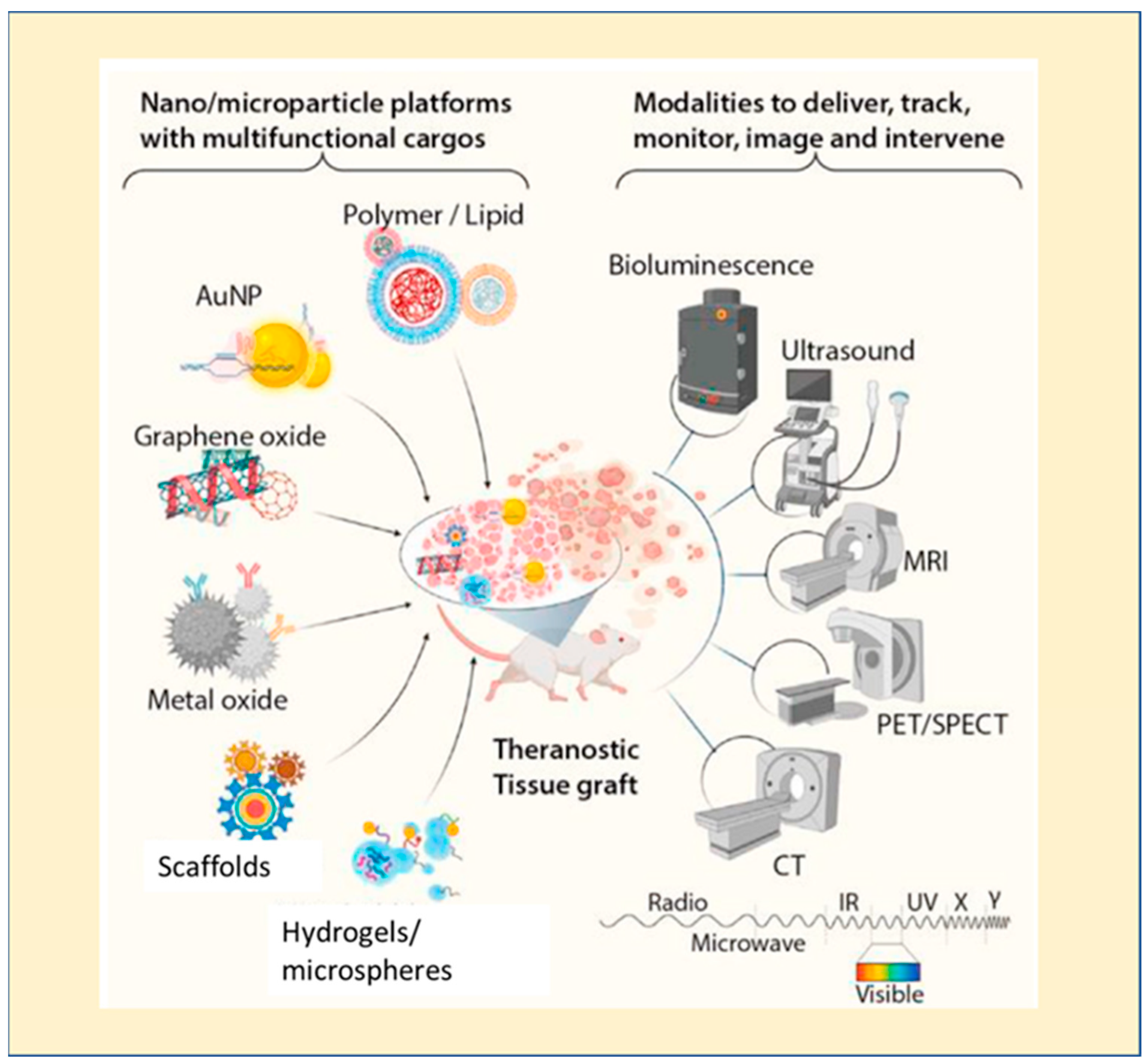
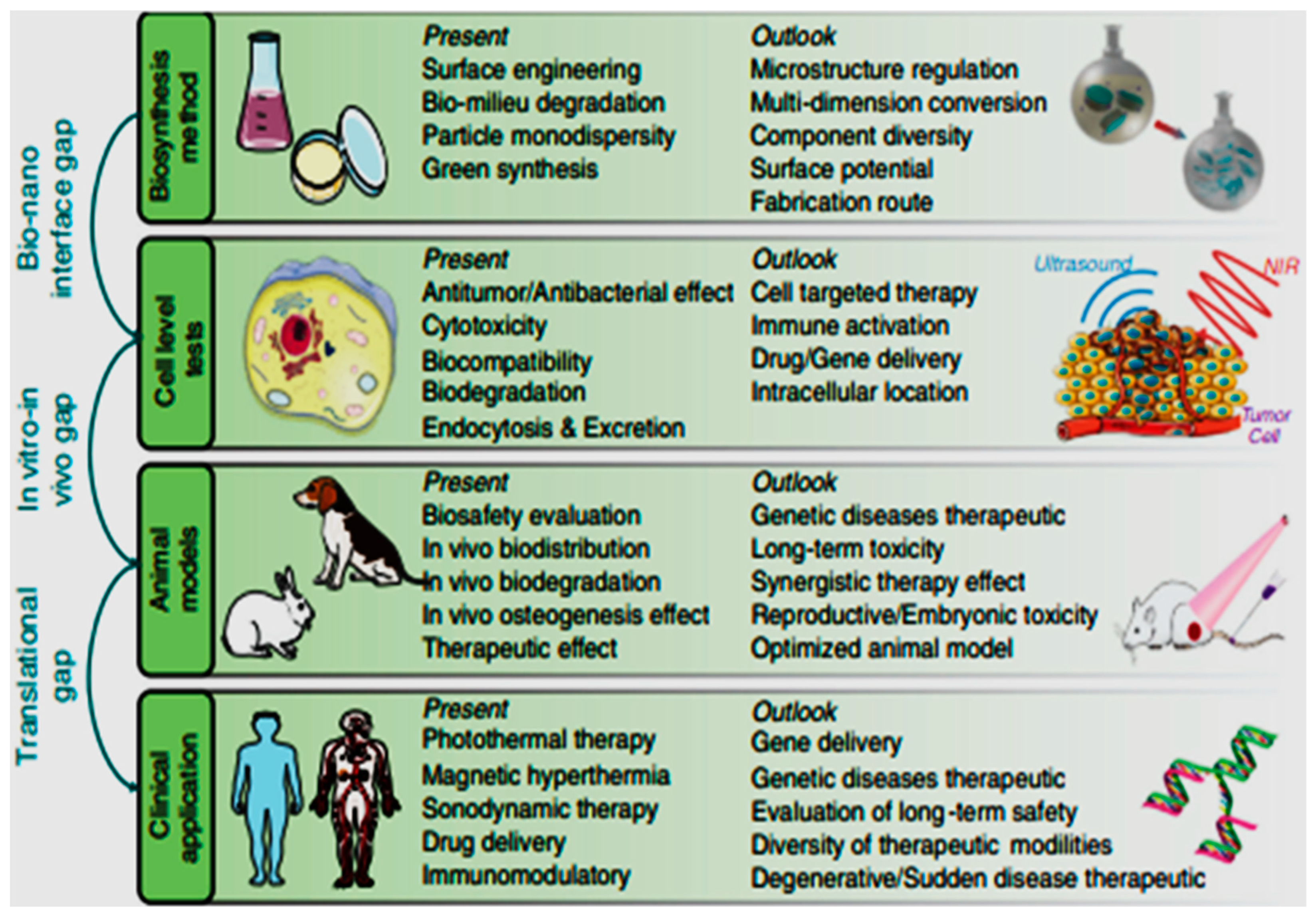
Table 1.
Different imaging techniques used as theragnostic tools for visualizing bone tissue, adapted from [14,25].
| Imaging Modality | Sensitivity (M) | Signal Type | Radiation Hazard | Spatial Resolution | Advantages | Disadvantages | Imaging Agent |
|---|---|---|---|---|---|---|---|
| Optical Imaging | Bioluminescence 10−15 to 10−17 & Fluorescence 10−9 to 10−12 | Visible light or near infrared | Nil | 2–5 nm | High sensitivity; ability to provide practical information without radiation exposure | Low tissue penetration; low resolution; limited potential for clinical translation | Fluorochrome/photoprotein |
| Computed tomography | Minimally characterized | X ray | Nil | 50–200 µm | High spatial resolution; capability of tissues differentiation without radiation exposure | High cost; required for contrasting agent; radiation and non-specificity | Iodine |
| Magnetic resonance imaging | 10−3 to 10−5 | Radio waves | Nil | 25–100 µm | High spatial resolution; no radiation exposure; ability to provide detailed functional information | High cost; patient with metallic implants not suitable | Iron oxide; Gadoliniu; manganese oxide; 19fluorine labelled compounds |
| Gamma scintigraphy | PET 10−11 to 10−12, SPECT 10−10 to 10−11 | Gamma rays | Nil | 1–2 nm | Capable of providing an image of the biochemical process | High cost; radiation; low resolution | 8F, 64Cu, 11C, 15O labelled compounds |
| Ultrasound | Minimally characterized | High frequency sound waves | Nil | 50–500 µm | Low cost; non-invasive; no radiation and ease of production | Low resolution | Microbubbles |
Disclaimer/Publisher’s Note: The statements, opinions and data contained in all publications are solely those of the individual author(s) and contributor(s) and not of MDPI and/or the editor(s). MDPI and/or the editor(s) disclaim responsibility for any injury to people or property resulting from any ideas, methods, instructions or products referred to in the content. |
© 2023 by the authors. Licensee MDPI, Basel, Switzerland. This article is an open access article distributed under the terms and conditions of the Creative Commons Attribution (CC BY) license (https://creativecommons.org/licenses/by/4.0/).
Share and Cite
MDPI and ACS Style
Banerjee, K.; Madhyastha, H. Polymeric Theragnostic Nanoplatforms for Bone Tissue Engineering. J. Nanotheranostics 2023, 4, 280-298. https://doi.org/10.3390/jnt4030013
AMA Style
Banerjee K, Madhyastha H. Polymeric Theragnostic Nanoplatforms for Bone Tissue Engineering. Journal of Nanotheranostics. 2023; 4(3):280-298. https://doi.org/10.3390/jnt4030013
Chicago/Turabian StyleBanerjee, Kaushita, and Harishkumar Madhyastha. 2023. "Polymeric Theragnostic Nanoplatforms for Bone Tissue Engineering" Journal of Nanotheranostics 4, no. 3: 280-298. https://doi.org/10.3390/jnt4030013