


Recent Advancement of Bio-Inspired Nanoparticles in Cancer Theragnostic

1
School of Health Sciences, University of Petroleum and Energy Studies, Dehradun 248007, Uttarakhand, India
2
Department of School of Engineering and Technology, The Assam Kaziranga University, Jorhat 785006, Assam, India
*
Author to whom correspondence should be addressed.
J. Nanotheranostics 2023, 4(3), 299-322; https://doi.org/10.3390/jnt4030014
Submission received: 31 May 2023
/
Revised: 7 July 2023
/
Accepted: 18 July 2023
/
Published: 24 July 2023
(This article belongs to the Special Issue Advanced Functional and Tunable Nano-Systems for High-Performance Theranostic and Tissue Engineering Applications)
Abstract
:The introduction of cancer therapeutics and nanotechnology has resulted in a paradigm shift from conventional therapy to precision medicine. Nanotechnology, an interdisciplinary field with a focus on biomedical applications, holds immense promise in bringing about novel approaches for cancer detection, diagnosis, and therapy. The past decade has witnessed significant research and material applications related to nanoparticles (NPs). NPs differ from small-molecule drugs as they possess unique physicochemical characteristics, such as a large surface-to-volume ratio, enabling them to penetrate live cells efficiently. Traditional cancer therapies, such as chemotherapy, radiation therapy, targeted therapy, and immunotherapy, have limitations, such as cytotoxicity, lack of specificity, and multiple drug resistance, which pose significant challenges for effective cancer treatment. However, nanomaterials have unique properties that enable new therapeutic modalities beyond conventional drug delivery in the fight against cancer. Moreover, nanoparticles (1–100 nm) have numerous benefits, such as biocompatibility, reduced toxicity, excellent stability, enhanced permeability and retention effect, and precise targeting, making them ideal for cancer treatment. The purpose of this article is to provide consolidated information on various bio-inspired nanoparticles that aid in cancer theranostics.
Keywords:
nanoparticles; quantum dots; radioisotopes; liposomes; micelles; nanobubbles; theranostics; therapeutics; toxicity1. Introduction
Nowadays, cancer is the second biggest cause for death amongst humans [1]. The World Health Organization estimates that cancer caused more than 10 million deaths globally in 2022, which equates to one in six fatalities [2]. Lung, skin, colorectal, breast, prostate, and stomach cancers are the most prevalent types of cancer, and they are listed chronologically from highest to lowest cancer case numbers. Cancer research has made significant strides, but a cure is still many years away [3].
Theranostics is a new biomedical procedure that combines diagnosis and treatment into one step. Targeting particular (diseased) tissues or cells with precision in order to improve the accuracy of diagnostic and therapeutic procedures is a distinctive aim of theranostics [4]. Theranostics’s main goal is to combine critical phases of medical care, like diagnosis and therapy, in order to speed up, make care safer, and improve outcomes [5]. Drug delivery using nanoparticles is a successful method for overcoming the difficulties of loose drug administration [6]. The chargeless chemotherapeutic agents have very slight bioavailability and, by activating various efflux mechanisms in cancer cells, cause drug resistance. Drug delivery using nanoparticles is advantageous for suppressing various drug resistance phenomena in cancer cells [7]. In contrast to small molecules, which enter cells through diffusion, receptor-mediated endocytosis is how nanoparticles are internalized [8]. The procedure enables the drug to be released from the lysosomal compartments by nanocarriers [9].
With each passing year, cancer’s global toll rises, and it is quickly overtaking all other causes of death. Natural compounds appear to be an option with fewer side effects than chemotherapy, though their bioavailability is still a concern. Chemotherapy plays an important role but causes side effects. Quantum dots (QDs) with bio-inspired designs derived from plant sources exhibit promising drug delivery and anticancer potential with minimal toxicity [10,11,12,13]. QDs are widely used in many different applications and can be specifically used for bio-imaging and drug delivery in cancer therapy due to their well-known optical activity. Additionally, using bio-inspired QDs as a delivery system for anticancer drugs is less studied [14].
Liposomes were first employed in few simple experiments, like the photochemistry of biological membranes, and are now mainly known for their amazing prospectus in the controlled release of a wide variety of drugs [15]. Liposomes used to be used to load a variety of PSs for photodynamic cancer therapy due to their many benefits and potentials. Combining photochemistry and photo physics, the multidisciplinary field of photodynamic therapy (PDT) has enormous potential for use in cancer treatment [16]. To create cytotoxic and reactive oxygen species as well as subsequently oxidize light-exposed tissues, PDT uses a photosensitizing agent (PS) and light. Available PSs are still far away from being an ideal therapy, despite the many benefits of PDT and the tremendous progress made in this area, due to its low permeability, side effects, non-specific phototoxicity, hydrophobicity, weak bioavailability, and propensity for self-aggregation [17,18,19]. PSs can be enclosed in such small liposomes, a cutting-edge drug delivering system that has been shown to improve drug permeability into the biological membranes and load both the lipophilic agents and hydrophobic, to get around these restrictions. To increase the effectiveness of delivery, liposomes can be covered with targeting agents [20].
The three main approaches to cancer therapy can be summarized as the delivery of biotherapeutics, tumor image analysis, and immunotherapy. Liposomes, which are artificial fat bubbles, have long been recognized for their ability to encapsulate various bioactive substances and release them gradually in response to stimuli. They have been extensively studied as a vehicle for delivering therapeutic drugs and imaging agents, and they are also widely employed in cancer immunotherapy. On the other hand, exosomes are naturally occurring nanosized vesicles found outside of cells that play a vital role in intercellular communication. Importantly, exosomes have demonstrated their capacity to transport a variety of pharmaceuticals and diagnostic molecules to tumor cells. Additionally, exosomes are enriched with tumor antigens and possess several immunomodulatory effects [21]. Recently, there has been growing interest in developing smart, bioengineered hybrid nanovesicles that combine the advantages of both exosomes and liposomes. These hybrid nanovesicles aim to leverage the benefits offered by both types of vesicular systems [22].
Exosomes play a crucial role in every stage of tumorigenesis, including initiation, growth, survival, and metastasis, making them an important focus in oncology. These organelles present exciting prospects for cancer therapy, and further advancements are needed to effectively apply the latest breakthroughs in clinical settings. Exosomes possess unique properties that make them an attractive target for cancer treatment, such as their inherent nature, small size, and ability to traverse biological barriers. Compared to cell therapy, the clinical use of exosomes offers several advantages, including simpler preparation and sterilization processes, reduced storage requirements, and significantly lower production costs. However, additional research is required to understand the signaling pathways and precise mechanisms through which exosomes contribute to tumorigenesis. It is also essential to develop practical techniques for isolating exosomes and determining the appropriate type and quantity of anticancer drug to load into them. Translating these discoveries into clinical practice will pave the way for new therapeutic approaches and potentially bring transformative changes to the lives of cancer patients in the future.
Drug delivery systems have been a main area of focus for pharmaceutical research from the start. In the current technological era of nanocarriers, polymeric micelles (also known more commonly as just PMs), have emerged as multifunctional nanoparticles that have shown promise in several scientific fields [23]. Due to their enhanced retention and permeability which enable them to change the release profile of the integrated pharmacological substances and concentrate them in the target zone, they are much better suited for the poorly soluble medications. PMs are anticipated to be a treatment option which is very successful for cancer therapy in coming times due to their amazing biocompatibility, enhanced permeability, much lower toxicity to healthy cells, and ability to solubilize several types of drugs in their micellar core [22,23,24,25,26]. They can assemble in the tumor microenvironment (TME) thanks to their nano size and the enhanced permeation and retention (EPR) effect. Additionally, PMs are used as “smart drug carriers,” which makes them target particular cancer sites by using several stimuli, enhancing the specificity and effectiveness of targeted drug delivery which is micelle-based [27,28,29].
2. Principle of Cancer Theranostics
Increased imaging capabilities and diagnostics have made it possible to identify cancers earlier and more thoroughly [30]. Recently, these methods have expanded into theranostics, in which the contrast agents that are typically used for imaging have been combined with therapeutics in order to diagnose and treat cancers simultaneously in a patient-specific manner [31].
Nanoparticles (NPs) play a wide range of roles in cancer diagnosis and have a number of advantages over other traditional methods of delivering chemotherapeutic drugs. Delivering drugs to the target tissue, or cells, or organs is more precise as well as effective with NPs, and the risk of side effects is reduced. With less drug removal from the system, NPs deliver drugs to tumor areas in passive and active modes [32]. The biodistribution of chemotherapy drugs in the body as well as the effectiveness of nanocarriers are significantly influenced by the size and surface properties of nanoparticles [33].
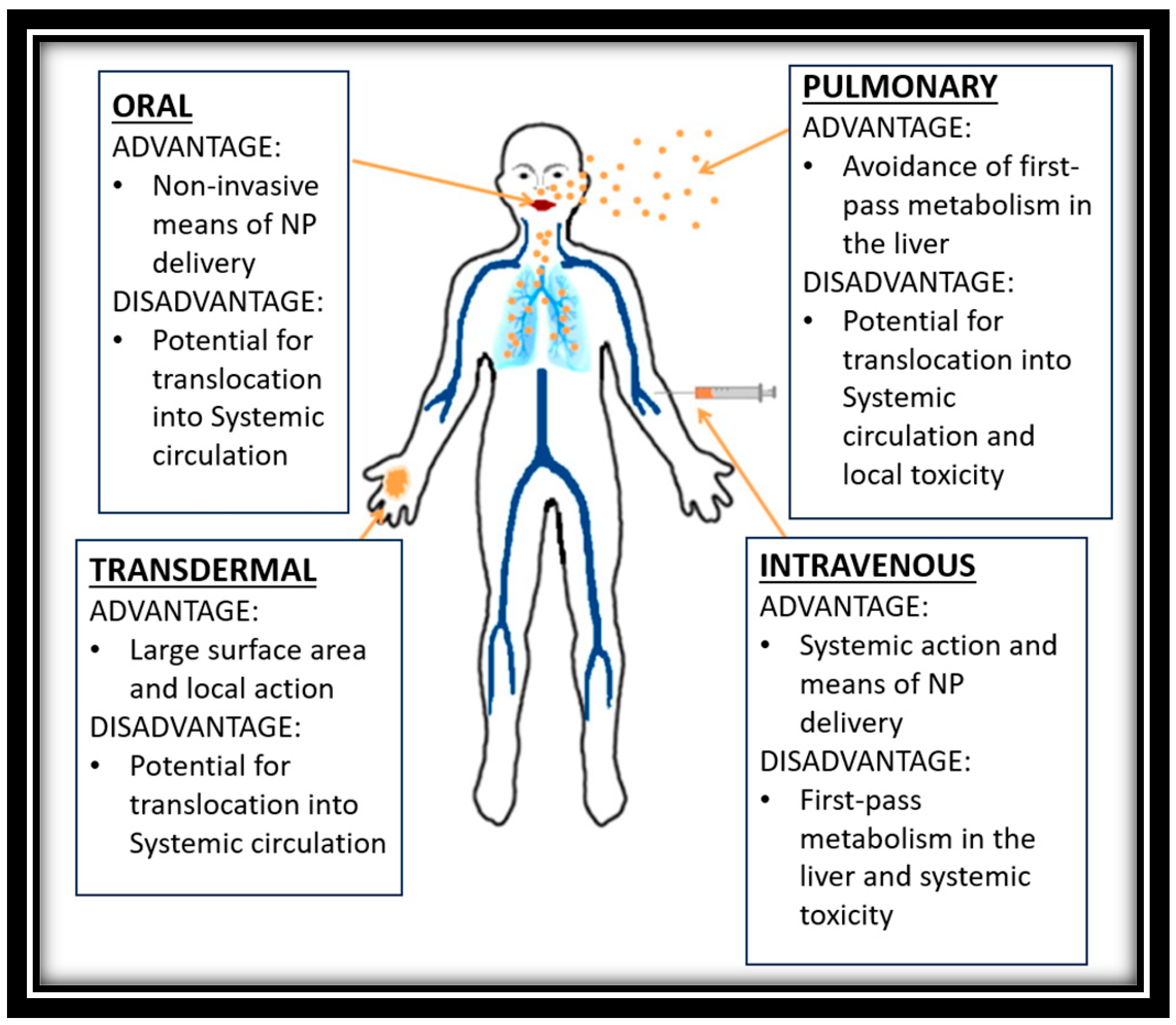
Phospholipids/surfactants are the primary constituents of self-assembled vesicular nanoparticles (NPs), which show promising potential for nanomedicine as novel biomedical nanocarriers. The two most common vesicle subcategories in recent decades, liposomes and noisome, have been utilized extensively in the treatment of cancer [34,35]. NPs can be administered into the body by several pathways having its own benefits, Figure 1 demonstrates some of them below.
3. Application of Cancer Theranostics
Drugs are more easily accessible in the cellular core, where they activate apoptotic signals for cell death, than in the peripheral cytoplasm, where they are less likely to activate efflux mechanisms [37]. As a result of their cellular internalization mechanisms, nanodrug delivery systems are less likely than free drugs to result in multiple drug resistance [38]. Another crucial fact is the capability of the nanocarriers to be surface-modified with additional ligands to provide active cancer cell targeting, straightforward internalization, and organelle-specific drug targeting [39].
Two-dimensional black phosphorus nanosheets (BPNSs), which have recently attracted a lot of interest, are best suited for theranostic nanomedicine. They are becoming more and more sought after as a potential replacement for the graphene-based nanomaterials used in biomedical applications, in part because of qualities like drug loading efficiency, biocompatibility, electrical, optical, and thermal and phototherapeutic characteristics [40,41].
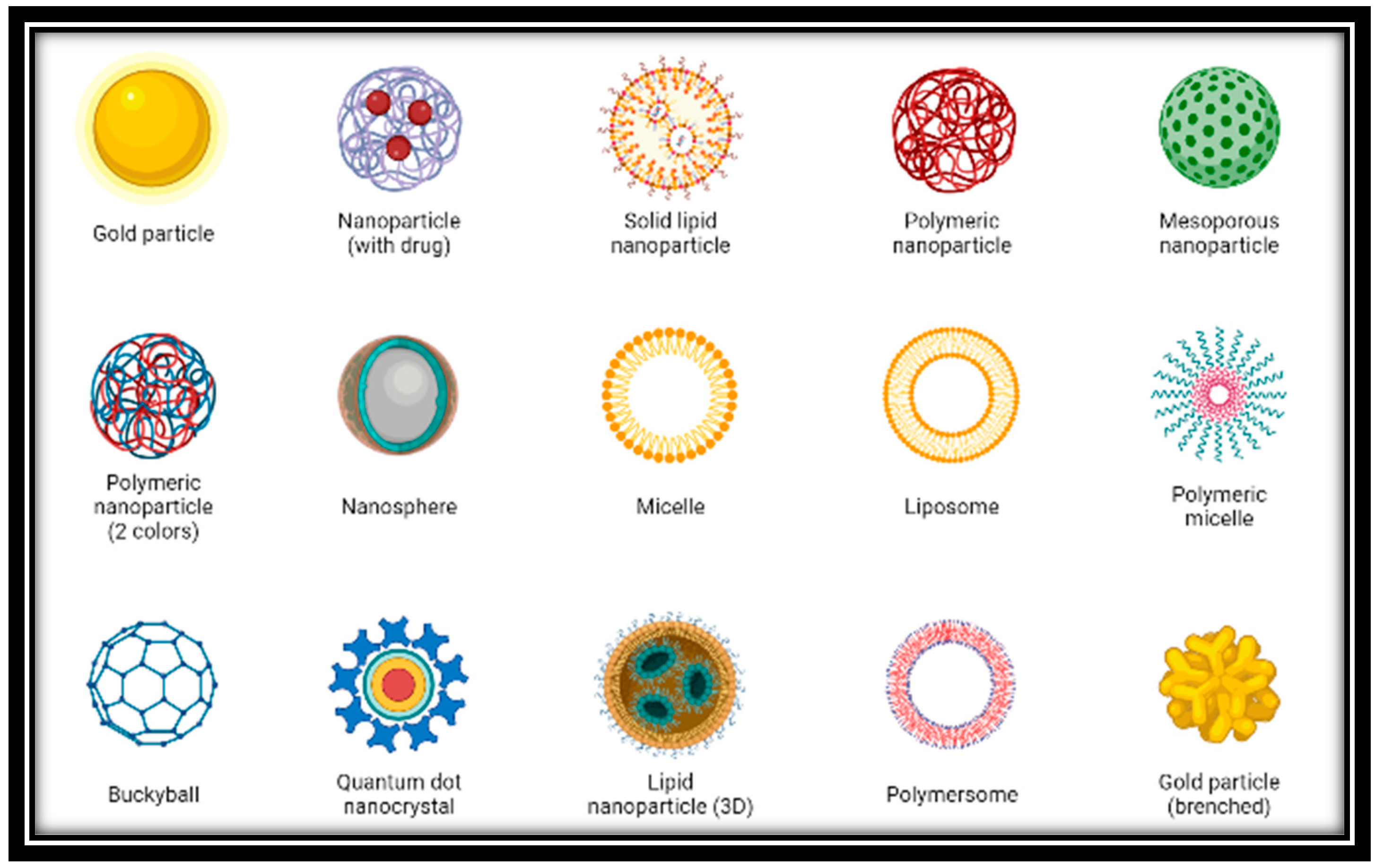
Cancer immunotherapies aim to amplify the immune system’s capacity to detect and combat cancer. Cancer immunotherapies try to “teach” the immune system to recognize and eliminate cancerous cells in the TME [42]. In order to improve cancer immunotherapies, nanomedicine-based delivery is crucial. These strategies either increase the antitumor response of the immune system or lessen their periodic side effects [43]. As we already know there are several types of nanoparticles that are used in the cancer treatment, some of them are shown below in Figure 2 with their diagrams and Table 1 highlights their application.
3.1. Quantum Dots
In anticancer studies, QDs are predominantly utilized for imaging purposes in conjunction with various nanoparticles to observe drug targeting and release. While QDs have been found to enhance drug activity in certain cases, there is limited research exploring their synergy with anticancer phytochemicals. It is important to note that QDs can exhibit cytotoxic effects, although coating the core can mitigate these toxicities. Nevertheless, the presence of heavy metals in QDs remains a health concern. On the other hand, carbon dots (CQDs) derived from natural sources offer a promising alternative as quasi-spherical particles with reduced health risks. These carbon dots possess optical properties and, if prepared from a natural product exhibiting anticancer activity, can independently elicit improved anticancer effects. Additionally, such carbon dots have demonstrated specificity towards cancer cells. To overcome the challenges associated with CQDs in drug delivery systems, comprehensive in vivo studies are needed, and the specific targeting can be achieved by attaching receptor ligands to the CQDs. Small semiconducting particles or nanocrystals known as QDs have extremely small diameters. They were initially found in 1980. The unbelievably high surface-to-volume ratios of these dots contribute to their unusual electronic properties, which are intermediate between those of discrete molecules and bulk semiconductors [45,46]. Fluorescence, where the nanocrystals can produce distinct colors depending on the size of the particles, is the most noticeable effect of this. The quantum dot is a type of nanoparticle with dimensions between tens of nanometers and a few hundredths of a nanometer, which are smaller than those of a typical nanoparticle. The tumor dot can be used in multiple fields, including solar cells, light-producing diodes, lasers, and biomedical applications because the quantum mechanical behavior associated with it exhibits various optical and electronic properties [47].
Due to their unusual properties, such as their small size, high surface area, photoluminescence, chemical stability, simple synthesis, and potential for functionalization, carbon quantum dots (CQDs) have attracted a lot of attention recently [48]. They are fluorescent carbon nanostructures with a size of less than 10 nm. Curiously, the scientific community has recently opted to use biomass precursors rather than chemical compounds to prepare CQDs. These biomass sources turn waste into useful materials while being cheap, accessible, environmentally friendly, and abundant [49,50]. Because of technological developments, we can detect tumors at the cellular level, making it simpler to study variations in the transcriptional gene and its genetic factors. Ideal chemotherapy is viewed as a non-specific therapy with the ability to kill healthy cells and harm a person systemically. Therefore, creating new technologies is now an urgent necessity. Semiconductor particles known as QDs have sizes between 2 and 10 nm. Many researchers are interested in QDs, for their distinctive qualities, like their small size, extensive surface area, charges in the surface, and precise targeting. The numerous nanocarriers include well-known QD-based drug carriers. Enhancing solubility, extending time for retention, and minimizing the negative effects of loaded medications are all benefits of using QDs as a delivery method [32]. Zero-dimensional carbon-based nanomaterials have shown clear structural and functional advantages for the use of nanotheranostics: (I) several luminescent models, like the PL, thermally activated delayed fluorescence (TADF), phosphorescence, hemiluminescence, etc., can be achieved by CDs and used for a variety of bio-imaging models; (II) compared to other imaging probes, CDs exhibit excellent biocompatibility and photostability, making them suitable for use as in vivo real-time imaging agents; (III) near-infrared (NIR) emission, in particular, is tunable in CDs and is applicable for tissue imaging alongside deep penetration; (IV) CDs have demonstrated very unique properties, like blood–brain barrier (BBB) penetration and novel cell targeting, which is extraordinarily advantageous for imaging; (V) CDs demonstrate clearable behavior in living organisms, which is great for long term imaging [51,52,53,54,55,56,57,58,59,60,61,62].
Even though research on QD nano-transporters is still ongoing, they have not yet made their benefits known to the public. There have already been a few beneficial developments as a result of the discovery of QDs [63]. The application of QD shows conversion in natural imaging and photography has shown incredibly amazing suitability in bio-imaging, the creation of novel drugs, the delivery of targeted genes, biosensing, as well as photodynamic therapy and diagnosis. The current review set out to prove the value of QD in the detection and management of cancer [64,65].
3.2. Liposomes
By increasing drug efficacy and enhancing cell type-specific delivery, liposomes have amazing benefits that could strengthen immune responses and cancer immunotherapy [66]. Liposomes are lipid-based nanoparticles that have a high potential for enhancing cancer immunotherapies because they can incorporate and/or associate a variety of cancer drug molecules [67]. The preparation of liposomes typically involves four fundamental stages. First, lipids are dried down from an organic solvent. Next, the dried lipids are dispersed in an aqueous medium. Subsequently, the resulting liposome formulation undergoes a purification process. Finally, the final product is analyzed to ensure its quality and characteristics. These four stages are commonly employed in various methods of liposome preparation. One commonly employed technique is the ether injection (solvent vaporization) method. In this approach, a solution of lipids dissolved in diethyl ether or an ether–methanol mixture is slowly injected into an aqueous solution containing the material to be encapsulated. This process typically takes place at temperatures ranging from 55 °C to 65 °C or under reduced pressure. As the ether is subsequently removed under vacuum, liposomes are formed. However, this method has some drawbacks, including the heterogeneity of the resulting population of liposomes (ranging from 70 to 200 nm) and the exposure of the compounds to be encapsulated to organic solvents at elevated temperatures.
Liposomes can be used for a variety of immunotherapeutic cancer treatments, such as vaccination or checkpoint blockade, making them very flexible. Low-molecular-weight anticancer drugs, immunomodulators, and antigens are frequently delivered using these mechanisms [68,69].
Anticancer agents have been formulated as liposomal formulations to increase antitumor efficacy by rising deposition of tumor drug, reducing toxicity of drug by avoiding critical normal tissues, and extend drug circulating lifetime [70]. The clinical application of micro- and nanoparticulate formulations and their development, along with the combination of the novel agents with conventional medications and standard-of-care therapies, still presents challenges in spite of clinical approval of chemotherapeutics which are multiple liposome-based [71]. Control over drug biodistribution, encapsulated drug release rates, and target cell uptake are all factors that need to be optimized. Several liposome-based goods have been authorized or put on the market. To improve liposomes’ therapeutic indices for applications in oncology, antineoplastic agent classes have been added. Anthracyclines, analogues of platinum, camptothecins, vinca alkaloid, and antimetabolite are a few of them. Many liposomal anticancer medications are either clinically approved or in late-stage clinical development [72,73,74,75].
There are a number of factors to consider when contrasting the traits of the drug and the liposome carrier. Before it can be approved, a drug must first show promise in treating the chosen tumor type [76]. A number of cancers have been successfully treated using anthracyclines and vinca alkaloids, two common examples [77]. The drug must be loaded into the liposome carrier adequately and effectively in order for the drug to be commercially viable [78]. To deliver a dose that is pharmacologically active in the proper quantity of carrier lipids, the carrier must have enough drug in it. Third, while the drug is being transported steadily by the carrier in the bloodstream, the drug must be released from the carrier at the tumor site at the appropriate rate. The release rate of agents which are chemotherapeutic to be encompassed into the carrier is significantly influenced by the physicochemical properties of the agent [79,80].
3.3. Extracellular Vesicles and Exosomes
Extracellular vesicles (EVs) are small biological particles that consist of exosomes, microvesicles, and various other types of nanoscopic vesicles. They are naturally produced by many different cell types and are believed to play important roles in intercellular communication. Recently, researchers have started exploring the potential of using EVs as vehicles for drug delivery. Initial findings suggest that EVs may offer several advantages over synthetic nanoparticle delivery systems for specific applications. They have emerged as a promising cell-free approach for the treatment of various diseases, including cancer. With their inherent ability to facilitate cell-to-cell communication, along with their remarkable physiochemical stability and biocompatibility, EVs have gained recognition as exceptional carriers for delivering therapeutic agents such as nucleic acids, proteins, drugs, and nanomaterials. Ongoing research indicates that EVs can be modified, engineered, or purposefully designed to enhance their effectiveness, specificity, and safety in cancer therapy [81].
Furthermore, the presence of heterogeneity and complex components within EVs poses challenges to their therapeutic efficacy and raises safety concerns when used as carriers for therapeutic cargos. Additionally, the lack of efficient methods for isolating and scaling up EV production, as well as accurately monitoring the dosage of therapeutic cargos within EVs, are potential issues in clinical applications. Consequently, engineered EVs have emerged as a promising alternative approach for EV-based therapy addressing these concerns [82]. Increasing evidence suggests that engineering EVs enhances their loading efficiency, targeting capability, and therapeutic effectiveness [83]. Currently, two primary strategies are employed to load desired cargos into EVs. One approach involves incorporating the cargo into producer cells, which then undergoes the natural biogenesis process to obtain cargo-loaded EVs. The other approach involves harvesting EVs from various sources (such as cultured cells, human blood, and milk) and introducing cargos into EVs using traditional and advanced biotechniques [84]. There is also a growing interest in modifying EV membranes to target specific tissues and combining EVs with other nanomaterials to achieve improved or synergistic therapeutic effects [85]. Extensive exploration has also been conducted to fabricate bio-inspired or biomimetic EVs with higher production yields and loading efficiencies [86].
Numerous studies have established a strong association between exosomes and the development and progression of cancers [87,88]. Within the TME, exosomes facilitate the transfer of bioactive molecules among tumor cells, immune cells, and stromal cells, enabling cancer cells to evade immune surveillance and induce immune tolerance. Conversely, exosomes originating from immune cells exhibit the ability to inhibit tumor growth, proliferation, and metastasis. Consequently, exosomes play a dual role as a double-edged sword in cancer immunity. The primary objective of cancer immunotherapy is to stimulate the immune-suppressed host to recognize and eliminate cancer cells. By modifying exosome-secreting cells, exosomes can be utilized as delivery systems for immunotherapy drugs, antigens, or genes [89]. Both immature and mature dendritic cells are capable of producing exosomes, and exosomes derived from dendritic cells (DEXs) have demonstrated the capacity to overcome tumor-induced immunosuppression through direct and indirect mechanisms.
TEXs, or tumor-derived exosomes, have been observed in various cancer types, including breast, ovary, prostate, pancreas, bladder, and melanoma [90]. Studies have revealed that tumor cells produce significantly higher quantities of exosomes (approximately ten times more) compared to normal cells, resulting in elevated exosome concentrations in the bloodstream of patients [91]. The membranes of exosomes contain adhesion receptors and ligands that enable interactions with specific cell types, facilitating the delivery of a diverse range of biomolecules [92]. Consequently, these organelles play a crucial role in numerous malignant processes, such as promoting angiogenesis, metastasis, malignant transformation, abnormal metabolism, proliferation, immune system dysregulation, and sustaining cancer cell immortality.
Tumors often exhibit altered metabolism, characterized by the “Warburg effect,” where cancer cells prefer fermentation over aerobic respiration. This metabolic shift leads to increased glucose consumption and lactic acid production compared to non-proliferating normal cells [93]. The elevated production of lactic acid contributes to a reduction in extracellular pH, which in turn promotes tumor proliferation, invasion, migration, and drug resistance [94]. Moreover, the high levels of lactic acid hinder the proliferation and cytokine output of human cytotoxic T lymphocytes while promoting the expansion of myeloid-derived suppressor cells, thereby facilitating tumor progression [95,96]. Parolini et al. discovered that an acidic TME plays a crucial role in the heightened secretion and uptake of exosomes by tumor cells. They found that exosomes released in an acidic environment contained higher levels of sphingomyelin/ganglioside GM3, facilitating fusion and uptake processes. Additionally, electrostatic charges on exosomes may enhance their fusion ability, and the content of exosomes released in low pH conditions is altered. For instance, exosomes released under acidic conditions contain higher levels of caveolin-1, which is implicated in melanoma progression [97].
3.4. Polymeric Nanoparticles
Polymeric nanoparticles (NPs) have a significant role in the precise delivery of cancer drugs. Polymeric nanocarriers, also referred to as polymeric nanomedicine, can attach or enclose anticancer drugs. The use of polymeric nanomedicine has demonstrated promising results by offering controlled drug release with decreased toxicity and improved tumor retention. However, there is still a scarcity of drug-loaded delivery systems that meet the requirements of clinical applications. Therefore, additional endeavors are necessary to address this gap [98].
They can be synthesized in two main ways: (I) The dialysis technique is a straightforward and efficient method used to prepare polymeric nanoparticles with a narrow size distribution. To summarize the process, both the drug and polymers are dissolved in water-miscible organic solvents of appropriate molecular weight cut-off and placed inside a dialysis tube or membrane. The organic phase gradually diffuses out through the dialysis tube or membrane into the aqueous phase, reducing the interfacial tension between the two. As a result, the solvent displacement within the membrane leads to the formation of a homogeneous nanoparticle suspension, followed by progressive polymer aggregation due to reduced solubility. To obtain a fine powder of nanoparticles, the resulting nanosuspension is freeze-dried using 5% mannitol as a cryoprotectant. (II) Polymers play a crucial role in the development of oral delivery systems. The utilization of natural polymers, such as chitosan and alginates, instead of toxic chemical polymers can enhance the drug’s permeation effect, enzyme inhibitory ability, and mucoadhesive properties. In brief, the drug and polymer are dissolved in a weak acidic medium or water, depending on their solubility. The resulting solution is then added drop by drop, with constant stirring, to a solution containing counter ions and a stabilizer. This process leads to the formation of spherical particles through the complexation of oppositely charged species, resulting in gelation and precipitation. To reduce the particle size to the nanometric range, the resulting solution is sonicated. The nanosuspension obtained is subsequently freeze-dried, using 5% mannitol as a cryoprotectant, to yield a fine powder of nanoparticles [99].
Currently, drug-loaded NPs have emerged as a promising approach for effective cancer therapy, offering improved drug delivery and therapeutic outcomes across different cancer types [100]. These polymeric nanocarriers, known as polymeric nanomedicines, exhibit diverse architectures such as polymer-drug conjugates, micelles, nanospheres, nanogels, vesicles, and dendrimers [101,102]. They have garnered significant attention for their ability to simultaneously deliver multiple therapeutic agents and target tumor cells [16]. A wide range of therapeutics, including cytotoxic agents, small interfering RNA (siRNA), chemosensitizers, and antiangiogenic agents, can be transported by these nanoparticle platforms [103,104].
Nanoparticles leverage the unique pathophysiology of cancer cells to enhance drug retention and permeability. They accumulate within cells without being recognized by P-glycoprotein, leading to increased intracellular drug concentrations. Furthermore, nanoparticles enable precise delivery of chemotherapies to tumor cells at the required time, thereby achieving optimal efficacy and reducing cytotoxicity in healthy peripheral tissues [105,106,107,108]. Among the various strategies employed, the use of multitargeting systems (MTS) as a targeting approach has gained significant attention [109]. In this strategy, the surface of nanoparticles is decorated with multiple functional ligands that can recognize different receptors on cells, thereby enhancing multivalent interactions between nanoparticles and cell surfaces and facilitating improved cell recognition and internalization. Additionally, other strategies, such as incorporating pH-sensitive groups to enhance drug efficacy, have also been explored [110]. As nanoparticles continue to revolutionize cancer treatment techniques, extensive research is underway to develop multifunctional nanoparticles with enhanced capabilities [111].
3.5. Radioisotopes
A radioisotope is an atom that is unique in that it has the ability to emit radiation. As a result of internal changes in unstable or radioactive atoms, radiations are waves or particles that are sent out in all directions [112]. For the large-scale development, high-current medical systems producing enhanced yields of radioisotopes, experimental thick target yields and functions of excitation—i.e., cross sections of isotope production that depend on energy—are crucial [113,114].
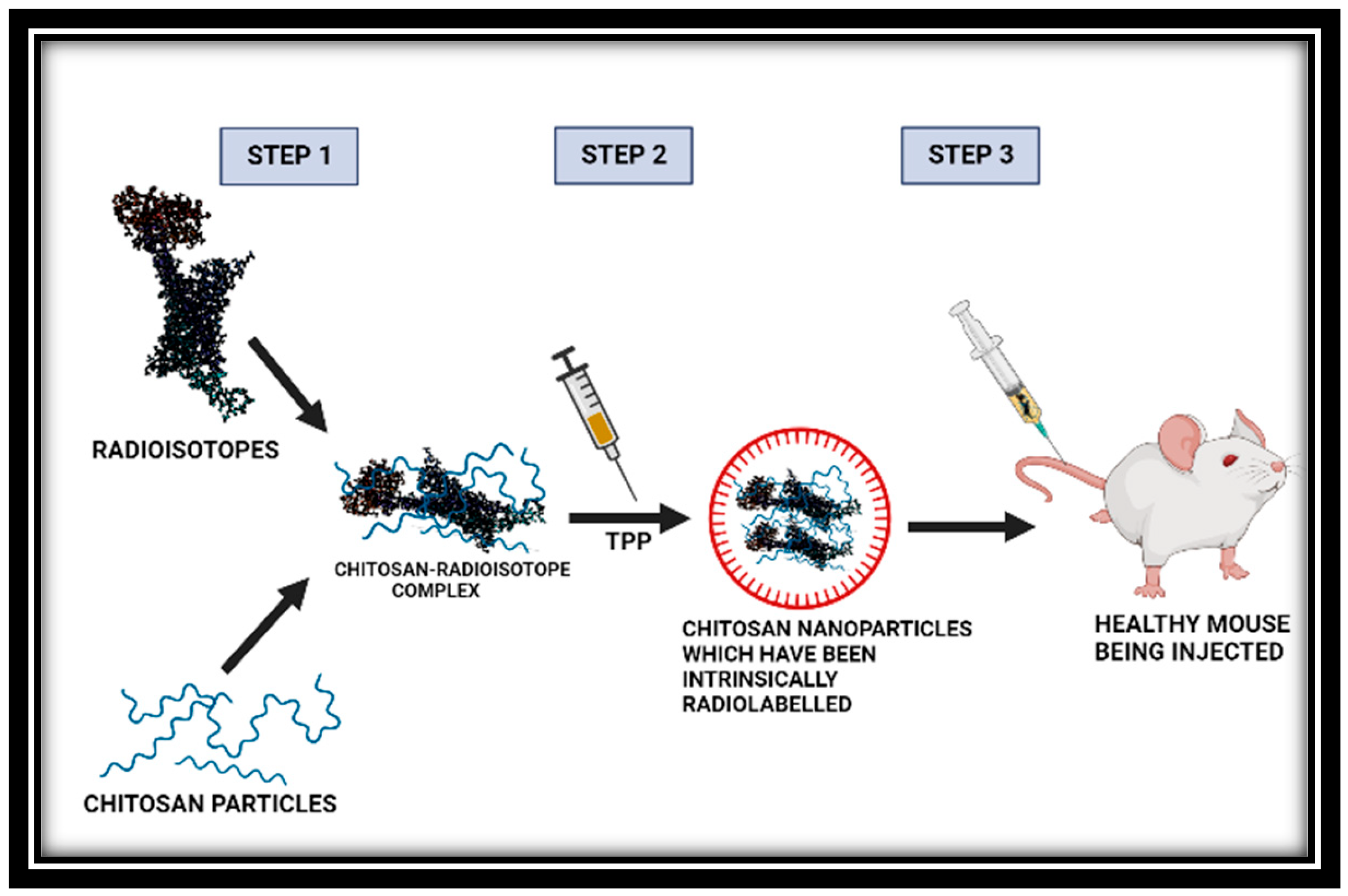
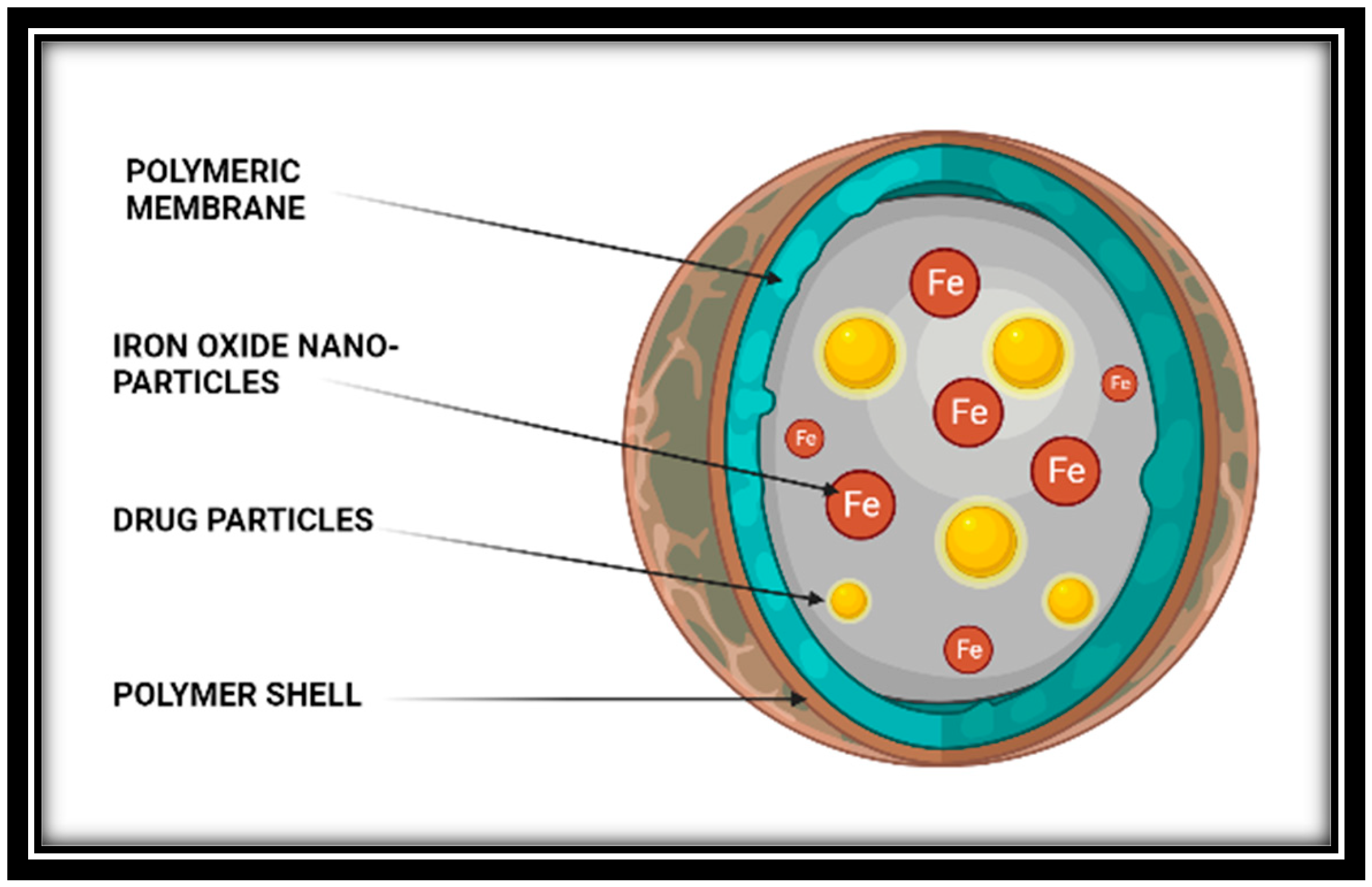
In modern medicine, accelerator-produced radioisotopes are frequently used for imaging, cancer therapy, and therapy and diagnostic combination (theranostics) [115]. Several radioisotope-based treatments are in advanced stages of clinical trials, which may pave the way for a strong demand for specific accelerator systems devoted to radioisotope production [116]. Although cyclotrons are the industry standard, we explore different options here using linear accelerators. Linacs have the advantage of modularity, compactness, and lower beam loss with less shielding needed when compared to cyclotrons [117]. Considerable attention has been given to the development of nanocarrier systems for drug delivery that utilize chitosan, which is a linear chain polysaccharide derived from the chitin of arthropods and occurs naturally. This non-toxic, biodegradable, and biocompatible polymer was combined with specific radioisotopes for diagnostic and therapeutic purposes (64Cu, 68Ga, 90Y, 153Sm, 166Ho and 177Lu) [118]. By using the ionotropic gelation method, radiolabeled chitosan was transformed into nanoparticles. In epithelial lung cancer cells, the surrogate nanoparticles’ cell uptake and cytotoxicity were assessed. The intrinsically radiolabeled nanoparticles had high in vivo stability, according to biodistribution studies carried out on healthy C57BL/6 mice; this is shown in Figure 3 below [119].
3.6. Micelles
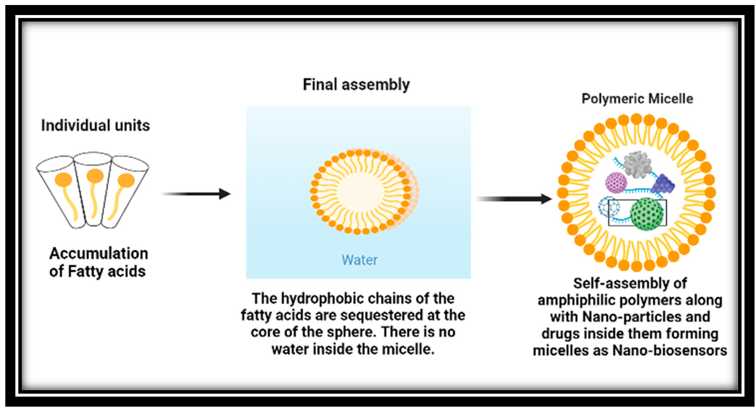
Polymeric micelles have emerged as a popular option for delivering chemotherapeutic agents that are difficult to dissolve for cancer patients in pre-clinical studies [120]. These micelles are formed by the self-assembly of amphiphilic polymers and can be customized with different polymeric combinations to achieve optimal loading, stability, systemic circulation, and delivery to the target cancer tissues as demonstrated below in Figure 4. Moreover, the surface of the nanocarriers can be modified with additional ligands to facilitate active cancer cell targeting, internalization, and organelle-specific drug targeting. Polymeric micelles offer several advantages over other types of nanoparticles. They are formed by amphiphilic polymers that self-assemble in an aqueous environment, and different polymeric building blocks can be used to achieve the desired hydrophobic/lipophilic balance, size, drug loading capacity, micellization ability, and stability in the systemic circulation [121,122,123].
Compared to other drug delivery methods, micelles have an advantage due to their small size, which enables them to extravasate more effectively through leaky vasculature. The hydrophilic polymeric coating on their surface also enables them to evade detection by the reticuloendothelial system during circulation [124]. Furthermore, micelles can be directed towards the tumor site by chemically conjugating tumor-homing ligands onto their surface [125].
Micelles are nanoparticles that self-assemble when amphiphilic block copolymers are dissolved in specific solvents at concentrations above the critical micelle concentration (CMC). In recent years, nanosized delivery systems like micelles have gained attention due to their improved in vivo stability, ability to protect entrapped drugs, release kinetics, ease of cellular penetration, and increased therapeutic efficacy [126].
Various immune cell types, such as effector T cells, regulatory T cells, dendritic cells, natural killer (NK) cells, myeloid-derived suppressor cells (MDSCs), and tumor-associated macrophages, play important roles in triggering and enhancing the immune response against tumors. Recent studies have shown the development of several methods, including cancer vaccines, antibodies, and immune-stimulating adjuvants, to trigger potent antitumor immunity against aberrant cancer cells. These approaches are enhancing the current antitumor immunity and reinvigorating an immunosuppressive TME [127,128,129]. Due to their distinct mesoporous channels that allow for encapsulation and the controlled release of anticancer medications, mesoporous silica nanoparticles serve as excellent nanocarriers in the development of drug delivery systems. The surface modification strategy is employed to prepare the nanoparticles, enabling the efficient release of KLAK and DOX within the acidic/reductive environment of TMEs. This facilitates their penetration into cell nuclei and results in the generation of the desired antitumor effect [130].
The initial requirement for effective systemic gene delivery is ensuring sufficient retention in the bloodstream to enable high availability at the desired target location. A promising approach for achieving this involves the formulation of polyplex micelles, which are formed through the self-assembly of oppositely charged block copolymers consisting of poly (ethylene glycol) (PEG) and polycations, along with plasmid DNA. These micelles possess a desirable core-shell architecture, with the outer hydrophilic PEG shell providing excellent stealth properties.
In order to further enhance the potential of these micelles for systemic applications, we strategically introduced hydrophobic cholesteryl moieties at the ω-terminus of the block copolymer. This addition was anticipated to improve the stability of the polyplex structure and increase the density of the tethered PEG chains. Additionally, the molecular weight of PEG in the PEGylated polyplex micelle was increased to 20 kDa, with the aim of further enhancing the density of PEG chains.
Furthermore, a cyclic RGD peptide was incorporated as a ligand molecule at the distal end of the PEG chains. This modification was intended to facilitate targeted delivery to the tumor site by interacting with integrin receptors, as well as promoting cellular uptake and intracellular trafficking. The resulting polyplex micelle, conjugated with cRGD and featuring an enhanced PEG shielding effect, was then tested in a mouse model of intractable pancreatic cancer. The micelle exhibited potent suppression of tumor growth through efficient gene expression of an antiangiogenic protein (sFlt-1) at the tumor site [131].
3.7. Nanobubbles
Nanobubbles, which are gas-filled cavities in aqueous solutions, have distinctive features attributed to their low internal pressure and surface tension resulting from the charged gas/liquid interface [132]. To induce cell death in the tumor lymph node by delivering particles, DOX medications are loaded into magnetic Poly (lactic-co-glycolic acid) PLGA microbubbles that contain perfluorocarbon gas. The results indicate that this approach can be utilized to improve lymphatic targeting and mitigate tumor metastasis [133]. Micro-nanobubble and nanobubble generators employ various methods such as pressurized dissolution, electrolysis, or swirling fluids in a mixing chamber to synthesize nanobubbles. These generators then pass the fluids through a single shear point, injector, or Venturi to produce air bubbles with an average particle size larger than 200 nm after this process the nanobubbles look like Figure 5. Most of these systems rely on an external air supply, and their primary function is not to enhance the electrochemical properties of water [134].
There are several ways to create plasmonic nanobubbles, including (1) using extracorporeal cell processing systems for gene therapy, monitoring, and eliminating cancer cells from bone marrow and blood; (2) detecting and eliminating residual cancer cells and microtumors on thin surfaces and in surgical beds during surgery; and (3) detecting and ablating residual cancer cells and metastases in deep tissues [135,136,137].
In the field of oncology, microbubbles (MBs) and nanobubbles (NBs) can serve as contrast agents that can be modified to meet theranostic requirements [136]. Thanks to the integration of nanotechnology with the biomedical sector, theranostic development has progressed rapidly, with numerous products currently in various stages of regulatory pre-clinical and clinical trials. MB and NB platforms stand out from other delivery systems due to their ability to perform traditional theranostic functions, as well as to enhance tissue permeation and deliver gas therapeutics, such as oxygen, to the tumoral architecture [137,138,139].
3.8. Diagnosis Using Cancer Theranostics
Nanoparticle-based therapy has been suggested as a potential solution for overcoming multiple drug resistance in various types of cancer, including breast cancer, ovarian cancer, and prostate cancer [140]. The intersection of nanotechnology and medicine has opened up a new phase in cancer treatment, and further exploration of this field is needed. This review explores current challenges, future research directions, and the underlying principles of using the nanocarrier system in cancer therapy [141,142].
Nanotheranostics is seen as a promising strategy for combating cancer by slowing down cancer progression during the initial diagnostic procedure, reducing the overall cancer burden and making the ensuing anticancer therapy easier [143]. Developing a molecular therapy system that can circulate undetected in the bloodstream, identify the target, and efficiently deliver drugs or silence genes is a significant challenge. Nanotechnology is essential for creating new types of nanotherapeutics that can provide efficient treatments with minimal side effects and high specificity [144,145].
Nanomedicine combines life sciences with nanoscience, nanoengineering, and nanotechnology to produce useful findings for healthcare. Nanotechnology-based drug delivery systems and nanoimaging agents are of great interest in medicine and pharmacy [146]. The simultaneous integration of diagnosis and treatment is known as “theragnostic”. The ideal nanotheranostic system should have long-lasting circulation in the body, adequate release behavior, tissue target specificity and penetration, imaging capability, and a high target-to-background ratio [147].
Polymeric nanoparticles, carbon-based nanomaterials, lipid-based nanovesicles, protein-based nanostructures, dendrimers, ceramic nanostructures, metallic inorganic nanocarriers, and graphene QDs are some of the technologies used to create theranostic nanomedicines. Magnetic nanoparticles (MNPs) are of particular interest due to their inherent magnetic properties, which enable them to be used as contrast agents in magnetic resonance imaging as well as a therapeutic system in conjunction with hyperthermia. MNPs also function effectively as drug carriers for specific therapeutic regimens [148,149].
Researchers are exploring more cost- and environmentally conscious large-scale technologies, including the environmentally friendly biological process known as “green synthesis” [150]. AuNPs are being synthesized using plants’ secondary metabolites, which has several advantages over traditional physical and chemical synthesis, including cost effectiveness, energy efficiency, and biocompatibility. Biological approaches, particularly the plant-based synthesis of metal nanoparticles, are considered the best course of action due to their environmental and in vivo safety and simplicity in synthesis [151].
Hydroxyapatite nanoparticles (Hap NPs) have shown great promise for enhanced cancer therapy due to their chemical structure, which provides excellent opportunities for loading and delivering a wide variety of anticancer drugs in a sustained, prolonged, and targeted manner. Hap NPs can also be modified by incorporating specific therapeutic elements into their composition, providing a strategy for delivering advanced anticancer effects [152].
BP nanomaterials have been used for managing various types of cancer due to their excellent optical characteristics and drug loading capability. Two-dimensional BP nanosheets are particularly suitable for theranostic nanomedicine due to their excellent biocompatibility and low toxicity, efficient drug loading from their large surface area, improved photothermal conversion efficiency, and high performance in photodynamic cancer therapy. BP materials also have excellent electrochemical catalytic activity and can be used as electrochemical biosensors. However, preventing BP nanomaterials’ degradation in water is a significant obstacle to their biomedical application [153,154].
4. Clinical Trials of Cancer Theranostics
Clinical trials have revealed the use of magnetic materials having very intriguing properties, as well as usage of nanometric materials in the form of carriers and releasers of fluorescent molecules and therapeutic agents [155]. Several projects these days have been using NPs for imaging and other purposes in the cancer therapy, an example is given in the form of Table 2 below. When compared to free molecular cargo, these Nano formulations have a number of benefits, including improved drug solubility, bioavailability, stability, and tumor specificity. In some instances, tumor multidrug resistance has also decreased, which has improved treatment efficiency by lowering drug dosage and averting potential side effects [156].
Gold nanoparticles are being investigated as effective tools for treating cancer, with studies being conducted to explore their potential as drug carriers, contrast agents, photothermal agents, and radiosensitizers. In 2004, the National Cancer Institute, the USFDA, and the National Institute of Standards and Technology collaborated to establish the Nanotechnology Characterisation Laboratory (NCL), which is responsible for conducting pre-clinical characterization and standardization of nanomaterials intended for cancer therapeutics. The NCL has tested more than 180 nanomaterials to date, conducting physicochemical in vitro and in vivo characterization. A pilot study of AuroShell® particles for photothermal therapy is currently underway, involving the intravenous administration of the particles to 15 patients with recurrent or resistant head and neck cancer. After the patients receive the AuroShell particles, they may undergo one or more interstitial illuminations using an 808-nm laser. Neutron-activated analysis will be used in post-treatment biopsies to evaluate the uptake of nanoparticles in tumors [157,158,159].
There are several methods for thermal therapy using nanoparticles, including infrared light absorption, radio frequency ablation, and magnetic heating [160,161]. Two of these methods are currently undergoing human clinical trials, and they have shown to be highly effective in animal models. Exogenous absorbers can be used to heat the tumor site while minimizing damage more precisely to healthy tissue and enabling non-invasive therapy [162]. Nanoparticles can frequently be engineered to produce local heating at the site of a tumor. Additionally, nanoparticles may present a chance to create versatile platforms for combined imaging and therapy [163,164,165].

Table 2.
A few projects being supported by European Commission’s Seventh Framework program that use iron oxide nanoparticles or imaging methods is given below [164].
Table 2.
A few projects being supported by European Commission’s Seventh Framework program that use iron oxide nanoparticles or imaging methods is given below [164].
| Project | Consortium | Aim |
|---|---|---|
| Vibrant | 10 groups | Contrast agent for pancreatic beta-cell imaging in diabetes Mellitus type I |
| Magnifyco | 11 groups | Magnetic nanoparticles have the potential to be used theranostically to treat ovarian cancer. |
| SaveMe | 19 groups | Nano core platforms to advance cancer treatment and diagnosis |
| Nicotinamide Adenine Dinucleotide (NAD) | 19 groups | Alzheimers’ treatment |
| Namdiatream | 22 groups | Molecular biomarker and detection |
| Multifun | 15 groups | Breast and pancreatic cancer early detection using iron oxide nanoparticles with cancer stem cells |
| Nanomagdye | 8 groups | Iron oxide nanoparticles as a new contrast agent in cancer patients’ lymph node imaging |
In 25 patients who went through high-intensity focused ultrasound therapy (HIFU) and transarterial chemoembolization (TACE) together from 32 HCCs, as well as 46 HCCs from 32 patients who underwent TACE alone, the clinical trial retrospectively reviewed the tumor responses. The TACE + HIFU group’s mean follow-up observation lasted an average of 31 months, while the TACE group’s lasted an average of 33 months. Amongst 25 patients, eighteen men and seven women—were in the TACE + HIFU group, and out of 32 there were twenty-three men and nine women—were in the TACE group. The average age of the TACE + HIFU group was 57 years (range: 44–67 years), while the average age of the TACE group was 65 years (range: 43–89 years). After this study the results are shown in Table 3.
A study was conducted where two types of polymers were created: mPEG-b-P(HEMA-ketal-VB6), which was an acid-linkage and cationic copolymer, and mPEG-b-P(MAAc-co-His), which contained histidine molecules [166]. These polymers were combined with Focal Adhesion Kinase (FAK) siRNA to form a polyplex-mixed micellar system. Inside endosomes or secondary lysosomes, the polymer design allowed for the cleavage of vitamin B6 derivatives (specifically pyridoxal), while the histidine molecules facilitated the release of siRNA into the cytosol by utilizing the proton sponge effect [167]. As a result, the polymers exhibited no toxicity, and the RNAi effect was quickly activated, effectively suppressing the expression of FAK proteins. Ultimately, the polyplex-mixed micelles demonstrated significant cytotoxic effects against HCT116, human colon cancer cells. The pH-responsive polyplex-mixed micelles containing vitamin B6 derivatives show great potential as fast-acting siRNA vectors for anticancer therapy [168,169].
5. Limitations and Challenges
To implement the concept of “safe-by-design”, guidelines have been developed by regulatory agencies and scientists to assess the toxicological characteristics of nanomaterials. However, combining nanotechnology with other technologies can be difficult due to the frequent emergence of new materials [170]. It is essential to focus on and address the toxicity of various types of QDs as numerous types of them are being produced. Each QD type possesses a unique set of physical and chemical properties, influenced by its structure and geometry, which can impact the probability of toxicity [171,172].
Even though the majority of CDs show low toxicity, there are still many people who are worried about the risk of using CDs in therapeutic applications. On the one hand, various animal studies have shown that CNTs and other members of the carbon family of nanomaterials can be ingested by macrophages, causing inflammation and damage to the respiratory system [173]. Numerous reports have been made about the novel cytotoxicity in some types of CDs, including their toxicity upon ROS generation and toxicity upon dose. As a result, there still need to be more worries and studies about the risk of CDs for applications in clinic therapy [174].
In anticancer studies, QDs are frequently combined with various nanoparticles for imaging purposes to monitor drug targeting and release. There have been instances where QDs enhanced the drug’s activity, but these studies with anticancer phytochemicals are uncommon. Effects of QDs are cytotoxic. Heavy metals continue to be a health concern even though coating the core helps to reduce the toxic effects of QDs [175,176,177].
Current research in nanomedicine is facing an exciting challenge with the development of imaging and therapeutics using nanostructure-based systems [178]. The reason being that a single platform must include multiple nanocarriers and active agents to enhance therapy, imaging, and controlled drug release [179]. Traditional methods of diagnosis and treatment, particularly for cancer, are associated with unfavorable outcomes such as serious side effects, drug resistance, high drug clearance rate, drug distribution to healthy tissues, and low drug concentration reaching diseased cells [180].
Theranostic nanomedicines have the ability to overcome the body’s defenses, achieve systemic circulation, and deliver both diagnostic agents and drugs to the targeted location at the cellular and molecular levels for treating and diagnosing diseases. Compared to conventional therapy, theranostic nanomedicines offer numerous advantages such as high cellular drug uptake, minimal side effects, and high drug release. Different nanocarriers used in nanotheranostics are capable of accumulating high drug concentrations at the intended site of action while having minimal toxic effects on healthy tissues.
6. Discussion
For addressing novel genomic biomarkers that alert cancer cells and do so with increased sensitivity, nanotechnology-based systems hold great promise for enabling early detection of genome/genetic alterations that are the root cause of cancer. Nanoscale structures and emerging technologies have been combined to deliver to the target site with fewer side effects. Based on the experience gained so far, it is obvious that the next step in successfully implementing nanotheranostics in clinics is the body’s response to the nanoconjugates.
The increasing prevalence of cancer necessitates the use of contrast agents to provide the highest sensitivity for detecting tumors and offering early treatment options. As a result, research focused on developing contrast agents can have a significantly positive impact on society in the long run. Moreover, iron oxide nanoparticles’ theranostic potential for upcoming cancer treatments is demonstrated by the recent approval of NanoTherm for hyperthermia. Iron oxide nanoparticles can offer new and effective treatment options for cancers that previously had limited treatment options, both as a contrast agent and by enhancing drug delivery. In response to the removal of some iron oxide nanoparticle products from development, regulatory agencies have implemented a range of safety measures to ensure the safe and efficient basic and clinical development of iron oxide nanoparticles. Photodynamic therapy (PDT) is a highly promising and minimally invasive cancer treatment approach known for its limited side effects. It can be used alone or in conjunction with surgery, chemotherapy, or radiation therapy to target and destroy cancerous cells, including those undetectable at the resection margins. PDT employs photosensitizing drugs that are pharmacologically inactive until they are exposed to a specific light wavelength in the presence of oxygen. This activation generates reactive oxygen species, leading to cell death and tissue necrosis. Some photosensitizing drugs, such as porphyrin and phthalocyanine derivatives, possess both imaging and therapeutic capabilities and have received clinical approval. However, their usage is often limited due to challenges related to water solubility, photostability, prolonged cutaneous photosensitivity, and low selectivity.
To address these limitations, various approaches have been explored to combine photosensitizing drugs with carriers such as liposomes, polymer nanoparticles, gold nanoparticles, carbon nanotubes, graphene, and carbon nanoparticles. For example, Huang et al. developed a novel theranostic system based on chlorin e6-conjugated carbon dots (C-dots-Ce6). In vitro experiments demonstrated that C-dots-Ce6 exhibited excellent stability, solubility, low cytotoxicity, good biocompatibility, enhanced photosensitizer fluorescence detection (PFD), and remarkable photodynamic efficacy compared to Ce6 alone. Moreover, in vivo studies indicated that the synthesized system possessed excellent imaging capabilities and tumor-targeting ability without compromising photodynamic efficacy, making it effective for simultaneous PFD and PDT of cancer.
It is easy to assume that combining TACE therapy with other treatments will be more effective in treating non-advanced medium-sized tumors, as TACE monotherapy has been successful in treating advanced-stage disease. Preliminary results suggest that TACE + HIFU combination therapy is more effective than TACE alone in treating non-advanced HCCs that are 5 cm or smaller. However, randomized controlled studies comparing TACE + HIFU with other traditional treatments are necessary to support this claim.
In the realm of personalized medicine, the “all-in-one” approach to developing theranostic agents is showing great promise. Such agents can aid in the early detection and monitoring of a patient’s cancer.
Administering anticancer agents over a longer period of time can increase their therapeutic effectiveness. While this approach is currently only applicable to imaging methods, faster and more accurate diagnostic techniques are being developed using multiple emulsion and Janus-like particle preparation techniques. Ongoing research aims to find a permanent cure for cancer, with the hope that the illness can soon be identified and effectively treated at an early stage.
7. Conclusions and Future Perspective
Copper radioisotopes are ideal candidates for theranostic applications because they can be used as diagnostic and therapeutic partners (for example, 61Cu and 67Cu) or to take advantage of 64Cu’s dual emissions. In the latter scenario, real-time therapy follow-up offers patients significant advantages. CDs have proven to be highly useful in various research fields, particularly in biomedical applications, serving as a viable alternative to traditional QDs that rely on heavy metals. In this review, we present a concise overview of recent advancements in the utilization of CDs as innovative instruments for diagnosis and therapy. Initially, we explore their physical–chemical properties, including stability and optical characteristics. Subsequently, we delve into their biological properties, encompassing cytotoxicity, biocompatibility, cellular internalization, and distribution. These findings demonstrate the immense potential of CDs as a promising tool for biomedical applications in cancer diagnosis and therapy, as well as their prospective clinical applications in the future.
The attachment of ligands onto the surface of polyplex micelles had a dual effect: it led to better accumulation at the tumor site and increased the expression of an antiangiogenic protein by aiding in cellular uptake and intracellular transport. These characteristics of the proposed polyplex micelle ultimately led to substantial suppression of tumor growth due to its antiangiogenic properties. The outcomes confirm the viability of using this system for gene therapy targeted specifically at tumors, which opens up exciting possibilities for its future development in various applications by selecting suitable therapeutic genes and targeting components.
Despite the potential of cancer immunotherapies, there are still numerous obstacles that must be addressed before they can be broadly applied in clinical settings. One issue is that certain immunotherapies have short half-lives and require large dosages, which can cause off-target toxicities and severe side effects, such as cytokine release and vascular leakage syndromes, in some patients. Furthermore, the effectiveness of cancer immunotherapies is limited to a small percentage of patients, with response rates ranging from 10 to 30% depending on the type of cancer.
Promising systems for use in the detection and treatment of cancer include theranostic nanogels. These systems’ high degree of adaptability enables the combination of various therapeutic agents and imaging modalities for improved theranostic functions. The creation of multifunctional theranostic nanogels has become a popular method for enhancing the diagnostic accuracy of imaging tests and the therapeutic efficacy of administered therapies. In particular, nanogels make a significant contribution to the creation of new theranostic platforms for several medical applications.
Nanocarrier systems have the ability to enhance drug delivery, primarily due to their responsiveness to stimuli. Polymeric micelles, which can react to different stimuli such as pH changes, temperature, light incidence, and reductive environmental conditions have been created. Polymeric micelles hold promise as a drug delivery option for cancer treatment as a result of their improved drug loading capability, simple scale-up methods, and better understanding of their fate in the biological system, enabling them to move from laboratory settings to the bedside of patients.
Nanotechnology provides a way to directly and specifically target cancer cells and tumors with therapeutic treatments. This approach enables clinicians to safely and effectively administer chemotherapy, radiotherapy, and advanced immuno- and gene therapies directly to the tumor site. By utilizing nanotechnology in cancer treatment, the surgical removal of tumors can be guided with higher precision, and the effectiveness of radiotherapies and other treatment options can be enhanced. As a result, patients face reduced risks and increased chances of survival. Scientists are currently developing innovative cancer therapies using newly discovered nanoparticles that possess unique properties applicable to medical science. Despite their small size, nanoparticles can encapsulate small pharmaceutical compounds. Additionally, their relatively large surface area allows for the attachment of ligands, DNA and RNA strands, peptides, or antibodies. These modifications provide the nanoparticles with added functionality, which improves the therapeutic effects and facilitates targeted delivery to specific locations.
Consequently, nanoparticles enable the delivery of combination drug treatments, multi-modality therapies, and “theranostic” actions that combine both therapy and diagnostics. Nanoparticles also possess energy absorption and re-radiation properties that can enhance laser ablation and hyperthermia techniques, which are used to disrupt diseased tissue. Currently, nanoparticle research in cancer therapy is rapidly advancing. This includes the development of nanoparticle packages and active pharmaceutical ingredients to explore a wider range of therapeutic components. Furthermore, researchers are investigating the use of immunogenic cargo and surface coatings as supplementary elements to nanoparticle-mediated therapy, as well as radiotherapy, chemotherapy, and independent therapies.
Author Contributions
Investigation, resources, data preparation, visualization and conceptualization were done by D.T.; methodology, software validation, investigation, writing-original draft preparation and data curation have been done by K.H.; proof writing, review and supervision, project administration has been done by D.M. All authors have read and agreed to the published version of the manuscript.
Funding
This research received no external funding.
Data Availability Statement
MDPI research data policies in several studies like Current trends in cancer nanotheranostics: Metallic, polymeric and lipid-based systems.
Conflicts of Interest
There are no conflict of interest declared by the authors. The funders did not participate in the study’s design, data collection, analysis, interpretation, manuscript writing, or decision to publish the findings.
References
- Siegel, R.L.; Miller, K.D.; Wagle, N.S.; Jemal, A. Cancer statistics, 2023. CA Cancer J. Clin. 2023, 73, 17–48. [Google Scholar] [CrossRef]
- Gavas, S.; Quazi, S.; Karpiński, T.M. Nanoparticles for cancer therapy: Current progress and challenges. Nanoscale Res. Lett. 2021, 16, 173. [Google Scholar] [CrossRef]
- Singh, N.; Kim, J.; Kim, J.; Lee, K.; Zunbul, Z.; Lee, I.; Kim, E.; Chi, S.G.; Kim, J.S. Covalent organic framework nanomedicines: Biocompatibility for advanced nanocarriers and cancer theranostics applications. Bioact. Mater. 2023, 21, 358–380. [Google Scholar] [CrossRef] [PubMed]
- Ding, M.; Liu, W.; Gref, R. Nanoscale MOFs: From synthesis to drug delivery and theranostics applications. Adv. Drug Deliv. Rev. 2022, 190, 114496. [Google Scholar] [CrossRef] [PubMed]
- Wang, J.; Li, J.; Li, M.; Ma, K.; Wang, D.; Su, L.; Zhang, X.; Tang, B.Z. Nanolab in a Cell: Crystallization-Induced In Situ Self-Assembly for Cancer Theranostic Amplification. J. Am. Chem. Soc. 2022, 144, 14388–14395. [Google Scholar] [CrossRef] [PubMed]
- Chen, L.L.; Zhao, L.; Wang, Z.G.; Liu, S.L.; Pang, D.W. Near-Infrared-II Quantum Dots for In Vivo Imaging and Cancer Therapy. Small 2022, 18, 2104567. [Google Scholar] [CrossRef] [PubMed]
- Barati, F.; Avatefi, M.; Moghadam, N.B.; Asghari, S.; Ekrami, E.; Mahmoudifard, M. A review of graphene quantum dots and their potential biomedical applications. J. Biomater. Appl. 2023, 37, 1137–1158. [Google Scholar] [CrossRef]
- Campora, S.; Ghersi, G. Recent developments and applications of smart nanoparticles in biomedicine. Nanotechnol. Rev. 2022, 11, 2595–2631. [Google Scholar] [CrossRef]
- Chavda, V.P.; Vihol, D.; Mehta, B.; Shah, D.; Patel, M.; Vora, L.K.; Pereira-Silva, M.; Paiva-Santos, A.C. Phytochemical-loaded liposomes for anticancer therapy: An updated review. Nanomedicine 2022, 17, 547–568. [Google Scholar] [CrossRef]
- Karges, J. Clinical development of metal complexes as photosensitizers for photodynamic therapy of cancer. Angew. Chem. Int. Ed. 2022, 61, e202112236. [Google Scholar] [CrossRef]
- Mou, Y.; Zhang, P.; Lai, W.F.; Zhang, D. Design and applications of liposome-in-gel as carriers for cancer therapy. Drug Deliv. 2022, 29, 3245–3255. [Google Scholar] [CrossRef]
- Kaur, J.; Gulati, M.; Jha, N.K.; Disouza, J.; Patravale, V.; Dua, K.; Singh, S.K. Recent advances in developing polymeric micelles for treating cancer: Breakthroughs and bottlenecks in their clinical translation. Drug Discov. Today 2022, 27, 1495–1512. [Google Scholar] [CrossRef] [PubMed]
- Long, H.; Tian, W.; Jiang, S.; Zhao, J.; Zhou, J.; He, Q.; Tang, Z.; Shen, W.; Wang, J. A dual drug delivery platform based on thermo-responsive polymeric micelle capped mesoporous silica nanoparticles for cancer therapy. Microporous Mesoporous Mater. 2022, 338, 111943. [Google Scholar] [CrossRef]
- Gautam, P.; Choudhary, S. Physicochemical insights into the micelle-based drug-delivery of bioactive compounds to the carrier protein. New J. Chem. 2022, 46, 19124–19135. [Google Scholar]
- Mishra, S.; Streeter, P.R. Micelle-Based Nanocarriers for Targeted Delivery of Cargo to Pancreas. In Type-1 Diabetes: Methods and Protocols; Springer: New York, NY, USA, 2022; pp. 175–184. [Google Scholar]
- Siddique, S.; Chow, J.C. Recent advances in functionalized nanoparticles in cancer theranostics. Nanomaterials 2022, 12, 2826. [Google Scholar] [CrossRef] [PubMed]
- Khursheed, R.; Dua, K.; Vishwas, S.; Gulati, M.; Jha, N.K.; Aldhafeeri, G.M.; Alanazi, F.G.; Goh, B.H.; Gupta, G.; Paudel, K.R.; et al. Biomedical applications of metallic nanoparticles in cancer: Current status and future perspectives. Biomed. Pharmacother. 2022, 150, 112951. [Google Scholar] [CrossRef]
- Raj, S.; Khurana, S.; Choudhari, R.; Kesari, K.K.; Kamal, M.A.; Garg, N.; Ruokolainen, J.; Das, B.C.; Kumar, D. February. Specific targeting cancer cells with nanoparticles and drug delivery in cancer therapy. In Seminars in Cancer Biology; Academic Press: Cambridge, MA, USA, 2021; Volume 69, pp. 166–177. [Google Scholar]
- Lombardo, D.; Kiselev, M.A. Methods of liposomes preparation: Formation and control factors of versatile nanocarriers for biomedical and nanomedicine application. Pharmaceutics 2022, 14, 543. [Google Scholar] [CrossRef]
- Ying, N.; Lin, X.; Xie, M.; Zeng, D. Effect of surface ligand modification on the properties of anti-tumor nanocarrier. Colloids Surf. B Biointerfaces 2022, 220, 112944. [Google Scholar] [CrossRef]
- Mukherjee, A.; Bisht, B.; Dutta, S.; Paul, M.K. Current advances in the use of exosomes, liposomes, and bioengineered hybrid nanovesicles in cancer detection and therapy. Acta Pharmacol. Sin. 2022, 43, 2759–2776. [Google Scholar] [CrossRef] [PubMed]
- Shin, H.E.; Oh, S.W.; Park, W. Hybrid nanovesicle of chimeric antigen receptor (CAR)-engineered cell-derived vesicle and drug-encapsulated liposome for effective cancer treatment. J. Ind. Eng. Chem. 2023, 122, 127–137. [Google Scholar] [CrossRef]
- Teixeira, M.I.; Lopes, C.M.; Amaral, M.H.; Costa, P.C. Surface-modified lipid nanocarriers for crossing the blood-brain barrier (BBB): A current overview of active targeting in brain diseases. Colloids Surf. B Biointerfaces 2022, 221, 112999. [Google Scholar] [CrossRef]
- Soman, S.; Kulkarni, S.; Pandey, A.; Dhas, N.; Subramanian, S.; Mukherjee, A.; Mutalik, S. 2D Hetero-Nanoconstructs of Black Phosphorus for Breast Cancer Theragnosis: Technological Advancements. Biosensors 2022, 12, 1009. [Google Scholar] [CrossRef]
- Park, W.; Heo, Y.J.; Han, D.K. New opportunities for nanoparticles in cancer immunotherapy. Biomater. Res. 2018, 22, 24. [Google Scholar] [CrossRef] [Green Version]
- Nasirmoghadas, P.; Mousakhani, A.; Behzad, F.; Beheshtkhoo, N.; Hassanzadeh, A.; Nikoo, M.; Mehrabi, M.; Kouhbanani, M.A.J. Nanoparticles in cancer immunotherapies: An innovative strategy. Biotechnol. Prog. 2021, 37, e3070. [Google Scholar] [CrossRef] [PubMed]
- Sargazi, S.; Laraib, U.; Er, S.; Rahdar, A.; Hassanisaadi, M.; Zafar, M.N.; Díez-Pascual, A.M.; Bilal, M. Application of green gold nanoparticles in cancer therapy and diagnosis. Nanomaterials 2022, 12, 1102. [Google Scholar] [CrossRef] [PubMed]
- Sreenivasan, M.S. Cytology of a spontaneous triploid Coffea canephora Pierre ex Froehner. Caryologia 1981, 34, 345–349. [Google Scholar] [CrossRef] [Green Version]
- Jortner, J.; Ratner, M.A. (Eds.) Molecular Electronics; Blackwell Science: Oxford, UK, 1997; p. 255. [Google Scholar]
- Kastner, M.A. Mesoscopic physics and artificial atoms. AIP Conf. Proc. 1993, 275, 573–586. [Google Scholar]
- Collier, C.P.; Vossmeyer, T.; Heath, J.R. Nanoparticles Superlattices. Anu. Rev. Phys. Phys. Chem. 1998, 49, 371. [Google Scholar] [CrossRef]
- Tiwari, K.P.; Sahu, M.; Kumar, G.; Ashourian, M. Pivotal Role of Quantum Dots in the Advancement of Healthcare Research. Comput. Intell. Neurosci. 2021, 2021, 2096208. [Google Scholar] [CrossRef]
- Malavika, J.P.; Shobana, C.; Sundarraj, S.; Ganeshbabu, M.; Kumar, P.; Selvan, R.K. Green synthesis of multifunctional carbon quantum dots: An approach in cancer theranostics. Biomater. Adv. 2022, 136, 212756. [Google Scholar] [CrossRef]
- Dhas, N.; Pastagia, M.; Sharma, A.; Khera, A.; Kudarha, R.; Kulkarni, S.; Soman, S.; Mutalik, S.; Barnwal, R.P.; Singh, G.; et al. Organic quantum dots: An ultrasmall nanoplatform for cancer theranostics. J. Control. Release 2022, 348, 798–824. [Google Scholar] [CrossRef] [PubMed]
- Shen, C.L.; Liu, H.R.; Lou, Q.; Wang, F.; Liu, K.K.; Dong, L.; Shan, C.X. Recent progress of carbon dots in targeted bioimaging and cancer therapy. Theranostics 2022, 12, 2860. [Google Scholar] [CrossRef] [PubMed]
- Yildirimer, L.; Thanh, N.T.; Loizidou, M.; Seifalian, A.M. Toxicology and clinical potential of nanoparticles. Nano Today 2011, 6, 585–607. [Google Scholar] [CrossRef] [PubMed] [Green Version]
- Devi, S.; Kumar, M.; Tiwari, A.; Tiwari, V.; Kaushik, D.; Verma, R.; Bhatt, S.; Sahoo, B.M.; Bhattacharya, T.; Alshehri, S.; et al. Quantum dots: An emerging approach for cancer therapy. Front. Mater. 2022, 8, 585. [Google Scholar] [CrossRef]
- Gu, Z.; Da Silva, C.G.; Van der Maaden, K.; Ossendorp, F.; Cruz, L.J. Liposome-Based Drug Delivery Systems in Cancer Immunotherapy. Pharmaceutics 2020, 12, 1054. [Google Scholar] [CrossRef]
- Gao, A.; Hu, X.L.; Saeed, M.; Chen, B.F.; Li, Y.P.; Yu, H.J. Overview of recent advances in liposomal nanoparticle-based cancer immunotherapy. Acta Pharmacol. Sin. 2019, 40, 1129–1137. [Google Scholar] [CrossRef] [Green Version]
- Vahed, S.Z.; Salehi, R.; Davaran, S.; Sharifi, S. Liposome-based drug co-delivery systems in cancer cells. Mater. Sci. Eng. C 2017, 71, 1327–1341. [Google Scholar] [CrossRef]
- Allen, T.M.; Cullis, P.R. Liposomal drug delivery systems: From concept to clinical applications. Adv. Drug Deliv. Rev. 2013, 65, 36–48. [Google Scholar] [CrossRef]
- Lim, H.J.; Masin, D.; Madden, T.D.; Bally, M.B. Influence of drug release characteristics on the therapeutic activity of liposomal mitoxantrone. J. Pharmacol. Exp. Ther. 1997, 281, 566–573. [Google Scholar]
- Forssen, E.A.; Male-Brune, R.; Adler-Moore, J.P.; Lee, M.J.A.; Schmidt, P.G.; Krasieva, T.B.; Shimizu, S.; Tromberg, B.J. Fluorescence imaging studies for the disposition of daunorubicin liposomes (DaunoXome) within tumor tissue. Cancer Res. 1996, 56, 2066–2075. [Google Scholar]
- Ali, A.; Ahmad, Z.; Ahmad, U.; Khan, M.M.; Haider, M.F.; Akhtar, J. Integrating Nanotherapeutic Platforms to Image Guided Approaches for Management of Cancer. In Molecular Pharmacology; Catala, A., Ahmad, U., Eds.; IntechOpen: London, UK, 2020. [Google Scholar]
- Krishna, R.; Webb, M.S.; Onge, G.S.; Mayer, L.D. Liposomal and nonliposomal drug pharmacokinetics after administration of liposome-encapsulated vincristine and their contribution to drug tissue distribution properties. J. Pharmacol. Exp. Ther. 2001, 298, 1206–1212. [Google Scholar] [PubMed]
- Zhigaltsev, I.V.; Maurer, N.; Akhong, Q.F.; Leone, R.; Leng, E.; Wang, J.; Semple, S.C.; Cullis, P.R. Liposome-encapsulated vincristine, vinblastine and vinorelbine: A comparative study of drug loading and retention. J. Control. Release 2005, 104, 103–111. [Google Scholar] [CrossRef] [PubMed]
- Young, R.C.; Ozols, R.F.; Myers, C.E. The anthracycline antineoplastic drugs. N. Engl. J. Med. 1981, 305, 139–153. [Google Scholar] [CrossRef] [PubMed]
- Nichols, J.W.; Deamer, D.W. Catecholamine uptake and concentration by liposomes maintaining pH gradients. Biochim. Biophys. Acta BBA Biomembr. 1976, 455, 269–271. [Google Scholar] [CrossRef]
- Mayer, L.D.; Bally, M.B.; Hope, M.J.; Cullis, P.R. Uptake of antineoplastic agents into large unilamellar vesicles in response to a membrane potential. Biochim. Biophys. Acta BBA Biomembr. 1985, 816, 294–302. [Google Scholar] [CrossRef]
- Madden, T.D.; Harrigan, P.R.; Tai, L.C.; Bally, M.B.; Mayer, L.D.; Redelmeier, T.E.; Loughrey, H.C.; Tilcock, C.P.; Reinish, L.W.; Cullis, P.R. The accumulation of drugs within large unilamellar vesicles exhibiting a proton gradient: A survey. Chem. Phys. Lipids 1990, 53, 37–46. [Google Scholar] [CrossRef]
- Lasic, D.D.; Frederik, P.M.; Stuart, M.C.A.; Barenholz, Y.; McIntosh, T.J. Gelation of liposome interior A novel method for drug encapsulation. FEBS Lett. 1992, 312, 255–258. [Google Scholar] [CrossRef] [Green Version]
- Andrews, G.A. A few notions involved in the clinical use of radioisotopes. Ann. Intern. Med. 1957, 47, 922–938. [Google Scholar]
- Otuka, N.; Takács, S. Definitions of radioisotope thick target yields. Radiochim. Acta 2015, 103, 1–6. [Google Scholar] [CrossRef] [Green Version]
- Pinnaduwage, D.S.; Srivastava, S.P.; Yan, X.; Jani, S.; Brachman, D.G.; Sorensen, S.P. Dosimetric impacts of source migration, radioisotope type, and decay with permanent implantable collagen tile brachytherapy for brain tumors. Technol. Cancer Res. Treat. 2022, 21, 15330338221106852. [Google Scholar] [CrossRef]
- Niculae, D.; Dusman, R.; Leonte, R.A.; Chilug, L.E.; Dragoi, C.M.; Nicolae, A.; Serban, R.M.; Niculae, D.A.; Dumitrescu, I.B.; Draganescu, D. Biological pathways as substantiation of the use of copper radioisotopes in cancer theranostics. Front. Phys. 2021, 8, 568296. [Google Scholar] [CrossRef]
- Vretenar, M.; Mamaras, A.; Bisoffi, G.; Foka, P. Production of radioisotopes for cancer imaging and treatment with compact linear accelerators. J. Phys. Conf. Ser. 2023, 2420, 012104. [Google Scholar] [CrossRef]
- Nikolaou, M.; Pavlopoulou, A.; Georgakilas, A.G.; Kyrodimos, E. The challenge of drug resistance in cancer treatment: A current overview. Clin. Exp. Metastasis 2018, 35, 309–318. [Google Scholar] [CrossRef]
- Sawant, R.R.; Torchilin, V.P. Multifunctionality of lipid-core micelles for drug delivery and tumour targeting. Mol. Membr. Biol. 2010, 27, 232–246. [Google Scholar] [CrossRef]
- Keskin, D.; Tezcaner, A. Micelles as delivery system for cancer treatment. Curr. Pharm. Des. 2017, 23, 5230–5241. [Google Scholar] [CrossRef]
- Nam, J.; Son, S.; Park, K.S.; Zou, W.; Shea, L.D.; Moon, J.J. Cancer nanomedicine for combination cancer immunotherapy. Nat. Rev. Mater. 2019, 4, 398–414. [Google Scholar] [CrossRef]
- Wan, Z.; Zheng, R.; Moharil, P.; Liu, Y.; Chen, J.; Sun, R.; Song, X.; Ao, Q. Polymeric micelles in cancer immunotherapy. Molecules 2021, 26, 1220. [Google Scholar] [CrossRef] [PubMed]
- Qiu, H.; Min, Y.; Rodgers, Z.; Zhang, L.; Wang, A.Z. Nanomedicine approaches to improve cancer immunotherapy. Wiley Interdiscip. Rev. Nanomed. Nanobiotechnol. 2017, 9, e1456. [Google Scholar] [CrossRef] [PubMed]
- Lapotko, D. Plasmonic nanobubbles as tunable cellular probes for cancer theranostics. Cancers 2011, 3, 802–840. [Google Scholar] [CrossRef] [Green Version]
- Jose, A.D.; Wu, Z.; Thakur, S.S. A comprehensive update of micro-and nanobubbles as theranostics in oncology. Eur. J. Pharm. Biopharm. 2022, 172, 123–133. [Google Scholar] [CrossRef]
- Yan, W.C.; Chua, Q.W.; Ong, X.J.; Sharma, V.K.; Tong, Y.W.; Wang, C.H. Fabrication of ultrasound-responsive microbubbles via coaxial electrohydrodynamic atomization for triggered release of tPA. J. Colloid Interface Sci. 2017, 501, 282–293. [Google Scholar] [CrossRef] [PubMed]
- Wande, D.P.; Trevaskis, N.; Farooq, M.A.; Jabeen, A.; Nayak, A.K. Theranostic nanostructures as nanomedicines: Benefits, costs, and future challenges. Des. Appl. Theranostic Nanomed. 2023, 1, 3–24. [Google Scholar]
- Thakkar, S.; Sharma, D.; Kalia, K.; Tekade, R.K. Tumor microenvironment targeted nanotherapeutics for cancer therapy and diagnosis: A review. Acta Biomater. 2020, 101, 43–68. [Google Scholar] [CrossRef] [PubMed]
- Yang, F.; Zhao, Z.; Sun, B.; Chen, Q.; Sun, J.; He, Z.; Luo, C. Nanotherapeutics for antimetastatic treatment. Trends Cancer 2020, 6, 645–659. [Google Scholar] [CrossRef] [PubMed]
- Muthu, M.S.; Leong, D.T.; Mei, L.; Feng, S.S. Nanotheranostics—Application and further development of nanomedicine strategies for advanced theranostics. Theranostics 2014, 4, 660. [Google Scholar] [CrossRef] [Green Version]
- Sabir, F.; Asad, M.I.; Qindeel, M.; Afzal, I.; Dar, M.J.; Shah, K.U.; Zeb, A.; Khan, G.M.; Ahmed, N.; Din, F.U. Polymeric nanogels as versatile nanoplatforms for biomedical applications. J. Nanomater. 2019, 2019, 1526186. [Google Scholar] [CrossRef] [Green Version]
- Shao, L.; Li, Q.; Zhao, C.; Lu, J.; Li, X.; Chen, L.; Deng, X.; Ge, G.; Wu, Y. Auto-fluorescent polymer nanotheranostics for self-monitoring of cancer therapy via triple-collaborative strategy. Biomaterials 2019, 194, 105–116. [Google Scholar] [CrossRef]
- Panigrahi, B.K.; Nayak, A.K. Carbon nanotubes: An emerging drug delivery carrier in cancer therapeutics. Curr. Drug Deliv. 2020, 17, 558–576. [Google Scholar] [CrossRef]
- Costa, P.M.; Wang, J.T.W.; Morfin, J.F.; Khanum, T.; To, W.; Sosabowski, J.; Tóth, E.; Al-Jamal, K.T. Functionalised carbon nanotubes enhance brain delivery of amyloid-targeting Pittsburgh compound B (PiB)-derived ligands. Nanotheranostics 2018, 2, 168. [Google Scholar] [CrossRef] [Green Version]
- Wang, J.T.; Hodgins, N.O.; Maher, J.; Sosabowski, J.K.; Al-Jamal, K.T. Organ biodistribution of radiolabelled γδ T cells following liposomal alendronate administration in different mouse tumour models. Nanotheranostics 2020, 4, 71. [Google Scholar] [CrossRef]
- Medalsy, I.; Dgany, O.; Sowwan, M.; Cohen, H.; Yukashevska, A.; Wolf, S.G.; Wolf, A.; Koster, A.; Almog, O.; Marton, I.; et al. SP1 protein-based nanostructures and arrays. Nano Lett. 2008, 8, 473–477. [Google Scholar] [CrossRef] [PubMed]
- Fu, F.; Wu, Y.; Zhu, J.; Wen, S.; Shen, M.; Shi, X. Multifunctional lactobionic acid-modified dendrimers for targeted drug delivery to liver cancer cells: Investigating the role played by PEG spacer. ACS Appl. Mater. Interfaces 2014, 6, 16416–16425. [Google Scholar] [CrossRef] [PubMed]
- Jang, D.; Meza, L.R.; Greer, F.; Greer, J.R. Fabrication and deformation of three-dimensional hollow ceramic nanostructures. Nat. Mater. 2013, 12, 893–898. [Google Scholar] [CrossRef] [PubMed] [Green Version]
- Mardhian, D.F.; Vrynas, A.; Storm, G.; Bansal, R.; Prakash, J. FGF2 engineered SPIONs attenuate tumor stroma and potentiate the effect of chemotherapy in 3D heterospheroidal model of pancreatic tumor. Nanotheranostics 2020, 4, 26. [Google Scholar] [CrossRef] [PubMed] [Green Version]
- Yang, M.; Deng, J.; Guo, D.; Zhang, J.; Yang, L.; Wu, F. A folate-conjugated platinum porphyrin complex as a new cancer-targeting photosensitizer for photodynamic therapy. Org. Biomol. Chem. 2019, 17, 5367–5374. [Google Scholar] [CrossRef]
- Zhang, X.; Zhang, H.; Gu, J.; Zhang, J.; Shi, H.; Qian, H.; Wang, D.; Xu, W.; Pan, J.; Santos, H.A. Engineered extracellular vesicles for cancer therapy. Adv. Mater. 2021, 33, 2005709. [Google Scholar] [CrossRef]
- Vader, P.; Mol, E.A.; Pasterkamp, G.; Schiffelers, R.M. Extracellular vesicles for drug delivery. Adv. Drug Deliv. Rev. 2016, 106, 148–156. [Google Scholar] [CrossRef]
- Yong, T.; Wang, D.; Li, X.; Yan, Y.; Hu, J.; Gan, L.; Yang, X. Extracellular vesicles for tumor targeting delivery based on five features principle. J. Control. Release 2020, 322, 555–565. [Google Scholar] [CrossRef]
- Yang, B.; Chen, Y.; Shi, J. Exosome biochemistry and advanced nanotechnology for next-generation theranostic platforms. Adv. Mater. 2019, 31, 1802896. [Google Scholar] [CrossRef]
- Tran, P.H.; Xiang, D.; Tran, T.T.; Yin, W.; Zhang, Y.; Kong, L.; Chen, K.; Sun, M.; Li, Y.; Hou, Y.; et al. Exosomes and nanoengineering: A match made for precision therapeutics. Adv. Mater. 2020, 32, 1904040. [Google Scholar] [CrossRef] [PubMed]
- Lu, M.; Huang, Y. Bioinspired exosome-like therapeutics and delivery nanoplatforms. Biomaterials 2020, 242, 119925. [Google Scholar] [CrossRef]
- LeBleu, V.S.; Kalluri, R. Exosomes as a multicomponent biomarker platform in cancer. Trends Cancer 2020, 6, 767–774. [Google Scholar] [CrossRef]
- Kim, H.; Kim, D.W.; Cho, J.Y. Exploring the key communicator role of exosomes in cancer microenvironment through proteomics. Proteome Sci. 2019, 17, 5. [Google Scholar] [CrossRef] [Green Version]
- Jella, K.K.; Nasti, T.H.; Li, Z.; Malla, S.R.; Buchwald, Z.S.; Khan, M.K. Exosomes, their biogenesis and role in inter-cellular communication, tumor microenvironment and cancer immunotherapy. Vaccines 2018, 6, 69. [Google Scholar] [CrossRef] [PubMed] [Green Version]
- Wang, Y.; Xu, F.; Zhong, J.Y.; Lin, X.; Shan, S.K.; Guo, B.; Zheng, M.H.; Yuan, L.Q. Exosomes as mediators of cell-to-cell communication in thyroid disease. Int. J. Endocrinol. 2020, 2020, 4378345. [Google Scholar] [CrossRef] [PubMed]
- Liu, H.; Chen, L.; Peng, Y.; Yu, S.; Liu, J.; Wu, L.; Zhang, L.; Wu, Q.; Chang, X.; Yu, X.; et al. Dendritic cells loaded with tumor derived exosomes for cancer immunotherapy. Oncotarget 2018, 9, 2887. [Google Scholar] [CrossRef] [PubMed] [Green Version]
- Zhou, Y.; Tian, T.; Zhu, Y.; Ali, D.J.; Hu, F.; Qi, Y.; Sun, B.; Xiao, Z. Exosomes transfer among different species cells and mediating miRNAs delivery. J. Cell. Biochem. 2017, 118, 4267–4274. [Google Scholar] [CrossRef]
- Vander Heiden, M.G.; Cantley, L.C.; Thompson, C.B. Understanding the Warburg effect: The metabolic requirements of cell proliferation. Science 2009, 324, 1029–1033. [Google Scholar] [CrossRef] [Green Version]
- Ward, C.; Meehan, J.; Gray, M.E.; Murray, A.F.; Argyle, D.J.; Kunkler, I.H.; Langdon, S.P. The impact of tumour pH on cancer progression: Strategies for clinical intervention. Explor. Target. Anti-Tumor Ther. 2020, 1, 71. [Google Scholar] [CrossRef]
- Fischer, K.; Hoffmann, P.; Voelkl, S.; Meidenbauer, N.; Ammer, J.; Edinger, M.; Gottfried, E.; Schwarz, S.; Rothe, G.; Hoves, S.; et al. Inhibitory effect of tumor cell–derived lactic acid on human T cells. Blood 2007, 109, 3812–3819. [Google Scholar] [CrossRef] [PubMed] [Green Version]
- Wang, P.; Zhao, X.H.; Wang, Z.Y.; Meng, M.; Li, X.; Ning, Q. Generation 4 polyamidoamine dendrimers is a novel candidate of nano-carrier for gene delivery agents in breast cancer treatment. Cancer Lett. 2010, 298, 34–49. [Google Scholar] [CrossRef]
- Rejinold, N.S.; Muthunarayanan, M.; Divyarani, V.V.; Sreerekha, P.R.; Chennazhi, K.P.; Nair, S.V.; Tamura, H.; Jayakumar, R. Curcumin-loaded biocompatible thermoresponsive polymeric nanoparticles for cancer drug delivery. J. Colloid Interface Sci. 2011, 360, 39–51. [Google Scholar] [CrossRef]
- Truong, N.P.; Whittaker, M.R.; Mak, C.W.; Davis, T.P. The importance of nanoparticle shape in cancer drug delivery. Expert Opin. Drug Deliv. 2015, 12, 129–142. [Google Scholar] [CrossRef] [PubMed]
- Alexis, F.; Pridgen, E.M.; Langer, R.; Farokhzad, O.C. Nanoparticle technologies for cancer therapy. Drug Deliv. 2010, 55–86. [Google Scholar] [CrossRef]
- Liu, Y.; Feng, L.; Liu, T.; Zhang, L.; Yao, Y.; Yu, D.; Wang, L.; Zhang, N. Multifunctional pH-sensitive polymeric nanoparticles for theranostics evaluated experimentally in cancer. Nanoscale 2014, 6, 3231–3242. [Google Scholar] [CrossRef] [PubMed]
- Liu, J.; Xu, M.; Yuan, Z. Immunoscore guided cold tumors to acquire “temperature” through integrating physicochemical and biological methods. BIO Integr. 2020, 1, 6–14. [Google Scholar] [CrossRef]
- Hu, C.M.J.; Aryal, S.; Zhang, L. Nanoparticle-assisted combination therapies for effective cancer treatment. Ther. Deliv. 2010, 1, 323–334. [Google Scholar] [CrossRef]
- Liao, L.; Liu, J.; Dreaden, E.C.; Morton, S.W.; Shopsowitz, K.E.; Hammond, P.T.; Johnson, J.A. A convergent synthetic platform for single-nanoparticle combination cancer therapy: Ratiometric loading and controlled release of cisplatin, doxorubicin, and camptothecin. J. Am. Chem. Soc. 2014, 136, 5896–5899. [Google Scholar] [CrossRef] [Green Version]
- Lammers, T.; Ferrari, M. The success of nanomedicine. Nano Today 2020, 31, 100853. [Google Scholar]
- Li, X.; He, G.; Jin, H.; Tao, J.; Li, X.; Zhai, C.; Luo, Y.; Liu, X. Dual-therapeutics-loaded mesoporous silica nanoparticles applied for breast tumor therapy. ACS Appl. Mater. Interfaces 2019, 11, 46497–46503. [Google Scholar] [CrossRef]
- Ge, Z.; Chen, Q.; Osada, K.; Liu, X.; Tockary, T.A.; Uchida, S.; Dirisala, A.; Ishii, T.; Nomoto, T.; Toh, K.; et al. Targeted gene delivery by polyplex micelles with crowded PEG palisade and cRGD moiety for systemic treatment of pancreatic tumors. Biomaterials 2014, 35, 3416–3426. [Google Scholar] [CrossRef] [PubMed] [Green Version]
- Gaitanis, A.; Staal, S. Liposomal doxorubicin and nab-paclitaxel: Nanoparticle cancer chemotherapy in current clinical use. Cancer Nanotechnol. Methods Protoc. 2010, 624, 385–392. [Google Scholar]
- Ambasta, R.K.; Sharma, A.; Kumar, P. Nanoparticle mediated targeting of VEGFR and cancer stem cells for cancer therapy. Vasc. Cell 2011, 3, 26. [Google Scholar] [CrossRef] [PubMed] [Green Version]
- Shao, K.; Singha, S.; Clemente-Casares, X.; Tsai, S.; Yang, Y.; Santamaria, P. Nanoparticle-based immunotherapy for cancer. ACS Nano 2015, 9, 16–30. [Google Scholar] [CrossRef] [PubMed]
- Purushottamachar, P.; Godbole, A.M.; Gediya, L.K.; Martin, M.S.; Vasaitis, T.S.; Kwegyir-Afful, A.K.; Ramalingam, S.; Ates-Alagoz, Z.; Njar, V.C. Systematic structure modifications of multitarget prostate cancer drug candidate galeterone to produce novel androgen receptor down-regulating agents as an approach to treatment of advanced prostate cancer. J. Med. Chem. 2013, 56, 4880–4898. [Google Scholar] [CrossRef] [Green Version]
- Sung, S.Y.; Su, Y.L.; Cheng, W.; Hu, P.F.; Chiang, C.S.; Chen, W.T.; Hu, S.H. Graphene quantum dots-mediated theranostic penetrative delivery of drug and photolytics in deep tumors by targeted biomimetic nanosponges. Nano Lett. 2018, 19, 69–81. [Google Scholar] [CrossRef]
- Singh, A.; Sahoo, S.K. Magnetic nanoparticles: A novel platform for cancer theranostics. Drug Discov. Today 2014, 19, 474–481. [Google Scholar] [CrossRef]
- Bharadwaj, K.K.; Rabha, B.; Pati, S.; Sarkar, T.; Choudhury, B.K.; Barman, A.; Bhattacharjya, D.; Srivastava, A.; Baishya, D.; Edinur, H.A.; et al. Green synthesis of gold nanoparticles using plant extracts as beneficial prospect for cancer theranostics. Molecules 2021, 26, 6389. [Google Scholar] [CrossRef]
- Kargozar, S.; Mollazadeh, S.; Kermani, F.; Webster, T.J.; Nazarnezhad, S.; Hamzehlou, S.; Baino, F. Hydroxyapatite nanoparticles for improved cancer theranostics. J. Funct. Biomater. 2022, 13, 100. [Google Scholar] [CrossRef]
- Yang, B.; Yin, J.; Chen, Y.; Pan, S.; Yao, H.; Gao, Y.; Shi, J. 2D-black-phosphorus-reinforced 3D-printed scaffolds: A stepwise countermeasure for osteosarcoma. Adv. Mater. 2018, 30, 1705611. [Google Scholar] [CrossRef]
- Wang, Z.; Zhao, J.; Tang, W.; Hu, L.; Chen, X.; Su, Y.; Zou, C.; Wang, J.; Lu, W.W.; Zhen, W.; et al. Multifunctional nanoengineered hydrogels consisting of black phosphorus nanosheets upregulate bone formation. Small 2019, 15, 1901560. [Google Scholar] [CrossRef]
- Geng, S.; Pan, T.; Zhou, W.; Cui, H.; Wu, L.; Li, Z.; Chu, P.K.; Yu, X.F. Bioactive phospho-therapy with black phosphorus for in vivo tumor suppression. Theranostics 2020, 10, 4720. [Google Scholar] [CrossRef] [PubMed]
- Wang, J.; Zhang, H.; Xiao, X.; Liang, D.; Liang, X.; Mi, L.; Wang, J.; Liu, J. Gold nanobipyramid-loaded black phosphorus nanosheets for plasmon-enhanced photodynamic and photothermal therapy of deep-seated orthotopic lung tumors. Acta Biomater. 2020, 107, 260–271. [Google Scholar] [CrossRef] [PubMed]
- Li, Z.; Xu, H.; Shao, J.; Jiang, C.; Zhang, F.; Lin, J.; Zhang, H.; Li, J.; Huang, P. Polydopamine-functionalized black phosphorus quantum dots for cancer theranostics. Appl. Mater. Today 2019, 15, 297–304. [Google Scholar] [CrossRef]
- Gaikwad, G.; Rohra, N.; Kumar, C.; Jadhav, S.; Sarma, H.D.; Borade, L.; Chakraborty, S.; Bhagwat, S.; Dandekar, P.; Jain, R.; et al. A facile strategy for synthesis of a broad palette of intrinsically radiolabeled chitosan nanoparticles for potential use in cancer theranostics. J. Drug Deliv. Sci. Technol. 2021, 63, 102485. [Google Scholar] [CrossRef]
- Li, Y.; Feng, P.; Wang, C.; Miao, W.; Huang, H. Black phosphorus nanophototherapeutics with enhanced stability and safety for breast cancer treatment. Chem. Eng. J. 2020, 400, 125851. [Google Scholar] [CrossRef]
- Yang, X.; Wang, D.; Shi, Y.; Zou, J.; Zhao, Q.; Zhang, Q.; Huang, W.; Shao, J.; Xie, X.; Dong, X. Black phosphorus nanosheets immobilizing Ce6 for imaging-guided photothermal/photodynamic cancer therapy. ACS Appl. Mater. Interfaces 2018, 10, 12431–12440. [Google Scholar] [CrossRef]
- Stern, S.T.; Hall, J.B.; Lee, L.Y.; Wood, L.J.; Paciotti, G.F.; Tamarkin, L.; Long, S.E.; McNeil, S.E. Translational considerations for cancer nanomedicine. J. Control. Release 2010, 146, 164–174. [Google Scholar] [CrossRef] [Green Version]
- Ghosh, B.; Biswas, S. Polymeric micelles in cancer therapy: State of the art. J. Control. Release 2021, 332, 127–147. [Google Scholar] [CrossRef]
- Jain, S.; Hirst, D.G.; O’Sullivan, J. Gold nanoparticles as novel agents for cancer therapy. Br. J. Radiol. 2012, 85, 101–113. [Google Scholar] [CrossRef] [Green Version]
- Staves, B. Pilot Study of AurolaseTM Therapy in Refractory and/or Recurrent Tumors of the Head and Neck,. 2010. ClinicalTrials.gov, Identifier: NCT00848042. Available online: https://classic.clinicaltrials.gov/ct2/show/NCT00848042 (accessed on 17 May 2023).
- El-Sayed, I.H. Nanotechnology in head and neck cancer: The race is on. Curr. Oncol. Rep. 2010, 12, 121–128. [Google Scholar] [CrossRef] [Green Version]
- Day, E.S.; Morton, J.G.; West, J.L. Nanoparticles for thermal cancer therapy. J. Biomech. Eng. 2009, 131, 074001. [Google Scholar] [CrossRef] [PubMed]
- Kim, J.; Chung, D.J.; Jung, S.E.; Cho, S.H.; Hahn, S.T.; Lee, J.M. Therapeutic effect of high-intensity focused ultrasound combined with transarterial chemoembolisation for hepatocellular carcinoma <5 cm: Comparison with transarterial chemoembolisation monotherapy—Preliminary observations. Br. J. Radiol. 2012, 85, e940–e946. [Google Scholar] [PubMed] [Green Version]
- Mokhosi, S.R.; Mdlalose, W.; Nhlapo, A.; Singh, M. Advances in the synthesis and application of magnetic ferrite nanoparticles for cancer therapy. Pharmaceutics 2022, 14, 937. [Google Scholar] [CrossRef] [PubMed]
- Ahmad, M.Z.; Rizwanullah, M.; Ahmad, J.; Alasmary, M.Y.; Akhter, M.H.; Abdel-Wahab, B.A.; Warsi, M.H.; Haque, A. Progress in nanomedicine-based drug delivery in designing of chitosan nanoparticles for cancer therapy. Int. J. Polym. Mater. Polym. Biomater. 2022, 71, 602–623. [Google Scholar] [CrossRef]
- Persano, F.; Gigli, G.; Leporatti, S. Lipid-polymer hybrid nanoparticles in cancer therapy: Current overview and future directions. Nano Express 2021, 2, 012006. [Google Scholar] [CrossRef]
- Moodley, T.; Singh, M. Current stimuli-responsive mesoporous silica nanoparticles for cancer therapy. Pharmaceutics 2021, 13, 71. [Google Scholar] [CrossRef] [PubMed]
- Bukhari, S.I.; Imam, S.S.; Ahmad, M.Z.; Vuddanda, P.R.; Alshehri, S.; Mahdi, W.A.; Ahmad, J. Recent progress in lipid nanoparticles for cancer theranostics: Opportunity and challenges. Pharmaceutics 2021, 13, 840. [Google Scholar] [CrossRef]
- Yusoff, A.H.M.; Salimi, M.N. Superparamagnetic nanoparticles for drug delivery. In Applications of Nanocomposite Materials in Drug Delivery; Woodhead Publishing: Cambridge, UK, 2018; pp. 843–859. [Google Scholar]
- Nayak, V.; Singh, K.R.; Verma, R.; Pandey, M.D.; Singh, J.; Singh, R.P. Recent advancements of biogenic iron nanoparticles in cancer theranostics. Mater. Lett. 2022, 313, 131769. [Google Scholar] [CrossRef]
- Cheng, H.W.; Tsao, H.Y.; Chiang, C.S.; Chen, S.Y. Advances in magnetic nanoparticle-mediated cancer immune-theranostics. Adv. Healthc. Mater. 2021, 10, 2001451. [Google Scholar] [CrossRef]
- Calero, M.; Chiappi, M.; Lazaro-Carrillo, A.; Rodríguez, M.J.; Chichón, F.J.; Crosbie-Staunton, K.; Prina-Mello, A.; Volkov, Y.; Villanueva, A.; Carrascosa, J.L. Characterization of interaction of magnetic nanoparticles with breast cancer cells. J. Nanobiotechnol. 2015, 13, 16. [Google Scholar] [CrossRef] [Green Version]
- Villanueva, A.; Canete, M.; Roca, A.G.; Calero, M.; Veintemillas-Verdaguer, S.; Serna, C.J.; del Puerto Morales, M.; Miranda, R. The influence of surface functionalization on the enhanced internalization of magnetic nanoparticles in cancer cells. Nanotechnology 2009, 20, 115103. [Google Scholar] [CrossRef]
- Flores-Rojas, G.G.; López-Saucedo, F.; Vera-Graziano, R.; Mendizabal, E.; Bucio, E. Magnetic Nanoparticles for Medical Applications: Updated Review. Macromol 2022, 2, 374–390. [Google Scholar] [CrossRef]
- Aslam, H.; Shukrullah, S.; Naz, M.Y.; Fatima, H.; Hussain, H.; Ullah, S.; Assiri, M.A. Current and future perspectives of multifunctional magnetic nanoparticles based controlled drug delivery systems. J. Drug Deliv. Sci. Technol. 2022, 67, 102946. [Google Scholar] [CrossRef]
- Stanicki, D.; Vangijzegem, T.; Ternad, I.; Laurent, S. An update on the applications and characteristics of magnetic iron oxide nanoparticles for drug delivery. Expert Opin. Drug Deliv. 2022, 19, 321–335. [Google Scholar] [CrossRef]
- Endo-Takahashi, Y.; Negishi, Y. Gene and oligonucleotide delivery via micro-and nanobubbles by ultrasound exposure. Drug Metab. Pharmacokinet. 2022, 44, 100445. [Google Scholar] [CrossRef] [PubMed]
- Bismuth, M.; Katz, S.; Mano, T.; Aronovich, R.; Hershkovitz, D.; Exner, A.A.; Ilovitsh, T. Low frequency nanobubble-enhanced ultrasound mechanotherapy for noninvasive cancer surgery. Nanoscale 2022, 14, 13614–13627. [Google Scholar] [CrossRef] [PubMed]
- Ghafary, S.M.; Rahimjazi, E.; Hamzehil, H.; Mousavi, S.M.M.; Nikkhah, M.; Hosseinkhani, S. Design and preparation of a theranostic peptideticle for targeted cancer therapy: Peptide-based codelivery of quaorubicin/curcumin and graphene quantum dots. Nanomed. Nanotechnol. Biol. Med. 2022, 42, 102544. [Google Scholar] [CrossRef] [PubMed]
- Alavi, M.; Webster, T.J.; Li, L. Theranostic safe quantum dots for anticancer and bioimaging applications. Micro Nano Bio Asp. 2022, 1, 1–11. [Google Scholar]
- Song, H.; Wang, J.; Xiong, B.; Hu, J.; Zeng, P.; Liu, X.; Liang, H. Biologically Safe, Versatile, and Smart Bismuthene Functionalized with a Drug Delivery System Based on Red Phosphorus Quantum Dots for Cancer Theranostics. Angew. Chem. 2022, 134, e202117679. [Google Scholar] [CrossRef]
- Salkho, N.M.; Awad, N.S.; Pitt, W.G.; Husseini, G.A. Photo-induced drug release from polymeric micelles and liposomes: Phototriggering mechanisms in drug delivery systems. Polymers 2022, 14, 1286. [Google Scholar] [CrossRef] [PubMed]
- Alavi, M.; Nokhodchi, A. Micro-and nanoformulations of paclitaxel based on micelles, liposomes, cubosomes, and lipid nanoparticles: Recent advances and challenges. Drug Discov. Today 2022, 27, 576–584. [Google Scholar] [CrossRef] [PubMed]
- Ashrafizadeh, M.; Delfi, M.; Zarrabi, A.; Bigham, A.; Sharifi, E.; Rabiee, N.; Paiva-Santos, A.C.; Kumar, A.P.; Tan, S.C.; Hushmandi, K.; et al. Stimuli-responsive liposomal nanoformulations in cancer therapy: Pre-clinical & clinical approaches. J. Control. Release 2022, 351, 50–80. [Google Scholar] [PubMed]
- Yang, L.; Zhang, Y.; Zhang, Y.; Xu, Y.; Li, Y.; Xie, Z.; Wang, H.; Lin, Y.; Lin, Q.; Gong, T.; et al. Live macrophage-delivered doxorubicin-loaded liposomes effectively treat triple-negative breast cancer. ACS Nano 2022, 16, 9799–9809. [Google Scholar] [CrossRef] [PubMed]
- Xu, S.Y.; Su, H.; Zhu, X.Y.; Li, X.Y.; Li, J.; Chen, X.; Wang, Q.; Hao, R.Y.; Yan, X.Y. Long-circulating doxorubicin and schizandrin A liposome with drug-resistant liver cancer activity: Preparation, characterization, and pharmacokinetic. J. Liposome Res. 2022, 32, 107–118. [Google Scholar] [CrossRef]
- De Oliveira Silva, J.; Fernandes, R.S.; Oda, C.M.R.; Ferreira, T.H.; Botelho, A.F.M.; Melo, M.M.; de Miranda, M.C.; Gomes, D.A.; Cassali, G.D.; Townsend, D.M.; et al. Folate-coated, long-circulating and pH-sensitive liposomes enhance doxorubicin antitumor effect in a breast cancer animal model. Biomed. Pharmacother. 2019, 118, 109323. [Google Scholar] [CrossRef]
- Nandi, U.; Onyesom, I.; Douroumis, D. Anti-cancer activity of sirolimus loaded liposomes in prostate cancer cell lines. J. Drug Deliv. Sci. Technol. 2021, 61, 102200. [Google Scholar] [CrossRef]
- Hu, Y.J.; Ju, R.J.; Zeng, F.; Qi, X.R.; Lu, W.L. Liposomes in drug delivery: Status and advances. In Liposome-Based Drug Delivery Systems; Springer: Berlin/Heidelberg, Germany, 2021; pp. 3–24. [Google Scholar]
- Terrisse, S.; Karamouza, E.; Parker, C.C.; Sartor, A.O.; James, N.D.; Pirrie, S.; Collette, L.; Tombal, B.F.; Chahoud, J.; Smeland, S.; et al. Overall survival in men with bone metastases from castration-resistant prostate cancer treated with bone-targeting radioisotopes: A meta-analysis of individual patient data from randomized clinical trials. JAMA Oncol. 2020, 6, 206–216. [Google Scholar] [CrossRef]
- Pei, P.; Liu, T.; Shen, W.; Liu, Z.; Yang, K. Biomaterial-mediated internal radioisotope therapy. Mater. Horiz. 2021, 8, 1348–1366. [Google Scholar] [CrossRef]
- Liang, C.; Chao, Y.; Yi, X.; Xu, J.; Feng, L.; Zhao, Q.; Yang, K.; Liu, Z. Nanoparticle-mediated internal radioisotope therapy to locally increase the tumor vasculature permeability for synergistically improved cancer therapies. Biomaterials 2019, 197, 368–379. [Google Scholar] [CrossRef]
- Xia, L.; Meng, X.; Wen, L.; Zhou, N.; Liu, T.; Xu, X.; Wang, F.; Cheng, Z.; Yang, Z.; Zhu, H. A Highly Specific Multiple Enhancement Theranostic Nanoprobe for PET/MRI/PAI Image-Guided Radioisotope Combined Photothermal Therapy in Prostate Cancer. Small 2021, 17, 2100378. [Google Scholar] [CrossRef] [PubMed]
- Choiński, J.; Łyczko, M. Prospects for the production of radioisotopes and radiobioconjugates for theranostics. Bio-Algorithms Med-Syst. 2021, 17, 241–257. [Google Scholar] [CrossRef]
- Jeyamogan, S.; Khan, N.A.; Siddiqui, R. Application and Importance of Theranostics in the Diagnosis and Treatment of Cancer. Arch. Med. Res. 2021, 52, 131–142. [Google Scholar] [CrossRef] [PubMed]
- Naskar, N.; Lahiri, S. Theranostic terbium radioisotopes: Challenges in production for clinical application. Front. Med. 2021, 8, 675014. [Google Scholar] [CrossRef]
- Karadag, S.N.; Ustun, O.; Yilmaz, A.; Yilmaz, M. The fabrication of excitation-dependent fluorescence boron/nitrogen co-doped carbon quantum dots and their employment in bioimaging. Chem. Phys. 2022, 562, 111678. [Google Scholar] [CrossRef]
- Yeroslavsky, G.; Umezawa, M.; Okubo, K.; Nigoghossian, K.; Dung, D.T.K.; Miyata, K.; Kamimura, M.; Soga, K. Stabilization of indocyanine green dye in polymeric micelles for NIR-II fluorescence imaging and cancer treatment. Biomater. Sci. 2020, 8, 2245–2254. [Google Scholar] [CrossRef]
- Gobbo, O.L.; Sjaastad, K.; Radomski, M.W.; Volkov, Y.; Prina-Mello, A. Magnetic nanoparticles in cancer theranostics. Theranostics 2015, 5, 1249. [Google Scholar] [CrossRef]
- Cuggino, J.C.; Picchio, M.L.; Gugliotta, A.; Bürgi, M.; Ronco, L.I.; Calderón, M.; Etcheverrigaray, M.; Igarzabal, C.I.A.; Minari, R.J.; Gugliotta, L.M. Crosslinked casein micelles bound paclitaxel as enzyme activated intracellular drug delivery systems for cancer therapy. Eur. Polym. J. 2021, 145, 110237. [Google Scholar] [CrossRef]
- Cavalcante, C.H.; Fernandes, R.S.; de Oliveira Silva, J.; Oda, C.M.R.; Leite, E.A.; Cassali, G.D.; Charlie-Silva, I.; Fernandes, B.H.V.; Ferreira, L.A.M.; de Barros, A.L.B. Doxorubicin-loaded pH-sensitive micelles: A promising alternative to enhance antitumor activity and reduce toxicity. Biomed. Pharmacother. 2021, 134, 111076. [Google Scholar] [CrossRef]
- Ghamkhari, A.; Pouyafar, A.; Salehi, R.; Rahbarghazi, R. Chrysin and docetaxel loaded biodegradable micelle for combination chemotherapy of cancer stem cell. Pharm. Res. 2019, 36, 165. [Google Scholar] [CrossRef]
- Junnuthula, V.; Kolimi, P.; Nyavanandi, D.; Sampathi, S.; Vora, L.K.; Dyawanapelly, S. Polymeric Micelles for Breast Cancer Therapy: Recent Updates, Clinical Translation and Regulatory Considerations. Pharmaceutics 2022, 14, 1860. [Google Scholar] [CrossRef] [PubMed]
- Wan, X.; Beaudoin, J.J.; Vinod, N.; Min, Y.; Makita, N.; Bludau, H.; Jordan, R.; Wang, A.; Sokolsky, M.; Kabanov, A.V. Co-delivery of paclitaxel and cisplatin in poly (2-oxazoline) polymeric micelles: Implications for drug loading, release, pharmacokinetics and outcome of ovarian and breast cancer treatments. Biomaterials 2019, 192, 1–14. [Google Scholar] [CrossRef] [PubMed]
- Yu, G.; Ning, Q.; Mo, Z.; Tang, S. Intelligent polymeric micelles for multidrug co-delivery and cancer therapy. Artif. Cells Nanomed. Biotechnol. 2019, 47, 1476–1487. [Google Scholar] [CrossRef]
- Gao, M.; Deng, J.; Liu, F.; Fan, A.; Wang, Y.; Wu, H.; Ding, D.; Kong, D.; Wang, Z.; Peer, D.; et al. Triggered ferroptotic polymer micelles for reversing multidrug resistance to chemotherapy. Biomaterials 2019, 223, 119486. [Google Scholar] [CrossRef] [PubMed]
- Angelopoulou, A.; Kolokithas-Ntoukas, A.; Fytas, C.; Avgoustakis, K. Folic acid-functionalized, condensed magnetic nanoparticles for targeted delivery of doxorubicin to tumor cancer cells overexpressing the folate receptor. ACS Omega 2019, 4, 22214–22227. [Google Scholar] [CrossRef] [Green Version]
- Farzin, A.; Etesami, S.A.; Quint, J.; Memic, A.; Tamayol, A. Magnetic nanoparticles in cancer therapy and diagnosis. Adv. Healthc. Mater. 2020, 9, 1901058. [Google Scholar] [CrossRef]
- Cai, Y.; Chen, X.; Si, J.; Mou, X.; Dong, X. All-in-One Nanomedicine: Multifunctional Single-Component Nanoparticles for Cancer Theranostics. Small 2021, 17, 2103072. [Google Scholar] [CrossRef]
- Zhou, H.; Ge, J.; Miao, Q.; Zhu, R.; Wen, L.; Zeng, J.; Gao, M. Biodegradable inorganic nanoparticles for cancer theranostics: Insights into the degradation behavior. Bioconjugate Chem. 2019, 31, 315–331. [Google Scholar] [CrossRef]
- Zavaleta, C.; Ho, D.; Chung, E.J. Theranostic nanoparticles for tracking and monitoring disease state. SLAS Technol. 2018, 23, 281–293. [Google Scholar] [CrossRef] [Green Version]
- Revia, R.A.; Stephen, Z.R.; Zhang, M. Theranostic nanoparticles for RNA-based cancer treatment. Acc. Chem. Res. 2019, 52, 1496–1506. [Google Scholar] [CrossRef]
- Boehnke, N.; Correa, S.; Hao, L.; Wang, W.; Straehla, J.P.; Bhatia, S.N.; Hammond, P.T. Theranostic Layer-by-Layer Nanoparticles for Simultaneous Tumor Detection and Gene Silencing. Angew. Chem. 2020, 132, 2798–2805. [Google Scholar] [CrossRef]
- Bukhari, S.Z.; Zeth, K.; Iftikhar, M.; Rehman, M.; Munir, M.U.; Khan, W.S.; Ihsan, A. Supramolecular lipid nanoparticles as delivery carriers for non-invasive cancer theranostics. Curr. Res. Pharmacol. Drug Discov. 2021, 2, 100067. [Google Scholar] [CrossRef] [PubMed]
- Cerqueira, M.; Belmonte-Reche, E.; Gallo, J.; Baltazar, F.; Bañobre-López, M. Magnetic solid nanoparticles and their counterparts: Recent advances towards cancer theranostics. Pharmaceutics 2022, 14, 506. [Google Scholar] [CrossRef] [PubMed]
Figure 1.
The NPs’ routes of administration in the body to treat cancer along with their advantages and disadvantages. Adapted from reference [36], source is open access.
Figure 1.
The NPs’ routes of administration in the body to treat cancer along with their advantages and disadvantages. Adapted from reference [36], source is open access.

Figure 2.
These are some of the nanoparticles which are being used in cancer theranostics.

Figure 3.
Radioisotope biodistribution on healthy C57BL/6 mice.

Figure 4.
The figure shows the formation of polymeric micelles used in cancer therapy.

Figure 5.
Schematic diagram of a magnetic Nano capsule which encapsulates the therapeutic agent.

Table 1.
The table below represents different nanoparticles being used in cancer therapy [44].
Table 1.
The table below represents different nanoparticles being used in cancer therapy [44].
| Contrast Agent | Drug Used | Applications |
|---|---|---|
| Gold | Doxorubicin (DOX) | Diagnosis, tumor targeting, and PTT |
| Silica | Pyro pheophorbide (HPPH), DOX | Drug carrier, X-ray/CT imaging, and photodynamic therapy |
| Manganese oxide | siRNA | MRI plus RNA delivery |
| CNTs | DNA plasmid, DOX, PTX | Diagnosis, DNA, and drug delivery |
| QDs | DOX, MTX | Imaging, therapy, and sensing |
| Iron oxide | siRNA, DOX, docetaxel | Targeting, MRI, and therapy |
Table 3.
The table below shows tumor responses from this trial [128].
Table 3.
The table below shows tumor responses from this trial [128].
| Responses | TACE Group (n = 32) | TACE + HIFU Group (n = 25) |
|---|---|---|
| SD | 4 (13%) | 5 (20%) |
| CR | 9 (28%) | 5 (20%) |
| PD | 17 (53%) | 13 (52%) |
| PR | 2 (6%) | 2 (8%) |
| Disease control rate | 15/32 (47%) | 12/25 (48%) |
Disclaimer/Publisher’s Note: The statements, opinions and data contained in all publications are solely those of the individual author(s) and contributor(s) and not of MDPI and/or the editor(s). MDPI and/or the editor(s) disclaim responsibility for any injury to people or property resulting from any ideas, methods, instructions or products referred to in the content. |
© 2023 by the authors. Licensee MDPI, Basel, Switzerland. This article is an open access article distributed under the terms and conditions of the Creative Commons Attribution (CC BY) license (https://creativecommons.org/licenses/by/4.0/).
Share and Cite
MDPI and ACS Style
Tripathi, D.; Hajra, K.; Maity, D. Recent Advancement of Bio-Inspired Nanoparticles in Cancer Theragnostic. J. Nanotheranostics 2023, 4, 299-322. https://doi.org/10.3390/jnt4030014
AMA Style
Tripathi D, Hajra K, Maity D. Recent Advancement of Bio-Inspired Nanoparticles in Cancer Theragnostic. Journal of Nanotheranostics. 2023; 4(3):299-322. https://doi.org/10.3390/jnt4030014
Chicago/Turabian StyleTripathi, Divya, Kasturee Hajra, and Dipak Maity. 2023. "Recent Advancement of Bio-Inspired Nanoparticles in Cancer Theragnostic" Journal of Nanotheranostics 4, no. 3: 299-322. https://doi.org/10.3390/jnt4030014