Review

Nanotheranostics: Platforms, Current Applications, and Mechanisms of Targeting in Breast and Prostate Cancers

by
Paul A. Akpa
1,2,
Ikechukwu E. Peter
2,3,*,
Akachukwu M. Onwuka
2,3,
Bonaventure C. Obi
2,3,4,
Maureen O. Akunne
2,5,
Chukwuemeka S. Nworu
3,
Paul M. Ejikeme
2,6,7,
Theophine C. Akunne
2,3,
Anthony A. Attama
1,2,* and
Peter A. Akah
2,3
1
Drug Delivery and Nanomedicines Research Laboratory, Department of Pharmaceutics, University of Nigeria, Nsukka 410105, Nigeria
2
Nanotheranostics Drug Discovery Research Group, Faculty of Pharmaceutical Sciences, University of Nigeria, Nsukka 410105, Nigeria
3
Department of Pharmacology and Toxicology, University of Nigeria, Nsukka 410105, Nigeria
4
School of Nutrition Sciences, Faculty of Health Sciences, University of Ottawa, Ottawa, ON K1H 8M5, Canada
5
Department of Clinical Pharmacy and Pharmacy Management, University of Nigeria, Nsukka 410105, Nigeria
6
Department of Pure and Industrial Chemistry, University of Nigeria, Nsukka 410105, Nigeria
7
Africa Centre of Excellence for Sustainable Power and Energy Development (ACE-SPED), University of Nigeria, Nsukka 410105, Nigeria
*
Authors to whom correspondence should be addressed.
J. Nanotheranostics 2023, 4(3), 346-383; https://doi.org/10.3390/jnt4030016
Submission received: 11 April 2023 / Revised: 9 July 2023 / Accepted: 4 August 2023 / Published: 10 August 2023

Abstract

:
Globally, cancer is one of the deadliest diseases, needing a meticulous diagnosis and targeted treatment plan to achieve an initial prognosis, followed by precision and optimization in treatment. Nonselective targeting, difficulty in accurately monitoring treatment end-results, serious drug side-effects, and severity of disease resulting in metastasis are the key flaws of traditional techniques. Nanotechnology and nanoparticles possess special features to completely transform the field of diagnosis and treatment of cancer. A holistic strategy that employs a dual function of diagnosis and therapy while utilizing a nanocarrier is referred to as a nanotheranostic. The nanotheranostic framework was created to surmount a variety of biological and physiological obstacles, effectively delivering the cargo to the intended target location, while simultaneously facilitating therapeutic intervention, surveillance, and validation to demonstrate improved treatment effectiveness. As a result, a nanotheranostic platform can be useful for targeted drug delivery, release, and distribution assessment, in addition to patient classification and survival. Nanotheranostic techniques also lead to reduced drug side-effects compared with conventional therapies. In this review, we outline current studies on nanotheranostics and their advantages over conventional treatment strategies, the applications and challenges/limitations of nanotheranostics, and the mechanisms of targeting in breast and prostate cancers.

Graphical Abstract

1. Introduction

According to the World Health Organization (WHO), cancer is one of the most important health challenges in both industrialized and developing countries. The rates of cancer morbidity and mortality are growing at an alarming level as a result of heightened pollution, aging population, and other factors. By 2030, the annual cancer incidence is predicted to reach 23.6 million [1]. The American Cancer Society rated the number of new cancer cases and deaths in the United States to be approximately 1.9 million and 608,000 in 2021 [2]. There are over 100 different forms of cancer, and they can affect any body part, including tissues and organs, due to rampant aberrant cell proliferation. The prostate and breast cancers are the most frequently diagnosed in men and women, respectively [3].
Breast cancer is the most often diagnosed cancer in women globally, accounting for one out of every four cancer cases. It is the prominent cause of cancer-related death in women. Breast cancer accounted for one out of every eight malignancies diagnosed in 2020, based on the expected 2.3 million new cases. Furthermore, statistics showed that breast cancer killed approximately 684,996 people in 2020, with a significant percentage of these fatalities happening in rural areas [4]. In 2022, 287,850 women and 2710 men were estimated to be diagnosed with breast cancer, with 43,250 women and 530 men estimated to die from the disease [5]. Today, the histopathologic type is frequently used as the first classification for breast cancer. The most common kind of breast cancer, invasive ductal carcinoma, accounts for most instances, but other, less common subtypes are nonetheless of interest due to their aggressiveness and prevalence in certain patient subpopulations (e.g., inflammatory breast cancer often occurs in younger patients) [6]. The tumor’s stage is typically the next significant worry. The original tumor in the breast (stage 1) often spreads to neighboring tissues and lymph nodes (stage 2–3) or distant organs as the disease advances (distant metastasis, i.e., stage 4) [7]. The most typical locations for breast cancer metastasis are the liver, lung, bone, and brain [8]. Due to the dramatic rise in mortality once the tumor has spread, staging is essential. Additionally, breast cancer is categorized according to its molecular subtypes, such as triple-negative, human epidermal growth factor receptor-2 (HER2), luminal A, and luminal B [9]. Hormone receptors (progesterone and estrogen) are expressed in luminal A and B. The most typical treatments for these two categories of breast cancer include estrogen antagonists such as tamoxifen and aromatase inhibitors. Estrogen and progesterone receptors are negative in breast cancers that are HER2-positive, whereas HER2 is overexpressed. The conventional treatment for this kind of breast cancer involves targeting HER2 with certain antibodies such as Trastuzumab. The most dangerous type of breast cancer is basal-like breast cancer, which is HER2- and hormone-negative (sometimes called triple-negative breast cancer, TNBC). TNBC currently has few treatment options available. The 5-year survival rate for metastatic breast cancer is below 30% [10]; despite significant breakthroughs in therapeutic approaches, 30% of patients with initial breast cancer may eventually proceed to the metastatic stage of this illness [11]. Exploring new therapies is, therefore, urgently needed to improve outcomes in various breast cancer subtypes.
Prostate cancer (PC or PCa), on the other hand, is the second most common cancer in men, with survival varying depending on the age and ethnicity of the victims, their daily routines, and the time since of detection. In the United States, prostate cancer is the most common malignancy and the second leading cause of cancer death in men [2]. It is estimated that 248,530 men were diagnosed with prostate cancer and 34,130 men died from the disease in 2021 [2]. The American cancer society speculated that in 2022 around 268,490 new cases and around 34,500 new deaths occurred because of PC in the USA [12]. PC is brought on by a mutation in the cells of the prostate gland, which causes them to multiply uncontrollably and eventually metastasize [13]. The etiology of prostate cancer is thought to be influenced by a number of factors, including radiation, toxins in the environment, age, genetics, and environmental pollutants. However, the precise mechanism is yet unknown [14]. A dismal prognosis of cancer is traced to the fact that PC is typically found when the cancer has spread to the bone or lymph, even though it is treatable if discovered early [15]. Prostate-specific antigen (PSA), which is frequently increased in prostate malignancies, is the most prevalent biomarker associated with prostate cancer [16]. Early detection of PC is essential for their prognosis because PC therapy largely relies on the stage of the cancer, with later stages being practically hard to cure [17]. It is exceedingly difficult to detect PC in patients at its early stage since it does not exhibit any symptoms, which is why it has a record of high mortality rate. If PC is to be cured, it has to be diagnosed at its budding stage. Traditional methods for diagnosing PC included rectal examination, tissue biopsy, and detection of prostate-specific antigen (PSA) from common biochemical assays [18,19,20].
Despite the fact that all three methods are frequently employed for PC detection, they all have some drawbacks. The sensitivity and specificity of the PSA are extremely low [21,22]. In a similar vein, DRE cannot offer early identification of PC regardless of the direct vision of the tumor gland status [23]. Additionally, the entire detection process is extremely uncomfortable and unpleasant due to direct touch with the patient’s prostate gland. The main downside of biopsies is the risk of infection from the bacterium inflaming the sick gland.
Generally, the cancer onus is increasing, putting forth enormous physical, psychological, and economic tension on persons, households, societies, and healthcare organizations. Several healthcare organizations in developing nations are ill-equipped to handle this onus, and many cancer sufferers around the world lack prompt access to high-quality diagnosis and therapy. However, with early diagnosis, superior therapies, and aftercare, cancer recovery rates have improved in nations with robust healthcare organizations.
Late detection and ineffective treatment account for the underlying determinants of greater death among cancer sufferers. Chemotherapy, surgery, radiation therapy, immunotherapy, and hormone therapy remain the conventional strategies in the treatment of cancer [1]. This strategy first requires diagnosing the patient, followed by treating the ailment or disorder using a well-known remedy. As a result, pharmaceutical and medical investigations have mostly relied on defining a disease type and then finding a drug or treatment modalities for that ailment [24]. However, due to their low specificity, these strategies are often constrained, as they might also harm normal tissue and/or the immune system, resulting in adverse effects. Furthermore, cancer therapies other than surgery can prompt resistance in tumor cells, reduced drug distribution, difficulty in permeating biological barriers, and inefficacy in tackling malignant ailment. Hence, the absence of standard modalities for therapeutic evaluation at the early stages is a significant drawback in outright cancer eradication [25]. As a result, anticancer investigations are always focused on finding more efficacious therapeutic techniques [26].
Nanomedicine is an interdisciplinary area that brings together nanotechnology, pharmaceutical science, and biomedical research to create new delivery frameworks for diagnostics and therapy [27,28]. The term “nano” refers to one-billionth of a meter. Any material employed in the formulation of medicine that results in a finished product from 1 to 100 nm in size is referred to as a nanoparticle [29]. The word “theranostics” refers to the concurrent convergence of diagnosis and therapy [30]. Therefore, nanotheranostics is the practice of combining the diagnostic and therapeutic activities of nanoscale materials or nanoparticles into a single entity (Figure 1). In cancer research, this technology has the possibility to transform the way we address the disease, not only in terms of treatment, but also in terms of diagnosis and prognosis.
In this review, we try to outline current studies on nanotheranostics platforms, advantages over conventional treatment strategies, applications of theranostic nanomedicines, challenges/limitations of nanotheranostics, mechanisms of targeting, and targeting moieties in breast and prostate cancers.

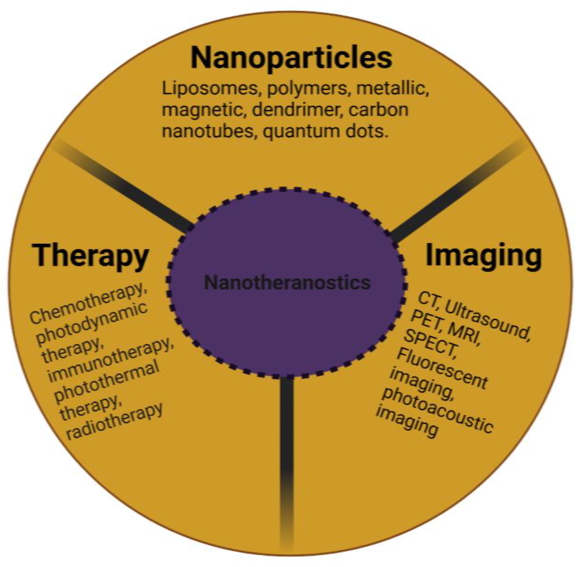
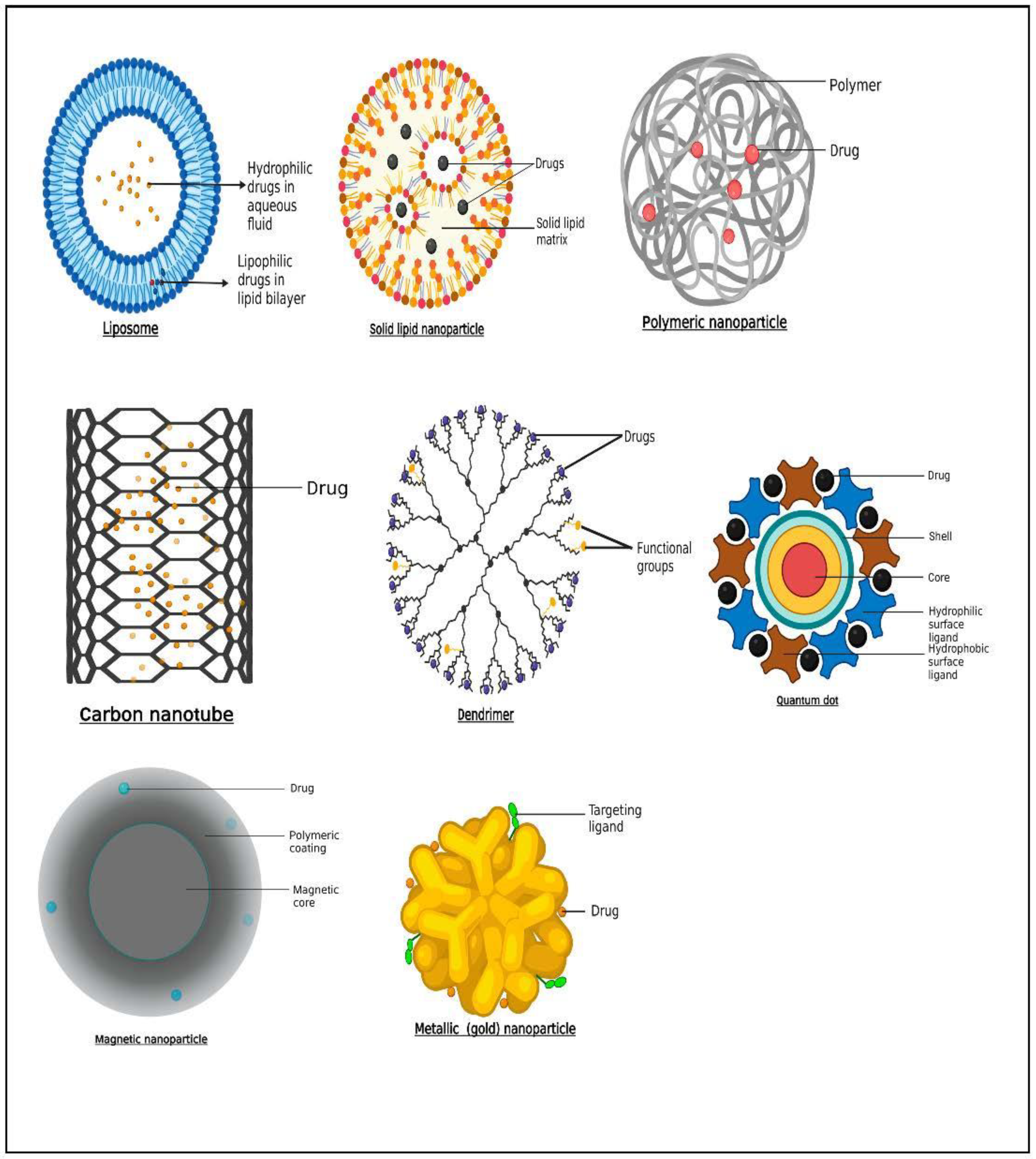
2. Nanotheranostic Platforms

The ideal nanoparticle should have biocompatible, biodegradable, nontoxic and non-immunogenic biological features, as well as the capacity to encapsulate both lipophobic and lipophilic drugs, diagnostic agents, and targeting moieties. These protect them against systemic inactivation and renal clearance in addition to enhancing their tumor targeting and real-time monitoring [31,32]. These nanotheranostic platforms are discussed below.

2.1. Liposomes

Liposomes and lipid nanocapsules (LNCs) are two lipid-based nanodelivery platforms. Liposomes, which are made up of a hydrophilic “core” surrounded by lipid bilayers, can contain hydrophobic or hydrophilic medicines in the lipid bilayer or the aqueous core, respectively [33] (Figure 2). Liposomes are frequently utilized as nanocarriers in drug delivery systems, while their size, biocompatibility, biodegradability, low toxicity, non-immunogenicity, and ability to encapsulate hydrophilic, lipophilic, and amphiphilic medicines give considerable benefits [34]. Liposomes possess the capability to incorporate both therapeutic and diagnostic agents which allow for a revolutionary use of liposomal delivery systems as theranostic platforms [35,36,37]. For instance, a hydrophilic gene probe for imaging hypoxia was loaded inside a PEGylated liposome that also contained a hydrophobic photosensitizer and the basic phospholipids lecithin and cholesterol. The liposomal transport of the probe was identified utilizing fluorescence imaging prior to undergoing therapeutic photodynamic therapy [38,39]. Liposomes and PEGylated liposomes were functionalized with gadolinium(III) diethylenetriamine pentaacetic acid salt, which served as a magnetic resonance imaging (MRI) contrast, and zinc phthalocyanine, which functioned as a model photosensitizer. The results showed how effective liposomal formulations could be as imaging agents [40]. A novel system containing near-infrared (NIR) carbon dots and cinobufagin, an anticancer drug, was developed and investigated as a potential anticancer nanotheranostic. Although cells could take up liposomes and deliver them to the tumor site, bioimaging of the created system was notably high. Furthermore, a significant anticancer activity and a protracted release pattern were observed [41]. As a preliminary measure of therapeutic response, multifunctional RNA-loaded magnetic liposomes were produced. The iron oxide-laden RNA liposomes activate dendritic cells after transferring RNA to them, thus enabling the prediction of tumor regression using MRI, according to Grippin et al. [42].
Currently, several kinds of cancer drugs have been applied to this lipid-based system using a variety of preparation methods. Among them, liposomal formulations of the anthracyclines doxorubicin (Doxil, Myocet) and daunorubicin (DaunoXome) are approved for the treatment of metastatic breast cancer and AIDS-related Kaposi’s sarcoma [44,45]. The research group of Rizzitelli [46,47] developed long-circulating liposomes loading both gadoteridol and the chemotherapeutic drug doxorubicin. After systemic injection, they successfully achieved a local tumor drug release from liposomes by ultrasound. This strategy significantly improved the therapeutic efficacy in a breast tumor model, demonstrated by almost complete tumor regression.
Yari et al. [48] also reported the development of a prostate-specific membrane antigen (PSMA)-tagged liposome for specific targeting of advanced prostate cancer tumoral cells. Their results showed the efficiency of active targeting of prostate cancer cells with PSMA ligand. Studies have demonstrated improved efficacy of liposomal drugs in prostate cancer models. For example, the combined effect of liposomal forms of curcumin and resveratrol more significantly inhibited tumor growth and induced apoptosis in PTEN-CaP8 cell lines, with a concomitant reduction in prostatic adenocarcinoma in vivo in PTEN mice [49]. Additionally, curcumin-loaded liposomes decorated with PSMA antibodies tested in LNCaP and C4-2B efficiently inhibited cellular proliferation at a very low dose (5–10 µM) compared to free curcumin [50]. Recently, epirubicin encapsulated by propylene glycol liposomes (EPI-PG-liposomes) was established as being effective in overcoming MDR in BC [51]. Another recent study also demonstrated that engineered liposomes using arginine8–glycine–aspartic acid (R8GD) encapsulated with daunorubicin and emodin selectively deposit at the tumor site, thereby demonstrating a distinct anti-BC effect [52].
Other examples of liposomes employed as cancer nanotheranostics are presented in Table 1. Magnetic resonance imaging (MRI), positron emission tomography (PET), and single-photon emission computed tomography (SPECT) are diagnostic techniques that can employ liposomes to diagnose cancer [53].

2.2. Solid Lipid Nanoparticles

Particles having a solid lipid matrix and a mean size of 50–500 nm are known as solid lipid nanoparticles (SLNs) (Figure 2). The liquid lipid (oil) in the structure of an oil-in-water emulsion is substituted for a solid lipid, or perhaps a combination of solid lipids, to produce SLNs. According to Lima et al. [54], one crucial characteristic of SLNs is that they remain solid at both room temperature and body temperature. Surface active agents as stabilizers in combination with the lipid and drug are present in the formulation. SLNs have the benefits of preserving labile medications from breakdown, improved physicochemical stability, ease of formulation, increased targeted delivery at tumor site, improved therapeutic outcome, and reduced systemic adverse effects [55]. Furthermore, because of the solid matrix, there is more flexibility in adjusting the release rate, a reduced breakdown rate, and no residual organic solvents during formulation [56]. Due to their composition of physiological lipids, solid lipid nanoparticles provide excellent opportunities to insert pharmaceuticals into nanosized targeted vehicles with high biotolerability and minimal biotoxicity [57]. According to Wissing et al. [58], SLNs are often made up of solid form lipids such as wax, triglycerides with a greater degree of purity, free fatty acids, free fatty alcohols, complex glyceride blends, and other well-known physiologic lipids. SLNs have many advantages, including the ability to include both hydrophilic and lipophilic medications, a nontoxic profile, and high drug stability [57]. Garg et al. [59] demonstrated in an in vivo study that there was accumulation of drug within breast cancer tissue after intravenous administration of methotrexate-loaded SLNs relative to methotrexate alone. Furthermore, significant lifespan prolongation was observed in mice treated with methotrexate-loaded SLNs. In another study by Guney et al. [60], tamoxifen, a hormonal compound, was loaded into SLNs for drug-resistant breast cancer cells. The findings revealed the cytotoxic and aggressive activity of tamoxifen-SLN was higher in the drug-resistant breast cancer cells than the free tamoxifen.
The biodistribution of resveratrol solid lipid nanoparticle (RSV-SLN) was found to be extremely high in prostate cells and accumulate 7.56 times greater than that of RSV solution. The developed RSV-SLN can be applied as potential carrier for delivery of drug of chemotherapeutics with an extended systemic circulation and targeting efficiency at tumor site [61]. Akanda et al. [62] showed that adjusting the process parameters (e.g., pressure/temperature) and using different lipids increased the anticancer activity of SLNs loaded with retinoic acid (RTA). The result further revealed that RTA-SLNs incubated in LNCap cell lines exhibited decreased cell viability and higher drug concentrations (e.g., 9.53% at 200 g/mL), but blank SLNs exhibited no cytotoxicity.

2.3. Polymers

Polymers of natural origin can be remodeled by chemical reactions to produce semisynthetic polymers, e.g., methylcellulose. Several polymers of natural sources that are employed in the synthesis of nanocarriers include chitosan, dextran, albumin, heparin, gelatin, and collagen [63,64]. These polymers possess biodegradable, biocompatible, non-immunogenic, and nontoxic features. Polyethylene glycol (PEG), polyvinylpyrrolidone (PVP), and polystyrene are some examples of synthetic polymers. In terms of drug distribution and therapy, these are comparable to natural polymers [65]. The ability to surface-modify polymer-based drug delivery systems allows for precise localization of therapeutic and/or diagnostic agents, enhancing the sensitivity, specificity, and efficacy of the therapeutic and diagnostic approach. They can also be easily modified to improve the theranostic conjugates’ biocompatibility and solubility [66]. Natural and synthetic macromolecules have both been employed as nanotheranostics. In a documented study for diagnosis and distribution of doxorubicin (DOX) in tumor cells, Zhao et al. [67] employed peptide aptamer-targeted polymers. As potential nanocarriers, these nanopolymers may penetrate biological membranes, preserve treatment payload, and distribute it to target tissue.
Glycol chitosan was coupled with the photosensitizer protoporphyrin IX and PEG to produce a polymeric core–shell nanosystem that can be activated by the plasma membrane. Jia et al. [68] reported that the generated nanoagents boosted tumor-related in vivo fluorescence and accumulated in tumor cells. Zhu et al. [69] developed bioresponsive and bright HA–iodixanol nanogels for application as targeted X-ray computed tomography imaging and chemotherapeutic drugs. To specifically target MCF-7 human breast tumors, the chemotherapeutic drug paclitaxel was added to the nanogels. It was demonstrated that the nanogel had a high rate of cell absorption and suppressed tumor growth. Moreover, MCF-7 breast tumors in nude mice were imaged using enhanced computed tomography (CT), and fluorescence showed that nanogels were disseminated throughout the whole tumor, suggesting deep tumor penetration [69]. In a study by Yu et al. [70], deoxycholic acid–HA–methotrexate was used to transport ICG and doxorubicin. The produced NPs demonstrated intracellular doxorubicin uptake by the CD44/folate receptor. The use of nanotheranostic technology for imaging-guided chemo-photothermal combination therapy, per the authors, is superior to more traditional approaches [70]. In a separate study, ZnS fluorescent quantum dots were altered using carboxymethylcellulose (CMC) to produce a nanocolloidal system, which was subsequently linked with doxorubicin. Nanocolloids with an average size of 3.6 nm were discovered to have both a photoluminescence emission property and to be biocompatible. The resulting system can, therefore, potentially serve as a fluorescent nanoprobe and drug nanocarrier with inhibitory ability [71]. The issues with using polymeric nanoparticles are related to the organic solvents that were used to formulate them, which might be present in the final product as residues and cause hazardous consequences (Figure 2).
The chemotherapy medicines docetaxel (DTX) and quercetin (QU) were combined to create chemically modified polymeric nanocapsules (NCs) that were specifically designed to target prostate cancer (PCa). Luteinizing hormone-releasing hormone (LHRH) ligands were attached to poly(propylene glycol-co-glycolide) (PLGA) utilizing polyethylene glycol (PEG) as a carrier to accomplish active targeting. An increase in cellular inhibitory activity and a considerable rise in cellular absorption of LHRH-targeted NCs were demonstrated by in vitro tests. The outcomes of tumor location and anticancer activity in in vivo tests complemented the in vitro findings, revealing the advantageous effects of NCs containing the combination of DTX and QU in the fight against PCa [72].

2.4. Metallic and Magnetic Nanoparticles

Nanoparticles of inorganic sources such as superparamagnetic iron oxides, gold nanoparticles (Figure 2), and other metallic and non-metallic nanoparticles or nanoclusters, increase radiation efficiency and tumor detection. Inorganic silver oxide (AgO) nanoparticles offer a great deal of promise for application as anticancer agents. Furthermore, they have antibacterial, antioxidant, anti-inflammatory, and anti-angiogenic properties [73]. Organic molecules such as proteins, deoxyribonucleic acid (DNA), and ribonucleic acid (RNA) can be delivered using gold nanoparticles [74]. Ionic or covalent interactions, as well as adhesion, are all easy ways to affix drugs. In order to improve the stability and half-life of metallic nanoparticles, polyethylene glycol (PEG) can be bonded to their surfaces. The physical characteristics of the gold nanoparticles and their potential for theranostic applications are influenced by their varied morphology, which can take the form of spheres, cubes, rods, clusters, or threads, necessitating strict form control [75]. Utilizing radioactive gold nanoparticles is another theranostic strategy (198AuNP and 199AuNP). Beta particles are employed to destroy tumor tissue, whereas gamma rays are used to acquire pictures for scintigraphy or single-photon emission computed tomography/computed tomography (SPECT/CT) [76]. Both radionuclides are beta (β) and gamma (γ) emitters. Target-specific drug delivery was shown by Kim et al. [77] to kill prostate cancer cells more efficiently than untargeted cells when doxorubicin was loaded onto aptamer-conjugated gold nanoparticles. By attaching gadolinium (Gd)(III) complexes and ligands that target PSMA to the surface of gold nanoparticles, Luo et al. [78] created gold nanoparticles for MR-guided prostate cancer targeted therapy. Higher binding affinity was produced as a result of the surface alteration, which quadrupled the rate of r1 relaxation. The outcomes demonstrated improved uptake of gold nanoparticles by prostate cancer cells expressing PSMA, good MRI contrast in vitro and in vivo, and improved suppression of prostate cancer following radiotherapy by binding of gold and Gd(III). The good selectivity of Au/Gd(III)–PSMA NPs for PSMA-expressing prostate cancer cells with improved cellular MR contrast and in vitro radiosensitization was validated [79,80]. PSMA-targeted gold nanoparticles’ ability to selectively target tumors enabled precise radiation therapy with a low irradiation dosage and little harm to healthy tissues [78].
Magnetic nanoparticles are thought to be safe because, in the serum iron pool, they are swiftly degraded and transformed into hemoglobin or other metabolic products [81,82]. Inorganic nanoparticles (IONPs), which are paramagnetic, can be used as imaging agents to identify and track pathological conditions, as well as release drugs [83]. This is accomplished by applying an external magnetic field to the target tissue. In tissues containing nanoparticles, selective cell death occurs, which lessens the detrimental effects. Superparamagnetic iron oxide nanoparticles (SPIONs), perfluorohexane, and paclitaxel were added to a carboxyl-modified PEGylated poly skeleton, on which the herceptin antibody was changed to show an HER—specific surface [84]. The perfluorohexane was vaporized, and paclitaxel was subsequently released after these SPIONs were activated by an NIR laser, which enabled the translation of laser energy into thermal energy. Consequently, this nanotheranostic incorporates a combination of chemotherapy and photothermal therapy for the treatment of breast cancer [84].

2.5. Carbon Nanotubes

Carbon nanomaterials are mostly made of carbon and come in the shape of hollow spheres, ellipsoids, or tubes with nanometer-sized diameters. Carbon nanotubes (CNTs) are remarkable in that the atoms’ bonds are extremely tough, allowing the tubes to exhibit extraordinary aspect ratios (Figure 2). CNTs come in a variety of shapes and sizes, but they are usually classified as single-walled (SWNTs) or multi-walled nanotubes (MWNTs). SWNTs are similar to a conventional straw in appearance. There is one layer, or wall, to it. Carbon nanotubes with multiple walls are made up of layered tubes with progressively smaller diameters. They can consist of as little as one exterior and one inner tube (a double-walled nanotube) to as many as 100 tubes (walls). Interatomic forces keep each tube apart from its neighbors. CNTs are fascinating because of their mechanical durability and electrical characteristics [85]. CNTs were also shown to be appropriate for theranostic utilization due to their vast surface area, capacity to enclose therapeutic/imaging agents, and adaptability to surface changes [86,87,88]. Chemotherapeutic agents such as doxorubicin and paclitaxel, as well as nucleic acids such as antisense oligonucleotides and siRNAs, have recently been included in nanotubes by several experts [89]. Moreover, many studies are looking at the possibility of using nanotubes as diagnostic agents. In order to attach paclitaxel (PTX) to the water-soluble carbon nanotubes, a branching polyethylene glycol chain was created by chemically altering single-walled carbon nanotubes with an ester bond. This substance demonstrated little toxicity and 10 times more tumor absorption than standard Taxol® in the mouse 4T1 breast triple-negative breast cancer (TNBC) model. The PEGylation was likely responsible for the enhanced circulation since it improved penetration and retention [90] and resulted in greater tumor growth suppression. Another fascinating application of these carbon nanotubes is based on photothermal-induced ablation, in which the nanotubes promote cell membrane permeabilization and necrosis, removing both the tumor mass and the breast cancer (BC) stem cells. This raises the possibility of this being a successful remedial option for tumor resistance and preventing recurrence [91].
A theranostic MWCNT was created by Das et al. [92] by combining acid-oxidized MWCNTs with four different functional moieties: a fluorochrome (Alexa-fluor, AF488/647), a targeting agent (folic acid), a radionuclide (Technitium-99m), and methotrexate. According to the cellular uptake investigations, human cancer cells (A549) and MCF-7 cells that express the folate receptor endocytose selectively internalize theranostic MWCNTs. In order to load the anticancer agent PTX, human serum albumin (HSA) nanoparticles were conjugated to create a single-walled carbon nanotube (SWNT)-based drug delivery system [93]. According to Shao et al. [93], the PTX formulated with SWNT-HSA inhibited proliferation in MCF-7 breast cancer cells more effectively than PTX made with HSA nanoparticle alone (cell viability of 63% versus 70% in 48 h and 53% versus 62% in 72 h).

2.6. Dendrimers

Dendrimers are monodispersed organic compounds that can create various polydisperse molecules (Figure 2). Low cytotoxicity, high solubility, chemical stability, and selective accumulation in tumor cells characterize the drug dendrimer conjugate [94]. Dendrimers’ cytotoxicity is the main cause for worry, although it depends on the type of polymer they are made of. It is frequently necessary to make specific structural changes, or new biocompatible polymers must be used to provide more physiologically acceptable alternatives [95]. The external active functional groups of dendrimers allow for the conjugation of biomolecules or contrast compounds to the surface while simultaneously loading medicines within. Dendrimers have been used to transport a variety of cargo, but nucleic acids and small molecules are the two types of cargo that have received the most attention [96,97]. For these reasons, charged polymers such as poly(ethylenimine) (PEI) and poly(amidoamine) (PAMAM) are widely utilized. Clinical trials are currently being conducted for a variety of dendrimer-based products, including contrast agents, topical gels, transfection agents, and theranostic agents [97,98,99]. Sweet lemon peel-derived carbon quantum dots and PAMAM dendrimers were used to treat triple-negative breast cancer. The system also contained the arginine–glycine–aspartic acid (RGD) peptide, targeting integrin, which was overexpressed in the specific cancer. This technology can successfully bind to cancer cells, according to research by Gosh et al. [100], making it a distinct theranostic option.
For the simultaneous delivery of cisplatin and doxorubicin, Guo et al. [101] produced a novel type of PAMAM dendrimer nanoparticle modified by HA (HA@PAMAM-Pt-Dox). HA@PAMAM-Pt-Dox demonstrated high potency in increasing the chemotherapeutic efficacy of cisplatin and DOX, according to the study’s findings. In both the androgen-sensitive prostate cancer cell line LNCaP and the androgen-independent prostate cancer cell line DU145, curcumin has been shown to have the ability to inhibit in vitro prostate cancer cell proliferation [102,103,104], as well as in vivo tumor growth in an LNCaP xenograft mouse model [105]. Using PAMAM dendrimers, Chittasupho et al. [106] created CXCR4-targeted dendrimers that were combined with the anticancer drug doxorubicin. The dendrimers were taken up by the T47D and BT-549-Luc breast carcinoma cells in a concentration- and time-dependent manner. In comparison to free doxorubicin and unloaded dendrimers, the polymeric dendrimer loaded with doxorubicin had a stronger cytotoxicity. Doxorubicin-loaded PAMAM dendrimers that conjugate with pluronic F68 incorporation were created by Wang et al. [107]. The in vivo and in vitro anticancer investigations showed that caveolae-mediated endocytosis boosted the antitumor activity of doxorubicin pluronic F68-PAMAM dendrimers against MCF-7/ADR cancer cells. By controlling gene expression and mitochondrial activity, they substantially enhanced apoptosis [107].
Dendrimers are used to deliver medications to patients with a variety of illnesses, including breast and prostate cancers, in a regulated and focused manner [94]. They are improvable, which means they can have their surfaces tweaked using targeted ligands including carrying both the therapeutic and the diagnostic agents [108,109].

2.7. Quantum Dots

Quantum dots (QDs) are light-emitting nanocrystals with a diameter of 2–10 nm manufactured from semiconductor substances such as selenides or cadmium or zinc sulfides. Depending on their size and makeup, they have distinct optical and electrical characteristics. QDs are safe, physiologically inert, and low-toxicity compounds employed as nanosensors and nanocarriers [110]. QDs are more durable than fluorescent proteins at varying temperatures and pH levels, making them ideal for long-term fluorescence tracking and image-guided treatment, and as potential substitutes for molecular fluorophores such as fluorescein and rhodamine [111]. QDs are attractive semiconductor nanoparticles for use in cancer nanotheranostics because of their unique capacity to see the tumor site (Figure 2). Functionalizing QD with targeting molecules, such as aptamers, may increase their affinity for certain body regions and enable targeted drug delivery [112]. Huang et al. [113] created a nanotheranostic platform by coating QDs with a polymer containing paclitaxel (a standard anticancer agent) and lipoic acid. Between the polymer and the drug, an ester bond was formed. This approach has been shown to be effective in the detection and treatment of cancer cell lines in vitro. AbdElhamid et al. [114] created highly fluorescent QDs that were layer by layer combined with gelatin/chondroitin and shown anti-breast cancer effectiveness, as well as non-immunogenicity. In this investigation, imaging and medication distribution were performed at the same time, typifying the nanotheranostic technique.
For intravenous castrate-resistant prostate cancer (CRPC) therapy, Jiang et al. [115] created a multifunctional enzalutamide-loaded graphene oxide nanosystem. Enzalutamide was then added to the 200 nm graphene quantum dot derivatives that were originally created via the disulfide cross-linking of graphene quantum dots. Tumor-targeting peptides and PEG were then used to further functionalize the created graphene quantum nano-drug system. This nano-drug carrier had a potent prostate cancer-targeting capacity which could be quickly internalized by CRPC cells through endocytosis, decrease the proliferation of prostate cancer cells, and lessen enzalutamine’s in vivo side-effects. The high photostability and brightness of QDs have led to their application in nanodevices for the detection of breast and prostate cancer. QD-conjugated protein microarrays for PSA detection with less nonspecific binding were created by Gokarna et al. [116]. Carbon quantum dots (CQDs) that were nitrogen-doped were created via a hydrothermal process by Samimi et al. [117]. In order to target breast cancer cells, the resultant nanoparticles were coupled with quinic acid. Electrostatic interactions allowed gemcitabine to be loaded onto the resultant nanoparticles. Through the use of the MCF-7 cell line, cell viability was assessed. Quinic acid-conjugated N-CQDs demonstrated intriguing properties such as outstanding luminous characteristics and high tumor accumulation when taken as a whole, highlighting them as great candidates for multifunctional theranostic agents [117].

3. Mechanisms of Targeting and Targeting Moieties/Ligands of Nanoparticles

Several barriers stand in the way of effective drug biodistribution to tumor site and positive treatment outcomes, including reduced bioaccessibility, which results in reduced chemotherapeutic agent concentration at target tissue [118]. Nanocarriers can be designed to employ passive and active targeting mode of actions to penetrate tumor tissue [119].

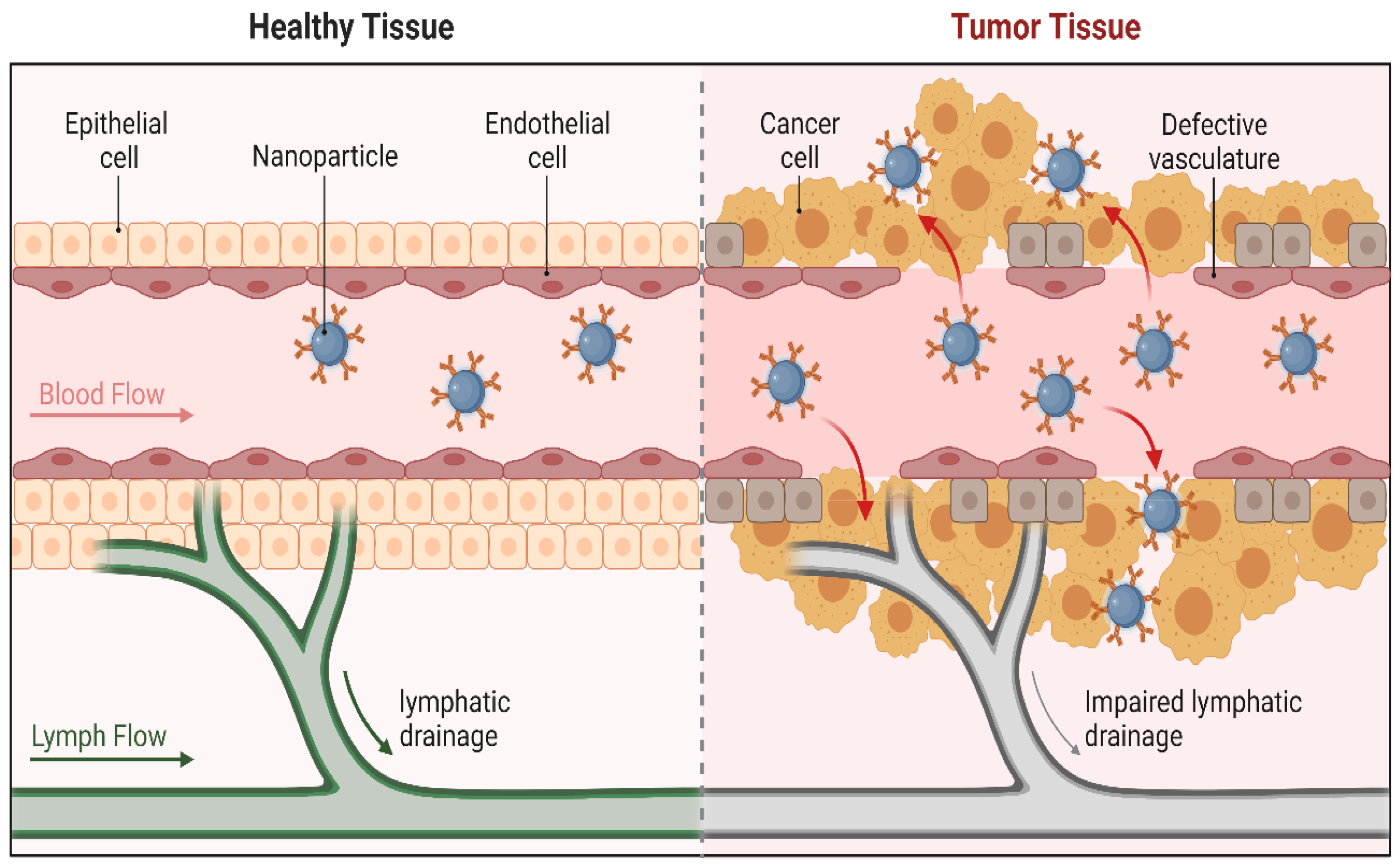
3.1. Passive Targeting

Nanoparticles can passively accumulate in porous vasculatures in tumors thanks to the enhanced permeability and retention (EPR) effect [120,121]. Tight junctions are 7–10 nm spaces between endothelial cells that can be seen in the blood vessels of healthy tissues. These spaces can reach hundreds of nanometers in tumor blood vessels, enabling nanosystems to selectively extravasate in tumor tissue [122,123]. In addition, macromolecule recovery via the vascular and lymphatic systems is impeded in tumor tissue, thereby leading to macromolecule retention inside tumor tissue [124]. As a result, passive targeting is a very efficient technique of targeted drug delivery, and nano-drug delivery systems can benefit from this occurrence (Figure 3). On the other hand, passive targeting cannot totally rule out the prospect of nanocarriers collecting in organs with fenestrated blood vessels, such as the liver or spleen [125]. Moreover, the microenvironments of various malignancies vary, which might be an impediment to the creation of nanosystems.
According to clinical therapy, there are various drawbacks to passive targeting techniques. As a result of some drugs’ inability to diffuse effectively, it is not always possible to target specific cells within tumors, and the random nature of the approach makes it challenging to control the process. As a result of this lack of control, multidrug resistance (MDR), which occurs when chemotherapy treatments fail patients and their cancer cells become resistant to one or more drugs, may develop. On the surface of cancer cells, drug-expelling transporter proteins are overexpressed, which leads to MDR [127,128,129]. The therapeutic impact of medications is necessarily reduced by drug expulsion, and cancer cells quickly develop treatment resistance. The passive targeting is further constrained by the fact that some tumors do not display the EPR effect, and that vascular permeability may vary within a single tumor [130]. To get over these restrictions, the nanocarriers can be modified so that, following extravasation, they actively bind to cells. This can be accomplished by attaching targeting agents, such as ligands, to the surface of the nanocarrier using a variety of conjugation techniques. Ligands are molecules that can bind to receptors on the cell surface. Through interactions between ligands and receptors, nanocarriers will recognize the receptor and connect to target cells, releasing the drug inside the cell. This can be tailored for some specific group of patients for successful clinical trials.

3.2. Active Targeting

In active targeting, ligands are attached to the surface of nanocarriers (Figure 4). These ligands are extremely selective for receptors and other cancer-specific targets such as glycans, which are abundant on tumor cell surfaces [131]. Nonspecific nanocarrier uptake in tissues apart from tumor tissue might be prevented by conjugating these ligands. Ligands include transferrin, folic acid, enzymes, modified antibodies, and macromolecules such as proteins and polysaccharides [120,125]. Another notable example is the creation of therapeutic monoclonal antibodies that target highly expressed membrane receptors on malignant cells. This was established when Herceptin (trastuzumab) was used to target the overexpressed HER2 receptor in a subtype of breast cancer [132]. Furthermore, the creation of antibodies or ligands that target the prostate-specific membrane antigen (PSMA) [133,134] are well-developed and used active drug targeting techniques.
The density of these ligands must be adjusted to minimize reticuloendothelial system (RES) recognition and contact with serum proteins, permitting nanocarriers to distribute for extended periods of time [125]. A receptor density of 105 copies of ErbB2 receptors per cell was required in a breast cancer model to increase the therapeutic effectiveness of an anti-ErbB2-targeted liposomal doxorubicin over its non-targeted counterpart [136]. The transferrin receptor, folate receptor, glycoproteins, and the epidermal growth factor receptor are among the best researched of these ligands, which bind to receptors on targeted cells. Targeting transferrin, a kind of serum glycoprotein that transports iron into cancer cells, is one instance. Transferrin receptors are overexpressed in most solid tumor cells, although they are expressed in modest quantities in normal cells. Therefore, transferrin-conjugated nanoparticles are being employed to deliver cancer drugs as an active targeting approach [137,138,139]. Targets for anticancer drugs should ideally be both vital for tumor cell existence and specific to tumor cells [140,141]. On the other hand, targets that match these requirements are few and challenging to identify and should serve as a research focus.

3.3. Transferrin

The monomeric glycoprotein, transferrin, has a molecular weight of 78 kDa and 679 amino acids. It is essential for iron delivery and metabolism, contributing to the body’s important iron pool. Transferrin receptors, which have a high affinity for transferrin, are often upregulated on the outside of various cancers in order to maintain the growing demands for iron for sustaining cell division and growth. As a result, researchers are developing new anticancer delivery systems that actively target the transferrin receptor. Transferrin receptors can be used to target and internalize therapeutic compounds via transferrin or to disrupt normal receptor activity, which causes cell death [142,143,144]. Jose et al. [145] showed that the transferrin-conjugated PLGA nanoparticles (NPs) loaded with docetaxel had extremely intriguing anticancer potential due to their capacity to stop cancer proliferation at the G2/M phase of mitosis. Cui et al. [146] additionally created PTX-conjugated magnetic PLGA NPs (PTX-MNP-PLGA NPs). In comparison to unchanged NPs or liberated PTX, they showed that transferrin-conjugated PTX-MNP-PLGA NPs possessed stronger antiproliferation and better absorption in U-87 cells [146]. Transferrin-conjugated NPs, such as polymeric NPs or liposomes, appear to have a great potential for targeting gliomas, according to several earlier studies [147,148]. In a fascinating study published recently, Qi et al. [144] created a novel transferrin protein corona (Tpc)-modified CuGd nanoplatform (Tpc-CuGd) for tumor-targeting photothermal and chemodynamic synergistic therapy [144].
Following coupling with transferrin, Sahoo et al. [149] created PLGA polymeric nanoparticles encapsulating paclitaxel (PTX). The results showed that the IC50 of the drug with Tf-conjugated nanoparticles (PTX-NPs-Tf) was around five times lower than that of the drug in solution or with unconjugated nanoparticles (PTX-NPs). PTX-NPs-Tf was administered intratumorally in a single dose (24 mg/kg) and showed complete tumor regression and a higher survival rate than either PTX-NPs or PTX-Cremophor® El formulation. Using poloxamer 407 (F127) and 123 (P123) as the building blocks, Soe et al. [150] successfully loaded DOX within transferrin (Tf)-conjugated polymeric nanoparticles (DOX/F127 and P123-Tf). By inducing cellular apoptosis, the DOX/F127 and P123-Tf compound increased DOX cytotoxicity in the OVCAR-3, MDA-MB-123, and MDA-MB-231(R) cell lines. An in vivo investigation using mice carrying MDA-MB-231 (R) tumors showed that coating nanoparticles with a targeting moiety improved their transport to the tumor site. Bioconjugated Tf-SLNs loaded with CRC were shown by Akanda et al. [151] to significantly reduce tumor growth in animals carrying prostate cancer. The therapeutic effectiveness of Tf-CRC-SLN had a significant impact on the mice that received CRC-SLN and CRC ethanol solutions, as it significantly reduced tumor development by 79% and increased survival rates.

3.4. Hyaluronic Acid

Hyaluronic acid (HA) is a naturally occurring polysaccharide made up of repeating D-glucuronic acid and N-acetyl-D-glucosamine disaccharide units joined by (1-β-4) and (1-β-3) glucosidic bonds [152,153]. According to Jiao et al. [154], hyaluronic acid is broadly distributed in the extracellular and synovial matrix and possesses biodegradability, high moisture retention, and tunable viscoelastic characteristics. Hyaluronic acid has been thoroughly investigated in the cosmetic and pharmaceutical industries due to its wide range of biological effects, including anti-inflammation, wound healing, antiaging, tissue regeneration, and skin-repairing characteristics [155,156]. Hyaluronic acid is a ligand for the cluster-determinant 44 receptor (CD44) [153,157]. In healthy tissues, CD44 is essential for cytokine release, lymphocyte activation, and control of hyaluronic metabolism [158]. CD44 is regarded as an early signal for cancer cell proliferation because of its tumorigenic properties and level of expression frequently related to tumor-initiating cells or cancer stem cells [159,160]. This makes CD44 a reliable biomarker for the detection of cancer, especially for the diagnosis of breast, colorectal, head and neck, pancreatic, bowel, and prostate cancers [160]. The tremendous potential of CD44 as a promising target for increasing tumor-selective medication delivery is indicated by both its overexpression in a variety of cancer cells and its affinity for hyaluronic acid. Due to its many benefits, hyaluronic acid is frequently used in nano-drug delivery systems. For instance, Seok et al. [161] found that coating the surface of curcumin-loaded zein nanoparticles with hyaluronic acid boosted the anticancer activity and improved the distribution of curcumin in CD44-overexpressed CT26 tumor cells. By combining paclitaxel (PTX)–hyaluronic acid prodrug with marimastat (MATT)-loaded thermal-sensitive liposomes, Lv et al. [162] created HA-PTX/MATT-LTSL HNPs. According to Lv et al. [163], these hybrid nanoparticles greatly inhibited the proliferation, metastasis, and angiogenesis of 4T1 tumor cells. Additionally, the hyaluronic acid–irinotecan phase 1 clinical trial showed that this combination was safe and tolerable without impairing the anticancer efficacy of irinotecan [163].
Doxorubicin (DOX) and miR-34a (a strong endogenous tumor suppressive molecule in breast cancer) were co-encapsulated into hyaluronic acid (HA)–chitosan (CS) nanoparticles (NPs) and simultaneously administered into breast cancer cells [164]. The expression of the non-pump-resistant and anti-apoptosis proto-oncogene Bcl-2 was shown to be suppressed in in vitro and in vivo tests, which improved the antitumor effects of DOX. Furthermore, miR-34a was restored intracellularly, which reduced the ability of breast cancer cells to migrate by concentrating on Notch-1 signaling. Through electrostatic self-assembly and subsequent spontaneous chemical cross-linking, Xia et al., [165] created thiol-hyaluronic acid (HA-SH)/chitosan (CS) nanoparticles with redox/pH dual responsiveness. Human breast cancer cells (SKBR3) ingested DOX-loaded HA-SH/CS particles, which then released the medication after binding to the cells and possibly exerting anticancer activity. Vogus et al. [166] examined the impact of timing the administration of the drugs doxorubicin (DOX) and gemcitabine (GEM) on drug synergy in a different study. To regulate the relative release kinetics of each medication, hyaluronic acid (HA) drug conjugates with unique linkers were created. The results demonstrated that triple-negative breast cancer cells are more efficiently killed in vitro by polymer conjugates that release GEM more quickly than DOX. Additionally, the optimal dual drug combination outperformed free DOX and GEM, as well as the single-drug HA conjugates, in their ability to limit the growth of an aggressive, orthotopic 4T1 tumor model in vivo.

3.5. Folic Acid

A low-molecular-weight vitamin (B9) called folic acid (FA) has a pterine moiety and a glutamate entity; these two components are connected by p-aminobenzoic acid. All eukaryotic cells require folic acid for the production of purines and pyrimidines, as well as 1-carbon metabolism [167]. Since mammals are unable to produce folate on their own, it must only be obtained through diet [168]. Folate receptors (FRs), a group of glycoproteins, are divided into the FRα, FRβ, and FRγ subtypes. According to several studies, FRα is the most frequently produced folate receptor subtype and is overexpressed in a variety of tumor cells, including those from breast, lung, kidney, cervical, and ovarian cancers [169,170,171]. Because folate receptors can transport folate into tumor cells via the receptor-mediated endocytosis process [172], several folic acid-based nanoplatforms have been created for increased internalization of therapeutic agents by tumor cells [173,174,175]. For folic acid-mediated targeted therapy, Sathiyaseelan et al. [176] developed a folic acid-conjugated chitosan-loaded rutin-prepared palladium nanoplatform. The endocytosis efficiency of the nanoparticles in breast cancer cells was dramatically increased by the addition of folic acid to the developed nanoplatform. A cell viability study demonstrated the proposed nanoplatform’s significant suppression of cell proliferation [176]. A water-soluble folic acid derivative called EC145 (vintafolide) has started phase 2 clinical trials for treating various tumors [172,177]. A variety of drug carriers, including liposomes, micelles, polymers, dendrimers, and inorganic nanomaterials, have been conjugated to folic acid in addition to direct conjugation to drug molecules. These drug carriers have then been tested for their potential as a tumor-selective drug delivery system by using different anticancer medications, including doxorubicin, paclitaxel, docetaxel, 5-fluorouracil, erlotinib, and curcumin. A variety of drug carriers, including liposomes, micelles, polymers, dendrimers, and inorganic nanomaterials, have been conjugated to folic acid in addition to direct conjugation to drug molecules. These drug carriers have then been tested for their potential as a tumor-selective drug delivery system by using different anticancer medications, including doxorubicin, paclitaxel, docetaxel, 5-fluorouracil, erlotinib, curcumin, and resveratrol [178,179].
Dhas et al. [180] conducted a study investigating the potential of folic acid (FA)-conjugated chitosan (CS)-functionalized poly (d,l-lactide-co-glycolide) (PLGA) nanocarriers for the treatment of prostate cancer. The nanoparticles (NPs) were coated with a folic acid–chitosan conjugate. Additionally, nanoparticles loaded with bicalutamide (BCL) were created. According to a cytotoxicity assay, the optimized coated batch’s IC50 value was under 80 µg/mL as opposed to over 80 µg/mL for the BCL solution. Patil et al. [181] investigated folate-targeted liposomes (FTL) as drug delivery systems for PSMA-positive cancer cells. The LNCaP prostate cancer cell line, which expresses PSMA but lacks the folate receptor, was utilized by the scientists together with FTL with co-entrapped mitomycin C lipophilic prodrug (MLP) and doxorubicin (DOX). When LNCaP cells were treated with FTL as opposed to non-targeted liposomes (NTL), the results showed a significant rise in cell drug levels. MLP was converted to mitomycin C, and intracellular and nuclear fluorescence of DOX were identified, confirming FTL processing and drug bioavailability. A particular PSMA inhibitor called PMPA (2-(phosphonomethyl)-pentanedioic acid) prevented the absorption of FTL into LNCaP cells, but not into KB cells with functional folate receptors or PSMA deficiency. When compared to NTL, drug-loaded FTL was found to have much greater cytotoxic action against LNCaP cells. Recent research by Essa et al. [182] produced a quercetin-loaded PLGA nanoparticle with chitosan coating and folic acid as a ligand. The study examined selective toxicity and improved absorption in LnCap prostate cancer cells. In comparison to the non-targeted nanosystem on LnCap cells, the results showed that the targeted nanosystem offered sustained, pH-dependent quercetin release, as well as increased cytotoxicity and cellular uptake.
Folic acid (FA) was coupled with a PLGA polymer and then used to create amodiaquine-loaded nanoparticles (FA-AQ NPs) in research by Parvathaneni et al. [183]. The conjugation of FA with PLGA was validated by the findings of the conjugation efficiency. The findings regarding cellular uptake indicated that FA modification might improve the cellular internalization of nanoparticulate systems in breast cancer cells and other cancer cell types. Additionally, cytotoxicity experiments on cancer cells including MDAMB-231 and HeLA demonstrated the greater efficacy of FA-AQ NPs. Faghfuri et al. [184] undertook a study to improve curcumin solubility and bioavailability by loading it on folic acid–selenium nanoparticles and evaluating their impact on STAT3 activation in breast cancer cell line. The results indicated a significant reduction of 49.62% ± 1.85% in the viability and 79% in the phospho-STAT3 (p-STAT3) level in the cell line tested.

3.6. Peptides

Peptides are extremely stable, with small molecules, great selectivity, and minimal immunogenicity [185]. They can be made to target particular cell receptors, preventing undesirable side-effects, and they can facilitate the selective intracellular delivery of a wide range of therapeutic medicines. They also have benefits over other targeting agents (such as antibodies). These benefits include their smaller size, greater stability, and easier-to-manufacture and easier-to-customize nature. Although numerous naturally occurring proteins and peptides are effective receptor ligands, their direct applications for targeted distribution are constrained by a number of factors, namely, poor biocompatibility, poor selectivity, high toxicity, and enormous size [186]. In particular, the phage display method has led to the continuous discovery of new peptide-based targeting medicines. However, there is still room for improvement in terms of the accuracy and efficacy of these delivery methods [187]. To find biomimetic peptide ligands that can compensate for the drawbacks of natural peptides and offer benefits such as high stability, improved specificity and affinity, and absence of toxicity, structure-based peptide optimization can be used [186]. For instance, the synthetic somatostatin analogue octreotide (SMS 201-995) has been used to transport liposomes, micelles, radiotherapeutic agents, and chemotherapeutic drugs to specific areas [186]. As a cancer cell-targeting peptide, Peptide 1 (or GE11) is a dodecapeptide that binds exclusively to the epidermal growth factor receptor (EGFR or ErbB1) overexpressed in a number of cancers of epithelial origin, including breast cancer [188].
Zahmatkeshan et al. [189] created anti-HER2 (ErbB2) peptide–liposomal formulations of the chemotherapy drug doxorubicin (DOX) using an engineered version of the breast tumor-targeting peptide ligand AHNP, anti-HER2/neu peptide (FCDGFYACYADV), which had three glycine amino acids added as a spacer before its initial sequencing. According to the results, SKBR3 and TUBO cells that overexpress HER2 exhibit increased cytotoxicity and cell uptake when the ligand density of AHNP is increased, but not in MDA-MB-231 cells with low HER2 expression profiles. Targeted liposomal DOX with higher AHNP density also had better antitumor efficacy. According to research by Bandekar et al. [190], anti-HER2 pH-tunable vesicles released doxorubicin in a pH-dependent manner and displayed a 233% rise in binding to HER2-overexpressing BT474 breast cancer cells with a pH decrease from 7.4 to 6.5, followed by significant (50%) internalization in vitro. When compared to non-targeted vesicles, targeted pH adjustable vesicles reduce tumor sizes by 159%, and they also display superior tumor control by 11% when compared to targeted vesicles without an unmasking property. Liu et al. [191] created RGD-modified long circulating liposomes (LCL) loading matrine (RGD-M-LCL) to enhance the tumor-targeting and effectiveness of matrine on the proliferation of Bcap-37, HT-29, and A375 cells. RGD-M-LCL demonstrated much greater in vitro activity when compared to free matrine, which resulted in actions against cancer cells that were both anti-proliferative and pro-apoptotic. A liposome system targeting tumor angiogenesis and tumor cells was created by Dai and co-authors [192] in order to demonstrate the concept. In the work, doxorubicin (DOX) served as the model medication, and N-acetyl-proline–histidine–serine–cysteine–asparagine–lysine (amide)-COOH (PHSCNK), a derivative of PHSCN, was first attached to the surface of PEGylated DOX liposomes (PL-DOX) via a unique method to produce the PHSCNK-modified and DOX-loaded PEGylated liposomes (PHSCNK-PL-DOX). It was discovered that, due to integrin-mediated endocytosis, PHSCNK-PL-DOX dramatically increased the cell absorption and cytotoxicity of DOX on breast cancer cell lines. Cantharidin was encapsulated into PEGylated liposomes by Chang et al. [193] who investigated the systemic toxicity of cantharidin in mice and its activity against human breast cancer MCF-7 cells in vitro. Cells were subjected to hyperbaric oxygen after the liposomal cantharidin was tagged with octreotide. Comparing PEGylated liposomal cantharidin to free cantharidin in vitro, the latter’s cytotoxic action was significantly diminished. Somatostatin receptors were precisely targeted by octreotide-labeled PEGylated liposomal cantharidin to cause cell death in MCF-7 cells. A low dose of cantharidin was added to hyperbaric oxygen to accelerate cell death. In vivo, liposomal cantharidin was substantially less hazardous to the body than free cantharidin, and it was highly effective in inhibiting tumor growth in nude mice [193].
Yeh et al. [194] created peptide–PEGylated lipids and post-inserted them into pre-formed liposomal doxorubicin and vinorelbine while conducting in vitro biopanning of the PC3 human prostate carcinoma cell line to choose prostate cancer-specific peptides using phage display. Cellular uptake and MTT assay results showed that peptide-conjugated liposomes had improved medication cytotoxicity and intracellular delivery. More precisely delivering these imaging agents to tumor areas was achieved by coupling a targeting peptide to imaging substances such as quantum dots (QDs) and superparamagnetic iron oxide nanoparticles (SPIONs). Additionally, it was discovered that administering liposomal doxorubicin and vinorelbine conjugated with targeting peptides significantly increased the suppression of human prostate tumor growth in mouse xenograft and orthotopic models. These results demonstrated that the targeting peptide SP204 has tremendous potential for molecular imaging and tailored therapy in prostate cancer. Recently, Nica and co-authors [195] created and improved unique shaped anisotropic core-shell–shell nanoparticles (also known as trimagnetic nanoparticles, or TMNPs), which were decorated on the surface of PCa cell membranes (CMs) and/or included LN1 cell-penetrating peptides (CPPs). Their results showed that the biomimetic dual CM–CPP targeting and responsiveness to alternating magnetic fields (AMFs) substantially trigger PCa cells to undergo caspase 9-mediated cell death. In addition, TMNP-assisted magnetic hyperthermia resulted in a downregulation of the cell-cycle progression markers and a reduction in the migration rate in surviving cells, which suggests a decrease in the aggressiveness of cancer cells [195].

3.7. Anisamide

Anisamide is a low-molecular-weight benzamide derivative that can be used as a tumor-targeting moiety in functionalized drug delivery systems because of its purported interaction with sigma receptors [196]. The transmembrane proteins sigma receptors (types 1 and 2) control ion channels [197]. According to Van Waarde et al. [197], sigma receptors are overexpressed in human cancers such as breast and prostate cancer. Due to anisamide’s strong affinity for sigma receptors, surface modification of drug carriers with anisamide has garnered significant interest for active tumor targeting. Anisamide-functionalized gold nanoparticles were created by Luan et al. [198] to target prostate tumors, and they successfully complexed with siRNA by electrostatic interaction. The created anisamide-targeted gold nanoparticles complexed with siRNA bonded to human prostate cancer PC-3 cells specifically, causing siRNA to efficiently exit endosomes [198]. In addition, the anisamide-targeted gold nanoparticles prolonged the systemic exposure to siRNA, which resulted in a considerable inhibition of tumor growth in a mouse xenograft model without causing an increase in toxicity [198]. According to a recent study by Yao et al. [199], anisamide-modified dual-responsive liposomes with MRI capabilities can be an effective tool for cancer-targeted therapy. Further study is required to elucidate the utility of anisamide as a ligand for tumor targeting. However, the antitumor efficacy of the anisamide-decorated drug delivery systems was shown to vary significantly in previous studies, and the interaction of anisamide with the sigma receptors is not yet well understood [199].
Anisamide (Anis) ligand, which can interact with the overexpressed sigma receptor on the prostate cancer cells, was coupled to the surface of SLNs by Jalilian et al. [200], in order to create targeted solid lipid nanoparticles (SLNs) containing docetaxel (DTX) for prostate cancer treatment. The results showed that the respective IC50 values for free drug, DTX-SLN, and DTX-SLN-Anis were 0.25 ± 0.01, 0.23 ± 0.02, and 0.12 ± 0.01 nM on the PC3 cell line and 20.9 ± 3.89, 18.74 ± 7.43, and 14.68 ± 5.70 nM on HEK293 cell line. Compared to DTX-SLN and free medication, targeted DTX-SLN-Anis had a greater effect on prostate cancer cells. A stable gold (Au) nanosphere covered in poly(ethylenimine) (PEI) was created by Fitzgerald et al. [201]. An anisamide-targeted nanoparticle (Au-PEI-AA) was produced by further conjugating the PEI with the targeting ligand anisamide (AA). By attaching to the sigma receptor, Au-PEI-AA facilitated siRNA absorption into PC3 prostate cancer cells. Additionally, when cells were transfected in serum-free medium, the Au-PEI-AAsiRNA complexes produced a highly effective suppression (70%) of the RelA gene. In contrast, no knockdown was seen when serum was present, indicating that serum proteins adsorb and prevent the anisamide moiety from attaching to the sigma receptor. A hepta-guanidino-cyclodextrin (G-CD), its hepta-PEG conjugate (G-CD-PEG), and the equivalent anisamide-terminated PEG conjugation (G-CD-PEG-AA) were created, and their effectiveness as siRNA delivery vectors to prostate cancer cells and tumors in vivo was evaluated [202]. In contrast to the non-targeted formulations, the G-CD-PEG-AA-siRNA formulations (in which anisamide targets the sigma receptor) caused prostate cell-specific internalization of siRNA, leading to an approximately 80% suppression of the reporter gene, luciferase, in vitro. In a mouse prostate tumor model, intravenous injection of the anisamide-targeted formulation led to considerable tumor inactivation and reductions in the amount of vascular endothelial growth factor (VEGF) mRNA without exhibiting increased toxicity.

3.8. Aptamers

Single-stranded DNA or RNA oligonucleotides known as aptamers have the ability to attach to a variety of target compounds, such as medicines, proteins, and receptors. They are created in vitro using a process called systematic evolution of ligands by exponential enrichment (SELEX). They have certain distinctive qualities, such as small size, biodegradability, low immunogenicity, a quick and easy synthetic procedure, low cost of manufacture, high specificity, and simplicity of labeling [203]. They have attracted interest as promising ligands for active cancer cell targeting because of these advantageous qualities [204]. The improved anticancer activity of aptamer-tagged nanoparticles was demonstrated by Taghavi et al. [205], who created chitosan-modified PLGA nanoparticles tagged with the 5TR1 aptamer. Epirubicin and antimir-21 were recently delivered in combination via a PLGA nanocomplex modified with the MUC1 aptamer, which demonstrated improved anticancer activity in tumor-bearing mice as compared to epirubicin alone and other therapies [206]. A number of other studies have also used aptamer–Dox conjugates for cancer treatment, including HER2 aptamer–Dox conjugates for the treatment of breast cancer [207] and PSMA aptamer–Dox conjugates for the treatment of prostate cancer [208].
For the purpose of focusing on prostate cancer stem cells, Ma et al. [209] created A15 curcumin liposomes (A15-Cur LPs) by altering curcumin liposomes (CSCs). It was shown that the drug–aptamer combination may precisely identify the cells that express CD44+/CD133+ on the surface of prostate cancer cells and limit the proliferation and trigger apoptosis of prostate stem cells by extending circulation and enhancing permeability and retention (EPR) effects. An aptamosome was created by Kim and Lee et al. [210,211], including an aptamer-conjugated liposome containing A9 (an aptamer that is a well-known PSMA-specific RNA sequence), vimentin siRNA, and Dox. They demonstrated that the aptamosome considerably increased the cytotoxicity of Dox against the targeted LNCaP cells compared to the untargeted PC-3 and A549 cells, indicating that the aptamosome enhanced Dox’s targeting efficiency. The A10 aptamer and poly(D,L-lactic-co-glycolic acid (PLGA)-b-poly(ethylene glycol) (PEG) (PLGAb-PEG) (PLGA) were used to create nanoparticles in the study by Dhar et al. [212], which were used to deliver cisplatin to prostate cancer cells that express PSMA. They later changed their method of administration to intravenous delivery and discovered LNCaP cells to be cytotoxic. They discovered that the A10 aptamer-targeted platinum(IV)-encapsulated PLGA-b-PEG nanoparticles were 80 times more lethal to LNCaP cells than free cisplatin. The effectiveness of cisplatin was significantly increased by the aptamer–drug delivery systems, and its cytotoxicity to untargeted cells was significantly decreased [212].
To cause tumor cells to undergo apoptosis in breast cancer, Bala and Yuhan et al. [213,214] used glutathione-attaching RNA aptamers. Reactive oxygen species (ROS), which control how caspases operate in breast cancers, were collected by the aptamers. In MDA-MB-231 and MCF-7 breast cancers, the AS1411 aptamer was tested for its capacity to bind the Bcl-2 mRNA-attaching protein nucleolin and induce Bcl-2 gene cytotoxicity and instability [215,216]. The AS1411 aptamer may inhibit MDA-MB-231 and MCF-7 cell homeostasis, shorten the Bcl-2 gene’s half-life in tumor cells, and prevent nucleolin from binding to the whole AU region of the Bcl-2 gene, which would otherwise start an apoptotic pathway. In their exploratory investigation, Li et al. [217] used the cell SELEX approach to precisely target a surface membrane protein on a TNBC tumor. With the aid of PDGF aptamer coupled to gold nanoparticles, Huang et al. [218] were able to identify differential overexpression of the PDGF receptor in the TNBC cell line. Mammaglobin A2 and B1 are reported to be overexpressed in MCF7 and MDA-MB-415 breast cancer cells. Using THz radiations and very sensitive THz chemical microscopy (TCM), Hassann et al. [219] identified the MAMA2 and MAMB1 aptamers as markers for metastatic breast cancer. Some breast cancer cells include the nucleolin receptor, which is preferentially targeted by a different 26-mer G-rich DNA aptamer [220].
Several obstacles still exist in the therapeutic application of aptamers, despite their great in vitro performance. Aptamers, for instance, are easily influenced by outside elements such as nonspecific serum-binding proteins, which reduces their ability to bind to the desired materials. The application of aptamers is additionally constrained by degradation during blood circulation [221]. For aptamers to be successful in clinical settings, their intrinsic physicochemical qualities must be enhanced, and a deeper comprehension of their pharmacokinetics, pharmacodynamics, and potential toxicity is required [222].

4. Monoclonal Antibodies

Antibodies are a sort of defense mechanism that can identify and eliminate invaders such as bacteria and viruses. Each of the “Y”-shaped antibodies can recognize a specific antigen that is specific to a target, because they each feature antigen-binding sites, or paratopes, placed at the higher ends of the molecules. Antibodies bind to surface antigens of pathogens, neutralizing their capacity to infect human cells, and to soluble toxins, limiting their action or marking them for destruction. Pathogens are eliminated by immune cells by the activation of complement, antibody-dependent cellular cytotoxicity (ADCC), or antibody-dependent cellular phagocytosis (ADCP). An antigen-binding fragment (Fab) that provides target specificity and a crystallizable component (Fc) that promotes biological activity make up the antibodies. The specificity, duration, and outcome of the antibody-dependent response are affected by changes to the Fab and Fc sections [223]. In recent years, monoclonal antibodies (mAbs), which are glycoproteins with the ability to bind an antigen to a specific epitope, have gained popularity in therapy [224]. Various antibodies (murine, chimeric, humanized, and others) have been approved for use in the treatment of a number of diseases by the Food and Drug Administration (FDA), the European Medicines Agency (EMA), and other federal agencies. The use of mAbs has steadily risen across all areas of medicine since the licensure of OKT3 [225].
Murine: Since they are formed entirely of mouse protein, murine antibodies were the first mAbs to be developed. Following their initial infusion, 2–3 weeks later, polyclonal human anti-mouse antibody responses were observed, which allowed them to be identified as allogeneic proteins on the basis of the place of their synthesis [226].
Chimeric: Because of the severe therapeutic limitations of murine antibodies, new medications with human components must be developed. Initially, a human constant portion was chemically substituted for the Fc region of the antibody molecule, which controls the activity of the antibody [227].
Humanized: The development of humanized mAbs, which are 90% human and 10% mouse protein, is the outcome of advancements in molecular biology technologies. Humanized MAbs are significantly less immunogenic than chimeric mAbs [228].
Human: Phage display technology and transgenic mice were used to make 100% human mAbs [229].
The application of mAbs in the treatment of various diseases, particularly cancer, has been at the frontline as a result of the creation of new phases of therapy in the area of medicine [230]. The United States Food and Drug Administration authorized the first human mAb for use in clinical practice in 2002. The market for manufacturing mAbs has grown dramatically since then [231]. The development of targetable tumor-specific antigens sparked interest in immunotherapies [232]. By causing cell cytotoxicity in response to antibodies, monoclonal antibody-mediated treatment attracts cytotoxic cells (monocytes and macrophages) [233]. A direct cell cytotoxicity that is complement-dependent occurs when mAbs attach to complement proteins in the therapy of cancer [234].
The primary direct mechanism used by many antibodies to cause the death of tumor cells is the blockage of growth factor receptor signaling. When mAbs engage their target growth factor receptors and modify their activation state or inhibit ligand binding, pro-tumor growth and survival signaling is disrupted. For instance, the overexpression of the EGFR (epidermal growth factor receptor) by a variety of malignancies promotes the proliferation, migration, and invasion of tumor cells. By preventing receptor dimerization and ligand interaction, the anti-EGFR monoclonal antibody cetuximab causes death in tumor cells [235,236]. Many malignancies, but especially breast carcinoma, overexpress the tyrosine kinase receptor human epidermal growth factor receptor 2 (HER2). It differs from EGFR in that it does not have a recognized ligand; instead, it heterodimerizes with other growth factor receptors to increase their activation [237]. Therefore, by preventing hetero-dimerization and internalization, antibodies against HER2 disrupt signaling. The first anti-HER2 mAb to receive FDA approval was trastuzumab, which is still a crucial part of therapies for breast cancer with increased HER2 expression. Sacituzumab, pertuzumab, and atezolizumab are other mAbs that have been approved for the treatment of breast cancer. Prostate-specific membrane antigen (PSMA) is an ideal target to develop monoclonal antibodies. Humanized J591 mAb binds specifically to the extracellular domain of PSMA [238]. J591 mAb was labeled with 177Lu at a high specific activity (10–30 mCi/mg) using DOTA as the bifunctional chelate. The preclinical data in PSMA-positive xenografts strongly suggested that 177Lu-J591 mAb is an ideal radiopharmaceutical for RIT of metastatic PCa [238].
Despite the fact that mAb therapy has been effective in treating cancer, clinical resistance to these medications remains a significant problem. The majority of patients acquire refractory disease after a year, and only a small percentage of individuals react [239,240,241]. Therapeutic resistance can be classified as innate (primary) or acquired (secondary), with various mechanisms depending on the situation. While acquired resistance is the consequence of immune selection pressure and immunoediting of the tumor during therapy, innate resistance is primarily caused by mutations that were previously present in the tumor cells prior to therapy. Epithelial-to-mesenchymal transition (EMT), activation of alternate growth signaling pathways, and reduced effector cell responses are only a few of the several mechanisms of resistance that have been identified by preclinical models and clinical trials of mAb treatment. The downregulation of HER2 expression has been hypothesized as a mechanism of resistance to trastuzumab-mediated ADCC [242], although this is still debatable. HER2 expression was not shown to be reduced in breast cancer patients who received trastuzumab, despite contradicting findings from in vitro investigations [243]. However, interferon gamma (IFN) exposure is known to cause STAT1-mediated pathways that reduce the expression of HER2 [244]. Additionally, trastuzumab-mediated ADCC causes IFN release from NK cells, which in turn causes HER2 expression to be downregulated in a STAT1-dependent manner and results in concurrent resistance to trastuzumab [245].
The future of monoclonal antibodies lies in the development and useful modification of therapeutic mAbs, which will lessen their negative effects as antibody-based therapies. Through the potential of conjugated antibodies with linking effector molecules, monoclonal antibodies can potentially be further altered to have improved effects [246]. In order to overcome resistance, future mAb therapy plans must include inhibitors of these additional signaling pathways. Monoclonal antibody-based therapeutic paradigms will develop further and have the potential to provide many breast and prostate cancer patients with curative therapy.

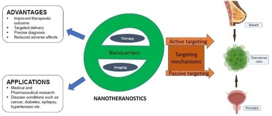
5. Advantages and Applications of Nanotheranostics

The nanotheranostic framework offers medication preference, optimization, and precise diagnosis, and it analyzes biopharmaceutical variables and drug release pattern in a timely manner for an intended targeted therapeutic result [247]. The majority of nanomaterials’ advantages over conventional anticancer medications are a result of their perfect size, which prevents renal removal and enhances tumor penetrability because of the aberrant blood vessels seen in cancer tissues. This shortens the time required to treat patients and tracks drug distribution throughout the body. In clinical trials, there are improved evaluation methods such as biomarker sensitivity and tracking actions aiding in getting the appropriate drug, estimating dosage, and reducing risk related to drug overdose or adverse effects. In cancer therapy, nanotheranostics is aimed at demonstrating the potential of merging cancer treatments, ensuring adequate financial returns in relation to outlays while enhancing quality living and having the capacity to be used in a variety of routes of administration. Furthermore, optimizing the pharmacokinetic and pharmacodynamic characteristics of current anticancer drugs and nanocarriers includes promoting effective vascular circulation and penetration [125,248]. Additionally, they are not intended to break down into potentially dangerous metabolites and are made to elicit fewer immunological responses [249].
The current applications of nanotheranostics in medical and pharmaceutical research embody disease conditions such as cancer, hypertension, diabetes, parkinsonism, epilepsy, hyperlipidemia, alopecia, hormone deficiency, topical inflammation, and ocular, hepatic, respiratory, and fungal diseases. Several nanotheranostics have been created and widely studied preclinically in breast and prostate cancers [250] (see Table 1). Nanocarriers such as liposomes, gold, quantum dots, bovine serum albumin, and polystyrene have been employed in such studies. In comparison, there are some nanomaterials in active clinical trials related to breast and prostate cancer (Table 2). In addition, a series of nanomedicines, namely, FDA (Food and Drug Administration)-approved paclitaxel albumin-bound nanoparticles (Abraxane), doxorubicin liposomal nanoparticles (Doxil), and irinotecan liposomal nanoparticles (Onivyde) and EMA (European Medicines Agency)-approved daunorubicin/cytarabine liposomal nanoparticles, amongst others (Table 3), are in clinical use for breast and prostate cancer treatment.
Table 1. Selected nanotheranostics used in preclinical breast and prostate cancer studies.
Nanoparticles Therapeutic AgentDiagnostic AgentCancer TypeObservationReferences
PolystyreneDoxorubicinCyanine dyeBreastImproved therapeutic index in vitro and reduced tumor volume in vivo[250]
Gold nanoclustersPaclitaxelIndocyanine green (ICG)BreastSuppression of tumor growth on mice breast cancer model[251]
Graphene quantum dotsDoxorubicin-BreastSynergistically enhanced anticancer strategy which provides treatment and diagnosis [252]
PEG liposomesDoxorubicinGadoteridolBreastIncreased intratumor drug concentration and complete regression of lesion [46,47]
Targeted PEG liposomesDoxorubicinFluorescent probe (PFBT)BreastInhibition of tumor-bearing mice[253]
Micelles DocetaxelNIR probe DiRBreastInhibition of tumor growth with little toxicity[254]
Targeted PEG liposomesMitoxantroneSPIONsBreastHigh cytotoxicity against MCF-7 human breast tumor cell line[255]
Iron oxideCurcumin-BreastDisplay of strong anticancer properties compared to free curcumin[256]
Poly(lactide-co-glycolic acid) PLGAResiquimodIndocyanine green (ICG)ProstateSignificant inhibition of PCa growth[257]
Bovine serum albuminCarbazitaxelGadoliniumProstateLower hemolysis, similar tumor inhibition and enhanced cellular uptake in vitro compared with CBZ–Tween-80 injection [258]
GoldAptamer/doxorubicin-ProstatePSMA aptamer showed more potency against targeted LNCaP cell lines than non-targeted PC3 cells[259]
Quantum dotsAptamer/doxorubicin-ProstateTargeted Qd-Apt (DOX) conjugate with reversible self-quenching properties[77]
PEG = polyethylene glycol; DiR = lipophilic fluorochrome (1,1-dioctadecyl-3,3,3,3-tetramethylindotricarbocyanine iodide); SPIONs = superparamagnetic nanoparticles; PFBT = poly(9,9-dioctylfluorene-2,7-diyl-co-benzothiadiazole).

5.1. Visualizing Drug Release

It is critical to make sure that medicaments are rightly being released because most of the therapeutic compounds employed in the formulation of nanotheranostics are inert when bound to or enmeshed in the nanocarrier. Drug release is typically quite simple to accomplish in vitro using tools such as high-performance liquid chromatography (HPLC). However, proving drug release in vivo is significantly more challenging. Nanotheranostics, in which therapeutic and imaging agents are integrated into the same formulation, have been created to address this problem and to facilitate noninvasive investigations on (the kinetics of) drug release in vivo. These imaging agents are not appropriate for observing medication release since radionuclides provide comparable signals when bound/entrapped and when unbound/free. Contrast agents used in magnetic resonance imaging, such as gadolinium and manganese, rely on their interactions with the water molecules in the area to produce a signal. Because the nature of these interactions differs significantly depending on whether the contrast agent is present inside or outside of water-impermeable vesicles such as liposomes, drug release monitoring can be accomplished with magnetic resonance probes. Dewhirst and colleagues presented a very clever strategy in this area, employing manganese sulfate (MnSO4) to load doxorubicin into liposomes (using a technique similar to ammonium sulfate/pH gradient loading), producing a substantial increase in magnetic resonance signal upon drug and contrast agent release [260,261,262].

5.2. Visualizing Biodistribution in Real Time

Systemically administered (chemo)therapeutic drugs can now be more effectively biodistributed and accumulated at the intended site due to nanotheranostics. It would be helpful if the circulation time and organ accumulation of nanomedicine systems, or even better, of the conjugated or entrapped therapeutic medicines, could be observed noninvasively in vivo in real time to enable biodistributional analysis. In order to monitor their pharmacokinetics and biodistribution, numerous different nanomedicines have been co-functionalized with contrast agents in addition to medicaments to accomplish this purpose. It is feasible to forecast and enhance the efficacy of an intervention by combining pharmacologically active compounds and imaging agents in a single nanomedicine formulation. It is also able to visualize and comprehend a number of crucial components of the drug delivery process. Seymour and colleagues, using γ-scintigraphy, demonstrated that an iodine 123-labeled polymeric prodrug of doxorubicin that contained galactosamine (i.e., was targeted to hepatocytes) was successfully localized to the liver, as opposed to a formulation that did not contain galactosamine [263]. However, intriguingly, the polymeric prodrug that was passively targeted against tumors and free of galactosamine was localized to tumors by EPR with a fair amount of success, as demonstrated by the fact that a patient with a major clavicular metastasis had significant retention in the shoulder area. Together, these ideas and initiatives show how theranostic strategies are ideal for assessing the in vivo possibilities of tumor-targeted nanomedicines because they highlight how co-functionalizing nanomedicine formulations with drugs and imaging agents facilitate a noninvasive evaluation of their biodistribution.

5.3. Noninvasively Assessing Target Site Accumulation

Nanotheranostics can be employed to noninvasively evaluate target site accumulation in addition to real-time tracking of the biodistribution of nanomedicine formulations. Nanomedicines that have been radionuclide- and magnetic resonance contrast agent-labeled are excellent candidates for tracking the precise medication distribution to diseased areas. Similar to this, it was demonstrated that the experimental monoclonal antibody U36, which is tagged with zirconium-89, can efficiently accumulate at the diseased location [264]. Similar results were recently reported by Wang and colleagues, who used N-(2-hydroxypropyl) methacrylamide (HPMA) copolymers as long-circulating and passively targeted disease site nanocarriers in place of PEGylated liposomes. They created a theranostic nanomedicine formulation containing gadolinium to provide proof of principle for target site accumulation. Using magnetic resonance imaging (MRI), they showed that Gd-DO3A-modified poly(HPMA) effectively accumulates in the ankles of rats with adjuvant-induced arthritis, whereas no accumulation was seen in the ankles of healthy rats [265]. Image-guided drug delivery can be utilized to certify (triggered) drug release at the target site, to noninvasively view and measure probe buildup at the target site, and to continuously monitor therapeutic efficacy [266].

5.4. Monitoring Drug Distribution at the Target Site

In addition to examining the overall levels of drug accumulation at the target site, it is crucial to visualize and analyze drug distribution at the target site in order to assess the spatial parameters of targeted drug delivery more thoroughly and precisely, as well as to better comprehend and foresee why some treatments are successful while others are not. This idea emphasizes the significance of tracking and measuring drug distribution at the target site and demonstrates the need for multiple imaging modalities that provide functional and/or molecular information in addition to anatomical information to accurately predict the effectiveness of targeted therapeutic interventions. Similar attempts have been made to use gadolinium-labeled liposomes to visualize drug distribution at the intended site. In this regard, Mulder and colleagues demonstrated that the distribution of control liposomes and liposomes modified with RGD and RAD differs significantly in malignancies [266,267].

5.5. Facilitating Triggered Drug Release

The capacity of nanotheranostics to assist triggered drug release is essentially a byproduct of the three nanotheranostics applications listed above, namely, visualizing target site accumulation, visualizing drug distribution at the target site, and visualizing drug release. It would be possible to visualize the accumulation and target site distribution of a stimulus-sensitive theranostic nanomedicine formulation before applying radiofrequency waves to those regions of the tumor in which high levels of the formulation have accumulated, rather than, for instance, using an invasive catheter to induce hyperthermia in a tumor. Instead, one could use an external heat-producing source, such as high-intensity focused ultrasound (HIFU). This allows for the trigger of drug release to have high temporal specificity (i.e., by applying the trigger at that time points at which the concentration of the formulation at the target site is optimal) in addition to high spatial specificity (i.e., those areas of the target tissue where the formulation is present in high amounts). To accomplish this, it is necessary to incorporate two different imaging agents: one that can be seen while the system is still intact, such as a positron emission tomography (PET) tracer or a magnetic resonance (MR) T1 contrast agent with free access to surrounding water molecules (for example, in the case of a liposome, bound to the external bilayer and not entrapped in the core), and one that only produces a signal after the system is released (e.g., an MR T1 contrast agent encapsulated in the core of a liposome, and shielded from the interaction with surrounding water molecules). Langereis and colleagues recently created a beautiful illustration of a theranostic nanomedicine formulation that can be utilized to facilitate triggered drug release. They created thermosensitive liposomes containing two different MR contrast agents [268]. In this system, a 19F probe (NH4PF6) and a CEST agent (chemical exchange saturation transfer; [Tm(HPDO3A)(H2O)]) are co-loaded into the aqueous interior of the liposomes [269]. The CEST agent produces a signal that can be used to monitor the biodistribution and target site accumulation of the formulation at temperatures below the transition temperature, when the liposomal bilayer is intact and there is no free access to surrounding water molecules, and it simultaneously quenches the 19F signal. The CEST signal vanishes when heated to temperatures above the transition temperature, and the 19F signal appears when both the CEST agent and the 19F probe are released from the aqueous interior of the liposome. The authors also worked to create a theranostic nanomedicine formulation in which doxorubicin and gadolinium are co-entrapped in the core of the liposomes using related temperature-sensitive liposomes [270]. In order to achieve this, liposomes containing the MR contrast agent ProHance (i.e., [Gd(HPDO3A)(H2O)]) and ammonium sulfate were formulated. Doxorubicin was then actively post-loaded into the liposomes using a pH gradient. For this formulation, cryo-TEM (transmission electron microscopy) images were taken both before and after heating. Hyperthermia caused the usual cigar-shaped doxorubicin crystals to vanish from the liposomes, indicating that the drug had been released. Fluorescence detection was used to further validate doxorubicin release, and this release corresponded well with the release of ProHance from the liposomes. In this connection, it should be highlighted that the release of doxorubicin as measured by fluorescence detection showed a very significant signal increase at temperatures close to the transition temperature, demonstrating the outstanding applicability of this formulation for hyperthermia-triggered drug release. However, this is because the liposomal bilayer’s permeability to water molecules already begins to increase at temperatures higher than 25 °C, which accounts for the less pronounced increase in the MR signal. De Smet and colleagues demonstrated that only the two temperature-sensitive formulations—namely, traditional thermosensitive liposomes (TTSL) and low-temperature-sensitive liposomes (LTSL)—observed the release of the gadolinium-based MR contrast agent upon hyperthermia, whereas heating had no effect on ProHance release from non-thermosensitive liposomes (NTSL) [270].

5.6. Predicting Drug Responses

The use of these systems for forecasting therapeutic responses is another, therapeutically very important application of nanotheranostics. If, for example, radionuclides or MR contrast agents can be used to label nanomedicines, particularly in the early stages of clinical evaluation, then crucial noninvasive information about target site accumulation can be obtained. Accordingly, logical predictions can be made about the potential efficacy of the targeted therapeutic interventions to be tested. One illustration of this theranostic nanomedicines benefit is the buildup of radiolabeled PEGylated liposomes in a patient with severe rheumatoid arthritis. Similar to this, patients receiving antibody-based chemo- or radio-immunotherapy or PEGylated liposomal corticosteroids (to lessen rheumatoid arthritis flare-ups) could be pre-screened using such theranostic nanomedicine formulations to distinguish between those showing high and those showing low (or no) target site accumulation, and to predict which patients are likely to respond to such treatments [271,272,273].

5.7. Evaluating Drug Efficacy Longitudinally

An additional crucial feature of nanotheranostics and image-guided drug delivery, particularly for preclinical applications, is that they greatly simplify longitudinal experimental setups, enabling not only more insightful and minimally invasive biodistribution studies but also more elegant and pertinent efficacy analyses, wherein, for example, genetically modified mice and orthotopic tumor models are used to study disease progression and treatment outcome in real time. Gadolinium- or radionuclide-labeled liposomes, polymers, micelles, and antibodies are also likely to have significant potential for evaluating drug efficacy longitudinally, for instance, by using such theranostic nanomedicines to determine tumor sizes, or by evaluating their ability to (a) measure tumor sizes using theranostic nanomedicines, or (b) measure tumor response to therapy using PET with 18F-FDG. Observing how the target site accumulation of theranostic nanomedicine formulations alters in response to therapy can also provide crucial information about visualizing the effectiveness of the intervention. For example, if PEGylated liposomal corticosteroids in rheumatoid arthritis significantly reduce local joint inflammation and disease severity, it is anticipated that their accumulation in these lesions will gradually diminish over time. Since the extent of the localization of their target site depends on the severity of the condition, it would be possible to utilize such formulations to track the effectiveness of the intervention in real time by radiolabeling a small portion of the corticosteroid-containing PEGylated liposomes. Similar results can be seen when using actively targeted nanotheranostics, such as receptor downregulation and/or the removal of diseased cells that express the receptor.

6. Limitations/Challenges of Nanotheranostics

Whereas these platforms have offered potential in in vivo studies, their inorganic or metallic makeup has generated concerns about toxicity, immunogenicity, and sluggish elimination profile from the body, all of which must be extensively investigated before clinical trials in humans. Adenosine 5′-triphosphate diminution and mitochondrial disruption, for instance, can be triggered by silver-based nanoparticles [274]. Single-walled carbon nanotubes, on the other hand, can produce oxidative stress and programmed cell death [275]. Magnetic nanoparticles can kill cells by damaging their membranes [276].
There is also the problem of overcoming multidrug resistance (MDR) [36]. Intriguingly, siRNA can be used in theranostic nanomedicine as a theranostic resistance suppressor. As a multifunctional therapy, siRNA-based theranostic nanomedicine has been demonstrated to considerably enhance diagnosis and treatment [277].
Autophagy is a breakdown process whereby defective cell components or extraneous bodies are destroyed intracellularly in lysosomes. In addition, intracellular autophagy influences nanomedicine after endocytosis and, therefore, its therapeutic efficacy by altering the nanomedicine’s intracellular pharmacokinetics. As a result, autophagy inhibitors embedded in theranostic nanomedicine can improve the efficacy of nanotheranostics [36].
The effectiveness, specificity, and sensitivity of encapsulating a single diagnostic or therapeutic substance in nanomedicine may be low. As a result, multifunctional nanotheranostics is created to combine various imaging and treatment modalities in a solitary platform [278,279].
The creation of novel molecular biomaterials with excellent efficiency in the co-formulation of imaging and treatment agents is a major issue in modern nanotheranostics. The discovery of novel biomarkers with high precision for the intended ailment, the possibility of agonism and antagonism between imaging and treatment agents co-produced in a solitary nanoparticle, and the presence of intracellular autophagy in nanotheranostics are other concerns and should be researched. Nanotheranostics may be capable of giving a plausible solution for cancer treatment, as well as other deadly illnesses, in the near future, allowing them to be cured or at least treated at an early stage.

7. Future Perspectives

Aside from the fact that a wide range of smart nanocarriers have been created in contemporary times, the inherent intricacy of biological surroundings has a significant impact on nanomaterial activity and frequently hinders their successful utilization for therapeutic interventions. The preference for nanoparticle characteristics (such as size, shape, material substrate, and surface chemistry) plays a vital role in the development of advanced nanocarriers for special purposes, because the microenvironment inside cancerous breast and prostate tissues has a significant influence on the effectiveness of delivery of nanoparticle frameworks. Hence, for the creation of innovative treatment techniques and regimens centered on the use of smart nanocarriers, a deep understanding of the genuine dynamics associated with malignant cells is essential.
Theranostic substances necessitate a high level of congeniality among the nanocarrier nucleus, the aiming molecule, the imaging component, and the therapeutic agent. The optimal nanoparticle for theranostic applications ought to have a formulation methodology with the least conceivable stages, be inexpensive and practical in budget, have excellent reproducibility and scale-up simplicity, and offer imaging and treatment effectiveness. Aiming molecules with diagnostic capabilities may increase the precision and specificity of both therapeutic and imaging results, allowing for actual monitoring, which is becoming more common in medicine, particularly in the treatment of breast and prostate cancers. Fluorescence imaging, scintillography, tomography (PET or SPECT), magnetic resonance, ultrasound, and upconversion imaging are but a few of the diagnostic modalities that can be used for theranostic applications. In order to maximize the effectiveness of nanoparticles, multiple techniques may be employed. As a result, theranostic nanomaterials must be synthesized with all these characteristics as a primary objective so as to improve treatment results whenever they are being used.
Despite numerous attempts to produce unique tailored nanocarriers, the FDA has only authorized a handful of them for medical use. As a result, more in vitro and in vivo models are needed to verify their aiming efficiency. Nanoparticles should be designed using multiscale modeling and computational models so as to achieve higher sophisticated targeted delivery of therapeutic agents. It is also conceivable to enhance clinical transposition by placing greater emphasis on advanced technologies and performing clinical trials to corroborate novel guidelines in the clinical stage. The clinical transposition and long-term viability of these “smart” systems require substantial and repeatable techniques that allow for comparatively simple and cost-effective scale-up and production of nanotheranostic frameworks for breast and prostate cancers.

8. Conclusions

Traditional cancer detection and treatment methods have failed to successfully eliminate malignant cells from the body. Formerly, the nanotheranostic system was solely employed as a stand-in for standard diagnostic and therapeutic procedures, but it is now becoming one of the most powerful multimodal instruments in pharmaceuticals for the mitigation of breast and prostate cancers. Nanotheranostics provides good prospects for simultaneously characterizing a disease and the medications utilized, as well as understanding the host–disease interaction. Moreover, cancer nanotherapeutics should be tailored to the needs of specific patients, resulting in the advancement of individualized medicine. Substantial development will be required to assist the clinical advancement of the hopeful preclinical findings in breast and prostate cancer treatment via nanotheranostic platforms.

Author Contributions

Conceptualization, I.E.P., T.C.A., A.M.O. and P.A.A. (Peter A. Akah); resources, T.C.A., A.A.A., P.M.E., P.A.A. (Paul A. Akpa), B.C.O., M.O.A. and P.A.A. (Peter A. Akah); writing—original draft preparation, I.E.P., T.C.A. and A.A.A.; writing—review and editing, P.A.A. (Paul A. Akpa), I.E.P., A.M.O., B.C.O., M.O.A., C.S.N., P.M.E., T.C.A., A.A.A. and P.A.A. (Peter A. Akah); supervision, A.A.A., P.M.E., T.C.A. and P.A.A. (Peter A. Akah); project administration, P.A.A. (Paul A. Akpa), T.C.A., A.A.A. and C.S.N.; funding acquisition, P.A.A. (Paul A. Akpa), T.C.A., A.A.A., P.M.E. and P.A.A. (Peter A. Akah). All authors have read and agreed to the published version of the manuscript.

Funding

This project was sponsored by TETFund NRF intervention 2019, code TETFUND/DR and D-CE/NRF/2019/STI/47/VOL-1.

Data Availability Statement

No new data were created or analyzed in this study. Data sharing is not applicable to this article.

Conflicts of Interest

The authors declare no conflict of interest.

References

  1. NCI. Available online: https://www.cancer.gov/about-cancer/understanding (accessed on 30 July 2020).
  2. Siegel, R.L.; Miller, K.D.; Fuchs, H.E.; Jemal, A. Cancer Statistics, 2021. CA Cancer J. Clin. 2021, 71, 7–33. [Google Scholar] [CrossRef] [PubMed]
  3. Ferlay, J.; Soerjomataram, I.; Dikshit, R.; Eser, S.; Mathers, C.; Rebelo, M.; Parkin, D.M.; Forman, D.; Bray, F. Cancer incidence and mortality worldwide: Sources, methods and major patterns in GLOBOCAN 2012. Int. J. Cancer 2015, 136, E359–E386. [Google Scholar] [CrossRef]
  4. WHO. Breast 2020. Available online: https://gco.iarc.fr/today/data/factsheets/cancers/20-Breast-fact-sheet.pdf (accessed on 2 June 2021).
  5. Giaquinto, A.N.; Sung, H.; Miller, K.D.; Kramer, J.L.; Newman, L.A.; Minihan, A.; Jemal, A.; Siegel, R.L. Breast Cancer Statistics, 2022. CA A Cancer J. Clin. 2022, 72, 524–541. [Google Scholar] [CrossRef]
  6. Altekruse, S.F.; Kosary, C.L.; Krapcho, M.; Neyman, N.; Aminou, R.; Waldron, W.; Ruhl, J.; Howlader, N.; Tatalovich, Z.; Cho, H.; et al. SEER Cancer Statistics Review, 1975–2007; National Cancer Institute: Bethesda, MD, USA, 2010. Available online: https://seer.cancer.gov/archive/csr/1975_2007/ (accessed on 17 May 2017).
  7. Sinn, H.P.; Kreipe, H. A Brief Overview of the WHO Classification of Breast Tumors, 4th Edition, Focusing on Issues and Updates from the 3rd Edition. Breast Care 2013, 8, 149–154. [Google Scholar] [CrossRef] [Green Version]
  8. Weigelt, B.; Peterse, J.L.; van’t Veer, L.J. Breast cancer metastasis: Markers and models. Nat. Rev. Cancer 2005, 5, 591–602. [Google Scholar] [CrossRef]
  9. Fragomeni, S.M.; Sciallis, A.; Jeruss, J.S. Molecular Subtypes and Local-Regional Control of Breast Cancer. Surg. Oncol. Clin. N. Am. 2018, 27, 95–120. [Google Scholar] [CrossRef] [PubMed]
  10. Bonotto, M.; Gerratana, L.; Poletto, E.; Driol, P.; Giangreco, M.; Russo, S.; Minisini, A.M.; Andreetta, C.; Mansutti, M.; Pisa, F.E.; et al. Measures of Outcome in Metastatic Breast Cancer: Insights from a Real-World Scenario. Oncology 2014, 19, 608–615. [Google Scholar] [CrossRef] [Green Version]
  11. Redig, A.J.; McAllister, S.S. Breast cancer as a systemic disease: A view of metastasis. J. Intern. Med. 2013, 274, 113–126. [Google Scholar] [CrossRef] [Green Version]
  12. Siegel, R.L.; Miller, K.D.; Fuchs, H.E.; Jemal, A. Cancer statistics, 2022. CA Cancer J. Clin. 2022, 72, 7–33. [Google Scholar] [CrossRef]
  13. Costello, L.C.; Franklin, R.B. A comprehensive review of the role of zinc in normal prostate function and metabolism; and its implications in prostate cancer. Arch. Biochem. Biophys. 2016, 611, 100–112. [Google Scholar] [CrossRef] [Green Version]
  14. Fujita, K.; Hayashi, T.; Matsushita, M.; Uemura, M.; Nonomura, N. Obesity, Inflammation, and Prostate Cancer. J. Clin. Med. 2019, 8, 201. [Google Scholar] [PubMed] [Green Version]
  15. Ferlay, J.; Shin, H.-R.; Bray, F.; Forman, D.; Mathers, C.; Parkin, D.M. Estimates of worldwide burden of cancer in 2008: GLOBOCAN 2008. Int. J. Cancer 2010, 127, 2893–2917. [Google Scholar] [CrossRef] [PubMed]
  16. Rawla, P. Epidemiology of Prostate Cancer. World J. Oncol. 2019, 10, 63–89. [Google Scholar] [CrossRef] [PubMed] [Green Version]
  17. Miller, K.D.; Fidler-Benaoudia, M.; Keegan, T.H.; Hipp, H.S.; Jemal, A.; Siegel, R.L.; Dvm, A.J. Cancer statistics for adolescents and young adults, 2020. CA A Cancer J. Clin. 2020, 70, 443–459. [Google Scholar] [CrossRef]
  18. Perdonà, S.; Cavadas, V.; Di Lorenzo, G.; Damiano, R.; Chiappetta, G.; Del Prete, P.; Franco, R.; Azzarito, G.; Scala, S.; Arra, C.; et al. Prostate cancer detection in the ‘‘grey area” of prostate-specific antigen below 10 ng/ml: Head-to-head comparison of the updated PCPT calculator and Chun’s nomogram, two risk estimators incorporating prostate cancer antigen 3. Eur. Urol. 2011, 59, 81–87. [Google Scholar]
  19. Shinohara, K. Improving cancer detection by prostate biopsy: The role of core number and site. Nat. Rev. Endocrinol. 2006, 3, 526–527. [Google Scholar] [CrossRef]
  20. Naji, L.; Randhawa, H.; Sohani, Z.; Dennis, B.; Lautenbach, D.; Kavanagh, O.; Bawor, M.; Banfield, L.; Profetto, J. Digital Rectal Examination for Prostate Cancer Screening in Primary Care: A Systematic Review and Meta-Analysis. Ann. Fam. Med. 2018, 16, 149–154. [Google Scholar] [CrossRef]
  21. Catalona, W.J.; Smith, D.S.; Ratliff, T.L.; Dodds, K.M.; Coplen, D.E.; Yuan, J.J.; Petros, J.A.; Andriole, G.L. Measurement of Prostate-Specific Antigen in Serum as a Screening Test for Prostate Cancer. N. Engl. J. Med. 1991, 324, 1156–1161. [Google Scholar] [CrossRef]
  22. Schröder, F.H.; van der Cruijsen-Koeter, I.; de Koning, H.J.; Vis, A.N.; Hoedemaeker, R.F.; Kranse, R. Prostate cancer detection at low prostate specific antigen. J. Urol. 2000, 163, 806–812. [Google Scholar]
  23. Lee, K.H.; Kang, B.J.; Jeun, M.; Jang, G.H.; Song, S.H.; Jeong, I.G.; Kim, C.-S.; Searson, P.C. Diagnosis of prostate cancer via nanotechnological approach. Int. J. Nanomed. 2015, 10, 6555–6569. [Google Scholar] [CrossRef] [Green Version]
  24. Nie, S.; Xing, Y.; Kim, G.J.; Simons, J.W. Nanotechnology Applications in Cancer. Annu. Rev. Biomed. Eng. 2007, 9, 257–288. [Google Scholar] [CrossRef] [PubMed] [Green Version]
  25. Melancon, M.P.; Stafford, R.J.; Li, C. Challenges to effective cancer nanotheranostics. J. Control. Release 2012, 164, 177–182. [Google Scholar] [CrossRef] [PubMed] [Green Version]
  26. Falzone, L.; Salomone, S.; Libra, M. Evolution of Cancer Pharmacological Treatments at the Turn of the Third Millennium. Front. Pharmacol. 2018, 9, 1300. [Google Scholar] [CrossRef] [Green Version]
  27. Chinen, A.B.; Guan, C.M.; Ferrer, J.R.; Barnaby, S.N.; Merkel, T.J.; Mirkin, C.A. Nanoparticle Probes for the Detection of Cancer Biomarkers, Cells, and Tissues by Fluorescence. Chem. Rev. 2015, 115, 10530–10574. [Google Scholar] [CrossRef] [PubMed] [Green Version]
  28. Shi, J.; Kantoff, P.W.; Wooster, R.; Farokhzad, O.C. Cancer nanomedicine: Progress, challenges and opportunities. Nat. Rev. Cancer 2016, 17, 20–37. [Google Scholar] [CrossRef] [Green Version]
  29. Banthia, P.; Gambhir, L.; Sharma, A.; Daga, D.; Kapoor, N.; Chaudhary, R.; Sharma, G. Nano to rescue: Repository of nanocarriers for targeted drug delivery to curb breast cancer. 3 Biotech 2022, 12, 1–23. [Google Scholar] [CrossRef]
  30. Funkhouser, J. Reinventing Pharma: The theranostic revolution. Curr. Drug Discov. 2002, 2, 17–19. [Google Scholar]
  31. Asem, H.; Malmström, E. Polymeric Nanoparticles Explored for Drug-Delivery Applications. In ACS Symposium Series; American Chemical Society: Washington, DC, USA, 2018; pp. 315–331. [Google Scholar]
  32. Kröger, A.P.P.; Hamelmann, N.M.; Juan, A.; Lindhoud, S.; Paulusse, J.M.J. Biocompatible Single-Chain Polymer Nanoparticles for Drug Delivery—A Dual Approach. ACS Appl. Mater. Interfaces 2018, 10, 30946–30951. [Google Scholar] [CrossRef] [Green Version]
  33. Akbarzadeh, A.; Rezaei-Sadabady, R.; Davaran, S.; Joo, S.W.; Zarghami, N.; Hanifehpour, Y.; Samiei, M.; Kouhi, M.; Nejati-Koshki, K. Liposome: Classification, preparation, and applications. Nanoscale Res. Lett. 2013, 8, 102. [Google Scholar] [CrossRef] [Green Version]
  34. Rehman, A.U.; Akram, S.; Seralin, A.; Vandamme, T.; Anton, N. Lipid nanocarriers: Formulation, properties, and applications. In Smart Nanocontainers; Elsevier: Amsterdam, The Netherlands, 2020; pp. 355–382. [Google Scholar]
  35. Bombelli, C.; Stringaro, A.; Borocci, S.; Bozzuto, G.; Colone, M.; Giansanti, L.; Sgambato, R.; Toccaceli, L.; Mancini, G.; Molinari, A. Efficiency of Liposomes in the Delivery of a Photosensitizer Controlled by the Stereochemistry of a Gemini Surfactant Component. Mol. Pharm. 2010, 7, 130–137. [Google Scholar] [CrossRef]
  36. Muthu, M.S.; Leong, D.T.; Mei, L.; Feng, S.-S. Nanotheranostics—Application and Further Development of Nanomedicine Strategies for Advanced Theranostics. Theranostics 2014, 4, 660–677. [Google Scholar] [CrossRef] [Green Version]
  37. Xing, H.; Hwang, K.; Lu, Y. Recent Developments of Liposomes as Nanocarriers for Theranostic Applications. Theranostics 2016, 6, 1336–1352. [Google Scholar] [CrossRef] [PubMed]
  38. Zhang, K.; Zhang, Y.; Meng, X.; Lu, H.; Chang, H.; Dong, H.; Zhang, X. Light-triggered theranostic liposomes for tumor diagnosis and combined photodynamic and hypoxia-activated prodrug therapy. Biomaterials 2018, 185, 301–309. [Google Scholar] [CrossRef] [PubMed]
  39. Karpuz, M.; Silindir-Gunay, M.; Ozer, A.Y.; Ozturk, S.C.; Yanik, H.; Tuncel, M.; Aydin, C.; Esendagli, G. Diagnostic and therapeutic evaluation of folate-targeted paclitaxel and vinorelbine encapsulating theranostic liposomes for non-small cell lung cancer. Eur. J. Pharm. Sci. 2020, 156, 105576. [Google Scholar] [CrossRef]
  40. Skupin-Mrugalska, P.; Sobotta, L.; Warowicka, A.; Wereszczynska, B.; Zalewski, T.; Gierlich, P.; Jarek, M.; Nowaczyk, G.; Kempka, M.; Gapinski, J.; et al. Theranostic liposomes as a bimodal carrier for magnetic resonance imaging contrast agent and photosensitizer. J. Inorg. Biochem. 2018, 180, 1–14. [Google Scholar] [CrossRef]
  41. Ren, W.; Chen, S.; Liao, Y.; Li, S.; Ge, J.; Tao, F.; Huo, Q.; Zhang, Y.; Zhao, Z. Near-infrared fluorescent carbon dots encapsulated liposomes as multifunctional nano-carrier and tracer of the anticancer agent cinobufagin in vivo and in vitro. Colloids Surfaces B Biointerfaces 2019, 174, 384–392. [Google Scholar] [CrossRef] [PubMed]
  42. Grippin, A.J.; Wummer, B.; Wildes, T.; Dyson, K.; Trivedi, V.; Yang, C.; Sebastian, M.; Mendez-Gomez, H.R.; Padala, S.; Grubb, M.; et al. Dendritic Cell-Activating Magnetic Nanoparticles Enable Early Prediction of Antitumor Response with Magnetic Resonance Imaging. ACS Nano 2019, 13, 13884–13898. [Google Scholar] [CrossRef]
  43. Gavas, S.; Quazi, S.; Karpiński, T.M. Nanoparticles for Cancer Therapy: Current Progress and Challenges. Nanoscale Res Lett. 2021, 16, 173. [Google Scholar] [CrossRef]
  44. Markman, M. Pegylated liposomal doxorubicin in the treatment of cancers of the breast and ovary. Expert Opin. Pharmacother. 2006, 7, 1469–1474. [Google Scholar] [CrossRef]
  45. Malik, N.; Evagorou, E.G.; Duncan, R. Dendrimer-platinate: A novel approach to cancer chemotherapy. Anti-Cancer Drugs 1999, 10, 767–776. [Google Scholar] [CrossRef]
  46. Rizzitelli, S.; Giustetto, P.; Cutrin, J.; Castelli, D.D.; Boffa, C.; Ruzza, M.; Menchise, V.; Molinari, F.; Aime, S.; Terreno, E. Sonosensitive theranostic liposomes for preclinical in vivo MRI-guided visualization of doxorubicin release stimulated by pulsed low intensity non-focused ultrasound. J. Control. Release 2015, 202, 21–30. [Google Scholar] [CrossRef]
  47. Rizzitelli, S.; Giustetto, P.; Faletto, D.; Castelli, D.D.; Aime, S.; Terreno, E. The release of Doxorubicin from liposomes monitored by MRI and triggered by a combination of US stimuli led to a complete tumor regression in a breast cancer mouse model. J. Control. Release 2016, 230, 57–63. [Google Scholar] [CrossRef] [PubMed] [Green Version]
  48. Yari, H.; Nkepang, G.; Awasthi, V. Surface Modification of Liposomes by a Lipopolymer Targeting Prostate Specific Membrane Antigen for Theranostic Delivery in Prostate Cancer. Materials 2019, 12, 756. [Google Scholar] [CrossRef] [PubMed] [Green Version]
  49. Narayanan, N.K.; Nargi, D.; Randolph, C.; Narayanan, B.A. Liposome encapsulation of curcumin and resveratrol in combination reduces prostate cancer incidence in PTEN knockout mice. Int. J. Cancer 2009, 125, 1–8. [Google Scholar] [CrossRef]
  50. Thangapazham, R.L.; Puri, A.; Tele, S.; Blumenthal, R.; Maheshwari, R.K. Evaluation of a nanotechnology-based carrier for delivery of curcumin in prostate cancer cells. Int. J. Oncol. 2008, 32, 1119–1123. [Google Scholar] [CrossRef] [Green Version]
  51. Zhao, Y.-Z.; Dai, D.-D.; Lu, C.-T.; Chen, L.-J.; Lin, M.; Shen, X.-T.; Li, X.-K.; Zhang, M.; Jiang, X.; Jin, R.-R.; et al. Epirubicin loaded with propylene glycol liposomes significantly overcomes multidrug resistance in breast cancer. Cancer Lett. 2013, 330, 74–83. [Google Scholar] [CrossRef]
  52. Fu, M.; Tang, W.; Liu, J.-J.; Gong, X.-Q.; Kong, L.; Yao, X.-M.; Jing, M.; Cai, F.-Y.; Li, X.-T.; Ju, R.-J. Combination of targeted daunorubicin liposomes and targeted emodin liposomes for treatment of invasive breast cancer. J. Drug Target. 2020, 28, 245–258. [Google Scholar] [CrossRef] [PubMed]
  53. Sisay, B.; Abrha, S.; Yilma, Z.; Assen, A.; Molla, F.; Tadese, E.; Wondimu, A.; Gebre-Samuel, N.; Pattnaik, G. Cancer nanotheranostics: A new paradigm of simultaneous diagnosis and therapy. J. Drug Deliv. Ther. 2014, 4, 79–86. [Google Scholar] [CrossRef]
  54. Lima, A.M.; Pizzol, C.D.; Monteiro, F.B.; Creczynski-Pasa, T.B.; Andrade, G.P.; Ribeiro, A.O.; Perussi, J.R. Hypericin encapsulated in solid lipid nanoparticles: Phototoxicity and photodynamic efficiency. J. Photochem. Photobiol. B Biol. 2013, 125, 146–154. [Google Scholar] [CrossRef]
  55. Wong, H.L.; Bendayan, R.; Rauth, A.M.; Li, Y.; Wu, X.Y. Chemotherapy with anticancer drugs encapsulated in solid lipid nanoparticles. Adv. Drug Deliv. Rev. 2007, 59, 491–504. [Google Scholar] [CrossRef]
  56. Fathi, M.; Mozafari, M.; Mohebbi, M. Nanoencapsulation of food ingredients using lipid based delivery systems. Trends Food Sci. Technol. 2012, 23, 13–27. [Google Scholar] [CrossRef]
  57. Mehnert, W.; Mader, K. Solid lipid nanoparticles: Production, characterization and applications. Adv. Drug Deliv. Rev. 2001, 47, 165–196. [Google Scholar] [CrossRef] [PubMed]
  58. Wissing, S.A.; Kayser, O.; Muller, R.H. Solid Lipid Nanoparticles for Parenteral Drug Delivery. Adv. Drug Deliv. Rev. 2004, 56, 1257–1272. [Google Scholar] [CrossRef] [PubMed]
  59. Garg, N.K.; Singh, B.; Jain, A.; Nirbhavane, P.; Sharma, R.; Tyagi, R.K.; Kushwah, V.; Jain, S.; Katare, O.P. Fucose decorated solid-lipid nanocarriers mediate efficient delivery of methotrexate in breast cancer therapeutics. Colloids Surfaces B Biointerfaces 2016, 146, 114–126. [Google Scholar] [CrossRef] [PubMed]
  60. Eskiler, G.G.; Cecener, G.; Dikmen, G.; Egeli, U.; Tunca, B. Solid lipid nanoparticles: Reversal of tamoxifen resistance in breast cancer. Eur. J. Pharm. Sci. 2018, 120, 73–88. [Google Scholar] [CrossRef]
  61. Sharma, A.N.; Upadhyay, P.K.; Dewangan, H.K. Development, evaluation, pharmacokinetic and biodistribution estimation of resveratrol-loaded solid lipid nanoparticles for prostate cancer targeting. J. Microencapsul. 2022, 39, 563–574. [Google Scholar] [CrossRef]
  62. Akanda, M.H.; Rai, R.; Slipper, I.J.; Chowdhry, B.Z.; Lamprou, D.; Getti, G.; Douroumis, D. Delivery of retinoic acid to LNCap human prostate cancer cells using solid lipid nanoparticles. Int. J. Pharm. 2015, 493, 161–171. [Google Scholar] [CrossRef] [Green Version]
  63. Janib, S.M.; Moses, A.S.; MacKay, J.A. Imaging and drug delivery using theranostic nanoparticles. Adv. Drug Deliv. Rev. 2010, 62, 1052–1063. [Google Scholar] [CrossRef] [Green Version]
  64. Grossman, J.H.; McNeil, S.E. Nanotechnology in Cancer Medicine. Phys. Today 2012, 65, 38–42. [Google Scholar] [CrossRef]
  65. Gardel, M.L. Synthetic polymers with biological rigidity. Nature 2013, 493, 619. [Google Scholar] [CrossRef]
  66. Ogay, V.; Mun, E.A.; Kudaibergen, G.; Baidarbekov, M.; Kassymbek, K.; Zharkinbekov, Z.; Saparov, A. Progress and Prospects of Polymer-Based Drug Delivery Systems for Bone Tissue Regeneration. Polymers 2020, 12, 2881. [Google Scholar] [CrossRef] [PubMed]
  67. Zhao, Y.; Houston, Z.H.; Simpson, J.D.; Chen, L.; Fletcher, N.L.; Fuchs, A.V.; Blakey, I.; Thurecht, K.J. Using Peptide Aptamer Targeted Polymers as a Model Nanomedicine for Investigating Drug Distribution in Cancer Nanotheranostics. Mol. Pharm. 2017, 14, 3539–3549. [Google Scholar] [CrossRef]
  68. Jia, H.-R.; Jiang, Y.-W.; Zhu, Y.-X.; Li, Y.-H.; Wang, H.-Y.; Han, X.; Yu, Z.-W.; Gu, N.; Liu, P.; Chen, Z.; et al. Plasma membrane activatable polymeric nanotheranostics with self-enhanced light-triggered photosensitizer cellular influx for photodynamic cancer therapy. J. Control. Release 2017, 255, 231–241. [Google Scholar] [CrossRef] [PubMed]
  69. Zhu, Y.; Wang, X.; Chen, J.; Zhang, J.; Meng, F.; Deng, C.; Cheng, R.; Feijen, J.; Zhong, Z. Bioresponsive and fluorescent hyaluronic acid-iodixanol nanogels for targeted X-ray computed tomography imaging and chemotherapy of breast tumors. J. Control. Release 2016, 244, 229–239. [Google Scholar] [CrossRef] [PubMed]
  70. Yu, F.; Zhu, M.; Li, N.; Ao, M.; Li, Y.; Zhong, M.; Yuan, Q.; Chen, H.; Fan, Z.; Wang, Y.; et al. Imaging-guided synergistic targeting-promoted photo-chemotherapy against cancers by methotrexate-conjugated hyaluronic acid nanoparticles. Chem. Eng. J. 2020, 380, 122426. [Google Scholar] [CrossRef]
  71. Mansur, A.A.; Caires, A.J.; Carvalho, S.M.; Capanema, N.S.; Carvalho, I.C.; Mansur, H.S. Dual-functional supramolecular nanohybrids of quantum dot/biopolymer/chemotherapeutic drug for bioimaging and killing brain cancer cells in vitro. Colloids Surfaces B Biointerfaces 2019, 184, 110507. [Google Scholar] [CrossRef]
  72. Shitole, A.A.; Sharma, N.; Giram, P.; Khandwekar, A.; Baruah, M.; Garnaik, B.; Koratkar, S. LHRH-conjugated, PEGylated, poly-lactide-co-glycolide nanocapsules for targeted delivery of combinational chemotherapeutic drugs Docetaxel and Quercetin for prostate cancer. Mater. Sci. Eng. C 2020, 114, 111035. [Google Scholar] [CrossRef]
  73. Zhang, X.-F.; Liu, Z.-G.; Shen, W.; Gurunathan, S. Silver nanoparticles: Synthesis, characterization, properties, applications, and therapeutic approaches. Int. J. Mol. Sci. 2016, 17, 1534. [Google Scholar]
  74. Libutti, S.K.; Paciotti, G.F.; Byrnes, A.A.; Alexander, H.R., Jr.; Gannon, W.E.; Walker, M.; Seidel, G.D.; Yuldasheva, N.; Tamarkin, L. Phase I and pharmacokinetic studies of CYT-6091, a novel PEGylated colloidal gold-rhTNF nanomedicine. Clin. Cancer Res. 2010, 16, 6139–6149. [Google Scholar] [CrossRef] [Green Version]
  75. Li, W.; Cao, Z.; Liu, R.; Liu, L.; Li, H.; Li, X.; Chen, Y.; Lu, C.; Liu, Y. AuNPs as an important inorganic nanoparticle applied in drug carrier systems. Artif. Cells Nanomed. Biotechnol. 2019, 47, 4222–4233. [Google Scholar] [CrossRef] [Green Version]
  76. Xu, H.; Jiang, S.; Wang, J.; Li, X.; Wu, T.; Xu, P.; Santos-Oliveira, R.; Zhang, A. Radioactive Gold Nanoparticle in Two Forms (19879Au GNPs and 99mTc-GNPs) for Lung Cancer Antiproliferative Induction and Intralesional Imaging: A Proof of Concept. Anti-Cancer Agents Med. Chem. 2020, 20, 1648–1653. [Google Scholar] [CrossRef] [PubMed]
  77. Kim, D.; Jeong, Y.Y.; Jon, S. A Drug-Loaded Aptamer−Gold Nanoparticle Bioconjugate for Combined CT Imaging and Therapy of Prostate Cancer. ACS Nano 2010, 4, 3689–3696. [Google Scholar] [CrossRef] [PubMed]
  78. Luo, D.; Johnson, A.; Wang, X.; Li, H.; Erokwu, B.O.; Springer, S.; Lou, J.; Ramamurthy, G.; Flask, C.A.; Burda, C.; et al. Targeted Radiosensitizers for MR-Guided Radiation Therapy of Prostate Cancer. Nano Lett. 2020, 20, 7159–7167. [Google Scholar] [CrossRef] [PubMed]
  79. Cunningham, C.; de Kock, M.; Engelbrecht, M.; Miles, X.; Slabbert, J.; Vandevoorde, C. Radiosensitization effect of gold nanoparticles in proton therapy. Front. Public Health. 2021, 9, 699822. [Google Scholar] [CrossRef] [PubMed]
  80. Bouché, M.; Hsu, J.C.; Dong, Y.C.; Kim, J.; Taing, K.; Cormode, D.P. Recent Advances in Molecular Imaging with Gold Nanoparticles. Bioconj. Chem. 2020, 31, 303–314. [Google Scholar] [CrossRef]
  81. Yang, S.-J.; Huang, C.-H.; Wang, C.-H.; Shieh, M.-J.; Chen, K.-C. The Synergistic Effect of Hyperthermia and Chemotherapy in Magnetite Nanomedicine-Based Lung Cancer Treatment. Int. J. Nanomed. 2020, 15, 10331–10347. [Google Scholar] [CrossRef]
  82. Liu, X.; Zhang, Y.; Wang, Y.; Zhu, W.; Li, G.; Ma, X.; Chen, S.; Tiwari, S.; Shi, K.; Zhang, S.; et al. Comprehensive understanding of magnetic hyperthermia for improving antitumor therapeutic efficacy. Theranostics 2020, 10, 3793–3815. [Google Scholar] [CrossRef]
  83. Ferreira, M.; Sousa, J.; Pais, A.; Vitorino, C. The Role of Magnetic Nanoparticles in Cancer Nanotheranostics. Materials 2020, 13, 266. [Google Scholar] [CrossRef] [Green Version]
  84. Guo, Y.; Wang, X.-Y.; Chen, Y.-L.; Liu, F.-Q.; Tan, M.-X.; Ao, M.; Yu, J.-H.; Ran, H.-T.; Wang, Z.-X. A light-controllable specific drug delivery nanoplatform for targeted bimodal imaging-guided photothermal/chemo synergistic cancer therapy. Acta Biomater. 2018, 80, 308–326. [Google Scholar] [CrossRef]
  85. Arndt, M.; Nairz, O.; Voss-Andreae, J.; Keller, C.; Van der Zouw, G.; Zeilinger, A. Wave-particle duality of C60. Nature 1999, 401, 680–682. [Google Scholar] [CrossRef]
  86. McDevitt, M.R.; Chattopadhyay, D.; Kappel, B.J.; Jaggi, J.S.; Schiffman, S.R.; Antczak, C.; Njardarson, J.T.; Brentjens, R.; Scheinberg, D.A. Tumor Targeting with Antibody-Functionalized, Radiolabeled Carbon Nanotubes. J. Nucl. Med. 2007, 48, 1180–1189. [Google Scholar] [CrossRef] [PubMed]
  87. Porter, A.E.; Gass, M.; Muller, K.; Skepper, J.N.; Midgley, P.A.; Welland, M. Direct imaging of single-walled carbon nanotubes in cells. Nat. Nanotechnol. 2007, 2, 713–717. [Google Scholar] [CrossRef] [PubMed]
  88. Fubini, B.; Ghiazza, M.; Fenoglio, I. Physio-chemical features of engineered nanoparticles relevant to their toxicity. Nanotoxicology 2010, 4, 347–363. [Google Scholar] [CrossRef] [PubMed]
  89. Fabbro, C.; Ali-Boucetta, H.; Da Ros, T.; Kostarelos, K.; Bianco, A.; Prato, M. Targeting carbon nanotubes against cancer. Chem. Commun. 2012, 48, 3911–3926. [Google Scholar] [CrossRef]
  90. Liu, Z.; Chen, K.; Davis, C.; Sherlock, S.; Cao, Q.; Chen, X.; Dai, H. Drug Delivery with Carbon Nanotubes for In vivo Cancer Treatment. Cancer Res. 2008, 68, 6652–6660. [Google Scholar] [CrossRef] [Green Version]
  91. Burke, A.R.; Singh, R.N.; Carroll, D.L.; Wood, J.C.S.; D’Agostino, R.B.; Ajayan, P.M.; Torti, F.M.; Torti, S.V. The resistance of breast cancer stem cells to conventional hyperthermia and their sensitivity to nanoparticle-mediated photothermal therapy. Biomaterials 2012, 33, 2961–2970. [Google Scholar] [CrossRef] [Green Version]
  92. Das, M.; Datir, S.R.; Singh, R.P.; Jain, S. Augmented anticancer activity of a targeted, intracellularly activatable, theranostic nanomedicine based a fluorescent and radiolabeled, methotrxate-folic acid-multiwalled carbon nanotube conjugate. Mol. Pharm. 2013, 10, 2543–2557. [Google Scholar] [CrossRef]
  93. Shao, W.; Paul, A.; Rodes, L.; Prakash, S. A New Carbon Nanotube-Based Breast Cancer Drug Delivery System: Preparation and In Vitro Analysis Using Paclitaxel. Cell Biochem. Biophys. 2015, 71, 1405–1414. [Google Scholar] [CrossRef]
  94. Abbasi, E.; Aval, S.F.; Akbarzadeh, A.; Milani, M.; Nasrabadi, H.T.; Joo, S.W.; Hanifehpour, Y.; Nejati-Koshki, K.; Pashaei-Asl, R. Dendrimers: Synthesis, applications, and properties. Nanoscale Res Lett. 2014, 9, 1–10. [Google Scholar] [CrossRef] [Green Version]
  95. Yan, X.; Yang, Y.; Sun, Y. Dendrimer Applications for Cancer Therapies. J. Phys. Conf. Ser. 2021, 1948, 012205. [Google Scholar] [CrossRef]
  96. Xu, L.; Zhang, H.; Wu, Y. Dendrimer Advances for the Central Nervous System Delivery of Therapeutics. ACS Chem. Neurosci. 2014, 5, 2–13. [Google Scholar] [CrossRef] [PubMed] [Green Version]
  97. Palmerston Mendes, L.; Pan, J.; Torchilin, V.P. Dendrimers as Nanocarriers for Nucleic Acid and Drug Delivery in Cancer Therapy. Molecules 2017, 22, 1401. [Google Scholar] [CrossRef] [PubMed] [Green Version]
  98. Menjoge, A.R.; Kannan, R.M.; Tomalia, D.A. Dendrimer-based drug and imaging conjugates: Design considerations for nanomedical applications. Drug Discov. Today 2010, 15, 171–185. [Google Scholar] [CrossRef]
  99. Kannan, R.M.; Nance, E.; Kannan, S.; Tomalia, D.A. Emerging concepts in dendrimer-based nanomedicine: From design principles to clinical applications. J. Intern. Med. 2014, 276, 579–617. [Google Scholar] [CrossRef]
  100. Ghosh, S.; Ghosal, K.; Mohammad, S.A.; Sarkar, K. Dendrimer functionalized carbon quantum dot for selective detection of breast cancer and gene therapy. Chem. Eng. J. 2019, 373, 468–484. [Google Scholar] [CrossRef]
  101. Guo, X.L.; Kang, X.X.; Wang, Y.Q.; Zhang, X.J.; Li, C.J.; Liu, Y.; Du, L.B. Codelivery of cisplatin and doxorubicin by covalently conjugating with polyamidoamine dendrimer for enhanced synergistic cancer therapy. Acta Biomater. 2019, 84, 367–377. [Google Scholar] [CrossRef] [PubMed]
  102. Dorai, T.; Gehani, N.; Katz, A. Therapeutic potential of curcumin in human prostate cancer—I. curcumin induces apoptosis in both androgen-dependent and androgen-independent prostate cancer cells. Prostate Cancer Prostatic Dis. 2000, 3, 84–93. [Google Scholar] [CrossRef] [Green Version]
  103. Dorai, T.; Gehani, N.; Katz, A. Therapeutic potential of curcumin in human prostate cancer. II. Curcumin inhibits tyrosine kinase activity of epidermal growth factor receptor and depletes the protein. Mol. Urol. 2000, 4, 1–6. [Google Scholar]
  104. Dorai, T.; Cao, Y.C.; Dorai, B.; Buttyan, R.; Katz, A.E. Therapeutic potential of curcumin in human prostate cancer. III. Curcumin inhibits proliferation, induces apoptosis, and inhibits angiogenesis of LNCaP prostate cancer cells in vivo. Prostate 2001, 47, 293–303. [Google Scholar] [CrossRef]
  105. Davis, J.N.; Muqim, N.; Bhuiyan, M.; Kucuk, O.; Pienta, K.J.; Sarkar, F.H. Inhibition of prostate specific antigen expression by genistein in prostate cancer cells. Int. J. Oncol. 2000, 16, 1091–1098. [Google Scholar] [CrossRef]
  106. Chittasupho, C.; Anuchapreeda, S.; Sarisuta, N. CXCR4 targeted dendrimer for anti-cancer drug delivery and breast cancer cell migration inhibition. Eur. J. Pharm. Biopharm. 2017, 119, 310–321. [Google Scholar] [CrossRef]
  107. Wang, M.; Li, Y.; HuangFu, M.; Xiao, Y.; Zhang, T.; Han, M.; Xu, D.; Li, F.; Ling, D.; Jin, Y.; et al. Pluronic-attached polyamidoamine dendrimer conjugates overcome drug resistance in breast cancer. Nanomedicine 2016, 11, 2917–2934. [Google Scholar] [CrossRef]
  108. Nottelet, B.; Darcos, V.; Coudane, J. Aliphatic polyesters for medical imaging and theranostic applications. Eur. J. Pharm. Biopharm. 2015, 97, 350–370. [Google Scholar] [CrossRef]
  109. Carvalho, M.R.; Reis, R.L.; Oliveira, J.M. Dendrimer nanoparticles for colorectal cancer applications. J. Mater. Chem. B 2020, 8, 1128–1138. [Google Scholar] [CrossRef]
  110. Granada-Ramirez, D.A.; Arias-Ceron, J.S.; Rodriguez-Fragoso, P.; Vazquez-Hernandez, F.; Luna-Arias, J.P.; Herrera-Perez, J.L.; Mendoza-Alvarez, J.G. Quantum dots for biomedical applications. In Nanobiomaterials; Narayan, R., Ed.; Woodhead Publishing: Sawston, UK, 2018; pp. 411–436. [Google Scholar]
  111. Zhao, M.-X.; Zeng, E.-Z. Application of functional quantum dot nanoparticles as fluorescence probes in cell labeling and tumor diagnostic imaging. Nanoscale Res. Lett. 2015, 10, 171. [Google Scholar] [CrossRef] [Green Version]
  112. Ahar, M.J. A Review on Aptamer-Conjugated Quantum Dot Nanosystems for Cancer Imaging and Theranostic. J. Nanomed. Res. 2017, 5, 1–9. [Google Scholar] [CrossRef] [Green Version]
  113. Huang, H.K.; Yan, J.; Liu, P.; Zhao, B.Y.; Cao, Y.; Zhang, X.F. A novel cancer nanotheranostics system based on quantum dots encapsulated by a polymer-prodrug with controlled release behavior. Aust. J. Chem. 2017, 70, 1302–1311. [Google Scholar] [CrossRef]
  114. AbdElhamid, A.S.; Helmy, M.W.; Ebrahim, S.M.; Bahey-El-Din, M.; Zayed, D.G.; Dein, E.A.Z.E.; El-Gizawy, S.; Elzoghby, A.O. Layer-by-layer gelatin/chondroitin quantum dots-based nanotheranostics: Combined rapamycin/celecoxib delivery and cancer imaging. Nanomedicine 2018, 13, 1707–1730. [Google Scholar] [CrossRef]
  115. Jiang, W.; Chen, J.; Gong, C.; Wang, Y.; Gao, Y.; Yuan, Y. Intravenous delivery of enzalutamide based on high drug loading multifunctional graphene oxide nanoparticles for castration-resistant prostate cancer therapy. J. Nanobiotechnol. 2020, 18, 50. [Google Scholar] [CrossRef]
  116. Gokarna, A.; Jin, L.-H.; Hwang, J.S.; Cho, Y.-H.; Lim, Y.T.; Chung, B.H.; Youn, S.H.; Choi, D.S.; Lim, J.H. Quantum dot-based protein micro- and nanoarrays for detection of prostate cancer biomarkers. Proteomics 2008, 8, 1809–1818. [Google Scholar] [CrossRef] [PubMed]
  117. Samimi, S.; Ardestani, M.S.; Dorkoosh, F.A. Preparation of carbon quantum dots- quinic acid for drug delivery of gemcitabine to breast cancer cells. J. Drug Deliv. Sci. Technol. 2021, 61, 102287. [Google Scholar] [CrossRef]
  118. Sambi, M.; Bagheri, L.; Szewczuk, M.R. Current Challenges in Cancer Immunotherapy: Multimodal Approaches to Improve Efficacy and Patient Response Rates. J. Oncol. 2019, 2019, 1–12. [Google Scholar] [CrossRef] [PubMed] [Green Version]
  119. Park, W.; Heo, Y.-J.; Han, D.K. New opportunities for nanoparticles in cancer immunotherapy. Biomater. Res. 2018, 22, 1–10. [Google Scholar] [CrossRef] [Green Version]
  120. Bregoli, L.; Movia, D.; Gavigan-Imedio, J.D.; Lysaght, J.; Reynolds, J.; Prina-Mello, A. Nanomedicine applied to translational oncology: A future perspective on cancer treatment. Nanomed. Nanotechnol. Biol. Med. 2016, 12, 81–103. [Google Scholar] [CrossRef] [Green Version]
  121. Kalyane, D.; Raval, N.; Maheshwari, R.; Tambe, V.; Kalia, K.; Tekade, R.K. Employment of enhanced permeability and retention effect (EPR): Nanoparticle-based precision tools for targeting of therapeutic and diagnostic agent in cancer. Mater. Sci. Eng. C 2019, 98, 1252–1276. [Google Scholar] [CrossRef]
  122. Lei, T.; Srinivasan, S.; Tang, Y.; Manchanda, R.; Nagesetti, A.; Fernandez-Fernandez, A.; McGoron, A.J. Comparing cellular uptake and cytotoxicity of targeted drug carriers in cancer cell lines with different drug resistance mechanisms. Nanomed. Nanotechnol. Biol. Med. 2011, 7, 324–332. [Google Scholar] [CrossRef] [Green Version]
  123. Tahmasbi Rad, A.; Chen, C.W.; Aresh, W.; Xia, Y.; Lai, P.S.; Nieh, M.P. Combinational effects of active targeting, shape, and enhanced permeability and retention for cancer theranostic nanocarriers, ACS Appl. Mater. Interfaces 2019, 11, 10505–10519. [Google Scholar] [CrossRef]
  124. Bort, G.; Lux, F.; Dufort, S.; Crémillieux, Y.; Verry, C.; Tillement, O. EPR-mediated tumor targeting using ultrasmall-hybrid nanoparticles: From animal to human with theranostic AGuIX nanoparticles. Theranostics 2020, 10, 1319–1331. [Google Scholar] [CrossRef]
  125. Wicki, A.; Witzigmann, D.; Balasubramanian, V.; Huwyler, J. Nanomedicine in cancer therapy: Challenges, opportunities, and clinical applications. J. Control. Release 2015, 200, 138–157. [Google Scholar] [CrossRef]
  126. Zhang, J.; Sun, J.; Li, C.; Qiao, H.; Hussain, Z. Functionalization of curcumin nanomedicines: A recent promising adaptation to maximize pharmacokinetic profile, specific cell internalization and anticancer efficacy against breast cancer. J. Nanobiotechnol. 2023, 21, 106. [Google Scholar] [CrossRef]
  127. Cho, K.; Wang, X.; Nie, S.; Chen, Z.; Shin, D.M. Therapeutic Nanoparticles for Drug Delivery in Cancer. Clin. Cancer Res. 2008, 14, 1310–1316. [Google Scholar] [CrossRef] [Green Version]
  128. Gottesman, M.M.; Fojo, T.; Bates, S.E. Multidrug resistance in cancer: Role of ATP–dependent transporters. Nat. Rev. Cancer 2002, 2, 48–58. [Google Scholar] [CrossRef] [Green Version]
  129. Peer, D.; Margalit, R. Fluoxetine and reversal of multidrug resistance. Cancer Lett. 2006, 237, 180–187. [Google Scholar] [CrossRef]
  130. Jain, R.K. Barriers to Drug Delivery in Solid Tumors. Sci. Am. 1994, 271, 58–65. [Google Scholar] [CrossRef]
  131. Lan, H.; Zhang, W.; Jin, K.; Liu, Y.; Wang, Z. Modulating barriers of tumor microenvironment through nanocarrier systems for improved cancer immunotherapy: A review of current status and future perspective. Drug Deliv. 2020, 27, 1248–1262. [Google Scholar] [CrossRef] [PubMed]
  132. Nahta, R. Molecular Mechanisms of Trastuzumab-Based Treatment in HER2-Overexpressing Breast Cancer. ISRN Oncol. 2012, 2012, 1–16. [Google Scholar] [CrossRef] [PubMed] [Green Version]
  133. Haberkorn, U.; Eder, M.; Kopka, K.; Babich, J.W.; Eisenhut, M. New Strategies in Prostate Cancer: Prostate-Specific Membrane Antigen (PSMA) Ligands for Diagnosis and Therapy. Clin. Cancer Res. 2016, 22, 9–15. [Google Scholar] [CrossRef] [PubMed] [Green Version]
  134. Wüstemann, T.; Haberkorn, U.; Babich, J.; Mier, W. Targeting prostate cancer: Prostate-specific membrane antigen based diagnosis and therapy. Med. Res. Rev. 2019, 39, 40–69. [Google Scholar] [CrossRef] [Green Version]
  135. Kumar, V.; Garg, V.; Dureja, H.; Ling, C. Nanomedicine-based approaches for delivery of herbal compounds. Tradit Med Res. 2022, 7, 48. [Google Scholar] [CrossRef]
  136. Siwak, D.R.; Tari, A.M.; Lopez-Berestein, G. The Potential of Drug-carrying Immunoliposomes as Anticancer Agents: Commentary re: JW Park et al., Anti-HER2 Immunoliposomes: Enhanced Efficacy due to Targeted Delivery. Clin. Cancer Res. 2002, 8, 1172–1181. [Google Scholar]
  137. Ramesh, R.; Amreddy, N.; Muralidharan, R.; Babu, A.; Mehta, M.; Johnson, E.V.; Munshi, A.; Zhao, Y.D. Tumor-targeted and pH-controlled delivery of doxorubicin using gold nanorods for lung cancer therapy. Int. J. Nanomed. 2015, 10, 6773–6788. [Google Scholar] [CrossRef] [PubMed] [Green Version]
  138. Liu, L.; Wei, Y.; Zhai, S.; Chen, Q.; Xing, D. Dihydroartemisinin and transferrin dual-dressed nano-graphene oxide for a pH-triggered chemotherapy. Biomaterials 2015, 62, 35–46. [Google Scholar] [CrossRef]
  139. Santi, M.; Maccari, G.; Mereghetti, P.; Voliani, V.; Rocchiccioli, S.; Ucciferri, N.; Luin, S.; Signore, G. Rational Design of a Transferrin-Binding Peptide Sequence Tailored to Targeted Nanoparticle Internalization. Bioconj. Chem. 2017, 28, 471–480. [Google Scholar] [CrossRef] [PubMed]
  140. Cho, Y.W.; Park, S.A.; Han, T.H.; Son, D.H.; Park, J.S.; Oh, S.J.; Moon, D.H.; Cho, K.-J.; Ahn, C.-H.; Byun, Y.; et al. In vivo tumor targeting and radionuclide imaging with self-assembled nanoparticles: Mechanisms, key factors, and their implications. Biomaterials 2007, 28, 1236–1247. [Google Scholar] [CrossRef] [PubMed]
  141. Lehner, R.; Wang, X.; Wolf, M.; Hunziker, P. Designing switchable nanosystems for medical application. J. Control. Release 2012, 161, 307–316. [Google Scholar] [CrossRef] [PubMed]
  142. Ge, P.; Liu, Y.; Chen, Q.; Su, Z.; Du, Y.; Luo, S.; Zhao, X.; Cao, X.; Song, H.; Zhu, X. Transferrin receptors/magnetic resonance dual-targeted nanoplatform for precise chemo-photodynamic synergistic cancer therapy. Nanomed. Nanotechnol. Biol. Med. 2022, 39, 102467. [Google Scholar] [CrossRef]
  143. Ramalho, M.J.; Loureiro, J.A.; Coelho, M.A.N.; Pereira, M.C. Transferrin Receptor-Targeted Nanocarriers: Overcoming Barriers to Treat Glioblastoma. Pharmaceutics 2022, 14, 279. [Google Scholar] [CrossRef]
  144. Qi, X.; Wang, G.; Wang, P.; Pei, Y.; Zhang, C.; Yan, M.; Wei, P.; Tian, G.; Zhang, G. Transferrin protein corona-modifed CuGd core-shell nanoplatform for tumor targeting photothermal and chemodynamic synergistic therapies. ACS Appl. Mater Interfaces 2022, 14, 7659–7670. [Google Scholar] [CrossRef]
  145. Cycle, M.-C.; Jose, S.; Cinu, T.A.; Sebastian, R.; Shoja, M.H.; Aleykutty, N.A.; Durazzo, A.; Lucarini, M.; Santini, A.; Souto, E.B. Transferrin-conjugated docetaxel–PLGA nanoparticles for tumor targeting: Influence on MCF-7 cell cycle. Polymers 2019, 11, 1905. [Google Scholar]
  146. Cui, Y.N.; Xu, Q.X.; Davoodi, P.; Wang, D.P.; Wang, C.H. Enhanced intracellular delivery and controlled drug release of magnetic PLGA nanoparticles modified with transferrin. Acta Pharmacol. Sin. 2017, 38, 943–953. [Google Scholar] [CrossRef] [Green Version]
  147. Lv, Q.; Li, L.-M.; Han, M.; Tang, X.-J.; Yao, J.-N.; Ying, X.-Y.; Li, F.-Z.; Gao, J.-Q. Characteristics of sequential targeting of brain glioma for transferrin-modified cisplatin liposome. Int. J. Pharm. 2013, 444, 1–9. [Google Scholar] [CrossRef]
  148. Sun, T.; Wu, H.; Li, Y.; Huang, Y.; Yao, L.; Chen, X.; Han, X.; Zhou, Y.; Du, Z. Targeting transferrin receptor delivery of temozolomide for a potential glioma stem cell-mediated therapy. Oncotarget 2017, 8, 74451–74465. [Google Scholar] [CrossRef] [PubMed] [Green Version]
  149. Sahoo, S.K.; Ma, W.; Labhasetwar, V. Efficacy of transferrin-conjugated paclitaxel-loaded nanoparticles in a murine model of prostate cancer. Int. J. Cancer 2004, 112, 335–340. [Google Scholar] [CrossRef] [PubMed]
  150. Soe, Z.C.; Kwon, J.B.; Thapa, R.K.; Ou, W.; Nguyen, H.T.; Gautam, M.; Oh, K.T.; Choi, H.-G.; Ku, S.K.; Yong, C.S.; et al. Transferrin-Conjugated Polymeric Nanoparticle for Receptor-Mediated Delivery of Doxorubicin in Doxorubicin-Resistant Breast Cancer Cells. Pharmaceutics 2019, 11, 63. [Google Scholar] [CrossRef] [PubMed] [Green Version]
  151. Akanda, M.; Getti, G.; Nandi, U.; Mithu, S.; Douroumis, D. Bioconjugated solid lipid nanoparticles (SLNs) for targeted prostate cancer therapy. Int. J. Pharm. 2021, 599, 120416. [Google Scholar] [CrossRef]
  152. Luo, Z.; Dai, Y.; Gao, H. Development and application of hyaluronic acid in tumor targeting drug delivery. Acta Pharm. Sin. B 2019, 9, 1099–1112. [Google Scholar] [CrossRef]
  153. Cho, H.-J. Recent progresses in the development of hyaluronic acid-based nanosystems for tumor-targeted drug delivery and cancer imaging. J. Pharm. Investig. 2020, 50, 115–129. [Google Scholar] [CrossRef]
  154. Jiao, Y.; Pang, X.; Zhai, G. Advances in Hyaluronic Acid-Based Drug Delivery Systems. Curr. Drug Targets 2016, 17. [Google Scholar] [CrossRef]
  155. Narurkar, V.A.; Cohen, J.L.; Dayan, S.; Kaminer, M.S.; Rivkin, A.; Shamban, A.; Sykes, J.M.; Teller, C.F.; Weinkle, S.H.; Werschler, W.P.; et al. A Comprehensive approach to multimodal facial aesthetic treatment: Injection techniques and treatment characteristics from the HARMONY study. Dermatol. Surg. 2016, 42, S177–S191. [Google Scholar] [CrossRef]
  156. Bukhari, S.N.A.; Roswandi, N.L.; Waqas, M.; Habib, H.; Hussain, F.; Khan, S.; Sohail, M.; Ramli, N.A.; Thu, H.E.; Hussain, Z. Hyaluronic acid, a promising skin rejuvenating biomedicine: A review of recent updates and pre-clinical and clinical investigations on cosmetic and nutricosmetic effects. Int. J. Biol. Macromol. 2018, 120, 1682–1695. [Google Scholar] [CrossRef]
  157. Wickens, J.M.; Alsaab, H.O.; Kesharwani, P.; Bhise, K.; Amin, M.C.I.M.; Tekade, R.K.; Gupta, U.; Iyer, A.K. Recent advances in hyaluronic acid-decorated nanocarriers for targeted cancer therapy. Drug Discov. Today 2017, 22, 665–680. [Google Scholar] [CrossRef] [PubMed] [Green Version]
  158. Senbanjo, L.T.; Chellaiah, M.A. CD44: A Multifunctional Cell Surface Adhesion Receptor Is a Regulator of Progression and Metastasis of Cancer Cells. Front. Cell Dev. Biol. 2017, 5, 18. [Google Scholar] [CrossRef] [PubMed] [Green Version]
  159. Basakran, N.S. CD44 as a potential diagnostic tumor marker. Saudi Med. J. 2015, 36, 273–279. [Google Scholar] [CrossRef] [PubMed]
  160. de la Rosa, J.M.R.; Tirella, A.; Tirelli, N. Receptor-Targeted Drug Delivery and the (Many) Problems We Know of: The Case of CD44 and Hyaluronic Acid. Adv. Biosyst. 2018, 2. [Google Scholar] [CrossRef]
  161. Seok, H.-Y.; Rejinold, N.S.; Lekshmi, K.M.; Cherukula, K.; Park, I.-K.; Kim, Y.-C. CD44 targeting biocompatible and biodegradable hyaluronic acid cross-linked zein nanogels for curcumin delivery to cancer cells: In vitro and in vivo evaluation. J. Control. Release 2018, 280, 20–30. [Google Scholar] [CrossRef]
  162. Lv, Y.; Xu, C.; Zhao, X.; Lin, C.; Yang, X.; Xin, X.; Zhang, L.; Qin, C.; Han, X.; Yang, L.; et al. Nanoplatform Assembled from a CD44-Targeted Prodrug and Smart Liposomes for Dual Targeting of Tumor Microenvironment and Cancer Cells. ACS Nano 2018, 12, 1519–1536. [Google Scholar] [CrossRef]
  163. Gibbs, P.; Clingan, P.R.; Ganju, V.; Strickland, A.H.; Wong, S.S.; Tebbutt, N.C.; Underhill, C.R.; Fox, R.M.; Clavant, S.P.; Leung, J.; et al. Hyaluronan-Irinotecan improves progression-free survival in 5-fluorouracil refractory patients with metastatic colorectal cancer: A randomized phase II trial. Cancer Chemother. Pharmacol. 2011, 67, 153–163. [Google Scholar] [CrossRef]
  164. Deng, X.W.; Cao, M.J.; Zhang, J.K.; Hu, K.; Yin, Z.; Zhou, Z.; Xiao, X.; Yang, Y.; Sheng, W.; Wu, Y.; et al. Hyaluronic Acid-Chitosan Nanoparticles for Co-Delivery of M1r-34a and Doxorubicin in Therapy against Triple Negative Breast Cancer. Biomaterials. 2014, 35, 4333–4344. [Google Scholar] [CrossRef]
  165. Xia, D.; Wang, F.; Pan, S.; Yuan, S.; Liu, Y.; Xu, Y. Redox/pH-Responsive Biodegradable Thiol-Hyaluronic Acid/Chitosan Charge-Reversal Nanocarriers for Triggered Drug Release. Polymers 2021, 13, 3785. [Google Scholar] [CrossRef]
  166. Vogus, D.R.; Evans, M.A.; Pusuluri, A.; Barajas, A.; Zhang, M.; Krishnan, V.; Nowak, M.; Menegatti, S.; Helgeson, M.E.; Squires, T.M.; et al. A hyaluronic acid conjugate engineered to synergistically and sequentially deliver gemcitabine and doxorubicin to treat triple negative breast cancer. J. Control. Release 2017, 267, 191–202. [Google Scholar] [CrossRef]
  167. Frigerio, B.; Bizzoni, C.; Jansen, G.; Leamon, C.P.; Peters, G.J.; Low, P.S.; Matherly, L.H.; Figini, M. Folate receptors and transporters: Biological role and diagnostic/therapeutic targets in cancer and other diseases. J. Exp. Clin. Cancer Res. 2019, 38, 1–12. [Google Scholar] [CrossRef] [Green Version]
  168. Ducker, G.S.; Rabinowitz, J.D. One-Carbon Metabolism in Health and Disease. Cell Metab. 2017, 25, 27–42. [Google Scholar] [CrossRef] [PubMed] [Green Version]
  169. Chen, C.; Ke, J.; Zhou, X.E.; Yi, W.; Brunzelle, J.S.; Li, J.; Yong, E.-L.; Xu, H.E.; Melcher, K. Structural basis for molecular recognition of folic acid by folate receptors. Nature 2013, 500, 486–489. [Google Scholar] [CrossRef] [PubMed] [Green Version]
  170. Assaraf, Y.G.; Leamon, C.P.; Reddy, J.A. The folate receptor as a rational therapeutic target for personalized cancer treatment. Drug Resist. Update 2014, 17, 89–95. [Google Scholar] [CrossRef] [PubMed]
  171. Scaranti, M.; Cojocaru, E.; Banerjee, S.; Banerji, U. Exploiting the folate receptor α in oncology. Nat. Rev. Clin. Oncol. 2020, 17, 349–359. [Google Scholar] [CrossRef]
  172. Fernández, M.; Javaid, F.; Chudasama, V. Advances in targeting the folate receptor in the treatment/imaging of cancers. Chem. Sci. 2017, 9, 790–810. [Google Scholar] [CrossRef] [Green Version]
  173. Hao, Y.; Li, H.; Zhao, H.; Liu, Y.; Ge, X.; Li, X.; Chen, H.; Yang, A.; Zou, J.; Li, X.; et al. An intelligent nanovehicle armed with multifunctional navigation for precise delivery of Toll-like receptor 7/8 agonist and immunogenic cell death amplifers to eliminate solid tumors and trigger durable antitumor immunity. Adv. Healthcare Mater. 2022, 11, e2102739. [Google Scholar] [CrossRef]
  174. Janardhanam, L.S.L.; Bandi, S.P.; Venuganti, V.V.K. Functionalized LbL flm for localized delivery of STAT3 siRNA and oxaliplatin combination to treat colon cancer. ACS Appl. Mater. Interfaces 2022, 14, 10030–10046. [Google Scholar] [CrossRef]
  175. Kefayat, A.; Hosseini, M.; Ghahremani, F.; Jolfaie, N.A.; Rafienia, M. Biodegradable and biocompatible subcutaneous implants consisted of pH-sensitive mebendazole-loaded/folic acid-targeted chitosan nanoparticles for murine triple-negative breast cancer treatment. J. Nanobiotechnol. 2022, 20, 169. [Google Scholar]
  176. Sathiyaseelan, A.; Saravanakumar, K.; Manivasagan, P.; Jeong, M.S.; Jang, E.-S.; Wang, M.-H. Folic acid conjugated chitosan encapsulated palladium nanoclusters for NIR triggered photothermal breast cancer treatment. Carbohydr. Polym. 2022, 280, 119021. [Google Scholar] [CrossRef]
  177. LoRusso, P.M.; Edelman, M.J.; Bever, S.L.; Forman, K.M.; Pilat, M.; Quinn, M.F.; Li, J.; Heath, E.I.; Malburg, L.M.; Klein, P.J.; et al. Phase I Study of Folate Conjugate EC145 (Vintafolide) in Patients with Refractory Solid Tumors. J. Clin. Oncol. 2012, 30, 4011–4016. [Google Scholar] [CrossRef]
  178. Narmani, A.; Rezvani, M.; Farhood, B.; Darkhor, P.; Mohammadnejad, J.; Amini, B.; Refahi, S.; Goushbolagh, N.A. Folic acid functionalized nanoparticles as pharmaceutical carriers in drug delivery systems. Drug Dev. Res. 2019, 80, 404–424. [Google Scholar] [CrossRef]
  179. Ebrahimnejad, P.; Taleghani, A.S.; Asare-Addo, K.; Nokhodchi, A. An updated review of folate-functionalized nanocarriers: A promising ligand in cancer. Drug Discov. Today 2022, 27, 471–489. [Google Scholar] [CrossRef] [PubMed]
  180. Dhas, N.L.; Ige, P.P.; Kudarha, R.R. Design, optimization and in-vitro study of folic acid conjugated-chitosan functionalized PLGA nanoparticle for delivery of bicalutamide in prostate cancer. Powder Technol. 2015, 283, 234–245. [Google Scholar] [CrossRef]
  181. Patil, Y.; Shmeeda, H.; Amitay, Y.; Ohana, P.; Kumar, S.; Gabizon, A. Targeting of folate-conjugated liposomes with co-entrapped drugs to prostate cancer cells via prostate-specific membrane antigen (PSMA). Nanomed. Nanotechnol. Biol. Med. 2018, 14, 1407–1416. [Google Scholar] [CrossRef] [PubMed]
  182. Essa, D.; Kondiah, P.P.D.; Kumar, P.; Choonara, Y.E. Design of Chitosan-Coated, Quercetin-Loaded PLGA Nanoparticles for Enhanced PSMA-Specific Activity on LnCap Prostate Cancer Cells. Biomedicines 2023, 11, 1201. [Google Scholar] [CrossRef] [PubMed]
  183. Parvathaneni, V.; Shukla, S.K.; Gupta, V. Development and Characterization of Folic Acid-Conjugated Amodiaquine-Loaded Nanoparticles–Efficacy in Cancer Treatment. Pharmaceutics 2023, 15, 1001. [Google Scholar] [CrossRef] [PubMed]
  184. Faghfuri, E.; Sagha, M.; Faghfouri, A.H. The Cytotoxicity Effect of Curcumin Loaded Folic Acid Conjugated-Nanoparticles on Breast Cancer Cells and Its Association with Inhibition of STAT3 Phosphorylation. J. Clust. Sci. 2022, 33, 2037–2044. [Google Scholar] [CrossRef]
  185. Reda, A.; Hosseiny, S.; El-Sherbiny, I.M. Next-generation nanotheranostics targeting cancer stem cells. Nanomedicine 2019, 14, 2487–2514. [Google Scholar] [CrossRef]
  186. Jiang, Z.; Guan, J.; Qian, J.; Zhan, C. Peptide ligand-mediated targeted drug delivery of nanomedicines. Biomater. Sci. 2019, 7, 461–471. [Google Scholar] [CrossRef]
  187. Mousavizadeh, A.; Jabbari, A.; Akrami, M.; Bardania, H. Cell targeting peptides as smart ligands for targeting of therapeutic or diagnostic agents: A systematic review. Colloids Surfaces B Biointerfaces 2017, 158, 507–517. [Google Scholar] [CrossRef] [PubMed]
  188. Genta, I.; Chiesa, E.; Colzani, B.; Modena, T.; Conti, B.; Dorati, R. GE11 Peptide as an Active Targeting Agent in Antitumor Therapy: A Minireview. Pharmaceutics 2017, 10, 2. [Google Scholar] [CrossRef] [PubMed] [Green Version]
  189. Zahmatkeshan, M.; Gheybi, F.; Rezayat, S.M.; Jaafari, M.R. Improved drug delivery and therapeutic efficacy of PEgylated liposomal doxorubicin by targeting anti-HER2 peptide in murine breast tumor model. Eur. J. Pharm. Sci. 2016, 86, 125–135. [Google Scholar] [CrossRef]
  190. Bandekar, A.; Zhu, C.; Gomez, A.; Menzenski, M.Z.; Sempkowski, M.; Sofou, S. Masking and Triggered Unmasking of Targeting Ligands on Liposomal Chemotherapy Selectively Suppress Tumor Growth in Vivo. Mol. Pharm. 2012, 10, 152–160. [Google Scholar] [CrossRef]
  191. Liu, X.-Y.; Ruan, L.-M.; Mao, W.-W.; Wang, J.-Q.; Shen, Y.-Q.; Sui, M.-H. Preparation of RGD-modified Long Circulating Liposome Loading Matrine, and its in vitro Anti-cancer Effects. Int. J. Med. Sci. 2010, 7, 197–208. [Google Scholar] [CrossRef] [Green Version]
  192. Dai, W.; Yang, T.; Wang, X.; Wang, J.; Zhang, X.; Zhang, Q. PHSCNK-Modified and doxorubicin-loaded liposomes as a dual targeting system to integrin-overexpressing tumor neovasculature and tumor cells. J. Drug Target. 2009, 18, 254–263. [Google Scholar] [CrossRef]
  193. Chang, C.-C.; Liu, D.-Z.; Lin, S.-Y.; Liang, H.-J.; Hou, W.-C.; Huang, W.-J.; Chang, C.-H.; Ho, F.-M.; Liang, Y.-C. Liposome encapsulation reduces cantharidin toxicity. Food Chem. Toxicol. 2008, 46, 3116–3121. [Google Scholar] [CrossRef] [PubMed]
  194. Yeh, C.-Y.; Hsiao, J.-K.; Wang, Y.-P.; Lan, C.-H.; Wu, H.-C. Peptide-conjugated nanoparticles for targeted imaging and therapy of prostate cancer. Biomaterials 2016, 99, 1–15. [Google Scholar] [CrossRef]
  195. Nica, V.; Marino, A.; Pucci, C.; Sen, O.; Emanet, M.; De Pasquale, D.; Carmignani, A.; Petretto, A.; Bartolucci, M.; Lauciello, S.; et al. Cell-Membrane-Coated and Cell-Penetrating Peptide-Conjugated Trimagnetic Nanoparticles for Targeted Magnetic Hyperthermia of Prostate Cancer Cells. ACS Appl. Mater. Interfaces 2023, 15, 30008–30028. [Google Scholar] [CrossRef]
  196. Dasargyri, A.; Kumin, C.D.; Leroux, J.C. Targeting Nanocarriers with anisamide: Fact or artifact? Adv. Mater. 2017, 29, 1603451. [Google Scholar] [CrossRef]
  197. van Waarde, A.; Rybczynska, A.A.; Ramakrishnan, N.K.; Ishiwata, K.; Elsinga, P.H.; Dierckx, R.A. Potential applications for sigma receptor ligands in cancer diagnosis and therapy. Biochim. Biophys. Acta Biomembr. 2015, 1848, 2703–2714. [Google Scholar] [CrossRef] [Green Version]
  198. Luan, X.; Rahme, K.; Cong, Z.; Wang, L.; Zou, Y.; He, Y.; Yang, H.; Holmes, J.D.; O’Driscoll, C.M.; Guo, J. Anisamide-targeted PEGylated gold nanoparticles designed to target prostate cancer mediate: Enhanced systemic exposure of siRNA, tumour growth suppression and a synergistic therapeutic response in combination with paclitaxel in mice. Eur. J. Pharm. Biopharm. 2019, 137, 56–67. [Google Scholar] [CrossRef] [PubMed]
  199. Yao, W.; Liu, C.; Wang, N.; Zhou, H.; Chen, H.; Qiao, W. Anisamide-modified dual-responsive drug delivery system with MRI capacity for cancer targeting therapy. J. Mol. Liq. 2021, 340, 116889. [Google Scholar] [CrossRef]
  200. Jalilian, M.; Derakhshandeh, K.; Kord, M.; Lashani, H. Targeting Solid Lipid Nanoparticles with Anisamide for Docetaxel Delivery to Prostate Cancer: Preparation, Optimization, and In-vitro Evaluation. Iran. J. Pharm. Res. 2021, 20, 327–338. [Google Scholar] [CrossRef]
  201. Fitzgerald, K.A.; Rahme, K.; Guo, J.; Holmes, J.D.; O’Driscoll, C.M. Anisamide-targeted gold nanoparticles for siRNA delivery in prostate cancer–synthesis, physicochemical characterisation and in vitro evaluation. J. Mater. Chem. B 2016, 4, 2242–2252. [Google Scholar]
  202. Guo, J.; Ogier, J.R.; Desgranges, S.; Darcy, R.; Caitriona, O. Anisamide-targeted cyclodextrin nanoparticles for siRNA delivery to prostate tumours in mice. Biomaterials. 2012, 33, 7775–7784. [Google Scholar] [CrossRef] [Green Version]
  203. Xi, Z.; Huang, R.; Deng, Y.; He, N. Progress in Selection and Biomedical Applications of Aptamers. J. Biomed. Nanotechnol. 2014, 10, 3043–3062. [Google Scholar] [CrossRef]
  204. Moosavian, S.A.; Sahebkar, A. Aptamer-functionalized liposomes for targeted cancer therapy. Cancer Lett. 2019, 448, 144–154. [Google Scholar] [PubMed]
  205. Taghavi, S.; Ramezani, M.; Alibolandi, M.; Abnous, K.; Taghdisi, S.M. Chitosan-modified PLGA nanoparticles tagged with 5TR1 aptamer for in vivo tumor-targeted drug delivery. Cancer Lett. 2017, 400, 1–8. [Google Scholar] [CrossRef]
  206. Bahreyni, A.; Alibolandi, M.; Ramezani, M.; Sadeghi, A.S.; Abnous, K.; Taghdisi, S.M. A novel MUC1 aptamer-modified PLGA-epirubicin-PβAE-antimir-21 nanocomplex platform for targeted co-delivery of anticancer agents in vitro and in vivo. Colloids Surf. B Biointerfaces 2019, 175, 231–238. [Google Scholar]
  207. Liu, Z.; Duan, J.-H.; Song, Y.-M.; Ma, J.; Wang, F.-D.; Lu, X.; Yang, X.-D. Novel HER2 Aptamer Selectively Delivers Cytotoxic Drug to HER2-positive Breast Cancer Cells in Vitro. J. Transl. Med. 2012, 10, 148. [Google Scholar] [CrossRef] [PubMed] [Green Version]
  208. Bagalkot, V.; Farokhzad, O.C.; Langer, R.; Jon, S. An Aptamer–Doxorubicin Physical Conjugate as a Novel Targeted Drug-Delivery Platform. Angew. Chem. Int. Ed. 2006, 45, 8149–8152. [Google Scholar] [CrossRef] [PubMed]
  209. Ma, Q.; Qian, W.; Tao, W.; Zhou, Y.; Xue, B. Delivery of Curcumin Nanoliposomes Using Surface Modified with CD133 Aptamers for Prostate Cancer. Drug Des. Dev. Ther. 2019, 13, 4021–4033. [Google Scholar] [CrossRef] [PubMed] [Green Version]
  210. Kim, K.S.; Lee, G.H.; Jeong, H.Y.; Park, Y.S.; Kim, D.E. Efficient and Specific Co-Delivery of Vimentin siRNA and Doxorubicin with Aptamosomes for Combination Cancer Therapy. Mol. Ther. 2012, 20, S158. [Google Scholar]
  211. Lee, G.H.; Jeong, H.Y.; Park, Y.S.; Kim, D.E.; Kim, K.S. Prostate Cancer Cell-Specific siRNA and Drug Co-Delivery with Aptamer-Functionalized Liposomes (Aptamosomes). Mol. Ther. 2013, 21, S63. [Google Scholar]
  212. Dhar, S.; Gu, F.X.; Langer, R.; Farokhzad, O.C.; Lippard, S.J. Targeted delivery of cisplatin to prostate cancer cells by aptamer functionalized Pt(IV) prodrug-PLGA–PEG nanoparticles. Proc. Natl. Acad. Sci. USA 2008, 105, 17356–17361. [Google Scholar] [CrossRef]
  213. Bala, J.; Bhaskar, A.; Varshney, A.; Singh, A.K.; Dey, S.; Yadava, P. In vitro selected RNA aptamer recognizing glutathione induces ROS mediated apoptosis in the human breast cancer cell line MCF 7. RNA Biol. 2011, 8, 101–111. [Google Scholar] [CrossRef] [Green Version]
  214. Yuhan, J.; Zhu, L.; Zhu, L.; Huang, K.; He, X.; Xu, W. Cell-specific aptamers as potential drugs in therapeutic applications: A review of current progress. J. Control. Release 2022, 346, 405–420. [Google Scholar] [CrossRef]
  215. Soundararajan, S.; Chen, W.; Spicer, E.K.; Courtenay-Luck, N.; Fernandes, D.J. The Nucleolin Targeting Aptamer AS1411 Destabilizes Bcl-2 Messenger RNA in Human Breast Cancer Cells. Cancer Res. 2008, 68, 2358–2365. [Google Scholar] [CrossRef] [Green Version]
  216. Ibarra, L.E.; Camorani, S.; Agnello, L.; Pedone, E.; Pirone, L.; Chesta, C.A.; Palacios, R.E.; Fedele, M.; Cerchia, L. Selective Photo-Assisted Eradication of Triple-Negative Breast Cancer Cells through Aptamer Decoration of Doped Conjugated Polymer Nanoparticles. Pharmaceutics 2022, 14, 626. [Google Scholar] [CrossRef]
  217. Li, X.; Zhang, W.; Liu, L.; Zhu, Z.; Ouyang, G.; An, Y.; Zhao, C.; Yang, C.J. In Vitro Selection of DNA Aptamers for Metastatic Breast Cancer Cell Recognition and Tissue Imaging. Anal. Chem. 2014, 86, 6596–6603. [Google Scholar] [CrossRef] [PubMed]
  218. Huang, Y.-F.; Lin, Y.-W.; Lin, Z.-H.; Chang, H.-T. Aptamer-modified gold nanoparticles for targeting breast cancer cells through light scattering. J. Nanoparticle Res. 2009, 11, 775–783. [Google Scholar] [CrossRef]
  219. Hassan, E.M.; Mohamed, A.; DeRosa, M.C.; Willmore, W.G.; Hanaoka, Y.; Kiwa, T.; Ozaki, T. High-sensitivity detection of metastatic breast cancer cells via terahertz chemical microscopy using aptamers. Sensors Actuators B Chem. 2019, 287, 595–601. [Google Scholar] [CrossRef]
  220. Tang, L.; Yang, X.; Dobrucki, L.W.; Chaudhury, I.; Yin, Q.; Yao, C.; Lezmi, S.; Helferich, W.G.; Fan, T.M.; Cheng, J. Aptamer-Functionalized, Ultra-Small, Monodisperse Silica Nanoconjugates for Targeted Dual-Modal Imaging of Lymph Nodes with Metastatic Tumors. Angew. Chem. Int. Ed. 2012, 51, 12721–12726. [Google Scholar] [CrossRef]
  221. Lakhin, A.V.; Tarantul, V.Z.; Gening, L.V. Aptamers: Problems, solutions and prospects. Acta Naturae 2013, 5, 34–43. [Google Scholar] [CrossRef] [PubMed] [Green Version]
  222. He, F.; Wen, N.; Xiao, D.; Yan, J.; Xiong, H.; Cai, S.; Liu, Z.; Liu, Y. Aptamer-Based Targeted Drug Delivery Systems: Current Potential and Challenges. Curr. Med. Chem. 2020, 27, 2189–2219. [Google Scholar] [CrossRef] [PubMed]
  223. Pecetta, S.; Finco, O.; Seubert, A. Quantum leap of monoclonal antibody (mAb) discovery and development in the COVID-19 era. Semin. Immunol. 2020, 50, 101427. [Google Scholar] [CrossRef]
  224. Santos-Neto, J.F.; Oliveira, F.O.; Hodel, K.V.S.; Fonseca, L.M.S.; Badaró, R.; Machado, B.A.S. Technological Advancements in Monoclonal Antibodies. Sci. World J. 2021, 2021, 1–19. [Google Scholar] [CrossRef]
  225. Gklinos, P.; Papadopoulou, M.; Stanulovic, V.; Mitsikostas, D.; Papadopoulos, D. Monoclonal Antibodies as Neurological Therapeutics. Pharmaceuticals 2021, 14, 92. [Google Scholar] [CrossRef]
  226. Devita, V.T.; Hellman, S.; Rosenberg, S.A. Cancer: Principles and Practice of Oncology, 6th ed.; Lippincott Williams & Wilkins Publishers: Philadelphia, PA, USA, 2001. [Google Scholar]
  227. Boulianne, G.L.; Hozumi, N.; Shulman, M.J. Production of functional chimaeric mouse/human antibody. Nature 1984, 312, 643–646. [Google Scholar] [CrossRef]
  228. Jones, P.T.; Dear, P.H.; Foote, J.; Neuberger, M.S.; Winter, G. Replacing the complementarity-determining regions in a human antibody with those from a mouse. Nature 1986, 321, 522–525. [Google Scholar] [CrossRef] [PubMed]
  229. Lu, R.-M.; Hwang, Y.-C.; Liu, I.-J.; Lee, C.-C.; Tsai, H.-Z.; Li, H.-J.; Wu, H.-C. Development of therapeutic antibodies for the treatment of diseases. J. Biomed. Sci. 2020, 27, 1–30. [Google Scholar] [CrossRef] [PubMed]
  230. Wang, S. Advances in the production of human monoclonal antibodies. Antib. Technol. J. 2011, 1, 1–4. [Google Scholar] [CrossRef] [Green Version]
  231. Nelson, A.L.; Dhimolea, E.; Reichert, J.M. Development trends for human monoclonal antibody therapeutics. Nat. Rev. Drug Discov. 2010, 9, 767–774. [Google Scholar] [CrossRef] [PubMed]
  232. Finn, O.J. Human Tumor Antigens Yesterday, Today, and Tomorrow. Cancer Immunol. Res. 2017, 5, 347–354. [Google Scholar] [CrossRef] [Green Version]
  233. Hazin, J.; Moldenhauer, G.; Altevogt, P.; Brady, N.R. A novel method for measuring cellular antibody uptake using imaging flow cytometry reveals distinct uptake rates for two different monoclonal antibodies targeting L1. J. Immunol. Methods 2015, 423, 70–77. [Google Scholar] [CrossRef]
  234. Ribatti, D. from the discovery of monoclonal antibodies to their therapeutic application: An historical reappraisal. Immunol. Lett. 2014, 161, 96–99. [Google Scholar] [CrossRef]
  235. Li, S.; Schmitz, K.R.; Jeffrey, P.D.; Wiltzius, J.J.W.; Kussie, P.; Ferguson, K.M. Structural basis for inhibition of the epidermal growth factor receptor by cetuximab. Cancer Cell 2005, 7, 301–311. [Google Scholar] [CrossRef] [Green Version]
  236. Patel, D.; Bassi, R.; Hooper, A.; Prewett, M.; Hicklin, D.J.; Kang, X. Anti-epidermal growth factor receptor monoclonal antibody cetuximab inhibits EGFR/HER-2 heterodimerization and activation. Int. J. Oncol. 2009, 34, 25–32. [Google Scholar] [CrossRef]
  237. Chen, J.S.; Lan, K.; Hung, M.C. Strategies to Target HER2/Neu Overexpression for Cancer Therapy. Drug Resist. Updates 2003, 6, 129–136. [Google Scholar]
  238. Vallabhajosula, S.; Nikolopoulou, A.; Jhanwar, Y.S.; Kaur, G.; Tagawa, S.T.; Nanus, D.M.; Bander, N.H.; Goldsmith, S.J. Radioimmunotherapy of metastatic prostate cancer with 177Lu-DOTAhuJ591 anti prostate specific membrane antigen specific monoclonal antibody. Curr. Radiopharm. 2016, 9, 44–53. [Google Scholar] [PubMed]
  239. McLaughlin, P.; Grillo-López, A.J.; Link, B.K.; Levy, R.; Czuczman, M.S.; Williams, M.E.; Heyman, M.R.; Bence-Bruckler, I.; White, C.A.; Cabanillas, F.; et al. Rituximab chimeric anti-CD20 monoclonal antibody therapy for relapsed indolent lymphoma: Half of patients respond to a four-dose treatment program. J. Clin. Oncol. 1998, 16, 2825–2833. [Google Scholar] [CrossRef] [PubMed]
  240. Benavente, S.; Huang, S.; Armstrong, E.A.; Chi, A.; Hsu, K.T.; Wheeler, D.L.; Harari, P.M. Establishment and Characterization of a Model of Acquired Resistance to Epidermal Growth Factor Receptor Targeting Agents in Human Cancer Cells. Clin. Cancer Res. 2009, 15, 1585–1592. [Google Scholar] [PubMed] [Green Version]
  241. Ahmad, A. Current Updates on Trastuzumab Resistance in HER2 Overexpressing Breast Cancers. In Advances in Experimental Medicine and Biology; Springer: Cham, Switzerland, 2019; Volume 1152, pp. 217–228. [Google Scholar] [CrossRef]
  242. Valabrega, G.; Montemurro, F.; Aglietta, M. Trastuzumab: Mechanism of action, resistance and future perspectives in HER2-overexpressing breast cancer. Ann. Oncol. 2007, 18, 977–984. [Google Scholar] [CrossRef] [PubMed]
  243. Gennari, R.; Menard, S.; Fagnoni, F.; Ponchio, L.; Scelsi, M.; Tagliabue, E.; Castiglioni, F.; Villani, L.; Magalotti, C.; Gibelli, N.; et al. Pilot Study of the Mechanism of Action of Preoperative Trastuzumab in Patients with Primary Operable Breast Tumors Overexpressing HER2. Clin. Cancer Res. 2004, 10, 5650–5655. [Google Scholar] [CrossRef] [Green Version]
  244. Kominsky, S.L.; Hobeika, A.C.; Lake, F.; Torres, B.; Johnson, H.M. Down-regulation of neu/HER-2 by interferon-gamma in prostate cancer cells. Cancer Res 2000, 60, 3904–3908. [Google Scholar] [PubMed]
  245. Shi, Y.; Fan, X.; Meng, W.; Deng, H.; Zhang, N.; An, Z. Engagement of Immune Effector Cells by Trastuzumab Induces HER2/ERBB2 Downregulation in Cancer Cells through STAT1 Activation. Breast Cancer Res. 2014, 16, 1–11. [Google Scholar]
  246. Teicher, B.A.; Chari, R.V. Antibody Conjugate Therapeutics: Challenges and Potential. Clin. Cancer Res. 2011, 17, 6389–6397. [Google Scholar] [CrossRef] [Green Version]
  247. Kalash, R.S.; Lakshmanan, V.K.; Cho, C.; Park, I.K. Biomaterials Nanoarchitectonics; William Andrew Publishing: Cambridge, MA, USA, 2016; pp. 197–215. [Google Scholar]
  248. Anselmo, A.C.; Mitragotri, S. Nanoparticles in the clinic. Bioeng. Transl. Med. 2016, 1, 10–29. [Google Scholar]
  249. Medavenkata, S.P.; Akshatha, H.S. Nano Theranostics—A Breakthrough in Cancer Diagnosis and Treatment and Regulations of Nano Technology Products. Int. J. Pharm. Sci. Res. 2018, 9, 3136–3149. [Google Scholar] [CrossRef]
  250. Cano-Cortes, M.V.; Navarro-Marchal, S.A.; Ruiz-Blas, M.P.; Diaz-Mochon, J.J.; Marchal, J.A.; Sanchez-Martin, R.M. A versatile theranostic nanodevice based on an orthogonal bioconjugation strategy for efficient targeted treatment and monitoring of triple negative breast cancer. Nanomed. Nanotechnol. Biol. Med. 2020, 24, 102120. [Google Scholar] [CrossRef]
  251. Liu, R.; Hu, C.; Yang, Y.; Zhang, J.; Gao, H. Theranostic nanoparticles with tumor-specific enzyme-triggered size reduction and drug release to perform photothermal therapy for breast cancer treatment. Acta Pharm. Sin. B 2019, 9, 410–420. [Google Scholar] [CrossRef] [PubMed]
  252. Ko, N.R.; Nafiujjaman, M.; Lee, J.S.; Lim, H.-N.; Lee, Y.-K.; Kwon, I.K. Graphene quantum dot-based theranostic agents for active targeting of breast cancer. RSC Adv. 2017, 7, 11420–11427. [Google Scholar] [CrossRef] [Green Version]
  253. Ma, M.; Lei, M.; Tan, X.; Tan, F.; Li, N. Theranostic liposomes containing conjugated polymer dots and doxorubicin for bio-imaging and targeted therapeutic delivery. RSC Adv. 2016, 6, 1945–1957. [Google Scholar] [CrossRef]
  254. Ma, M.; Zhang, X.; Hao, Y.; Liu, N.; Yin, Z.; Wang, L. A novel lipid-based nanomicelle of docetaxel: Evaluation of antitumor activity and biodistribution. Int. J. Nanomed. 2012, 7, 3389–3398. [Google Scholar] [CrossRef] [Green Version]
  255. He, Y.; Zhang, L.; Song, C.; Zhu, D. Design of multifunctional magnetic iron oxide nanoparticles/mitoxantrone-loaded liposomes for both magnetic resonance imaging and targeted cancer therapy. Int. J. Nanomed. 2014, 9, 4055–4066. [Google Scholar] [CrossRef] [Green Version]
  256. Yallapu, M.M.; Othman, S.F.; Curtis, E.T.; Bauer, N.A.; Chauhan, N.; Kumar, D.; Jaggi, M.; Chauhan, S.C. Curcumin-loaded magnetic nanoparticles for breast cancer therapeutics and imaging applications. Int. J. Nanomed. 2012, 7, 1761–1779. [Google Scholar] [CrossRef] [Green Version]
  257. Lin, W.; Li, C.; Xu, N.; Watanabe, M.; Xue, R.; Xu, A.; Araki, M.; Sun, R.; Liu, C.; Nasu, Y.; et al. Dual-Functional PLGA Nanoparticles Co-Loaded with Indocyanine Green and Resiquimod for Prostate Cancer Treatment. Int. J. Nanomed. 2021, 16, 2775–2787. [Google Scholar] [CrossRef]
  258. Wan, Z.; Xie, F.; Wang, L.; Zhang, G.; Zhang, H. Preparation and Evaluation of Cabazitaxel-Loaded Bovine Serum Albumin Nanoparticles for Prostate Cancer. Int. J. Nanomed. 2020, 15, 5333–5344. [Google Scholar] [CrossRef]
  259. Bagalkot, V.; Zhang, L.; Levy-Nissenbaum, E.; Jon, S.; Kantoff, P.W.; Langer, R.; Farokhzad, O.C. Quantum Dot−Aptamer Conjugates for Synchronous Cancer Imaging, Therapy, and Sensing of Drug Delivery Based on Bi-Fluorescence Resonance Energy Transfer. Nano Lett. 2007, 7, 3065–3070. [Google Scholar] [CrossRef]
  260. Viglianti, B.L.; Abraham, S.A.; Michelich, C.R.; Yarmolenko, P.S.; MacFall, J.R.; Bally, M.B.; Dewhirst, M.W. In vivo monitoring of tissue pharmacokinetics of liposome/drug using MRI: Illustration of targeted delivery. Magn. Reson. Med. 2004, 51, 1153–1162. [Google Scholar] [CrossRef] [PubMed]
  261. Viglianti, B.L.; Ponce, A.M.; Michelich, C.R.; Yu, D.; Abraham, S.A.; Sanders, L.; Yarmolenko, P.S.; Schroeder, T.; MacFall, J.R.; Barboriak, D.P.; et al. Chemodosimetry of in vivo tumor liposomal drug concentration using MRI. Magn. Reson. Med. 2006, 56, 1011–1018. [Google Scholar] [CrossRef] [PubMed]
  262. Ponce, A.M.; Viglianti, B.L.; Yu, D.; Yarmolenko, P.S.; Michelich, C.R.; Woo, J.; Bally, M.B.; Dewhirst, M.W. Magnetic Resonance Imaging of Temperature-Sensitive Liposome Release: Drug Dose Painting and Antitumor Effects. Gynecol. Oncol. 2007, 99, 53–63. [Google Scholar] [CrossRef] [Green Version]
  263. Seymour, L.W.; Ferry, D.R.; Anderson, D.; Hesslewood, S.; Julyan, P.J.; Poyner, R.; Doran, J.; Young, A.M.; Burtles, S.; Kerr, D.J. Hepatic drug targeting: Phase I evaluation of polymer-bound doxorubicin. J. Clin. Oncol. 2002, 20, 1668–1676. [Google Scholar] [PubMed]
  264. Van Dongen, G.A.M.S.; Visser, G.W.M.; Lub-De Hooghe, M.N.; De Vries, E.G.; Perk, L.R. Immuno-PET: A navigator in monoclonal antibody development and applications. Oncologist 2007, 12, 1379–1389. [Google Scholar]
  265. Wang, D.; Miller, S.C.; Sima, M.; Parker, D.; Buswell, H.; Goodrich, K.C.; Kopečková, P.; Kopeček, J. The Arthrotropism of Macromolecules in Adjuvant-Induced Arthritis Rat Model: A Preliminary Study. Pharm. Res. 2004, 21, 1741–1749. [Google Scholar] [CrossRef]
  266. Mulder, W.J.M.; Strijkers, G.; Van Tilborg, G.A.F.; Griffioen, A.W.; Nicolay, K. Lipid-based nanoparticles for contrast-enhanced MRI and molecular imaging. NMR Biomed. 2006, 19, 142–164. [Google Scholar] [CrossRef]
  267. Mulder, W.J.M.; Strijkers, G.J.; Habets, J.W.; Bleeker, E.J.W.; Schaft, D.W.J.; Storm, G.; Koning, G.A.; Griffioen, A.W.; Nicolay, K. MR molecular imaging and fluorescence microscopy for identification of activated tumor endothelium using a bimodal lipidic nanoparticle. FASEB J. 2005, 19, 2008–2010. [Google Scholar] [CrossRef]
  268. Langereis, S.; Keupp, J.; van Velthoven, J.L.; de Roos, I.H.; Burdinski, D.; Pikkemaat, J.A.; Grull, H. A temperature-sensitive liposomal 1H CEST and 19F contrast agent for MR image-guided drug delivery. J. Am. Chem. Soc. 2009, 131, 1380–1381. [Google Scholar]
  269. Terreno, E.; Cabella, C.; Carrera, C.; Castelli, D.D.; Mazzon, R.; Rollet, S.; Stancanello, J.; Visigalli, M.; Aime, S. from Spherical to Osmotically Shrunken Paramagnetic Liposomes: An Improved Generation of LIPOCEST MRI Agents with Highly Shifted Water Protons. Angew. Chem. Int. Ed. 2007, 46, 966–968. [Google Scholar] [CrossRef]
  270. de Smet, M.; Langereis, S.; van den Bosch, S.V.D.; Grüll, H. Temperature-sensitive liposomes for doxorubicin delivery under MRI guidance. J. Control. Release 2010, 143, 120–127. [Google Scholar] [CrossRef] [PubMed]
  271. AshaRrani, P.V.; Low Kah Mun, G.; Hande, M.P.; Valiyaveettil, S. Cytotoxicity and Genotoxicity of Silver Nanoparticles in Human Cells. ACS Nano 2009, 3, 279–290. [Google Scholar] [CrossRef]
  272. Lammers, T.; Rizzo, L.Y.; Storm, G.; Kiessling, F. Personalized Nanomedicine. Clin. Cancer Res. 2012, 18, 4889–4894. [Google Scholar] [CrossRef] [PubMed] [Green Version]
  273. Harrington, K.J.; Mohammadtaghi, S.; Uster, P.S.; Glass, D.; Peters, A.M.; Vile, R.G.; Stewart, J.S. Effective targeting of solid tumors in patients with locally advanced cancers by radiolabeled pegylated liposomes. Clin. Cancer Res. 2001, 7, 243–254. [Google Scholar] [PubMed]
  274. Harris, L.; Batist, G.; Belt, R.; Rovira, D.; Navari, R.; Azamia, N.; Welles, L.; Winer, E.; TLC D-99 Study Group. Liposome-encapsulated doxorubicin compared with conventional doxorubicin in a randomized multicentre trial as first-line therapy of metastatic breast carcinoma. Cancer 2002, 94, 25–36. [Google Scholar] [PubMed]
  275. Lison, D.; Muller, J. Lung and Systemic Responses to Carbon Nanotubes (CNT) in Mice. Toxicol. Sci. 2008, 101, 179–180, author reply, pp. 181−182. [Google Scholar]
  276. Choi, S.-J.; Oh, J.-M.; Choy, J.-H. Toxicological effects of inorganic nanoparticles on human lung cancer A549 cells. J. Inorg. Biochem. 2009, 103, 463–471. [Google Scholar] [CrossRef]
  277. Kamaruzman, N.I.; Aziz, N.A.; Poh, C.L.; Chowdhury, E.H. Oncogenic Signaling in Tumorigenesis and Applications of siRNA Nanotherapeutics in Breast Cancer. Cancers 2019, 11, 632. [Google Scholar] [CrossRef] [Green Version]
  278. Muthu, M.S.; Rajesh, C.V.; Mishra, A.; Singh, S. Stimulus-responsive targeted nanomicelles for effective cancer therapy. Nanomedicine 2009, 4, 657–667. [Google Scholar] [CrossRef]
  279. Ma, X.; Zhao, Y.; Liang, X.-J. Theranostic Nanoparticles Engineered for Clinic and Pharmaceutics. Accounts Chem. Res. 2011, 44, 1114–1122. [Google Scholar] [CrossRef]
Figure 1. Schematic representation of the composition of a nanotheranostic system.
Jnt 04 00016 g001
Figure 2. Schematic representation of some nanocarriers employed for theranostic purposes in breast and prostate cancer. Adapted with permission from Ref. [43]. Copyright 2021, Springer.
Jnt 04 00016 g002
Figure 3. Schematic representation of passive targeting. Passive targeting uses the tumor vascular system to enhance the permeability and retention effect (EPR), which passively accumulates at the pathological site and allows the drug to be released near the tumor. Adapted from the Ref. [126]. Copyright 2023, BioMed Central Ltd.
Jnt 04 00016 g003
Figure 4. Schematic representation of active targeting. Active targeting is achieved by functionalizing the surface of drug-containing nanocarriers, whose targeting component provides selective recognition of different receptors or antigens overexpressed in breast and prostate cancer cells, improving their therapeutic efficacy and overcoming multidrug resistance. Adapted with permission from Ref. [135]. Copyright 2022, Chinese Anti-Cancer Association (CACA).
Jnt 04 00016 g004
Table 2. Selected clinical trials using nanomaterials for breast and prostate cancers applications (according to clinicaltrials.gov).
NanoparticlesDrugApplicationsClinical PhaseCT Identifiers
LiposomesIVAC_W_bre1_uID and IVAC_M_uIDBreast cancer1NCT 02316457
Quantum dotsVeldoreotideBreast cancer1NCT 04138342
LiposomesDaunorubicinBreast cancer1NCT 00004207
Magnetic-Prostate cancerEarly phase 1NCT 02033447
Silica64Cu-NOTA-PSMAi-PEG-Cy5.5-C’ dotsProstate cancer1NCT 04167969
PolymerCRLX101 and enzalutamideProstate cancer2NCT 03531827
PolymerBIND-014Prostate cancer2NCT 01812746
PolymerPTX and Durvalumab with or without neoantigen vaccineBreast cancer2NCT 03606967
PolymerDOX, cyclophosphamide and filgrastim followed by PTXBreast cancer2NCT 00407888
Polymer Carboplatin and nab-paclitaxel with or without vorinostatBreast cancer2NCT 00616967
PolymerPTX and cyclophosphamideBreast cancer2NCT 00629499P
Hafnium oxide-Prostate cancer1 and 2NCT 02805894
Albumin boundPTX and cyclophosphamideBreast cancer2NCT 00629499
Albumin stabilizedPTX, gemcitabine and bevacizumabBreast cancer2NCT 00662129
MagneticSuperparamagnetic iron oxideBreast cancer1 and 2NCT 05359783
DOX = doxorubicin; PTX = paclitaxel.
Table 3. Selected clinically used nanomedicines in cancer therapy.
ProductTMCompanyNanoparticleDrugIndicationApproval (Year)
DoxilOrtho Biotech (Bridgewater, NJ, United States)LiposomeDoxorubicinBreast cancerFDA (1995)
CaelyxSchering-Plough (Newton, NJ, United States)LiposomeDoxorubicinBreast cancerEMA (1996)
MyocetTeva UK (Castleford UK)LiposomeDoxorubicinBreast cancerEMA (2000)
Lipo-DoxSun Pharmaceuticals (Princeton, NJ, United States) LiposomeDoxorubicinBreast cancerTaiwan (1998)
EligardRecordati Industria
Chimicae Farmaceutica (Milan, Italy)
PolymerLeuprorelin acetateProstate cancerFDA (2002)
AbraxaneAmerican Biosciences, Inc. (Blauvelt, NY, United States)AlbuminPaclitaxelBreast cancerFDA (2005)
Lipusu LiposomePaclitaxelBreast cancerEMA (2013)
Nano ThermMagforce (Berlin, Gemany)MetallicFe2O3Prostate cancer, Prostrate cancerEMA (2013)
KadcylaGenentech (San Francisco, CA, United States)Trastuzumab linked to DM1 via thioether linker MCCDM1Breast cancerFDA, EMA (2013)
PazenirRatiopharm GmbH (Ulm, Germany)AlbuminPaclitaxel Breast cancerEMA (2019)
FDA = Food and Drug Administration, EMA = European Medicines Agency.
Disclaimer/Publisher’s Note: The statements, opinions and data contained in all publications are solely those of the individual author(s) and contributor(s) and not of MDPI and/or the editor(s). MDPI and/or the editor(s) disclaim responsibility for any injury to people or property resulting from any ideas, methods, instructions or products referred to in the content.

