
Smart Platinum Nanostructures: A Journey from Synthesis to Advanced Theranostic Applications

1
Nanomaterial Toxicology Laboratory, Drug and Chemical Toxicology Group, Food, Drug & Chemical, Environment and Systems Toxicology (FEST) Division, CSIR-Indian Institute of Toxicology Research (CSIR-IITR), Vishvigyan Bhawan, 31 Mahatma Gandhi Marg, Lucknow 226001, India
2
Academy of Scientific and Innovative Research (AcSIR), Ghaziabad 201002, India
*
Author to whom correspondence should be addressed.
J. Nanotheranostics 2023, 4(3), 384-407; https://doi.org/10.3390/jnt4030017
Submission received: 5 July 2023
/
Revised: 4 August 2023
/
Accepted: 8 August 2023
/
Published: 10 August 2023
(This article belongs to the Special Issue Role of Nano-Bio Interaction for Nanotheranostic Applications)
Abstract
:A significant paradigm shift has been observed in the past decade in the area of theranostics owing to the development of various isotropic and anisotropic metal nanostructures, simultaneous with improved imaging modalities. Platinum-based nanostructures are advancing in a plethora of clinical applications as theranostics tools owing to their unique behavior concerning their size, shape, and surface chemistry at the nanoscale regime. Platinum nanostructures are optically active and provide significant potential to the field of theranostics by simplifying diagnosis and therapeutics, thus providing key solutions through nano-enabled technologies. The review emphasizes the potential of platinum nanostructures that have immense potential in vitro and in vivo scenarios as nanocarriers. Still, their potential in terms of photothermal active agents has not been well explored or reported. Nanotheranostics has emerged as a platform where various noble metal nanoparticles are effectively efficient as photothermal agents in bringing precision to therapy and diagnostics. Platinum, as an antioxidant and a stable nanocarrier, will enable them to act as photosensitizers when conjugated to affinity molecules and plays a key role in efficient treatment and diagnosis. The review envisions bringing together the possibilities of the safe-by-design synthesis of platinum nanostructures and their potential role in both in vitro and in vivo applications. A roadmap describing the challenges, pitfalls, and possibilities of influencing platinum nanostructures to overcome the existing biological/targeting barriers is elaborated. This review provides a literature survey on platinum nanostructures in theranostics, providing novel strategies in bio-imaging, diagnostics, and nanomedicine.
1. Introduction
Theranostics is an emerging medical specialty that combines therapeutic and diagnostic skills in a single approach. The term theranostics combines therapy and diagnostics, reflecting its dual-purpose approach. The simultaneous diagnosis and treatment of a disease condition through a theranostic approach have been applauded worldwide. Theranostic applications often use biomarkers as measurable indicators in the body that can provide information about a disease or response to treatment [1]. They can also guide treatment strategies, such as predicting a patient’s response to a particular drug or therapy. In recent times, theranostic-based applications have been widely explored in oncology, where certain types of cancer may express specific biomarkers that indicate their responsiveness to targeted therapies [2].
Cancer is one of the major diseases responsible for human death, excluding infectious diseases. Almost 10 million people were killed by cancer in 2020, as per a World Health Organization (WHO) report [3]. Cancer commonly occurs in the lung, breast, prostate, skin, blood, liver, colorectal, and stomach [3]. These can be diagnosed with existing modalities that have certain limitations and treated with existing therapies, including chemotherapy, radiotherapy, and surgery [4]. The availability of sufficient resources and current technology is not enough to prevent the relapse of the disease condition due to certain factors. The contributing factors are the unavailability of a suitable pre-diagnostic system, target uncertainty, the low enhanced permeability and retention effect (EPR) of the drug, a lack of a targeted drug delivery system, and resistance to multiple drugs. Early diagnosis and specialized medicine are needed to manage and cure cancer successfully [5]. Therefore, a novel approach utilizing modern technology is urgently required, and nanotechnology-based theranostic applications are the silver lining in the cloud [6]. However, certain criteria must be met before any conclusive assumptions are made: biocompatibility, target selectivity, and specificity. The safety of the designed material is critical in meeting the criteria for reduced biodistribution and a distant target range.
The advent of nanotechnology has emerged as a promising technology in delivering a tool to fight against cancer [7].The uniqueness of this tool is due to its size in the nanoscale domain since materials in this size range exhibit unique and advanced physicochemical properties [7]. Nanoparticles’ unique physical and chemical properties are attributed to their size–shape, high surface-area-to-volume ratio, strong catalytic activity, and optical and magnetic properties. [8]. These properties of nanoparticles have been utilized to diagnose and treat deadly diseases such as heart disease, diabetes, malaria, HIV, rheumatoid arthritis, and cancer [9]. As per existing studies, 2D nanomaterials have been extensively explored and have emerged as a potential candidate in the field of cancer theranostics [10]. Among existing nanomaterials, noble metal nanoparticles have gained tremendous interest in various biomedical applications, including diagnosis and enhanced radiotherapy, drug delivery, etc. [11]. These noble metal nanoparticles can be functionalized with various functional moieties such as drugs, peptides, DNA/RNA, and antibodies. However, functionalized nanoparticles were additionally encapsulated with polymers to increase the circulating half-life of nanoparticles together with functional moieties [11]. In addition, these nanoparticles can convert light energy into heat upon laser irradiation, which induces the heat-generated killing of target tumor cells [11]. Noble metal nanoparticles consisting of particularly silver (Ag), gold (Au), palladium (Pd), and platinum (Pt) are used individually or in combination to combat disease conditions such as cancer and act as a potential theranostics agent [11]. Among noble metals, Platinum-based materials/nanomaterials (PtNPs) are also a potential candidate for theranostics because of their small size, high catalytic activity, and strong anticancer and antioxidant effects [12]. The properties of platinum nanostructures can be designed during the synthesis process depending on the desired application. The synthesis processes for PtNPs are physical methods (laser ablation and microwave radiation), chemical methods (wet chemical reduction, thermal decomposition, precipitation, and micro-emulsion), and biological (bacteria, fungus, and plant extracts) methods [12]. Indeed, various options are available, although each technique has limitations and advantages; for example, physical processes developed nanoparticles of controlled size and shape, but the methods lack cost-effectiveness and are time-consuming [13]. Likewise, chemical methods are simple and inexpensive, offering monodispersity and easy surface functionalization opportunities. However, there are safety concerns about the developed material due to the use of toxic chemicals in the synthesis process [13]. In order to overcome the limitations of chemical and physical processes, biological methods based on natural sources have recently emerged, where natural methods are considered non-toxic. However, they do not provide monodisperse nanoparticles, the most critical parameter for the biomedical application of a nanomaterial [13]. The PtNPs used for theranostic applications in cancer were synthesized using all the mentioned methods. Among all the above techniques, chemical approaches are preferred because of their wide range of customizable options, such as size/shape adjustment, surface functionalization, drug loading, conjugation of targeting moieties, etc. [14]. However, with the synthesis of PtNPs, the toxicity concern arises from either the nanoparticles or methods used in nanoparticle synthesis. The incorporation of several biomolecules during synthesis can limit the toxicity concern generated via the wet chemical method, which is further assessed for its safe use prior to clinical application. In order to assess nanoparticle-mediated toxicity, various in vitro and in vivo model systems are used. The nanoparticle’s toxicity arises mainly from its surface morphology, surface complexity, dose concentration, exposure route, and, finally, interaction with cells [15,16]. In a dose-dependent manner, the less toxic effect of PtNPs was assessed in vitro and in vivo, which showed the safe nature of PtNPs at particular concentrations [17]. The PtNPs show a potent cytotoxic effect against the cancer cells, while the normal cells remained unaffected at a particular dose [18]. The PtNPs release platinum ions that internalize into the cells and cause DNA breakage, thus acting as a potential theranostic agent for cancer [19]. PtNPs are passively internalized by cancer cells, showing size-, dose-, and time-dependent toxicity, leading to DNA strand breaks. In addition, DNA damage results in replication inhibition, cell arrest, and the induction of apoptosis at the tumor site. Another possible PtNP mechanism involves inhibiting the cells’ metabolic activity, forming hydroxyl radicals, and releasing active Pt2+ ions [20]. PtNPs showed strong catalytic activity against peroxidase substrate 3,3′,5,5′ tetramethylbenzidine (TMB) and confirmed its enzymatic potential, which is used in cancer theranostics [21]. PtNPs have also demonstrated clinical efficacy by treating multidrug-resistant cancer cells [22]. PtNP provides a platform for targeted drug delivery and gene silencing, thus acting as a potent theranostic agent for cancer [23,24]. These unique properties of PtNPs make them a safe and alternative therapeutic agent for cancer with minimum side effects compared to traditionally used chemotherapeutic drugs. Furthermore, PtNPs also confirmed their potential as an imaging agent for cancer cell imaging [25]. For this reason, much research has been devoted to developing nanocarriers based on PtNPs and their potential applications in cancer theranostics. Figure 1 shows the possible potential of platinum nanostructures in cancer theranostics.
This review article represented a comprehensive literature review focused on the potential application of PtNPs in theranostics, particularly in cancer. The possible toxic effects of platinum nanostructures are discussed using in vitro and in vivo model systems. Furthermore, theranostic applications of PtNPs have been elaborated, including therapeutic and diagnostic applications. The report concludes by discussing the current challenges of using platinum nanostructures in cancer theranostics.
2. Synthesis of Platinum Nanoparticles
The synthesis of platinum nanoparticles can be achieved through different methods, each of which has advantages and limitations. Platinum nanoparticles of controlled size and shape for desired applications can be synthesized using physical, chemical, and biological methods, which are discussed below. Figure 2 shows a schematic representation of different synthesis approaches for PtNPs.
2.1. Physical Method
The physical method is one of the widely used methods for producing nanoparticles with controlled dimensions and morphologies. The physical techniques follow a top-down approach to generate nanoparticles based on the mechanical degradation of bulk materials. The generation of PtNPs using physical methods involves laser ablation, physical vapor deposition, solvothermal techniques, ball milling, electric discharge, devolatilization, condensation, and ion sputtering [26,27,28,29,30,31,32]. However, producing PtNPs using various physical techniques requires high thermal or electrical energy, mechanical pressure, material abrasion, condensation, or evaporation to produce nano-regime particles [33].
The laser ablation method is a simple, rapid, and costly technique of removing material from a primarily solid surface using laser irradiation. The laser ablation method involves the use of a high-intensity beam for the volatilization of PtNPs from the solid surface. The laser can be operated continuously or pulsed depending on the requirements. This method relies on molecular vibrations, atmospheric pressure, and temperature to obtain PtNPs with desired properties [33]. The PtNPs of the sizes 10 and 20 nm were generated using a laser based on neodymium yttrium aluminum garnet (Nd-YAG, 1064 nm) in pure water at 100 and 150 pulses, respectively [34]. Similarly, in another study, PtNPs of 8 and 9 nm were obtained by changing the parameter of a laser source (Nd: YAG laser, 1064 nm, 7 ns, and 30 mJ) and irradiating on a platinum sheet (99.9%) in water with a frequency of 10 and 15 Hz, respectively [35]. Laser ablation has several advantages since it eliminates several factors involved in other methods, such as undesirable coatings, solvent contamination, and the necessity of stabilizers. On the other hand, the method has limited use due to difficulties in obtaining the desirable size, shape, and yield [36,37,38]. The ball milling approach includes the mechanical breakdown of bulk material and reduces its size into a nano region [29]. The nano-size material can be removed from the surface of a substrate using a potent reducing agent and a suitable medium. The most common reducing agents are sodium citrate (TSC), sodium borohydride, and hydrazine hydrate. Miyazawa et al. synthesized carbon-supported PtNPs of a mean size of 3.7 ± 1.1 nm using a ball milling process [39]. In the thermal process (solvothermal), increasing the reactants’ solubility enabled the reaction to occur at low temperatures. In this process, preference is given to polar solvents, which can be used under high pressure and temperature above their flash point [40]. The inert gas condensation (ICG) process vaporizes metals in the presence of inert gas at 100 Pascal pressure. IGC is a highly efficient process for synthesizing silver and platinum nanoparticles [32]. In IGC, collisions occur between gas and metal atoms, causing the metal atoms to vaporize and lose their kinetic energy, followed by condensing to generate tiny crystals that finally accumulate in liquid nitrogen. The other method is physical vapor deposition (PVD), in which metal is vaporized and can be deposited on conductive materials as a thin film—for example, the synthesis of platinum nanoparticles on a carbon template. PtNPs of sizes ranging from 5 to 20 nm were synthesized using a PVD coater at a temperature of 700 °C for 90 min [41]. Recently, introducing a low-pressure liquid matrix using ion sputtering has been introduced as an efficient method for synthesizing and stabilizing PtNPs. Using polyethylene glycol (PEG) as the liquid matrix resulted in synthesizing nanoparticles ranging in size from 0.9 to 1.4 nm [42].
2.2. Chemical Method
The chemical synthesis of nanoparticles follows a bottom-up approach. In this approach, individual atoms and molecules are self-assembled into nanoparticles [43,44]. Also, the chemical process is used to synthesize PtNPs of different morphologies supported on solid or aqueous phases, such as carbon-based or silica, as solid supporting material for synthesizing PtNPs [45]. The chemical approaches are cost-effective and rapid. The platinum nanoparticles were synthesized using a precursor metal salt such as platinum chloride or chloroplatinic acid with the help of a reductant [46,47]. However, the nanoparticle morphology, monodispersity, and yield highly depend on experimental parameters such as the temperature, pH, reaction solvent, and capping/reducing agent ratio [48]. Various chemical methods are employed for nanoparticle synthesis, including wet chemical reduction, electrochemical methods, hydrothermal methods, photochemical methods, polyol synthesis, micro-emulsion, precipitation, the sol-gel process, laser pyrolysis, sono decomposition, and chemical vapor deposition [49,50,51,52,53,54,55,56,57,58].
The wet chemical reduction method for nanoparticle synthesis is the most widely used method for synthesizing controlled-sized nanoparticles. For instance, Ye et al. prepared glutathione (GSH)-stabilized PtNPs using the potent reducing agent NaBH4 [59]. GSH is a tripeptide that comprises three amino acids: glycine, cysteine, and glutamic acid. Among them, cysteine contains a thiol group and forms a composite with the platinum metal surface, allowing platinum nanoparticles for their advanced theranostics applications [59]. Likewise, Puja et al. prepared PtNPs capped with PVP (polyvinyl pyrrolidone), using ethanol as a mild reductant. The obtained size of the PVP-coated PtNPs was 2 to 10 nm, and they showed potent anticancer activity [60]. In the sonochemical method, the high-frequency sound is used in a mixture of precursor metal salt, capping agents, and reducing agents. The PtNPs (2.3 nm) have been synthesized using platinum tetrachloride as a metal precursor and sodium tetrahydroborate (NaBH4) as a potent reductant [61]. The size-controlled synthesis of PtNPs is highly dependent on the ratio of the reducing agent to the capping agent [62]. In a typical study, a controlled size of platinum nanoparticles was synthesized using ethylene glycol both as a solvent and reducing agent. Subsequently, PVP was added as a capping agent in a time-bound manner to regulate the size and shape of PtNPs [63]. Furthermore, PtNPs in the presence of NaBH4 (reductant) and polyvinyl alcohol (PVA) were synthesized on the surface of carbon fibers using a chemical deposition method with the help of a controlled flow Microreactor system [64]. The microreactor systems provided a rapid synthesis of PtNPs through regulated temperature. The microreactor synthesis of PtNPs using vitamin C (reducing agent) and PVP (stabilizer) could reduce the synthesis time from 40 min to a few seconds upon increasing the temperature from 40 °C to 105 °C [65]. The photochemical reduction of metal precursors to platinum nanoparticles in methanol was reported elsewhere [66]. Similarly, monodisperse PtNPs were prepared using poly(ethylenimine) (PEI) as a surface stabilizer [67]. Gamma radiation has also been studied to reduce Pt-tetraammine to PtNPs using PVP as a stabilizing molecule. The size of the synthesized nanoparticles is between 2.8 and 4.4 nm, respectively, depending on the strength of the radiation [68]. The platinum nanocube and nano octahedra of sizes 5–7 nm and 8–12 nm were prepared by adding a platinum precursor to silver salt, where silver ions significantly induced anisotropy [69,70]. PtNPs have also been generated using different reactions for advanced theranostic applications.
2.3. Biological Method
The biological synthesis of nanoparticles involves using natural sources such as plant extracts, bacteria, and fungi. Naturally originating unicellular and multicellular organisms serve the dual purpose of reduction and stabilization. Previously, several studies reported the synthesis of the monodisperse and stable PtNP using bacteria, cyanobacteria, algae, fungi, and plants [47,71,72,73,74].
Bacteria contain enzymes and produce metabolites that are responsible for reducing metal ions. In a typical study, PtNPs of 20–50 nm were synthesized using Streptomyces sp., which exhibited enhanced anticancer properties. The author also found that amino acid moieties in the protein reduce the precursor platinum to platinum nanoparticles [75]. Similarly, PtNPs of 2–3 nm in size were prepared using Acinetobacter calcoaceticus, whose protein is mainly responsible for the metal reduction leading to nanoparticle synthesis [71]. Indeed, in bacterial synthesis, cyanobacteria are most commonly utilized in synthesizing nanoparticles. Cyanobacteria showed metal ion-reducing properties to generate PtNPs of specific sizes [47]. Different strains of cyanobacteria, such as Anabaena, Calothrix, and Leptolyngbya, have been used to synthesize PtNPs in an intracellular medium mediated by a nitrogenase enzyme. The internally synthesized nanoparticles were then released into the extracellular medium and stabilized using bacterial polysaccharides [76]. In addition to the bacterially mediated synthesis, the fungus has also been reported in nanoparticle synthesis at a large scale. The fungus Neurospora crassa showed an enhanced reduction in hexachloroplatinic acid (which served as a metal precursor) for obtaining PtNPs of 40–50 nm in size [77]. However, the synthesis required a dark environment, continuous agitation, and 24 h of duration for generating stable nanoparticles [77]. The PtNPs of sizes of 8.5–15 nm and 50–315 nm were synthesized using cell filtrate of the fungi Penicillium chrysogenum and Alternaria alternate, respectively [78,79]. Another fungus, Fusarium oxysporum, has also been reported to produce PtNPs with the enzymatic metal reduction of their respective ions, which were stabilized by the proteins present in fungi [80]. The plant-mediated synthesis also belongs to the category of bio-based methods for nanoparticle synthesis. Plant-mediated synthesis includes plant-originated molecules such as parts of plants, plant extracts, and molecules isolated from plants in nanoparticle synthesis. Plant extracts are often used for metal nanoparticle synthesis [81]. Also, plant resources have been used for the synthesis of PtNPs—for example, the plant extract of Gloriosa superba has been used to synthesize PtNPs of sizes of 0.8–3 nm over the duration of 5 h [82]. Similarly, the leaf extract of Barleria prionitis was used to synthesize PtNPs of 1–2 nm in size [83]. Anisotropic-shaped PtNPs of a size of 30 nm were synthesized using an extract of Fumariae herba [84]. Likewise, Jatropha gossypifoliaa and Jatropha glaundulifera leaf extracts were used to synthesize cubic and dodecahedral-shaped PtNPs of sizes ranging from 100 to 200 nm [85].
Each method offers unique advantages and may be preferred based on the application’s specific requirements, such as the particle size, shape, dispersibility, scalability, and toxicity. For example, PtNPs synthesized using physical methods have several advantages, such as rapid synthesis, monodispersity, and a high nanoparticle yield. In addition, the physical processes do not utilize toxic chemicals for nanoparticle synthesis. Besides advantages, the methods have disadvantages, such as less production, high temperatures, and radiation, along with them being expensive and less stable and having high wastage. These limitations of the techniques restrict their use due to the effect of the surface chemistry and the physical and chemical properties of prepared PtNPs in terms of their desired applications. The chemical reduction processes have several advantages, including cost-effectiveness, easy surface functionalization, high yields, monodispersity, and thermal as well as chemical stability. Despite several advantages, the synthetic reagents and solvents used during synthesis raised certain speculations regarding toxicity concerns. The biological process has several advantages, including cost-effectiveness, easy synthesis, less toxicity/non-toxicity, less wastage, and scaling-up the production of nanoparticles. The limitation associated with the biological method for synthesizing nanoparticles is controlling the nanoparticle’s size and shape. Plant extracts used for nanoparticle synthesis based on a greener approach consist of several active components that can raise a concern regarding the purity of nanoparticles.
3. Toxicity Assessment, Cellular Uptake, and Biodistribution Studies on Platinum Nanoparticles
Platinum nanoparticles have shown their enhanced potential in biomedical science due to their outstanding functional physicochemical properties. However, like any other nanomaterial, their potential toxicity must be thoroughly investigated to establish their safe practice. However, using nano platinum for biomedical applications is still restricted for safety reasons, despite the FDA statement on platinum safety in its zero oxidation state after reduction [86]. The toxicity of metallic nanoparticles can be observed based on increased oxidative stress markers, DNA breakage, and cell growth arrest, which lead to organ/organ system failure [86]. Nanoparticle toxicity is mainly governed by the morphology (size and shape), surface properties (area, complexity, charges, and composition), dose, exposure time, and route of administration [15,16]. However, it was confirmed that the cell function impairment was sometimes due to different contaminants in the nanoparticles, such as endotoxins, toxic capping/reducing agents, and solvents in which nanoparticles are dispersed [87,88,89,90]. Although the reports confirmed that metallic nanoparticles release ions inside the cellular system, no definitive data demonstrated platinum ions-dependent toxicity after PtNPs administration [91,92,93,94]. In particular, the safety of PtNPs is evaluated based on responses obtained from the in vitro and in vivo model systems after exposure. The toxicity evaluation of different platinum nanostructures was performed on in vitro and in vivo models, which is discussed in detail in the following sections.
3.1. In Vitro
3.1.1. Cytotoxicity on Cancer Cell Lines
In vitro toxicity studies are essential in evaluating platinum nanoparticles’ potential adverse effects on human cells or tissues outside a living organism. These studies involve exposing specific cell lines or tissue cultures to varying concentrations of platinum nanoparticles and assessing their effects on cellular viability, morphology, proliferation, and other relevant parameters. The in vitro toxicity evaluation is the first step toward obtaining any material’s safest dose, i.e., the LC50 value, for further investigation. The in vitro assessment is the first test system for assessing the toxic potential of any nanoparticle intended for theranostic applications. The PtNP safety was evaluated using human umbilical vein endothelial cells (HUVEC) and lung carcinoma epithelial cells (A549) subjected to nano platinum after acellular oxidation potential analysis [95]. Despite cellular uptake, the PtNPs showed no cytotoxic effect or oxidative damage in either cell type [95]. In other scenarios, Tea polyphenol (TPP) capped PtNPs when exposed to cervical cancer (SiHa) ) cells led to morphological changes in the nucleus, an abnormal cell cycle distribution, and an inhibition in cellular growth [96]. Onizawa et al. used PtNPs capped with polyacrylate (PAA-PtNPs) for the inhibition of ROS generated by cigarette smoke (CS) at a 1% concentration in an alveolar-type-II-like epithelial cell line (A549). The author found that PAA-PtNPs reduced cell death by two times that of N-acetylcysteine (a potent antioxidant) after exposure to CS [97]. In a typical study, Teow et al. conducted a capping-dependent cytotoxicity assessment of PtNPs (capped with folic acid and PVP) on breast cancer cell lines (MCF7). The study concluded that folic acid-capped PtNPs (FA-PtNPs) reduced cell viability by up to 24% for an exposed duration of 72 h at a 100 μg/mL dose compared to PVP-capped PtNPs [98]. Asharani et al. reported that PVP-capped PtNPs (5–8 nm) diffused into the human cellular system and resulted in increased DNA impairment, cellular growth inhibition, and the induction of p21 activation, resulting in proliferative nuclear antigens-mediated proliferation inhibition and apoptosis [92]. Apart from size, PtNPs’ exposure to fibroblast cells (L929) or macrophages (RAW264) showed a significant loss in cell viability and DNA damage along with inhibition matrix metalloprotease (MMP) activity [99]. The biobased synthesized PtNPs using pomegranate extract demonstrated reduced cell viability, increased apoptosis, and DNA damage as cytotoxic effects against breast cancer cells (MCF-7) [100]. Two types of PtNPs, namely, NG-PtNPs (normal gravity) and MG-PtNPs (microgravity), were synthesized using culture filtrate from P. chrysogenum. The NG-PtNPs and MG-PtNPs of the sizes 15 nm and 8.5 nm, respectively, showed a dose-dependent decrease in the cellular viability of myoblast cells (C2C12) with increased ROS generation. In addition, both PtNPs increase the expression of caspases 3 and 9 and the expression of commonly present proinflammatory proteins (TNF-α, TGF-β, and NF-kB) [79]. PtNPs induced apoptosis by changing the cell morphology, activating caspases 3 and 7, and inducing DNA fragmentation upon exposure to Raw 264.7 cells [101]. The Azadirachta indica leaf extract-stabilized PtNPs showed alternation in the cellular responses upon exposure to kidney cells (HEK293 cells). PtNPs induced cytotoxicity by increasing the expression of caspase-3, depolarizing the mitochondrial membrane potential (MMP), and DNA-fragmenting HEK293 cells in a dose- and exposure time-dependent manner [102]. Recently, Gurunathan et al. reported that PtNPs stabilized with apigenin demonstrated cytotoxicity, genotoxicity, and pro-inflammatory responses upon exposure to the human monocytic cell line (THP-1). The authors also found increased lactate dehydrogenase (LDH) levels, ROS, malondialdehyde production, nitric oxide (NO), protein carbonylation, apoptosis, and oxidative DNA damage. An increased expression of various pro-inflammatory cytokines such as interleukin-1 (IL-1), IL-6, IL-8, tumor necrosis factor (TNF), granulocyte-macrophage colony-stimulating factor (GM-CSF), and monocyte chemoattractant protein 1 (MCP-1) was also observed in the study [103]. These results confirmed that PtNPs could induce cytotoxicity in cellular/in vitro systems. Indeed, the toxicity of PtNPs also hinders biological functions via the induction of cell death, ROS, and proinflammatory cytokines, which was observed in HepG2 cell lines upon increasing the dose [17] (as shown in Figure 3).
3.1.2. Cytotoxicity on Normal Cells
The size-dependent effects of PtNPs of 5.8 nm and 57 nm in size coated with PVP were evaluated using primary keratinocytes, and the cellular responses were analyzed. Particles of 57 nm in size showed a reduction in cellular metabolism, but no cell viability or migration was significantly affected, although nanoparticles of 5.8 nm had more severe effects on DNA stability and generated caspases-mediated apoptosis [104]. The plant-mediated platinum nanoparticles showed no toxicity in normal peripheral blood mononuclear cells (PBMC) at a dose of 200 μg/mL. However, it significantly induced cell death in different cancer cell lines [105]. Platinum nanoparticles capped with folic acid showed a more considerable IC50 value in normal keratinocytes than in breast cancer cells (MCF-7). The study concluded that folic acid plays a significant role in cellular uptake via receptor-mediated endocytosis [106]. However, further research is needed to understand platinum nanoparticle toxicity mechanisms better and assess their potential long-term effects. In vivo studies, including clinical trials, are also crucial for comprehensively evaluating their safety profile when intended for theranostic applications.
3.2. In Vivo
The in vivo toxicity of platinum nanoparticles involves investigating their potential adverse effects when introduced into living organisms. These studies aim to understand platinum nanoparticles’ biodistribution, systemic toxicity, organ-specific effects, and overall safety profile. It is worth noting that PtNP toxicity can depend on various parameters, including size, shape, surface coating, surface charge, administration route, and exposure duration [107]. Additionally, different animal models and species may exhibit variations in the sensitivity and response to platinum nanoparticles [108]. Asharani et al. synthesized various types of nanoparticles such as gold (AuNPs), silver (AgNPs), and platinum (PtNPs), having different sizes of 15–35 nm, 3–10 nm, and 5–35 nm, respectively, with the same capping agent, polyvinyl alcohol (PVA). AgNPs and PtNPs showed toxicity in zebrafish, a hatching delay, a decreased heart rate upon increasing the dose, a change in the touch response, and axis curvatures. AgNPs also induced changes in pericardial effusion, malfunctioning in the heart and circulatory system, and the eyes’ absence or malformation, while AuNPs showed no evidence of toxicity [109]. Similarly, PtNPs of 5 and 70 nm in size, in a dose-dependent manner (3–10 mg/kg body weight, intravenously), showed a decreased heart rate and the induction of a complete atrioventricular conduction block (AVB) in the mice model. PtNPs of both sizes in the range of 10−9–10−5 g/mL did not show increased ROS formation and LDH leakage from cardiomyocytes within 5 min after the challenge, except that PtNP (5 nm) at 10−5 g/mL showed slight leakage in LDH from the cells. PtNPs of both sizes (5 and 70 nm) require 1 h to internalize into cells after exposure [110]. Yamagishi et al. investigated platinum particles’ (sizes of 1 nm and 15 nm) toxic effects in mouse livers. After the intravenous administration of PtNPs (size 1 nm) at a concentration of 15 mg/kg b.wt to BALB/c mice, histopathological analysis confirmed the acute liver injury, and the biochemical analysis revealed an increase in serum markers responsible for liver injury along with the cytokines for inflammation. In addition, the administration of PtNPs (15 nm) did not show any malfunctioning. Furthermore, small-sized PtNPs induced cytotoxicity when applied directly to primary hepatocytes [111]. Park et al. tested PVP-capped PtNPs (21 nm) on mice after a single intratracheal administration and measured their inflammatory response. The author found elevated levels of various types of cytokines, including pro-inflammatory cytokines (IL-1, IL-6, and TNF-α), TH0 (IL-2), TH1 (IL-12), and TH2 (IL-4 and IL-5), in the fluid obtained from bronchoalveolar lavage, which was due to PtNPs exposure. The study concluded that the tumor growth factor (TGF-β) and serum IgE level increased along with a reduction in Intracellular glutathione (GSH). Also, the T-cell population’s ratio decreased (CD4+:CD8+) and increased MMP expression in the mice model [112]. However, Park et al. performed experiments to comprehend the PtNPs’ impact on mice generations. The author administered PVP-capped PtNPs (20 nm size) to the mice orally at different doses from 0.25 to 1 mg/kg b.wt during the premating, gestation, and lactation periods. As a result, PVP-PtNPs were found in the mothers’ lungs at 0.5 and 1 mg/kg doses, but the same was not observed in the pups’ bodies, probably due to the particle’s inability to cross the placental barrier. PtNPs have also increased offspring mortality during lactation and decreased mouse pups’ infant growth rate [113]. Similarly, Prasek et al. confirmed that PtNPs (2–19 nm) in concentrations ranging from 1 to 20 mg/mL (i.e., 0.3 mL per egg) had no mutagenic or detrimental effects on chick embryogenesis; however, the particles triggered apoptosis and reduced the number of proliferating cells in brain tissue [114]. The administration of PEG-coated PtNPs at 10 and 50 mg/kg b.wt to mice with an intraperitoneal route showed no pathological changes in mice. In addition, moderate nephrotoxicity was observed at higher doses, with no significant differences in hematological, serum biochemical, and histopathological parameters. Furthermore, a pharmacokinetics study revealed that nanoparticles could retain for a longer duration along with their elimination half-life at a 10 mg/kg dose. Biodistribution studies indicated that the maximum accumulation of nano-platinum occurs in the spleen and tail of mice. However, the presence of platinum in feces and urine confirmed hepatobiliary system-mediated excretion. This study concluded that a 10 mg/kg dose is safe for mice for further therapeutic applications [115]. In another study, PtNP micelles were prepared and evaluated for their dose-dependent biodistribution in different organs of the mice. The study also confirmed that up to 15 mg/kg of platinum was safe and showed no cytotoxic effects [116] (as shown in Figure 4). In vivo toxicity studies provide critical insights into platinum nanoparticles’ safety and potential risks. These findings contributed to developing guidelines and safety regulations for their use in biomedical applications and other fields.
4. Platinum Nanoparticles in Diagnostics
Platinum nanoparticles have shown significant potential for diagnostic and therapeutic applications owing to their inherent anticancer properties. The properties of PtNPs provide their use in biosensors and imaging modalities for the early diagnosis of particular disease conditions. The development of colorimetric biosensors depends on the principle of color changes upon the interaction of targeted biological or chemical molecules [117]. Colorimetric sensors are efficient in terms of their cost-effectiveness, reaction speed, sensitivity, specificity, and lack of complex instrumentation. However, specific nanomaterials, such as platinum-based ones, act as an intermediate for target molecule detection. For example, Zhang et al. engineered monodispersed porous PtNPs (50 nm) with a high porosity synthesized on graphene oxide (GO) plates with 1 nm thicknesses. The prepared nanoparticles were functionalized with folic acid and utilized for the colorimetric detection of human breast cancer cells (MCF-7). Folic acid can efficiently bind to numerous tumor cells due to overexpressed folate receptors in cancer cells [118]. Folate receptors are one of the prognostic markers that can selectively present on tumor cells in three forms, namely, FR-α, FR-β, FR-δ, and FR-γ. Only FR- α and FR-β have GPI anchors that facilitate easy detection by folic acid as a nanoparticles probe [119]. A similar study with folic acid-functionalized PVP-capped platinum nanoparticles was also used to target the MCF-7 cell line actively [98]. In addition to detecting cancer cells, various biomarkers are present on the surface of cancer cells, which can serve as prognostic markers for the early detection of tumorigenesis. The prognostic marker can be selectively targeted using antibodies against that marker in combination with nanoparticles as an indicator between the ligand and receptor. For example, a nanocomposite was developed by combining PtNPs and magnetic nanoparticles and their incorporation into GO. The nanocomposite was conjugated with the HER2 antibody for the selective colorimetric targeting of human breast adenocarcinoma cells (SKBR-3). The limit of the detection (LOD) of the designed nanocomposite was 10-fold lower than that of previous assays due to the peroxidase mimicking property of GO with 3,3′,5,5′-tetramethylbenzidine (TMB) as a substrate (specific for peroxidase only) [120]. Besides the detection of cancer, untagged PtNPs of a dendritic shape (13–23 nm in size) act as potent bioimaging agents in the breast cancer (BT-20) cell line using differential interference contrast (DIC) microscopy. Interestingly, dendritic PtNPs showed no transparency and appeared black inside the cell which was also confirmed by the DAPI staining [121]. Ye et al. synthesized trimetallic Cu/Au/Pt nanoparticles of a size of 10 nm to develop effective biosensors based on the property of catalysis. The copper (Cu) significantly enhanced the catalytic property of PtNPs upon doping. At the same time, Au-doped trimetallic nanoparticles suggested the synergistic effect of trimetallic (Cu/Au/Pt) nanoparticles. The Cu/Au/PtNPs, upon conjugation with the DNA aptamer (Sgc8c), spectrophotometrically analyzed targeted CCRF-CEM tumor cells visually using the H2O2-TMB reaction system [122]. Cai et al. conjugated aptamers with bimetallic platinum cobalt (PtCo) nanoparticles with high oxidase-like activity for developing a magnetically enhanced colorimetric assay. The deposition of magnetic cobalt atoms onto PtNPs enhances oxidase-like activities in detecting MCF-7 and A549. Therefore, multifunctional bimetallic nanoparticles, along with aptamer magnetic conjugates, can effectively achieve the fast and selective colorimetric detection of tumor cells and other medical conditions [123]. Chu et al. synthesized silica nanoparticles (MSNs) with a mesoporous structure and used them as a contrasting imaging agent in optical imaging (OI) computed tomography (CT) and near-infrared (NIR) imaging. The pristine platinum nanoparticles (PtNPs) on MSNs (MSNs-PtNPs) were prepared for cellular imaging in breast cancer cells (MDA-MB-231). PtNPs on the surface of MSNs significantly enhanced the CT contrast, followed by the conjugation of Dy800 (NIR fluorescent dye) to the MSN-PtNPs (MSN-PtNPs-Dy800) for improving the optical imaging in vitro. PEG was used for in vivo imaging to coat over MSN-PtNPs-Dy800 to increase bioavailability and minimize the developed material’s toxicity (PEG-MSN-PtNPs-Dy800). In vivo imaging of tumor-bearing mice using PEG-MSNs-PtNPs-Dy800 at 1.8 mg/mL showed a significant increase in contrast CT and fluorescence imaging. The intensity of the imaging signal within the tumor was retained for 24 h post-administration [124] (as shown in Figure 5). Besides the conjugation of fluorescent dye, PtNPs also possess inherent fluorescent properties, which arise in particles of sizes less than 2 nm, also known as nanoclusters. In a study, platinum nanoclusters (PtNCs) of a size of 1.4 nm were synthesized using glutathione (GSH) and ascorbic acid for cellular imaging. The synthesized PtNC showed enhanced fluorescent properties under the excitation of 390 nm with an emission at 456 nm and was used for cellular imaging of the liver cell line (HepG2 and L02) [125]. It is worth noting that the application of platinum nanoparticles in diagnostics is an active area of research, and ongoing studies continue to explore their potential to improve diagnostic accuracy, sensitivity, and specificity.
5. Platinum Nanoparticles in Therapeutics
The PtNPs have emerged as theranostic tools over the decades. According to the reported studies, PtNPs have shown positive results in combating diseases such as cancer. The physicochemical properties of PtNPs offer a more significant advantage than other metal nanoparticles in curing various cancers owing to their enhanced anticancer and antioxidant properties [126,127]. The therapeutic potential of PtNPs as therapeutic agents and drug delivery vehicles has been explored in treating various cancers such as breast, brain, blood, skin, lung, cervical, liver, ovarian, colon, and oral cancers [128]. Rokade et al. synthesized monodisperse PtNPs (1–2 nm) using Barleria prionitis leaf extract for the first time. The cytotoxic potential of PtNPs was evaluated using a cell viability assay (MTT) against breast cancer (MCF-7) cells, which showed 60.08 ± 2.4% viable cells after exposure. Also, 21.48% of the cells underwent apoptosis, as confirmed by the flow cytometric analysis [83]. Another study showed spherical PtNPs prepared using Gloriosa superba tuber extract with a diameter range of 0.8–3 nm. The cell viability assay (MTT) using nanoparticles performed on MCF-7 cells showed 49.65 ± 1.99% viable cells, suggesting an efficient anticancer property. The PtNPs induced 12.32% cellular apoptosis post-48 h of exposure at 200 μg/mL and were confirmed by membrane breakdown, the externalization of phosphatidylserine, and a bulge in the cell membrane with nuclear chromosome condensation. This study concluded that the high cytotoxicity of PtNPs arises due to various functional groups including phosphate groups and nitrogen bases, in coordination with DNA proteins inducing cell death [129]. Similarly, MCF-7 showed decreased cell proliferation activity due to biogenic synthesized PtNPs of 20.12 nm from Punica granatum crusts, which have anti-inflammatory and antioxidant potential. The morphology of cells treated with PtNPs appeared narrow and spherical due to the anticancer effects of biologically synthesized PtNPs. Dose-mediated (2.5 to 50 μg/mL) ones showed decreased cell viability up to 80%, with an IC50 of 17.84 μg/mL in MCF-7 cells. Propidium staining (PI) and flow cytometry studies revealed a reduction in the DNA content in the cell cycle of the G0/G1 phase, indicating that cells in the Gap 1 phase have undergone apoptosis via molecular DNA breakage. The induction of oxidative pressure damage to DNA in cells upon treatment with PtNPs was confirmed using a comet assay [100]. Fu et al. prepared Pluronic F127-stabilized PtNPs (mesoporous) and modified them with polyethylene glycol (PEG). Doxorubicin was loaded onto PEG-PtNPs (PEG-PtNPs/Dox). The designed nanoconstruct was exposed to doxorubicin-resistant breast cancer cells (MCF-7/ADR). PEG-PtNPs/Dox, efficiently internalized by breast cancer cells, transported doxorubicin into the cytoplasm and released it into the nucleus. In addition, the cells treated with PEG-PtNPs/Dox and a laser with a wavelength of 808 nm reduced cell viability by up to 84% at a concentration of 8 μg/mL doxorubicin in the nanoconstruct. PEG-PtNPs/Dox demonstrated an effective chemotherapy and photothermal therapy (PTT) strategy in drug-resistant cancer [130] (as shown in Figure 6). Odayar et al. engineered gemcitabine-conjugated PVP-capped PtNPs (1.3–2.66 nm) and assessed their efficacy on breast cancer cells (MCF-7), the melanoma cancer cell (UACC-62), and healthy human peripheral blood mononuclear cells (PBMC). The PtNPs conjugated with gemcitabine were confirmed using UV–Vis spectrophotometry with a strong plasmon at 379 nm. The MTT assay was performed at different doses (0.10 to 100 µg/mL) and confirmed that gemcitabine-conjugated PtNPs exhibit the highest cytotoxicity towards MCF-7 (IC50 of 9.20 ± 0.04 μg/mL) and UACC-62 cells (IC50 of 8.03 ± 0.03 μg/mL). The PBMC exposed to PtNPs conjugated with gemcitabine at 50 µg/mL (1.11% cytotoxicity) had a significantly lower cytotoxic effect as compared to cancer cells; however, at 100 µg/mL, no significant decrease in the cell number (1.26% cytotoxicity) was observed. This study concluded that PtNPs and gemcitabine alone and in combination (PtNPs–gemcitabine conjugate) exhibited minimum toxicity to healthy cells and induced apoptosis by causing changes in the membrane morphology, mitochondrial membrane disruption, and the cleavage of caspase-3 in cancer cells [18]. Bao et al. designed platinum-doped PEG-coated carbon nanoparticles (PEG-PtCNPs) and tested their photothermal efficiency on an A549-derived U14 xenograft model, normal type 2 lung cells, and murine cervical carcinoma cells (U14), respectively. Under continuous laser irradiation (808 nm, 1.0 W/cm2), PEG-PtCNPs showed a 41.4% conversion efficiency of light into heat and were dependent on the concentration of nanoparticles. The functionalization of a nanoconstruct with a red fluorescent dye (RITC) was observed in the cytoplasm, mitochondria, endoplasmic reticulum (ER), Golgi apparatus, and lysosome, but not the nucleus. The designed nanoconstruct was solely responsible for decreasing the cell viability by 35% at 20 μg/mL. In addition, mild laser irradiation at 0.3 W/cm2 for 10 min along with the nanoconstruct at 5, 10, 15, and 20 μg/mL doses significantly inhibited the cell migration by 27.6%, 50.5%, 69.5%, and 96.0%, respectively. An increase in lamin A/C expression by up to 1.4-fold was observed upon combined treatment using PEG-PtCNPs with laser irradiation. In addition, the cell viability was only 10% when combining the 20 μg/mL of PEG-PtCNPs with 1.0 W/cm2 laser irradiation for 10 min. In vivo studies in mice bearing U14 tumors confirmed that PEG-PtCNPs inhibited the tumor spread, while laser-irradiated PEG-PtCNPs inhibited tumor migration in the liver [131]. Ali et al. biologically prepared PtNPs (size 30–45 nm) using Iraqi Zahidi dates as a reductant as well as a stabilizer. The synthesized PtNPs at different concentrations (0.00125, 0.0025, 0.005, 0.01 M) were exposed to SKO-3 ovarian cells, which, at a concentration of 0.01 M, showed decreased cell proliferation and induced cytotoxicity, killing 75% of the cells [132]. It is important to note that while platinum nanoparticles hold great potential as therapeutic agents, further research is needed to understand their safety, efficacy, and long-term effects. Regulatory approval and extensive clinical trials have been conducted before their wide use in medical treatments. Overall, the application of designed platinum nanoparticles in theranostics holds great promise for personalized medicine, as it allows for concurrent therapy and real-time monitoring of the treatment response.
However, it is essential to note that the development of theranostic platforms using platinum nanoparticles is an active area of research, and more studies are needed to optimize their design, biocompatibility, and therapeutic efficacy. The biocompatibility and theranostic potential of designed PtNPs can be changed while changing the physicochemical properties such as the size, shape, and surface cappings. Apart from physicochemical properties, experimental model systems, which are either cell-based or animal-based, showed different responses after exposure to the same nanoparticles. The reported studies also confirmed the theranostic potential of platinum nanoparticles in the last decades. Table 1 includes various complications from previous studies that showed different platinum nanostructures with their size, shape, capping molecule, toxicity, LD50 value, and potential applications [18,75,79,93,101,102,104,123,131,133,134,135,136,137,138,139,140,141,142,143,144,145,146,147,148,149,150,151].
6. Current Challenges and Translational Aspects of Platinum Nanoparticles as Theranostic Agents
Platinum nanoparticles, like any other nanomaterial, present certain challenges that need to be considered in their synthesis, characterization, toxicity, and applications. Here are some of the challenges associated with platinum nanoparticles from their synthesis to their theranostic application: first of all, nanoparticles, including PtNPs, can be challenging due to the need for precise control over the particle size, shape, surface coating, and distribution [87]. Additionally, platinum nanoparticles can be prone to agglomeration, which reduces their stability and dispersibility [152,153]. Maintaining the stability of platinum nanoparticles is crucial for their successful use in various applications. Cost is another parameter for designing platinum nanoparticles as a theranostic tool. Platinum is a precious metal and has a high cost, which can limit the scalability and commercial viability of platinum nanoparticle-based technologies. The cost of platinum nanoparticles can hinder their widespread adoption in large-scale applications of biomedical science. The precise characterization of platinum nanoparticles is essential to understanding their physicochemical properties and optimizing their performance in various applications. However, characterizing nanoparticles at the nanoscale can be challenging due to their small size and the need for specialized techniques. Accurate characterization methods are required to determine the particle size, shape, surface chemistry, and stability [154]. The development of scalable and cost-effective manufacturing processes is necessary to meet the increasing demand for platinum nanoparticles in various industries: another crucial concern of the platinum nanoparticle’s safety and biocompatibility. Although platinum nanoparticles are widely used in various fields, their potential toxicity and biocompatibility must be thoroughly evaluated [87]. Nanoparticles’ small size and large surface area can enhance their reactivity and potentially adversely affect biological systems. It is crucial to assess the safety profile of platinum nanoparticles before their application in areas such as medicine and consumer products. Platinum nanoparticles may have environmental implications due to their potential release into the environment during their manufacturing, use, and disposal. The long-term effects of platinum nanoparticles on ecosystems and their potential to accumulate in organisms are still being studied. Considering the environmental impact and implementing proper waste management strategies when working with platinum nanoparticles is essential. Different applications of platinum nanoparticles may have specific challenges. For example, surface activity and property loss due to particle sintering can be a significant concern for biological applications. The various challenges associated with PtNPs from their synthesis to cancer theranostic applications are shown in Figure 7. Issues such as nanoparticle targeting, therapeutic potential (anticancer and antioxidant), controlled drug release, and long-term stability in biological environments must be addressed for biomedical applications [155]. One of the major issues related to stability in biological media is due to the formation of protein corona, thus creating a major hindrance regarding achieving the maximum potential for theranostic applications. Addressing these challenges requires interdisciplinary research efforts, including materials science, chemistry, biology, toxicology, and engineering, to overcome the limitations and unlock the full potential of platinum nanoparticles in biomedical science as advanced theranostic agents.
7. Conclusions
Traditionally, the prognosis/diagnosis of cancer has significantly advanced in the past decade, but it still faces certain challenges involving non-specificity, drug resistance, late-stage diagnosis, invasive procedures, costs, and point-of-care options. Therefore, there is a need to develop a robust and affordable system or material that can be advanced enough to provide rapid, sensitive, target-specific, non-invasive, and cost-effective diagnostic and therapeutic potential for deadly diseases such as cancer.
Platinum nanoparticles can improve and facilitate such possibilities to be developed as theranostic agents, given their excellent stability in vivo compared to other known noble metals owing to their intrinsic antioxidant and anticancer properties. These properties play a key role in both in vitro and in vivo scenarios, along with plausible imaging capabilities during therapeutics for unraveling the complex stages of cancer. The controlled-sized and -shaped platinum nanoparticles can be prepared using physical, chemical, and biological methods. However, each technique possesses its unique advantages and limitations in relation to the other ones. The different synthesis method leads to nanoparticle formation for desired applications with a concern for their safety or toxicity. The nanoparticle-mediated cytotoxicity highly depends on the size, shape, surface complexity, dose, administration route, and exposure time.
The nanoparticle-mediated toxicity was assessed using an in vitro (cell-based) and in vivo (animal-based) model system. These models help researchers evaluate the potential adverse effects of nanoparticles and understand their impact on biological systems. The nano–bio interaction is an essential parameter for PtNPs-based theranostic applications in cancer. The theranostic application of PtNPs has advanced enough to combat cancer in terms of diagnosis, targeted drug delivery, and photodynamic and photothermal therapy. The versatile structure of PtNPs provides a platform for conjugating several functional moieties on the nanoparticle surface, such as antibodies, fluorescence agents, DNA/RNA, peptides, and anticancer drugs for efficient imaging and targeted drug delivery in tumor cells. Conjugation to PtNPs makes them suitable as biosensors based on colorimetric or fluorometric change while detecting the targeted molecules.
Despite the various applications of platinum nanoparticles, the particles still face certain challenges that restrict their use in clinics. The challenges can be divided into different steps, from synthesis to application. The challenges include stability, controlled size, scale-up, cost, nano–bio interaction, and post-synthesis functional modification with DNA, RNA, drugs, and antibodies for targeted therapeutics and imaging. Addressing these challenges requires interdisciplinary research and collaboration between scientists, engineers, toxicologists, and regulatory bodies to lay a roadmap for future translational value.
Future research might see new developments in nanoparticle synthesizing processes that allow for precise control over the nanoparticle size, shape, surface complexity, and functional properties, with minimum toxicity for formulating highly stable multifunctional nanocarriers with enhanced biocompatibility. Controlling platinum nanoparticles’ physicochemical properties leads to an increase in the efficiency and safety of nanoparticles. Platinum nanoparticles could emerge as a potent anticancer agent along with targeted therapeutic and diagnostic systems with considerable side effects. Although platinum nanoparticles have shown significant potential in preclinical studies, more research is needed to understand their long-term safety, biodistribution, and potential side effects in human clinical trials.
Author Contributions
Conceptualization provided by A.K. and R.G.R.; A.K., N.D. and R.G.R. contributed towards the writing of the review content and the structuring of the manuscript. All authors have read and agreed to the published version of the manuscript.
Funding
This research received no external funding.
Institutional Review Board Statement
Not applicable.
Informed Consent Statement
Not applicable.
Data Availability Statement
Not applicable.
Acknowledgments
The authors acknowledge the Council for Scientific and Industrial Research (CSIR), New Delhi, India, for providing the fellowship and CSIR-IITR for providing the facilities and infrastructure. The manuscript was approved by the CSIR-IITR Institutional Publication Committee and was granted the communication number IITR/SEC/MS/2023/57.
Conflicts of Interest
The authors declare no conflict of interest.
References
- Rush, A.J.; Ibrahim, H.M. A clinician’s perspective on biomarkers. Focus 2018, 16, 124–134. [Google Scholar] [CrossRef] [PubMed]
- Liu, D. Cancer biomarkers for targeted therapy. Biomark. Res. 2019, 7, 1–7. [Google Scholar]
- Sung, H.; Ferlay, J.; Siegel, R.L.; Laversanne, M.; Soerjomataram, I.; Jemal, A.; Bray, F. Global cancer statistics 2020: GLOBOCAN estimates of incidence and mortality worldwide for 36 cancers in 185 countries. CA Cancer J. Clin. 2021, 71, 209–249. [Google Scholar] [PubMed]
- Chakraborty, S.; Rahman, T. The difficulties in cancer treatment. Ecancermedicalscience 2012, 6, ed16. [Google Scholar] [PubMed]
- Lammers, T.; Kiessling, F.; Hennink, W.E.; Storm, G. Drug targeting to tumors: Principles, pitfalls and (pre-) clinical progress. J. Control Release 2012, 161, 175–187. [Google Scholar]
- Jagtap, P.; Sritharan, V.; Gupta, S. Nanotheranostic approaches for management of bloodstream bacterial infections. Nanomed. Nanotechnol. Biol. Med. 2017, 13, 329–341. [Google Scholar]
- Kher, C.; Kumar, S. The application of nanotechnology and nanomaterials in cancer diagnosis and treatment: A review. Cureus 2022, 14, e29059. [Google Scholar]
- Khan, I.; Saeed, K.; Khan, I. Nanoparticles: Properties, applications and toxicities. Arab. J. Chem. 2019, 12, 908–931. [Google Scholar]
- Yetisgin, A.A.; Cetinel, S.; Zuvin, M.; Kosar, A.; Kutlu, O. Therapeutic nanoparticles and their targeted delivery applications. Molecules 2020, 25, 2193. [Google Scholar] [CrossRef]
- Zhu, W.; Li, H.; Luo, P. Emerging 2D Nanomaterials for Multimodel Theranostics of Cancer. Front. Bioeng. Biotechnol. 2021, 9, 769178. [Google Scholar] [CrossRef]
- Klębowski, B.; Depciuch, J.; Parlińska-Wojtan, M.; Baran, J. Applications of noble metal-based nanoparticles in medicine. Int. J. Mol. Sci. 2018, 19, 4031. [Google Scholar] [CrossRef] [Green Version]
- Abed, A.; Derakhshan, M.; Karimi, M.; Shirazinia, M.; Mahjoubin-Tehran, M.; Homayonfal, M.; Hamblin, M.R.; Mirzaei, S.A.; Soleimanpour, H.; Dehghani, S. Platinum nanoparticles in biomedicine: Preparation, anti-cancer activity, and drug delivery vehicles. Front. Pharmacol. 2022, 13, 797804. [Google Scholar] [PubMed]
- Bloch, K.; Pardesi, K.; Satriano, C.; Ghosh, S. Bacteriogenic platinum nanoparticles for application in nanomedicine. Front. Chem. 2021, 9, 624344. [Google Scholar] [CrossRef]
- Sau, T.K.; Pal, A.; Jana, N.; Wang, Z.; Pal, T. Size controlled synthesis of gold nanoparticles using photochemically prepared seed particles. J. Nanopart. Res. 2001, 3, 257–261. [Google Scholar] [CrossRef]
- Sukhanova, A.; Bozrova, S.; Sokolov, P.; Berestovoy, M.; Karaulov, A.; Nabiev, I. Dependence of nanoparticle toxicity on their physical and chemical properties. Nanoscale Res. Lett. 2018, 13, 1–21. [Google Scholar]
- Das, N.; Kumar, A.; Kumar Roy, S.; Kumar Satija, N.; Raja Gopal, R. Bare plasmonic metal nanoparticles: Synthesis, characterisation and in vitro toxicity assessment on a liver carcinoma cell line. IET Nanobiotechnol. 2020, 14, 851–857. [Google Scholar]
- Labrador-Rached, C.J.; Browning, R.T.; Braydich-Stolle, L.K.; Comfort, K.K. Toxicological implications of platinum nanoparticle exposure: Stimulation of intracellular stress, inflammatory response, and akt signaling in vitro. J. Toxicol. 2018, 2018, 1367801. [Google Scholar]
- Odayar, K. Active Targeting of Cancer Cells Using Gemcitabine Conjugated Platinum Nanoparticles. Ph.D. Dissertation, Durban University of Technology, Durban, South Africa, 2017. [Google Scholar]
- Nejdl, L.; Kudr, J.; Moulick, A.; Hegerova, D.; Ruttkay-Nedecky, B.; Gumulec, J.; Cihalova, K.; Smerkova, K.; Dostalova, S.; Krizkova, S. Platinum nanoparticles induce damage to DNA and inhibit DNA replication. PLoS ONE 2017, 12, e0180798. [Google Scholar]
- Borowik, A.; Banasiuk, R.; Derewonko, N.; Rychlowski, M.; Krychowiak-Masnicka, M.; Wyrzykowski, D.; Ziabka, M.; Woziwodzka, A.; Krolicka, A.; Piosik, J. Interactions of newly synthesized platinum nanoparticles with ICR-191 and their potential application. Sci. Rep. 2019, 9, 4987. [Google Scholar] [CrossRef] [Green Version]
- Lian, M.; Shao, S.; Liu, M.; Shi, Y.; Zhang, H.; Chen, D. Cell membrane-coated nanoparticles as peroxidase mimetics for cancer cell targeted detection and therapy. Talanta 2022, 238, 123071. [Google Scholar]
- Kang, S.; Kang, K.; Chae, A.; Kim, Y.-K.; Jang, H.; Min, D.-H. Fucoidan-coated coral-like Pt nanoparticles for computed tomography-guided highly enhanced synergistic anticancer effect against drug-resistant breast cancer cells. Nanoscale 2019, 11, 15173–15183. [Google Scholar] [PubMed] [Green Version]
- Ruiz, A.L.; Arribas, E.V.; McEnnis, K. Poly (lactic-co-glycolic acid) encapsulated platinum nanoparticles for cancer treatment. Mater. Adv. 2022, 3, 2858–2870. [Google Scholar] [CrossRef]
- Manikandan, M.; Hasan, N.; Wu, H.-F. Platinum nanoparticles for the photothermal treatment of Neuro 2A cancer cells. Biomaterials 2013, 34, 5833–5842. [Google Scholar] [CrossRef]
- Jameel, M.S.; Aziz, A.A.; Dheyab, M.A.; Mehrdel, B.; Khaniabadi, P.M.; Khaniabadi, B.M. Green sonochemical synthesis platinum nanoparticles as a novel contrast agent for computed tomography. Mater. Today Commun. 2021, 27, 102480. [Google Scholar]
- Mendivil Palma, M.I.; Krishnan, B.; Rodriguez, G.A.C.; Das Roy, T.K.; Avellaneda, D.A.; Shaji, S. Synthesis and properties of platinum nanoparticles by pulsed laser ablation in liquid. J. Nanomater. 2016, 2016, 9651637. [Google Scholar] [CrossRef] [Green Version]
- Xu, C.; Yang, J.; Liu, E.; Jia, Q.; Veith, G.M.; Nair, G.; DiPietro, S.; Sun, K.; Chen, J.; Pietrasz, P. Physical vapor deposition process for engineering Pt based oxygen reduction reaction catalysts on NbOx templated carbon support. J. Power Sources 2020, 451, 227709. [Google Scholar] [CrossRef]
- Carpenter, M.K.; Moylan, T.E.; Kukreja, R.S.; Atwan, M.H.; Tessema, M.M. Solvothermal synthesis of platinum alloy nanoparticles for oxygen reduction electrocatalysis. J. Am. Chem. Soc. 2012, 134, 8535–8542. [Google Scholar] [CrossRef]
- Yadav, T.P.; Yadav, R.M.; Singh, D.P. Mechanical milling: A top down approach for the synthesis of nanomaterials and nanocomposites. Nanosci. Nanotechnol. 2012, 2, 22–48. [Google Scholar]
- Yoshii, K.; Tsuda, T.; Arimura, T.; Imanishi, A.; Torimoto, T.; Kuwabata, S. Platinum nanoparticle immobilization onto carbon nanotubes using Pt-sputtered room-temperature ionic liquid. RSC Adv. 2012, 2, 8262–8264. [Google Scholar] [CrossRef]
- Hamdan, A.; Noel, C.; Ghanbaja, J.; Migot-Choux, S.; Belmonte, T. Synthesis of platinum embedded in amorphous carbon by micro-gap discharge in heptane. Mater. Chem. Phys. 2013, 142, 199–206. [Google Scholar] [CrossRef]
- Maicu, M.; Schmittgens, R.; Hecker, D.; Glöß, D.; Frach, P.; Gerlach, G. Synthesis and deposition of metal nanoparticles by gas condensation process. J. Vac. Sci. Technol. A Vac. Surf. Film 2014, 32, 02B113. [Google Scholar]
- Dhand, C.; Dwivedi, N.; Loh, X.J.; Ying, A.N.J.; Verma, N.K.; Beuerman, R.W.; Lakshminarayanan, R.; Ramakrishna, S. Methods and strategies for the synthesis of diverse nanoparticles and their applications: A comprehensive overview. RSC Adv. 2015, 5, 105003–105037. [Google Scholar]
- N Madlum, K.; Jasim Khamees, E.; Abdulridha Ahmed, S.; Akram Naji, R. Antimicrobial and cytotoxic activity of platinum nanoparticles synthesized by laser ablation technique. J. Nanostruct. 2021, 11, 13–19. [Google Scholar]
- Prasetya, O.D.; Khumaeni, A. Synthesis of colloidal platinum nanoparticles using pulse laser ablation method. In Proceedings of the International Conference on Science and Applied Science (ICSAS) 2018, Surakarta, Indonesia, 12 May 2018; p. 020050. [Google Scholar]
- Adil, S.F.; Assal, M.E.; Khan, M.; Al-Warthan, A.; Siddiqui, M.R.H.; Liz-Marzán, L.M. Biogenic synthesis of metallic nanoparticles and prospects toward green chemistry. Dalton Trans. 2015, 44, 9709–9717. [Google Scholar]
- Kshirsagar, P.; Sangaru, S.S.; Malvindi, M.A.; Martiradonna, L.; Cingolani, R.; Pompa, P.P. Synthesis of highly stable silver nanoparticles by photoreduction and their size fractionation by phase transfer method. Colloids Surf. Physicochem. Eng. Asp. 2011, 392, 264–270. [Google Scholar]
- Scaramuzza, S.; Zerbetto, M.; Amendola, V. Synthesis of gold nanoparticles in liquid environment by laser ablation with geometrically confined configurations: Insights to improve size control and productivity. J. Phys. Chem. C 2016, 120, 9453–9463. [Google Scholar]
- Miyazawa, K.i.; Shimomura, S.; Yoshitake, M.; Tanaka, Y. HRTEM structural characterization of platinum nanoparticles loaded on carbon black particles using focused ion beam milling. Mater. Lett. 2019, 237, 96–100. [Google Scholar]
- Das, S.; Dhara, S. Chemical Solution Synthesis for Materials Design and Thin Film Device Applications; Elsevier: Amsterdam, The Netherlands, 2021. [Google Scholar]
- Dobrzański, L.A.; Szindler, M.; Pawlyta, M.; Szindler, M.M.; Boryło, P.; Tomiczek, B. Synthesis of Pt nanowires with the participation of physical vapour deposition. Open Phys. 2016, 14, 159–165. [Google Scholar] [CrossRef]
- Deng, L.; Nguyen, M.T.; Yonezawa, T. Sub-2 nm single-crystal Pt nanoparticles via sputtering onto a liquid polymer. Langmuir 2018, 34, 2876–2881. [Google Scholar]
- Fahmy, S.A.; Preis, E.; Bakowsky, U.; Azzazy, H.M.E.-S. Platinum nanoparticles: Green synthesis and biomedical applications. Molecules 2020, 25, 4981. [Google Scholar]
- Dang-Bao, T.; Favier, I.; Gómez, M. Metal nanoparticles in polyols: Bottom-up and top-down syntheses and catalytic applications. In Nanoparticles in Catalysis: Advances in Synthesis and Application; WILEY-VCH GmbH: Hoboken, NJ, USA, 2021; pp. 99–122. [Google Scholar]
- Sweetman, M.J.; May, S.; Mebberson, N.; Pendleton, P.; Vasilev, K.; Plush, S.E.; Hayball, J.D. Activated carbon, carbon nanotubes and graphene: Materials and composites for advanced water purification. C J. Carbon Res. 2017, 3, 18. [Google Scholar]
- Prabhu, N.; Gajendran, T. Green synthesis of noble metal of platinum nanoparticles from Ocimum sanctum (Tulsi) Plant-extracts. IOSR J. Biotechnol. Biochem. 2017, 3, 107–112. [Google Scholar] [CrossRef]
- Lengke, M.F.; Fleet, M.E.; Southam, G. Synthesis of platinum nanoparticles by reaction of filamentous cyanobacteria with platinum (IV)–chloride complex. Langmuir 2006, 22, 7318–7323. [Google Scholar] [PubMed]
- Brown, R.; Lonn, B.; Pfeiffer, R.; Frederiksen, H.; Wickman, B. Plasma-Induced Heating Effects on Platinum Nanoparticle Size During Sputter Deposition Synthesis in Polymer and Ionic Liquid Substrates. Langmuir 2021, 37, 8821–8828. [Google Scholar] [PubMed]
- Garlyyev, B.; Watzele, S.; Fichtner, J.; Michalička, J.; Schökel, A.; Senyshyn, A.; Perego, A.; Pan, D.; El-Sayed, H.A.; Macak, J.M. Electrochemical top-down synthesis of C-supported Pt nano-particles with controllable shape and size: Mechanistic insights and application. Nano Res. 2020, 14, 2762–2769. [Google Scholar]
- Daka, M.; Ferrara, M.; Bevilacqua, M.; Pengo, P.; Rajak, P.; Ciancio, R.; Montini, T.; Pasquato, L.; Fornasiero, P. Wet-chemical synthesis of porous multifaceted platinum nanoparticles for oxygen reduction and methanol oxidation reactions. ACS Appl. Nano Mater. 2022, 5, 4710–4720. [Google Scholar]
- Ji, W.; Qi, W.; Tang, S.; Peng, H.; Li, S. Hydrothermal synthesis of ultrasmall Pt nanoparticles as highly active electrocatalysts for methanol oxidation. Nanomaterials 2015, 5, 2203–2211. [Google Scholar]
- Tjoa, V.; Chua, J.; Pramana, S.S.; Wei, J.; Mhaisalkar, S.G.; Mathews, N. Facile photochemical synthesis of graphene-Pt nanoparticle composite for counter electrode in dye sensitized solar cell. ACS Appl. Mater. Interfaces 2012, 4, 3447–3452. [Google Scholar]
- Sharma, R.; Wang, Y.; Li, F.; Chamier, J.; Andersen, S.M. Particle size-controlled growth of carbon-supported platinum nanoparticles (Pt/C) through water-assisted polyol synthesis. ACS Omega 2019, 4, 15711–15720. [Google Scholar]
- Martínez-Rodríguez, R.A.; Vidal-Iglesias, F.J.; Solla-Gullon, J.; Cabrera, C.R.; Feliu, J.M. Synthesis of Pt nanoparticles in water-in-oil microemulsion: Effect of HCl on their surface structure. J. Am. Chem. Soc. 2014, 136, 1280–1283. [Google Scholar]
- Liu, W.; Chen, Q.; Huang, Y.; Wang, D.; Li, L.; Liu, Z. In situ laser synthesis of Pt nanoparticles embedded in graphene films for wearable strain sensors with ultra-high sensitivity and stability. Carbon 2022, 190, 245–254. [Google Scholar] [CrossRef]
- Ingale, S.; Wagh, P.; Bandyopadhyay, D.; Singh, I.; Tewari, R.; Gupta, S. Synthesis of nanosized platinum based catalyst using sol-gel process. In IOP Conference Series: Materials Science and Engineering; IOP Publishing: Bristol, UK, 2015; p. 012076. [Google Scholar]
- Okitsu, K.; Yue, A.; Tanabe, S.; Matsumoto, H. Sonochemical preparation and catalytic behavior of highly dispersed palladium nanoparticles on alumina. Chem. Mater. 2000, 12, 3006–3011. [Google Scholar] [CrossRef]
- JH, C.D.R.A.W.; Low-Temperature, K.S.K.H. Chemical Vapor Deposition Synthesis of Pt–Co Alloyed Nanoparticles with Enhanced Oxygen Reduction Reaction Catalysis. Adv. Mater. 2016, 28, 7115–7122. [Google Scholar]
- Ye, Q.; Yin, Z.-Z.; Wu, H.; Wu, D.; Tao, Y.; Kong, Y. Decoration of glutathione with copper-platinum nanoparticles for chirality sensing of tyrosine enantiomers. Electrochem. Commun. 2020, 110, 106638. [Google Scholar]
- Puja, P.; Vinita, N.M.; Devan, U.; Velangani, A.J.; Srinivasan, P.; Yuvakkumar, R.; Arul Prakash, P.; Kumar, P. Fluorescence microscopy-based analysis of apoptosis induced by platinum nanoparticles against breast cancer cells. Appl. Organomet. Chem. 2020, 34, e5740. [Google Scholar] [CrossRef]
- Hansen, H.E.; Seland, F.; Sunde, S.; Burheim, O.S.; Pollet, B.G. Two routes for sonochemical synthesis of platinum nanoparticles with narrow size distribution. Mater. Adv. 2021, 2, 1962–1971. [Google Scholar] [CrossRef]
- Stepanov, A.; Golubev, A.; Nikitin, S.; Osin, Y. A review on the fabrication and properties of platinum nanoparticles. Rev. Adv. Mater. Sci. 2014, 38, 160–175. [Google Scholar]
- Gopal, J.; Hasan, N.; Manikandan, M.; Wu, H.-F. Bacterial toxicity/compatibility of platinum nanospheres, nanocuboids and nanoflowers. Sci. Rep. 2013, 3, 1260. [Google Scholar]
- Luty-Błocho, M.; Wojnicki, M.; Pacławski, K.; Fitzner, K. The synthesis of platinum nanoparticles and their deposition on the active carbon fibers in one microreactor cycle. Chem. Eng. J. 2013, 226, 46–51. [Google Scholar] [CrossRef]
- Luty-Błocho, M.; Wojnicki, M.; Grzonka, J.; Kurzydłowski, K. The synthesis of stable platinum nanoparticles in the microreactor. Arch. Metall. Mater. 2014, 59, 509–512. [Google Scholar]
- Wojnicki, M.; Kwolek, P. Reduction of hexachloroplatinate (IV) ions with methanol under UV radiation. J. Photochem. Photobiol. A Chem. 2016, 314, 133–142. [Google Scholar]
- Bai, L.; Zhu, H.; Thrasher, J.S.; Street, S.C. Synthesis and electrocatalytic activity of photoreduced platinum nanoparticles in a poly (ethylenimine) matrix. ACS Appl. Mater. Interfaces 2009, 1, 2304–2311. [Google Scholar]
- Gharibshahi, E.; Saion, E.; Ashraf, A.; Gharibshahi, L. Size-controlled and optical properties of platinum nanoparticles by gamma radiolytic synthesis. Appl. Radiat. Isot. 2017, 130, 211–217. [Google Scholar] [PubMed]
- Long, N.V.; Chien, N.D.; Hayakawa, T.; Hirata, H.; Lakshminarayana, G.; Nogami, M. The synthesis and characterization of platinum nanoparticles: A method of controlling the size and morphology. Nanotechnology 2009, 21, 035605. [Google Scholar]
- Wang, J.; Li, B.; Yersak, T.; Yang, D.; Xiao, Q.; Zhang, J.; Zhang, C. Recent advances in Pt-based octahedral nanocrystals as high performance fuel cell catalysts. J. Mater. Chem. A 2016, 4, 11559–11581. [Google Scholar]
- Gaidhani, S.V.; Yeshvekar, R.K.; Shedbalkar, U.U.; Bellare, J.H.; Chopade, B.A. Bio-reduction of hexachloroplatinic acid to platinum nanoparticles employing Acinetobacter calcoaceticus. Process Biochem. 2014, 49, 2313–2319. [Google Scholar]
- Ramkumar, V.S.; Pugazhendhi, A.; Prakash, S.; Ahila, N.; Vinoj, G.; Selvam, S.; Kumar, G.; Kannapiran, E.; Rajendran, R.B. Synthesis of platinum nanoparticles using seaweed Padina gymnospora and their catalytic activity as PVP/PtNPs nanocomposite towards biological applications. Biomed. Pharmacother. 2017, 92, 479–490. [Google Scholar]
- Syed, A.; Ahmad, A. Extracellular biosynthesis of platinum nanoparticles using the fungus Fusarium oxysporum. Colloids Surf. B Biointerfaces 2012, 97, 27–31. [Google Scholar]
- Eltaweil, A.S.; Fawzy, M.; Hosny, M.; Abd El-Monaem, E.M.; Tamer, T.M.; Omer, A.M. Green synthesis of platinum nanoparticles using Atriplex halimus leaves for potential antimicrobial, antioxidant, and catalytic applications. Arab. J. Chem. 2022, 15, 103517. [Google Scholar]
- Baskaran, B.; Muthukumarasamy, A.; Chidambaram, S.; Sugumaran, A.; Ramachandran, K.; Rasu Manimuthu, T. Cytotoxic potentials of biologically fabricated platinum nanoparticles from Streptomyces sp. on MCF-7 breast cancer cells. IET Nanobiotechnol. 2017, 11, 241–246. [Google Scholar]
- Brayner, R.; Barberousse, H.; Hemadi, M.; Djedjat, C.; Yéprémian, C.; Coradin, T.; Livage, J.; Fiévet, F.; Couté, A. Cyanobacteria as bioreactors for the synthesis of Au, Ag, Pd, and Pt nanoparticles via an enzyme-mediated route. J. Nanosci. Nanotechnol. 2007, 7, 2696–2708. [Google Scholar] [PubMed]
- Longoria, E.C.; Velasquez, S.M.; Nestor, A.V.; Berumen, E.A.; Borja, M.A. Production of platinum nanoparticles and nanoaggregates using Neurospora crassa. J. Microbiol. Biotechnol. 2012, 22, 1000–1004. [Google Scholar] [CrossRef] [PubMed] [Green Version]
- Sarkar, J.; Acharya, K. Alternaria alternata culture filtrate mediated bioreduction of chloroplatinate to platinum nanoparticles. Inorg. Nano-Met. Chem. 2017, 47, 365–369. [Google Scholar] [CrossRef]
- Subramaniyan, S.A.; Sheet, S.; Vinothkannan, M.; Yoo, D.J.; Lee, Y.S.; Belal, S.A.; Shim, K.S. One-pot facile synthesis of Pt nanoparticles using cultural filtrate of microgravity simulated grown P. chrysogenum and their activity on bacteria and cancer cells. J. Nanosci. Nanotechnol. 2018, 18, 3110–3125. [Google Scholar] [PubMed]
- Govender, Y.; Riddin, T.; Gericke, M.; Whiteley, C.G. Bioreduction of platinum salts into nanoparticles: A mechanistic perspective. Biotechnol. Lett. 2009, 31, 95–100. [Google Scholar] [PubMed]
- Kumar, V.; Yadav, S.K. Plant-mediated synthesis of silver and gold nanoparticles and their applications. J. Chem. Technol. Biotechnol. 2009, 84, 151–157. [Google Scholar]
- Thirumurugan, A.; Aswitha, P.; Kiruthika, C.; Nagarajan, S.; Christy, A.N. Green synthesis of platinum nanoparticles using Azadirachta indica–An eco-friendly approach. Mater. Lett. 2016, 170, 175–178. [Google Scholar]
- Rokade, S.S.; Joshi, K.A.; Mahajan, K.; Tomar, G.; Dubal, D.S.; Singh, V.; Kitture, R.; Bellare, J.; Ghosh, S. Novel anticancer platinum and palladium nanoparticles from Barleria prionitis. Glob. J. Nanomed. 2017, 2, 555600. [Google Scholar]
- Dobrucka, R. Biofabrication of platinum nanoparticles using Fumariae herba extract and their catalytic properties. Saudi J. Biol. Sci. 2019, 26, 31–37. [Google Scholar]
- Jeyapaul, U.; Kala, M.J.; BOSCO, A.; Piruthiviraj, P.; Easuraja, M. An Eco-friendly Approach for Synthesis of Platinum Nanoparticles Using Leaf Extracts of Jatropa gossypifolia and Jatropa glandulifera and their Antibacterial Activity. Orient. J. Chem. 2018, 34, 783–790. [Google Scholar]
- Pedone, D.; Moglianetti, M.; De Luca, E.; Bardi, G.; Pompa, P.P. Platinum nanoparticles in nanobiomedicine. Chem. Soc. Rev. 2017, 46, 4951–4975. [Google Scholar] [CrossRef] [PubMed]
- Crist, R.M.; Grossman, J.H.; Patri, A.K.; Stern, S.T.; Dobrovolskaia, M.A.; Adiseshaiah, P.P.; Clogston, J.D.; McNeil, S.E. Common pitfalls in nanotechnology: Lessons learned from NCI’s Nanotechnology Characterization Laboratory. Integr. Biol. 2013, 5, 66–73. [Google Scholar]
- Oostingh, G.J.; Casals, E.; Italiani, P.; Colognato, R.; Stritzinger, R.; Ponti, J.; Pfaller, T.; Kohl, Y.; Ooms, D.; Favilli, F. Problems and challenges in the development and validation of human cell-based assays to determine nanoparticle-induced immunomodulatory effects. Part. Fibre Toxicol. 2011, 8, 1–21. [Google Scholar]
- McNeil, S.E. Characterization of Nanoparticles Intended for Drug Delivery; Springer: Berlin/Heidelberg, Germany, 2011; Volume 697. [Google Scholar]
- Dobrovolskaia, M.A.; Neun, B.W.; Clogston, J.D.; Grossman, J.H.; McNeil, S.E. Choice of method for endotoxin detection depends on nanoformulation. Nanomedicine 2014, 9, 1847–1856. [Google Scholar] [CrossRef]
- Gao, J.; Liang, G.; Zhang, B.; Kuang, Y.; Zhang, X.; Xu, B. FePt@CoS2 yolk–shell nanocrystals as a potent agent to kill HeLa cells. J. Am. Chem. Soc. 2007, 129, 1428–1433. [Google Scholar] [PubMed]
- Asharani, P.; Xinyi, N.; Hande, M.P.; Valiyaveettil, S. DNA damage and p53-mediated growth arrest in human cells treated with platinum nanoparticles. Nanomedicine 2010, 5, 51–64. [Google Scholar]
- Kutwin, M.; Sawosz, E.; Jaworski, S.; Hinzmann, M.; Wierzbicki, M.; Hotowy, A.; Grodzik, M.; Winnicka, A.; Chwalibog, A. Investigation of platinum nanoparticle properties against U87 glioblastoma multiforme. Arch. Med. Sci. 2017, 13, 1322–1334. [Google Scholar]
- Gehrke, H.; Pelka, J.; Hartinger, C.G.; Blank, H.; Bleimund, F.; Schneider, R.; Gerthsen, D.; Bräse, S.; Crone, M.; Türk, M. Platinum nanoparticles and their cellular uptake and DNA platination at non-cytotoxic concentrations. Arch. Toxicol. 2011, 85, 799–812. [Google Scholar]
- Elder, A.; Yang, H.; Gwiazda, R.; Teng, X.; Thurston, S.; He, H.; Oberdörster, G. Testing nanomaterials of unknown toxicity: An example based on platinum nanoparticles of different shapes. Adv. Mater. 2007, 19, 3124–3129. [Google Scholar]
- Kostova, I. Platinum complexes as anticancer agents. Recent Pat. Anticancer Drug Discov. 2006, 1, 1–22. [Google Scholar] [CrossRef]
- Onizawa, S.; Aoshiba, K.; Kajita, M.; Miyamoto, Y.; Nagai, A. Platinum nanoparticle antioxidants inhibit pulmonary inflammation in mice exposed to cigarette smoke. Pulm. Pharmacol. Ther. 2009, 22, 340–349. [Google Scholar] [PubMed]
- Teow, Y.; Valiyaveettil, S. Active targeting of cancer cells using folic acid-conjugated platinum nanoparticles. Nanoscale 2010, 2, 2607–2613. [Google Scholar] [PubMed]
- Hashimoto, M.; Kawai, K.; Kawakami, H.; Imazato, S. Matrix metalloproteases inhibition and biocompatibility of gold and platinum nanoparticles. J. Biomed. Mater. Res. Part A 2016, 104, 209–217. [Google Scholar] [CrossRef] [PubMed]
- Şahin, B.; Aygün, A.; Gündüz, H.; Şahin, K.; Demir, E.; Akocak, S.; Şen, F. Cytotoxic effects of platinum nanoparticles obtained from pomegranate extract by the green synthesis method on the MCF-7 cell line. Colloids Surf. B Biointerfaces 2018, 163, 119–124. [Google Scholar] [PubMed]
- Loan, T.T.; Do, L.T.; Yoo, H. Platinum nanoparticles induce apoptosis on raw 264.7 macrophage cells. J. Nanosci. Nanotechnol. 2018, 18, 861–864. [Google Scholar] [PubMed]
- Almeer, R.S.; Ali, D.; Alarifi, S.; Alkahtani, S.; Almansour, M. Green platinum nanoparticles interaction with HEK293 cells: Cellular toxicity, apoptosis, and genetic damage. Dose-Response 2018, 16, 1559325818807382. [Google Scholar]
- Gurunathan, S.; Jeyaraj, M.; Kang, M.-H.; Kim, J.-H. The effects of apigenin-biosynthesized ultra-small platinum nanoparticles on the human monocytic THP-1 cell line. Cells 2019, 8, 444. [Google Scholar]
- Konieczny, P.; Goralczyk, A.G.; Szmyd, R.; Skalniak, L.; Koziel, J.; Filon, F.L.; Crosera, M.; Cierniak, A.; Zuba-Surma, E.K.; Borowczyk, J. Effects triggered by platinum nanoparticles on primary keratinocytes. Int. J. Nanomed. 2013, 8, 3963–3975. [Google Scholar]
- Bendale, Y.; Bendale, V.; Paul, S. Evaluation of cytotoxic activity of platinum nanoparticles against normal and cancer cells and its anticancer potential through induction of apoptosis. Integr. Med. Res. 2017, 6, 141–148. [Google Scholar]
- Mironava, T.; Simon, M.; Rafailovich, M.H.; Rigas, B. Platinum folate nanoparticles toxicity: Cancer vs. normal cells. Toxicol. Vitro 2013, 27, 882–889. [Google Scholar]
- Abbasi, R.; Shineh, G.; Mobaraki, M.; Doughty, S.; Tayebi, L. Structural parameters of nanoparticles affecting their toxicity for biomedical applications: A review. J. Nanopart. Res. 2023, 25, 43. [Google Scholar] [PubMed]
- Sohaebuddin, S.K.; Thevenot, P.T.; Baker, D.; Eaton, J.W.; Tang, L. Nanomaterial cytotoxicity is composition, size, and cell type dependent. Part. Fibre Toxicol. 2010, 7, 1–17. [Google Scholar] [CrossRef] [PubMed] [Green Version]
- Asharani, P.; Lianwu, Y.; Gong, Z.; Valiyaveettil, S. Comparison of the toxicity of silver, gold and platinum nanoparticles in developing zebrafish embryos. Nanotoxicology 2011, 5, 43–54. [Google Scholar] [PubMed]
- Lin, C.-X.; Gu, J.-L.; Cao, J.-M. The acute toxic effects of platinum nanoparticles on ion channels, transmembrane potentials of cardiomyocytes in vitro and heart rhythm in vivo in mice. Int. J. Nanomed. 2019, 14, 5595–5609. [Google Scholar]
- Yamagishi, Y.; Watari, A.; Hayata, Y.; Li, X.; Kondoh, M.; Tsutsumi, Y.; Yagi, K. Hepatotoxicity of sub-nanosized platinum particles in mice. Pharmazie 2013, 68, 178–182. [Google Scholar]
- Park, E.-J.; Kim, H.; Kim, Y.; Park, K. Intratracheal instillation of platinum nanoparticles may induce inflammatory responses in mice. Arch. Pharm. Res. 2010, 33, 727–735. [Google Scholar] [CrossRef]
- Park, E.-J.; Kim, H.; Kim, Y.; Park, K. Effects of platinum nanoparticles on the postnatal development of mouse pups by maternal exposure. Environ. Anal. Health Toxicol. 2010, 25, 279–286. [Google Scholar]
- Prasek, M.; Sawosz, E.; Jaworski, S.; Grodzik, M.; Ostaszewska, T.; Kamaszewski, M.; Wierzbicki, M.; Chwalibog, A. Influence of nanoparticles of platinum on chicken embryo development and brain morphology. Nanoscale Res. Lett. 2013, 8, 251. [Google Scholar] [CrossRef] [Green Version]
- Mukherjee, S.; Bollu, V.S.; Roy, A.; Nethi, S.K.; Madhusudana, K.; Kumar, J.M.; Sistla, R.; Patra, C.R. Acute toxicity, biodistribution, and pharmacokinetics studies of pegylated platinum nanoparticles in mouse model. Adv. NanoBiomed Res. 2021, 1, 2000082. [Google Scholar]
- Brown, A.L.; Kai, M.P.; DuRoss, A.N.; Sahay, G.; Sun, C. Biodistribution and toxicity of micellar platinum nanoparticles in mice via intravenous administration. Nanomaterials 2018, 8, 410. [Google Scholar]
- Wu, Y.; Feng, J.; Hu, G.; Zhang, E.; Yu, H.-H. Colorimetric Sensors for Chemical and Biological Sensing Applications. Sensors 2023, 23, 2749. [Google Scholar] [PubMed]
- Zhang, L.-N.; Deng, H.-H.; Lin, F.-L.; Xu, X.-W.; Weng, S.-H.; Liu, A.-L.; Lin, X.-H.; Xia, X.-H.; Chen, W. In situ growth of porous platinum nanoparticles on graphene oxide for colorimetric detection of cancer cells. Anal. Chem. 2014, 86, 2711–2718. [Google Scholar] [PubMed]
- Holm, J.; Bruun, S.W.; Hansen, S.I. The complex interplay between ligand binding and conformational structure of the folate binding protein (folate receptor): Biological perspectives. Biochim Biophys. Acta 2015, 1854, 1249–1259. [Google Scholar] [PubMed]
- Kim, M.I.; Kim, M.S.; Woo, M.-A.; Ye, Y.; Kang, K.S.; Lee, J.; Park, H.G. Highly efficient colorimetric detection of target cancer cells utilizing superior catalytic activity of graphene oxide–magnetic-platinum nanohybrids. Nanoscale 2014, 6, 1529–1536. [Google Scholar]
- Hong, C.-Y.; Yamauchi, Y.; Wu, K.C.-W. In vitro cytotoxicity and intracellular bioimaging of dendritic platinum nanoparticles by differential interference contrast (DIC). Chem. Lett. 2011, 40, 408–409. [Google Scholar]
- Ye, X.; He, X.; Lei, Y.; Tang, J.; Yu, Y.; Shi, H.; Wang, K. One-pot synthesized Cu/Au/Pt trimetallic nanoparticles with enhanced catalytic and plasmonic properties as a universal platform for biosensing and cancer theranostics. Chem. Commun. 2019, 55, 2321–2324. [Google Scholar]
- Cai, S.; Qi, C.; Li, Y.; Han, Q.; Yang, R.; Wang, C. PtCo bimetallic nanoparticles with high oxidase-like catalytic activity and their applications for magnetic-enhanced colorimetric biosensing. J. Mater. Chem. B 2016, 4, 1869–1877. [Google Scholar]
- Chu, C.-H.; Cheng, S.-H.; Chen, N.-T.; Liao, W.-N.; Lo, L.-W. Microwave-synthesized platinum-embedded mesoporous silica nanoparticles as dual-modality contrast agents: Computed tomography and optical imaging. Int. J. Mol. Sci. 2019, 20, 1560. [Google Scholar]
- Chen, D.; Gao, S.; Ge, W.; Li, Q.; Jiang, H.; Wang, X. One-step rapid synthesis of fluorescent platinum nanoclusters for cellular imaging and photothermal treatment. RSC Adv. 2014, 4, 40141–40145. [Google Scholar]
- Gurunathan, S.; Jeyaraj, M.; Kang, M.-H.; Kim, J.-H. Anticancer properties of platinum nanoparticles and retinoic acid: Combination therapy for the treatment of human neuroblastoma cancer. Int. J. Mol. Sci. 2020, 21, 6792. [Google Scholar]
- Ismail, N.A.S.; Lee, J.X.; Yusof, F. Platinum nanoparticles: The potential antioxidant in the human lung cancer cells. Antioxidants 2022, 11, 986. [Google Scholar] [PubMed]
- Pawar, A.A.; Sahoo, J.; Verma, A.; Lodh, A.; Lakkakula, J. Usage of Platinum Nanoparticles for Anticancer Therapy over Last Decade: A Review. Part. Part. Syst. Charact. 2021, 38, 2100115. [Google Scholar]
- Rokade, S.S.; Joshi, K.A.; Mahajan, K.; Patil, S.; Tomar, G.; Dubal, D.S.; Parihar, V.S.; Kitture, R.; Bellare, J.R.; Ghosh, S. Gloriosa superba mediated synthesis of platinum and palladium nanoparticles for induction of apoptosis in breast cancer. Bioinorg. Chem. Appl. 2018, 2018, 4924186. [Google Scholar] [PubMed] [Green Version]
- Fu, B.; Dang, M.; Tao, J.; Li, Y.; Tang, Y. Mesoporous platinum nanoparticle-based nanoplatforms for combined chemo-photothermal breast cancer therapy. J. Colloid. Interface Sci. 2020, 570, 197–204. [Google Scholar] [PubMed]
- Bao, Y.-W.; Hua, X.-W.; Chen, X.; Wu, F.-G. Platinum-doped carbon nanoparticles inhibit cancer cell migration under mild laser irradiation: Multi-organelle-targeted photothermal therapy. Biomaterials 2018, 183, 30–42. [Google Scholar]
- Ali, N.H.; Mohammed, A.M. Biosynthesis and characterization of platinum nanoparticles using Iraqi Zahidi dates and evaluation of their biological applications. Biotechnol. Rep. 2021, 30, e00635. [Google Scholar] [CrossRef] [PubMed]
- Zhu, S.; Zeng, M.; Feng, G.; Wu, H. Platinum nanoparticles as a therapeutic agent against dextran sodium sulfate-induced colitis in mice. Int. J. Nanomed. 2019, 14, 8361–8378. [Google Scholar]
- Loan, T.T.; Do, L.T.; Yoo, H. Different cellular effects of platinum nanoparticles on RAW 264.7 cells. J. Nanosci. Nanotechnol. 2019, 19, 709–712. [Google Scholar] [CrossRef]
- Gurunathan, S.; Kang, M.-H.; Jeyaraj, M.; Kim, J.-H. Platinum nanoparticles enhance exosome release in human lung epithelial adenocarcinoma cancer cells (A549): Oxidative stress and the ceramide pathway are key players. Int. J. Nanomed. 2021, 16, 515. [Google Scholar]
- Gurunathan, S.; Jeyaraj, M.; Kang, M.-H.; Kim, J.-H. Tangeretin-assisted platinum nanoparticles enhance the apoptotic properties of doxorubicin: Combination therapy for osteosarcoma treatment. Nanomaterials 2019, 9, 1089. [Google Scholar]
- Alyami, N.M.; Almeer, R.; Alyami, H.M. Role of green synthesized platinum nanoparticles in cytotoxicity, oxidative stress, and apoptosis of human colon cancer cells (HCT-116). Heliyon 2022, 8, e11917. [Google Scholar] [PubMed]
- Yang, C.; Wang, M.; Zhou, J.; Chi, Q. Bio-synthesis of peppermint leaf extract polyphenols capped nano-platinum and their in-vitro cytotoxicity towards colon cancer cell lines (HCT 116). Mater. Sci. Eng. C Mater. Biol. Appl. 2017, 77, 1012–1016. [Google Scholar] [PubMed]
- Yin, T.; Wang, Z.; Li, X.; Li, Y.; Bian, K.; Cao, W.; He, Y.; Liu, H.; Niu, K.; Gao, D. Biologically inspired self-assembly of bacitracin-based platinum nanoparticles with anti-tumor effects. New J. Chem. 2017, 41, 2941–2948. [Google Scholar]
- Medhat, A.; Mansour, S.; El-Sonbaty, S.; Kandil, E.; Mahmoud, M. Evaluation of the antitumor activity of platinum nanoparticles in the treatment of hepatocellular carcinoma induced in rats. Tumor Biol. 2017, 39, 1010428317717259. [Google Scholar]
- Wei, Z.; Yin, X.; Cai, Y.; Xu, W.; Song, C.; Wang, Y.; Zhang, J.; Kang, A.; Wang, Z.; Han, W. Antitumor effect of a Pt-loaded nanocomposite based on graphene quantum dots combats hypoxia-induced chemoresistance of oral squamous cell carcinoma. Int. J. Nanomed. 2018, 13, 1505. [Google Scholar]
- Chen, X.; Zhou, J.; Yue, X.; Wang, S.; Yu, B.; Luo, Y.; Huang, X. Selective bio-labeling and induced apoptosis of hematopoietic cancer cells using dual-functional polyethylenimine-caged platinum nanoclusters. Biochem. Biophys. Res. Commun. 2018, 503, 1465–1470. [Google Scholar]
- Liang, S.; Zhou, Q.; Wang, M.; Zhu, Y.; Wu, Q.; Yang, X. Water-soluble L-cysteine-coated FePt nanoparticles as dual MRI/CT imaging contrast agent for glioma. Int. J. Nanomed. 2015, 10, 2325. [Google Scholar]
- Zhu, Y.; Li, W.; Zhao, X.; Zhou, Z.; Wang, Y.; Cheng, Y.; Huang, Q.; Zhang, Q. Hyaluronic acid-encapsulated platinum nanoparticles for targeted photothermal therapy of breast cancer. J. Biomed. Nanotechnol. 2017, 13, 1457–1467. [Google Scholar]
- Depciuch, J.; Stec, M.; Klebowski, B.; Maximenko, A.; Drzymała, E.; Baran, J.; Parlinska-Wojtan, M. Size effect of platinum nanoparticles in simulated anticancer photothermal therapy. Photodiagnosis Photodyn. Ther. 2020, 29, 101594. [Google Scholar]
- Klebowski, B.; Depciuch, J.; Stec, M.; Krzempek, D.; Komenda, W.; Baran, J.; Parlinska-Wojtan, M. Fancy-Shaped Gold–Platinum Nanocauliflowers for Improved Proton Irradiation Effect on Colon Cancer Cells. Int. J. Mol. Sci. 2020, 21, 9610. [Google Scholar]
- Phan, T.T.V.; Bui, N.Q.; Moorthy, M.S.; Lee, K.D.; Oh, J. Synthesis and in vitro performance of polypyrrole-coated iron–platinum nanoparticles for photothermal therapy and photoacoustic imaging. Nanoscale Res. Lett. 2017, 12, 1–12. [Google Scholar]
- Yang, X.; Salado-Leza, D.; Porcel, E.; González-Vargas, C.R.; Savina, F.; Dragoe, D.; Remita, H.; Lacombe, S. A facile one-pot synthesis of versatile PEGylated platinum nanoflowers and their application in radiation therapy. Int. J. Mol. Sci. 2020, 21, 1619. [Google Scholar] [PubMed] [Green Version]
- Hullo, M.; Grall, R.; Perrot, Y.; Mathé, C.; Ménard, V.; Yang, X.; Lacombe, S.; Porcel, E.; Villagrasa, C.; Chevillard, S. Radiation enhancer effect of platinum nanoparticles in breast cancer cell lines: In vitro and in silico analyses. Int. J. Mol. Sci. 2021, 22, 4436. [Google Scholar] [PubMed]
- Li, Y.; Yun, K.-H.; Lee, H.; Goh, S.-H.; Suh, Y.-G.; Choi, Y. Porous platinum nanoparticles as a high-Z and oxygen generating nanozyme for enhanced radiotherapy in vivo. Biomaterials 2019, 197, 12–19. [Google Scholar] [PubMed]
- Shoshan, M.S.; Vonderach, T.; Hattendorf, B.; Wennemers, H. Peptide-coated platinum nanoparticles with selective toxicity against liver cancer cells. Angew. Chem. Int. Ed. 2019, 58, 4901–4905. [Google Scholar]
- Hansen, H.E.; Seland, F.; Sunde, S.; Burheim, O.S.; Pollet, B.G. Frequency controlled agglomeration of Pt-nanoparticles in sonochemical synthesis. Ultrason. Sonochem. 2022, 85, 105991. [Google Scholar]
- Yan, F.; Zhang, H.; Zhang, Y.; Yang, F. Agglomeration of Pt nanoparticles on the g-C3N4 surface dominated by oriented attachment mechanism and way of inhibition. Mater. Res. Express 2021, 8, 055504. [Google Scholar]
- Jeyaraj, M.; Gurunathan, S.; Qasim, M.; Kang, M.-H.; Kim, J.-H. A comprehensive review on the synthesis, characterization, and biomedical application of platinum nanoparticles. Nanomaterials 2019, 9, 1719. [Google Scholar]
- Pasut, G. Grand challenges in nano-based drug delivery. Front. Med. Technol. 2019, 1, 1. [Google Scholar]
Figure 1.
Schematic representation of different platinum nanostructures providing a single platform for the simultaneous diagnosis and treatment of a particular disease.
Figure 1.
Schematic representation of different platinum nanostructures providing a single platform for the simultaneous diagnosis and treatment of a particular disease.

Figure 2.
Synthesis of platinum nanoparticles through various methods.

Figure 3.
Safety assessment of citrate-stabilized platinum nanoparticles (70 nm) using a liver cell line (HepG2). (a,b) Cell viability assessment using an MTS assay and ROS evaluation using a DCFH-DA dye of PtNPs with different concentrations. (c–f) Assessment of different pro-inflammatory cytokines (IL-1β, IL-6, IL-8, and TNF-α) upon exposure to various concentrations of PtNPs. * denotes statistical significance, where p < 0.05. Adapted from ref. [17] with permission. Copyright © 2018 Hindawi.

Figure 4.
Safety assessment of synthesized platinum nanoparticles in an in vivo model system: (a) synthesis procedure for obtaining less toxic PtNPs; (b) assessment of a common toxicity marker from serum after the termination of a tolerated dose (3 weeks); (c) histopathological analysis of different organs using H & E stain after the termination of the study; (d) change in mice weight after the injection of PtNP:DSPE-PEG micelles at different doses while phosphate-buffered saline acts as a control; (e) biodistribution analysis of PtNP:DSPE-PEG in BALB/c mice, 4T1 tumors-bearing BALB/c mice, and the control (BALB/c mice receiving saline). The PtNP:DSPE-PEG was injected at 10 mg Pt/kg b.wt, and tissues were collected after 24 h of injection. (f) Bioaccumulation of PtNP:DSPE-PEG in mice at concentrations ranging from 5 to 20 mg Pt/kg was studied using inductively coupled plasma mass spectroscopy (ICP-MS), and the platinum content was quantified in different organs. Adapted from ref. [114] with permission, MDPI 2018, Basel, Switzerland.

Figure 5.
Diagnostic potential of designed platinum nanoparticles: (a) preparation steps for the synthesis of PEG-stabilized platinum nanoparticles over mesoporous silica nanoparticles, conjugated with imaging dye Dy800 (PEG-MSNs-Pt-Dy800); (b) optical image of a tumor-bearing mouse before (control) and after 24 h of exposure (sample, injected with PEG-MSNs-Pt-Dy800); (c) CT images of a tumor-bearing mouse (encircled in red) after 24 h of exposure (sample, injected with PEG-MSNs-Pt-Dy800). Adapted from ref. [117] with permission, MDPI 2019, Basel, Switzerland.

Figure 6.
Therapeutic potential of platinum nanoparticles: (a) synthesis steps of doxorubicin-loaded Peglayted mesoporous platinum nanoparticles (PEG@PtNP/Dox); (b) confocal images of PEG@PtNP/Dox in MCF-7/ADR cells at 1 h, 12 h, and 24 h, respectively. z stack images with different cross-sections in XY, XZ, and YZ plane PEG@Pt/Dox-treated MCF-7/ADR cells for (c) 1 h, (d) 12 h, and (e) 24 h, respectively. Blue: DAPI (nucleus), Red: Doxorubicin. (f) TEM images of PEG@Pt/Dox-treated cells treated for 12 h (red arrows confirmed PEG@Pt/Dox’s presence in cells). (g) Flow cytometric analysis of PEG@Pt/Dox-treated MCF-7/ADR cells for 1 h, 12 h, and 24 h, respectively. (h) The corresponding cellular fluorescence intensity. The concentration of Doxorubicin is 5 μg/mL (cells without treatment were considered as the control). (i) Thermal images and (j) change in temperature of PEG@Pt/Dox solutions with different nanoparticle concentrations and irradiation intensities. (k) Change in temperature of PEG@Pt/Dox solution with fixed 20 μg/mL of PEG@Pt over five ON/OFF cycles of irradiation at 1.0 W/cm2. (l) Cell viability of the MCF-7/ADR cells line after exposure to free Doxorubicin, PEG@Pt/Dox + laser, and PEG@Pt/Dox after 24 h of incubation. Adapted from ref. [130] with permission. Copyright 2020, Elsevier.

Figure 7.
Various challenges associated with platinum nanoparticles for their clinical use in cancer theranostics. The challenges include the level of synthesis, toxicity/safety, application, and regulation.
Figure 7.
Various challenges associated with platinum nanoparticles for their clinical use in cancer theranostics. The challenges include the level of synthesis, toxicity/safety, application, and regulation.


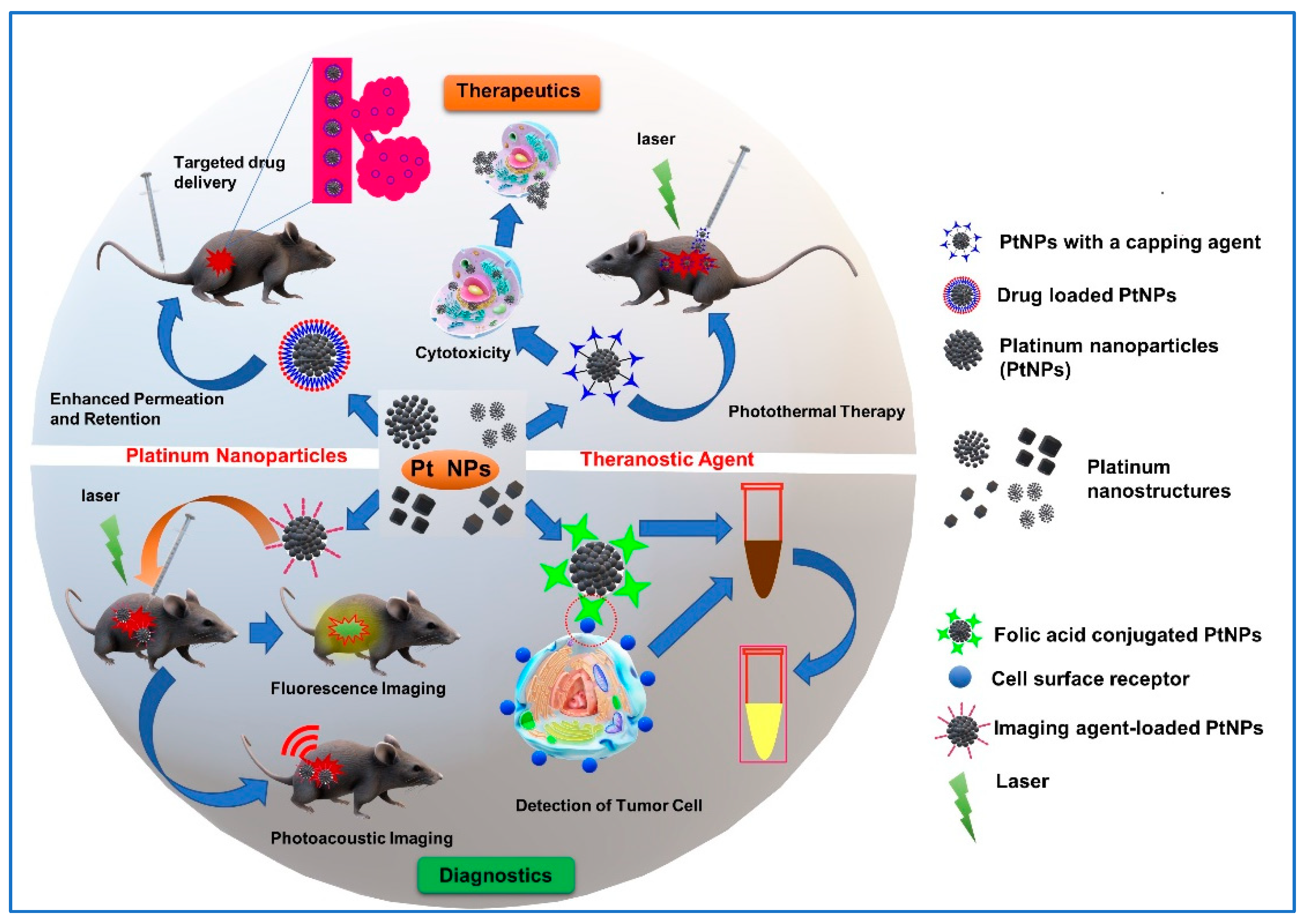
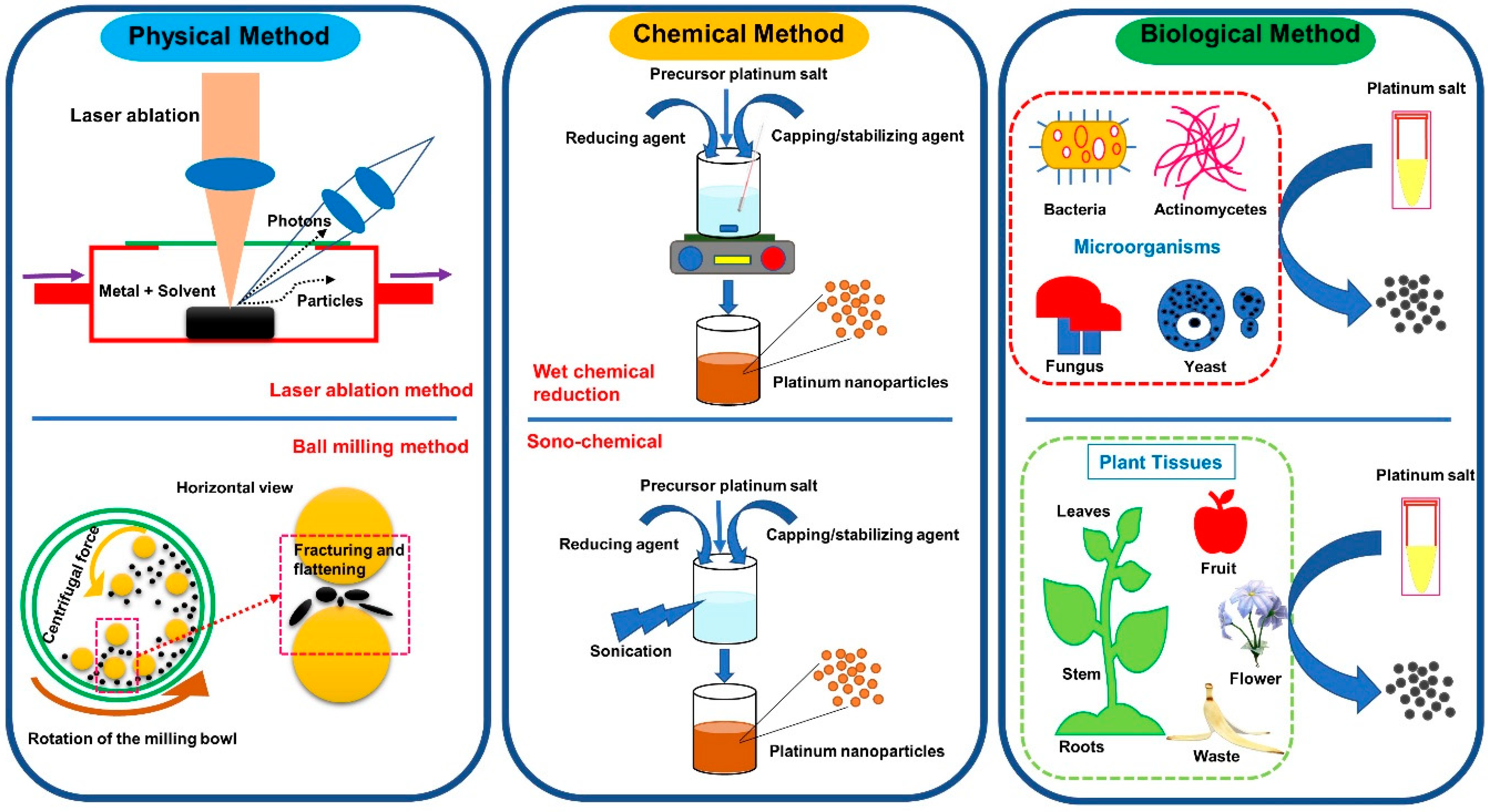
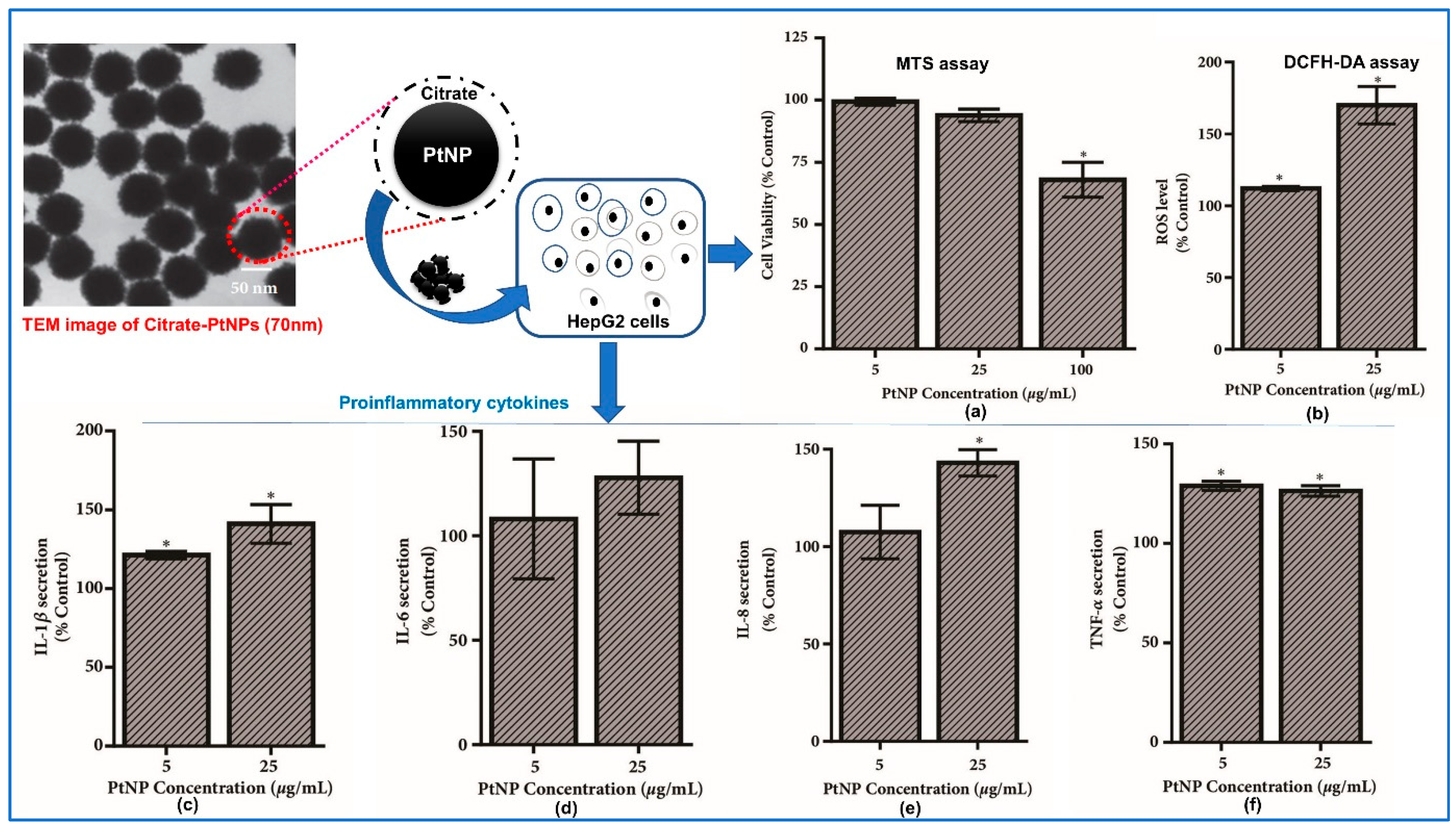
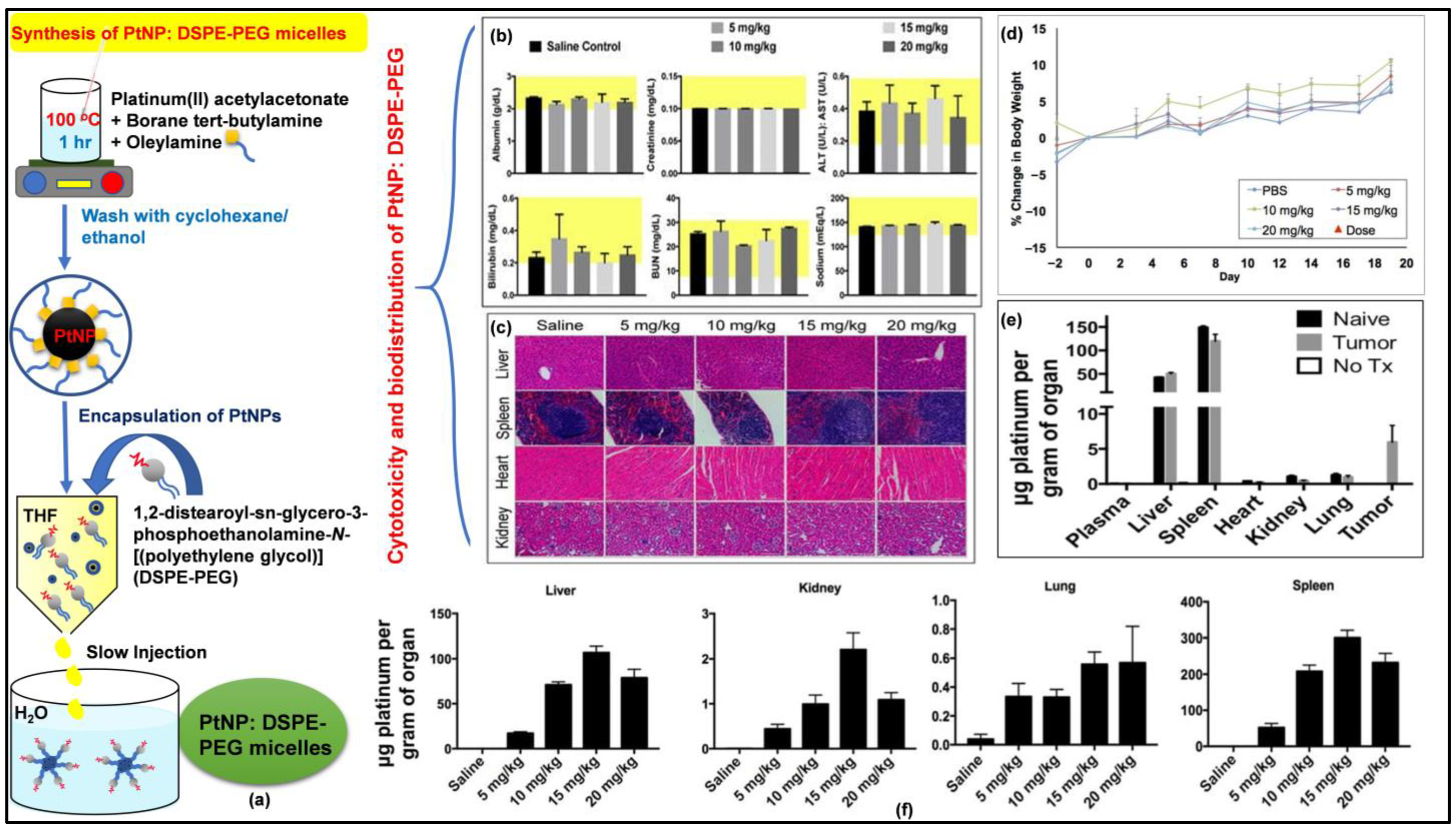
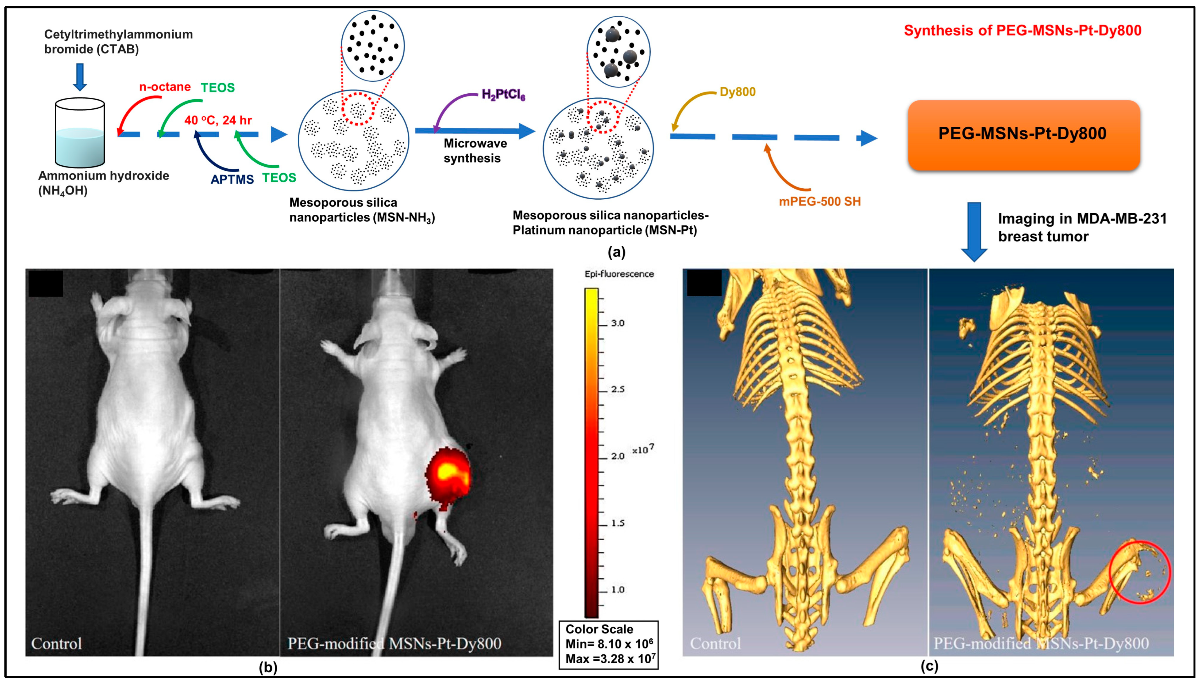
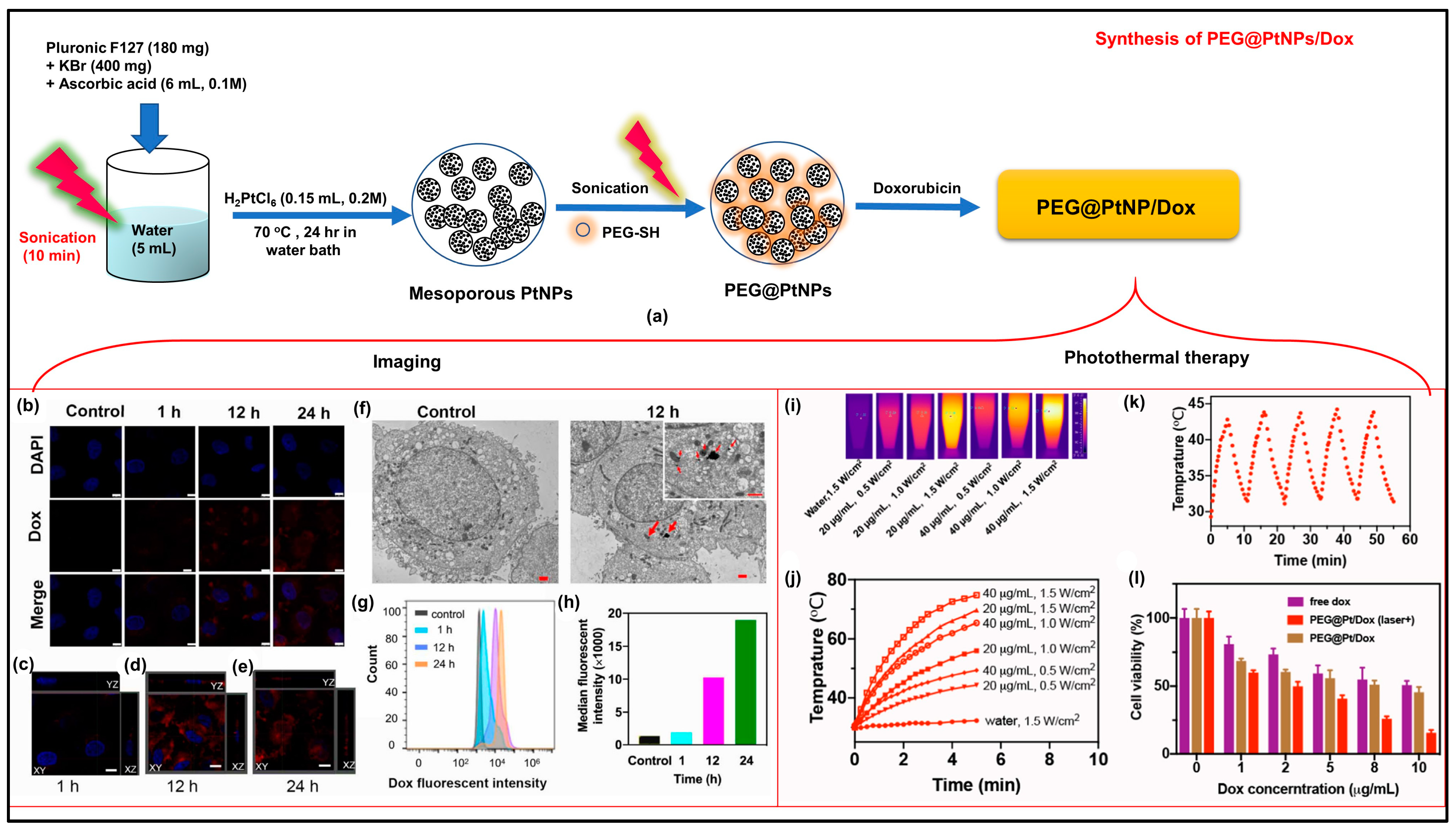
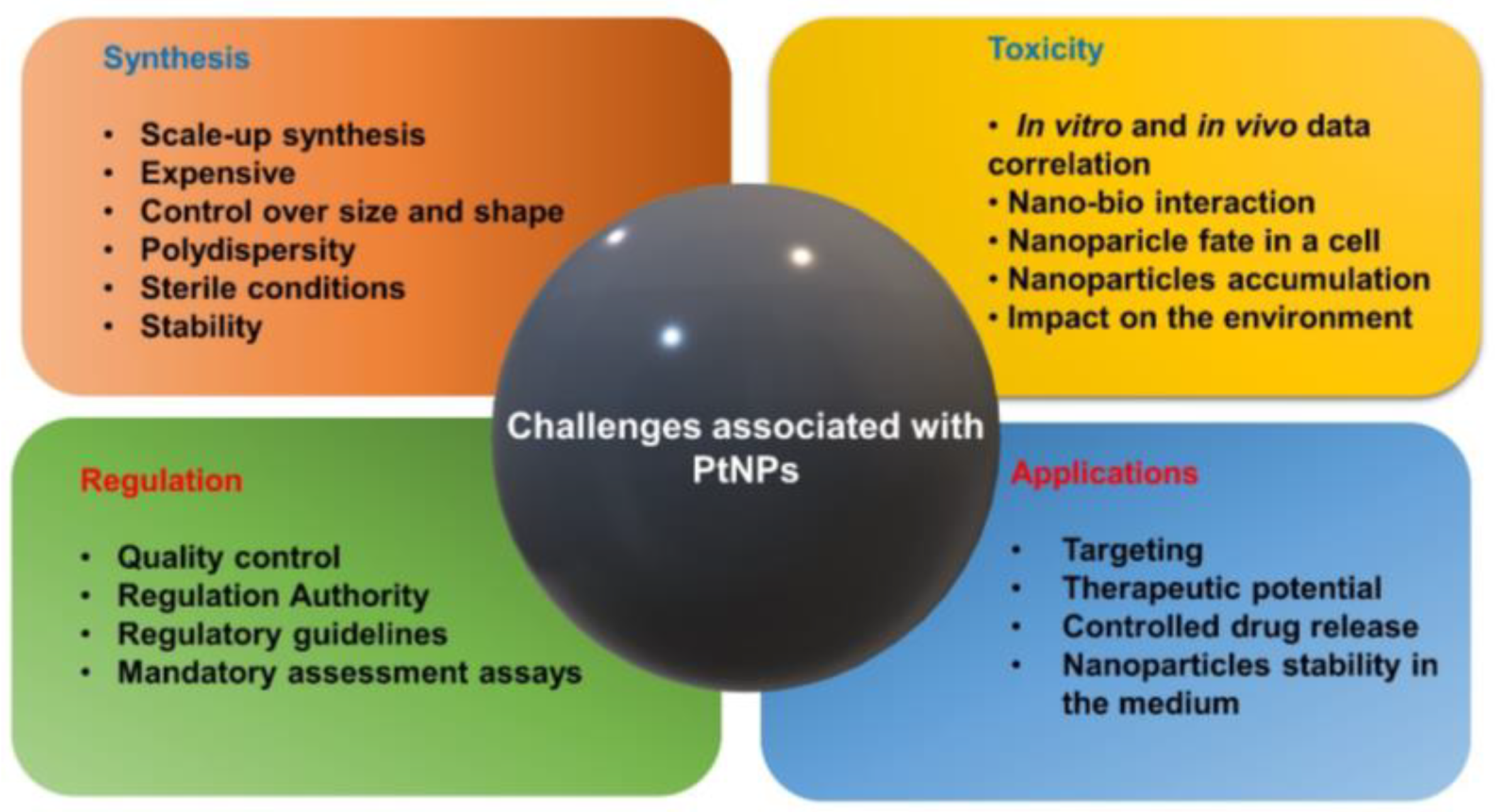
Table 1.
Studies on the platinum nanostructures’ potential in cancer theranostics concerning their size, shape, surface capping, toxicity assessment, and LD50.
Table 1.
Studies on the platinum nanostructures’ potential in cancer theranostics concerning their size, shape, surface capping, toxicity assessment, and LD50.
| Types (Shape and Size) | Capping Agent | Model System | Dose (LD50) | Effects on the Model System | Potential Applications | References |
|---|---|---|---|---|---|---|
| PtNPs (sphere, 1.14–1.65 nm) and gemcitabine-conjugated PtNPs (hybrid, 1.53–2.66 nm) | PVP | MCF-7 and Skin cancer (UACC-62) | 9.20 ± 0.04 µg/mL and 8.03 ± 0.03 µg/mL | Conjugated nanoparticles showed cell inhibition, the activation of Caspases 3, apoptosis, and a loss of MMP | Targeted therapy | [18] |
| PtNPs (spherical, 20–50 nm) | Streptomyces sp. | MCF-7 | 31.2 µg/mL | Cancer therapeutics | Chemotherapeutic agent | [75] |
| PtNPs (spherical, 15 and 8.5 nm) | Penicillium chrysogenum | Skin cancer (C2C12) | Induced ROS and the upregulation of the apoptosis marker (cas-3 and cas-9) and inflammatory markers (TNF-, TGF-, and NF-kB) | Anticancer | [79] | |
| PtNPs (spherical, 2–19 nm) | Brain cancer (U87 cell) | Induced genotoxicity and the pro-apoptotic marker and upregulated P53 and caspase-3 expression | Anticancer agent | [93] | ||
| PtNPs (spherical 20–100 nm) | Punica granatum crusts | Breast cancer cell (MCF-7) | 17.48 μg/mL | Anti-tumor agent | Chemotherapeutic agent | [100] |
| PtNPs (spherical, 5 and 30 nm) | Blood cancer (Raw 264.7) | 10 ppm | Anticancer agent | Anticancer agent | [101] | |
| PtNPs (1–2 nm) | Apigenin | Blood cancer (THP-1) | 150 μg/mL | Increased LDH, ROS, NO, malondialdehyde, and carbonylated protein. Reduced GSH, GSH:GSSG, GPx, SOD, CAT, and TRX. Upregulated the pregulation of (IL-1β), IL-6, IL-8, tumor necrosis factor-α (TNF-α), (GM-CSF), and MCP-1 | Anticancer | [103] |
| Cu/Au/Pt MNPs (anisotpoic, >10 nm) | Aptamer (Sgc8c) | Blood cancer (CCRF-CEM cells) | Photothermal therapy and biosensing | [122] | ||
| Pt-doped carbon nanoparticles (PtCNPs) (12.4 ± 2.4 nm, spherical) | Polyethylene glycol | A549 | Fragmented cytoskeletal structures and overexpression of lamin A/C were observed, thus inhibiting cancer cell migration | Photothermal therapy and imaging | [131] | |
| PtNPS (spherical, 5, 30 and 70 nm) | Citrate | C57BL/6 mice | Anti-colitis agent | [133] | ||
| PtNPs (spherical, 5 and 30 nm) | Raw 264.7 | 6 µg/mL | Cytotoxicity; suppressed the expression of iNOS and COX-2 proteins | Anticancer agent | [134] | |
| PtNPs (flower-like structure, 40 nm) | Lutein | Lung cancer (A549) | Increased exosomes’ biogenesis | Anticancer agent | [135] | |
| PtNPs (cubic and tetrahedral particles, 10–22 nm) | Tangeretin | Bone cancer (U2OS) | 15 μg/mL | Promoted cell death; increased LDH, ROS, NO, malondialdehyde and carbonylated protein; reduced MMP and ATP levels, | Chemotherapeutic agent | [136] |
| PtNPs (spherical, >50 nm) | Lycopene | Colon cancer (HCT-116 cells) | 14.62 μM/mL | Reduced cell proliferation and viability; increased ROS and oxidative stress; increased pro-apoptotic Bax and caspase-3; decreased anti-apoptotic Bcl-2 | Anticancer | [137] |
| PtNPs (spherical, 54.3 nm) | Mentha piperita (Peppermint) | HCT-116 | 20 μg/mL | Decreased cell viability at lower concentrations | Anticancer | [138] |
| PtNPs (spherical chain, 50 ± 5 nm) | Bacitracin | Hepatoma cell, A549, HCT-116 cells, and Kunming female mice | Cytotoxicity | Anti-tumor agent | [139] | |
| PtNPs (spherical, 20–40 nm) | Bacillus sp. | Liver cancer (HepG2) and rats | 10.3 × 10⁻⁶ m | Reduced GSH, SOD, and malondialdehyde | Cancer therapeutic | [140] |
| PtNPs (graphene quantum dots -pt, spherical, 5 nm) | Polyethylene glycol | Oral cancer (HSC3, SCC4, and CAL-27 cells) | 7.15 ± 0.99 × 10⁻⁶ m (HSC3 cells), 2.77 ± 0.84 × 10⁻⁶ m (SCC4 cells), and 6.19 ± 1.25 × 10⁻⁶ m (CAL-27 cells) | Induced apoptosis, less systemic drug toxicity, tumor suppression | Cancer therapy | [141] |
| Platinum nanocluster (PtNCs) | Polyethyleneimine | Blood cancer (K562, BV173) | 5 × 10⁻⁶ m | Induction of pro-apoptotic protein expression (p53, PUMA, cleaved caspase) | Cellular imaging and anticancer effects | [142] |
| Fe-PtNPs (4.8 ± 0.6 nm) | Cysteine | Gliomas cells (C6, SGH44, U251), ECV304, L929, and HEK293 | Imaging | MRI/CT contrast imaging agent | [143] | |
| PtNPs (sphere, 36 ± 6 nm) | Hyaluronic acid and ascorbic acid | Breast cancer (MDA-MB231 cells), mouse fibroblast (NIH3T3), and BALB/c nude mice | Targeted therapy | Photothermal therapy | [144] | |
| PtNPs (spherical, 2 and 80 nm) | Ethylene glycol | Colon cancer (SW480 and SW620) | Cell death with small-sized particles | Photothermal therapy | [145] | |
| Au-PtNP (cauliflowers, 66 nm) | Gallic acid | SW480, SW620, HCT116, and FHC | Selective cancer cell death | Photothermal therapy | [146] | |
| Iron -PtNPs (spherical, 42 nm) | Polypyrrole | Breast cancer (MDA-MB-231 cells) and female BALB/c nude mouse | Kill cancer cells selectively | Photothermal therapy and photoacoustic imaging | [147] | |
| PtNPs (flower, 14.6 ± 7.4 nm) | Poly(Ethylene Glycol) diamine | Cervical cancer (Hela cells) | Cytotoxic against cancer cells | Radiotherapy | [148] | |
| PtNPs (3.2 nm) | PEG | Breast cancer (T47D and MDA-MB-231) | Radiotherapy | [149] | ||
| PtNPs (spherical, 65 ± 6.68 nm) | PEG | Lung cancer (NCI-H460 cells) and Male athymic nude mice | Induced DNA damage, cell cycle arrest, and ROS stress | Radiotherapy | [150] | |
| PtNPs (spherical, 2.5 nm) | Peptide (H-Lys-Pro-Gly-DLys-NH2) | HepG2 | 2.9 ± 0.3 mgL−1 | Cell death due to the generation of oxidative stress | Selective cancer therapy | [151] |
Disclaimer/Publisher’s Note: The statements, opinions and data contained in all publications are solely those of the individual author(s) and contributor(s) and not of MDPI and/or the editor(s). MDPI and/or the editor(s) disclaim responsibility for any injury to people or property resulting from any ideas, methods, instructions or products referred to in the content. |
© 2023 by the authors. Licensee MDPI, Basel, Switzerland. This article is an open access article distributed under the terms and conditions of the Creative Commons Attribution (CC BY) license (https://creativecommons.org/licenses/by/4.0/).
Share and Cite
MDPI and ACS Style
Kumar, A.; Das, N.; Rayavarapu, R.G. Smart Platinum Nanostructures: A Journey from Synthesis to Advanced Theranostic Applications. J. Nanotheranostics 2023, 4, 384-407. https://doi.org/10.3390/jnt4030017
AMA Style
Kumar A, Das N, Rayavarapu RG. Smart Platinum Nanostructures: A Journey from Synthesis to Advanced Theranostic Applications. Journal of Nanotheranostics. 2023; 4(3):384-407. https://doi.org/10.3390/jnt4030017
Chicago/Turabian StyleKumar, Akash, Nabojit Das, and Raja Gopal Rayavarapu. 2023. "Smart Platinum Nanostructures: A Journey from Synthesis to Advanced Theranostic Applications" Journal of Nanotheranostics 4, no. 3: 384-407. https://doi.org/10.3390/jnt4030017